Evaluation of the Physicochemical Properties, Pharmacokinetics, and In Vitro Anticancer Effects of Docetaxel and Osthol Encapsulated in Methoxy Poly(ethylene glycol)-b-Poly(caprolactone) Polymeric Micelles

 ,
, 

Abstract
1. Introduction
2. Results
2.1. Evaluation of Synergistic Effects of DTX and OTH
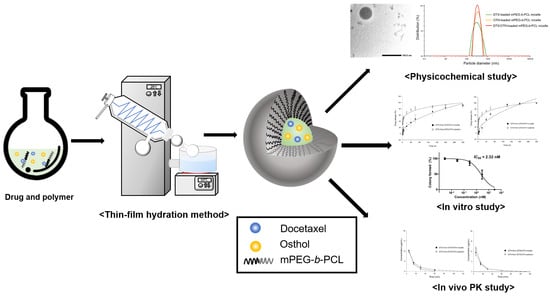
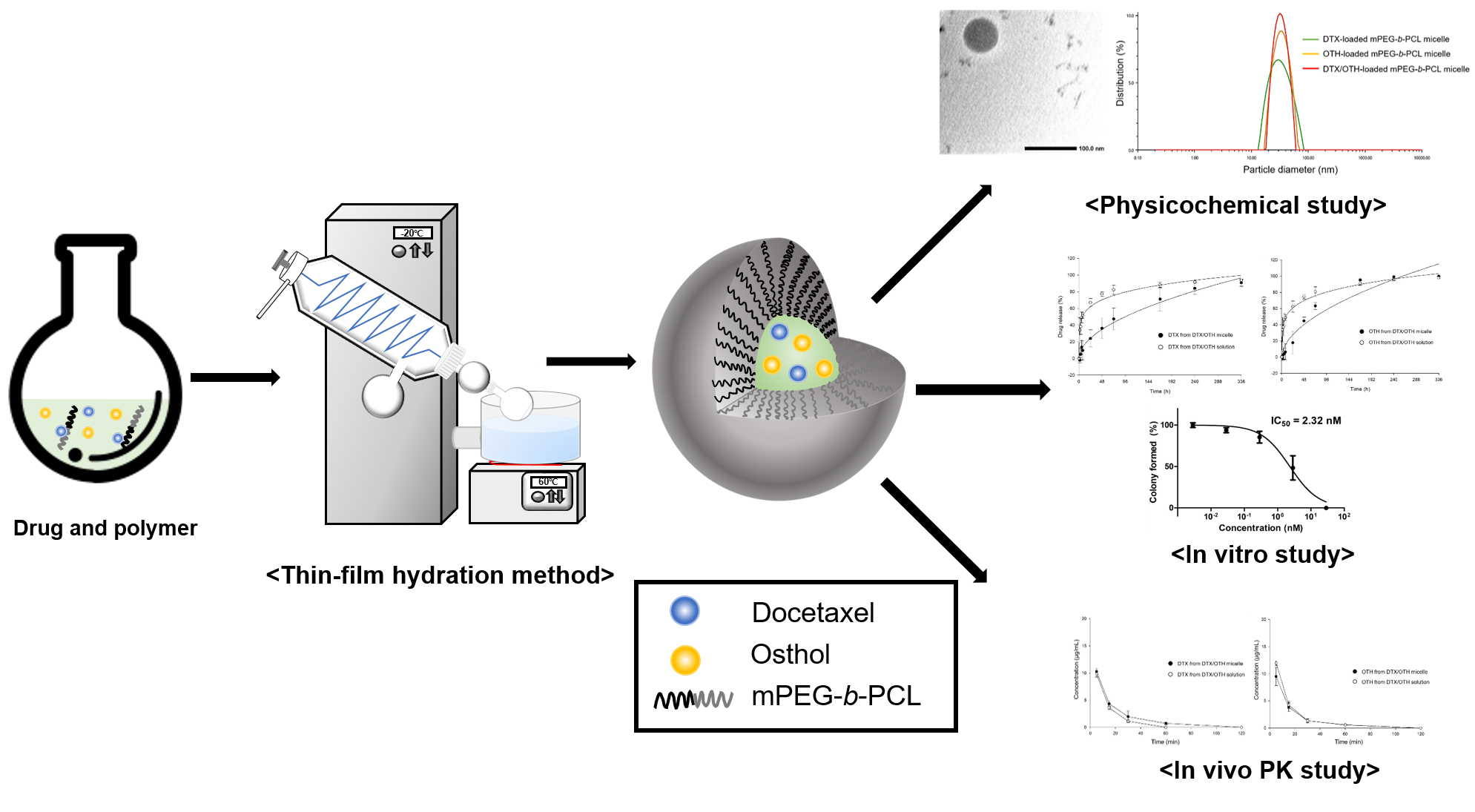
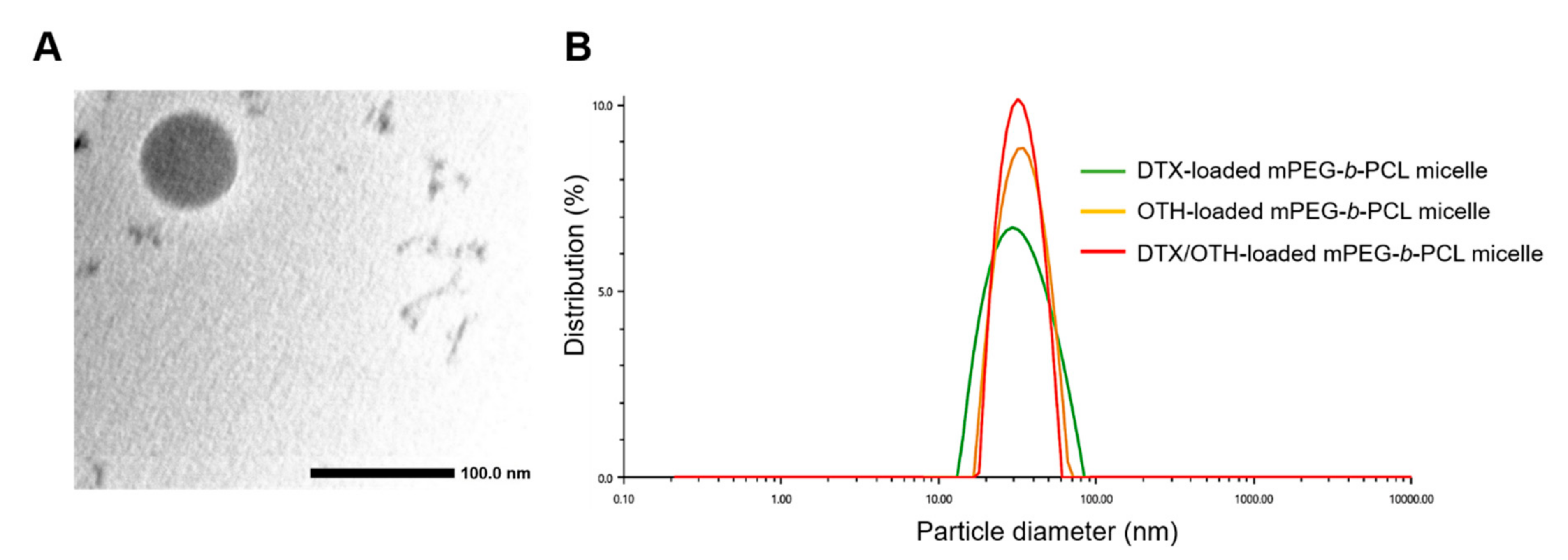
2.2. Preparation and Characterization of DTX/OTH-Loaded mPEG-b-PCL Micelles
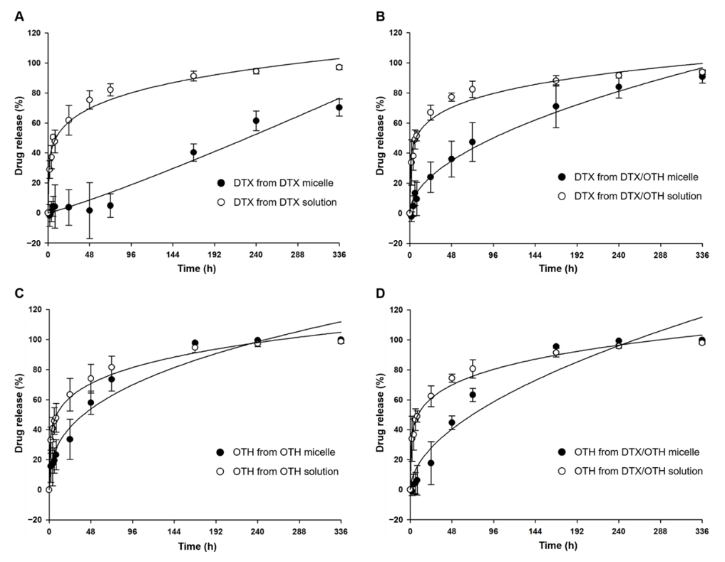
2.3. In Vitro Drug Release Assay
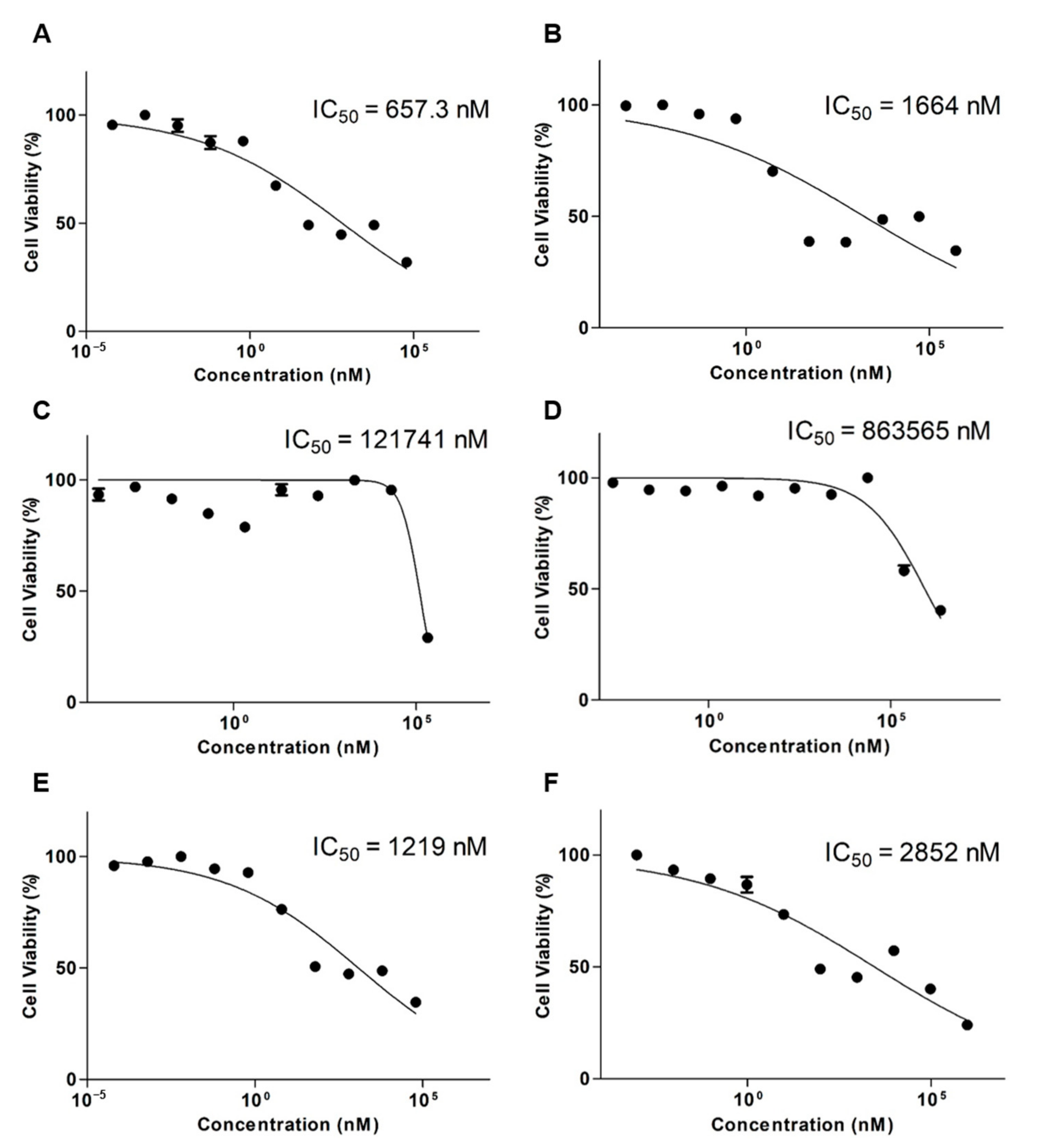
2.4. Cytotoxicity of DTX/OTH-Loaded mPEG-b-PCL Micelles in A549 Cells
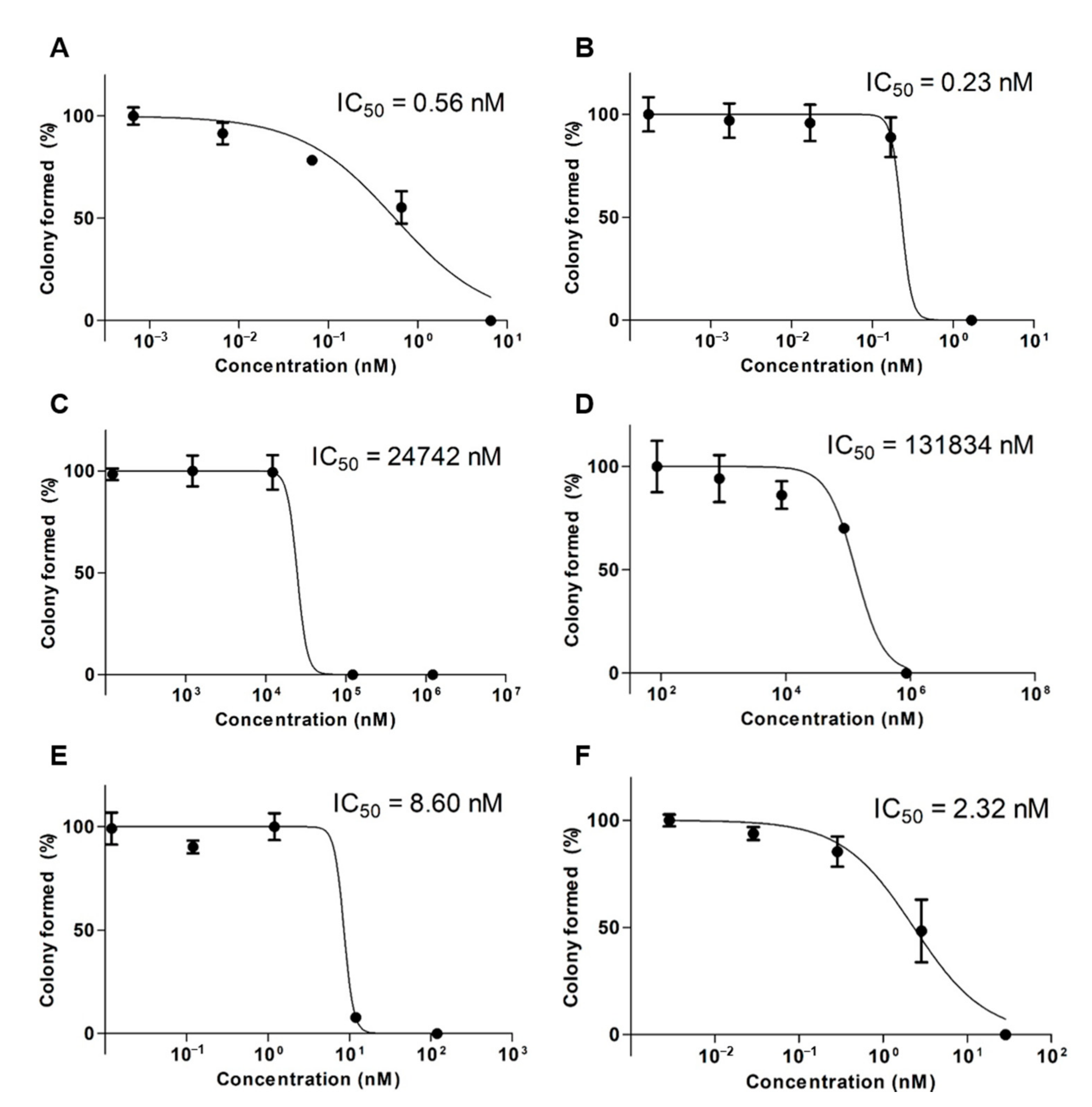
2.5. Clonogenic Assay
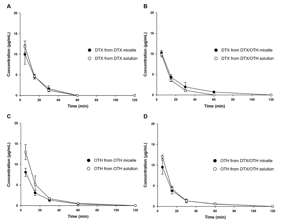
2.6. In Vivo Pharmacokinetic Study
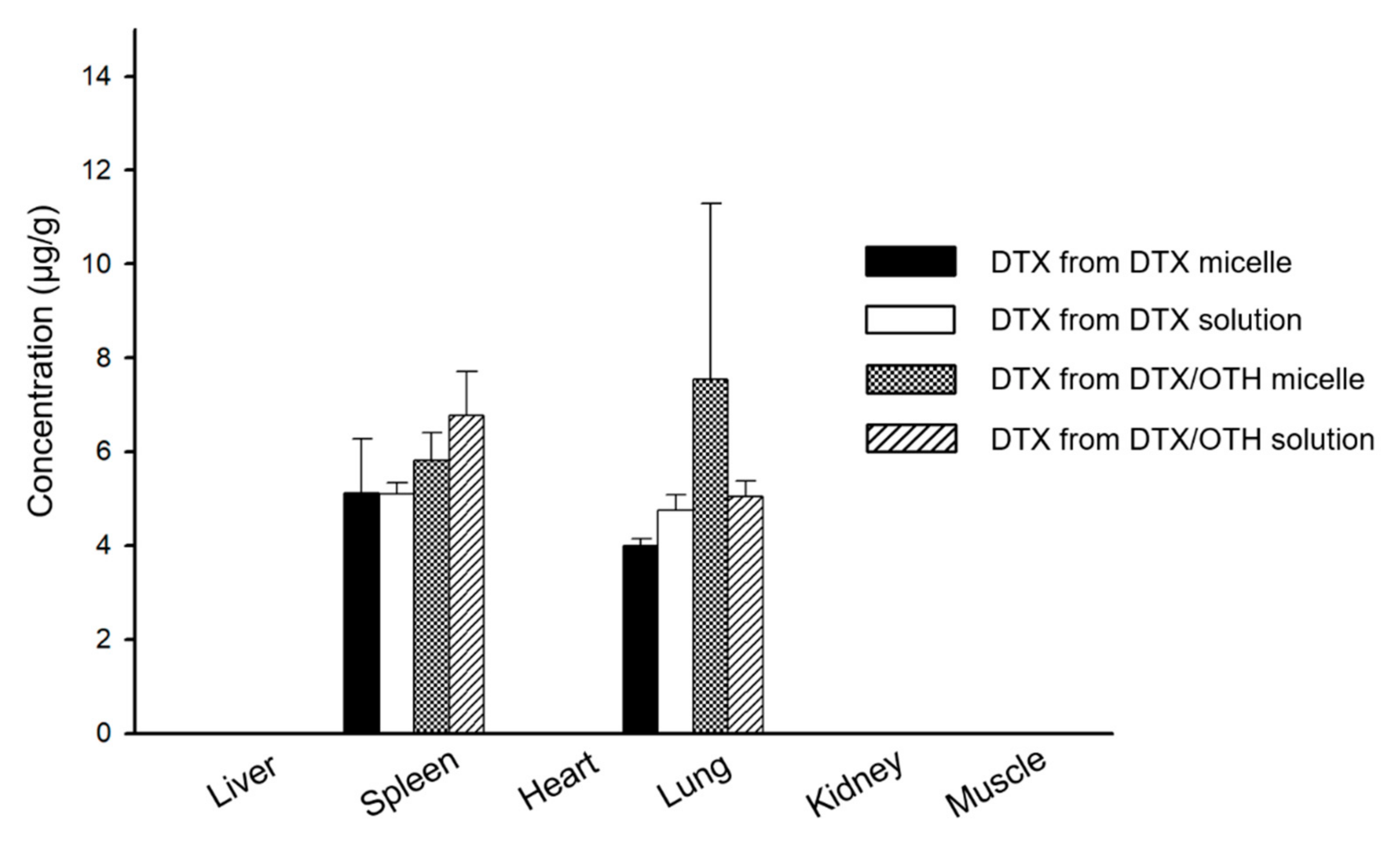
2.7. Biodistribution Study
3. Discussion
4. Materials and Methods
4.1. Materials and Reagents
4.2. Methods
4.2.1. Cell Line and Cell Culture
4.2.2. High-Performance Liquid Chromatography (HPLC) Analysis
4.2.3. In Vitro Cytotoxicity Assay
4.2.4. Combination Index (CI) Analysis
4.2.5. Clonogenic Assay
4.2.6. Preparation of DTX- and OTH-Loaded Polymeric Micelles
4.2.7. Physicochemical Characterization of Micelles
4.2.8. In Vitro Drug Release Assay
4.2.9. Pharmacokinetic Study
4.2.10. Biodistribution Study
4.2.11. Biological Sample Pretreatment for HPLC Analysis
4.2.12. Statistical Analysis
5. Conclusions
Author Contributions
Funding
Institutional Review Board Statement
Informed Consent Statement
Data Availability Statement
Acknowledgments
Conflicts of Interest
Abbreviations
| DTX | Docetaxel |
| OTH | Osthol |
| mPEG-b-PCL | Methoxy poly(ethylene glycol)-b-poly(caprolactone) |
| EE | Encapsulation efficiency |
| DL | Drug loading |
| CI | Combination index |
| LOD | Limit of detection |
| PDI | Poly-dispersity index |
| RES | Reticuloendothelial system |
References
- Mei, D.; Zhao, L.; Chen, B.; Zhang, X.; Wang, X.; Yu, Z.; Ni, X.; Zhang, Q. α-Conotoxin ImI-modified polymeric micelles as potential nanocarriers for targeted docetaxel delivery to α7-nAChR overexpressed non-small cell lung cancer. Drug Deliv. 2018, 25, 493–503. [Google Scholar] [CrossRef] [PubMed]
- Zhang, L.; Liu, Z.; Kong, C.; Liu, C.; Yang, K.; Chen, H.; Huang, J.; Qian, F. Improving Drug Delivery of Micellar Paclitaxel against Non-Small Cell Lung Cancer by Coloading Itraconazole as a Micelle Stabilizer and a Tumor Vascular Manipulator. Small (Weinheim an der Bergstrasse, Germany) 2018, 14, e1802112. [Google Scholar] [CrossRef] [PubMed]
- Zhuang, B.; Du, L.; Xu, H.; Xu, X.; Wang, C.; Fan, Y.; Cong, M.; Yin, J.; Li, H.; Guan, H. Self-assembled Micelle Loading Cabazitaxel for therapy of Lung Cancer. Int. J. Pharm. 2016, 499, 146–155. [Google Scholar] [CrossRef] [PubMed]
- Bao, Y.; Deng, Q.; Li, Y.; Zhou, S. Engineering docetaxel-loaded micelles for non-small cell lung cancer: A comparative study of microfluidic and bulk nanoparticle preparation. RSC Advances 2018, 8, 31950–31966. [Google Scholar] [CrossRef]
- Brahmer, J.R.; Govindan, R.; Anders, R.A.; Antonia, S.J.; Sagorsky, S.; Davies, M.J.; Dubinett, S.M.; Ferris, A.; Gandhi, L.; Garon, E.B.; et al. The Society for Immunotherapy of Cancer consensus statement on immunotherapy for the treatment of non-small cell lung cancer (NSCLC). Journal for immunotherapy of cancer 2018, 6, 75. [Google Scholar] [CrossRef]
- De Weger, V.A.; Beijnen, J.H.; Schellens, J.H. Cellular and clinical pharmacology of the taxanes docetaxel and paclitaxel--a review. Anticancer Drugs 2014, 25, 488–494. [Google Scholar] [CrossRef]
- Hirsch, F.R.; Suda, K.; Wiens, J.; Bunn, P.A., Jr. New and emerging targeted treatments in advanced non-small-cell lung cancer. Lancet 2016, 388, 1012–1024. [Google Scholar] [CrossRef]
- Rowinsky, E.K. The development and clinical utility of the taxane class of antimicrotubule chemotherapy agents. Annu. Rev. Med. 1997, 48, 353–374. [Google Scholar] [CrossRef]
- Thomas, A.; Liu, S.V.; Subramaniam, D.S.; Giaccone, G. Refining the treatment of NSCLC according to histological and molecular subtypes. Nat. Rev. Clin. Oncol. 2015, 12, 511–526. [Google Scholar] [CrossRef]
- Jinturkar, K.A.; Anish, C.; Kumar, M.K.; Bagchi, T.; Panda, A.K.; Misra, A.R. Liposomal formulations of Etoposide and Docetaxel for p53 mediated enhanced cytotoxicity in lung cancer cell lines. Biomaterials 2012, 33, 2492–2507. [Google Scholar] [CrossRef]
- Zarogoulidis, P.; Chatzaki, E.; Porpodis, K.; Domvri, K.; Hohenforst-Schmidt, W.; Goldberg, E.P.; Karamanos, N.; Zarogoulidis, K. Inhaled chemotherapy in lung cancer: Future concept of nanomedicine. International journal of nanomedicine 2012, 7, 1551–1572. [Google Scholar] [CrossRef] [PubMed]
- Chen, X.; Zhao, L.; Kang, Y.; He, Z.; Xiong, F.; Ling, X.; Wu, J. Significant Suppression of Non-small-cell Lung Cancer by Hydrophobic Poly(ester amide) Nanoparticles with High Docetaxel Loading. Front. Pharmacol. 2018, 9, 118. [Google Scholar] [CrossRef] [PubMed]
- Lee, S.W.; Yun, M.H.; Jeong, S.W.; In, C.H.; Kim, J.Y.; Seo, M.H.; Pai, C.M.; Kim, S.O. Development of docetaxel-loaded intravenous formulation, Nanoxel-PM™ using polymer-based delivery system. J. Control. Release 2011, 155, 262–271. [Google Scholar] [CrossRef] [PubMed]
- Herbst, R.S.; Khuri, F.R. Mode of action of docetaxel - a basis for combination with novel anticancer agents. Cancer Treat. Rev. 2003, 29, 407–415. [Google Scholar] [CrossRef]
- Horwitz, S.B. Mechanism of action of taxol. Trends Pharmacol. Sci. 1992, 13, 134–136. [Google Scholar] [CrossRef]
- Lyseng-Williamson, K.A.; Fenton, C. Docetaxel: A review of its use in metastatic breast cancer. Drugs 2005, 65, 2513–2531. [Google Scholar] [CrossRef]
- Qi, D.; Gong, F.; Teng, X.; Ma, M.; Wen, H.; Yuan, W.; Cheng, Y.; Lu, C. Design and evaluation of mPEG-PLA micelles functionalized with drug-interactive domains as improved drug carriers for docetaxel delivery. J. Biomater. Sci. Polym. Ed. 2017, 28, 1538–1555. [Google Scholar] [CrossRef]
- Su, C.Y.; Liu, J.J.; Ho, Y.S.; Huang, Y.Y.; Chang, V.H.; Liu, D.Z.; Chen, L.C.; Ho, H.O.; Sheu, M.T. Development and characterization of docetaxel-loaded lecithin-stabilized micellar drug delivery system (L(sb)MDDs) for improving the therapeutic efficacy and reducing systemic toxicity. Eur. J. Pharm. Biopharm. 2018, 123, 9–19. [Google Scholar] [CrossRef]
- Galletti, E.; Magnani, M.; Renzulli, M.L.; Botta, M. Paclitaxel and docetaxel resistance: Molecular mechanisms and development of new generation taxanes. ChemMedChem 2007, 2, 920–942. [Google Scholar] [CrossRef]
- Zhang, Z.R.; Leung, W.N.; Cheung, H.Y.; Chan, C.W. Osthole: A Review on Its Bioactivities, Pharmacological Properties, and Potential as Alternative Medicine. Evid. Based Complement. Alternat. Med. 2015, 2015, 919616. [Google Scholar] [CrossRef]
- Liu, W.B.; Zhou, J.; Qu, Y.; Li, X.; Lu, C.T.; Xie, K.L.; Sun, X.L.; Fei, Z. Neuroprotective effect of osthole on MPP+-induced cytotoxicity in PC12 cells via inhibition of mitochondrial dysfunction and ROS production. Neurochem. Int. 2010, 57, 206–215. [Google Scholar] [CrossRef] [PubMed]
- Liu, J.H.; Zschocke, S.; Reininger, E.; Bauer, R. Inhibitory effects of Angelica pubescens f. biserrata on 5-lipoxygenase and cyclooxygenase. Planta Med. 1998, 64, 525–529. [Google Scholar] [CrossRef] [PubMed]
- Zhang, Q.; Qin, L.; He, W.; Van Puyvelde, L.; Maes, D.; Adams, A.; Zheng, H.; De Kimpe, N. Coumarins from Cnidium monnieri and their antiosteoporotic activity. Planta Med. 2007, 73, 13–19. [Google Scholar] [CrossRef] [PubMed]
- Matsuda, H.; Tomohiro, N.; Ido, Y.; Kubo, M. Anti-allergic effects of cnidii monnieri fructus (dried fruits of Cnidium monnieri) and its major component, osthol. Biol. Pharm. Bull. 2002, 25, 809–812. [Google Scholar] [CrossRef] [PubMed]
- Jiang, G.; Liu, J.; Ren, B.; Tang, Y.; Owusu, L.; Li, M.; Zhang, J.; Liu, L.; Li, W. Anti-tumor effects of osthole on ovarian cancer cells in vitro. J. Ethnopharmacol. 2016, 193, 368–376. [Google Scholar] [CrossRef] [PubMed]
- Wang, L.; Yang, L.; Lu, Y.; Chen, Y.; Liu, T.; Peng, Y.; Zhou, Y.; Cao, Y.; Bi, Z.; Liu, T.; et al. Osthole Induces Cell Cycle Arrest and Inhibits Migration and Invasion via PTEN/Akt Pathways in Osteosarcoma. Cell. Physiol. Biochem. 2016, 38, 2173–2182. [Google Scholar] [CrossRef] [PubMed]
- Xu, X.M.; Zhang, M.L.; Zhang, Y.; Zhao, L. Osthole induces lung cancer cell apoptosis through inhibition of inhibitor of apoptosis family proteins. Oncol. Lett. 2016, 12, 3779–3784. [Google Scholar] [CrossRef]
- Yang, Y.; Ren, F.; Tian, Z.; Song, W.; Cheng, B.; Feng, Z. Osthole Synergizes With HER2 Inhibitor, Trastuzumab in HER2-Overexpressed N87 Gastric Cancer by Inducing Apoptosis and Inhibition of AKT-MAPK Pathway. Front. Pharmacol. 2018, 9, 1392. [Google Scholar] [CrossRef]
- Sánchez, B.G.; Bort, A.; Mateos-Gómez, P.A.; Rodríguez-Henche, N.; Díaz-Laviada, I. Combination of the natural product capsaicin and docetaxel synergistically kills human prostate cancer cells through the metabolic regulator AMP-activated kinase. Cancer Cell Int. 2019, 19, 54. [Google Scholar] [CrossRef]
- Singh, S.K.; Apata, T.; Gordetsky, J.B.; Singh, R. Docetaxel Combined with Thymoquinone Induces Apoptosis in Prostate Cancer Cells via Inhibition of the PI3K/AKT Signaling Pathway. Cancers (Basel) 2019, 11, 1390. [Google Scholar] [CrossRef]
- Hu, X.J.; Liu, Y.; Zhou, X.F.; Zhu, Q.L.; Bei, Y.Y.; You, B.G.; Zhang, C.G.; Chen, W.L.; Wang, Z.L.; Zhu, A.J.; et al. Synthesis and characterization of low-toxicity N-caprinoyl-N-trimethyl chitosan as self-assembled micelles carriers for osthole. International journal of nanomedicine 2013, 8, 3543–3558. [Google Scholar] [CrossRef] [PubMed]
- Ten Tije, A.J.; Verweij, J.; Loos, W.J.; Sparreboom, A. Pharmacological effects of formulation vehicles: Implications for cancer chemotherapy. Clin. Pharmacokinet. 2003, 42, 665–685. [Google Scholar] [CrossRef] [PubMed]
- Schwartzberg, L.S.; Navari, R.M. Safety of Polysorbate 80 in the Oncology Setting. Adv. Ther. 2018, 35, 754–767. [Google Scholar] [CrossRef] [PubMed]
- Jo, M.J.; Jo, Y.H.; Lee, Y.J.; Park, C.W.; Kim, J.S.; Hong, J.T.; Chung, Y.B.; Lee, M.K.; Shin, D.H. Physicochemical, Pharmacokinetic, and Toxicity Evaluation of Methoxy Poly(ethylene glycol)-b-Poly(d,l-Lactide) Polymeric Micelles Encapsulating Alpinumisoflavone Extracted from Unripe Cudrania tricuspidata Fruit. Pharmaceutics 2019, 11, 366. [Google Scholar] [CrossRef] [PubMed]
- Su, Y.; Wang, K.; Li, Y.; Song, W.; Xin, Y.; Zhao, W.; Tian, J.; Ren, L.; Lu, L. Sorafenib-loaded polymeric micelles as passive targeting therapeutic agents for hepatocellular carcinoma therapy. Nanomedicine (London, England) 2018, 13, 1009–1023. [Google Scholar] [CrossRef] [PubMed]
- Zamani, M.; Shirinzadeh, A.; Aghajanzadeh, M.; Andalib, S.; Danafar, H. In vivo study of mPEG-PCL as a nanocarriers for anti-inflammatory drug delivery of simvastatin. Pharm. Dev. Technol. 2019, 24, 663–670. [Google Scholar] [CrossRef]
- Wei, W.; Li, S.; Xu, H.; Zhou, F.; Wen, Y.; Song, Z.; Feng, S.; Feng, R. MPEG-PCL Copolymeric Micelles for Encapsulation of Azithromycin. AAPS PharmSciTech 2018, 19, 2041–2047. [Google Scholar] [CrossRef]
- Kheiri Manjili, H.; Ghasemi, P.; Malvandi, H.; Mousavi, M.S.; Attari, E.; Danafar, H. Pharmacokinetics and in vivo delivery of curcumin by copolymeric mPEG-PCL micelles. Eur. J. Pharm. Biopharm. 2017, 116, 17–30. [Google Scholar] [CrossRef]
- Cho, H.; Lai, T.C.; Kwon, G.S. Poly(ethylene glycol)-block-poly(ε-caprolactone) micelles for combination drug delivery: Evaluation of paclitaxel, cyclopamine and gossypol in intraperitoneal xenograft models of ovarian cancer. J. Control. Release 2013, 166, 1–9. [Google Scholar] [CrossRef]
- Dou, J.; Zhang, H.; Liu, X.; Zhang, M.; Zhai, G. Preparation and evaluation in vitro and in vivo of docetaxel loaded mixed micelles for oral administration. Colloids Surf. B. Biointerfaces 2014, 114, 20–27. [Google Scholar] [CrossRef]
- Dai, J.D.; Wang, X.Q.; Zhang, T.; Meng, M.; Zhang, X.; Lü, W.L.; Zhang, Q. [Preparation of cyclosporine A pH sensitive nanoparticles and oral pharmacokinetics in rats]. Yao Xue Xue Bao 2004, 39, 1023–1027. [Google Scholar] [PubMed]
- Aw, M.S.; Kurian, M.; Losic, D. Polymeric micelles for multidrug delivery and combination therapy. Chemistry (Weinheim an der Bergstrasse, Germany) 2013, 19, 12586–12601. [Google Scholar] [CrossRef] [PubMed]
- Chen, L.; Sha, X.; Jiang, X.; Chen, Y.; Ren, Q.; Fang, X. Pluronic P105/F127 mixed micelles for the delivery of docetaxel against Taxol-resistant non-small cell lung cancer: Optimization and in vitro, in vivo evaluation. International journal of nanomedicine 2013, 8, 73–84. [Google Scholar] [CrossRef] [PubMed]
- Liao, J.; Song, Y.; Liu, C.; Li, D.; Zheng, H.; Lu, B. Dual-drug delivery based charge-conversional polymeric micelles for enhanced cellular uptake and combination therapy. Polymer Chemistry 2019, 10. [Google Scholar] [CrossRef]
- Rezazadeh, M.; Emami, J.; Hasanzadeh, F.; Sadeghi, H.; Minaiyan, M.; Mostafavi, A.; Rostami, M.; Lavasanifar, A. In vivo pharmacokinetics, biodistribution and anti-tumor effect of paclitaxel-loaded targeted chitosan-based polymeric micelle. Drug Deliv. 2016, 23, 1707–1717. [Google Scholar] [CrossRef]
- Emami, J.; Rezazadeh, M.; Mashayekhi, M.; Rostami, M.; Jahanian-Najafabadi, A. A novel mixed polymeric micelle for co-delivery of paclitaxel and retinoic acid and overcoming multidrug resistance: Synthesis, characterization, cytotoxicity, and pharmacokinetic evaluation. Drug Dev. Ind. Pharm. 2018, 44, 729–740. [Google Scholar] [CrossRef]
- Kim, S.C.; Kim, D.W.; Shim, Y.H.; Bang, J.S.; Oh, H.S.; Wan Kim, S.; Seo, M.H. In vivo evaluation of polymeric micellar paclitaxel formulation: Toxicity and efficacy. J. Control. Release 2001, 72, 191–202. [Google Scholar] [CrossRef]
- Liu, J.; Li, H.; Chen, D.; Jin, X.; Zhao, X.; Zhang, C.; Ping, Q. In vivo evaluation of novel chitosan graft polymeric micelles for delivery of paclitaxel. Drug Deliv. 2011, 18, 181–189. [Google Scholar] [CrossRef]
- Zhang, C.; Qu, G.; Sun, Y.; Wu, X.; Yao, Z.; Guo, Q.; Ding, Q.; Yuan, S.; Shen, Z.; Ping, Q.; et al. Pharmacokinetics, biodistribution, efficacy and safety of N-octyl-O-sulfate chitosan micelles loaded with paclitaxel. Biomaterials 2008, 29, 1233–1241. [Google Scholar] [CrossRef]
- Esmaeili, F.; Dinarvand, R.; Ghahremani, M.H.; Ostad, S.N.; Esmaily, H.; Atyabi, F. Cellular cytotoxicity and in-vivo biodistribution of docetaxel poly(lactide-co-glycolide) nanoparticles. Anticancer Drugs 2010, 21, 43–52. [Google Scholar] [CrossRef]
- Twentyman, P.R.; Luscombe, M. A study of some variables in a tetrazolium dye (MTT) based assay for cell growth and chemosensitivity. Br. J. Cancer 1987, 56, 279–285. [Google Scholar] [CrossRef] [PubMed]
- Chou, T.C. Drug combination studies and their synergy quantification using the Chou-Talalay method. Cancer Res. 2010, 70, 440–446. [Google Scholar] [CrossRef] [PubMed]
- Zhang, H. Thin-Film Hydration Followed by Extrusion Method for Liposome Preparation. Methods Mol. Biol. 2017, 1522, 17–22. [Google Scholar] [CrossRef]
- Xu, H.; Hou, Z.; Zhang, H.; Kong, H.; Li, X.; Wang, H.; Xie, W. An efficient Trojan delivery of tetrandrine by poly(N-vinylpyrrolidone)-block-poly(ε-caprolactone) (PVP-b-PCL) nanoparticles shows enhanced apoptotic induction of lung cancer cells and inhibition of its migration and invasion. International journal of nanomedicine 2014, 9, 231–242. [Google Scholar] [CrossRef] [PubMed]
- Sun, C.; Liang, Y.; Hao, N.; Xu, L.; Cheng, F.; Su, T.; Cao, J.; Gao, W.; Pu, Y.; He, B. A ROS-responsive polymeric micelle with a π-conjugated thioketal moiety for enhanced drug loading and efficient drug delivery. Organic & biomolecular chemistry 2017, 15, 9176–9185. [Google Scholar] [CrossRef]
- Zhang, W.; Li, C.; Jin, Y.; Liu, X.; Wang, Z.; Shaw, J.P.; Baguley, B.C.; Wu, Z.; Liu, J. Multiseed liposomal drug delivery system using micelle gradient as driving force to improve amphiphilic drug retention and its anti-tumor efficacy. Drug Deliv. 2018, 25, 611–622. [Google Scholar] [CrossRef]
- Modi, S.; Anderson, B.D. Determination of Drug Release Kinetics from Nanoparticles: Overcoming Pitfalls of the Dynamic Dialysis Method. Mol. Pharm. 2013, 10, 3076–3089. [Google Scholar] [CrossRef]
- Shin, D.H.; Park, S.H.; Jeong, S.W.; Park, C.W.; Han, K.; Chung, Y.B. Hepatic uptake of epirubicin by isolated rat hepatocytes and its biliary excretion after intravenous infusion in rats. Arch. Pharm. Res. 2014, 37, 1599–1606. [Google Scholar] [CrossRef]


| DTX:OTH (Molar Ratio) | IC50 (nM) | CI Value | |
|---|---|---|---|
| DTX | OTH | ||
| 11:1 | 1214 ± 705 | 110 ± 64.1 | 1.85 |
| 4:1 | 1947 ± 550 | 487 ± 137 | 2.97 |
| 2:1 | 131 ± 60.9 | 65.5 ± 30.5 | 0.20 |
| 1:1 | 404 ± 206 | 404 ± 206 | 0.62 |
| 1:2 | 343 ± 278 | 686 ± 557 | 0.53 |
| 1:4 | 244 ± 101 | 975 ± 402 | 0.38 |
| 1:11 | 68.8 ± 23.1 | 757 ± 232 | 0.11 |
| Formulation | Polymer Amount Used (mg) | DTX Amount Used (mg) | OTH Amount Used (mg) | DTX Encapsulation Efficiency (EE %) | OTH Encapsulation Efficiency (EE %) | DTX Drug Loading (DL %) | OTH Drug Loading (DL %) | Particle Size (nm) | Poly-Dispersity Index (PDI) | Zeta Potential (mV) |
|---|---|---|---|---|---|---|---|---|---|---|
| DTX:OTH 1:2 | 50 | 4 | 2.4 | 73.4 ± 9.81 | 80.8 ± 12.6 | 5.43 ± 0.73 | 3.70 ± 0.58 | 30.9 ± 0.85 | 0.13 ± 0.65 | 0.97 ± 0.21 |
| 100 | 4 | 2.4 | 76.2 ± 7.27 | 82.3 ± 10.4 | 2.93 ± 0.28 | 1.93 ± 0.24 | 30.9 ± 0.99 | 0.16 ± 4.00 | 2.17 ± 1.05 | |
| 150 | 4 | 2.4 | 69.6 ± 5.72 | 77.3 ± 11.8 | 1.81 ± 0.15 | 1.22 ± 0.19 | 30.1 ± 1.06 | 0.08 ± 2.31 | 2.40 ± 1.21 | |
| DTX:OTH 2:1 | 50 | 6.62 | 1.0 | 54.0 ± 18.1 | 109 ± 4.16 | 6.31 ± 2.12 | 2.14 ± 0.08 | 31.3 ± 1.52 | 0.12 ± 7.75 | −1.03 ± 2.12 |
| 100 | 6.62 | 1.0 | 65.4 ± 5.31 | 82.2 ± 11.7 | 4.06 ± 0.33 | 0.81 ± 0.12 | 31.4 ± 2.22 | 0.12 ± 7.54 | 1.73 ± 0.76 | |
| 150 | 6.62 | 1.0 | 67.4 ± 0.79 | 95.8 ± 32.6 | 2.85 ± 0.03 | 0.63 ± 0.22 | 31.1 ± 0.97 | 0.12 ± 4.04 | 6.07 ± 2.37 | |
| DTX:OTH 1:4 | 50 | 3 | 3.6 | 72.2 ± 10.8 | 77.2 ± 11.8 | 4.09 ± 0.61 | 5.19 ± 0.80 | 31.4 ± 0.20 | 0.08 ± 0.38 | 0.53 ± 1.46 |
| 100 | 3 | 3.6 | 70.5 ± 5.01 | 78.1 ± 5.93 | 2.05 ± 0.15 | 2.72 ± 0.21 | 31.6 ± 0.97 | 0.10 ± 1.14 | 1.37 ± 2.80 | |
| 150 | 3 | 3.6 | 74.2 ± 8.55 | 80.6 ± 1.95 | 1.45 ± 0.17 | 1.89 ± 0.05 | 31.5 ± 1.29 | 0.13 ± 0.82 | 4.03 ± 2.32 | |
| 50 | 6 | 7.2 | 72.2 ± 4.53 | 78.6 ± 2.38 | 7.74 ± 0.49 | 9.90 ± 0.30 | 33.3 ± 0.28 | 0.09 ± 0.67 | −0.60 ± 0.78 | |
| 150 | 6 | 7.2 | 69.1 ± 4.27 | 77.7 ± 3.47 | 2.66 ± 0.16 | 3.56 ± 0.16 | 33.5 ± 2.25 | 0.17 ± 5.55 | 2.67 ± 0.40 | |
| DTX:OTH 1:11 | 50 | 2 | 6.6 | 74.1 ± 5.25 | 78.9 ± 1.68 | 2.85 ± 0.20 | 9.20 ± 0.20 | 32.6 ± 0.53 | 0.11 ± 2.35 | −0.47 ± 0.70 |
| 100 | 2 | 6.6 | 70.5 ± 3.11 | 76.8 ± 5.03 | 1.38 ± 0.06 | 4.75 ± 0.31 | 32.4 ± 0.36 | 0.10 ± 2.81 | 3.47 ± 0.96 | |
| 150 | 2 | 6.6 | 70.8 ± 5.44 | 74.5 ± 5.86 | 0.93 ± 0.07 | 3.14 ± 0.25 | 32.2 ± 1.00 | 0.16 ± 3.59 | 4.17 ± 1.03 |
| Parameters | DTX Solution | DTX Micelle | OTH Solution | OTH Micelle | DTX in Combination Solution | DTX in Combination Micelle | OTH in Combination Solution | OTH in Combination Micelle |
|---|---|---|---|---|---|---|---|---|
| Dose (µg·kg−1) | 10,000 | 10,000 | 12,000 | 12,000 | 10,000 | 10,000 | 12,000 | 12,000 |
| AUC (min·µg·mL−1) | 222 ± 16.6 | 205 ± 43.3 | 274 ± 41.6 | 177 ± 23.5 | 183 ± 11.7 | 244 ± 42.4 | 246 ± 7.3 | 212 ± 24.2 |
| CLt (mL·kg−1·min) | 45.2 ± 3.44 | 50.1 ± 9.47 | 44.6 ± 7.37 | 68.5 ± 9.3 | 54.8 ± 3.65 | 41.8 ± 7.4 | 48.7 ± 1.45 | 57.1 ± 6.21 |
Publisher’s Note: MDPI stays neutral with regard to jurisdictional claims in published maps and institutional affiliations. |
© 2020 by the authors. Licensee MDPI, Basel, Switzerland. This article is an open access article distributed under the terms and conditions of the Creative Commons Attribution (CC BY) license (http://creativecommons.org/licenses/by/4.0/).
Share and Cite
Jo, M.J.; Lee, Y.J.; Park, C.-W.; Chung, Y.B.; Kim, J.-S.; Lee, M.K.; Shin, D.H. Evaluation of the Physicochemical Properties, Pharmacokinetics, and In Vitro Anticancer Effects of Docetaxel and Osthol Encapsulated in Methoxy Poly(ethylene glycol)-b-Poly(caprolactone) Polymeric Micelles. Int. J. Mol. Sci. 2021, 22, 231. https://doi.org/10.3390/ijms22010231
Jo MJ, Lee YJ, Park C-W, Chung YB, Kim J-S, Lee MK, Shin DH. Evaluation of the Physicochemical Properties, Pharmacokinetics, and In Vitro Anticancer Effects of Docetaxel and Osthol Encapsulated in Methoxy Poly(ethylene glycol)-b-Poly(caprolactone) Polymeric Micelles. International Journal of Molecular Sciences. 2021; 22(1):231. https://doi.org/10.3390/ijms22010231
Chicago/Turabian StyleJo, Min Jeong, Yu Jin Lee, Chun-Woong Park, Youn Bok Chung, Jin-Seok Kim, Mi Kyeong Lee, and Dae Hwan Shin. 2021. "Evaluation of the Physicochemical Properties, Pharmacokinetics, and In Vitro Anticancer Effects of Docetaxel and Osthol Encapsulated in Methoxy Poly(ethylene glycol)-b-Poly(caprolactone) Polymeric Micelles" International Journal of Molecular Sciences 22, no. 1: 231. https://doi.org/10.3390/ijms22010231
APA StyleJo, M. J., Lee, Y. J., Park, C.-W., Chung, Y. B., Kim, J.-S., Lee, M. K., & Shin, D. H. (2021). Evaluation of the Physicochemical Properties, Pharmacokinetics, and In Vitro Anticancer Effects of Docetaxel and Osthol Encapsulated in Methoxy Poly(ethylene glycol)-b-Poly(caprolactone) Polymeric Micelles. International Journal of Molecular Sciences, 22(1), 231. https://doi.org/10.3390/ijms22010231