Review

Reactive Oxygen Species: Modulators of Phenotypic Switch of Vascular Smooth Muscle Cells

1
Department of Nutrition, University of Petra, P.O. Box 961343, Amman 11196, Jordan
2
Department of Pharmacology and Therapeutics, Beirut Arab University, P.O. Box 11-5020, Beirut 1107-2809, Lebanon
3
Department of Biology, American University of Beirut, P.O. Box 11-0236, Beirut 1107-2020, Lebanon
4
Department of Pharmacology and Toxicology, American University of Beirut, P.O. Box 11-0236, Beirut 1107-2020, Lebanon
5
Department of Pharmacology and Toxicology, Alexandria University, Alexandria 21526, Egypt
6
Department of Clinical and Experimental Medicine, University of Messina, 98125 Messina, Italy
7
Department of Basic Medical Sciences, College of Medicine, QU Health, Qatar University, Doha P.O. Box 2713, Qatar
8
Biomedical and Pharmaceutical Research Unit, QU Health, Qatar University, Doha P.O. Box 2713, Qatar
*
Authors to whom correspondence should be addressed.
These authors contributed equally to this work.
Int. J. Mol. Sci. 2020, 21(22), 8764; https://doi.org/10.3390/ijms21228764
Submission received: 30 July 2020 / Revised: 29 September 2020 / Accepted: 7 October 2020 / Published: 20 November 2020

Abstract

:
Reactive oxygen species (ROS) are natural byproducts of oxygen metabolism in the cell. At physiological levels, they play a vital role in cell signaling. However, high ROS levels cause oxidative stress, which is implicated in cardiovascular diseases (CVD) such as atherosclerosis, hypertension, and restenosis after angioplasty. Despite the great amount of research conducted to identify the role of ROS in CVD, the image is still far from being complete. A common event in CVD pathophysiology is the switch of vascular smooth muscle cells (VSMCs) from a contractile to a synthetic phenotype. Interestingly, oxidative stress is a major contributor to this phenotypic switch. In this review, we focus on the effect of ROS on the hallmarks of VSMC phenotypic switch, particularly proliferation and migration. In addition, we speculate on the underlying molecular mechanisms of these cellular events. Along these lines, the impact of ROS on the expression of contractile markers of VSMCs is discussed in depth. We conclude by commenting on the efficiency of antioxidants as CVD therapies.

1. Introduction

Reactive oxygen species are oxygen byproducts of metabolic reactions taking place in the cell. They include superoxide anion (O2), hydrogen peroxide (H2O2) and hydroxyl radical (HO). Superoxide anions are produced in a controlled manner by NADPH oxidases. They undergo dismutation by superoxide dismutase (SOD), leading to the production of hydrogen peroxide. In turn, hydrogen peroxide may be converted to HO, a highly reactive ROS. At the cellular level, ROS play the role of secondary messengers of signaling pathways that underlie key events, such as cell differentiation, growth and death [1]. In addition, ROS are implicated in several physiological processes, such as the regulation of vasotone, immune responses, and others [2,3]. Notably, an imbalance between pro- and anti-oxidants leads to exaggerated ROS production [4]. This increase in ROS levels results in oxidative stress, which induces damage to cellular components such as DNA, lipids and proteins [5,6]. Furthermore, the resulting disturbance in the cellular redox balance mediates the pathogenesis of many diseases [4,7].
An increasing body of evidence shows that oxidative stress is strongly involved in the pathophysiology of cardiovascular diseases (CVD), including hypertension, atherosclerosis, aortic aneurysms and vascular restenosis [8,9,10]. In fact, NAD(P)H oxidases (NOX), which are expressed in vascular cells [9], are responsive to many chemical stimuli, such as angiotensin II (Ag-II), physical stimuli, including mechanical stretch and pressure, and hypoxia [7,11]. Consequently, the activation of these enzymes leads to excessive ROS production [12]. The resulting oxidative stress sets the stage for CVD by reducing the bioavailability of nitric oxide (NO), promoting endothelial dysfunction and altering vascular response [9]. Importantly, oxidative stress induces vascular smooth muscle cell (VSMC) proliferation and migration, thus contributing to atheroma formation and restenosis.
VSMCs are crucial components of blood vessels and the major determinants of vasotone [13,14]. This critical and tightly regulated function is granted by the contractile phenotype of VSMCs [13,14,15]. In response to certain cues, VSMCs switch to a synthetic dedifferentiated phenotype characterized by increased proliferative and migratory capabilities [16,17]. In addition, synthetic VSMCs show an increased secretion of extracellular matrix (ECM) proteins [16,17].
Several factors that modulate VSMC phenotypic switch have been reported. These include growth factors such as transforming growth factor (TGF)-β [18] and fibroblast growth factor (FGF) [19], cytokines such as monocyte chemokine protein 1 (MCP-1) [20], and endothelial peptides such as endothelin-1 [21]. Prostaglandin D2 [22], microRNAs [23], hyperhomocysteinemia [24] and cyclic stretch [25] have emerged as more recent non-canonical modulators. Estrogen, especially by virtue of its ability to increase the intracellular pool of cAMP, has also been shown to modulate VSMC function, and thus phenotype [26,27,28,29,30]. Indeed, in addition to its role in promoting vasorelaxation [31], cAMP has been shown to modulate the expression of adrenergic receptors as well as cellular phenotypes [26,32,33,34,35,36]. Importantly, this cAMP, which can act through its downstream PKA or Epac pathways [37,38], elicits the aforementioned effects in microvascular smooth muscle cells mainly via Epac [26,32,37,38].
A rather controversial modulator of VSMC phenotypic switch is ROS [39]. In fact, the literature presents some inconsistency regarding the role of ROS in VSMC differentiation. While a substantial number of studies report a proliferative effect of ROS on VSMCs, thus inducing a dedifferentiated phenotype [40], other studies show that ROS significantly increases the expression of VSMC differentiation markers [41].
Several sources of ROS in VSMCs have been reported [42,43,44]. These include mitochondrial respiratory chain, xanthine oxidase, lipoxygenases and NOXs [42,43,44]. Indeed, NOX1 and NOX4 are expressed in the VSMCs of large arteries, while NOX2 is expressed in the VSMCs of resistance and coronary arteries [45,46]. In addition, NOX5 has been found to be expressed in the VSMCs of human aorta [47]. Interestingly, vascular NOXs differ in their subcellular localizations, responsiveness to agonists, and amount of ROS released [42].
In this review, we highlight the effect of ROS on VSMC phenotype, a critical determinant of vascular function and physiology [29]. Specifically, we examine the major hallmarks of VSMC phenotypic switch, namely cell proliferation, migration and expression of contractile markers. In addition, we speculate on the underlying signaling pathways and factors accounting for the differential response of VSMCs to ROS.

2. Effect of ROS on VSMC Proliferation and Migration

Cell proliferation is a major hallmark of VSMC phenotypic switch [16]. The effect of ROS on this cellular process has been overwhelmingly documented [7]. Contextually, most reports indicate that ROS promote VSMC proliferation [7,8,48,49]. Furthermore, ROS mediate the proliferative effects of hormones and growth factors on VSMCs. For instance, H2O2 facilitates the proliferative effects of bradykinin, Ag-II and growth factors such as platelet-derived growth factor (PDGF) and thrombin [50,51,52,53,54], whereas O2 mediates plasminogen urokinase-induced VSMC proliferation [55].
The proliferative effect of ROS may be achieved by activating distinct signaling pathways (Figure 1). For example, O2, but not H2O2, stimulates VSMC proliferation via the rapid PKC-dependent activation of ERK1/2 [49]. Alternatively, O2-induced cell proliferation has been reported to be mediated by the dominant negative helix–loop–helix protein, Id3 [56]. On the other hand, cyclophilin A (CyPA), a chaperone protein secreted in response to oxidative stress, dictates the proliferative effect of H2O2 on VSMCs [57,58]. This H2O2-induced proliferation is associated with the upregulation of proto-oncogenes c-myc, c-fos and c-jun [59,60]. The aforementioned ROS-induced proliferative effect has been contradicted by some reports. The HO production by H2O2 treatment provokes growth arrest by gut-enriched Kruppel-like factor (GKLF) (Figure 1) [56]. The apparent discrepancies may be attributed to the differentially regulated redox sensitive genes, Id3 and GKLF, which provide new insights towards understanding the regulatory effect of ROS on VSMC proliferation and potential differentiation [56]. In addition, the source of ROS, whether endogenous or exogenous, may also contribute to the differential effects of ROS. In agreement with this, it has been shown that treatment with H2O2 triggers growth arrest [56], while the inhibition of basal H2O2 attenuates VSMC proliferation [61]. Furthermore, ROS concentration may represent an important factor in determining the role of ROS in VSMC proliferation. Relevantly, H2O2 induces VSMC proliferation at a concentration of 200 μmol/L [58], but it arrests cell growth at 100 μmol/L [56]. In line with these observations, a 100 μM concentration of H2O2 has been shown to arrest cell cycle, while the endogenous H2O2 levels have proven to be crucial for cell proliferation [9].
Importantly, the evidence shows that cardiovascular pathologies involving vascular remodeling are accompanied by the upregulation of NOX subunits [62,63,64] and increased ROS release [65,66]. The association of oxidative stress with vascular remodeling reflects a relation between ROS and remodeling events, including migration [43]. Indeed, ROS modulates several events critical for VSMC migration, a characteristic feature of dedifferentiated VSMC [16]. These events include lamellipodia formation, focal adhesion kinase activation and actin polymerization. In response to a certain chemoattractant, ROS mediates Rac-induced actin polymerization, leading to lamellipodia formation [67]. Subsequently, ROS mediates the activation/deactivation of several focal adhesion proteins, which form sites of cell attachment to ECM [43]. Finally, ROS alter actin polymerization by oxidizing the thiols of cytoskeletal reorganization proteins Src [68] and actin [69]. Notably, the rate and the extent of actin polymerization are increased under oxidative conditions [70].
In the vasculature, ROS are implicated in the migratory effects of growth factors and hypertrophic hormones [7]. These include phenylephrine, thrombin, vascular epidermal growth factor (VEGF), basic fibroblast growth factor (bFGF), PDGF, insulin-like growth factor-I–induced (IGF-I), and Ag-II [71,72,73]. Moreover, ROS mediate bradykinin-induced VSMC migration and collagen production [53]. Given that the signaling pathways activated by ROS greatly overlap with those driven by the aforementioned ligands, it has been speculated that ROS act as second messengers for growth factors and hypertrophic hormones [40]. Consistently, it has been shown that phenylephrine- and VEGF-induced VSMC migration is mitigated by the antioxidants N-acetylcysteine (NAC) and pyrrolidine dithiocarbamate [71,72]. In addition, the NOX inhibitor apocynin attenuates VSMC migration, suggesting that NOXs serve as important mediators in the VSMC migratory signaling pathway [74]. Contextually, NOX4 mediates Ag-II- and IGF-I- induced VSMC migration [75,76], while NOX1 facilitates migration stimulated by bFGF and PDGF [73,77].
The mechanism of PDGF-induced VSMC migration has been extensively studied (Figure 2). It is mainly mediated by H2O2 [50,78], and occurs through the PDGF-β receptor [79], as α and β receptors are barely expressed in VSMCs [80,81]. One study showed that ROS mediates PDGF-induced VSMC migration by activating the ROS/NF-κB/mTOR/P70S6K signaling cascade, which also induces VSMC proliferation [82]. In addition, ROS mediates PDGF-induced Slingshot1L (SSH1L) phosphatase and LIM kinase (LIMK) activation [77,83]. In turn, SSH1L and LIMK catalyze cofilin phosphorylation/dephosphorylation, leading to VSMC migration [83]. Interestingly, cofilin is involved in lamellipodium protrusion and actin filaments reorganization, crucial events in VSMC migration [84]. Furthermore, PDGF-induced ROS activate the Src/phosphoinositide-dependent kinase-1 (PDK1)/Rac-effector p21-activated protein kinase (PAK1) signaling pathway [78]. PAK1 activation potentially leads to cytoskeletal rearrangements, and thus facilitates VSMC migration. Src seems to represent a regulatory point from which another pathway diverges. PDGF-activated Src phosphorylates the CaV1.2 channel, leading to an increased intracellular Ca2+ concentration. The intracellular Ca2+ increase causes actinomyosin rearrangement, culminating in VSMC migration [85]. Finally, the PDGF-mediated migratory signaling cascade is known to comprise the mitogen-activated protein kinases (MAPKs) ERK1/2, JNK and p38 [86]. Whether these pathways are ROS-dependent is still to be determined.
It is worth mentioning that VSMC migration and proliferation is facilitated by matrix degradation and reorganization impelled by matrix metalloproteinases (MMPs) [87,88,89]. Interestingly, MMP expression and activation are regulated by ROS. Indeed, stress-induced ROS upregulate the transcription of MMP-2 [90]. In addition, ROS promote the expression of MMP-2, MMP-9, MMP-14, collagen, fibronectin, integrin α5 and β1 [91]. The secretion of these proteins leads to ECM disorganization, characteristic of synthetic VSMCs [91]. Additionally, ROS activate the pro- MMP-2 and pro-MMP-9 secreted by VSMCs [92]. Remarkably, H2O2 activates MMP-2 at a concentration of 4 mM, while higher doses (10–50 mM) lead to MMP-2 inactivation [92]. The biphasic response of MMP to oxidative stress may further contribute to our understanding of the differential effects of ROS on VSMC migration and proliferation.

3. Effect of ROS on VSMC Cell Cycle and Cell Fate

Increasing evidence highlights the role of ROS in the VSMC cell cycle and cell fate, whether senescence or apoptosis [93,94]. These events play key roles in the development of atherosclerosis and restenosis [95,96]. Of note, increased VSMC apoptosis and senescence promote plaque rupturing in the atherosclerotic vessels [96,97].
Several studies have shown that ROS exhibit an apoptotic effect in VSMC (Figure 3) [98,99]. In this regard, ROS are pivotal mediators of NO-induced apoptosis in VSMCs [100]. Conversely, ROS inhibition by melatonin decreases VSMC apoptosis by upregulation of sestrin2, a ROS scavenger [101]. However, contradictory results regarding the role of ROS in apoptosis have been reported by many studies [59,102]. For instance, it has been shown that basal ROS inhibition by catalase overexpression triggers the apoptosis of rat aortic smooth muscle cells [61,103], indicating that endogenous ROS is important for cell survival and proliferation. Similar results have been obtained upon decreasing ROS levels using the antioxidants pyrrolidinedithiocarbamate (PDTC) and NAC [61,103]. Interestingly, these antioxidants have been used to prevent apoptosis in other cell types, such as lymphocytes, neurons and vascular endothelial cells [104,105,106], suggestive of the cell-type-specific effects of ROS [103]. Taken together, these results selectively highlight the potential benefits of ROS suppression in an atherosclerotic setting.
ROS have been depicted as critical regulators of DNA synthesis in VSMC (Figure 3). In this context, O2 induces DNA synthesis in VSMC, leading to cell proliferation [49]. Likewise, H2O2 provokes DNA synthesis in rat aortic VSMCs by upregulating c-fos and c-jun via the PKC and PLA2 release of arachidonic acid [107,108]. However, in contrast to O2, H2O2-induced DNA synthesis results in VSMC death [109]. This has been explained by the fact that H2O2 downregulates α, δ and ϵ isoforms, but upregulates the ζ isoform of PKC, which stimulates DNA synthesis [109]. The stimulatory effect of H2O2 on cell cycle progression and VSMC proliferation has been reported elsewhere, with the implication of several intracellular proteins as mediators [110]. These include an upregulated cyclin B1 and cyclin-dependent kinase (Cdk1), and a downregulated cyclin-dependent kinase inhibitor, Kip1/p27 [110]. Concomitantly, an increased expression of c-myc, an oncogene that promotes cell growth, has been shown to associate with H2O2-induced VSMC proliferation [110]. On the contrary, H2O2 has been reported to trigger cell cycle arrest by inhibiting Cdk2 and cyclin A, and upregulating the cell cycle inhibitors p21 and p53 [99]. Thus, H2O2, by activating various signaling pathways, may positively or negatively regulate the cell cycle, leading to different cellular events.
Accumulating evidence demonstrates a role for ROS in VSMC senescence. In fact, the treatment of human primary VSMCs with H2O2 induces their premature senescence (Figure 3), which is accompanied by a decreased expression of ROS-producing NADPH oxidase, NOX4 [111]. The attenuated activity or expression of NOX4 results in not only cellular senescence but also the secretion of pro-inflammatory cytokines [111]. Thus, further research must be conducted to elucidate the interplay between ROS and NOX4 in inducing senescence in VSMC. In addition, the safety of using NOX4 inhibitors in treating redox-related vascular diseases should be assessed.
The apparent discrepancy in ROS-induced cell fate may be due to several factors, including ROS species, processing and concentration (Figure 4) [40]. While moderate H2O2 concentrations (100 μmol/L) promote cell cycle arrest, high concentrations (500 μmol/L to 1 mmol/L) lead to apoptosis [99]. Furthermore, the level of redox state is another contributing factor, determining the effect of ROS on VSMC [112]. While the basic ROS level appears to promote VSMC proliferation [7,103], a high oxidative stress setting paradoxically induces cell death [112]. In addition, the VSMC source seems to be a key determinant of the response to ROS. Treatment with H2O2 (100 μmol/L) induces the senescence of human primary VSMCs [111], while the same concentration provokes the cell proliferation of rat aortic VSMCs [110]. Moreover, the ROS-activated pathway probably depends on the ROS-generating stimulus, as well as their source and localization (Figure 5) [39,113]. Whereas H2O2 mediates glucose oxidase/glucose (GO/G) or diethylmaleate (DEM)-induced VSMC apoptosis [94], the Ang II-induced H2O2 leads to VSMC hypertrophy, a hallmark of many vascular diseases [112]. Furthermore, xanthine/xanthine oxidase-produced H2O2 induces DNA synthesis via the PKC-upregulated expression of proto-oncogenes c-myc and c-fos [59]. A similar proliferative effect is promoted by PDGF- and braydakinin-induced H2O2 [50,53]. The presence of redox-sensitive genes in VSMCs adds another level of complexity to the effect of ROS on these cells. An interesting example is the differential activation of genes encoding for the redox-sensitive transcription factors Id3 and GKLF [56,114]. In this respect, Ang II-induced O2 increases the expression of Id3, which in turn inhibits the transcription factor E2A, resulting in cell cycle progression [114]. This was accompanied by the downregulation of cell cycle proteins p21WAF1/Cip1, p27Kip1, p53 and Rb [114]. On the other hand, H2O2 activates p38, which in turn upregulates GKLF, leading to cell cycle arrest [56] (Figure 3).

4. Effect of ROS on VSMC Differentiation Markers

It is well-established that ROS have a direct effect on VSMC phenotypic plasticity. Several lines of evidence show that ROS can induce contractile, synthetic and osteogenic phenotypes. The variation in the ROS-induced phenotypes appears to be dependent on the nature of the VSMC microenvironment—whether quiescent, differentiating, atherosclerotic or diabetic.
In quiescent VSMCs, oxidant activity is required for the increased expression of differentiation markers, including calponin, smooth muscle (SM1 and SM2) myosins and α-actin (Figure 6) [41]. This ROS-induced upregulation is mediated via p38 [41]. Given that p38 increases the transcriptional activity of the SMC-specific transcription factor serum response factor (SRF) [115,116], the potential involvement of SRF in the ROS/p38-mediated increase in VSMC differentiation seems likely [41]. It is worth mentioning that ROS in quiescent VSMCs are derived from NOX4 [117]. In addition to its functional role in inducing a contractile phenotype, Nox4 is thought to play a structural role by maintaining VSMC differentiation [118]. This role stems from the observation that during the early de-differentiation process, Nox4 translocates from the α-actin stress fibers in contractile VSMCs to focal adhesions in de-differentiated cells [118,119]. Interestingly, an attenuated Nox4 activity decreases the level of endogenous H2O2, and induces a synthetic VSMC phenotype and increased ECM deposition [119]. Thus it appears that a basal level of ROS is needed to maintain a contractile phenotype.
Another niche where ROS induces contractile gene expression is during the differentiation of embryonic stem cells to VSMCs (Figure 6) [42]. Indeed, NOX4-produced H2O2 induces the activation of SRF, which translocates to the nucleus and recruits the muscle-specific co-activator myocardin [42]. The resulting SRF/myocardin complex binds to the promoter-enhancing region of the VSMC contractile marker genes, such as calponin and myosin heavy chain, and induces their expression [120].
In the context of atherosclerosis, SRF plays the opposite role by attenuating the expression of contractile markers genes [121], and promoting VSMC dedifferentiation (Figure 6). In fact, SRF may bind to one of two competing partners, myocardin or Elk-1. Whereas the SRF–myocardin complex induces the transcription of smooth muscle differentiation genes, the SRF–Elk-1 complex represses the transcription of these genes [91]. Interestingly, ELK-1 is greatly upregulated in thoracic aortic aneurysm (TAA) tissues and is undetectable in normal aortic tissues [91]. As such, in TAA patients, ROS rather induce the VSMC synthetic phenotype via the Elk-1/SRF signaling pathway, by upregulating the connective tissue growth factor (CTGF) [91]. The VSMC synthetic induction is accompanied by an increase in the dedifferentiation markers, osteopontin and vimentin, as well as a decrease in the contractile markers, smoothelin B and smooth muscle α [91]. Alternatively, in response to inflammatory cues, ROS may induce VSMC dedifferentiation by activating the key transcriptional factor, NF-κB [53] (Figure 6). NF-κB upregulates the transcription of osteopontin [122], a glycoptrotein involved in the phenotypic transition of VSMCs towards the synthetic phenotype [123]. Additionally, NF-κB suppresses the myocardin-dependent expression of the contractile marker, SM22 [124].
Advanced stages of atherosclerosis are characterized by vascular calcification [125]. Notably, ROS, namely H2O2, have the potential to promote calcification by inducing the VSMC switch to an osteoblast-like cell phenotype in a defined osteogenic medium [126]. This H2O2-induced phenotypic switch is mediated via AKT-activated Runx2, a key transcription factor for osteogenic differentiation [126].
Under diabetic conditions, excessive ROS production affects the VSMC phenotype [95,127,128]. Mesenteric VSMCs from type 2 diabetic Goto-Kakizaki rats show a decreased expression of calponin (Figure 6) [128]. The downregulation of this contractile marker is associated with increased ROS levels and enhanced ERK1/2 activation [128]. However, in aortic VSMCs extracted from the same diabetic model, treatment with H2O2 upregulates microRNA-145, which increases the activity of myocardin, thus inducing contractile gene transcription [127]. These effects are mediated via ERK1/2 [127]. The inconsistency in diabetic VSMC redox response could be explained by the distinct VSMC origin—whether extracted from large vessels or microvessels.
Moreover, VSMC phenotypic switch has been reported to be influenced by microenvironment mechanical cues, specifically cyclic stretch [25]. In fact, cyclic stretch upregulates myocyte enhancer factor 2B (MEF2), a transcription factor known for its role in cell proliferation and differentiation [129]. MEF2B then potentiates NOX1-mediated ROS release, leading to VSMC phenotypic switch, as evidenced by the upregulation of osteopontin and the downregulation of contractile markers, calponin1 and smoothelin B [25].
In addition to VSMC microenvironment, the differential phenotypic response to ROS may be explained by other inhibitory pathways that may override the stimulatory effect of ROS [41]. For instance, although PDGF increases ROS in VSMCs [50], it attenuates the expression of differentiation markers [130]. As a consequence, the PDGF-generated high oxidant activity in proliferating VSMCs fails to stimulate differentiation [41]. Congruently, increased H2O2 production decreases and increases the expression of contractile and synthetic markers, respectively [131].

5. ROS and VSMC Epigenetics

Evidence shows that epigenetic modifications regulate the phenotype of VSMCs [132,133]. These alterations, including DNA modifications, histone modifications and ATP-dependent chromatin remodeling, affect the gene expression pattern contributing to the VSMC phenotypic switch [134]. For instance, the histone modifying enzymes, histone acetyltransferases (HATs) and histone deacetylases (HDACs), potentially play a role in atherosclerosis and restenosis after coronary intervention by promoting neointima formation, smooth muscle cell proliferation and inflammation [135]. In addition, an increased susceptibility to atherosclerotic changes in the VSMCs of ApoE+/− mice is associated with altered histone methylation and lysine methyltransferase expression [136]. Notably, epigenetic pathways in the nuclear and mitochondrial genome involve ROS as signaling messengers [134]. Here, we present some reports that support the role of ROS in coordinating the effects of epigenetic modifications in VSMC phenotypic switch.
ROS is known to affect DNA methylation by targeting the expression and/or activity of the DNA-modifying enzyme ten-eleven translocation-2 (TET2) [134,137,138]. In human atherosclerosis and in a mouse model of vascular injury, conditions known for their increased oxidative stress, the activity of TET2 is reduced in VSMCs, contributing to their switch to the synthetic phenotype [133]. Thus, it is tempting to speculate that TET2 activity is reduced by the increased ROS level in atherosclerotic VSMCs. Furthermore, proliferating VSMCs exhibit decreased whole genome methylation content in atherosclerotic aortas as compared with normal arteries [139]. VSMCs in atherosclerotic plaque show also a reduced methylation in the extracellular superoxide dismutase (EC-SOD) gene [139]. Although these observations do not provide a causal relationship between hypomethylation and atherosclerosis, they unquestionably reveal that EC-SOD hypomethylation is associated with atherosclerosis [139] and consequently VSMC phenotypic switch [139,140]. Given that ROS are increased in atherosclerosis and regulate SOD [141], it is plausible to hypothesize that ROS signaling is involved in the hypomethylation profile of SOD, and potentially in the whole genome.
Histone modifications contribute to VSMC’s phenotypic switch during atherosclerosis and intimal injury [142]. In response to these conditions, SRF causes post-translational histone modifications, suppressing smooth muscle cell differentiation [121]. Knowing that atherosclerosis and intimal injury are characterized by high levels of ROS and that ROS regulate SRF, it can be assumed that ROS are potentially implicated in the SRF-induced suppression of VSMC differentiation markers.

6. Conclusions

It is now evident that a basal ROS level is crucial for vascular homeostasis, particularly in the maintenance of VSMC survival, contractility and intracellular signaling [143]. However, the dysregulation in ROS generating- and/or ROS-scavenging enzymes leads to oxidative stress, which induces VSMC proliferation and dedifferentiation [9]. The VSMC phenotypic switch is implicated in the pathogenesis of vascular diseases [9]. Thus, it is reasonable to assume that antioxidants play a vasculoprotective role. In support of this assumption, clinical trials have shown that administering antioxidants such as vitamin C, vitamin E and resveratrol improves vascular function [144,145,146,147]. On the other hand, other clinical trials have not succeeded in finding a relation between antioxidants consumption and cardiovascular morbidity [148,149,150,151]. The failure of antioxidants to grant cardiovascular protection could be ascribed to several factors, pertinent to the antioxidant agent used, such as its pharmacokinetics, dosage, efficacy and selective scavenging activity [9,152,153,154], and/or to the patients, such as their age, disease stage and presence of oxidative stress [9].
To date, there is no well-defined therapeutic strategy for the clinical prescription of antioxidants [143], as the results of clinical trials in this aspect are not encouraging. In addition, caution must be taken when using antioxidants, as they are able to play a pro-oxidant role by reducing iron ions [155]. Iron reduction by antioxidants leads to ROS production, resulting in oxidative damage to proteins, lipids and DNA [156,157]. Another cautionary note is that antioxidants may halt vital ROS-dependent signaling pathways [143]. For instance, cold-induced vasoconstriction is mediated by the α2C-adrenergic receptor, a receptor functionally rescued by ROS [158]. Thus, more investigations and research must be conducted to specify the nature of the ROS implicated in a given CVD, on the one hand, and to augment the efficiency of antioxidants on the other hand.

Author Contributions

A.H.E. conceived of the paper. M.M.F., A.H.E. and E.B. designed and outlined the first draft. A.B. (Adnan Badran), M.M.F. and S.A.N. drafted the first version of the paper. All other authors worked on this draft (added sections, edited parts, drew diagrams) and generated a second much improved draft. A.H.E. did the final editing and preparation of the final version. All authors approved the final submitted version. A.H.E. responded to reviewers’ comments and submitted the different versions of the manuscript. All authors have read and agreed to the published version of the manuscript.

Funding

This work was supported by the American University of Beirut (Grant # MPP 320133 and Farouk Jabre Award to A.E.), University of Petra (Grant #: 5/4/2019) to A.B., E.B., and A.E., and the National Council for Scientific Research (CNRS) to M.M.F.

Conflicts of Interest

The authors declare no conflict of interest.

Abbreviations

Ag-IIAngiotensin II
bFGFBasic fibroblast growth factor
CdkCyclin-dependent kinase
CTGFConnective tissue growth factor
CVD Cardiovascular disease
CyPA Cyclophilin A
ECMExtracellular matrix
EC-SODExtracellular superoxide dismutase
FGFFibroblast growth factor
GKLFGut-enriched Kruppel-like factor
H2O2Hydrogen peroxide
HATAcetyltransferase
HDACHistone deacetylase
HOHydroxyl radical
IGF-IInsulin-like growth factor-I
LIMKLIM kinase
MEF2BMyocyte enhancer factor 2B
MCP-1 Monocyte chemokine protein 1
MMPMatrix metalloproteinases
NACN-acetylcysteine
NONitric oxide
NOXNADPH Oxidase
O2 Superoxide anion
PAK1Rac-effector p21-activated protein kinase
PDGFPlatelet-derived growth factor
PDK-1Phosphoinositide-dependent kinase-1
PTDCPyrrolidinedithiocarbamate
TGF-βTransforming growth factor
ROS Reactive oxygen species
SMSmooth muscle
SODSuperoxide dismutase
SRFSerum response factor
SSHL1Slingshot1L
TAAThoracic aortic aneurysm
TET2Ten-eleven translocation-2
VEGFVascular epidermal growth factor
VSMCsVascular smooth muscle cells

References

  1. Zhang, J.; Wang, X.; Vikash, V.; Ye, Q.; Wu, D.; Liu, Y.; Dong, W. ROS and ROS-mediated cellular signaling. Oxidative Med. Cell. Longev. 2016, 2016. [Google Scholar] [CrossRef] [PubMed] [Green Version]
  2. Liu, Y.; Zhao, H.; Li, H.; Kalyanaraman, B.; Nicolosi, A.C.; Gutterman, D.D. Mitochondrial sources of H2O2 generation play a key role in flow-mediated dilation in human coronary resistance arteries. Circ. Res. 2003, 93, 573–580. [Google Scholar] [CrossRef] [PubMed] [Green Version]
  3. Droge, W. Free radicals in the physiological control of cell function. Physiol. Rev. 2002, 82, 47–95. [Google Scholar] [CrossRef] [PubMed]
  4. Alfadda, A.A.; Sallam, R.M. Reactive oxygen species in health and disease. J. Biomed. Biotechnol. 2012, 2012. [Google Scholar] [CrossRef] [PubMed]
  5. Finkel, T.; Holbrook, N.J. Oxidants, oxidative stress and the biology of ageing. Nature 2000, 408, 239–247. [Google Scholar] [CrossRef]
  6. Wu, J.Q.; Kosten, T.R.; Zhang, X.Y. Free radicals, antioxidant defense systems, and schizophrenia. Prog. Neuro-Psychopharmacol. Biol. Psychiatry 2013, 46, 200–206. [Google Scholar] [CrossRef]
  7. Taniyama, Y.; Griendling, K.K. Reactive oxygen species in the vasculature: molecular and cellular mechanisms. Hypertension 2003, 42, 1075–1081. [Google Scholar] [CrossRef] [Green Version]
  8. Omar, H.; Cherry, P.; Mortelliti, M.; Burke-Wolin, T.; Wolin, M. Inhibition of coronary artery superoxide dismutase attenuates endothelium-dependent and-independent nitrovasodilator relaxation. Circ. Res. 1991, 69, 601–608. [Google Scholar] [CrossRef] [Green Version]
  9. Vokurkova, M.; Xu, S.; Touyz, R.M. Reactive oxygen species, cell growth, cell cycle progression and vascular remodeling in hypertension. Future Cardiol. 2007, 3, 53–63. [Google Scholar] [CrossRef]
  10. Guzik, B.; Sagan, A.; Ludew, D.; Mrowiecki, W.; Chwała, M.; Bujak-Gizycka, B.; Filip, G.; Grudzien, G.; Kapelak, B.; Żmudka, K. Mechanisms of oxidative stress in human aortic aneurysms—association with clinical risk factors for atherosclerosis and disease severity. Int. J. Cardiol. 2013, 168, 2389–2396. [Google Scholar] [CrossRef] [Green Version]
  11. Lassègue, B.; San Martín, A.; Griendling, K.K. Biochemistry, physiology, and pathophysiology of NADPH oxidases in the cardiovascular system. Circ. Res. 2012, 110, 1364–1390. [Google Scholar] [CrossRef] [PubMed]
  12. Griendling, K.K.; Minieri, C.A.; Ollerenshaw, J.D.; Alexander, R.W. Angiotensin II stimulates NADH and NADPH oxidase activity in cultured vascular smooth muscle cells. Circ. Res. 1994, 74, 1141–1148. [Google Scholar] [CrossRef] [PubMed] [Green Version]
  13. Brozovich, F.; Nicholson, C.; Degen, C.; Gao, Y.Z.; Aggarwal, M.; Morgan, K.G. Mechanisms of vascular smooth muscle contraction and the basis for pharmacologic treatment of smooth muscle disorders. Pharmacol. Rev. 2016, 68, 476–532. [Google Scholar] [CrossRef] [PubMed] [Green Version]
  14. Zhou, N.; Stoll, S.; Leimena, C.; Qiu, H. Vascular Smooth Muscle Cell. Vasc. Smooth Muscle Cell. Muscle Cell Tissue–Curr. Status Res. Field 2018, 209–227. [Google Scholar]
  15. Fardoun, M.; Al-Shehabi, T.; El-Yazbi, A.; Issa, K.; Zouein, F.; Maaliki, D.; Iratni, R.; Eid, A.H. Ziziphus nummularia Inhibits Inflammation-Induced Atherogenic Phenotype of Human Aortic Smooth Muscle Cells. Oxidative Med. Cell. Longev. 2017, 2017, 4134093. [Google Scholar] [CrossRef] [Green Version]
  16. Rensen, S.; Doevendans, P.; Van Eys, G. Regulation and characteristics of vascular smooth muscle cell phenotypic diversity. Neth. Heart J. 2007, 15, 100–108. [Google Scholar] [CrossRef] [Green Version]
  17. Saleh Al-Shehabi, T.; Iratni, R.; Eid, A.H. Anti-atherosclerotic plants which modulate the phenotype of vascular smooth muscle cells. Phytomedicine Int. J. Phytother. Phytopharm. 2016, 23, 1068–1081. [Google Scholar] [CrossRef]
  18. Zhu, S.B.; Zhu, J.; Zhou, Z.Z.; Xi, E.P.; Wang, R.P.; Zhang, Y. TGF-beta1 induces human aortic vascular smooth muscle cell phenotype switch through PI3K/AKT/ID2 signaling. Am. J. Transl. Res. 2015, 7, 2764–2774. [Google Scholar]
  19. Qi, M.; Xin, S. FGF signaling contributes to atherosclerosis by enhancing the inflammatory response in vascular smooth muscle cells. Mol. Med. Rep. 2019, 20, 162–170. [Google Scholar] [CrossRef]
  20. Ramel, D.; Gayral, S.; Sarthou, M.K.; Auge, N.; Negre-Salvayre, A.; Laffargue, M. Immune and Smooth Muscle Cells Interactions in Atherosclerosis: How to Target a Breaking Bad Dialogue? Front. Pharmacol. 2019, 10, 1276. [Google Scholar] [CrossRef] [Green Version]
  21. Ljuca, F.; Drevensek, G. Endothelin-1 induced vascular smooth muscle cell proliferation is mediated by cytochrome p-450 arachidonic acid metabolites. Bosn. J. Basic Med Sci. 2010, 10, 223–226. [Google Scholar] [CrossRef] [PubMed] [Green Version]
  22. Lee, H.S.; Yun, S.J.; Ha, J.M.; Jin, S.Y.; Ha, H.K.; Song, S.H.; Kim, C.D.; Bae, S.S. Prostaglandin D2 stimulates phenotypic changes in vascular smooth muscle cells. Exp. Mol. Med. 2019, 51, 1–10. [Google Scholar] [CrossRef] [PubMed] [Green Version]
  23. Wang, Y.; Dong, C.Q.; Peng, G.Y.; Huang, H.Y.; Yu, Y.S.; Ji, Z.C.; Shen, Z.Y. MicroRNA-134-5p Regulates Media Degeneration through Inhibiting VSMC Phenotypic Switch and Migration in Thoracic Aortic Dissection. Mol. Ther. Nucleic Acids 2019, 16, 284–294. [Google Scholar] [CrossRef] [PubMed] [Green Version]
  24. Chen, Y.; Su, X.; Qin, Q.; Yu, Y.; Jia, M.; Zhang, H.; Li, H.; Pei, L. New insights into phenotypic switching of VSMCs induced by hyperhomocysteinemia: Role of endothelin-1 signaling. Biomed. Pharmacother. 2020, 123, 109758. [Google Scholar] [CrossRef] [PubMed]
  25. Rodriguez, A.I.; Csanyi, G.; Ranayhossaini, D.J.; Feck, D.M.; Blose, K.J.; Assatourian, L.; Vorp, D.A.; Pagano, P.J. MEF2B-Nox1 signaling is critical for stretch-induced phenotypic modulation of vascular smooth muscle cells. Arter. Thromb Vasc Biol 2015, 35, 430–438. [Google Scholar] [CrossRef] [Green Version]
  26. Fardoun, M.M.; Issa, K.; Maaliki, D.; Nasser, S.A.; Baydoun, E.; Eid, A.H. Estrogen increases expression of vascular alpha 2C adrenoceptor through the cAMP/Epac/JNK/AP-1 pathway and potentiates cold-induced vasoconstriction. Vasc. Pharmacol. 2020, 131, 106690. [Google Scholar] [CrossRef]
  27. Wehbe, Z.; Nasser, S.A.; El-Yazbi, A.; Nasreddine, S.; Eid, A.H. Estrogen and Bisphenol A in Hypertension. Curr. Hypertens. Rep. 2020, 22, 23. [Google Scholar] [CrossRef]
  28. Fardoun, M.; Dehaini, H.; Shaito, A.; Mesmar, J.; El-Yazbi, A.; Badran, A.; Beydoun, E.; Eid, A.H. The hypertensive potential of estrogen: An untold story. Vasc. Pharmacol. 2020, 124, 106600. [Google Scholar] [CrossRef]
  29. Dehaini, H.; Fardoun, M.; Abou-Saleh, H.; El-Yazbi, A.; Eid, A.A.; Eid, A.H. Estrogen in vascular smooth muscle cells: A friend or a foe? Vasc. Pharmacol. 2018, 111, 15–21. [Google Scholar] [CrossRef]
  30. Eid, A.H.; Maiti, K.; Mitra, S.; Chotani, M.A.; Flavahan, S.; Bailey, S.R.; Thompson-Torgerson, C.S.; Flavahan, N.A. Estrogen increases smooth muscle expression of alpha2C-adrenoceptors and cold-induced constriction of cutaneous arteries. Am. J. Physiol. Heart Circ. Physiol. 2007, 293, H1955–H1961. [Google Scholar] [CrossRef] [Green Version]
  31. Anwar, M.A.; Samaha, A.A.; Baydoun, S.; Iratni, R.; Eid, A.H. Rhus coriaria L. (Sumac) Evokes Endothelium-Dependent Vasorelaxation of Rat Aorta: Involvement of the cAMP and cGMP Pathways. Front. Pharmacol. 2018, 9, 688. [Google Scholar] [CrossRef] [PubMed]
  32. Eid, A.H. cAMP induces adhesion of microvascular smooth muscle cells to fibronectin via an Epac-mediated but PKA-independent mechanism. Cell. Physiol. Biochem. Int. J. Exp. Cell. Physiol. Biochem. Pharmacol. 2012, 30, 247–258. [Google Scholar] [CrossRef] [PubMed]
  33. Chotani, M.A.; Mitra, S.; Eid, A.H.; Han, S.A.; Flavahan, N.A. Distinct cAMP signaling pathways differentially regulate alpha2C-adrenoceptor expression: role in serum induction in human arteriolar smooth muscle cells. Am. J.Physiol. Heart Circ. Physiol. 2005, 288, H69–H76. [Google Scholar] [CrossRef] [PubMed] [Green Version]
  34. Motawea, H.K.; Jeyaraj, S.C.; Eid, A.H.; Mitra, S.; Unger, N.T.; Ahmed, A.A.; Flavahan, N.A.; Chotani, M.A. Cyclic AMP-Rap1A signaling mediates cell surface translocation of microvascular smooth muscle alpha2C-adrenoceptors through the actin-binding protein filamin-2. Am. J. Physiol. Cell Physiol. 2013, 305, C829–C845. [Google Scholar] [CrossRef] [PubMed] [Green Version]
  35. Jeyaraj, S.C.; Unger, N.T.; Eid, A.H.; Mitra, S.; Paul El-Dahdah, N.; Quilliam, L.A.; Flavahan, N.A.; Chotani, M.A. Cyclic AMP-Rap1A signaling activates RhoA to induce alpha(2c)-adrenoceptor translocation to the cell surface of microvascular smooth muscle cells. Am. J. Physiol. Cell Physiol. 2012, 303, C499–C511. [Google Scholar] [CrossRef] [PubMed] [Green Version]
  36. Eid, A.H.; Chotani, M.A.; Mitra, S.; Miller, T.J.; Flavahan, N.A. Cyclic AMP acts through Rap1 and JNK signaling to increase expression of cutaneous smooth muscle alpha2C-adrenoceptors. Am. J. Physiol. Heart Circ. Physiol. 2008, 295, H266–H272. [Google Scholar] [CrossRef] [Green Version]
  37. Wehbe, N.; Slika, H.; Mesmar, J.; Nasser, S.A.; Pintus, G.; Baydoun, S.; Badran, A.; Kobeissy, F.; Eid, A.H.; Baydoun, E. The Role of Epac in Cancer Progression. Int. J. Mol. Sci. 2020, 21. [Google Scholar] [CrossRef]
  38. Wehbe, N.; Nasser, S.A.; Al-Dhaheri, Y.; Iratni, R.; Bitto, A.; El-Yazbi, A.F.; Badran, A.; Kobeissy, F.; Baydoun, E.; Eid, A.H. EPAC in Vascular Smooth Muscle Cells. Int. J. Mol. Sci. 2020, 21. [Google Scholar] [CrossRef]
  39. Clempus, R.E.; Griendling, K.K. Reactive oxygen species signaling in vascular smooth muscle cells. Cardiovasc. Res. 2006, 71, 216–225. [Google Scholar] [CrossRef]
  40. Griendling, K.K.; Ushio-Fukai, M. Redox control of vascular smooth muscle proliferation. J. Lab. Clin. Med. 1998, 132, 9–15. [Google Scholar] [CrossRef]
  41. Su, B.; Mitra, S.; Gregg, H.; Flavahan, S.; Chotani, M.; Clark, K.; Goldschmidt-Clermont, P.; Flavahan, N. Redox regulation of vascular smooth muscle cell differentiation. Circ. Res. 2001, 89, 39–46. [Google Scholar] [CrossRef] [PubMed] [Green Version]
  42. Burtenshaw, D.; Hakimjavadi, R.; Redmond, E.M.; Cahill, P.A. Nox, Reactive Oxygen Species and Regulation of Vascular Cell Fate. Antioxidants (Basel) 2017, 6. [Google Scholar] [CrossRef] [Green Version]
  43. San Martin, A.; Griendling, K.K. Redox control of vascular smooth muscle migration. Antioxid Redox Signal 2010, 12, 625–640. [Google Scholar] [CrossRef]
  44. Konior, A.; Schramm, A.; Czesnikiewicz-Guzik, M.; Guzik, T.J. NADPH oxidases in vascular pathology. Antioxid Redox Signal 2014, 20, 2794–2814. [Google Scholar] [CrossRef] [PubMed] [Green Version]
  45. Gupte, S.A.; Kaminski, P.M.; George, S.; Kouznestova, L.; Olson, S.C.; Mathew, R.; Hintze, T.H.; Wolin, M.S. Peroxide generation by p47phox-Src activation of Nox2 has a key role in protein kinase C-induced arterial smooth muscle contraction. Am. J. Physiol. Heart Circ. Physiol. 2009, 296, H1048–H1057. [Google Scholar] [CrossRef] [Green Version]
  46. Touyz, R.M.; Chen, X.; Tabet, F.; Yao, G.; He, G.; Quinn, M.T.; Pagano, P.J.; Schiffrin, E.L. Expression of a functionally active gp91phox-containing neutrophil-type NAD(P)H oxidase in smooth muscle cells from human resistance arteries: regulation by angiotensin II. Circ. Res. 2002, 90, 1205–1213. [Google Scholar] [CrossRef] [PubMed] [Green Version]
  47. Banfi, B.; Molnar, G.; Maturana, A.; Steger, K.; Hegedus, B.; Demaurex, N.; Krause, K.H. A Ca(2+)-activated NADPH oxidase in testis, spleen, and lymph nodes. J. Biol. Chem. 2001, 276, 37594–37601. [Google Scholar] [CrossRef] [Green Version]
  48. Alexander, R.W. Hypertension and the pathogenesis of atherosclerosis: oxidative stress and the mediation of arterial inflammatory response: a new perspective. Hypertension 1995, 25, 155–161. [Google Scholar] [CrossRef]
  49. Baas, A.S.; Berk, B.C. Differential activation of mitogen-activated protein kinases by H2O2 and O2− in vascular smooth muscle cells. Circ. Res. 1995, 77, 29–36. [Google Scholar] [CrossRef]
  50. Sundaresan, M.; Yu, Z.-X.; Ferrans, V.J.; Irani, K.; Finkel, T. Requirement for generation of H2O2 for platelet-derived growth factor signal transduction. Science 1995, 270, 296–299. [Google Scholar] [CrossRef] [Green Version]
  51. Ushio-Fukai, M.; Alexander, R.; Akers, M.; Griendling, K. p38 MAP kinase is a critical component of the redox-sensitive signaling pathways by angiotensin II: role in vascular smooth muscle cell hypertrophy. J. Biol. Chem. 1998, 273, 15022–15029. [Google Scholar] [CrossRef] [Green Version]
  52. Patterson, C.; Ruef, J.; Madamanchi, N.R.; Barry-Lane, P.; Hu, Z.; Horaist, C.; Ballinger, C.A.; Brasier, A.R.; Bode, C.; Runge, M.S. Stimulation of a vascular smooth muscle cell NAD(P)H oxidase by thrombin. Evidence that p47(phox) may participate in forming this oxidase in vitro and in vivo. J. Biol. Chem. 1999, 274, 19814–19822. [Google Scholar] [CrossRef] [Green Version]
  53. Velarde, V.; De La Cerda, P.M.; Duarte, C.; Arancibia, F.; Abbott, E.; Gonzalez, A.; Moreno, F.; Jaffa, A.A. Role of reactive oxygen species in bradykinin-induced proliferation of vascular smooth muscle cells. Biol. Res. 2004, 37, 419–430. [Google Scholar] [CrossRef] [PubMed] [Green Version]
  54. Zhou, Y.; Zhang, M.J.; Li, B.H.; Chen, L.; Pi, Y.; Yin, Y.W.; Long, C.Y.; Wang, X.; Sun, M.J.; Chen, X.; et al. PPARgamma Inhibits VSMC Proliferation and Migration via Attenuating Oxidative Stress through Upregulating UCP2. PLoS ONE 2016, 11, e0154720. [Google Scholar] [CrossRef] [Green Version]
  55. Menshikov, M.; Plekhanova, O.; Cai, H.; Chalupsky, K.; Parfyonova, Y.; Bashtrikov, P.; Tkachuk, V.; Berk, B.C. Urokinase plasminogen activator stimulates vascular smooth muscle cell proliferation via redox-dependent pathways. Arterioscler. Thromb. Vasc. Biol. 2006, 26, 801–807. [Google Scholar] [CrossRef] [Green Version]
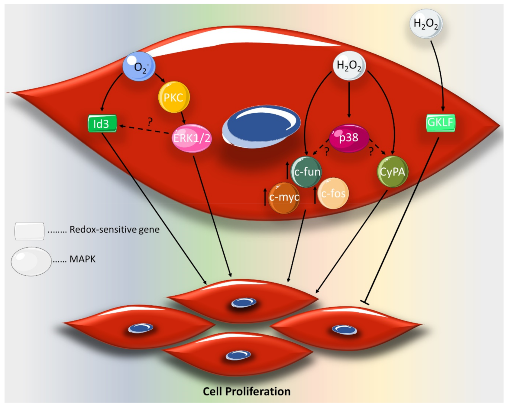
  56. Nickenig, G.; Baudler, S.; Müller, C.; Werner, C.; Werner, N.; Welzel, H.; STREHLOW, K.; BöHM, M. Redox-sensitive vascular smooth muscle cell proliferation is mediated by GKLF and Id3 in vitro and in vivo. Faseb J. 2002, 16, 1077–1086. [Google Scholar] [CrossRef] [PubMed] [Green Version]
  57. Satoh, K.; Nigro, P.; Berk, B.C. Oxidative stress and vascular smooth muscle cell growth: a mechanistic linkage by cyclophilin A. Antioxid. Redox Signal. 2010, 12, 675–682. [Google Scholar] [CrossRef] [PubMed] [Green Version]
  58. Jin, Z.-G.; Melaragno, M.G.; Liao, D.-F.; Yan, C.; Haendeler, J.; Suh, Y.-A.; Lambeth, J.D.; Berk, B.C. Cyclophilin A is a secreted growth factor induced by oxidative stress. Circ. Res. 2000, 87, 789–796. [Google Scholar] [CrossRef] [PubMed] [Green Version]
  59. Rao, G.N.; Berk, B.C. Active oxygen species stimulate vascular smooth muscle cell growth and proto-oncogene expression. Circ. Res. 1992, 70, 593–599. [Google Scholar] [CrossRef] [Green Version]
  60. Rao, G.N. Hydrogen peroxide induces complex formation of SHC-Grb2-SOS with receptor tyrosine kinase and activates Ras and extracellular signal-regulated protein kinases group of mitogen-activated protein kinases. Oncogene 1996, 13, 713. [Google Scholar]
  61. Brown, M.R.; Miller, F.J., Jr.; Li, W.G.; Ellingson, A.N.; Mozena, J.D.; Chatterjee, P.; Engelhardt, J.F.; Zwacka, R.M.; Oberley, L.W.; Fang, X.; et al. Overexpression of human catalase inhibits proliferation and promotes apoptosis in vascular smooth muscle cells. Circ. Res. 1999, 85, 524–533. [Google Scholar] [CrossRef] [PubMed] [Green Version]
  62. Fukui, T.; Ishizaka, N.; Rajagopalan, S.; Laursen, J.B.; Capers, Q.t.; Taylor, W.R.; Harrison, D.G.; de Leon, H.; Wilcox, J.N.; Griendling, K.K. p22phox mRNA expression and NADPH oxidase activity are increased in aortas from hypertensive rats. Circ. Res. 1997, 80, 45–51. [Google Scholar] [CrossRef] [PubMed]
  63. Szocs, K.; Lassegue, B.; Sorescu, D.; Hilenski, L.L.; Valppu, L.; Couse, T.L.; Wilcox, J.N.; Quinn, M.T.; Lambeth, J.D.; Griendling, K.K. Upregulation of Nox-based NAD(P)H oxidases in restenosis after carotid injury. Arter. Thromb Vasc Biol 2002, 22, 21–27. [Google Scholar] [CrossRef] [PubMed] [Green Version]
  64. Souza, H.P.; Souza, L.C.; Anastacio, V.M.; Pereira, A.C.; Junqueira, M.L.; Krieger, J.E.; da Luz, P.L.; Augusto, O.; Laurindo, F.R. Vascular oxidant stress early after balloon injury: evidence for increased NAD(P)H oxidoreductase activity. Free Radic. Biol. Med. 2000, 28, 1232–1242. [Google Scholar] [CrossRef]
  65. Bauersachs, J.; Bouloumie, A.; Fraccarollo, D.; Hu, K.; Busse, R.; Ertl, G. Endothelial dysfunction in chronic myocardial infarction despite increased vascular endothelial nitric oxide synthase and soluble guanylate cyclase expression: role of enhanced vascular superoxide production. Circulation 1999, 100, 292–298. [Google Scholar] [CrossRef]
  66. Rajagopalan, S.; Kurz, S.; Munzel, T.; Tarpey, M.; Freeman, B.A.; Griendling, K.K.; Harrison, D.G. Angiotensin II-mediated hypertension in the rat increases vascular superoxide production via membrane NADH/NADPH oxidase activation. Contribution to alterations of vasomotor tone. J. Clin. Invest. 1996, 97, 1916–1923. [Google Scholar] [CrossRef] [Green Version]
  67. Moldovan, L.; Moldovan, N.I.; Sohn, R.H.; Parikh, S.A.; Goldschmidt-Clermont, P.J. Redox changes of cultured endothelial cells and actin dynamics. Circ. Res. 2000, 86, 549–557. [Google Scholar] [CrossRef] [Green Version]
  68. Giannoni, E.; Buricchi, F.; Raugei, G.; Ramponi, G.; Chiarugi, P. Intracellular reactive oxygen species activate Src tyrosine kinase during cell adhesion and anchorage-dependent cell growth. Mol. Cell. Biol. 2005, 25, 6391–6403. [Google Scholar] [CrossRef] [Green Version]
  69. DalleDonne, I.; Milzani, A.; Colombo, R. H2O2-treated actin: assembly and polymer interactions with cross-linking proteins. Biophys. J. 1995, 69, 2710–2719. [Google Scholar] [CrossRef] [Green Version]
  70. Hinshaw, D.B.; Burger, J.M.; Beals, T.F.; Armstrong, B.C.; Hyslop, P.A. Actin polymerization in cellular oxidant injury. Arch. Biochem. Biophys. 1991, 288, 311–316. [Google Scholar] [CrossRef] [Green Version]
  71. Nishio, E.; Watanabe, Y. The involvement of reactive oxygen species and arachidonic acid in alpha 1-adrenoceptor-induced smooth muscle cell proliferation and migration. Br. J. Pharm. 1997, 121, 665–670. [Google Scholar] [CrossRef] [PubMed] [Green Version]
  72. Wang, Z.; Castresana, M.R.; Newman, W.H. Reactive oxygen and NF-kappaB in VEGF-induced migration of human vascular smooth muscle cells. Biochem. Biophys. Res. Commun. 2001, 285, 669–674. [Google Scholar] [CrossRef] [PubMed]
  73. Schroder, K.; Helmcke, I.; Palfi, K.; Krause, K.H.; Busse, R.; Brandes, R.P. Nox1 mediates basic fibroblast growth factor-induced migration of vascular smooth muscle cells. Arter. Thromb Vasc Biol 2007, 27, 1736–1743. [Google Scholar] [CrossRef] [PubMed] [Green Version]
  74. Wang, Z.; Castresana, M.R.; Newman, W.H. Reactive oxygen species-sensitive p38 MAPK controls thrombin-induced migration of vascular smooth muscle cells. J. Mol. Cell. Cardiol. 2004, 36, 49–56. [Google Scholar] [CrossRef] [PubMed]
  75. Meng, D.; Lv, D.D.; Fang, J. Insulin-like growth factor-I induces reactive oxygen species production and cell migration through Nox4 and Rac1 in vascular smooth muscle cells. Cardiovasc. Res. 2008, 80, 299–308. [Google Scholar] [CrossRef] [Green Version]
  76. Haurani, M.J.; Cifuentes, M.E.; Shepard, A.D.; Pagano, P.J. Nox4 oxidase overexpression specifically decreases endogenous Nox4 mRNA and inhibits angiotensin II-induced adventitial myofibroblast migration. Hypertension 2008, 52, 143–149. [Google Scholar] [CrossRef] [PubMed] [Green Version]
  77. Lee, M.Y.; San Martin, A.; Mehta, P.K.; Dikalova, A.E.; Garrido, A.M.; Datla, S.R.; Lyons, E.; Krause, K.H.; Banfi, B.; Lambeth, J.D.; et al. Mechanisms of vascular smooth muscle NADPH oxidase 1 (Nox1) contribution to injury-induced neointimal formation. Arter. Thromb. Vasc. Biol. 2009, 29, 480–487. [Google Scholar] [CrossRef] [PubMed] [Green Version]
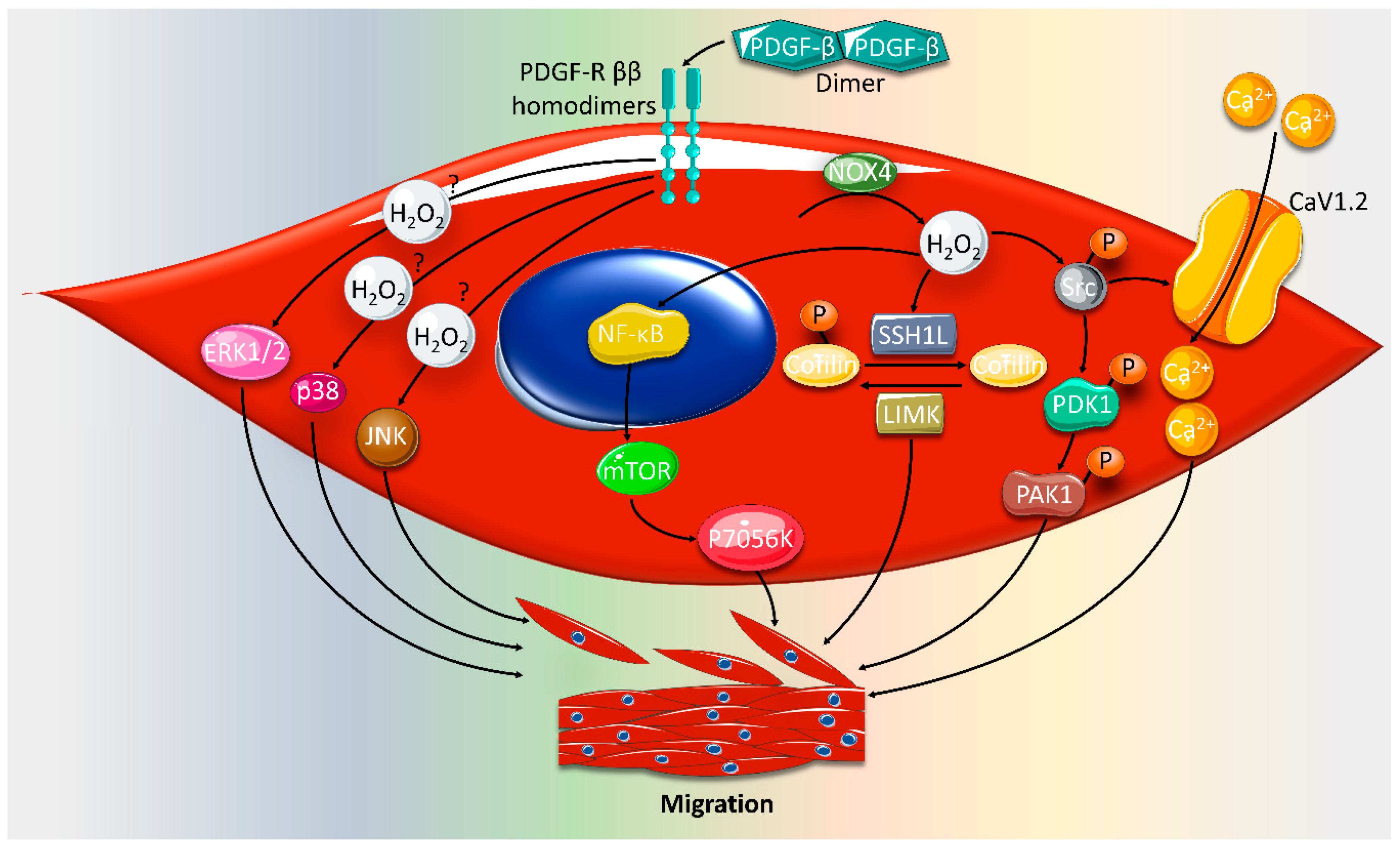
  78. Weber, D.S.; Taniyama, Y.; Rocic, P.; Seshiah, P.N.; Dechert, M.A.; Gerthoffer, W.T.; Griendling, K.K. Phosphoinositide-dependent kinase 1 and p21-activated protein kinase mediate reactive oxygen species-dependent regulation of platelet-derived growth factor-induced smooth muscle cell migration. Circ. Res. 2004, 94, 1219–1226. [Google Scholar] [CrossRef] [Green Version]
  79. Buetow, B.S.; Tappan, K.A.; Crosby, J.R.; Seifert, R.A.; Bowen-Pope, D.F. Chimera analysis supports a predominant role of PDGFRbeta in promoting smooth-muscle cell chemotaxis after arterial injury. Am. J. Pathol. 2003, 163, 979–984. [Google Scholar] [CrossRef]
  80. Floege, J.; Hudkins, K.L.; Davis, C.L.; Schwartz, S.M.; Alpers, C.E. Expression of PDGF alpha-receptor in renal arteriosclerosis and rejecting renal transplants. J. Am. Soc. Nephrol. Jasn 1998, 9, 211–223. [Google Scholar]
  81. Rubin, K.; Tingstrom, A.; Hansson, G.K.; Larsson, E.; Ronnstrand, L.; Klareskog, L.; Claesson-Welsh, L.; Heldin, C.H.; Fellstrom, B.; Terracio, L. Induction of B-type receptors for platelet-derived growth factor in vascular inflammation: possible implications for development of vascular proliferative lesions. Lancet 1988, 1, 1353–1356. [Google Scholar] [CrossRef]
  82. Lu, Q.-B.; Wan, M.-Y.; Wang, P.-Y.; Zhang, C.-X.; Xu, D.-Y.; Liao, X.; Sun, H.-J. Chicoric acid prevents PDGF-BB-induced VSMC dedifferentiation, proliferation and migration by suppressing ROS/NFκB/mTOR/P70S6K signaling cascade. Redox Biol. 2018, 14, 656–668. [Google Scholar] [CrossRef] [PubMed]
  83. San Martin, A.; Lee, M.Y.; Williams, H.C.; Mizuno, K.; Lassegue, B.; Griendling, K.K. Dual regulation of cofilin activity by LIM kinase and Slingshot-1L phosphatase controls platelet-derived growth factor-induced migration of human aortic smooth muscle cells. Circ Res 2008, 102, 432–438. [Google Scholar] [CrossRef] [PubMed] [Green Version]
  84. Bravo-Cordero, J.J.; Magalhaes, M.A.; Eddy, R.J.; Hodgson, L.; Condeelis, J. Functions of cofilin in cell locomotion and invasion. Nat. Rev. Mol. Cell Biol. 2013, 14, 405–415. [Google Scholar] [CrossRef] [Green Version]
  85. Guo, X.; Kashihara, T.; Nakada, T.; Aoyama, T.; Yamada, M. PDGF-induced migration of synthetic vascular smooth muscle cells through c-Src-activated L-type Ca(2+) channels with full-length CaV1.2 C-terminus. Pflug. Arch. Eur. J. Physiol. 2018, 470, 909–921. [Google Scholar] [CrossRef] [Green Version]
  86. Zhan, Y.; Kim, S.; Izumi, Y.; Izumiya, Y.; Nakao, T.; Miyazaki, H.; Iwao, H. Role of JNK, p38, and ERK in platelet-derived growth factor-induced vascular proliferation, migration, and gene expression. Arter. Thromb. Vasc. Biol. 2003, 23, 795–801. [Google Scholar] [CrossRef] [Green Version]
  87. Uzui, H.; Lee, J.-D.; Shimizu, H.; Tsutani, H.; Ueda, T. The role of protein-tyrosine phosphorylation and gelatinase production in the migration and proliferation of smooth muscle cells. Atherosclerosis 2000, 149, 51–59. [Google Scholar] [CrossRef]
  88. Mason, D.P.; Kenagy, R.D.; Hasenstab, D.; Bowen-Pope, D.F.; Seifert, R.A.; Coats, S.; Hawkins, S.M.; Clowes, A.W. Matrix metalloproteinase-9 overexpression enhances vascular smooth muscle cell migration and alters remodeling in the injured rat carotid artery. Circ. Res. 1999, 85, 1179–1185. [Google Scholar] [CrossRef]
  89. Southgate, K.M.; Davies, M.; Booth, R.; Newby, A. Involvement of extracellular-matrix-degrading metalloproteinases in rabbit aortic smooth-muscle cell proliferation. Biochem. J. 1992, 288, 93–99. [Google Scholar] [CrossRef] [Green Version]
  90. Grote, K.; Flach, I.; Luchtefeld, M.; Akin, E.; Holland, S.M.; Drexler, H.; Schieffer, B. Mechanical stretch enhances mRNA expression and proenzyme release of matrix metalloproteinase-2 (MMP-2) via NAD (P) H oxidase–derived reactive oxygen species. Circ. Res. 2003, 92, e80–e86. [Google Scholar] [CrossRef] [Green Version]
  91. Branchetti, E.; Poggio, P.; Sainger, R.; Shang, E.; Grau, J.B.; Jackson, B.M.; Lai, E.K.; Parmacek, M.S.; Gorman, R.C.; Gorman, J.H. Oxidative stress modulates vascular smooth muscle cell phenotype via CTGF in thoracic aortic aneurysm. Cardiovasc. Res. 2013, 100, 316–324. [Google Scholar] [CrossRef] [PubMed] [Green Version]
  92. Rajagopalan, S.; Meng, X.P.; Ramasamy, S.; Harrison, D.G.; Galis, Z.S. Reactive oxygen species produced by macrophage-derived foam cells regulate the activity of vascular matrix metalloproteinases in vitro. Implications for atherosclerotic plaque stability. J. Clin. Investig. 1996, 98, 2572–2579. [Google Scholar] [CrossRef] [Green Version]
  93. Brown, G.C. Regulation of mitochondrial respiration by nitric oxide inhibition of cytochrome c oxidase. Biochim. Et Biophys. Acta 2001, 1504, 46–57. [Google Scholar] [CrossRef] [Green Version]
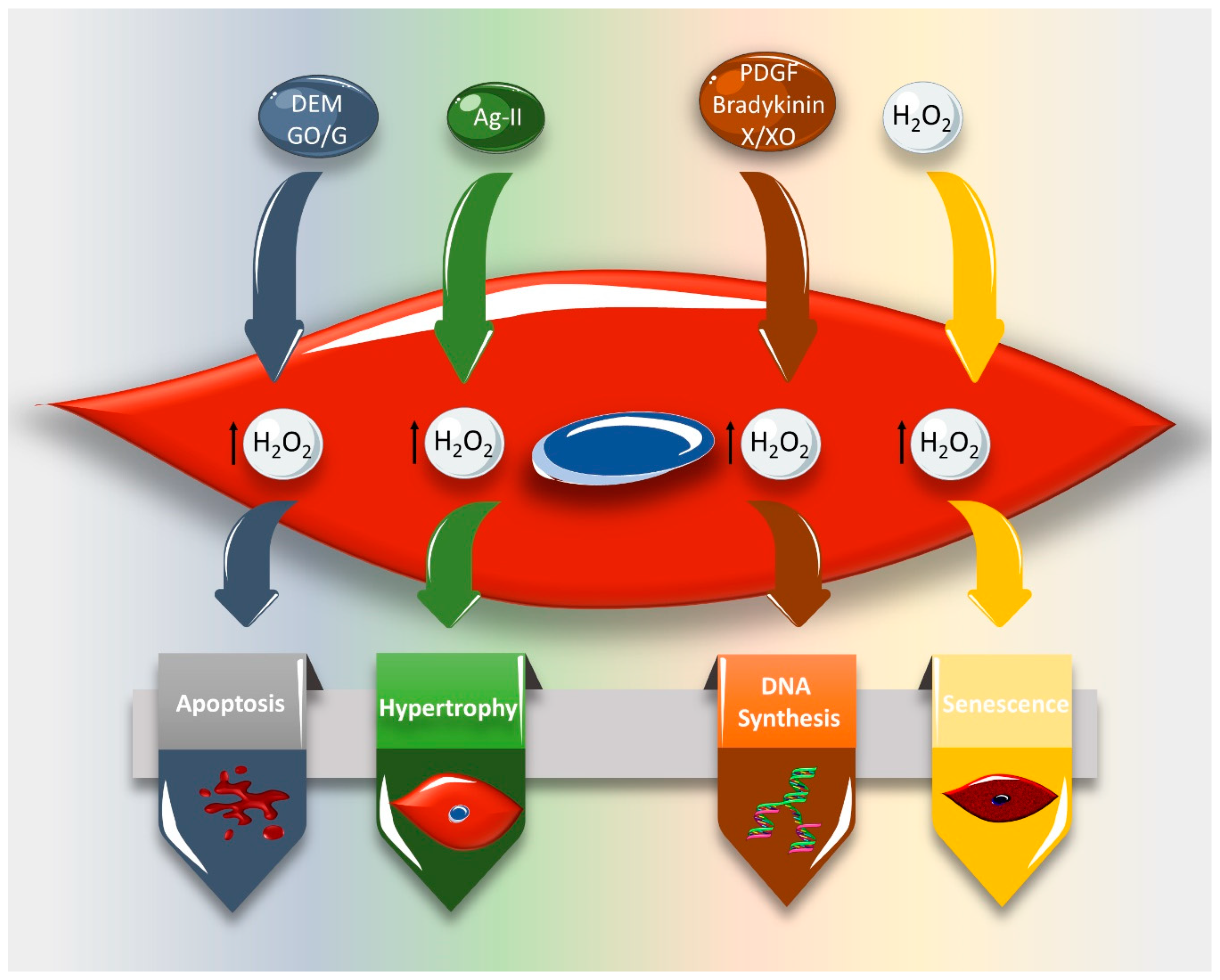
  94. Li, P.F.; Dietz, R.; von Harsdorf, R. Reactive oxygen species induce apoptosis of vascular smooth muscle cell. Febs Lett. 1997, 404, 249–252. [Google Scholar] [CrossRef] [Green Version]
  95. Kaneto, H.; Katakami, N.; Matsuhisa, M.; Matsuoka, T.-a. Role of reactive oxygen species in the progression of type 2 diabetes and atherosclerosis. Mediat. Inflamm. 2010, 2010. [Google Scholar] [CrossRef] [Green Version]
  96. Wang, J.; Uryga, A.K.; Reinhold, J.; Figg, N.; Baker, L.; Finigan, A.; Gray, K.; Kumar, S.; Clarke, M.; Bennett, M. Vascular smooth muscle cell senescence promotes atherosclerosis and features of plaque vulnerability. Circulation 2015, 132, 1909–1919. [Google Scholar] [CrossRef]
  97. Bennett, M.R. Apoptosis of vascular smooth muscle cells in vascular remodelling and atherosclerotic plaque rupture. Cardiovasc. Res. 1999, 41, 361–368. [Google Scholar] [CrossRef]
  98. Li, P.-F.; Dietz, R.; von Harsdorf, R.d. Differential effect of hydrogen peroxide and superoxide anion on apoptosis and proliferation of vascular smooth muscle cells. Circulation 1997, 96, 3602–3609. [Google Scholar] [CrossRef]
  99. Deshpande, N.N.; Sorescu, D.; Seshiah, P.; Ushio-Fukai, M.; Akers, M.; Yin, Q.; Griendling, K.K. Mechanism of hydrogen peroxide-induced cell cycle arrest in vascular smooth muscle. Antioxid. Redox Signal. 2002, 4, 845–854. [Google Scholar] [CrossRef]
  100. Popowich, D.A.; Vavra, A.K.; Walsh, C.P.; Bhikhapurwala, H.A.; Rossi, N.B.; Jiang, Q.; Aalami, O.O.; Kibbe, M.R. Regulation of reactive oxygen species by p53: implications for nitric oxide-mediated apoptosis. Am. J. Physiol.. Heart Circ. Physiol. 2010, 298, H2192–H2200. [Google Scholar] [CrossRef] [Green Version]
  101. Lee, S.; Byun, J.K.; Park, M.; Woo Kim, S.; Lee, S.; Kim, J.G.; Lee, I.K.; Choi, Y.K.; Park, K.G. Melatonin inhibits vascular smooth muscle cell proliferation and apoptosis through upregulation of Sestrin2. Exp. Ther. Med. 2020, 19, 3454–3460. [Google Scholar] [CrossRef] [PubMed] [Green Version]
  102. Griendling, K.K.; Harrison, D.G. Dual role of reactive oxygen species in vascular growth. Circ Res 1999, 85, 562–563. [Google Scholar] [CrossRef] [PubMed] [Green Version]
  103. Tsai, J.-C.; Jain, M.; Hsieh, C.-M.; Lee, W.-S.; Yoshizumi, M.; Patterson, C.; Perrella, M.A.; Cooke, C.; Wang, H.; Haber, E. Induction of apoptosis by pyrrolidinedithiocarbamate and N-acetylcysteine in vascular smooth muscle cells. J. Biol. Chem. 1996, 271, 3667–3670. [Google Scholar] [CrossRef] [Green Version]
  104. Abello, P.A.; Fidler, S.A.; Bulkley, G.B.; Buchman, T.G. Antioxidants modulate induction of programmed endothelial cell death (apoptosis) by endotoxin. Arch. Surg. 1994, 129, 134–140. [Google Scholar] [CrossRef] [PubMed]
  105. Rothstein, J.D.; Bristol, L.A.; Hosler, B.; Brown, R.H., Jr.; Kuncl, R.W. Chronic inhibition of superoxide dismutase produces apoptotic death of spinal neurons. Proc. Natl. Acad. Sci. USA 1994, 91, 4155–4159. [Google Scholar] [CrossRef] [Green Version]
  106. Roederer, M.; Staal, F.J.; Ela, S.W.; Herzenberg, L.A.; Herzenberg, L.A. N-acetylcysteine: potential for AIDS therapy. Pharmacology 1993, 46, 121–129. [Google Scholar] [CrossRef]
  107. Rao, G.; Lassegue, B.; Griendling, K.; Alexander, R.W. Hydrogen peroxide stimulates transcription of c-jun in vascular smooth muscle cells: role of arachidonic acid. Oncogene 1993, 8, 2759–2764. [Google Scholar]
  108. Rao, G.N.; Lassegue, B.; Griendling, K.K.; Alexander, R.W.; Berk, B.C. Hydrogen peroxide-induced c-fos expression is mediated by arachidonic acid release: role of protein kinase C. Nucleic Acids Res. 1993, 21, 1259–1263. [Google Scholar] [CrossRef] [Green Version]
  109. Fiorani, M.; Cantoni, O.; Tasinato, A.; Boscoboinik, D.; Azzi, A. Hydrogen peroxide-and fetal bovine serum-induced DNA synthesis in vascular smooth muscle cells: positive and negative regulation by protein kinase C isoforms. Biochim. Et Biophys. Acta (Bba)-Mol. Cell Res. 1995, 1269, 98–104. [Google Scholar] [CrossRef] [Green Version]
  110. Ma, Y.; Gong, X.; Mo, Y.; Wu, S. Polydatin inhibits the oxidative stress-induced proliferation of vascular smooth muscle cells by activating the eNOS/SIRT1 pathway. Int. J. Mol. Med. 2016, 37, 1652–1660. [Google Scholar] [CrossRef] [Green Version]
  111. Przybylska, D.; Janiszewska, D.; Gozdzik, A.; Bielak-Zmijewska, A.; Sunderland, P.; Sikora, E.; Mosieniak, G. NOX4 downregulation leads to senescence of human vascular smooth muscle cells. Oncotarget 2016, 7, 66429–66443. [Google Scholar] [CrossRef] [PubMed] [Green Version]
  112. Zafari, A.M.; Ushio-Fukai, M.; Akers, M.; Yin, Q.; Shah, A.; Harrison, D.G.; Taylor, W.R.; Griendling, K.K. Role of NADH/NADPH oxidase-derived H2O2 in angiotensin II-induced vascular hypertrophy. Hypertension 1998, 32, 488–495. [Google Scholar] [CrossRef] [PubMed] [Green Version]
  113. Hilenski, L.L.; Clempus, R.E.; Quinn, M.T.; Lambeth, J.D.; Griendling, K.K. Distinct subcellular localizations of Nox1 and Nox4 in vascular smooth muscle cells. Arterioscler. Thromb. Vasc. Biol. 2004, 24, 677–683. [Google Scholar] [CrossRef] [PubMed]
  114. Mueller, C.; Baudler, S.; Welzel, H.; Böhm, M.; Nickenig, G. Identification of a novel redox-sensitive gene, Id3, which mediates angiotensin II–induced cell growth. Circulation 2002, 105, 2423–2428. [Google Scholar] [CrossRef] [Green Version]
  115. Heidenreich, O.; Neininger, A.; Schratt, G.; Zinck, R.; Cahill, M.A.; Engel, K.; Kotlyarov, A.; Kraft, R.; Kostka, S.; Gaestel, M. MAPKAP kinase 2 phosphorylates serum response factor in vitro and in vivo. J. Biol. Chem. 1999, 274, 14434–14443. [Google Scholar] [CrossRef] [Green Version]
  116. Thuerauf, D.J.; Arnold, N.D.; Zechner, D.; Hanford, D.S.; DeMartin, K.M.; McDonough, P.M.; Prywes, R.; Glembotski, C.C. P38 mitogen-activated protein kinase mediates the transcriptional induction of the atrial natriuretic factor gene through a serum response element a potential role for the transcription factor atf6. J. Biol. Chem. 1998, 273, 20636–20643. [Google Scholar] [CrossRef] [Green Version]
  117. Clempus, R.E.; Sorescu, D.; Dikalova, A.E.; Pounkova, L.; Jo, P.; Sorescu, G.P.; Schmidt, H.H.; Lassegue, B.; Griendling, K.K. Nox4 is required for maintenance of the differentiated vascular smooth muscle cell phenotype. Arter. Thromb. Vasc. Biol. 2007, 27, 42–48. [Google Scholar] [CrossRef] [Green Version]
  118. Du, K.L.; Chen, M.; Li, J.; Lepore, J.J.; Mericko, P.; Parmacek, M.S. Megakaryoblastic leukemia factor-1 transduces cytoskeletal signals and induces smooth muscle cell differentiation from undifferentiated embryonic stem cells. J. Biol. Chem. 2004, 279, 17578–17586. [Google Scholar] [CrossRef] [Green Version]
  119. Sutliff, R.L.; Hilenski, L.L.; Amanso, A.M.; Parastatidis, I.; Dikalova, A.E.; Hansen, L.; Datla, S.R.; Long, J.S.; El-Ali, A.M.; Joseph, G. Polymerase delta interacting protein 2 sustains vascular structure and function. Arterioscler. Thromb. Vasc. Biol. 2013, 33, 2154–2161. [Google Scholar] [CrossRef] [Green Version]
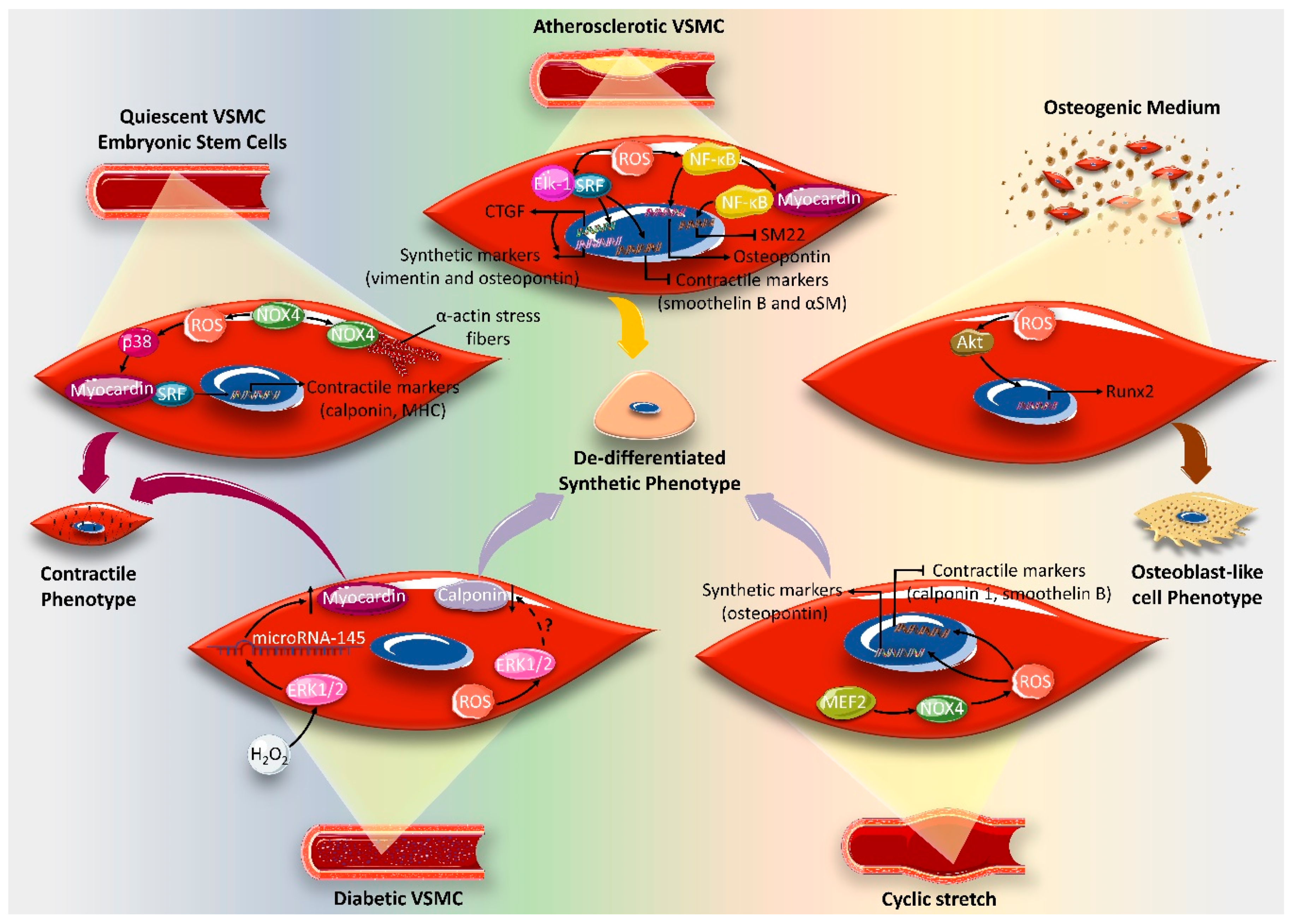
  120. Xiao, Q.; Luo, Z.; Pepe, A.E.; Margariti, A.; Zeng, L.; Xu, Q. Embryonic stem cell differentiation into smooth muscle cells is mediated by Nox4-produced H2O2. Am. J. Physiol. Cell Physiol. 2009, 296, C711–C723. [Google Scholar] [CrossRef] [Green Version]
  121. McDonald, O.G.; Wamhoff, B.R.; Hoofnagle, M.H.; Owens, G.K. Control of SRF binding to CArG box chromatin regulates smooth muscle gene expression in vivo. J. Clin. Investig. 2006, 116, 36–48. [Google Scholar] [CrossRef] [PubMed] [Green Version]
  122. Williams, E.S.; Wilson, E.; Ramos, K.S. NF-kappaB and matrix-dependent regulation of osteopontin promoter activity in allylamine-activated vascular smooth muscle cells. Oxid. Med. Cell. Longev. 2012, 2012, 496540. [Google Scholar] [CrossRef] [PubMed] [Green Version]
  123. O’Brien, E.R.; Garvin, M.R.; Stewart, D.K.; Hinohara, T.; Simpson, J.B.; Schwartz, S.M.; Giachelli, C.M. Osteopontin is synthesized by macrophage, smooth muscle, and endothelial cells in primary and restenotic human coronary atherosclerotic plaques. Arterioscler. Thromb. A J. Vasc. Biol. 1994, 14, 1648–1656. [Google Scholar]
  124. Tang, R.-H.; Zheng, X.-L.; Callis, T.E.; Stansfield, W.E.; He, J.; Baldwin, A.S.; Wang, D.-Z.; Selzman, C.H. Myocardin inhibits cellular proliferation by inhibiting NF-κB (p65)-dependent cell cycle progression. Proc. Natl. Acad. Sci. USA 2008, 105, 3362–3367. [Google Scholar] [CrossRef] [PubMed] [Green Version]
  125. Trion, A.; van der Laarse, A. Vascular smooth muscle cells and calcification in atherosclerosis. Am. Heart J. 2004, 147, 808–814. [Google Scholar] [CrossRef]
  126. Byon, C.H.; Javed, A.; Dai, Q.; Kappes, J.C.; Clemens, T.L.; Darley-Usmar, V.M.; McDonald, J.M.; Chen, Y. Oxidative stress induces vascular calcification through modulation of the osteogenic transcription factor Runx2 by AKT signaling. J. Biol. Chem. 2008, 283, 15319–15327. [Google Scholar] [CrossRef] [Green Version]
  127. Chettimada, S.; Ata, H.; Rawat, D.K.; Gulati, S.; Kahn, A.G.; Edwards, J.G.; Gupte, S.A. Contractile protein expression is upregulated by reactive oxygen species in aorta of Goto-Kakizaki rat. Am. J. Physiol. Heart Circ. Physiol. 2014, 306, H214–H224. [Google Scholar] [CrossRef] [Green Version]
  128. Carrillo-Sepulveda, M.A.; Matsumoto, T. Phenotypic modulation of mesenteric vascular smooth muscle cells from type 2 diabetic rats is associated with decreased caveolin-1 expression. Cell. Physiol. Biochem. 2014, 34, 1497–1506. [Google Scholar] [CrossRef]
  129. Katoh, Y.; Molkentin, J.D.; Dave, V.; Olson, E.N.; Periasamy, M. MEF2B is a component of a smooth muscle-specific complex that binds an A/T-rich element important for smooth muscle myosin heavy chain gene expression. J. Biol. Chem. 1998, 273, 1511–1518. [Google Scholar] [CrossRef] [Green Version]
  130. Blank, R.; Owens, G. Platelet-derived growth factor regulates actin isoform expression and growth state in cultured rat aortic smooth muscle cells. J. Cell. Physiol. 1990, 142, 635–642. [Google Scholar] [CrossRef]
  131. Sung, H.-J.; Eskin, S.G.; Sakurai, Y.; Yee, A.; Kataoka, N.; McIntire, L.V. Oxidative stress produced with cell migration increases synthetic phenotype of vascular smooth muscle cells. Ann. Biomed. Eng. 2005, 33, 1546–1554. [Google Scholar] [CrossRef] [PubMed]
  132. Alexander, M.R.; Owens, G.K. Epigenetic control of smooth muscle cell differentiation and phenotypic switching in vascular development and disease. Annu. Rev. Physiol. 2012, 74, 13–40. [Google Scholar] [CrossRef] [PubMed]
  133. Liu, R.; Jin, Y.; Tang, W.H.; Qin, L.; Zhang, X.; Tellides, G.; Hwa, J.; Yu, J.; Martin, K.A. Ten-eleven translocation-2 (TET2) is a master regulator of smooth muscle cell plasticity. Circulation 2013, 128, 2047–2057. [Google Scholar] [CrossRef] [PubMed] [Green Version]
  134. Kietzmann, T.; Petry, A.; Shvetsova, A.; Gerhold, J.M.; Görlach, A. The epigenetic landscape related to reactive oxygen species formation in the cardiovascular system. Br. J. Pharmacol. 2017, 174, 1533–1554. [Google Scholar] [CrossRef]
  135. Pons, D.; de Vries, F.R.; van den Elsen, P.J.; Heijmans, B.T.; Quax, P.H.; Jukema, J.W. Epigenetic histone acetylation modifiers in vascular remodelling: new targets for therapy in cardiovascular disease. Eur. Heart J. 2009, 30, 266–277. [Google Scholar] [CrossRef] [Green Version]
  136. Alkemade, F.E.; van Vliet, P.; Henneman, P.; van Dijk, K.W.; Hierck, B.P.; van Munsteren, J.C.; Scheerman, J.A.; Goeman, J.J.; Havekes, L.M.; Gittenberger-de Groot, A.C. Prenatal exposure to apoE deficiency and postnatal hypercholesterolemia are associated with altered cell-specific lysine methyltransferase and histone methylation patterns in the vasculature. Am. J. Pathol. 2010, 176, 542–548. [Google Scholar] [CrossRef] [Green Version]
  137. Bishop, T.; Ratcliffe, P.J. HIF hydroxylase pathways in cardiovascular physiology and medicine. Circ. Res. 2015, 117, 65–79. [Google Scholar] [CrossRef] [Green Version]
  138. Salminen, A.; Kaarniranta, K.; Kauppinen, A. Hypoxia-inducible histone lysine demethylases: impact on the aging process and age-related diseases. Aging Dis. 2016, 7, 180. [Google Scholar]
  139. Laukkanen, M.O.; Mannermaa, S.; Hiltunen, M.O.; Aittomäki, S.; Airenne, K.; Jänne, J.; Ylä-Herttuala, S. Local hypomethylation in atherosclerosis found in rabbit ec-sod gene. Arterioscler. Thromb. Vasc. Biol. 1999, 19, 2171–2178. [Google Scholar] [CrossRef] [Green Version]
  140. Ross, R. The pathogenesis of atherosclerosis: a perspective for the 1990s. Nature 1993, 362, 801–809. [Google Scholar] [CrossRef]
  141. Juarez, J.C.; Manuia, M.; Burnett, M.E.; Betancourt, O.; Boivin, B.; Shaw, D.E.; Tonks, N.K.; Mazar, A.P.; Doñate, F. Superoxide dismutase 1 (SOD1) is essential for H2O2-mediated oxidation and inactivation of phosphatases in growth factor signaling. Proc. Natl. Acad. Sci. 2008, 105, 7147–7152. [Google Scholar] [CrossRef] [PubMed] [Green Version]
  142. McDonald, O.G.; Owens, G.K. Programming smooth muscle plasticity with chromatin dynamics. Circ. Res. 2007, 100, 1428–1441. [Google Scholar] [CrossRef] [PubMed]
  143. Shimokawa, H. Reactive oxygen species promote vascular smooth muscle cell proliferation. Circ. Res. 2013, 113, 1040–1042. [Google Scholar] [CrossRef] [PubMed] [Green Version]
  144. Stampfer, M.J.; Hennekens, C.H.; Manson, J.E.; Colditz, G.A.; Rosner, B.; Willett, W.C. Vitamin E consumption and the risk of coronary disease in women. New Engl. J. Med. 1993, 328, 1444–1449. [Google Scholar] [CrossRef] [PubMed]
  145. Losonczy, K.G.; Harris, T.B.; Havlik, R.J. Vitamin E and vitamin C supplement use and risk of all-cause and coronary heart disease mortality in older persons: the Established Populations for Epidemiologic Studies of the Elderly. Am. J. Clin. Nutr. 1996, 64, 190–196. [Google Scholar] [CrossRef]
  146. Marques, B.; Trindade, M.; Aquino, J.; Cunha, A.; Gismondi, R.; Neves, M.; Oigman, W. Beneficial effects of acute trans-resveratrol supplementation in treated hypertensive patients with endothelial dysfunction. Clin. Exp. Hypertens. 2018, 40, 218–223. [Google Scholar] [CrossRef]
  147. Dyck, G.J.; Raj, P.; Zieroth, S.; Dyck, J.R.; Ezekowitz, J.A. The effects of resveratrol in patients with cardiovascular disease and heart failure: a narrative review. Int. J. Mol. Sci. 2019, 20, 904. [Google Scholar] [CrossRef] [Green Version]
  148. Greenberg, E.R.; Baron, J.A.; Karagas, M.R.; Stukel, T.A.; Nierenberg, D.W.; Stevens, M.M.; Mandel, J.S.; Haile, R.W. Mortality associated with low plasma concentration of beta carotene and the effect of oral supplementation. Jama 1996, 275, 699–703. [Google Scholar] [CrossRef]
  149. Rapola, J.M.; Virtamo, J.; Ripatti, S.; Huttunen, J.K.; Albanes, D.; Taylor, P.R.; Heinonen, O.P. Randomised trial of α-tocopherol and β-carotene supplements on incidence of major coronary events in men with previous myocardial infarction. Lancet 1997, 349, 1715–1720. [Google Scholar] [CrossRef] [Green Version]
  150. Eidelman, R.S.; Hollar, D.; Hebert, P.R.; Lamas, G.A.; Hennekens, C.H. Randomized trials of vitamin E in the treatment and prevention of cardiovascular disease. Arch. Intern. Med. 2004, 164, 1552–1556. [Google Scholar] [CrossRef] [Green Version]
  151. Stephens, N. Randomised controlled trial of vitamin E in patients with coronary disease: Cambrige Heart Antioxidant Study (CHAOS). Lancet 1996, 347, 408–416. [Google Scholar] [CrossRef]
  152. Stoner, G.D.; Sardo, C.; Apseloff, G.; Mullet, D.; Wargo, W.; Pound, V.; Singh, A.; Sanders, J.; Aziz, R.; Casto, B. Pharmacokinetics of anthocyanins and ellagic acid in healthy volunteers fed freeze-dried black raspberries daily for 7 days. J. Clin. Pharmacol. 2005, 45, 1153–1164. [Google Scholar] [CrossRef] [PubMed]
  153. Pellegrino, D. Antioxidants and cardiovascular risk factors. Diseases 2016, 4, 11. [Google Scholar] [CrossRef] [Green Version]
  154. Aruoma, O.I.; Halliwell, B.; Hoey, B.M.; Butler, J. The antioxidant action of N-acetylcysteine: its reaction with hydrogen peroxide, hydroxyl radical, superoxide, and hypochlorous acid. Free Radic. Biol. Med. 1989, 6, 593–597. [Google Scholar] [CrossRef]
  155. Griffiths, H.; Lunec, J. Ascorbic acid in the 21st century–more than a simple antioxidant. Environ. Toxicol. Pharmacol. 2001, 10, 173–182. [Google Scholar] [CrossRef]
  156. Duarte, T.L.; Lunec, J. When is an antioxidant not an antioxidant? A review of novel actions and reactions of vitamin C. Free Radic. Res. 2005, 39, 671–686. [Google Scholar] [CrossRef]
  157. Goszcz, K.; Deakin, S.J.; Duthie, G.G.; Stewart, D.; Leslie, S.J.; Megson, I.L. Antioxidants in cardiovascular therapy: panacea or false hope? Front. Cardiovasc. Med. 2015, 2, 29. [Google Scholar] [CrossRef]
  158. Bailey, S.R.; Mitra, S.; Flavahan, S.; Flavahan, N.A. Reactive oxygen species from smooth muscle mitochondria initiate cold-induced constriction of cutaneous arteries. Am. J. Physiol. Heart Circ. Physiol. 2005, 289, H243–H250. [Google Scholar] [CrossRef]
Figure 1. Redox signaling pathways regulating vascular smooth muscle cell (VSMC) proliferation. Superoxide anion, O2, induces cell proliferation by activating the mitogen-activated protein kinase (MAPK), ERK1/2, or upregulating the transcription factor Id3. Hydrogen peroxide, H2O2, promotes VSMC proliferation by activating the p38 MAPK, the CypA chaperone protein and the proto-oncogenes c-myc, c-fos and c-jun. The inhibitory actions of H2O2 are elicited via the redox sensitive transcription factor gut-enriched Kruppel-like factor, GKLF. Figure key: arrow: activation, block arrow: inhibition, up-arrow: upregulation, question mark: potential crosstalk.
Ijms 21 08764 g001
Figure 2. Platelet-derived growth factor-β (PDGF-β)-activated pathways mediating vascular smooth muscle cell (VSMC) migration. After platelet-derived growth factor receptor-β (PDGF-R ββ ) activation, NADPH oxidase-1 (Nox1)-released peroxide (H2O2) activates Slingshot1L (SSLH1) and LIM kinase (LIMK), cofilin phosphatase and kinase, respectively. The net result is cofilin.dephosphorylation leading to actin reorganization and ultimately migration. Furthermore, PDGF-induced H2O2 activates the Src/ (phosphoinositide-dependent kinase-1) PDK1/ (p21-activated protein kinase) PAK1 signaling pathway mediating VSMC migration. Additionally, Src activation increases L-type voltage-dependent calcium channel (CaV1.2) activity leading to increased intracellular calcium (Ca2+) concentration, and consequently VSMC migration. In addition, ERK1/2, JNK and p38 mediate PDGF-induced migration. Whether the activation of these MAPKs is ROS-dependent is yet to be determined. Figure key: arrow: activation, question mark: potential crosstalk.
Ijms 21 08764 g002
Figure 3. Reactive oxygen species (ROS) regulation of cellular signaling pathways involved in vascular smooth muscle cell (VSMC) fate and cell cycle progression. Superoxides anion, O2, -induces DNA synthesis, and consequently cell cycle progression, by activating ERK1/2. Alternatively, O2 mediates angiotensin II (Ang-II)-induced cell cycle progression via upregulating Id3 transcription factor, which in turn downregulates cell cycle proteins, including p27, p53 and p21, leading to cell cycle progression. Nitric oxide (NO)-induced ROS induce VSMC apoptosis associated with DNA synthesis inhibition. Hydrogen peroxide (H2O2) downregulates NADPH oxidase (NOX4), leading to cell senescence and DNA synthesis inhibition. H2O2 also attenuates cell cycle progression by upregulating the transcription factor gut-enriched Kruppel-like factor (GKLF) via p38. H2O2 regulates cell cycle proteins and proto-oncogenes to induce cell cycle progression or cell death. Melatonin upregulates sestrin2 leading to ROS inhibition, which decreases VSMC apoptosis. Basal ROS inhibition by catalase overexpression or antioxidants, (Pyrrolidinedithiocarbamate) PDTC or (N-acetylcysteine) NAC, initiates VSMC apoptosis.
Ijms 21 08764 g003
Figure 4. Various factors determining the effect of ROS on VSMC fate. ROS-inducing stimulus, ROS concentrations, as well as ROS type, play a major role in VSMC response. ROS-initiated signaling pathways involving redox-sensitive genes underwrite VSMC cell fate regulation. The vascular beds from which VSMCs are isolated contribute to their differential responses to ROS.
Ijms 21 08764 g004
Figure 5. Diverse vascular smooth muscle cell (VSMC) responses to different hydrogen peroxide (H2O2)-generating stimuli. Glucose oxidase/glucose (GO/G)- or diethylmaleate (DEM)-induced H2O2 leads to VSMC apoptosis, while angiotensin II (Ang II)-induced H2O2 causes VSMC hypertrophy. H2O2 released in response to xanthine/xanthine oxidase, platelet-derived growth factor (PDGF), or bradykinin promotes DNA synthesis and, consequently, VSMC proliferation. Treating human VSMCs with H2O2 induces cell senescence. Figure key: up-arrow: increment.
Ijms 21 08764 g005
Figure 6. The impact of the vascular smooth muscle cell (VSMC) microenvironment on determining the effect of reactive oxygen species (ROS) on phenotypic switch. Oxidative stress seems to be crucial for maintaining the contractile phenotype of quiescent VSMCs and for the differentiation of embryonic contractile VSMCs. NAPDH oxidase (NOX4)-produced hydrogen peroxide (H2O2) activates the transcription complex serum response factor (SRF)/myocardin via p38. This complex translocates to the nucleus and upregulates the transcription of contractile markers, such as calponin and myosin heavy chain. NOX4 also seems to play a structural role in maintaining a contractile phenotype by binding to α-actin stress fibers, characteristic of this phenotype. In the context of atherosclerosis, ROS induces VSMC dedifferentiation by activating the NF-κB and/or Elk-1/SRF signaling pathways. NF-κB upregulates the transcription of the synthetic marker osteopontin, and associates with myocardin to repress the myocardin-dependent contractile gene expression of smooth muscle 22 (SM22). Alternatively, ROS promote Elk-1/SRF complex formation, which activates the transcription of synthetic markers, vimentin and osteopontin, via connective tissue growth factor (CTGF). The Elk-1/SRF complex, alternatively, downregulates the contractile markers smoothelin B and α-smooth muscle (αSM). In an osteogenic medium, ROS induce VSMCs, which induces transition to the osteoblast-like cell phenotype, characteristic of vessel calcification in advanced atherosclerosis. The ROS-induced osteoblast-like cell phenotype is mediated via AKT-activated Runx2, a key transcription factor for osteogenic differentiation. In diabetic VSMC, ROS induce a synthetic phenotype by decreasing calponin, probably via ERK1/2. Conversely, ROS provoke the contractile phenotypic switch of diabetic VSMC by upregulating microRNA-145, which in turn increases the activity of myocardin in an ERK1/2-dependent manner. Cyclic stretch evokes a VSMC synthetic phenotypic switch through NOX1-derived ROS release via myocyte enhancer factor 2B (MEF2B), resulting in the upregulation of osteopontin and the downregulation of contractile markers calponin1 and smoothelin B. Figure key: up-arrow: upregulation, down-arrow: downregulation, arrow: activation, block arrow: inhibition, question mark: potential crosstalk.
Ijms 21 08764 g006
Publisher’s Note: MDPI stays neutral with regard to jurisdictional claims in published maps and institutional affiliations.

