Review

Transcription Factors in the Development and Function of Group 2 Innate Lymphoid Cells

Laboratory for Transcriptional Regulation, RIKEN Center for Integrative Medical Sciences (IMS), 1-7-22 Suehiro-cho, Tsurumi-ku, Yokohama 230-0045, Japan
*
Author to whom correspondence should be addressed.
Present address: Department of Medical Biology, Akita University School of Medicine, 1-1-1 Hondo, Akita 010-8543, Japan
Int. J. Mol. Sci. 2019, 20(6), 1377; https://doi.org/10.3390/ijms20061377
Submission received: 28 February 2019 / Revised: 15 March 2019 / Accepted: 18 March 2019 / Published: 19 March 2019

Abstract

:
Group 2 innate lymphoid cells (ILC2s) are tissue-resident cells and are a major source of innate TH2 cytokine secretion upon allergen exposure or parasitic-worm infection. Accumulating studies have revealed that transcription factors, including GATA-3, Bcl11b, Gfi1, RORα, and Ets-1, play a role in ILC2 differentiation. Recent reports have further revealed that the characteristics and functions of ILC2 are influenced by the physiological state of the tissues. Specifically, the type of inflammation strongly affects the ILC2 phenotype in tissues. Inhibitory ILC2s, memory-like ILC2s, and ex-ILC2s with ILC1 features acquire their characteristic properties following exposure to their specific inflammatory environment. We have recently reported a new ILC2 population, designated as exhausted-like ILC2s, which emerges after a severe allergic inflammation. Exhausted-like ILC2s are featured with low reactivity and high expression of inhibitory receptors. Therefore, for a more comprehensive understanding of ILC2 function and differentiation, we review the recent knowledge of transcriptional regulation of ILC2 differentiation and discuss the roles of the Runx transcription factor in controlling the emergence of exhausted-like ILC2s. The concept of exhausted-like ILC2s sheds a light on a new aspect of ILC2 biology in allergic diseases.

1. Introduction

Innate lymphoid cells (ILCs) are lymphocytes responsible for innate cytotoxicity or helper functions [1,2,3]. While ILCs with cytotoxic capability are conventional natural killer (NK) cells which do not have tissue-resident properties, ILCs with helper functions generally reside in the tissue, provoke immune responses against pathogens, and maintain mucosal integrity [4,5]. Helper ILCs are unable to directly sense pathogenic antigens because they do not express antigen-specific receptors such as T cell receptor and B cell receptor. Instead, helper ILCs are generally stimulated by cytokines released from damaged-epithelial cells, phagocytes, and dendritic cells [6,7,8,9,10,11]. Based on cytokine production and requirements of transcription factors, helper ILCs are classified into three distinct subsets; ILC1s, ILC2s, and ILC3s which mirror TH1, TH2, and TH17, respectively [3,12]. ILC1s secrete interferon g (IFNγ) and require T-bet for their differentiation [13]. T-bet is also important for the final maturation of conventional NK cells, which is highly dependent also on eomesodermin (Eomes) [14]. ILC2s are characterized by the production of the TH2 cytokines interleukin 4 (IL-4), IL-5, IL-13, and granulocyte-macrophage colony-stimulating factor (GM-CSF). All of these expressions are positively regulated by the transcription factor GATA binding protein 3 (GATA-3) [15]. Both RAR-related orphan receptor gamma t (RORγt) and aryl hydrocarbon receptor (AhR) are required for the differentiation of ILC3s as well as for the production of IL-17 or IL-22 by ILC3s [16,17,18]. A substantial number of ILC1s are found in the liver, intestinal intraepithelial layer, intestinal lamina propria, adipose tissue, and the skin [13,19,20,21]. ILC2s are preferentially localized in the lung, skin, adipose tissue, fat-associated lymphoid clusters, and the intestinal lamina propria [7,8,11,22]. ILC3s are abundant in the intestinal lamina propria, skin, and secondary lymphoid organs, such as Peyer’s patches and mesenteric lymph nodes [16,18,23].
All ILC subsets develop from common lymphoid progenitor cells (CLPs) [24]. During the first commitment process from CLPs to the ILC lineage, early innate lymphoid progenitor cells (EILPs) are generated [25]. EILPs give rise to conventional NK cells and all helper ILC subsets. EILPs are marked by the expression of T cell factor 1 (TCF-1), thymocyte selection associated high mobility group box (Tox), and nuclear factor, interleukin 3 regulated (NFIL3) [25,26,27]. Progenitors specific to helper ILCs are Id2+ common helper innate lymphoid progenitors (CHILPs) that lose the developmental potential for conventional NK cells [13]. Half of all CHILPs express promyelocytic leukemia zinc finger (PLZF) and are designated as innate lymphoid cell precursors (ILCPs) [10,24]. ILCPs can differentiate into helper ILCs except for a subpopulation of ILC3s, lymphoid tissue inducer-like (Lti-like) cells.
ILC2s have been extensively studied in the research field of allergy or parasitic worm infection [9]. Physiological roles of ILC2s are also implicated in metabolic homeostasis and virus infection [22,28,29]. Steady state ILC2s in tissues constitutively produce IL-5, which recruits eosinophils to the tissues [30]. Eosinophils are one of the essential components of allergy, as well as cytotoxic effector cells against parasitic worms. When mucosal tissues are injured by allergens or worms, IL-33, IL-25, and thymic stromal lymphopoietin (TSLP) are released from the damaged epithelial cells [31]. ILCs sense these signal cytokines by their cognate receptors and produce a variety of TH2 cytokines, including IL-5, IL-9, and IL-13. IL-9 is necessary for the maintenance of ILC2 activity in an autocrine manner during the early phase of an allergic reaction [32]. IL-13 from ILC2s induces chemokine CCL17 expression from dendritic cells to attract TH2 cells, resulting in enhanced TH2 responses [33]. ILC2s are an early source of amphiregulin, which is a ligand of the epidermal growth factor receptor and plays a critical role in the repair of damaged epithelial cells [28]. Therefore, ILC2s are involved in the initiation of allergy as well as tissue-repair.
The functional state of ILC2s is influenced by environmental cues. ILC2s acquire ILC1-like properties and produce IFNγ when mice are infected with viruses and bacteria or are exposed to cigarette smoke to induce chronic obstructive pulmonary-like disease [34,35]. IL-1b, IL-12, and IL-18 are involved in the phenotypic conversion from ILC2s to ILC1-like cells. ILC2s also acquire “memory-like” signatures [36]. Once ILC2s are stimulated with IL-33, the activated ILC2s live long in the tissue and produce increased levels of TH2 cytokines upon a second challenge. Systemic IL-25 treatment and helminth infection induce inflammatory ILC2s (iILC2s), which are characterized by IL-17 production [37]. iILC2s migrate from the site of inflammation to the systemic tissues through lymphatics in an S1P-depenent manner [38]. Despite such migratory capacity of iILC2s, the majority of ILC2s reside in the tissue during and after helminth infection [4].
Transcription factors specifically required for ILC2 differentiation have been studied. Previous studies demonstrated the importance of transcription factors, such as GATA-3, B-Cell lymphoma/leukaemia 11B (Bcl11b), growth factor independent 1 transcription repressor (Gfi-1), RAR related orphan receptor A (RORα), and v-ets erythroblastosis virus E26 oncogene homolog 1 (Ets-1), for ILC2 differentiation [7,15,39,40,41,42,43,44,45,46]. ILC2s change their characteristics in response to environments. The mechanism by which transcription factor networks dictate phenotypical changes of ILC2s remains to be determined. For a comprehensive understanding of physiological roles of ILC2s in the allergic pathology, we summarize the current understanding of the transcriptional control of ILC2 differentiation and function, followed by our recent findings regarding how Runt-related transcription factor (Runx) proteins regulate the functional state of ILC2.

2. Transcription Factors Involved in the Development of Group 2 Innate Lymphoid Cells (ILC2s)

2.1. GATA-3

GATA-3 is necessary for the differentiation of CHILPs, which specifically give rise to all helper ILC subsets (Figure 1) [13]. While ILC1s and ILC3s express GATA-3, the expression level of GATA-3 in ILC2s is significantly higher compared to other ILCs. GATA-3 binds the gene loci which are critical for ILC2 activity. The genes positively regulated by GATA-3 include Il5, Il13, Areg encoding amphiregulin, Il1rl1 encoding IL-33 receptor ST2, Il9, and Il2ra [15]. The conditional deletion of GATA-3 in mature ILC2s leads to reduced cytokine production and hypo proliferation [15,39,40]. Therefore, GATA-3 governs ILC2 identity, differentiation, homeostasis, and function.

2.2. Bcl11b

Bcl11b is the earliest marker for the ILC2-lineage. The Bcl11b-expressing cells among the CHILP population specifically develop into ILC2s (Figure 1) [41]. Bcl11b ablation in hematopoietic stem cells impairs ILC2 differentiation in the bone marrow and peripheral tissues. Bcl11b expression in ILC subsets is controversial. While Yu et al. suggested that Bcl11b expression among ILC subsets was restricted to ILC2s [43], and Walker et al. showed that ILC1s and ILC3s expressed Bcl11b [42]. Conditional deletion of Bcl11b in ILC2s results in the generation of ILC2s with ILC3 gene signatures [41]. Thus, Bcl11b-deficient ILC2s have a reduction in GATA-3, Gfi-1, and RORα expression, and an increase in sex determining region Y-box 4 (Sox4), RORγt, and AhR expression. Bcl11b is associated with the Gfi1 and Ahr gene loci, but not the Gata3 or Rorc gene loci, suggesting that Gfi1 and AhR expression may be directly regulated by Bcl11b. Enhanced Gfi-1 expression in Bcl11b-deficient ILC2s restores GATA-3 expression and down-regulates RORγt expression. Therefore, Bcl11b positively regulates GATA-3 expression and negatively regulates RORγt expression through Gfi-1 induction.

2.3. Gfi-1

Gfi-1 expression in ILC2 is relatively higher than other ILCs. Gfi-1 is required for ILC2 differentiation and optimal TH2 cytokine responses to IL-33 [44]. Gfi-1 positively regulates the expression of Gata3 and Il1rl1 (Figure 1). Deletion of Gfi-1 causes aberrant IL-17a production by ILC2s due to dysregulated expression of RORγt [44]. Increased RORγt expression in Gfi-1-deficient ILC2s may be explained by increased expression of Sox4, which is known to induce RORγt expression. Thus, Gfi-1 suppresses the machinery for the ILC3 lineage and maintains the effector state of ILC2s in allergic diseases.

2.4. RORα

Although RORα is expressed in CHILPs and all ILC subsets, it is specifically necessary for ILC2 differentiation among ILC subsets (Figure 1) [7,45,47]. A RORα-dependent ILC2 differentiation mechanism has not been described yet. Differential interaction of RORα with the gene loci in ILC2s and other ILCs should be examined to understand RORα function in ILC2 differentiation and function. RORα-deficient staggerer mice have been used to observe physiological effects resulting from ILC2 deficiency [7,45]. However, deletion of RORα dampens regulatory T cell (Treg) and ILC3 function, but not their differentiation [48,49]. These data suggest that there might be other unknown immunological defects in the RORα-deficient staggerer mice and the related mice with conditional deletion of RORα.

2.5. Ets1

Ets-1 induces Id2 which is critical for differentiation of ILC progenitors, CHILPs [46]. However, for unknown reasons, ILC2s without Ets-1 are reduced only in the bone marrow, but not in the lung. Ets-1 is required for ILC2s to expand and produce IL-5 and IL-13 in response to IL-33. In contrast, IL-6 and IL-9 production are up-regulated in Ets-1-deficient ILC2s cultured with IL-33. It remains to be elucidated how Ets-1 regulates the ILC2 fitness and function.

3. Runx Proteins and Immune Cells

3.1. Global Effects of Runx Proteins in Immune Cells

Runx proteins are a family of transcription factors necessary for many biological processes ranging from differentiation, function, proliferation, tumorigenesis, and cellular identity [50,51]. There are three mammal members in the Runx protein family: Runx1, Runx2, and Runx3. All Runx proteins require heterodimer formation with Subunit b of core binding factor (Cbfβ) to exert their function as transcription factors. All three Runx genes generate two transcripts from distal (P1) and proximal (P2) promoters [52]. These two transcripts generate Runx protein isoforms with distinct N-terminal sequences. Utilization of the promoters differs among cell types. Runx1 and Runx3 genes are expressed in many types of immune cells [50]. Runx1 is required for the emergence of hematopoietic stem cells from hemogenic endothelium. Loss of Runx1 causes embryonic lethality due to bleeding in the brain caused by the absence of platelets [53,54]. Runx1 is also involved in the early thymocyte differentiation and development of invariant natural killer T (iNKT) cells, regulatory T cells and TH17 cells [51,55,56,57,58]. Runx3 is a critical transcription factor for the differentiation of CD8+ T cells and TH1 cells [56,59]. Runx3 is crucial to induce effector genes such as Ifng, Gzmb, Eomes, and to repress Il4 gene in TH1 cells. Runx3 transcript from the P1 promoter is correlated with protein expression level in T cells because the P2-Runx3 transcript utilizes IRES-mediated translation [52,60]. Runx2 is required for the differentiation of osteoblasts and bone formation [61,62]. Runx2 expression in hematopoietic cells is rather restricted to plasmacytoid dendritic cells [63]. Runx2-deficient plasmacytoid dendritic cells have deficits in migrating to the peripheral tissues.

3.2. Runx3 Is Required for the Differentiation of ILC1s and ILC3s

Helper ILCs and conventional NK cells preferentially express Runx3 mRNA among all Runx family genes [64,65]. However, ILC subsets are characterized by the levels of Runx3 P1 transcripts and Runx3 protein expression. High Runx3 protein expression is observed in NK cells, which are innate counterparts of CD8+T cells, and in ILC1s, innate counterparts of TH1 cells. ILC3s, innate counterpart for TH17 cells, express an intermediate level of Runx3, while Runx1 is likely to be a major Runx protein to drive TH17 program. Runx3 expression is very low in ILC2s, innate lymphoid cells. Ablation of Runx3 in all hematopoietic cells leads to a reduction of NK cells, ILC1s, and ILC3s but not ILC2s. Runx3 is necessary for optimal IFNγ production by ILC1s. Ablation of Cbfβ or a combined loss of both Runx1 and Runx3 result in a dramatic reduction of NK cells and ILC1s, suggesting that Runx1 compensates for the loss of Runx3 in these cells. ILC1s fall into an apoptotic state in the absence of Runx proteins, which is a result of low expression of anti-apoptotic Bcl2. For the development of ILC3s, Runx3 induces expression of an ILC3 master regulator gene, RORγt mainly by direct binding to the intronic enhancer region of the RORγt [66]. AhR is also required for full production of IL-17 and IL-22 together with RORγt [17]. Runx3 deletion impairs expression of Ahr in ILC3s. Upon a half dosage of RORγt expression caused by heterozygous gene knock-out, Ahr expression is reduced by half in the ILC3s. Thus, Runx3 regulates RORγt expression and its downstream target, AhR.
Lymphoid tissue-inducer cells (Lti) cells are indispensable for secondary lymphoid organogenesis and are considered as a subpopulation of ILC3s because Lti cells are characterized by and are dependent on RORγt expression. Lti cells appear in the embryonic intestine at embryonic day 12. An early study clarified the importance of Runx1 for Lti cell differentiation via RORγt induction in the embryos [67]. Although RORγt expression in Lti-cells is reduced by Runx3 deficiency, the number of Lti cells in the embryonic intestine is normal in the absence of Runx3 [64]. These data suggest that there might be distinct functions of Runx proteins for the generation of Lti cells.
Runx proteins also regulate early ILC differentiation. Runx1 and Runx3 expression in ILC progenitors increases as CLPs give rise to CHILPs and ILCPs (Figure 1) [64]. Expression of Runx1 and Runx3 reaches the highest level at an ILCP stage in the course of ILC differentiation from CLPs to ILC subsets. Runx proteins are essential to induce PLZF expression, which is the marker for ILCPs, by direct interaction with an enhancer of the Zbtb16 locus encoding PLZF [68]. ILC1s and ILC3s down-regulate Runx1 expression but maintain intermediate to high levels of Runx3 expression [64]. In contrast, down-regulation of Runx3 occurs in ILC2 progenitor cells and ILC2s which express more Runx1 than other ILCs. Therefore, the levels of Runx3 expression specifies ILC lineages.

4. The Function of Runx Proteins in ILC2s

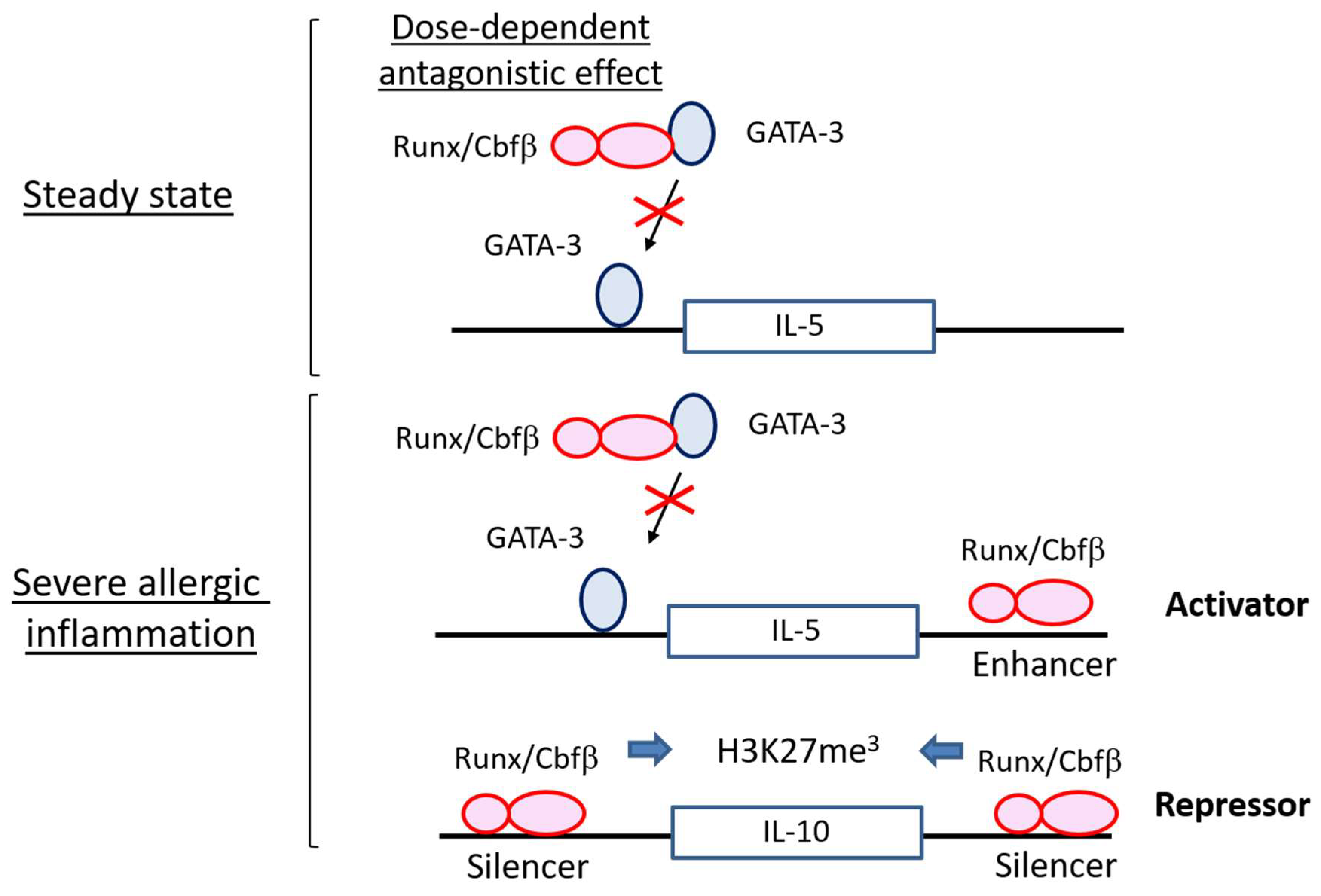
4.1. Runx Proteins Prevent Steady-State ILC2s from Overactivation

Runx3 deletion is not enough for obvious phenotypic changes in ILC2s [64,69]. ILC2s lacking Runx3 normally populate the peripheral tissues and are functionally intact because Runx1 is also expressed and compensate Runx3 function in ILC2s. When the function of all Runx proteins is abrogated by ablation of Cbfβ, a binding partner of Runx proteins, in the ILC2s, the ILC2s are normally present in the tissues but exhibit an activated phenotype even in the steady-state condition. Killer cell lectin-like receptor G1 (KLRG1) is a known marker for activated ILC2s, and Thy1 is down-regulated in the ILC2s when they are stimulated by IL-25. Interestingly, loss of Runx function in ILC2s induces high KLRG1 expression and low Thy1 expression with unleashed IL-5 production. As ILC2s are a major source of IL-5, which recruits eosinophils to the lung, eosinophils in the bronchoalveolar space are increased by overproduction of IL-5 from the activated ILC2s lacking Runx function. The infiltration of eosinophils to the bronchoalveolar space seems to occur at subclinical levels because lung epithelial cells are not significantly damaged by the adoptive transfer of the Cbfβ-deficient ILC2s.
The activated ILC2 phenotype caused by Cbfβ deficiency is reminiscent of those observed in the T cells lacking Cbfβ function. TH2 skewing occurs in CD4 T cells in the absence of Cbfβ, resulting in asthma-like symptoms in the lung [59]. Runx/Cbfβ complexes associate and antagonize GATA-3, a master regulator of TH2 cells, in T cells [70]. Runx/Cbfβ complexes also suppress the expression of IL-4, which is a critical TH2 cytokine for type 2 immunity, through binding to a silencer region in the Il4 gene locus [59]. Therefore, the balance between Runx and GATA-3 dictates the functional state of TH2 cells.
ILC2 activity at steady state is also determined by the balancing action of Runx proteins against GATA-3 (Figure 2). While GATA-3 overexpression increases IL-5 production in ILC2s, Runx3 cancels such effects in ILC2s [69]. In addition to IL-5, transcriptome analysis demonstrates that many genes regulated by GATA-3 are inversely regulated by Runx proteins. In steady-state ILC2s, IL-5 production is increased by the absence of Cbfβ, presumably via antagonizing GATA-3 function. However, we cannot deny the possibility that Runx/Cbfβ complexes work as a direct repressor for the Il5 gene as is the case for Il4, because Cbfβ associates with several genomic regions around the Il5 gene in ILC2s. Deletion of either Runx1 or Runx3 alone does not induce IL-5 overproduction from ILC2s. However, when both Runx1 and Runx3 are deleted, ILC2s show the activated phenotype, suggesting that the redundant function of Runx proteins restrains the basic activity of ILC2s in steady-state.

4.2. Runx Proteins Inhibit the Emergence of Exhausted-Like ILC2s during Allergic Inflammation

ILC2s are activated by IL-33, IL-25, TSLP, TNF superfamily ligand TL1A (TL1A), and neuropeptides such as vasoactive intestinal polypeptide (VIP), neuromedin U and, calcitonin gene-related peptide [6,11,30,31,71,72,73]. Furthermore, IL-33 is the most potent ILC2 stimulator in vitro and in vivo and plays an important role in the pathogenesis of allergy. Interestingly, Cbfβ-deficient ILC2s have defects in ILC2 cytokine production and proliferation during in vitro responses to IL-33 [69]. Expression of Il5, Il9, Il13, Csf2 encoding GM-CSF, and Areg encoding amphiregulin upon IL-33 stimulation is all downregulated in Cbfβ-deficient ILC2s. In addition, the expression of many molecules critical for ILC2 activity is dampened in the Cbfβ-deficient ILC2s. ILC2s have machinery for extending immune responses upon their activation. For instance, IL-9 produced by ILC2s binds IL-9 receptor on ILC2s and stimulates their expansion [32]. Inducible costimulatory ligand (ICOSL) induced on the activated ILC2s contributes to their survival and cytokine production by receiving ICOS signals [74]. Activated ILC2s express a variety of neuropeptide receptors including Vipr2, a VIP receptor, and Nmur1, a neuromedin receptor for expansion and cytokine production of ILC2s during allergy [30,71,72,73]. Surprisingly, expression of Il9, Il9r, Icos, Vipr2, and Nmur1 in activated ILC2 are all impaired by Cbfβ-deficiency [69]. Furthermore, ILC2s lacking Cbfβ show up-regulated expressions of a series of T-cell exhaustion marker genes, such as Tigit, Il10, Prdm1, Ctla4, and Lag3. It has been documented that T cells acquire a state of dysfunction, called T cell exhaustion, after chronic exposure to cancer and viruses as a result of continuous antigen stimulation [75,76,77]. Exhausted T cells are characterized by impaired ability of proliferation, cytokine production and, effector function after re-stimulation. Expression of a variety of inhibitory receptors such as programmed cell death 1 (PD-1), T cell immunoreceptor with Ig and ITIM domains (Tigit), and lymphocyte Activation Gene-3 (Lag3) are highly induced on exhausted T cells. We, therefore, define hyporesponsive ILC2s with increased expression of T cell exhaustion markers as “exhausted-like” ILC2s.
Since both Tigit and IL-10 are highly expressed in Cbfβ-deficient ILC2s, these two molecules could serve as good markers to define ILC2s with hyporesponsive characteristics. Indeed, a low number of hyporesponsive Tigit+IL-10+ ILC2s emerge at the site of severe allergic inflammation even in wild-type mice [69]. Severe allergy is known to induce IL-10-producing ILC2s, which are associated with decreased eosinophil recruitment in the lung [26]. Tigit expression is restricted to high IL-10-producers in the activated ILC2s [69]. The low reactive Tigit+IL-10+ ILC2s also express high levels of ILC2 activation markers such as PD-1, KLRG1, and glucocorticoid-induced tumor necrosis factor receptor (GITR). When mice are intranasally administrated with papain, a proteinase allergen, the bronchoalveolar space is the most inflamed site in the lung. The Tigit+IL-10+ ILC2s with low reactivity can be found only in the bronchoalveolar space, but not in the lung during severe subacute allergy induced by high doses of papain. The absence of Cbfβ in ILC2s enhances the generation of the Tigit+IL-10+ ILC2s and ameliorates allergic inflammation induced by papain inhalation in the subacute phase. However, when mice are chronically inoculated with papain for a month, increased low reactive Tigit+IL-10+ ILC2s from Cbfβ deficiency do not reduce the degree of chronic inflammation. If mice have the papain treatment interval for recovery after the first course of challenges, the emergence of the low reactive Tigit+IL-10+ ILC2s enhanced by Cbfβ deficiency is associated with attenuation of allergic inflammation induced by second challenges of papain treatment. Therefore, the presence of exhausted-like Tigit+IL-10+ ILC2s is likely to reduce acute deterioration of chronic allergy.
Our data clearly show that pre-activated Cbfβ-deficient ILC2s are hyporesponsive to IL-33 stimulation in vitro and in vivo [69]. If enhanced GATA-3 activation by the absence of Cbfβ is involved in the generation of exhausted-like ILC2s, GATA-3 overexpression alone would lead to the emergence of exhausted-like ILC2s. However, ectopic expression of GATA-3 conversely elicits increased ILC2 reactivity upon IL-33 stimulation in vitro and in vivo. Of note, Runx3 overexpression over GATA-3 expression down-modulates ILC2 responsiveness, suggesting that the reactivity of ILC2s to IL33 stimulation is regulated by the balance between GATA-3 and Runx3 even in the activation phase (Figure 3). Conversely, transcription factors regulate gene expression through association with regulatory genomic regions such as promoters, enhancers, or repressors. Chromatin immunoprecipitation sequencing data suggest that the Cbfβ binding pattern in ILC2s activated with IL-33 differs from that in ILC2s without IL-33 stimulation. Interestingly, Cbfβ-binding peaks induced specifically upon IL-33 stimulation are enriched in regions nearby the signature genes for exhausted-like ILC2s. For example, the Il5, Il13, Nmur1, Vipr2, Il10, Tigit, Prdm1, Lag3, and Ctla4 gene loci are marked by Cbfβ-bindings specific to IL-33 stimulation. These Cbfβ-binding peaks are not bound by GATA-3, suggesting that regulation of the activity of those possible regulatory regions by Runx/Cbfβ is likely to be independent of GATA-3. Furthermore, Runx/Cbfβ complexes bind the presumed enhancers of ILC2 functional gene loci marked by H3K27 acetylation in the IL-33-stimulated ILC2s (Figure 3). Cbfβ binding peaks overlapping with H3K27 acetylation peaks are found around the Il5, Il9, Il13, Areg, and Nmur1 gene loci. A decrease of H3K27 tri-methylation at the Il10 gene by the absence of Cbfb suggests that Cbfβ could repress the Il10 locus through epigenetic mechanisms (Figure 3). Therefore, Runx/Cbfβ complexes directly contribute to the phenotypical gene expression of exhausted like ILC2s.

5. Conclusions

We reviewed how transcription factors regulate ILC2 differentiation and function. A new mechanism by which Runx proteins confer sustained ILC2 reactivity during allergic inflammation was identified. Exhausted-like ILC2s are a small population of over-activated ILC2s expressing inhibitory molecules. PD-1 is a good example of an inhibitory receptor that is induced on activated ILC2s and downmodulate ILC2 activity against worm infection [78]. However, PD-1 on ILC2s could be an activation marker rather than exhaustion marker, because PD-1+ ILC2s generally produce more TH2 cytokines than PD-1- ILC2s. ILC2s produce IL-10 when the cells are exposed to severe allergic inflammation. However, higher TH2 cytokine production by IL-10-producing ILC2s suggests that IL-10-producing ILC2s are likely to be in more activated than non-IL-10 producing ILC2s [26]. We revealed that Tigit+IL-10+ ILC2s have exhausted-like features at the site of severe inflammation. Exhausted-like ILC2s are very rare in the mouse model of subacute allergy as long as Runx proteins are functional. Whether exhausted-like ILC2s are present in humans remains elusive. However, if they are present, exhausted-like ILC2s might be accumulated in patients with chronic allergy and contribute to constraining allergic responses. Thus, exhausted-like ILC2s may serve as beneficial cells to those patients and have the therapeutic potency to be designated a new target for allergic disorders. Clinical trials are warranted to clarify the physiological effects of exhausted-like ILC2s in chronic allergy.

Author Contributions

T.E and I.T. discuss the concept and wrote this review article.

Funding

This research was funded by “Kibou Project 2016” Startup Support for Young Researchers in Immunology (T.E.), JSPS KAKENHI grant number 18H02647 (T.E.), Riken (Incentive Research Project, FY2017) (T.E.), Takeda Science Foundation (T.E.), Mochida Memorial Foundation for Medical and Pharmaceutical Research (T.E.), and Suzuken Memorial Foundation (T.E.).

Conflicts of Interest

The authors declare no conflict of interest.

Abbreviations

GATA-3GATA binding protein 3
Bcl11bB-Cell lymphoma/leukaemia 11B
Gfi1Growth factor independent 1 transcription repressor
RORαRAR related orphan receptor A
Ets-1v-ets erythroblastosis virus E26 oncogene homolog 1
CbfbSubunit b of core binding factor
RunxRunt-related transcription factor
ToxThymocyte selection associated high mobility group box
NFIL3Nuclear factor, interleukin 3 regulated
PLZFPromyelocytic leukemia zinc finger
T-betT-box–containing protein expressed in T cells
EomesEomesodermin
RORγtRAR-related orphan receptor gamma t
AhRAryl hydrocarbon receptor
TCF-1T cell factor 1
ILCInnate lymphoid cell
Sox4Sex determining region Y-box 4
THHelper T cell
CLPCommon lymphoid progenitor
EILPEarly innate lymphoid progenitor
CHILPCommon helper innate lymphoid progenitors
ILCPInnate lymphoid cell precursors
LtiLymphoid tissue inducer
IFNγInterferon gamma
ILInterleukin
GM-CSFGranulocyte-macrophage colony-stimulating factor
TSLPThymic stromal lymphopoietin
TL1ATNF superfamily ligand TL1A
VipVasoactive intestinal peptide
PD-1Programmed cell death 1
TigitT cell immunoreceptor with Ig and ITIM domains
ICOSInducible costimulator
Lag3Lymphocyte activation gene-3
KLRG1Killer cell lectin-like receptor G1
GITRGlucocorticoid-induced tumor necrosis factor receptor
ICOSLInducible costimulatory ligand
CCL17CC chemokine ligand 17

References

  1. Ebbo, M.; Crinier, A.; Vely, F.; Vivier, E. Innate lymphoid cells: major players in inflammatory diseases. Nat. Rev. Immunol. 2017, 17, 665–678. [Google Scholar] [CrossRef] [PubMed]
  2. Sonnenberg, G.F.; Artis, D. Innate lymphoid cells in the initiation, regulation and resolution of inflammation. Nat. Med. 2015, 21, 698–708. [Google Scholar] [CrossRef] [Green Version]
  3. Vivier, E.; Artis, D.; Colonna, M.; Diefenbach, A.; di Santo, J.P.; Eberl, G.; Koyasu, S.; Locksley, R.M.; McKenzie, A.N.J.; Mebius, R.E.; et al. Innate Lymphoid Cells: 10 Years On. Cell 2018, 174, 1054–1066. [Google Scholar] [CrossRef] [PubMed]
  4. Gasteiger, G.; Fan, X.; Dikiy, S.; Lee, S.Y.; Rudensky, A.Y. Tissue residency of innate lymphoid cells in lymphoid and nonlymphoid organs. Science 2015, 350, 981–985. [Google Scholar] [CrossRef] [PubMed]
  5. Klose, C.S.; Artis, D. Innate lymphoid cells as regulators of immunity, inflammation and tissue homeostasis. Nat. Immunol. 2016, 17, 765–774. [Google Scholar] [CrossRef] [PubMed]
  6. Von Moltke, J.; Ji, M.; Liang, H.E.; Locksley, R.M. Tuft-cell-derived IL-25 regulates an intestinal ILC2-epithelial response circuit. Nature 2016, 529, 221–225. [Google Scholar] [CrossRef] [PubMed]
  7. Neill, D.R.; Wong, S.H.; Bellosi, A.; Flynn, R.J.; Daly, M.; Langford, T.K.; Bucks, C.; Kane, C.M.; Fallon, P.G.; Pannell, R.; et al. Nuocytes represent a new innate effector leukocyte that mediates type-2 immunity. Nature 2010, 464, 1367–1370. [Google Scholar] [CrossRef] [PubMed] [Green Version]
  8. Moro, K.; Yamada, T.; Tanabe, M.; Takeuchi, T.; Ikawa, T.; Kawamoto, H.; Furusawa, J.; Ohtani, M.; Fujii, H.; Koyasu, S. Innate production of T(H)2 cytokines by adipose tissue-associated c-Kit(+)Sca-1(+) lymphoid cells. Nature 2010, 463, 540–544. [Google Scholar] [CrossRef]
  9. Kim, B.S.; Artis, D. Group 2 innate lymphoid cells in health and disease. Cold Spring Harb. Perspect. Biol. 2015, 7. [Google Scholar] [CrossRef]
  10. Longman, R.S.; Diehl, G.E.; Victorio, D.A.; Huh, J.R.; Galan, C.; Miraldi, E.R.; Swaminath, A.; Bonneau, R.; Scherl, E.J.; Littman, D.R. CX(3)CR1(+) mononuclear phagocytes support colitis-associated innate lymphoid cell production of IL-22. J. Exp. Med. 2014, 211, 1571–1583. [Google Scholar] [CrossRef]
  11. Kim, B.S.; Siracusa, M.C.; Saenz, S.A.; Noti, M.; Monticelli, L.A.; Sonnenberg, G.F.; Hepworth, M.R.; van Voorhees, A.S.; Comeau, M.R.; Artis, D. TSLP elicits IL-33-independent innate lymphoid cell responses to promote skin inflammation. Sci. Transl. Med. 2013, 5. [Google Scholar] [CrossRef] [PubMed]
  12. Zook, E.C.; Kee, B.L. Development of innate lymphoid cells. Nat. Immunol. 2016, 17, 775–782. [Google Scholar] [CrossRef] [PubMed]
  13. Klose, C.S.; Flach, M.; Mohle, L.; Rogell, L.; Hoyler, T.; Ebert, K.; Fabiunke, C.; Pfeifer, D.; Sexl, V.; Fonseca-Pereira, D.; et al. Differentiation of type 1 ILCs from a common progenitor to all helper-like innate lymphoid cell lineages. Cell 2014, 157, 340–356. [Google Scholar] [CrossRef] [PubMed]
  14. Daussy, C.; Faure, F.; Mayol, K.; Viel, S.; Gasteiger, G.; Charrier, E.; Bienvenu, J.; Henry, T.; Debien, E.; Hasan, U.A.; et al. T-bet and Eomes instruct the development of two distinct natural killer cell lineages in the liver and in the bone marrow. J. Exp. Med. 2014, 211, 563–577. [Google Scholar] [CrossRef] [PubMed] [Green Version]
  15. Yagi, R.; Zhong, C.; Northrup, D.L.; Yu, F.; Bouladoux, N.; Spencer, S.; Hu, G.; Barron, L.; Sharma, S.; Nakayama, T.; et al. The transcription factor GATA3 is critical for the development of all IL-7Ralpha-expressing innate lymphoid cells. Immunity 2014, 40, 378–388. [Google Scholar] [CrossRef] [PubMed]
  16. Lee, J.S.; Cella, M.; McDonald, K.G.; Garlanda, C.; Kennedy, G.D.; Nukaya, M.; Mantovani, A.; Kopan, R.; Bradfield, C.A.; Newberry, R.D.; et al. AHR drives the development of gut ILC22 cells and postnatal lymphoid tissues via pathways dependent on and independent of Notch. Nat. Immunol. 2012, 13, 144–151. [Google Scholar] [CrossRef]
  17. Kiss, E.A.; Diefenbach, A. Role of the Aryl Hydrocarbon Receptor in Controlling Maintenance and Functional Programs of RORgammat(+) Innate Lymphoid Cells and Intraepithelial Lymphocytes. Front. Immunol. 2012, 3, 124. [Google Scholar] [CrossRef]
  18. Sawa, S.; Cherrier, M.; Lochner, M.; Satoh-Takayama, N.; Fehling, H.J.; Langa, F.; di Santo, J.P.; Eberl, G. Lineage relationship analysis of RORgammat+ innate lymphoid cells. Science 2010, 330, 665–669. [Google Scholar] [CrossRef]
  19. O’Sullivan, T.E.; Rapp, M.; Fan, X.; Weizman, O.E.; Bhardwaj, P.; Adams, N.M.; Walzer, T.; Dannenberg, A.J.; Sun, J.C. Adipose-Resident Group 1 Innate Lymphoid Cells Promote Obesity-Associated Insulin Resistance. Immunity 2016, 45, 428–441. [Google Scholar] [CrossRef]
  20. Sojka, D.K.; Plougastel-Douglas, B.; Yang, L.; Pak-Wittel, M.A.; Artyomov, M.N.; Ivanova, Y.; Zhong, C.; Chase, J.M.; Rothman, P.B.; Yu, J.; et al. Tissue-resident natural killer (NK) cells are cell lineages distinct from thymic and conventional splenic NK cells. eLife 2014, 3, e01659. [Google Scholar] [CrossRef] [PubMed]
  21. Fuchs, A.; Vermi, W.; Lee, J.S.; Lonardi, S.; Gilfillan, S.; Newberry, R.D.; Cella, M.; Colonna, M. Intraepithelial type 1 innate lymphoid cells are a unique subset of IL-12- and IL-15-responsive IFN-gamma-producing cells. Immunity 2013, 38, 769–781. [Google Scholar] [CrossRef] [PubMed]
  22. Lee, M.W.; Odegaard, J.I.; Mukundan, L.; Qiu, Y.; Molofsky, A.B.; Nussbaum, J.C.; Yun, K.; Locksley, R.M.; Chawla, A. Activated type 2 innate lymphoid cells regulate beige fat biogenesis. Cell 2015, 160, 74–87. [Google Scholar] [CrossRef] [PubMed]
  23. Pantelyushin, S.; Haak, S.; Ingold, B.; Kulig, P.; Heppner, F.L.; Navarini, A.A.; Becher, B. Rorgammat+ innate lymphocytes and gammadelta T cells initiate psoriasiform plaque formation in mice. J. Clin. Investig. 2012, 122, 2252–2256. [Google Scholar] [CrossRef] [PubMed]
  24. Constantinides, M.G.; McDonald, B.D.; Verhoef, P.A.; Bendelac, A. A committed precursor to innate lymphoid cells. Nature 2014, 508, 397–401. [Google Scholar] [CrossRef] [PubMed]
  25. Yang, Q.; Li, F.; Harly, C.; Xing, S.; Ye, L.; Xia, X.; Wang, H.; Wang, X.; Yu, S.; Zhou, X.; et al. TCF-1 upregulation identifies early innate lymphoid progenitors in the bone marrow. Nat. Immunol. 2015, 16, 1044–1050. [Google Scholar] [CrossRef] [PubMed] [Green Version]
  26. Seehus, C.R.; Kadavallore, A.; Torre, B.; Yeckes, A.R.; Wang, Y.; Tang, J.; Kaye, J. Alternative activation generates IL-10 producing type 2 innate lymphoid cells. Nat. Commun. 2017, 8, 1900. [Google Scholar] [CrossRef] [Green Version]
  27. Yu, X.; Wang, Y.; Deng, M.; Li, Y.; Ruhn, K.A.; Zhang, C.C.; Hooper, L.V. The basic leucine zipper transcription factor NFIL3 directs the development of a common innate lymphoid cell precursor. eLife 2014, 3. [Google Scholar] [CrossRef] [PubMed] [Green Version]
  28. Monticelli, L.A.; Sonnenberg, G.F.; Abt, M.C.; Alenghat, T.; Ziegler, C.G.; Doering, T.A.; Angelosanto, J.M.; Laidlaw, B.J.; Yang, C.Y.; Sathaliyawala, T.; et al. Innate lymphoid cells promote lung-tissue homeostasis after infection with influenza virus. Nat. Immunol. 2011, 12, 1045–1054. [Google Scholar] [CrossRef]
  29. Chang, Y.J.; Kim, H.Y.; Albacker, L.A.; Baumgarth, N.; McKenzie, A.N.; Smith, D.E.; Dekruyff, R.H.; Umetsu, D.T. Innate lymphoid cells mediate influenza-induced airway hyper-reactivity independently of adaptive immunity. Nat. Immunol. 2011, 12, 631–638. [Google Scholar] [CrossRef] [Green Version]
  30. Nussbaum, J.C.; van Dyken, S.J.; von Moltke, J.; Cheng, L.E.; Mohapatra, A.; Molofsky, A.B.; Thornton, E.E.; Krummel, M.F.; Chawla, A.; Liang, H.E.; et al. Type 2 innate lymphoid cells control eosinophil homeostasis. Nature 2013, 502, 245–248. [Google Scholar] [CrossRef] [Green Version]
  31. Halim, T.Y. Group 2 innate lymphoid cells in disease. Int. Immunol. 2016, 28, 13–22. [Google Scholar] [CrossRef]
  32. Wilhelm, C.; Hirota, K.; Stieglitz, B.; van Snick, J.; Tolaini, M.; Lahl, K.; Sparwasser, T.; Helmby, H.; Stockinger, B. An IL-9 fate reporter demonstrates the induction of an innate IL-9 response in lung inflammation. Nat. Immunol. 2011, 12, 1071–1077. [Google Scholar] [CrossRef]
  33. Halim, T.Y.; Hwang, Y.Y.; Scanlon, S.T.; Zaghouani, H.; Garbi, N.; Fallon, P.G.; McKenzie, A.N. Group 2 innate lymphoid cells license dendritic cells to potentiate memory TH2 cell responses. Nat. Immunol. 2016, 17, 57–64. [Google Scholar] [CrossRef]
  34. Silver, J.S.; Kearley, J.; Copenhaver, A.M.; Sanden, C.; Mori, M.; Yu, L.; Pritchard, G.H.; Berlin, A.A.; Hunter, C.A.; Bowler, R.; et al. Inflammatory triggers associated with exacerbations of COPD orchestrate plasticity of group 2 innate lymphoid cells in the lungs. Nat. Immunol. 2016, 17, 626–635. [Google Scholar] [CrossRef] [Green Version]
  35. Ohne, Y.; Silver, J.S.; Thompson-Snipes, L.; Collet, M.A.; Blanck, J.P.; Cantarel, B.L.; Copenhaver, A.M.; Humbles, A.A.; Liu, Y.J. IL-1 is a critical regulator of group 2 innate lymphoid cell function and plasticity. Nat. Immunol. 2016, 17, 646–655. [Google Scholar] [CrossRef]
  36. Martinez-Gonzalez, I.; Matha, L.; Steer, C.A.; Ghaedi, M.; Poon, G.F.; Takei, F. Allergen-Experienced Group 2 Innate Lymphoid Cells Acquire Memory-like Properties and Enhance Allergic Lung Inflammation. Immunity 2016, 45, 198–208. [Google Scholar] [CrossRef]
  37. Huang, Y.; Guo, L.; Qiu, J.; Chen, X.; Hu-Li, J.; Siebenlist, U.; Williamson, P.R.; Urban, J.F., Jr.; Paul, W.E. IL-25-responsive, lineage-negative KLRG1(hi) cells are multipotential ‘inflammatory’ type 2 innate lymphoid cells. Nat. Immunol. 2015, 16, 161–169. [Google Scholar] [CrossRef]
  38. Huang, Y.; Mao, K.; Chen, X.; Sun, M.A.; Kawabe, T.; Li, W.; Usher, N.; Zhu, J.; Urban, J.F., Jr.; Paul, W.E.; et al. S1P-dependent interorgan trafficking of group 2 innate lymphoid cells supports host defense. Science 2018, 359, 114–119. [Google Scholar] [CrossRef]
  39. Klein Wolterink, R.G.; Serafini, N.; van Nimwegen, M.; Vosshenrich, C.A.; de Bruijn, M.J.; Fonseca Pereira, D.; Veiga Fernandes, H.; Hendriks, R.W.; di Santo, J.P. Essential, dose-dependent role for the transcription factor Gata3 in the development of IL-5+ and IL-13+ type 2 innate lymphoid cells. Proc. Natl. Acad. Sci. USA 2013, 110, 10240–10245. [Google Scholar] [CrossRef] [Green Version]
  40. Hoyler, T.; Klose, C.S.; Souabni, A.; Turqueti-Neves, A.; Pfeifer, D.; Rawlins, E.L.; Voehringer, D.; Busslinger, M.; Diefenbach, A. The transcription factor GATA-3 controls cell fate and maintenance of type 2 innate lymphoid cells. Immunity 2012, 37, 634–648. [Google Scholar] [CrossRef]
  41. Califano, D.; Cho, J.J.; Uddin, M.N.; Lorentsen, K.J.; Yang, Q.; Bhandoola, A.; Li, H.; Avram, D. Transcription Factor Bcl11b Controls Identity and Function of Mature Type 2 Innate Lymphoid Cells. Immunity 2015, 43, 354–368. [Google Scholar] [CrossRef]
  42. Walker, J.A.; Oliphant, C.J.; Englezakis, A.; Yu, Y.; Clare, S.; Rodewald, H.R.; Belz, G.; Liu, P.; Fallon, P.G.; McKenzie, A.N. Bcl11b is essential for group 2 innate lymphoid cell development. J. Exp. Med. 2015, 212, 875–882. [Google Scholar] [CrossRef] [Green Version]
  43. Yu, Y.; Wang, C.; Clare, S.; Wang, J.; Lee, S.C.; Brandt, C.; Burke, S.; Lu, L.; He, D.; Jenkins, N.A.; et al. The transcription factor Bcl11b is specifically expressed in group 2 innate lymphoid cells and is essential for their development. J. Exp. Med. 2015, 212, 865–874. [Google Scholar] [CrossRef] [Green Version]
  44. Spooner, C.J.; Lesch, J.; Yan, D.; Khan, A.A.; Abbas, A.; Ramirez-Carrozzi, V.; Zhou, M.; Soriano, R.; Eastham-Anderson, J.; Diehl, L.; et al. Specification of type 2 innate lymphocytes by the transcriptional determinant Gfi1. Nat. Immunol. 2013, 14, 1229–1236. [Google Scholar] [CrossRef]
  45. Halim, T.Y.; MacLaren, A.; Romanish, M.T.; Gold, M.J.; McNagny, K.M.; Takei, F. Retinoic-acid-receptor-related orphan nuclear receptor alpha is required for natural helper cell development and allergic inflammation. Immunity 2012, 37, 463–474. [Google Scholar] [CrossRef]
  46. Zook, E.C.; Ramirez, K.; Guo, X.; van der Voort, G.; Sigvardsson, M.; Svensson, E.C.; Fu, Y.X.; Kee, B.L. The ETS1 transcription factor is required for the development and cytokine-induced expansion of ILC2. J. Exp. Med. 2016, 213, 687–696. [Google Scholar] [CrossRef] [Green Version]
  47. Robinette, M.L.; Fuchs, A.; Cortez, V.S.; Lee, J.S.; Wang, Y.; Durum, S.K.; Gilfillan, S.; Colonna, M.; Immunological Genome, C. Transcriptional programs define molecular characteristics of innate lymphoid cell classes and subsets. Nat. Immunol. 2015, 16, 306–317. [Google Scholar] [CrossRef] [Green Version]
  48. Malhotra, N.; Leyva-Castillo, J.M.; Jadhav, U.; Barreiro, O.; Kam, C.; O’Neill, N.K.; Meylan, F.; Chambon, P.; von Andrian, U.H.; Siegel, R.M.; et al. RORalpha-expressing T regulatory cells restrain allergic skin inflammation. Sci. Immunol. 2018, 3. [Google Scholar] [CrossRef]
  49. Lo, B.C.; Gold, M.J.; Hughes, M.R.; Antignano, F.; Valdez, Y.; Zaph, C.; Harder, K.W.; McNagny, K.M. The orphan nuclear receptor ROR alpha and group 3 innate lymphoid cells drive fibrosis in a mouse model of Crohn’s disease. Sci. Immunol. 2016, 1. [Google Scholar] [CrossRef]
  50. Voon, D.C.; Hor, Y.T.; Ito, Y. The RUNX complex: reaching beyond haematopoiesis into immunity. Immunology 2015, 146, 523–536. [Google Scholar] [CrossRef] [Green Version]
  51. Collins, A.; Littman, D.R.; Taniuchi, I. RUNX proteins in transcription factor networks that regulate T-cell lineage choice. Nat. Rev. Immunol. 2009, 9, 106–115. [Google Scholar] [CrossRef] [Green Version]
  52. Bone, K.R.; Gruper, Y.; Goldenberg, D.; Levanon, D.; Groner, Y. Translation regulation of Runx3. Blood Cells Mol. Dis. 2010, 45, 112–116. [Google Scholar] [CrossRef]
  53. Okuda, T.; van Deursen, J.; Hiebert, S.W.; Grosveld, G.; Downing, J.R. AML1, the target of multiple chromosomal translocations in human leukemia, is essential for normal fetal liver hematopoiesis. Cell 1996, 84, 321–330. [Google Scholar] [CrossRef]
  54. Wang, Q.; Stacy, T.; Miller, J.D.; Lewis, A.F.; Gu, T.L.; Huang, X.; Bushweller, J.H.; Bories, J.C.; Alt, F.W.; Ryan, G.; et al. The CBFbeta subunit is essential for CBFalpha2 (AML1) function in vivo. Cell 1996, 87, 697–708. [Google Scholar] [CrossRef]
  55. Taniuchi, I.; Osato, M.; Egawa, T.; Sunshine, M.J.; Bae, S.C.; Komori, T.; Ito, Y.; Littman, D.R. Differential requirements for Runx proteins in CD4 repression and epigenetic silencing during T lymphocyte development. Cell 2002, 111, 621–633. [Google Scholar] [CrossRef]
  56. Egawa, T.; Tillman, R.E.; Naoe, Y.; Taniuchi, I.; Littman, D.R. The role of the Runx transcription factors in thymocyte differentiation and in homeostasis of naive T cells. J. Exp. Med. 2007, 204, 1945–1957. [Google Scholar] [CrossRef] [Green Version]
  57. Kitoh, A.; Ono, M.; Naoe, Y.; Ohkura, N.; Yamaguchi, T.; Yaguchi, H.; Kitabayashi, I.; Tsukada, T.; Nomura, T.; Miyachi, Y.; et al. Indispensable role of the Runx1-Cbfbeta transcription complex for in vivo-suppressive function of FoxP3+ regulatory T cells. Immunity 2009, 31, 609–620. [Google Scholar] [CrossRef]
  58. Zhang, F.; Meng, G.; Strober, W. Interactions among the transcription factors Runx1, RORgammat and Foxp3 regulate the differentiation of interleukin 17-producing T cells. Nat. Immunol. 2008, 9, 1297–1306. [Google Scholar] [CrossRef]
  59. Naoe, Y.; Setoguchi, R.; Akiyama, K.; Muroi, S.; Kuroda, M.; Hatam, F.; Littman, D.R.; Taniuchi, I. Repression of interleukin-4 in T helper type 1 cells by Runx/Cbf beta binding to the Il4 silencer. J. Exp. Med. 2007, 204, 1749–1755. [Google Scholar] [CrossRef]
  60. Kim, B.; Sasaki, Y.; Egawa, T. Restriction of Nonpermissive RUNX3 Protein Expression in T Lymphocytes by the Kozak Sequence. J. Immunol. 2015, 195, 1517–1523. [Google Scholar] [CrossRef]
  61. Komori, T.; Yagi, H.; Nomura, S.; Yamaguchi, A.; Sasaki, K.; Deguchi, K.; Shimizu, Y.; Bronson, R.T.; Gao, Y.H.; Inada, M.; et al. Targeted disruption of Cbfa1 results in a complete lack of bone formation owing to maturational arrest of osteoblasts. Cell 1997, 89, 755–764. [Google Scholar] [CrossRef]
  62. Otto, F.; Thornell, A.P.; Crompton, T.; Denzel, A.; Gilmour, K.C.; Rosewell, I.R.; Stamp, G.W.; Beddington, R.S.; Mundlos, S.; Olsen, B.R.; et al. Cbfa1, a candidate gene for cleidocranial dysplasia syndrome, is essential for osteoblast differentiation and bone development. Cell 1997, 89, 765–771. [Google Scholar] [CrossRef]
  63. Sawai, C.M.; Sisirak, V.; Ghosh, H.S.; Hou, E.Z.; Ceribelli, M.; Staudt, L.M.; Reizis, B. Transcription factor Runx2 controls the development and migration of plasmacytoid dendritic cells. J. Exp. Med. 2013, 210, 2151–2159. [Google Scholar] [CrossRef] [Green Version]
  64. Ebihara, T.; Song, C.; Ryu, S.H.; Plougastel-Douglas, B.; Yang, L.; Levanon, D.; Groner, Y.; Bern, M.D.; Stappenbeck, T.S.; Colonna, M.; et al. Runx3 specifies lineage commitment of innate lymphoid cells. Nat. Immunol. 2015, 16, 1124–1133. [Google Scholar] [CrossRef] [Green Version]
  65. Levanon, D.; Negreanu, V.; Lotem, J.; Bone, K.R.; Brenner, O.; Leshkowitz, D.; Groner, Y. Transcription factor Runx3 regulates interleukin-15-dependent natural killer cell activation. Mol. Cell. Biol. 2014, 34, 1158–1169. [Google Scholar] [CrossRef]
  66. Tenno, M.; Kojo, S.; Lawir, D.F.; Hess, I.; Shiroguchi, K.; Ebihara, T.; Endo, T.A.; Muroi, S.; Satoh, R.; Kawamoto, H.; et al. Cbfbeta2 controls differentiation of and confers homing capacity to prethymic progenitors. J. Exp. Med. 2018, 215, 595–610. [Google Scholar] [CrossRef]
  67. Tachibana, M.; Tenno, M.; Tezuka, C.; Sugiyama, M.; Yoshida, H.; Taniuchi, I. Runx1/Cbfbeta2 complexes are required for lymphoid tissue inducer cell differentiation at two developmental stages. J. Immunol. 2011, 186, 1450–1457. [Google Scholar] [CrossRef]
  68. Mao, A.P.; Ishizuka, I.E.; Kasal, D.N.; Mandal, M.; Bendelac, A. A shared Runx1-bound Zbtb16 enhancer directs innate and innate-like lymphoid lineage development. Nat. Commun. 2017, 8, 863. [Google Scholar] [CrossRef]
  69. Miyamoto, C.; Kojo, S.; Yamashita, M.; Moro, K.; Lacaud, G.; Shiroguchi, K.; Taniuchi, I.; Ebihara, T. Runx/Cbfbeta complexes protect group 2 innate lymphoid cells from exhausted-like hyporesponsiveness during allergic airway inflammation. Nat. Commun. 2019, 10, 447. [Google Scholar] [CrossRef]
  70. Yagi, R.; Junttila, I.S.; Wei, G.; Urban, J.F., Jr.; Zhao, K.; Paul, W.E.; Zhu, J. The transcription factor GATA3 actively represses RUNX3 protein-regulated production of interferon-gamma. Immunity 2010, 32, 507–517. [Google Scholar] [CrossRef] [Green Version]
  71. Wallrapp, A.; Riesenfeld, S.J.; Burkett, P.R.; Abdulnour, R.E.; Nyman, J.; Dionne, D.; Hofree, M.; Cuoco, M.S.; Rodman, C.; Farouq, D.; et al. The neuropeptide NMU amplifies ILC2-driven allergic lung inflammation. Nature 2017, 549, 351–356. [Google Scholar] [CrossRef] [Green Version]
  72. Cardoso, V.; Chesne, J.; Ribeiro, H.; Garcia-Cassani, B.; Carvalho, T.; Bouchery, T.; Shah, K.; Barbosa-Morais, N.L.; Harris, N.; Veiga-Fernandes, H. Neuronal regulation of type 2 innate lymphoid cells via neuromedin U. Nature 2017, 549, 277–281. [Google Scholar] [CrossRef] [Green Version]
  73. Klose, C.S.N.; Mahlakoiv, T.; Moeller, J.B.; Rankin, L.C.; Flamar, A.L.; Kabata, H.; Monticelli, L.A.; Moriyama, S.; Putzel, G.G.; Rakhilin, N.; et al. The neuropeptide neuromedin U stimulates innate lymphoid cells and type 2 inflammation. Nature 2017, 549, 282–286. [Google Scholar] [CrossRef]
  74. Maazi, H.; Patel, N.; Sankaranarayanan, I.; Suzuki, Y.; Rigas, D.; Soroosh, P.; Freeman, G.J.; Sharpe, A.H.; Akbari, O. ICOS:ICOS-ligand interaction is required for type 2 innate lymphoid cell function, homeostasis, and induction of airway hyperreactivity. Immunity 2015, 42, 538–551. [Google Scholar] [CrossRef]
  75. Wherry, E.J.; Kurachi, M. Molecular and cellular insights into T cell exhaustion. Nat. Rev. Immunol. 2015, 15, 486–499. [Google Scholar] [CrossRef] [Green Version]
  76. Crespo, J.; Sun, H.; Welling, T.H.; Tian, Z.; Zou, W. T cell anergy, exhaustion, senescence, and stemness in the tumor microenvironment. Curr. Opin. Immunol. 2013, 25, 214–221. [Google Scholar] [CrossRef]
  77. Yi, J.S.; Cox, M.A.; Zajac, A.J. T-cell exhaustion: characteristics, causes and conversion. Immunology 2010, 129, 474–481. [Google Scholar] [CrossRef] [Green Version]
  78. Taylor, S.; Huang, Y.; Mallett, G.; Stathopoulou, C.; Felizardo, T.C.; Sun, M.A.; Martin, E.L.; Zhu, N.; Woodward, E.L.; Elias, M.S.; et al. PD-1 regulates KLRG1(+) group 2 innate lymphoid cells. J. Exp. Med. 2017, 214, 1663–1678. [Google Scholar] [CrossRef]
Figure 1. Group 2 innate lymphoid cells (ILC2) differentiation and transcription factors. GATA-3 is expressed in early innate lymphoid progenitor cell (EILP), and its expression is maintained in all ILC subsets. Ets-1 up-regulates Id2 in common helper innate lymphoid progenitors (CHILP). Bcl11b-expressing progenitors in the CHILP population give rise to ILC2 (green blocked arrow). Bcl11b positively regulates RORα and Gfi-1 both of which are indispensable for ILC2 differentiation (black blocked arrow from Bcl11b). Gfi-1 induces GATA-3 for ILC2 specification (black blocked arrow from Gfi-1). Runx proteins repress GATA-3 activity by antagonistic binding (red T-bar).
Ijms 20 01377 g001
Figure 2. Runx/Cbfβ complexes differentially regulate ILC2 effector function at steady state and in severe inflammation. In the steady state of the lung, Runx/Cbfβ complexes suppress ILC2 activity through antagonistic binding to GATA-3 which positively regulates ILC2 effector function including constitutive IL-5 production (left red T-bar). During severe allergic airway inflammation, Runx/Cbfβ complexes are essential for ILC2s to respond to IL-33 and maintain their activity. Runx/Cbfβ complexes inhibit the emergence of exhausted-like ILC2s which are normally rare at the site of severe inflammation (right red T-bar). Red dots: IL-5. Blue dots: IL-13.
Ijms 20 01377 g002
Figure 3. Transcriptional regulation by Runx/Cbfβ complexes in ILC2s. Runx/Cbfβ complexes antagonize GATA-3 (red cross) and suppress the ILC2 activity irrespective of allergic inflammation in a dose dependent manner. The presence of Runx/Cbfβ complexes is indispensable for ILC2s not to acquire a gene expression profile of exhausted-like ILC2s. Runx/Cbfβ complexes function as activators for gene expression of ILC2 cytokines and receptors, and as repressors for inhibitory molecules, such as IL-10.
Ijms 20 01377 g003

