Article

Low Expression of IL-10 in Circulating Bregs and Inverted IL-10/TNF-α Ratio in Tears of Patients with Perennial Allergic Conjunctivitis: A Preliminary Study

by
Alberto Salazar
1,2,
Israel Casanova-Méndez
2,
Michele Pacheco-Quito
3,
Henry Velázquez-Soto
2,
Julio Ayala-Balboa
2,
Enrique O. Graue-Hernández
3,
Jeanet Serafín-López
1 and
María C. Jiménez-Martínez
2,4,*
1
Departamento de Inmunología, ENCB, Instituto Politécnico Nacional, 11340 Ciudad de México, Mexico
2
Department of Immunology and Research Unit, Institute of Ophthalmology “Conde de Valenciana Foundation”, 06800 Mexico City, Mexico
3
Cornea and Refractive Surgery Department, Institute of Ophthalmology “Conde de Valenciana Foundation”, 06800 Mexico City, Mexico
4
Department of Biochemistry, Faculty of Medicine, National Autonomous University of Mexico. P.O. Box 70159, 04510 Mexico City, Mexico
*
Author to whom correspondence should be addressed.
Int. J. Mol. Sci. 2019, 20(5), 1035; https://doi.org/10.3390/ijms20051035
Submission received: 6 January 2019 / Revised: 12 February 2019 / Accepted: 22 February 2019 / Published: 27 February 2019
(This article belongs to the Special Issue The Interleukins in Health and Disease 2019)

Abstract

:
Allergic conjunctivitis (AC) is one of the most common ophthalmological disorders seen in clinical practice. Growing evidence from recent years suggests that a subset of IL-10-expressing B cells is involved in inflammatory allergic diseases. In this study, we aimed to evaluate the potential involvement of blood Bregs cells in perennial allergic conjunctivitis (PAC), and interleukins (IL)-1β, IL-6, IL-8, IL-10, and IL-12, and tumor necrosis factor (TNF)-α, were measured in tear samples and compared with healthy controls (HC) using flow cytometry. Non-significant differences in CD19+IL-10+ cell frequency between PAC patients and healthy controls (HC) were observed. Nevertheless, when we analyzed the mean fluorescence intensity (MFI) of IL-10 on CD19+CD38Lo/Med/Hi-gated cells, we observed a significant decrease in MFI in all Bregs subsets in PAC patients. Additionally, tear cytokines showed 2.8 times lower levels of IL-10 than TNF-α in PAC patients when compared to HC. Our findings demonstrate an immunological dysregulation in patients with allergic conjunctivitis, characterized by the low expression of IL-10 in circulating CD19+CD38+ Bregs subsets and an inverted tear IL-10/TNF-α ratio, promoting a local pro-inflammatory microenvironment. These findings highlight the novel pathologic changes involved in ocular allergic diseases. Understanding systemic and local mechanisms will aid the design of immunomodulating therapeutics at different levels.

1. Introduction

Allergic conjunctivitis (AC) is one of the most common ophthalmological conditions seen in clinical practice [1]. The prevalence varies from country to country, with rates between 15% and 40% [2], and children being the most affected population [3]. Clinically, it is characterized by bilateral injection of the conjunctiva with itching as the predominant symptom, and it is often also associated with nasal symptoms [4]. Ocular allergy may present as severe forms of chronic inflammation affecting the cornea and conjunctiva in atopic keratoconjunctivitis (AKC) and vernal keratoconjunctivitis (VKC); or mild forms of conjunctival inflammation, with periods of absence of damage in seasonal allergic conjunctivitis (SAC); or with mild persistent inflammation in perennial allergic conjunctivitis (PAC) as the acute form and the most frequent type of AC [1].
Damage to the conjunctiva appears to be mediated by the activation of CD4+ T cells by environmental allergens [2,5]. Circulating T-helper cells from patients with chronic forms express CD30 on their cell surface and produce large quantities of IL-4 and IL-5 compared with IFN-γ after allergen-specific stimulation, favoring a microenvironment related to Th2 over a Th1 response [6]. The Th1/Th2 microenvironment explains the injury mechanisms to the conjunctiva and cornea reported in those patients [1]. On the other hand, in acute forms, the cytokines IL-5, IL-6, and IL-8 are released after allergen-specific stimulation, favoring a microenvironment related to Th2 inflammation, which is characteristic in SAC and PAC [7,8].
Recently, we reported an imbalance between helper effector cells and Treg cells in PAC patients, with a higher frequency of Th2 cells in transit to the conjunctiva (CD4+CCR4+CCR9+) than circulating Treg (CD4+CD25+FoxP3+) cells [8]. The role of Tregs cells in the regulation of allergic diseases has been well recognized, and their lack of function or absence contributes to Th2-mediated inflammation [9,10]. In the last few years, growing evidence has suggested that the induction of Tregs is controlled by a subset of IL-10-expressing B cells (Bregs or B10 cells) [11,12,13]. Inhibiting the activation and expansion of pathogenic cells through IL-10 is the main function of Bregs; remarkably, a lack of Bregs cells is involved in autoimmunity [14,15], and a functional deficiency of IL-10+ B cells occurs in allergic airway inflammation [16]. This work aims to evaluate the potential involvement of IL-10+ B cells in allergic conjunctivitis and their impact on ocular changes.

2. Results

2.1. Frequency of CD19+IL-10+ B Cell Subsets in Peripheral Blood

We began by determining the percentage of CD19+IL-10+ B cells in the peripheral blood of 16 patients with PAC and 8 healthy controls (HC). Demographical characteristics of both groups are depicted in Table 1, and ophthalmological characteristics of patients and healthy controls are described in Tables S1 and S2. As expected, the percentage of CD19+ B cells was similar among patients with PAC and HC (MD 18.75% IQR 12.2–26.2 vs. MD 17.65% IQR 14.7–23.1, p = 0.5177). No significant differences were observed in absolute counts for CD19+IL-10+ cells between PAC patients and HC (MD (IQR) 131 (62–197), vs. 128 (79–141), respectively, p = 0.3214).
To identify the percentage of Bregs cells, we determined the percentage of double positive cells to IL-10 and CD19 staining, but we did not find significant differences between groups (PAC = MD 3.35% IQR 1.4–4.5 vs. HC = M 3.35% IQR 1.5–3.5, p = 0.6744) (Figure 1). The mean fluorescence intensity (MFI) for IL-10 was evaluated in both groups, and we did not find any significant difference between PAC and HC (MFI MD (IQR) 9207 (5818–19,126), vs. MFI 14460 (12,392–23,392), respectively, p = 0.2193).

2.2. Diminished Expression of IL-10 in CD19+CD38+ B Cells in Patients with Perennial Allergic Conjunctivitis

In addition to determining the percentage of CD19+IL-10+ B cells, we included identification of the CD38 ectoenzyme on CD19+IL-10+ B cells, since the density of CD38 expression catalogs three cell subpopulations (CD38Lo, CD38Med, and CD38Hi) on B cells. We did not find differences in CD19+CD38Lo, CD19+CD38Med, and CD19+CD38Hi, nor in CD19+CD38Lo/Med/Hi IL-10+ cells, between PAC patients and healthy controls. The results are depicted in Table 2. Interestingly, when we analyzed MFI for IL-10 on CD19+CD38Lo/Med/Hi-gated cells, we observed a significant decrease of MFI in all B cell subsets in patients when compared with HC. Values of MFI were as follows: CD19+CD38Lo IL-10+ 2866 (PAC) vs. 5406 (HC), p = 0.026; CD19+CD38Med IL-10+ 1417 (PAC) vs. 6153 (HC), p = 0.0092; CD19+CD38Hi IL-10+ 2462 (PAC) vs. 6357 (HC), p = 0.0245 (Figure 2).

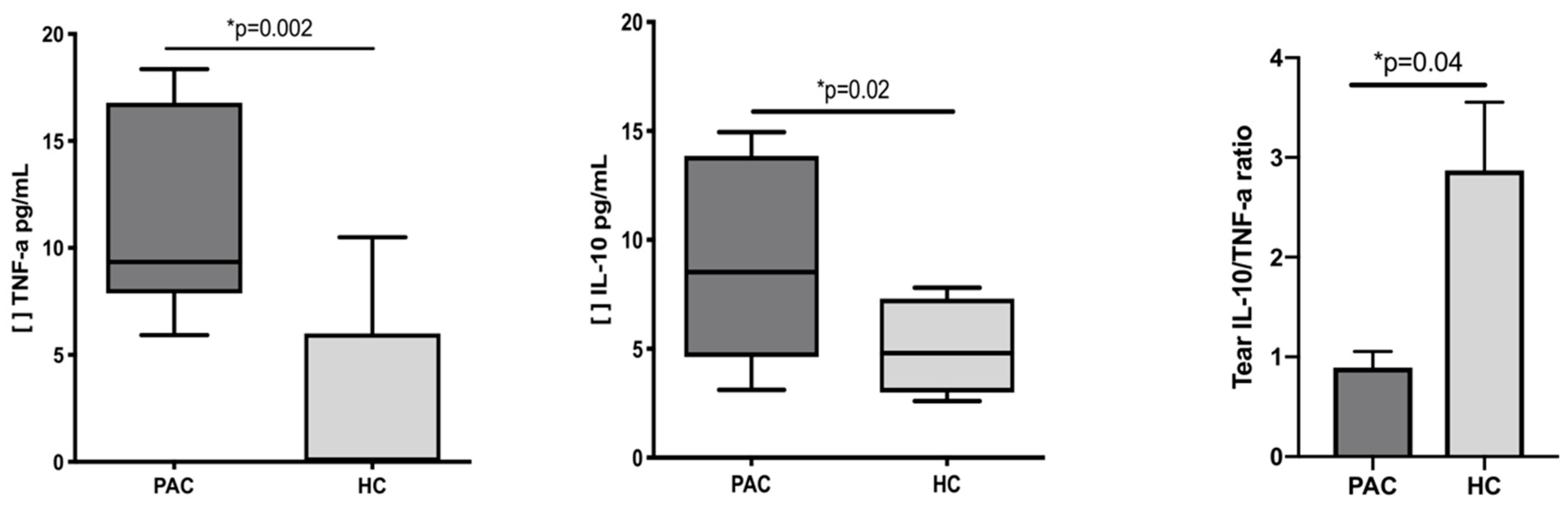
2.3. Increased Concentration of TNF-α and IL-10 in the Tears of Patients with Perennial Allergic Conjunctivitis

Tear cytokines were determined in both groups, and the results are presented in Table 3. We observed 2.4 times more tear TNF-α (p = 0.002) and 1.9 times more tear IL-10 in PAC patients when compared with healthy controls (p = 0.02). Interestingly, when we analyzed the ratio of anti-inflammatory tear IL-10 vs. pro-inflammatory tear TNF-α, we observed 2.8 more times IL-10 than TNF-α in healthy controls when compared with PAC patients (Figure 3). No significant correlations were found between tear cytokines and CD19+IL-10+ cells, with CD38Lo/Med/Hi IL-10+ cell subsets, or with IL-10 MFI.

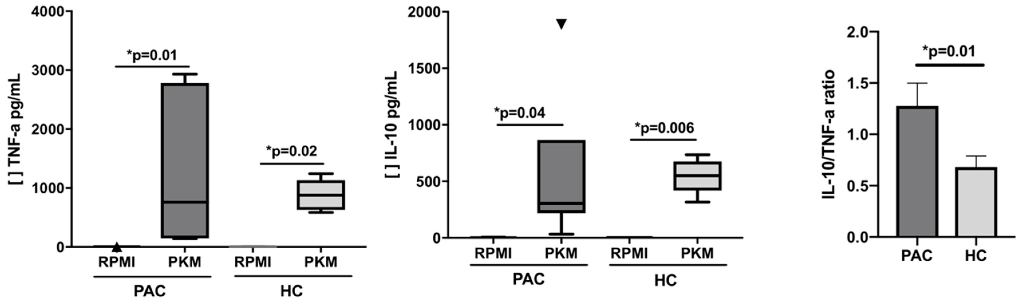
2.4. Functional Evaluation of B Cells after Mitogen Stimulation

In order to evaluate the secretion of cytokines by B cells, mononuclear cells were stimulated with Pokeweed mitogen (PKM), a known B cell mitogen, over 24 h [17]. No significant differences were observed between PAC patients and HC after stimulation; when the ratio of anti-inflammatory IL-10 vs. pro-inflammatory TNF-α was analyzed in the supernatant of cultured cells, we observed 1.2 times more TNF-α than IL-10 in PAC patients when compared with HC (p = 0.01) (Figure 4). The other cytokines evaluated after PKM stimulation are depicted in Table 4.

3. Discussion

Allergic conjunctivitis is an inflammation of the conjunctiva secondary to contact with an allergen at the conjunctiva in a previously sensitized individual [18]. Two forms of AC have been described: VKC and AKC are the chronic forms, and can lead to permanent visual impairment, while SAC and PAC are the acute forms, and are the most frequent clinical presentations. Immunologically, CD4 T cells have been described as the main cell subsets that control conjunctival inflammation in both the acute and chronic forms [5,6,19], but a diminished frequency of Tregs has been reported in PAC patients [8]. This is remarkable due to the growing evidence that suggests that the induction of Tregs is maintained by a subset of IL-10-expressing B cells (Bregs) [11,12,13]. Underlining the importance of Bregs in allergic conjunctivitis, Miyazaki et al. demonstrated in a mice model of IL-10 deficient B cells an exacerbated late-phase inflammation, whereas transfer of IL-10 secreting B cells protected against inflammation [20], suggesting a regulatory function for B cells in allergic conjunctivitis. Nonetheless, the evaluation of Bregs in patients with PAC had not been described yet.
In this work, we analyzed the frequency of Bregs and the expression of IL-10 in Bregs subsets in patients with PAC. Our results are in accordance with those of other authors that have reported changes in the frequency of B cell subsets involved in allergic inflammation, including CD27+CD24hi, CD24hiCD38hi and CD73CD25+CD71+ cells [15]. Remarkably, these Bregs populations have been observed after allergen-specific desensitization, or in nonallergic individuals highly exposed to allergens [15]. In our work, we did not find differences in the percentage or absolute numbers of CD19+IL-10+ cells between patients and controls, but analysis of IL-10 MFI in circulating CD19+CD38Lo/Med/Hi B cell subsets showed they were markedly diminished in PAC patients. The MFI result is translated as low expression of IL-10 in CD19+CD38+ B cells [21]. The absolute numbers and percentage of CD19+ cells observed in our groups are in accordance with other authors [22,23,24] who have reported normal variations in the reference values for B cell subpopulations from infancy to adulthood. Those reports recognized that B cells decline with age, showing greater percentages at birth and infancy, declining gradually during childhood, and after adolescence, B cells reach values similar to those found in adults [22,23,24]. Also, CD38 has similar expression kinetics with higher values at infancy, declining with age [23,24]. Iwata et al. reported a “rare” IL-10 B cell subpopulation in blood from adult donors, similar to the spontaneous IL-10 producing B cells in mice, that corresponded to the 0.6% of total B cells [25]. Thus, it is possible that the percentage of IL-10+ cells in the circulating CD19+CD38+ cells observed in our study would be the biological reflex of a normal variation during childhood. Blair et al. have suggested that CD24 and CD38 define a subset of B cells with regulatory characteristics [26], and in mice models, it has been recognized that CD38 is a protein that enhances IL-10 production by regulatory B cells [27]. Circulating phenotypes of IL-10+ B cells in both human and mice models have been described, and changes in the frequency of circulating IL-10+ B cells are associated with dysregulation of immune responses [28,29]. Moreover, it has been reported that human CD19+CD24hiCD38hi B cells exhibit regulatory capacity in healthy individuals, while the same B cells produce less IL-10 and lack suppressive activity in SLE patients. In line with the findings of these authors, when we analyzed IL-10/TNF-α production in B stimulated cells, we observed an inverted IL-10/TNF-α ratio in PAC patients when compared to healthy controls. These differences between groups indirectly indicate saccharidic changes in the cells of patients with PAC, since polyclonal activation of PKM occurs in their lectin binding to (GlcNAc)3 structures that are mainly expressed on B cells, but also on some subsets of T and NK cells [30]. Although saccharidic specificity is essential for cell activation, proliferating activity and immunoglobulin production depend on the interaction with TLRs, mainly TLR2 and TLR9 [17]. Glycophenotype evaluation could give new insights into the ocular allergy pathophysiology, since cytokine production is a direct consequence of cell activation induced by PKM.
In line with this, changes in cytokines also induce glycosylation changes in cells, regulating cell-cell interaction and inflammation [31]. Unfortunately, in this study, neither proliferative activity nor induced immunoglobulin production by PKM stimulated cells were evaluated. The lack of a complete B cell evaluation in this work is a limitation to being able to conclude that the differences observed are associated with differential expression of TLRs in PKM stimulated cells, and further assessment is needed. It is important to note that stimulation assays were performed with peripheral blood monononuclear cells (PBMC) thus, the cytokines reported in this work were produced not only by B cells, but also other cells such as T cells and macrophages also contributed to the observed result. Regardless, this does not change the biological fact that PAC patients responded immunologically differently compared with HC during PKM stimuli, however, additional assays are needed to find whether the circulating CD19+CD38+IL-10+ cells from PAC patients are dysfunctional.
IL-10 is a cytokine that is described as a potent anti-inflammatory molecule with paradoxical functions [32]. Its anti-inflammatory functions include the downregulation of T cells and monocyte activation, inhibiting Th1 mediated inflammation. Nonetheless, IL-10 does not impair the ability of B cells to function as antigen presenting cells. Moreover, IL-10 contributes to the stimulation of Th2 cytokines [33]. In line with its Th2-promoting functions, IL-10 has been recognized as a potent stimulatory factor for mast cells and their progenitors [34]. The activation of mast cells in a Th2 microenvironment is a critical characteristic in allergic conjunctivitis inflammation [35]. At the ocular surface level, IL-10 is produced by conjunctival fibroblasts [36], by blood inflammatory cells such as basophils and eosinophils, and by local mast cells after an allergenic challenge [37,38]. Some authors have suggested a pathogenic role of IL-10 in ocular surface inflammatory diseases in which the Th2 profile is associated with ocular damage [39,40], and in a mice model of induced allergic conjunctivitis, IL-10 contributed to the development of the acute effector phase, augmenting the infiltration of eosinophils into the conjunctiva [41]. These paradoxical functions of IL-10 reinforce our observation of increasing tear IL-10 in patients when compared with control subjects, and at the same time, an inverted IL-10/TNF-α ratio, approximating to the Th2/Th9 mediated inflammation reported in experimental conjunctivitis [42]. TNF is one of the major mediators of inflammation and is a pleiotropic cytokine produced by diverse cell types such as macrophages, NK cells, T cells, fibroblasts, and mast cells [43]. Conjunctival epithelial cells stimulated with TNF-α induce the secretion of MCP, IL-1β, and IL-6 [44], all of which are molecules involved in the pathogenesis of human ocular allergy [45]. Remarkably, stimulation of corneal and conjunctival fibroblasts with TNF- α induces eosinophil chemotaxis [46], and at the ocular level, the resident mast cells are the primary source of TNF-α in the allergic process [47], sustaining the ocular allergic inflammation. On the contrary, increased IL-10 has been reported in the contralateral healthy eye when infection is occurring in the other eye, supporting the idea that IL-10 has an anti-inflammatory function [48]. Whether tear IL-10 is a participant in the local Th2 inflammation or is an attempt to inhibit the pro-inflammatory actions of TNF-α at the ocular surface is not known, and further investigations are needed to clearly define the role of tear IL-10 in human allergic conjunctivitis, as well as the involvement of IL-10 activated mast cells.
Understanding systemic and local mechanisms involved in allergic conjunctivitis will aid in designing immunomodulating therapies at different levels. Our results ratify the notion of the medical ophthalmological use of specific immune therapy (SIT) for acute types of ocular allergy (SAC and PAC), as it has been reported for other allergies. In asthma, SIT with allergens induces IL-10 producing B cells [15]. The induced Bregs suppress the allergen-specific immune response, contributing to tolerance [49,50]. Luo et al. reported changes in B cell subsets in allergic rhinitis, associating an increased percentage of terminally differentiated CD19+CD24hiCD38+cells, a subset of Bregs, with better clinical outcomes in patients receiving immunotherapy [51]. On the other hand, at the ocular level, the addition of IL-10 could have paradoxical results. For example, in a mouse model of uveitis, distinct doses of IL-10 showed contradictory effects, and low doses of IL-10 potentiated LPS induced inflammation, while high IL-10 doses induced anti-inflammation [52]. In contrast, using adenovirus vectors encoding for IL-10 in human conjunctival cells [53] and lacrimal gland epithelial cells [54] has demonstrated control of lymphocyte proliferation. Thus, IL-10 seems to play a dual role in the ocular microenvironment by simultaneously suppressing proinflammatory pathways and enhancing others, depending on the dose if IL-10 is administered intraocularly or induced at the surface. These opposite roles must be taken into account during the development of biological therapies using IL-10 at the ocular level.
Our findings demonstrate an immunological dysregulation in patients with allergic conjunctivitis, characterized by the low expression of IL-10 in circulating CD19+CD38+ Bregs subsets, and an inverted tear IL-10/TNF-α ratio (Figure 5). Ophthalmological therapeutics should consider systemic and local interventions to reduce ocular damage in patients with allergic conjunctivitis.

4. Materials and Methods

4.1. Patients and Health Controls

Sixteen patients with perennial allergic conjunctivitis (PAC) (11 males and 5 females, mean age 12.1 years, range 6–15) were included in the study. PAC diagnosis was based on clinical ophthalmological history (mean disease duration 3.5 (SD 3.1) years) and allergo/immunological examination. All patients were classified as having active forms of PAC. The allergic condition was confirmed with a skin-prick test positive for Der p (wheal ≥ 3 mm diameter, and compared with histamine control) and determination of serum total IgE (tIgE) and specific IgE (sIgE) to Dermatophagoides pteronyssinus 1 (Der p 1).
Eight healthy volunteers were used as controls (4 males and 4 females, mean age 12.5 years, range 7–17). All participants in this study gave their assent consent for blood sampling after written information was provided. The study adhered to the ethical principles of the Declaration of Helsinki and the E11 Statements of the International Conference of Harmonisation (E11-ICH). This work was approved by the Scientific (CI-001-2016, 08/February/2016), Bioethics (CEI-2016/01/01, 09/March/2016), and Biosafety (CB-001-2016, 29/March/2016) Institutional Committees at the Institute of Ophthalmology, “Foundation Conde de Valenciana”, Mexico City.

4.2. Monoclonal Antibodies and Reagents

Allophycocyanin (APC)-labeled-mouse monoclonal antibodies (mAbs) against human CD19, and phycoerythrin (PE)-labeled mAbs anti-human IL-10, were purchased from BioLegend (San Diego, CA, USA). PE-Cy7-labeled mAbs anti-human CD38 were acquired from BD Biosciences (San Jose, CA, USA), and BD™ CompBeads (San Diego, CA, USA). BD FACS™ Lysing Solution, BD Cytofix/Cytoperm™ solution, and BD FACSFlow™ Sheat Fluid were purchased from BD Biosciences (San Jose, CA, USA). RPMI-1640 culture medium, Pokeweed mitogen (PKW), and salts were purchased from Sigma Chemical Co. (St. Louis, MO, USA). Sodium pyruvate, L-glutamine, and 2-mercaptoethanol were purchased from Gibco BRL (Rockville, MD, USA). Fetal calf serum was obtained from HyClone Labs (Logan, UT, USA), and Dermatophagoides pteronyssinus (Der p) was purchased from Allerstand Co. (Mexico City, Mexico).

4.3. Peripheral Blood Samples

Blood samples were collected by venipuncture into a BD Vacutainer™ K2 EDTA tube (BD, Franklin Lakes, NJ, USA). The sample was kept in gentle agitation and at room temperature (25 °C) for ~30 min for subsequent immunofluorescence staining of the cell surface and intracellular markers.

4.4. Immunofluorescence Staining of Cell Surface Markers

Two-color staining was performed on the peripheral blood cells with direct immunofluorescence, using either APC-mAb anti-CD19 or PE-Cy7-mAb anti-CD38. Briefly, 20 µL of whole peripheral blood was incubated with fluorochrome-labeled mAb for 30 min at 25 °C in darkness. After incubation, the red blood cells were lysated with BD FACS™ Lysing Solution according to the manufacturer’s instructions; then, the cells were washed twice with BD FACSFlow™ Sheat Fluid and processed with intracellular staining.

4.5. Immunofluorescence Staining of Intracellular Markers

After extracellular staining was performed, the cells were fixed and permeabilized with BD Cytofix/Cytoperm™ solution according to the manufacturer´s instructions. The cells were then incubated with PE-labelled anti-human IL-10 antibody, and immediately acquired by flow cytometry. In all cases, Fluorescence Minus One (FMO) controls and anti-mouse Ig and κ/Negative control compensation particle sets (BD™ CompBeads) were used. All samples were immediately acquired after immunoflurescence staining.

4.6. Flow Cytometric Analysis

All cells were analyzed for the expression of phenotypic markers on a BD FACSVerse™ (BD Biosciences, San Jose, CA, USA) flow cytometer using FACSuite Software version 1.0.5.3841 (BD Biosciences, San Jose, CA, USA), and 10,000 events were counted. To analyze the staining of the cell-surface markers, single cells were first gated (FSC-H-forward height vs. FSC-A-forward area); subsequently, a second gate was drawn according to the physical properties (FSC-forward and SSC-scatter) that corresponded to lymphocytes. Then, CD19+ cells were selected in a third FSC-CD19 dot. To determine the subsets of Bregs cells, a new dot plot was obtained showing CD19+ cells (y-axis) and CD38+ cells (x-axis). Subsequently, we identified CD38 expression as low, medium, and high sub-populations on CD19+ B cells (see Figure S1). Finally, using intracellular IL-10 staining on gated CD19+ and CD19+CD38Lo/Med/Hi B cells, a dot plot and/or a histogram was drawn to analyze the frequency or the mean fluorescence intensity (MFI) of IL-10+ cells. Control staining was performed using fluorescence minus one (FMO) and BD™ CompBeads.

4.7. Tear Samples

Tear samples from healthy and allergic eyes were obtained from the conjunctival fornix by addition of 20 µL of BSS™ sterile saline solution (Alcon Laboratories, Inc., Fort Worth, Texas, USA) at the ocular surface, according to Santacruz´method [55]. Then, the ocular wash was immediately recovered and stored at −20 °C until cytometric analysis.

4.8. Serum Samples

A sample of peripheral blood was taken into a BD Vacutainer™ serum tube (BD, Franklin Lakes, NJ, USA), allowing the blood to clot for ~20 min. Then, the serum was obtained by centrifugation at 130 g × 10 min and stored at −20 °C until tIgE and sIgE determination by fluoroenzymo immuno assay with the ImmunoCAP Phadia® 100 Laboratory System (Thermo Fisher Scientific Inc., Portage, MI, USA).

4.9. Cell Cultures

PBMC were cultured in 24-well flat bottomed cell culture plates (Costar, Cambridge, MA, USA) at 5 × 10⁵ cells/well in Roswell Park Memorial Institute (RPMI) 1640 (RPMI-1640) medium, supplemented with 1 mM sodium pyruvate, 2 mM L-glutamine, and 0.5% heat-inactivated fetal calf serum, and incubated at 37 °C in a 5% CO2 humidified chamber. After 24 h the culture medium was removed, and fresh culture medium was supplemented with 10% heat-inactivated fetal calf serum and Pokeweed mitogen (PKW) (5 µg/mL). After 24 h, the supernatants were collected and stored at −70 °C to measure the soluble cytokines.

4.10. Determination of Tear and Serum Cytokines

IL-8, IL-1β, IL-6, IL-10, TNF-α, and IL-12p70 (Human Inflammatory Cytokine Kit, BD Biosciences, San Jose, CA, USA) were measured with Cytometric Bead Arrays (CBA) in tear and serum samples, according to the manufacturer´s instructions (BD Biosciences, San Jose, CA, USA). The samples were processed by flow cytometry, and the results were analyzed with FCAP Array™ Software version 3.0 (BD Biosciences, San Jose, CA, USA).

4.11. Determination of Total IgE (tIgE) and Specific IgE (sIgE)

The serum samples were processed to determine tIgE and sIgE to Der p 1 by fluorenzymeimmunoassay (FEIA), following the manufacturer’s instructions, in an ImmunoCAP Phadia® 100 Laboratory System. The results were analyzed by ImmunoCAP® v.4.13 software (Thermo Fisher Scientific Inc., Portage, MI, USA), and the limits of detection were <2 kUI/L for Total IgE, and <0.1 kUA/L for sIgE.

4.12. Statistical Analysis

A Mann–Whitney U test was used to determine significant differences; correlations between the studied variables were performed with the Spearman rank test; and p < 0.05 was considered as statistically significant. The analysis was performed using GraphPad Prism software version 7.00 for Windows (GraphPad Software, La Jolla, CA, USA).

5. Conclusions

Our findings demonstrate an immunological dysregulation in patients with allergic conjunctivitis, characterized by the low expression of IL-10 in circulating CD19+CD38+ Bregs subsets, and an inverted tear IL-10/TNF-α ratio. These findings highlight the novel pathologic changes involved in ocular allergic diseases.

Supplementary Materials

The supplementary materials can be found at https://www.mdpi.com/1422-0067/20/5/1035/s1.

Author Contributions

Conceptualization, A.S. and M.C.J.-M.; Data curation, A.S., I.C.-M. and H.V.-S.; Formal analysis, A.S., I.C.-M., M.P.-Q., H.V.-S., J.A.-B., E.O.G.-H., J.S.-L. and M.C.J.-M.; Funding acquisition, E.O.G.-H. and M.C.J.-M.; Investigation, A.S., I.C.-M., M.P.-Q., H.V.-S., J.A.-B.; Methodology, A.S., I.C.-M., M.P.-Q., H.V.-S., J.A.-B., E.O.G.-H. and J.S.-L.; Supervision, E.O.G.-H., J.S.-L. and M.C.J.-M.; Validation, E.O.G.-H., J.S.-L. and M.C.J.-M.; Writing—original draft, A.S. and I.C.-M.; Writing—review & editing, J.S.-L. and M.C.J.-M.

Funding

This work was supported in part by Conde de Valenciana Foundation and Department of Biochemistry, Faculty of Medicine, National Autonomous University of Mexico. A.S. is a doctoral student from Programa de Posgrado en Inmunología del Instituto Politécnico Nacional; H.V.-S. is a doctoral student from Programa de Doctorado en Ciencias Médicas, Odontológicas y de la Salud (Farmacología Clínica), Universidad Nacional Autónoma de México (UNAM), and received fellowship 294674 from CONACYT.

Acknowledgments

Thanks are due to F. Raúl Chávez by the constant support and working encouragement to A.S. Thanks are due to Patricia Herrera, Veronica Martinez, and Olga Tamayo for technical contribution; and Jesus Esteban Mendoza for the art in Figure 5.

Conflicts of Interest

The authors declare no conflict of interest.

References

  1. Miraldi Utz, V.; Kaufman, A.R. Allergic eye disease. Pediatr. Clin. N. Am. 2014, 61, 607–620. [Google Scholar] [CrossRef] [PubMed]
  2. Rosario, N.; Bielory, L. Epidemiology of allergic conjunctivitis. Curr. Opin. Allergy Clin. Immunol. 2011, 11, 471–476. [Google Scholar] [CrossRef] [PubMed]
  3. Calderon-Ezquerro, M.C.; Guerrero-Guerra, C.; Galán, C.; Serrano-Silva, N.; Guidos-Fogelbach, G.; Jiménez-Martínez, M.C.; Larenas-Linnemann, D.; López-Espinosa, E.D.; Ayala-Balboa, J. Pollen in the atmosphere of Mexico City and its impact on the health of the pediatric population. Atmos. Environ. 2018, 186, 198–208. [Google Scholar] [CrossRef]
  4. Passali, D.; Cingi, C.; Staffa, P.; Passali, F.; Muluk, N.B.; Bellussi, M.L. The International Study of the Allergic Rhinitis Survey: Outcomes from 4 geographical regions. Asia Pac. Allergy 2018, 8, e7. [Google Scholar] [CrossRef] [PubMed]
  5. Shaker, M.; Salcone, E. An update on ocular allergy. Curr. Opin. Allergy Clin. Immunol. 2016, 16, 505–510. [Google Scholar] [CrossRef] [PubMed]
  6. Magaña, D.; Aguilar, G.; Linares, M.; Ayala-Balboa, J.; Santacruz, C.; Chávez, R.; Estrada-Parra, S.; Garfias, Y.; Lascurain, R.; Jiménez-Martínez, M.C. Intracellular IL-4, IL-5 and IFN-g as the main characteristic of CD4+CD30+ T cells after allergen-stimulation, in patients with vernal keratoconjunctivitis. Mol. Vis. 2015, 21, 443–450. [Google Scholar] [PubMed]
  7. Anderson, A.E.; Mackerness, K.J.; Aizen, M.; Carr, V.A.; Nguyen, D.; Du Pre, F.; Durham, S.R.; Robinson, D.S. Seasonal changes in suppressive capacity of CD4+ CD25+ T cells from patients with hayfever are allergen-specific and may result in part from expansion of effector T cells among the CD25+ population. Clin. Exp. Allergy 2009, 39, 1693–1699. [Google Scholar] [PubMed]
  8. Galicia-Carreón, J.; Santacruz, C.; Ayala-Balboa, J.; Robles-Contreras, A.; Perez-Tapia, S.M.; Garfias, Y.; Hong, E.; Jiménez-Martínez, M.C. An Imbalance Between Frequency of CD4+CD25+FOXP3+ Regulatory T Cells, and CCR4+ and CCR9+ Circulating Helper T cells is Associated with Active Perennial Allergic Conjunctivitis. Clin. Dev. Immunol. 2013. [Google Scholar] [CrossRef] [PubMed]
  9. Palomares, O.; Martín-Fontecha, M.; Lauener, R.; Traidl-Hoffmann, C.; Cavkaytar, O.; Akdis, M.; Akdis, C.A. Regulatory T cells and immune regulation of allergic diseases: Roles of IL-10 and TGF-β. Genes Immunol. 2014, 15, 511–520. [Google Scholar] [CrossRef] [PubMed]
  10. Poojary, K.V.; Kong, Y.M.; Farrar, M.A. Control of Th2-Mediated Inflammation by Regulatory T Cells. Am. J. Pathol. 2010, 177, 525–531. [Google Scholar] [CrossRef] [PubMed] [Green Version]
  11. Ashour, H.M.; Niederkorn, J.Y. Expansion of B cells is necessary for the induction of T-cell tolerance elicited through the anterior chamber of the eye. Int. Arch Allergy Immunol. 2007, 144, 343–346. [Google Scholar] [CrossRef] [PubMed]
  12. Pennati, A.; Ng, S.; Wu, Y.; Murphy, J.R.; Deng, J.; Rangaraju, S.; Asress, S.; Blanchfield, J.L.; Evavold, B.; Galipeau, J. Regulatory B Cells Induce Formation of IL-10-Expressing T Cells in Mice with Autoimmune Neuroinflammation. J. Neurosci. 2016, 36, 12598–12610. [Google Scholar] [CrossRef] [PubMed]
  13. Flores-Borja, F.; Bosma, A.; Ng, D.; Reddy, V.; Ehrenstein, M.R.; Isenberg, D.A.; Mauri, C. CD19+CD24hiCD38hi B cells maintain regulatory T cells while limiting TH1 and TH17 differentiation. Sci. Transl. Med. 2013, 5, 173ra23. [Google Scholar] [CrossRef] [PubMed]
  14. Fillatreau, S.; Sweenie, C.H.; McGeachy, M.J.; Gray, D.; Anderton, S.M. B cells regulate autoimmunity by provision of IL-10. Nat. Immunol. 2002, 3, 944. [Google Scholar] [CrossRef] [PubMed]
  15. Van de Veen, W.; Stanic, B.; Wirz, O.F.; Jansen, K.; Globinska, A.; Akdis, M. Role of regulatory B cells in immune tolerance to allergens and beyond. J. Allergy Clin. Immunol. 2016, 138, 654–665. [Google Scholar] [CrossRef] [PubMed]
  16. Lundy, S.K.; Berlin, A.A.; Martens, T.F.; Lukacs, N.W. Deficiency of regulatory B cells increases allergic airway inflammation. Inflamm. Res. 2005, 54, 514–521. [Google Scholar] [CrossRef] [PubMed] [Green Version]
  17. Bekeredjian-Ding, I.; Foermer, S.; Kirschning, C.J.; Parcina, M.; Heeg, K. Poke weed mitogen requires Toll-like receptor ligands for proliferative activity in human and murine B lymphocytes. PLoS ONE 2012, 7, e29806. [Google Scholar] [CrossRef] [PubMed]
  18. Bielory, L. Allergic and immunologic disorders of the eye. Part II: Ocular allergy. J. Allergy Clin. Immunol. 2000, 106, 1019–1032. [Google Scholar] [CrossRef] [PubMed]
  19. Galicia-Carreón, J.; Santacruz, C.; Hong, E.; Jiménez-Martínez, M.C. The ocular surface: From physiology to the ocular allergic diseases. Rev. Alerg. Mexico 2013, 60, 172–183. [Google Scholar]
  20. Miyazaki, D.; Kuo, C.H.; Tominaga, T.; Inoue, Y.; Ono, S.J. Regulatory function of CpG-activated B cells in late-phase experimental allergic conjunctivitis. Investig. Ophthalmol. Vis. Sci. 2009, 50, 1626–1635. [Google Scholar] [CrossRef] [PubMed]
  21. Jaroszeski, M.J.; Radcliff, G. Fundamentals of flow cytometry. Mol. Biotechnol. 1999, 11, 37–53. [Google Scholar] [CrossRef] [PubMed]
  22. Hulstaert, F.; Hannet, I.; Deneys, V.; Munhyeshuli, V.; Reichert, T.; De Bruyere, M.; Strauss, K. Age-related changes in human blood lymphocyte subpopulations. II. Varying kinetics of percentage and absolute count measurements. Clin. Immunol. Immunopathol. 1994, 70, 152–158. [Google Scholar] [CrossRef] [PubMed]
  23. Morbach, H.; Eichhorn, E.M.; Liese, J.G.; Girschick, H.J. Reference values for B cell subpopulations from infancy to adulthood. Clin. Exp. Immunol. 2010, 162, 271–279. [Google Scholar] [CrossRef] [PubMed] [Green Version]
  24. Valiathan, R.; Ashman, M.; Asthana, D. Effects of Ageing on the Immune System: Infants to Elderly. Scand. J. Immunol. 2016, 83, 255–266. [Google Scholar] [CrossRef] [PubMed]
  25. Iwata, Y.; Matsushita, T.; Horikawa, M.; Dilillo, D.J.; Yanaba, K.; Venturi, G.M.; Szabolcs, P.M.; Bernstein, S.H.; Magro, C.M.; Williams, A.D.; Hall, R.P.; St Clair, E.W.; Tedder, T.F. Characterization of a rare IL-10-competent B-cell subset in humans that parallels mouse regulatory B10 cells. Blood 2011, 117, 530–541. [Google Scholar] [CrossRef] [PubMed]
  26. Blair, P.A.; Noreña, L.Y.; Flores-Borja, F.; Rawlings, D.J.; Isenberg, D.A.; Ehrenstein, M.R.; Mauri, C. CD19(+)CD24(hi)CD38(hi) B cells exhibit regulatory capacity in healthy individuals but are functionally impaired in systemic Lupus Erythematosus patients. Immunity 2010, 32, 129–140. [Google Scholar] [CrossRef] [PubMed]
  27. Domínguez-Pantoja, M.; López-Herrera, G.; Romero-Ramírez, H.; Santos-Argumedo, L.; Chávez-Rueda, A.K.; Hernández-Cueto, Á.; Flores-Muñoz, M.; Rodríguez-Alba, J.C. CD38 protein deficiency induces autoimmune characteristics and its activation enhances IL-10 production by regulatory B cells. Scand. J. Immunol. 2018, 87, e12664. [Google Scholar] [CrossRef] [PubMed]
  28. Wang, L.; Qiu, J.; Yu, L.; Hu, X.; Zhao, P.; Jiang, Y. Increased numbers of CD5+CD19+CD1dhighIL-10+ Bregs, CD4+Foxp3+ Tregs, CD4+CXCR5+Foxp3+ follicular regulatory T (TFR) cells in CHB or CHC patients. J. Transl. Med. 2014, 9, 251. [Google Scholar] [CrossRef] [PubMed]
  29. Jeong, Y.I.; Hong, S.H.; Cho, S.H.; Lee, W.J.; Lee, S.E. Induction of IL-10-producing CD1dhighCD5+ regulatory B cells following Babesia microti-infection. PLoS ONE 2012, 7, e46553. [Google Scholar] [CrossRef] [PubMed]
  30. Yokoyama, K.; Terao, T.; Osawa, T. Carbohydrate-binding specificity of pokeweed mitogens. Biochim. Biophys. Acta 1978, 538, 384–396. [Google Scholar] [CrossRef]
  31. Dewald, J.H.; Colomb, F.; Bobowski-Gerard, M.; Groux-Degroote, S.; Delannoy, P. Role of Cytokine-Induced Glycosylation Changes in Regulating Cell Interactions and Cell Signaling in Inflammatory Diseases and Cancer. Cells 2016, 29, 43. [Google Scholar] [CrossRef] [PubMed]
  32. Saxena, A.; Khosraviani, S.; Noel, S.; Mohan, D.; Donner, T.; Hamad, A.R. Interleukin-10 paradox: A potent immunoregulatory cytokine that has been difficult to harness for immunotherapy. Cytokine 2014, 74, 27–34. [Google Scholar] [CrossRef] [PubMed]
  33. Fiorentino, D.F.; Zlotnik, A.; Vieira, P.; Mosmann, T.R.; Howard, M.; Moore, K.W.; O’Garra, A. IL-10 acts on the antigen-presenting cell to inhibit cytokine production by Th1 cells. J. Immunol. 1991, 146, 3444–3451. [Google Scholar] [PubMed]
  34. Thompson-Snipes, L.; Dhar, V.; Bond, M.W.; Mosmann, T.R.; Moore, K.W.; Rennick, D.M. Interleukin 10: A novel stimulatory factor for mast cells and their progenitors. J. Exp. Med. 1991, 173, 507–510. [Google Scholar] [CrossRef] [PubMed]
  35. Elieh Ali Komi, D.; Rambasek, T.; Bielory, L. Clinical implications of mast cell involvement in allergic conjunctivitis. Allergy 2018, 73, 528–539. [Google Scholar] [CrossRef] [PubMed]
  36. Leonardi, A.; Curnow, S.J.; Zhan, H.; Calder, V.L. Multiple cytokines in human tear specimens in seasonal and chronic allergic eye disease and in conjunctival fibroblast cultures. Clin. Exp. Allergy 2006, 36, 777–784. [Google Scholar] [CrossRef] [PubMed]
  37. Pelikan, Z. Cytological changes in tears during the secondary conjunctival response induced by nasal allergy. Br. J. Ophthalmol. 2012, 96, 941–948. [Google Scholar] [CrossRef] [PubMed]
  38. Pelikan, Z. Cytokine profiles in tears accompanying the secondary conjunctival responses induced by nasal allergy. Curr. Eye Res. 2014, 39, 120–132. [Google Scholar] [CrossRef] [PubMed]
  39. Skwor, T.A.; Atik, B.; Kandel, R.P.; Adhikari, H.K.; Sharma, B.; Dean, D. Role of secreted conjunctival mucosal cytokine and chemokine proteins in different stages of trachomatous disease. PLoS Negl. Trop. Dis. 2008, 2, e264. [Google Scholar] [CrossRef] [PubMed]
  40. Lee, S.Y.; Han, S.J.; Nam, S.M.; Yoon, S.C.; Ahn, J.M.; Kim, T.I.; Kim, E.K.; Seo, K.Y. Analysis of tear cytokines and clinical correlations in Sjögren syndrome dry eye patients and non-Sjögren syndrome dry eye patients. Am. J. Ophthalmol. 2013, 156, 247.e1–253.e1. [Google Scholar] [CrossRef] [PubMed]
  41. Fukushima, A.; Sumi, T.; Fukuda, K.; Kumagai, N.; Nishida, T.; Yagita, H.; Ueno, H. Interleukin 10 and transforming growth factor beta contribute to the development of experimentally induced allergic conjunctivitis in mice during the effector phase. Br. J. Ophthalmol. 2006, 90, 1535–1541. [Google Scholar] [CrossRef] [PubMed]
  42. Tan, C.; Wandu, W.S.; St Leger, A.; Kielczewski, J.; Wawrousek, E.F.; Chan, C.C.; Gery, I. Unlike Th1/Th17 cells, Th2/Th9 cells selectively migrate to the limbus/conjunctiva and initiate an eosinophilic infiltration process. Exp. Eye Res. 2018, 166, 116–119. [Google Scholar] [CrossRef] [PubMed]
  43. Bemelmans, M.H.A.; van Tits, L.J.H.; Buurman, W.A. Tumor Necrosis Factor: Function, Release and Clearance. Crit. Rev. Immunol. 2017, 37, 249–259. [Google Scholar] [CrossRef] [PubMed]
  44. Enríquez-de-Salamanca, A.; Calder, V.; Gao, J.; Galatowicz, G.; García-Vázquez, C.; Fernández, I.; Stern, M.E.; Diebold, Y.; Calonge, M. Cytokine responses by conjunctival epithelial cells: An in vitro model of ocular inflammation. Cytokine 2008, 44, 160–167. [Google Scholar] [CrossRef] [PubMed]
  45. Leonardi, A.; Motterle, L.; Bortolotti, M. Allergy and the eye. Clin. Exp. Immunol. 2008, 153 (Suppl. 1), 17–21. [Google Scholar] [CrossRef] [PubMed] [Green Version]
  46. Fukagawa, K.; Okada, N.; Fujishima, H.; Nakajima, T.; Takano, Y.; Tanaka, M.; Dogru, M.; Satake, Y.; Tsubota, K.; Saito, H. Corneal and conjunctival fibroblasts are major sources of eosinophil-recruiting chemokines. Allergol. Int. 2009, 58, 499–508. [Google Scholar] [CrossRef] [PubMed]
  47. Leonardi, A. The central role of conjunctival mast cells in the pathogenesis of ocular allergy. Curr. Allergy Asthma Rep. 2002, 2, 325–331. [Google Scholar] [CrossRef] [PubMed]
  48. Yamaguchi, T.; Hamrah, P.; Shimazaki, J. Bilateral Alterations in Corneal Nerves, Dendritic Cells, and Tear Cytokine Levels in Ocular Surface Disease. Cornea 2016, 35 (Suppl. 1), S65–S70. [Google Scholar] [CrossRef] [PubMed]
  49. Van de Veen, W.; Stanic, B.; Yaman, G.; Wawrzyniak, M.; Söllner, S.; Akdis, D.G.; Rückert, B.; Akdis, C.A.; Akdis, M. IgG4 production is confined to human IL-10-producing regulatory B cells that suppress antigen-specific immune responses. J. Allergy Clin. Immunol. 2013, 131, 1204–1212. [Google Scholar] [CrossRef] [PubMed]
  50. Van de Veen, W. The role of regulatory B cells in allergen immunotherapy. Curr. Opin. Allergy Clin. Immunol. 2017, 17, 447–452. [Google Scholar] [CrossRef] [PubMed]
  51. Luo, J.; Guo, H.; Liu, Z.; Peng, T.; Hu, X.; Han, M.; Yang, X.; Zhou, X.; Li, H. Analysis of Peripheral B Cell Subsets in Patients With Allergic Rhinitis. Allergy Asthma Immunol. Res. 2018, 10, 236–243. [Google Scholar] [CrossRef] [PubMed] [Green Version]
  52. Rosenbaum, J.T.; Angell, E. Paradoxical effects of IL-10 in endotoxin-induced uveitis. J. Immunol. 1995, 15, 4090–4094. [Google Scholar]
  53. Shen, J.; Taylor, N.; Duncan, L.; Kovesdi, I.; Bruder, J.T.; Forrester, J.V.; Dick, A.D. Ex vivo adenovirus mediated gene transfection of human conjunctival epithelium. Br. J. Ophthalmol. 2001, 85, 861–867. [Google Scholar] [CrossRef] [PubMed] [Green Version]
  54. Zhu, Z.; Stevenson, D.; Ritter, T.; Schechter, J.E.; Mircheff, A.K.; Kaslow, H.R.; Trousdale, M.D. Expression of IL-10 and TNF-inhibitor genes in lacrimal gland epithelial cells suppresses their ability to activate lymphocytes. Cornea 2002, 21, 210–214. [Google Scholar] [CrossRef] [PubMed]
  55. Santacruz, C.; Linares, M.; Garfias, Y.; Loustaunau, L.M.; Pavon, L.; Perez-Tapia, S.M.; Jimenez-Martinez, M.C. Expression of IL-8, IL-6 and IL-1β in tears as a main characteristic of the immune response in human microbial keratitis. Int. J. Mol. Sci. 2015, 16, 4850–4864. [Google Scholar] [CrossRef] [PubMed]
Figure 1. The percentage of CD19+IL-10+ cells. B cells were identified as the CD19+ population, as explained in Figure S1D. (A) Representative dot plots of CD19+IL-10+ cells in PAC and HC. Comparison of the frequency of B cells positive to IL-10 staining (B) in both groups. Data are expressed as the mean ± standard deviation (SD).
Ijms 20 01035 g001
Figure 2. Expression of IL-10 in CD19+CD38 cell subsets in patients with perennial allergic conjunctivitis (PAC) and healthy controls (HC). Representative dot plots from gates performed according to the CD38 expression on B cells (see Figure S1 to consult the hierarchical analysis performed). Fluorescence minus one (FMO) (A); CD38 Lo/Med/Hi on gated CD19+ cells (B). Representative dot plots of CD19+ CD38 Lo/Med/Hi vs. IL-10 staining (C); median fluorescence expression (MFI) of IL-10 on gated CD19+CD38+Lo/Med/Hi cells (D). The dark-grey histogram corresponds to PAC patients; the light-grey histogram corresponds to healthy controls; (E) column shows the IL-10 MFI comparison between the two groups. See Figure S2 for cytometric control of the IL-10 analysis. Data are expressed as the mean ± standard deviation (SD). Statistical differences were considered when p < 0.05. The black dot above PAC columns corresponded to the outlier values.
Ijms 20 01035 g002
Figure 3. Tear cytokines in patients with perennial allergic conjunctivitis (PAC) and healthy controls (HC). The cytokines IL-10 and TNF were measured by cytometric bead arrays in tear samples from patients with PAC and HC. Significant differences were observed in IL-10 and TNF-α in PAC patients. Analysis of the IL-10/TNF-α ratio showed 2.8 more times tear IL-10 than tear TNF-α in HC. The ratio was calculated using the mean values for IL-10 and TNF-α. See Table 3 for additional determined cytokines. Statistical differences were considered when p < 0.05. [ ] Concentration of cytokine.
Ijms 20 01035 g003
Figure 4. Determination of TNF-α and IL-10 secretion after B cell-stimulation. B cells were stimulated for 24 h with Pokeweed mitogen (PKM). After stimulation, supernatants were collected and cytokines measured by cytometric bead arrays. Significant differences were observed in IL-10 and TNF-α in both groups. Analysis of the IL-10/TNF-α ratio showed 1.2 times more TNF-α than IL-10 in PAC patients when compared with HC. The ratio was calculated using the mean values for IL-10 and TNF-α. See Table 4 for additional determined cytokines. Statistical differences were considered when p < 0.05. Ijms 20 01035 i001 and Ijms 20 01035 i002 Corresponded to the outliers values. [ ] Concentration of cytokine.
Ijms 20 01035 g004
Figure 5. Systemic and local immune dysregulation observed in patients with perennial allergic conjunctivitis (PAC). The immune dysregulation in PAC is characterized by the low expression of IL-10 in circulating CD19+CD38+ B cells (Bregs), favoring a systemic Th2 response that is clinically observable with higher concentrations of IgE in the serum of patients with PAC. Whether these IL-10+ B cells are migrating to the conjunctiva or are dysfunctional B cells unable to down-regulate the Th2 immune response is unknown, and needs further investigation. IL-10 predominates over TNF-α in tears under physiological conditions of the ocular surface; nevertheless, in PAC patients, an inverted IL-10/TNF-α ratio is observed. Tear TNF-α facilitates the migration of circulating blood cells to the conjunctiva, and at the same time, the activation of local sensitized mast cells by allergens contributes to more inflammation through the secretion of TNF-α. Amplification of the ocular damage is observed each time that the allergens cross the conjunctival epithelium, activating the sensitized local o migratory cells. For example, eosinophils are one such cell of this type which injure the cornea and other structures of the ocular surface. Thus, a damaging circle is created in which the tear cytokines IL-10 and TNF-α appear to be the biomolecules directing the ocular allergic process; this circle is reinforced with the systemic feedback of possible dysfunctional circulating Bregs. CALT: Conjunctival Associated Lymphoid Tissue; B: B cells; T: T cells; NK: Natural Killer cells.
Ijms 20 01035 g005
Table 1. Demographics characteristics of patients with PAC and healthy controls.
Demographic CharacteristicsPACHCp Value
(n = 16) MD (IQR)(n = 8) MD (IQR)
Age 12.5 (11–14)12 (11–15.5) NS
Male13 (11–14)11 (11–13.3)NS
Female11 (10.5–13.5)14.5 (8.5–16.8)NS
TBUT (sg)4.5 (3–6)6 (3.5–8)NS
Schirmer Test (mm)16.5 (9–27)20 (14.8–29.8)NS
SPT (mm) to Der p6 (3–8)0 (0–0)0.0006
IgA174 (140–211)171 (86–213)NS
IgG1230 (1169–1377)1310 (649–1933)NS
IgM116 (85–146)127 (103–516)NS
tIgE 404 (164–585)58.3 (29.3–182.5)0.002
sIgE 29.9 (11.4–62.9)0 (0–0) *<0.0001
PAC: Perennial Allergic Conjunctivitis; HC: Healthy Controls; MD: Median; IQR: Interquartile Range; NS: Not Significant; TBUT: Tear Breakup Tear; SPT: Skin Prick Test; Der p: Dermatophagoides pteronyssinus; tIgE: Total IgE; sIgE: Specific IgE. * Results below the limit of detection were considered as 0 for statistical analysis. See Table S2 for a detailed description of demographic characteristics.
Table 2. Percentages of B cell subsets in patients with perennial allergic conjunctivitis (PAC) and healthy controls (HC).
B Cell SubsetsPACHCp Value
(n = 16)(n = 8)
MD (IQR)MD (IQR)
CD19+18.8 (16.9–21.9)17.7 (15.7–20.1)0.5177
CD19+CD38+84.4 (77.9–88.6)86 (71.9–91.8)0.9649
CD19+CD38Lo61.5 (36.8–64.1)55.2 (40.6–60.5)0.2597
CD19+CD38Med23.5 (14.2–31.3)17.5 (7.2–36.7)0.6650
CD19+CD38Hi5 (3.0–11.9)5.25 (1.8–14.9)0.2834
CD19+IL-10+3.8 (2.7–4.6)3.3 (1.9–3.5)0.2198
CD19+CD38Lo IL-10+2.5 (1.6–4.2)2.3 (1.0–7.0)0.500
CD19+CD38Med IL-10+5 (2.6–6.7)4.8 (3.7–5.4)0.449
CD19+CD38Hi IL-10+4.7 (3.2–7.3)10.4 (3.9–15.3)0.419
PAC: Perennial Allergic Conjunctivitis; HC: Healthy Controls; MD: Median; IQR: Interquartile Range.
Table 3. Tear cytokines in patients with perennial allergic conjunctivitis (PAC) and healthy controls (HC).
Tear CytokinesPAC (n = 16)HC (n = 8)p Value
MD (IQR)MD (IQR)
pg/mLpg/mL
IL-1β32.46 (19.85–47.63)33.8 (18.02–38.79)0.4435
IL-625.46 (14.61–40.41)19.95 (17.36–25.28)0.3333
IL-8167 (127–222.7)176 (158–245)0.3031
IL-108.5 (4.6–13.9)4.8 (3.0–7.3)0.02
IL-12p7027.17 (7.9–39.53)18.6 (7.9–22.6)0.1750
TNF-α9.34 (7.88–16.79)0 (0–6.0) *0.002
Kit detection limits were as follows: IL-8, 3.8 pg/mL; IL-1β, 7.2 pg/mL; IL-6, 2.5 pg/mL; IL-10, 3.3 pg/mL; TNF-α, 3.7 pg/mL; and IL-12p70, 1.9 pg/mL. * Results below the limit of detection were considered as 0 for comparison between groups.
Table 4. Supernatant (SN) cytokines after Pokeweed mitogen (PKM) stimuli in cells from patients with perennial allergic conjunctivitis (PAC) and healthy controls (HC).
SN
Cytokines
PAC (n = 16)HC (n = 8)
MD (IQR)MD (IQR)
pg/mLpg/mL
RPMIPKMpRPMIPKMp
IL-1β6.6 (5.2–7.6)454.7 (108.8–1342)0.036.3 (5.9–6.9)830 (380–1161)0.01
IL-63.8 (3.1–4.6)3078 (2137–8638)0.034.8 (4–9.8)5084 (2508–6197)0.002
IL-8919 (318–1043)20,237 (10,558–21,513)0.00031223 (501.5–1654)22,075 (17,873–24,910)0.004
IL-100 (0–0.3) *304.8 (218–866)0.0063.3 (0–4.6) *550.2 (418–675) 0.04
IL-12p704.6 (2.2–4.9)21 (4.5–50)0.023.5 (2.2–4.5)8.9 (4.8–47.3)0.03
TNF-α0 (0–1.9) *877 (627–1131)0.010 (0–1.2) *757.6 (144.9–2932)0.02
Kit detection limits were as follows: IL-8, 3.8 pg/mL; IL-1β, 7.2 pg/mL; IL-6, 2.5 pg/mL; IL-10, 3.3 pg/mL; TNF-α, 3.7 pg/mL; and IL-12p70, 1.9 pg/mL. * Results below the limit of detection were considered as 0 for comparision between groups.

