Review

Aberrant Calcium Signals in Reactive Astrocytes: A Key Process in Neurological Disorders

1
Department of Neuropharmacology, Interdisciplinary Graduate School of Medicine, University of Yamanashi Chuo, Yamanashi 409-3898, Japan
2
Department of Neurology, Graduate School of Medical Science, Kyoto Prefectural University of Medicine, Kyoto 602-8566, Japan
3
Department of Pediatrics, Interdisciplinary Graduate School of Medicine, University of Yamanashi, Chuo, Yamanashi 409-3898, Japan
*
Author to whom correspondence should be addressed.
Int. J. Mol. Sci. 2019, 20(4), 996; https://doi.org/10.3390/ijms20040996
Submission received: 1 February 2019 / Revised: 20 February 2019 / Accepted: 21 February 2019 / Published: 25 February 2019

Abstract

:
Astrocytes are abundant cells in the brain that regulate multiple aspects of neural tissue homeostasis by providing structural and metabolic support to neurons, maintaining synaptic environments and regulating blood flow. Recent evidence indicates that astrocytes also actively participate in brain functions and play a key role in brain disease by responding to neuronal activities and brain insults. Astrocytes become reactive in response to injury and inflammation, which is typically described as hypertrophy with increased expression of glial fibrillary acidic protein (GFAP). Reactive astrocytes are frequently found in many neurological disorders and are a hallmark of brain disease. Furthermore, reactive astrocytes may drive the initiation and progression of disease processes. Recent improvements in the methods to visualize the activity of reactive astrocytes in situ and in vivo have helped elucidate their functions. Ca2+ signals in reactive astrocytes are closely related to multiple aspects of disease and can be a good indicator of disease severity/state. In this review, we summarize recent findings concerning reactive astrocyte Ca2+ signals. We discuss the molecular mechanisms underlying aberrant Ca2+ signals in reactive astrocytes and the functional significance of aberrant Ca2+ signals in neurological disorders.

1. Introduction

Astrocytes constitute approximately 30% of the cells of the brain and occupy non-overlapping spatial domains in the central nervous system. Astrocytes not only provide structural, metabolic and homeostatic support for neurons but also actively participate in brain functions [1,2]. Astrocytes do not fire action potentials but are excitable with respect to intracellular signaling. Intracellular ions (e.g., Ca2+, Na+) and second messengers (e.g., cAMP) of astrocytes change dynamically in space and time in response to stimuli. Recent improvements in methodologies to visualize changes in signaling molecules have revealed novel functions of astrocytes in neuronal circuits [3,4].
Ca2+ signals have been extensively studied and well characterized since they were discovered to be fundamental to intracellular signaling and intercellular communication [5]. Ca2+ signal in astrocytes may reflect local consumption of energy, circuit activity, and brain states. Numerous proteins that regulate Ca2+ transport support the dynamic features of Ca2+ signals [6]. The form of Ca2+ signals in astrocytes changes acutely and chronically in response to brain insult, such as injury, inflammation or hyperexcitability [7]. Reactive phenotype of astrocytes frequently found in disease shows altered Ca2+ signals in response to damaged neuronal tissues. In this review, we focus on recent progress in the understanding of Ca2+ signals in reactive astrocytes and we discuss mechanisms underlying aberrant Ca2+ signals in reactive astrocytes and their functional significance in disease pathogenesis. We focus on recent findings made in the last five years and, therefore, we do not reference many of the prior studies that are fundamental to our understanding of Ca2+ signaling in astrocytes. There are many excellent reviews explaining the roles of astrocytes and reactive astrocytes that should be referred to for further understanding of these topics [1,2,3,4,7,8,9].

2. Measurement of Ca2+ Signals in Astrocytes

Organic Ca2+ indicator dyes have been widely used to measure Ca2+ in astrocytes. Bulk-loading of acetoxymethyl (AM) ester forms of indicator dyes is used in many studies because they are easy to load into cells in situ and in vivo and reliably report Ca2+ signals. Some AM dyes (e.g., Fluo-4/AM) are preferentially loaded into astrocytes in optimal conditions. However, in theory, these dyes can also enter other cells, therefore, these data must be analyzed with caution. To confirm whether signals are derived from astrocytes, sulforhodamine 101, a red fluorescent dye that selectively labels astrocytes at low concentration is used. Bulk-loading of Ca2+ indicator dyes can report Ca2+ signals in relatively large regions such as somata and major branches. However, astrocytes are morphologically very complex with many other cellular compartments. Thousands of astrocytic branchlets and leaflets are closely associated with synapses. Most of an astrocyte’s surface area (90–95%) consists of branchlets and leaflets [1]. In these fine structures, Ca2+ signals cannot be reliably measured by bulk-loading of a Ca2+ indicator. Therefore, most astrocyte territories, especially fine processes that are relevant to interactions with synapses, cannot be observed by this method [10]. Genetically encoded Ca2+ indicators (GECIs), such as Yellow Cameleon, GCaMP and GECO, are more suitable for the measurement of Ca2+ signals at fine structures because GECIs can be introduced specifically into astrocytes under the control of astrocyte specific promoters. GCaMP3 and membrane tethered GCaMP3 (Lck-GCaMP3) are able to indicate many microdomain Ca2+ signals in situ in entire astrocyte territories and significantly improve the detection of Ca2+ signals that are missed by bulk-loading of organic Ca2+ indicators [11]. Similar microdomain Ca2+ signals can be observed in vivo using the latest versions of GECIs [12,13,14,15,16,17]. GECIs can be introduced into astrocytes using adeno-associated viral vectors or transgenic mouse lines. Many transgenic lines are available to drive the expression of GECIs using the Cre-lox and Tet-systems [6,14].
GECIs are superior to organic Ca2+ indicator dyes with respect to specificity. Recently, Smith et al. found that high concentrations of organic Ca2+ indicators (Fura-2, Fluo-4, and Rhod-2), but not GCaMP3, inhibit Na,K-ATPase. Loading organic Ca2+ indicator dyes (or BAPTA) into mice increases intracellular K+, probably through reduction of K+ uptake. Dye loading also increases extracellular ATP, possibly through dying cells [18]. These findings indicate that data obtained using organic Ca2+ indicators should be interpreted with caution.
GECIs can be stably expressed in certain cell types allowing the chronic monitoring of Ca2+ activities using two-photon microscopy. However, high expression levels of GECIs can cause cellular damage [19]. Because of their brightness and photostability, GECIs are suitable for wide-field imaging, revealing global Ca2+ elevation in astrocytes, which occurs synchronously in many astrocytes throughout the cortex [20]. Noradrenaline, derived from locus coeruleus neurons in response to arousal or startle, causes global Ca2+ signals [20,21,22].

3. Reactive Astrocytes in Disease

Astrocytes become reactive in response to injury and inflammation. There are at least two distinct categories of reactive astrocytes: hypertrophic reactive astrocytes and scar-forming astrocytes [1]. In this review, we focus on hypertrophic reactive astrocytes, which we term, hereafter, reactive astrocytes. Reactive astrocytes are found in many neurological diseases, such as Alzheimer’s disease (AD), amyotrophic lateral sclerosis (ALS), multiple sclerosis (MS), epilepsy, stroke and traumatic brain injury (TBI) [9,23]. Upregulation of glial fibrillary acidic protein (GFAP) is widely used as a marker of reactive astrocytes. Reactive astrocytes show hypertrophy with thicker processes. Roles of reactive astrocytes can be neuroprotective or neurotoxic, depending on the context. In brain or spinal cord injuries (SCIs), astrocytes become reactive astrocytes with morphological changes. At the site of injury, scar-forming astrocytes form glial scars. Preventing glial scar formation leads to infiltration of circulating immune cells and subsequent neuronal cell damage. Therefore, glial scars provide a barrier around injury sites to protect intact tissues from damage. However, conversely, glial scars are thought to prevent axonal regeneration and functional recovery of neural circuits [24]. Anderson et al. showed that prevention of scar-forming astrocytes do not promote axonal regrowth but reduce stimulated axon regrowth, indicating that scar formation aids rather than prevents recovery of neural circuits from SCI [25]. Thus, glial scars play both beneficial and detrimental roles in axonal regeneration and functional recovery of neural circuits [26].
Zamanian et al. performed transcriptome analysis of reactive astrocytes isolated from inflamed brain after intraperitoneal injection of lipopolysaccharide (LPS), a main component of the outer membrane of gram negative bacteria, or damaged brain following middle cerebral artery occlusion (MCAO) [27]. Differentially expressed genes in LPS-induced reactive astrocytes included genes with potentially detrimental effects, such as C1r, C1s, C3 and C4, which can cause synapse loss and neuronal damage. Meanwhile, in MCAO-induced reactive astrocytes, differentially expressed genes included neuroprotective genes, such as genes encoding neurotrophic factors, thrombospondins, and cytokines including IL-6. Neurotoxic reactive astrocytes and neuroprotective reactive astrocytes were termed A1 and A2, respectively [28]. These data show the heterogeneity of reactive astrocytes induced by distinct brain insults.
A follow-up study showed that microglia-derived signals (IL-1α, TNF-α and C1q) induce neurotoxic A1 astrocytes. A1 astrocytes release unidentified toxic molecules onto neurons and oligodendrocytes, lose ability to promote neuronal survival, outgrowth and synaptogenesis and impair phagocytosis [28]. Recent evidence shows that astrocytes are heterogeneous with respect to transcriptome, proteome and function; region- and circuit-specific functions have been described [29,30,31]. Therefore, response to injury or inflammation is likely to be distinct depending on the circuits involved and the phenotypes of reactive astrocytes may be more heterogeneous than currently recognized.
Many studies over the past few decades have indicated that reactive astrocytes lose homeostatic functions, including excitatory neurotransmitter uptake, potassium ion uptake, and ion buffering and thus passively contribute to disease pathogenesis [32]. However, recent evidence indicates that reactive astrocytes actively contribute to disease processes even in the presymptomatic phase (see below). A key process in this active role is Ca2+ signaling.

4. Ca2+ Signals in Reactive Astrocytes

In parallel with morphological changes, reactive astrocytes demonstrate dynamic, aberrant Ca2+ signals. In most cases, Ca2+ signals increase in terms of amplitude, duration and frequency. There is huge variation in the Ca2+ dynamics of reactive astrocytes in distinct pathological models, phases (acute or chronic) and regions, indicating diverse underlying mechanisms for aberrant Ca2+ signals that are dependent on the conditions.
Astrocytes respond rapidly to injury and hyperexcitability to generate Ca2+ signals [33,34,35,36,37,38]. For example, astrocytes rapidly increase their Ca2+ in response to hyperexcitability in drug (kainate and pilocarpine)-induced seizure model. In adult mice, simultaneous Ca2+ imaging from both neurons and astrocytes using two different colored GECIs revealed that astrocytes are activated earlier than neurons following seizure induced by intraperitoneal administration of kainate. Although the detailed mechanism underlying this astrocyte response is not clear, suppression of Ca2+ responses by deletion of inositol-1,4,5 trisphosphate receptor type 2 (IP3R2, see below), in which astrocytes lack a major intracellular Ca2+ release pathway, resulted in less kainate-induced epileptic activity recorded by electroencephalogram telemetry [38]. This indicates that astrocyte Ca2+ signals are proconvulsive, which is consistent with previous reports [36]. In adult mice, 2–3 days after pilocarpine-induced status epilepticus (SE), astrocytes started to show larger Ca2+ signals, which may contribute to the delayed loss of neurons because suppression of enhanced Ca2+ signals by BAPTA-AM, a membrane permeable Ca2+ chelator, which presumably was preferentially loaded into astrocytes in the condition, reduced neuronal damage [39]. Astrocyte Ca2+ signals recovered to control levels five days after SE. Interestingly, four weeks after pilocarpine-induced SE, reactive astrocytes showed large Ca2+ signals, which may regulate seizure susceptibility in adult mice (Sano et al., University of Yamanashi; unpublished observation). It appears that reactive astrocytes regain aberrant Ca2+ activities during the recovery from the damage caused by the initial SE. Thus, the role of Ca2+ signals in reactive astrocytes may be distinct during various phases after initial SE (e.g., neuron loss, seizure threshold). Enhanced Ca2+ signals in reactive astrocytes are frequently described; however, astrocytes in seizure models do not always show enhanced Ca2+ signals. Plata et al. reported that 2-4 weeks after pilocarpine-induced SE in young rats (3–6 weeks old), hippocampal astrocytes showed fewer Ca2+ signals [40]. These astrocytes also showed atrophy, but not hypertrophy. Sholl analysis showed reduced complexity of structures in atrophic reactive astrocytes, which may decrease support for synapses from astrocyte processes [41]. Astrocytes can, therefore, be hypertrophic and atrophic in response to hyperexcitable neurons. It is unclear what determines astrocyte phenotypes after SE. The mechanism underlying functional and structural changes in astrocytes in response to hyperexcitability may provide useful information for the etiology of epilepsy.
Aberrant Ca2+ signals are preferentially observed in the area where the tissue is strongly affected and where hypertrophic astrocytes are located. For example, in an in vivo adult mouse model of familial AD, reactive astrocytes displayed frequent Ca2+ signals near amyloid plaques and Ca2+ waves that originated from plaques [42,43]. In an acute stroke model, reactive astrocytes in the ischemic core displayed much larger Ca2+ signals in terms of ΔF/F compared with those in the penumbra region [44]. Suppression of such large Ca2+ signals reduced the extent of the damaged areas and the number of injured cells. Therefore, these Ca2+ signals in reactive astrocytes have a harmful effect in neuronal tissues.
Differences in brain regions and experimental models dramatically affect the Ca2+ signals observed. Even in the same ischemia model, the age of animals significantly affects Ca2+ signals in reactive astrocytes in the penumbra, where neurons lose the ability to generate spontaneous or evoked electrical activity and astrocytes become reactive. In an MCAO model using adult (3–4-month-old) and aged (18–24-month-old) mice, Fordsmann et al. observed Ca2+ activities in both neurons and astrocytes. In adult mice, Ca2+ signals in both neurons and astrocytes were suppressed 2–4 h after MCAO, while, in aged mice, Ca2+ signals in astrocytes were enhanced and Ca2+ in neurons was unchanged. Enhanced Ca2+ signals in aged mouse astrocytes were action potential-dependent and occurred through P2 receptor activation, which may be harmful to neurons [45]. These observations clearly show that astrocyte Ca2+ dynamics and their roles are distinct at different ages.
Severity of a disease relates to augmentation of aberrant Ca2+ signals in reactive astrocytes. In a model of Alexander disease (AxD), a rare neurodegenerative disease caused by autosomal dominant gain of function mutations in GFAP, we recently found extraordinarily large Ca2+ signals in astrocytes, whose areas were over 300 μm2. In contrast, local Ca2+ signals (<300 μm2) were mainly observed in control mice [46]. We called the large Ca2+ signals seen in AxD model mice, aberrant extra-large Ca2+ signals (AxCa). Reactive astrocytes in homozygotes showed a higher frequency of AxCa and higher GFAP expression compared with those in hemizygotes. Astrocytes derived from older mice showed higher AxCa frequency and higher GFAP expression in the same genotype. Interestingly, there was a strong positive correlation between AxCa frequency and GFAP expression, a hallmark of disease severity of AxD. Genetic deletion of IP3R2 abolished AxCa and reduced GFAP expression in AxD astrocytes, indicating a causal relationship between AxCa frequency and GFAP expression. These data indicate that aberrant Ca2+ signals are not just epiphenomena of the disease, but actually partly determine the severity of the disease [46].
Overall, the spatiotemporal dynamics of Ca2+ in reactive astrocytes are highly diverse. The frequency and size of Ca2+ signals may be good indicators of the phenotype of reactive astrocytes and of disease severity. Ca2+ signals should not be interpreted in a binary manner for the downstream signaling of astrocytes [47,48]. Ca2+ dynamics, including amplitude, duration, and frequency, may encode information of astrocytes. For example, in situ imaging data indicate that Ca2+ oscillations are associated with hypertrophy of astrocytes [49]. Therefore, exploring Ca2+ signals in reactive astrocytes provides insight into functional changes.

5. Mechanisms of Aberrant Ca2+ Signals

In normal physiology, astrocytes display Ca2+ signals spontaneously and in response to receptor activation. The most well-defined Ca2+ mechanism is Ca2+ release from the endoplasmic reticulum (ER) through IP3R. There are three isoforms of mammalian IP3R. Among them, IP3R type2 (IP3R2) is well characterized and thought to be a major isoform in astrocytes. Petravicz et al. found that astrocytes in IP3R2 knockout mice (IP3R2KO) have almost no spontaneous or evoked Ca2+ signals [50]. IP3R2KO mice have, therefore, been frequently used to remove Ca2+ signals from astrocytes. However, IP3R2 is not the sole IP3R in astrocytes. Recently, IP3R1 and IP3R3 have also been revealed to contribute to Ca2+ signals in astrocytes [51]. Their contribution to Ca2+ signals seems to be much smaller than that of IP3R2. IP3R1 and IP3R3 are likely to contribute to locally confined Ca2+ signals, but not to spreading Ca2+ signals. In contrast, IP3R2 contributes to spatially spreading Ca2+ signals in astrocytes. The IP3R2KO mouse is a useful tool to analyze global Ca2+ signals in astrocytes in vivo. However, data obtained from IP3R2KO mice should be carefully interpreted. Firstly, a substantial amount of Ca2+ signal remains in astrocytic processes in IP3R2KO mice [14,52]. Secondly, Ca2+ release from the ER is retained in the KO [53], probably through other IP3Rs or rynanodine receptors. Thirdly, the conventional IP3R2KO, which is widely used in the field, may show increased innate immunity [54], which may complicate data interpretation especially in disease models.

5.1. Receptor-Mediated Ca2+ Signals

Astrocytes express a plethora of Gq-protein coupled receptors (GqPCRs), activation of which leads to Ca2+ release from the ER via IP3Rs. Among GqPCRs in astrocytes, metabotropic glutamate receptor 5 (mGluR5) has attracted much attention because it is a receptor for the major excitatory synaptic neurotransmitter, glutamate, and is thought to be central to gliotransmitter release. mGluR5 expression is mainly observed in developmental stages and its expression is dramatically decreased in the adult. Expression of mGluR5 in astrocytes is negligible in the adult brain [55]. Instead of mGluR5, adult astrocytes express mGluR2/3 to receive neuronal information [56]. Interestingly, mGluR5 expression reemerges in reactive astrocytes in pathophysiological conditions such as AD [57,58], epilepsy [59] and neuropathic pain [60].
A few days after pilocarpine-induced SE, cortical astrocytes displayed massive Ca2+ signals in vivo, which were inhibited by MPEP, a mGluR5-specific antagonist, indicating that astrocytes express functional mGluR5 after SE [39]. Consistent with this, mGluR5 immunoreactivity was upregulated in reactive astrocytes in a temporal lobe epilepsy rat model [59]. Umpierre et al. confirmed the reemergence of mGluR5 expression in reactive astrocytes in an SE model using conditional mGluR5KO mice [61]. The authors selectively deleted mGluR5 expression from astrocytes using the Cre-lox system with Aldh1l1-CreERT2 or GFAP-CreERT2. In these animals, only a small proportion of astrocytes responded to DHPG, a mGluR5 agonist, after SE, while in control mice, most astrocytes responded to the mGluR5 agonist. The authors demonstrated slow clearance of glutamate released from synapses in conditional mGluR5 KO mice after SE, indicating that mGluR5 upregulation in astrocytes after SE may be beneficial for glutamate uptake. In neuropathic pain model, astrocytes in the primary somatosensory (S1) cortex became reactive and showed robust mGluR5-mediated Ca2+ signals in vivo, 3–6 days after peripheral nerve injury [60]. Interestingly, the reemergence of mGluR5 in astrocytes requires a few days later after initial insults [39,60,61]. The mechanism of its upregulation in vivo is currently unknown. Signaling molecules, such as Aβ and neurotrophic factors, induce mGluR5 expression in cultured astrocytes [58,62].
mGluR5 signaling in astrocytes is altered by not only mGluR5 gene expression but also the protein interaction with a scaffold protein, Homer1. Homer1a, a splice variant of Homer1, was upregulated in reactive astrocytes, causing a reduction of mGluR5-mediated Ca2+ signals and its downstream mechanisms [63].
The purinergic receptor, P2Y1, is another important GqPCR in reactive astrocytes. In contrast to the mGluR5 receptor, the P2Y1 receptor is expressed in astrocytes throughout life. Endogenous ligands for the P2Y1 receptor are ATP and ADP. ATP can be released from various brain cell in the brain through various mechanisms, such as channels, transporters and exocytosis [64]. P2Y1 receptor-mediated Ca2+ signals occur in response to increased neuronal excitability or even spontaneously. Overexpression of the P2Y1 receptor specifically in astrocytes preferentially increased Ca2+ wave-like signals rather than microdomain Ca2+ signals [65]. This is consistent with reports showing that P2Y1 receptors mediate Ca2+ waves [35,64]. The P2Y1 receptor is upregulated in pathophysiology, including in AD, stroke and epilepsy [42,66,67]. In familial AD model mice, ~38% of in vivo cortical astrocytes were defined to be hyperactive displaying a high frequency of Ca2+ signals. Hyperactive astrocytes were found near amyloid plaques. Ca2+ signals in hyperactive astrocytes were independent from action potential and inhibited by MRS 2179, a P2Y1 receptor antagonist, indicating that ATP or ADP release near plaques contributed to the signals [42]. Carbenoxolone reduced the number of hyperactive astrocytes, indicating that ATP may be released via hemichannels. MPEP did not affect Ca2+ signals in either astrocytes or neurons, indicating that mGluR5 does not contribute to the hyperactivity [42], in contrast to immunohistochemical data showing upregulation of mGluR5 in reactive astrocytes near plaques [57,58]. A follow-up report by the same group showed that pharmacological blockade of P2Y1 receptors chronically ameliorated synaptic deficits and restored spatial learning and memory in the mice. Deletion of IP3R2, a downstream molecule in the P2Y1 receptor pathway, from the AD model also improved spatial memory [68]. This series of studies indicates that the P2Y1 receptor mediates Ca2+ signals and may be a therapeutic target to treat some symptoms of AD.
In kindled rats, reactive astrocytes in the hippocampus showed longer duration Ca2+ signals, termed slow transients, which were not observed in control rats [66]. Similar to Ca2+ signals in the AD model, slow transients were independent of action potentials and mediated by the P2Y1 receptor because slow transients were reduced by MRS 2179 but not by MPEP. The ligands to activate P2Y1 receptors may be released through pannnexin-1 channels [69]. Overall, the P2Y1 receptor is upregulated in reactive astrocytes. ATP or ADP seem to be released via hemichannels/pannexins. What triggers the release of ATP? Nikolic et al. found that a puff of TNF-α, a cytokine that regulates synapse functions and cell death, caused Ca2+ elevation in the astrocyte molecular layer of the dentate gyrus [70]. This elevation was reduced by MRS 2179, indicating that autocrine or paracrine release of ATP activated P2Y1 receptors to cause TNF-α-induced Ca2+ elevation. This indicates that TNF-α can trigger ATP release from astrocytes via unknown mechanism. TNFα-activated P2Y1 receptors contribute to enhancement of excitatory synaptic transmission onto granule neurons in seizure [70]. It would be interesting to determine if enhanced Ca2+ signals in reactive astrocytes in a seizure model are actually mediated by TNF-α-P2Y1 receptor signaling.
GABA released from neurons activates astrocytic GABAB receptors resulting in Ca2+ signals [71,72]. Optogenetic activation of interneurons caused interneuron subtype-specific Ca2+ elevation in astrocytes [72]. In pathological conditions, GABAB receptors in astrocytes contribute to Ca2+ oscillations after cortical spreading depression (CSD) [73] or stroke [44], resulting in increased neuronal excitability and damage. In a CSD model, neither mGluR5 nor P2 receptors contributed to the Ca2+ oscillation, but a GABAB antagonist reduced the oscillation. This indicates that GABA may be preferentially released in the phase when astrocytes display Ca2+ oscillations in the CSD model [73].
The adenosine A2A receptor is a Gs-protein coupled receptor. There is no clear evidence to show that activation of the A2A receptor can lead to Ca2+ signals in astrocytes in vivo. Chemogenetic activation in astrocytes via Gs-DREADD causes Ca2+ signals [29]. The A2A receptor is upregulated in human AD tissues and upregulation of A2A in AD model mice contributes to reduce memory performance [74].

5.2. Transmembrane Ca2+ Pathways

Ca2+ influx through the plasma membrane is also relevant to Ca2+ signals in astrocytes. Srinivasan et al. showed that processes but not soma of cortical astrocytes in behaving mice display Ca2+ signals that are independent of IP3R2 and GqPCR activation [14]. The authors also found that Ca2+ influx contributed to Ca2+ signals in astrocytes because nominally Ca2+-free conditions strongly reduced Ca2+ signals at processes. Rungta et al. also found that Ca2+ signals at fine astrocyte processes occur through Ca2+ influx pathways in the hippocampus [52]. Many types of Ca2+-permeable channels are thought to be expressed in astrocytes, such as AMPA receptors, NMDA receptors, α7 nicotinic receptor, P2X1 receptor, P2X7 receptor, TRPA1, TRPCs and TRPV4 [6,8]. The TPRA1 channel partly contributes to microdomain Ca2+ signals in astrocytes, which can be detected by Lck-GCaMP, a membrane-targeted GECI [15,52,75].
In contrast to aged mice, young (one-month-old) familial AD mice do not show reactive astrocytes, which was assessed by GFAP expression. This early phase should be considered a presymptomatic phase. However, approximately 20% of astrocytes display Ca2+ frequency hyperactivity in situ. This hyperactivity is reduced by HC030031, a TRPA1 channel blocker, indicating that TRPA1 contributes to aberrant Ca2+ signals in an early phase of the disease [76]. The Aβ oligomer triggers Ca2+ signals in naïve mice. Therefore, production/accumulation of Aβ oligomer may be a key process for such astrocyte hyperactivity in the AD model. The TRPA1-mediatd Ca2+ signal of astrocytes in the AD model contributes to increased excitatory synaptic transmission [76]. TRPA1 in astrocytes also contributes to the expression of proinflammatory cytokine genes in the AD model [77]. Furthermore, Aβ1-42 caused Ca2+ elevation in astrocytes through α7 nicotinic receptor causing gliotransmission [78]. Thus, multiple Ca2+ flux pathway may contribute to AD disease process.
TRPC4 channels in astrocytes were upregulated in MeCP2-deficient astrocytes, also termed Rett syndrome (RTT) astrocytes. Ca2+ content in the ER of RTT astrocytes was highly elevated (i.e., Ca2+ overload), resulting in highly frequent Ca2+ signals that occur spontaneously [79]. Knockdown of TRPC4 in RTT astrocytes ameliorated the Ca2+ overload in the ER and resulted in fewer abnormal Ca2+ signals in RTT astrocytes. TRPC4-mediated abnormal Ca2+ signals in RTT astrocytes trigger astrocytic glutamate release to activate extrasynaptic NMDA receptors in neurons leading to network hyperexcitability in RTT mice [79].
Ischemic stroke causes irreversible damage to neuronal tissues. However, mild ischemia that does not cause severe symptoms in animals makes these tissues/animals more tolerant to subsequent, more severe ischemia. This phenomenon is called ischemic tolerance. Hirayama et al. found that short-term MCAO, which triggers ischemic tolerance, induced reactive astrocytes, while impairment of reactive astrocytes abolished the ischemic tolerance. The purinergic P2X7 receptor, an ATP-gated Ca2+ permeable channel, is selectively upregulated in these astrocytes and is essential for astrocyte-mediated ischemic tolerance [80]. P2X7 receptor upregulation in reactive astrocytes leads to HIF1α induction for long-lasting neuroprotection [81]. This phenotypic change following mild ischemia is consistent with the idea that A2 astrocytes induced by MCAO upregulate neuroprotective genes [27].

5.3. Ca2+ Release from Mitochondria

Agarwal et al. found that a substantial portion (~55%) of microdomain Ca2+ signals occurred via Ca2+ efflux from mitochondria via mitochondrial permeability transition pores (mPTPs). Mitochondrial Ca2+ efflux was increased by reactive oxygen species production. In an ALS model, mutations in the mitochondrial enzyme gene, superoxide dismutase 1, caused more microdomain Ca2+ signals, indicating that mitochondrial stress causes mPTP opening [15]. Ca2+ signals through mPTP openings are suggested to relate to the metabolic demands of neurons. Removal of external Ca2+ reduced microdomain Ca2+ signals by 52–90% [14,52]. This indicates that mitochondrial Ca2+ may be immediately replenished by Ca2+ from the extracellular space.

5.4. Other Mechanisms

As mentioned above, we found aberrant Ca2+ signals in astrocytes termed AxCa in an AxD model. Pharmacological profiles of Ca2+ suggest that AxCa is independent of action potentials, P2 receptors, mGluR5, mGluR2/3, adrenergic α1 receptors and A2A receptors. The mechanisms underlying AxCa action are still not clear, but our data indicate that Ca2+ handling at the ER may be important [46]. Consistently, Jones et al. recently found that disrupted ER distribution and abnormal Ca2+ transport by GFAP mutation in AxD patients [82]. Accordingly, Ca2+ release through IP3R2 is a major pathway for the generation of AxCa [46].
Similarly, cultured Down syndrome (DS) astrocytes, generated from human DS stem cells, show aberrant Ca2+ signals. These Ca2+ signals are also independent of GPCRs, such as mGluR5, P2 receptors and adenosine A1 receptors, but dependent on IP3R2. The S100β gene, which encodes a Ca2+ binding protein that is preferentially expressed in astrocytes, is located on human chromosome 21, and is, therefore, overexpressed in DS. S100β causes the aberrant Ca2+ signals by acting on intracellular rather than extracellular targets, resulting in suppression of neuronal activities via A1 receptors [83].

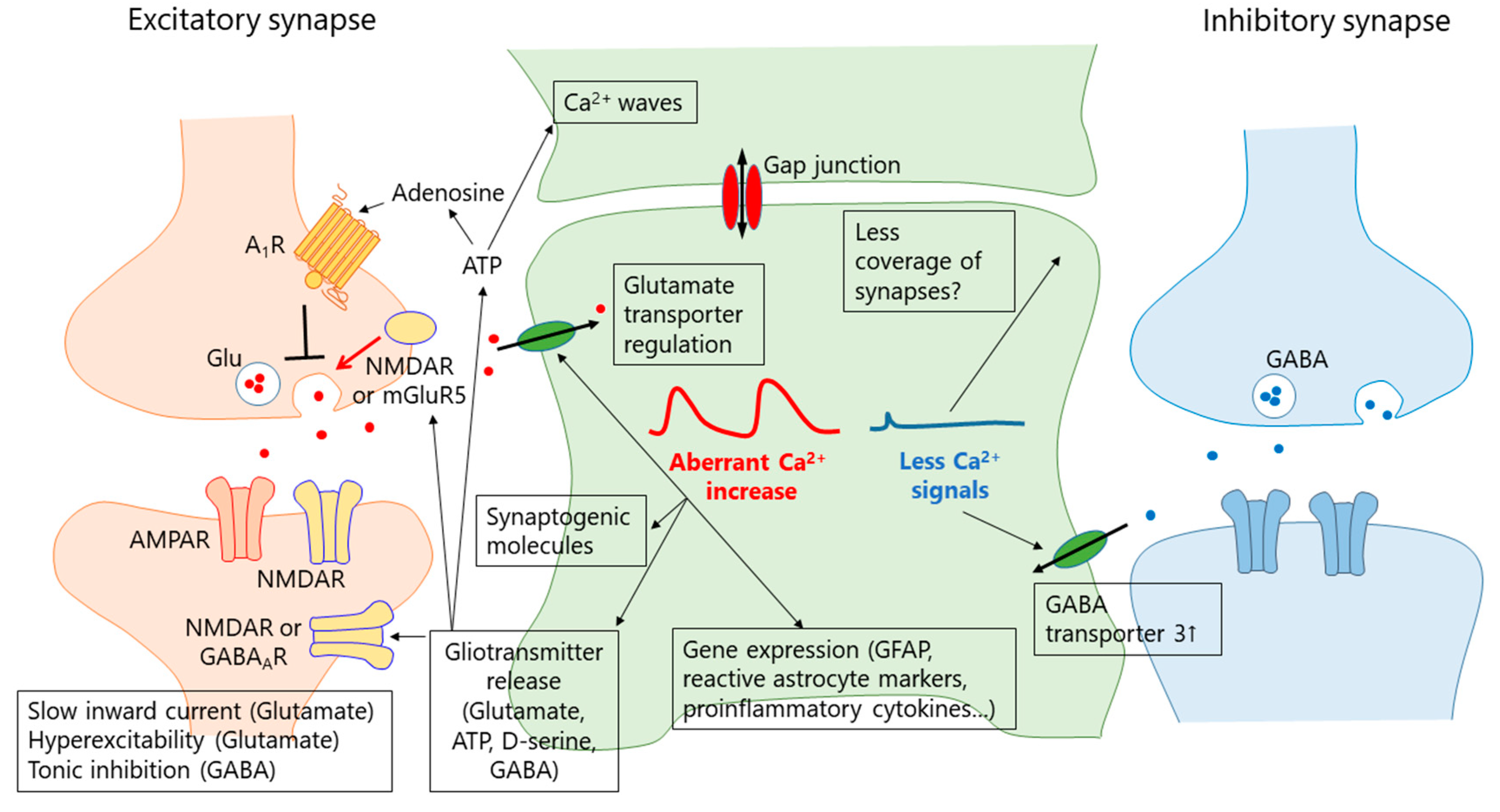
6. What Is the Function of the Aberrant Ca2+ Signal in Reactive Astrocytes?

Ca2+ is a ubiquitous second messenger regulating multiple aspects of cellular signaling. There are many mechanisms proposed for the functions of aberrant Ca2+ signals in reactive astrocytes that are summarized in Figure 1. Ca2+-dependent gliotransmission has attracted much interest because it is a well-known and well-characterized feature of astrocytes, although its relevance and mechanism are still under debate [84,85].

6.1. Gliotransmission

Astrocytes can release gliotransmitters, such as glutamate, ATP, D-serine and GABA in a Ca2+-dependent manner. Glutamate derived from astrocytes activates NMDA receptor on neurons in epilepsy [36,39,66,70,86], ischemia [34,87], CSD [73], Alzheimer’s disease [68] and Rett syndrome [79]. Activation of presynaptic NMDA receptors or mGluR5 by glutamate from astrocytes enhances excitatory synaptic transmission [66,70,88,89], while postsynaptic activation of NMDA receptors may lead to hyperexcitablity [34,36,39,73]. Glial-dependent presynaptic NMDA receptor activation is enhanced by TNF-α [90], which contributes to cognitive impairment in experimental autoimmune encephalitis (EAE), an animal model of MS [88]. Thus, NMDA receptor activation presumably triggers increased excitation of networks and neuronal death. One of the important issues in the field is whether or not astrocytes release glutamate in a Ca2+-dependent manner [29,84,85,91,92]. The machinery for glutamate release is undefined because astrocytes lack the molecules for vesicular glutamate release [29,93], although exocytosis of glutamate from astrocytes has been proposed. Another important issue is the functional significance of glutamate release from astrocytes. In many cases, slow inward currents (SIC) are recorded as an indicator of glial-derived glutamate release. SICs are thought to be caused by activation of extrasynaptic NMDA receptors in postsynaptic sites via glutamate released from astrocytes. SICs are blocked by antagonists against NMDA receptor containing NR2B subunit such as D-AP5 and Ro 25-6981. Recently, Gomez-Gonzalo et al. found that spontaneous SICs were mediated by a channel sensitive to 4,4’-Diisothiocyano-2,2’-stilbenedisulfonic acid (DIDS), quinine and fluoxetine but not by Ca2+-dependent vesicular glutamate release from astrocytes [94]. It has been suggested that glutamate derived from astrocytes, which cause SICs, contributes to many neurological diseases, such as epilepsy [36,39,66], stroke [34,87] and neurodevelopmental disorders [79]. Currently, there is no specific way to inhibit glutamate release mechanism underlying SICs without affecting other cell types/mechanisms. Therefore, the pathophysiological significance of SICs has not been tested directly. These two issues need to be solved to understand the role of Ca2+-dependent glutamate release from reactive astrocytes. In addition to a Ca2+-dependent mechanism, astrocytes can release glutamate in an intracellular pH-dependent manner. Oxygen glucose deprivation (OGD) reduces intracellular pH to cause glutamate release, which underlies ischemic brain damage [95].
In an APP/PS1 familial AD mouse model, reactive astrocytes in the dentate gyrus upregulate monoamine oxidase B, which contributes to GABA synthesis in reactive astrocytes. This astrocytic GABA is released through BEST1 channels in a Ca2+-dependent manner, causing intense tonic GABA currents. A strong inhibitory effect of astrocyte-derived GABA impaired neurotransmitter release, action-potential firing, synaptic plasticity and memory, which may underlie cognitive impairment in AD [96]. In other AD mouse model, tonic GABA inhibition was also enhanced and impaired synaptic plasticity, although GABA was released by the reverse mode of GABA transporters [97]. Increased GABA expression is also found in hypertrophic astrocytes induced by a stab wound brain injury model. Then, increased GABA is considered as a maker for reactive astrocytes [98].
ATP is also released from reactive astrocytes. ATP activates purinergic receptors in reactive astrocytes in an autocrine/paracrine fashion to contribute to Ca2+ waves [42,64]. These Ca2+ waves propagate to areas remote from the initiation site to transmit the information to synapses and microglia [99], which may cause hyperexcitability/damage and microglial chemotaxis to the injury site [35,64,100]. Many studies suggest that ATP acts on P2Y1 receptors on astrocytes to induce glutamate release in a Ca2+-dependent manner [64,66,70,89,90]. P2Y1 receptor-mediated glutamate release could be relevant to pathological conditions because TNF-α, an inflammatory cytokine induced by injury and seizure, is able to enhance P2Y1 receptor-mediated Ca2+ signals and gliotransmission [70,90]. In EAE, TNF-α levels are increased. Pathological levels of TNF-α altered synaptic transmission in the dentate gyrus, contributing to memory deficits in EAE [88]. It is not clear whether P2Y1 receptor-mediated Ca2+ signals are involved in this TNF-α effect. However, it is intriguing to see whether P2Y1 receptor-mediated Ca2+ signals contribute to the memory deficits in EAE. ATP is degraded into adenosine via ecto-nucleotidase, which in turn activates adenosine A1 receptors to inhibit neuronal excitability and excitatory synaptic transmission [83]. Thus, ATP derived from astrocytes either excites or inhibits neuronal networks, which depends on the context.

6.2. Synapse Remodeling

Developmental stage astrocytes express synaptogenic molecules, such as thrombospondin 1 (TSP1) [101]. TSP1 expression is low in adult mice but upregulated in pathophysiology. In a neuropathic pain model following peripheral nerve injury, TSP1 was upregulated in astrocytes of the S1 cortex through mGluR5-mediated Ca2+ signals in astrocytes [60], blockade of which reduced chronic pain. Also, pharmacological inhibition of the TSP1 receptor or TSP1 knockdown reduced chronic pain. Furthermore, TSP1 contributes to synapse remodeling in S1 cortex in neuropathic pain models. Thus, TSP1 upregulation through mGluR5-mediated Ca2+ signals in astrocytes cause synaptic rewiring in the S1 circuits by forming novel connections between neurons that underlie neuropathic pain [60,102].

6.3. GFAP Upregulation

GFAP is the most established maker for astrocytes and its upregulation is generally considered as a maker for reactive astrocytes. GFAP accumulation/upregulation is found in many neurological diseases, including AD [103]. Aberrant Ca2+ signal is essential for GFAP upregulation in AxD [46], TBI [37] and photothrombosis [104], since deletion of aberrant Ca2+ signals strongly suppressed the GFAP accumulation/upregulation. Thus, Ca2+ signals in astrocytes seem to be common events to induce GFAP upregulation. Because chronic treatment with a P2Y1 receptor antagonist reduced astrocyte Ca2+ signals in AD model mice but not GFAP upregulation, astrocyte Ca2+ signals may be relevant to induction of GFAP expression rather than maintenance of the expression.
In addition to GFAP upregulation, IP3R2-mediated Ca2+ signals regulate gene expression of other genes. AxCa regulate Lcn2, a pan reactive astrocyte maker, and C3, an A1 (neurotoxic) astrocyte marker [46]. Kanemaru et al. show that TBI-induced Ca2+ signals via IP3R2 inhibit the translational repressor, Pum2, to upregulate N-cadherin expression, which prevents infiltration of leukocytes and is neuroprotective [37]. Thus, IP3R2-mediated Ca2+ signals are essential for GFAP upregulation in pathological conditions.

6.4. Neuronal Damage

Infarct volume following MCAO or photothrombosis is significantly smaller in IP3R2KO mice compared with controls [34,87,104]. IP3R2 is upregulated in the penumbra by photothrombosis [54], which may contribute to aberrant Ca2+ signals in astrocytes of this region. Rakers et al. found that IP3R2 contributes to peri-infarct depolarization (PID), which is thought to enhance neurodegeneration and expand infarct size. The authors imaged Ca2+ in neurons and astrocytes in vivo after permanent MCAO and found PID-related Ca2+ elevations in both cell types. Ca2+ elevations in astrocytes were significantly reduced in IP3R2KO. Interestingly, PID-related Ca2+ elevations in neurons were also reduced in IP3R2KO, suggesting that astrocytic Ca2+ waves enhance neuronal Ca2+ elevations in PID. Astrocyte Ca2+ elevation is positively correlated with extracellular glutamate increase [87]. Lack of IP3R2 shortened both the duration of astrocyte Ca2+ elevation and duration of glutamate increase. Thus, glutamate levels are further increased by glutamate derived from Ca2+-elevated astrocytes, which contributes to excitotoxicity following ischemia. Similarly, Dong et al. found that in OGD, an in vitro model of ischemia, Ca2+ waves were induced in astrocytes in an IP3R2-dependent manner. SICs induced by OGD were also reduced in IP3R2KO mice, indicating that glutamate release from astrocytes may be augmented [34]. Thus, ischemia causes extracellular glutamate levels to rise by Ca2+ signals via IP3R2 in astrocytes, which contribute to glutamate toxicity through the activation of extrasynaptic NMDA receptors. These findings indicate that augmented IP3R2-mediated Ca2+ signals exacerbate damage by ischemia probably through elevation of extracellular glutamate [34,87]. These lines of evidence indicate that IP3R2-mediated Ca2+ signals can be a therapeutic target to protect neurons from ischemic damage. A transmembrane pathway through TRPV4 also contributes to astrocyte Ca2+ elevation following permanent MCAO. Although its contribution is smaller than that of IP3R2-mediated Ca2+ signals [105], recent evidence indicates that TRPV4 activation is a key determinant in brain edema induced by ischemia [106].

7. Role of Reduced Astrocyte Ca2+ Signals in Disease

Generally, reactive astrocytes display augmented Ca2+ signals; however, Ca2+ signals in astrocytes can also be reduced in pathophysiology [40,107]. There are several ways to reduce Ca2+ signals experimentally. Firstly, IP3R2KO mice have strongly reduced spontaneous and evoked Ca2+ signals in the cytosol, as described above [50]. Secondary, the “IP3 sponge”, an IP3 absorber, is another way to reduce IP3-mediated Ca2+ signals. Selective introduction of an IP3 sponge into astrocytes leads to reduced coverage of synapses by astrocytes and facilitated spillover of glutamate from synapses [41]. Thirdly, IP3 5-phosphatase, an IP3 hydrolyzing enzyme, is a useful tool to suppress IP3-mediated Ca2+ signals in astrocytes [108]. Fourthly, Yu et al. showed that overexpression of human PMCA2w/b (hPMCA2w/b), which constitutively excludes Ca2+ from the cytoplasm in astrocytes, strongly reduces spontaneous and evoked Ca2+ signals [109]. Overexpression of hPMCA2w/b in striatal astrocytes caused an increase in self-grooming behavior. Detailed molecular and functional analysis of astrocytes and medium spiny neurons (MSNs) in the striatum revealed altered MSN activity resulting from increased expression of GABA transporter 3 (GAT-3) in the plasma membrane through downregulation of Rab11a. Huntington’s disease model astrocytes also showed reduced Ca2+ signaling and excessive self-grooming behaviors in a GAT-3 dependent manner, indicating that attenuation of Ca2+ signals by enhancing Ca2+ efflux through the plasma membrane affects the function of astrocytes, circuits and behaviors [109]. The effect of hPMCA2w/b on reducing astrocytic cytosolic Ca2+ was strong but smaller than that in IP3R2KO mice. However, IP3R2KO mice are not reported to display abnormal grooming behavior. Why is the grooming behavior of IP3R2KO mice normal? In conventional IP3R2KO mice, IP3R2 is deleted from astrocytes throughout the brain and at all times; therefore, compensatory mechanisms might maintain biological functions of astrocytes. A more intriguing possibility is that subcellular differences in Ca2+ dynamics are relevant to the functions of astrocyte Ca2+ signals. hPMCA2w/b mice have highly reduced Ca2+ near the plasma membrane, while IP3R2KO mice lack the major Ca2+ release pathway from the ER. Therefore, subcellular Ca2+ dynamics may differ between hPMCA2w/b astrocytes and IP3R2KO astrocytes, which may explain the difference in behavioral phenotypes and indicate functional diversity of Ca2+ signals in astrocytes. In theory, the methods described above can be applied to astrocytes in specific circuits rather than the entire brain. Such approaches may help to more specifically elucidate the role of Ca2+ signals in reactive astrocytes and may reveal the detailed mechanisms underlying aberrant Ca2+ signals in reactive astrocytes.

8. Conclusions

Reactive astrocytes display spatiotemporally dynamic Ca2+ signals. Advanced methods such as the use of GECIs, two-photon microscopy and novel transgenic approaches have revealed the molecular mechanisms of the dynamic features of Ca2+ signals. Although it is difficult to generalize on the role of aberrant Ca2+ signals in astrocytes considering the heterogeneity of reactive astrocytes, Ca2+ signals in reactive astrocytes clearly indicate certain disease states and disease severity. Reactive astrocytes showing enhanced Ca2+ signals probably acquire “gain of toxicity”, which plays deleterious roles in disease progression, even in the presymptomatic phase of disease. There are many mechanisms underlying aberrant Ca2+ signals in reactive astrocytes. Different disease models may share a similar mechanism, while other models of similar diseases may use distinct mechanisms. Some of these mechanisms are potentially targets to treat the disease. Why are so many different mechanisms involved in aberrant Ca2+ signals in astrocytes? We don’t have a clear answer for this right now. However, it may be a consequence of astrocyte heterogeneity in different circumstances (e.g., young vs. old, early phase vs. late phase, hippocampus vs. striatum); reactive astrocytes may show a broad and graded spectrum of molecular, cellular and functional changes [9] that produce distinct phenotypes depending on the stimuli [28].
To understand the functional implications of aberrant Ca2+ signals, experiments need to be stringently designed to take into account astrocyte heterogeneity in different circuits, and during development and aging. Even in the same experimental settings, astrocytes react immediately to brain insults to change their properties. To understand how Ca2+ signals are regulated in reactive astrocytes, it is therefore important to analyze astrocyte properties by non-biased methods for each experimental setting. This approach will allow us to understand not only the mechanisms underlying aberrant Ca2+ signals but also downstream signaling. In addition, it is important to develop a method to precisely manipulate Ca2+ signals in (patho-)physiological states without affecting unwanted targets [91]. A combination of the approaches described above may elucidate the role of reactive astrocytes in neurological diseases and help to find novel therapeutic targets to treat such diseases. Since astrocytes change their morphology and gene expression immediately in response to environmental changes [27,110], both in vitro and in vivo experiments are needed to understand the cellular functions of reactive astrocytes.
Ca2+ signal is one type of cellular activities. Other signals, such as cAMP and Na+, are also dynamically regulated in astrocytes. Improvement of Ca2+ imaging techniques has advanced our understanding of Ca2+ signals in astrocytes. Similarly, refinement of sensors for other signals will reveal novel insights into astrocyte activities enabling investigation of how distinct activities interact and cooperate in physiology and pathophysiology.

Author Contributions

All of the authors wrote and contributed to the final version of the manuscript.

Funding

JSPS KAKENHI 17K01974 (E.S.), 15KK0340 (E.S.), 16H04669 (S.K.), 18H05121 (S.K.), 257117003 (S.K.) and 18K15701 (F.S.). The Takeda Science Foundation (E.S.).

Acknowledgments

E.S. is supported by JSPS KAKENHI 17K01974 and 15KK0340 and the Takeda Science Foundation. S.K. is supported by JSPS KAKENHI 16H04669, 18H05121 and 257117003. F.S. is supported by JSPS KAKENHI 18K15701. We thank Bijay Parajuli, PhD for comments on this manuscript. We thank Jeremy Allen, PhD, from Edanz Group (www.edanzediting.com/ac) for editing a draft of this manuscript.

Conflicts of Interest

The authors declare no conflict of interest.

Abbreviations

ADAlzheimer’s disease
ALSAmyotrophic lateral sclerosis
AMAcetoxymethyl
AxCaAberrant extra-large Ca2+ signal
AxDAlexander disease
CSDCortical spreading depression
DSDown syndrome
EAEExperimental autoimmune encephalitis
EREndoplasmic reticulum
GAT-3GABA transporter 3
GECIGenetically encoded Ca2+ indicator
GFAPGlial fibrillary acidic protein
GqPCRGq-protein coupled receptor
hPMCA2w/bHuman PMCA2w/b
IP3R2Inositol-1,4,5 trisphosphate receptor type2
IP3R2KOIP3R type2 knockout
MCAOMiddle cerebral artery occlusion
mGluR5Metabotropic glutamate receptor 5
MSNMedium spiny neuron
mPTPMitochondrial permeability transition pore
MSMultiple sclerosis
OGDOxygen glucose deprivation
PIDPeri-infarct depolarization
RTTRett syndrome
SCISpinal cord injury
SEStatus epilepticus
SICSlow inward current
TBITraumatic brain injury
TSP1Thrombospondin 1

References

  1. Khakh, B.S.; Sofroniew, M.V. Diversity of astrocyte functions and phenotypes in neural circuits. Nat. Neurosci. 2015, 18, 942–952. [Google Scholar] [CrossRef] [PubMed] [Green Version]
  2. Verkhratsky, A.; Nedergaard, M. Physiology of astroglia. Physiol. Rev. 2018, 98, 239–389. [Google Scholar] [CrossRef] [PubMed]
  3. Dallerac, G.; Zapata, J.; Rouach, N. Versatile control of synaptic circuits by astrocytes: Where, when and how? Nat. Rev. Neurosci. 2018, 19, 729–743. [Google Scholar] [CrossRef] [PubMed]
  4. Ben Haim, L.; Rowitch, D.H. Functional diversity of astrocytes in neural circuit regulation. Nat. Rev. Neurosci. 2017, 18, 31–41. [Google Scholar] [CrossRef] [PubMed]
  5. Araque, A.; Carmignoto, G.; Haydon, P.G.; Oliet, S.H.; Robitaille, R.; Volterra, A. Gliotransmitters travel in time and space. Neuron 2014, 81, 728–739. [Google Scholar] [CrossRef] [PubMed]
  6. Shigetomi, E.; Patel, S.; Khakh, B.S. Probing the complexities of astrocyte calcium signaling. Trends Cell Biol. 2016, 26, 300–312. [Google Scholar] [CrossRef] [PubMed]
  7. Pekny, M.; Pekna, M.; Messing, A.; Steinhauser, C.; Lee, J.M.; Parpura, V.; Hol, E.M.; Sofroniew, M.V.; Verkhratsky, A. Astrocytes: A central element in neurological diseases. Acta Neuropathol. 2016, 131, 323–345. [Google Scholar] [CrossRef] [PubMed]
  8. Bazargani, N.; Attwell, D. Astrocyte calcium signaling: The third wave. Nat. Neurosci. 2016, 19, 182–189. [Google Scholar] [CrossRef] [PubMed]
  9. Burda, J.E.; Sofroniew, M.V. Reactive gliosis and the multicellular response to CNS damage and disease. Neuron 2014, 81, 229–248. [Google Scholar] [CrossRef] [PubMed]
  10. Reeves, A.M.; Shigetomi, E.; Khakh, B.S. Bulk loading of calcium indicator dyes to study astrocyte physiology: Key limitations and improvements using morphological maps. J. Neurosci. 2011, 31, 9353–9358. [Google Scholar] [CrossRef] [PubMed]
  11. Shigetomi, E.; Bushong, E.A.; Haustein, M.D.; Tong, X.; Jackson-Weaver, O.; Kracun, S.; Xu, J.; Sofroniew, M.V.; Ellisman, M.H.; Khakh, B.S. Imaging calcium microdomains within entire astrocyte territories and endfeet with GCaMPs expressed using adeno-associated viruses. J. Gen. Physiol. 2013, 141, 633–647. [Google Scholar] [CrossRef] [PubMed] [Green Version]
  12. Kanemaru, K.; Sekiya, H.; Xu, M.; Satoh, K.; Kitajima, N.; Yoshida, K.; Okubo, Y.; Sasaki, T.; Moritoh, S.; Hasuwa, H.; et al. In vivo visualization of subtle, transient, and local activity of astrocytes using an ultrasensitive Ca(2+) indicator. Cell Rep. 2014, 8, 311–318. [Google Scholar] [CrossRef] [PubMed]
  13. Gee, J.M.; Gibbons, M.B.; Taheri, M.; Palumbos, S.; Morris, S.C.; Smeal, R.M.; Flynn, K.F.; Economo, M.N.; Cizek, C.G.; Capecchi, M.R.; et al. Imaging activity in astrocytes and neurons with genetically encoded calcium indicators following in utero electroporation. Front. Mol. Neurosci. 2015, 8, 10. [Google Scholar] [CrossRef] [PubMed]
  14. Srinivasan, R.; Huang, B.S.; Venugopal, S.; Johnston, A.D.; Chai, H.; Zeng, H.; Golshani, P.; Khakh, B.S. Ca(2+) signaling in astrocytes from Ip3r2(-/-) mice in brain slices and during startle responses in vivo. Nat. Neurosci. 2015, 18, 708–717. [Google Scholar] [CrossRef] [PubMed]
  15. Agarwal, A.; Wu, P.H.; Hughes, E.G.; Fukaya, M.; Tischfield, M.A.; Langseth, A.J.; Wirtz, D.; Bergles, D.E. Transient opening of the mitochondrial permeability transition pore induces microdomain calcium transients in astrocyte processes. Neuron 2017, 93, 587.e7–605.e7. [Google Scholar] [CrossRef] [PubMed]
  16. Bindocci, E.; Savtchouk, I.; Liaudet, N.; Becker, D.; Carriero, G.; Volterra, A. Three-dimensional Ca2+ imaging advances understanding of astrocyte biology. Science 2017, 356, eaai8185. [Google Scholar] [CrossRef] [PubMed]
  17. Stobart, J.L.; Ferrari, K.D.; Barrett, M.J.P.; Gluck, C.; Stobart, M.J.; Zuend, M.; Weber, B. Cortical circuit activity evokes rapid astrocyte calcium signals on a similar timescale to neurons. Neuron 2018, 98, 726–735. [Google Scholar] [CrossRef] [PubMed]
  18. Smith, N.A.; Kress, B.T.; Lu, Y.; Chandler-Militello, D.; Benraiss, A.; Nedergaard, M. Fluorescent Ca(2+) indicators directly inhibit the Na,K-ATPase and disrupt cellular functions. Sci. Signal. 2018, 11, eaal2039. [Google Scholar] [CrossRef] [PubMed]
  19. Zariwala, H.A.; Borghuis, B.G.; Hoogland, T.M.; Madisen, L.; Tian, L.; De Zeeuw, C.I.; Zeng, H.; Looger, L.L.; Svoboda, K.; Chen, T.W. A Cre-dependent GCaMP3 reporter mouse for neuronal imaging in vivo. J. Neurosci. 2012, 32, 3131–3141. [Google Scholar] [CrossRef] [PubMed]
  20. Monai, H.; Ohkura, M.; Tanaka, M.; Oe, Y.; Konno, A.; Hirai, H.; Mikoshiba, K.; Itohara, S.; Nakai, J.; Iwai, Y.; et al. Calcium imaging reveals glial involvement in transcranial direct current stimulation-induced plasticity in mouse brain. Nat. Commun. 2016, 7, 11100. [Google Scholar] [CrossRef] [PubMed] [Green Version]
  21. Ding, F.; O’Donnell, J.; Thrane, A.S.; Zeppenfeld, D.; Kang, H.; Xie, L.; Wang, F.; Nedergaard, M. Alpha1-adrenergic receptors mediate coordinated Ca2+ signaling of cortical astrocytes in awake, behaving mice. Cell Calcium 2013, 54, 387–394. [Google Scholar] [CrossRef] [PubMed]
  22. Paukert, M.; Agarwal, A.; Cha, J.; Doze, V.A.; Kang, J.U.; Bergles, D.E. Norepinephrine controls astroglial responsiveness to local circuit activity. Neuron 2014, 82, 1263–1270. [Google Scholar] [CrossRef] [PubMed]
  23. Pekny, M.; Pekna, M. Astrocyte reactivity and reactive astrogliosis: Costs and benefits. Physiol. Rev. 2014, 94, 1077–1098. [Google Scholar] [CrossRef] [PubMed]
  24. Hara, M.; Kobayakawa, K.; Ohkawa, Y.; Kumamaru, H.; Yokota, K.; Saito, T.; Kijima, K.; Yoshizaki, S.; Harimaya, K.; Nakashima, Y.; et al. Interaction of reactive astrocytes with type I collagen induces astrocytic scar formation through the integrin-N-cadherin pathway after spinal cord injury. Nat. Med. 2017, 23, 818–828. [Google Scholar] [CrossRef] [PubMed]
  25. Anderson, M.A.; Burda, J.E.; Ren, Y.; Ao, Y.; O’Shea, T.M.; Kawaguchi, R.; Coppola, G.; Khakh, B.S.; Deming, T.J.; Sofroniew, M.V. Astrocyte scar formation aids central nervous system axon regeneration. Nature 2016, 532, 195. [Google Scholar] [CrossRef] [PubMed]
  26. Adams, K.L.; Gallo, V. The diversity and disparity of the glial scar. Nat. Neurosci. 2018, 21, 9–15. [Google Scholar] [CrossRef] [PubMed]
  27. Zamanian, J.L.; Xu, L.; Foo, L.C.; Nouri, N.; Zhou, L.; Giffard, R.G.; Barres, B.A. Genomic analysis of reactive astrogliosis. J. Neurosci. 2012, 32, 6391–6410. [Google Scholar] [CrossRef] [PubMed]
  28. Liddelow, S.A.; Guttenplan, K.A.; Clarke, L.E.; Bennett, F.C.; Bohlen, C.J.; Schirmer, L.; Bennett, M.L.; Munch, A.E.; Chung, W.S.; Peterson, T.C.; et al. Neurotoxic reactive astrocytes are induced by activated microglia. Nature 2017, 541, 481–487. [Google Scholar] [CrossRef] [PubMed] [Green Version]
  29. Chai, H.; Diaz-Castro, B.; Shigetomi, E.; Monte, E.; Octeau, J.C.; Yu, X.; Cohn, W.; Rajendran, P.S.; Vondriska, T.M.; Whitelegge, J.P.; et al. Neural circuit-specialized astrocytes: Transcriptomic, proteomic, morphological, and functional evidence. Neuron 2017, 95, 531.e9–549.e9. [Google Scholar] [CrossRef] [PubMed]
  30. John Lin, C.C.; Yu, K.; Hatcher, A.; Huang, T.W.; Lee, H.K.; Carlson, J.; Weston, M.C.; Chen, F.; Zhang, Y.; Zhu, W.; et al. Identification of diverse astrocyte populations and their malignant analogs. Nat. Neurosci. 2017, 20, 396–405. [Google Scholar] [CrossRef] [PubMed] [Green Version]
  31. Morel, L.; Chiang, M.S.R.; Higashimori, H.; Shoneye, T.; Iyer, L.K.; Yelick, J.; Tai, A.; Yang, Y. Molecular and functional properties of regional astrocytes in the adult brain. J. Neurosci. 2017, 37, 8706–8717. [Google Scholar] [CrossRef] [PubMed]
  32. Rossi, D.; Volterra, A. Astrocytic dysfunction: Insights on the role in neurodegeneration. Brain Res. Bull. 2009, 80, 224–232. [Google Scholar] [CrossRef] [PubMed]
  33. Duffy, S.; MacVicar, B.A. In vitro ischemia promotes calcium influx and intracellular calcium release in hippocampal astrocytes. J. Neurosci. 1996, 16, 71–81. [Google Scholar] [CrossRef] [PubMed]
  34. Dong, Q.P.; He, J.Q.; Chai, Z. Astrocytic Ca(2+) waves mediate activation of extrasynaptic nmda receptors in hippocampal neurons to aggravate brain damage during ischemia. Neurobiol. Dis. 2013, 58, 68–75. [Google Scholar] [CrossRef] [PubMed]
  35. Choo, A.M.; Miller, W.J.; Chen, Y.C.; Nibley, P.; Patel, T.P.; Goletiani, C.; Morrison, B., 3rd; Kutzing, M.K.; Firestein, B.L.; Sul, J.Y.; et al. Antagonism of purinergic signalling improves recovery from traumatic brain injury. Brain 2013, 136, 65–80. [Google Scholar] [CrossRef] [PubMed] [Green Version]
  36. Tian, G.F.; Azmi, H.; Takano, T.; Xu, Q.; Peng, W.; Lin, J.; Oberheim, N.; Lou, N.; Wang, X.; Zielke, H.R.; et al. An astrocytic basis of epilepsy. Nat. Med. 2005, 11, 973–981. [Google Scholar] [CrossRef] [PubMed] [Green Version]
  37. Kanemaru, K.; Kubota, J.; Sekiya, H.; Hirose, K.; Okubo, Y.; Iino, M. Calcium-dependent n-cadherin up-regulation mediates reactive astrogliosis and neuroprotection after brain injury. Proc. Natl. Acad. Sci. USA 2013, 110, 11612–11617. [Google Scholar] [CrossRef] [PubMed]
  38. Heuser, K.; Nome, C.G.; Pettersen, K.H.; Abjorsbraten, K.S.; Jensen, V.; Tang, W.; Sprengel, R.; Tauboll, E.; Nagelhus, E.A.; Enger, R. Ca2+ signals in astrocytes facilitate spread of epileptiform activity. Cereb. Cortex 2018, 28, 4036–4048. [Google Scholar] [CrossRef] [PubMed]
  39. Ding, S.; Fellin, T.; Zhu, Y.; Lee, S.Y.; Auberson, Y.P.; Meaney, D.F.; Coulter, D.A.; Carmignoto, G.; Haydon, P.G. Enhanced astrocytic Ca2+ signals contribute to neuronal excitotoxicity after status epilepticus. J. Neurosci. 2007, 27, 10674–10684. [Google Scholar] [CrossRef] [PubMed]
  40. Plata, A.; Lebedeva, A.; Denisov, P.; Nosova, O.; Postnikova, T.Y.; Pimashkin, A.; Brazhe, A.; Zaitsev, A.V.; Rusakov, D.A.; Semyanov, A. Astrocytic atrophy following status epilepticus parallels reduced Ca(2+) activity and impaired synaptic plasticity in the rat hippocampus. Front. Mol. Neurosci. 2018, 11, 215. [Google Scholar] [CrossRef] [PubMed]
  41. Tanaka, M.; Shih, P.Y.; Gomi, H.; Yoshida, T.; Nakai, J.; Ando, R.; Furuichi, T.; Mikoshiba, K.; Semyanov, A.; Itohara, S. Astrocytic Ca2+ signals are required for the functional integrity of tripartite synapses. Mol. Brain 2013, 6, 6. [Google Scholar] [CrossRef] [PubMed]
  42. Delekate, A.; Fuchtemeier, M.; Schumacher, T.; Ulbrich, C.; Foddis, M.; Petzold, G.C. Metabotropic P2Y1 receptor signalling mediates astrocytic hyperactivity in vivo in an Alzheimer’s disease mouse model. Nat. Commun. 2014, 5, 5422. [Google Scholar] [CrossRef] [PubMed]
  43. Kuchibhotla, K.V.; Lattarulo, C.R.; Hyman, B.T.; Bacskai, B.J. Synchronous hyperactivity and intercellular calcium waves in astrocytes in Alzheimer mice. Science 2009, 323, 1211–1215. [Google Scholar] [CrossRef] [PubMed]
  44. Ding, S.; Wang, T.; Cui, W.; Haydon, P.G. Photothrombosis ischemia stimulates a sustained astrocytic Ca2+ signaling in vivo. Glia 2009, 57, 767–776. [Google Scholar] [CrossRef] [PubMed]
  45. Fordsmann, J.C.; Murmu, R.P.; Cai, C.; Brazhe, A.; Thomsen, K.J.; Zambach, S.A.; Lonstrup, M.; Lind, B.L.; Lauritzen, M. Spontaneous astrocytic Ca(2+) activity abounds in electrically suppressed ischemic penumbra of aged mice. Glia 2019, 67, 37–52. [Google Scholar] [CrossRef] [PubMed]
  46. Saito, K.; Shigetomi, E.; Yasuda, R.; Sato, R.; Nakano, M.; Tashiro, K.; Tanaka, K.F.; Ikenaka, K.; Mikoshiba, K.; Mizuta, I.; et al. Aberrant astrocyte Ca(2+) signals “axca signals” exacerbate pathological alterations in an Alexander disease model. Glia 2018, 66, 1053–1067. [Google Scholar] [CrossRef] [PubMed]
  47. Shigetomi, E.; Bowser, D.N.; Sofroniew, M.V.; Khakh, B.S. Two forms of astrocyte calcium excitability have distinct effects on NMDA receptor-mediated slow inward currents in pyramidal neurons. J. Neurosci. 2008, 28, 6659–6663. [Google Scholar] [CrossRef] [PubMed]
  48. Khakh, B.S.; McCarthy, K.D. Astrocyte calcium signaling: From observations to functions and the challenges therein. Cold Spring Harb. Perspect. Biol. 2015, 7, a020404. [Google Scholar] [CrossRef] [PubMed]
  49. Ujita, S.; Sasaki, T.; Asada, A.; Funayama, K.; Gao, M.; Mikoshiba, K.; Matsuki, N.; Ikegaya, Y. cAMP-dependent calcium oscillations of astrocytes: An implication for pathology. Cereb. Cortex 2017, 27, 1602–1614. [Google Scholar] [CrossRef] [PubMed]
  50. Petravicz, J.; Fiacco, T.A.; McCarthy, K.D. Loss of IP3 receptor-dependent Ca2+ increases in hippocampal astrocytes does not affect baseline Ca1 pyramidal neuron synaptic activity. J. Neurosci. 2008, 28, 4967–4973. [Google Scholar] [CrossRef] [PubMed]
  51. Sherwood, M.W.; Arizono, M.; Hisatsune, C.; Bannai, H.; Ebisui, E.; Sherwood, J.L.; Panatier, A.; Oliet, S.H.; Mikoshiba, K. Astrocytic IP3 Rs: Contribution to Ca(2+) signalling and hippocampal LTP. Glia 2017, 65, 502–513. [Google Scholar] [CrossRef] [PubMed]
  52. Rungta, R.L.; Bernier, L.P.; Dissing-Olesen, L.; Groten, C.J.; LeDue, J.M.; Ko, R.; Drissler, S.; MacVicar, B.A. Ca2+ transients in astrocyte fine processes occur via Ca2+ influx in the adult mouse hippocampus. Glia 2016, 64, 2093–2103. [Google Scholar] [CrossRef] [PubMed]
  53. Okubo, Y.; Kanemaru, K.; Suzuki, J.; Kobayashi, K.; Hirose, K.; Iino, M. Inositol 1,4,5-trisphosphate receptor type 2-independent Ca(2+) release from the endoplasmic reticulum in astrocytes. Glia 2018, 67, 113–124. [Google Scholar] [CrossRef] [PubMed]
  54. Staats, K.A.; Humblet-Baron, S.; Bento-Abreu, A.; Scheveneels, W.; Nikolaou, A.; Deckers, K.; Lemmens, R.; Goris, A.; Van Ginderachter, J.A.; Van Damme, P.; et al. Genetic ablation of IP3 receptor 2 increases cytokines and decreases survival of SOD1G93A mice. Hum. Mol. Genet. 2016, 25, 3491–3499. [Google Scholar] [CrossRef] [PubMed]
  55. Sun, W.; McConnell, E.; Pare, J.F.; Xu, Q.; Chen, M.; Peng, W.; Lovatt, D.; Han, X.; Smith, Y.; Nedergaard, M. Glutamate-dependent neuroglial calcium signaling differs between young and adult brain. Science 2013, 339, 197–200. [Google Scholar] [CrossRef] [PubMed]
  56. Haustein, M.D.; Kracun, S.; Lu, X.H.; Shih, T.; Jackson-Weaver, O.; Tong, X.; Xu, J.; Yang, X.W.; O’Dell, T.J.; Marvin, J.S.; et al. Conditions and constraints for astrocyte calcium signaling in the hippocampal mossy fiber pathway. Neuron 2014, 82, 413–429. [Google Scholar] [CrossRef] [PubMed]
  57. Shrivastava, A.N.; Kowalewski, J.M.; Renner, M.; Bousset, L.; Koulakoff, A.; Melki, R.; Giaume, C.; Triller, A. Beta-amyloid and ATP-induced diffusional trapping of astrocyte and neuronal metabotropic glutamate type-5 receptors. Glia 2013, 61, 1673–1686. [Google Scholar] [CrossRef] [PubMed]
  58. Lim, D.; Iyer, A.; Ronco, V.; Grolla, A.A.; Canonico, P.L.; Aronica, E.; Genazzani, A.A. Amyloid beta deregulates astroglial mGluR5-mediated calcium signaling via calcineurin and nf-kb. Glia 2013, 61, 1134–1145. [Google Scholar] [CrossRef] [PubMed]
  59. Aronica, E.; van Vliet, E.A.; Mayboroda, O.A.; Troost, D.; da Silva, F.H.; Gorter, J.A. Upregulation of metabotropic glutamate receptor subtype mGluR3 and mGluR5 in reactive astrocytes in a rat model of mesial temporal lobe epilepsy. Eur. J. Neurosci. 2000, 12, 2333–2344. [Google Scholar] [CrossRef] [PubMed]
  60. Kim, S.K.; Hayashi, H.; Ishikawa, T.; Shibata, K.; Shigetomi, E.; Shinozaki, Y.; Inada, H.; Roh, S.E.; Kim, S.J.; Lee, G.; et al. Cortical astrocytes rewire somatosensory cortical circuits for peripheral neuropathic pain. J. Clin. Investig. 2016, 126, 1983–1997. [Google Scholar] [CrossRef] [PubMed] [Green Version]
  61. Umpierre, A.D.; West, P.J.; White, J.A.; Wilcox, K.S. Conditional knockout of mGluR5 from astrocytes during epilepsy development impairs high-frequency glutamate uptake. J. Neurosci. 2019, 39, 727–742. [Google Scholar] [CrossRef] [PubMed]
  62. Miller, S.; Romano, C.; Cotman, C.W. Growth factor upregulation of a phosphoinositide-coupled metabotropic glutamate receptor in cortical astrocytes. J. Neurosci. 1995, 15, 6103–6109. [Google Scholar] [CrossRef] [PubMed] [Green Version]
  63. Buscemi, L.; Ginet, V.; Lopatar, J.; Montana, V.; Pucci, L.; Spagnuolo, P.; Zehnder, T.; Grubisic, V.; Truttman, A.; Sala, C.; et al. Homer1 scaffold proteins govern Ca2+ dynamics in normal and reactive astrocytes. Cereb. Cortex 2017, 27, 2365–2384. [Google Scholar] [CrossRef] [PubMed]
  64. Koizumi, S. Synchronization of Ca2+ oscillations: Involvement of atp release in astrocytes. FEBS J. 2010, 277, 286–292. [Google Scholar] [CrossRef] [PubMed]
  65. Shigetomi, E.; Hirayama, Y.J.; Ikenaka, K.; Tanaka, K.F.; Koizumi, S. Role of purinergic receptor P2Y1 in spatiotemporal Ca(2+) dynamics in astrocytes. J. Neurosci. 2018, 38, 1383–1395. [Google Scholar] [CrossRef] [PubMed]
  66. Alvarez-Ferradas, C.; Morales, J.C.; Wellmann, M.; Nualart, F.; Roncagliolo, M.; Fuenzalida, M.; Bonansco, C. Enhanced astroglial Ca2+ signaling increases excitatory synaptic strength in the epileptic brain. Glia 2015, 63, 1507–1521. [Google Scholar] [CrossRef] [PubMed]
  67. Kuboyama, K.; Harada, H.; Tozaki-Saitoh, H.; Tsuda, M.; Ushijima, K.; Inoue, K. Astrocytic P2y(1) receptor is involved in the regulation of cytokine/chemokine transcription and cerebral damage in a rat model of cerebral ischemia. J. Cereb. Blood Flow Metab. 2011, 31, 1930–1941. [Google Scholar] [CrossRef] [PubMed]
  68. Reichenbach, N.; Delekate, A.; Breithausen, B.; Keppler, K.; Poll, S.; Schulte, T.; Peter, J.; Plescher, M.; Hansen, J.N.; Blank, N.; et al. P2Y1 receptor blockade normalizes network dysfunction and cognition in an Alzheimer’s disease model. J. Exp. Med. 2018, 215, 1649–1663. [Google Scholar] [CrossRef] [PubMed]
  69. Wellmann, M.; Alvarez-Ferradas, C.; Maturana, C.J.; Saez, J.C.; Bonansco, C. Astroglial Ca(2+)-dependent hyperexcitability requires P2Y1 purinergic receptors and pannexin-1 channel activation in a chronic model of epilepsy. Front. Cell. Neurosci. 2018, 12, 446. [Google Scholar] [CrossRef] [PubMed]
  70. Nikolic, L.; Shen, W.; Nobili, P.; Virenque, A.; Ulmann, L.; Audinat, E. Blocking TNFα-driven astrocyte purinergic signaling restores normal synaptic activity during epileptogenesis. Glia 2018, 66, 2673–2683. [Google Scholar] [CrossRef] [PubMed]
  71. Perea, G.; Gomez, R.; Mederos, S.; Covelo, A.; Ballesteros, J.J.; Schlosser, L.; Hernandez-Vivanco, A.; Martin-Fernandez, M.; Quintana, R.; Rayan, A.; et al. Activity-dependent switch of gabaergic inhibition into glutamatergic excitation in astrocyte-neuron networks. Elife 2016, 5, e20362. [Google Scholar] [CrossRef] [PubMed]
  72. Mariotti, L.; Losi, G.; Lia, A.; Melone, M.; Chiavegato, A.; Gomez-Gonzalo, M.; Sessolo, M.; Bovetti, S.; Forli, A.; Zonta, M.; et al. Interneuron-specific signaling evokes distinctive somatostatin-mediated responses in adult cortical astrocytes. Nat. Commun. 2018, 9, 82. [Google Scholar] [CrossRef] [PubMed] [Green Version]
  73. Wu, D.C.; Chen, R.Y.; Cheng, T.C.; Chiang, Y.C.; Shen, M.L.; Hsu, L.L.; Zhou, N. Spreading depression promotes astrocytic calcium oscillations and enhances gliotransmission to hippocampal neurons. Cereb. Cortex 2018, 28, 3204–3216. [Google Scholar] [CrossRef] [PubMed]
  74. Orr, A.G.; Hsiao, E.C.; Wang, M.M.; Ho, K.; Kim, D.H.; Wang, X.; Guo, W.; Kang, J.; Yu, G.Q.; Adame, A.; et al. Astrocytic adenosine receptor A2A and GS-coupled signaling regulate memory. Nat. Neurosci. 2015, 18, 423–434. [Google Scholar] [CrossRef] [PubMed]
  75. Shigetomi, E.; Jackson-Weaver, O.; Huckstepp, R.T.; O’Dell, T.J.; Khakh, B.S. TRPA1 channels are regulators of astrocyte basal calcium levels and long-term potentiation via constitutive D-serine release. J. Neurosci. 2013, 33, 10143–10153. [Google Scholar] [CrossRef] [PubMed]
  76. Bosson, A.; Paumier, A.; Boisseau, S.; Jacquier-Sarlin, M.; Buisson, A.; Albrieux, M. TRPA1 channels promote astrocytic Ca(2+) hyperactivity and synaptic dysfunction mediated by oligomeric forms of amyloid-beta peptide. Mol. Neurodegener. 2017, 12, 53. [Google Scholar] [CrossRef] [PubMed]
  77. Lee, K.I.; Lee, H.T.; Lin, H.C.; Tsay, H.J.; Tsai, F.C.; Shyue, S.K.; Lee, T.S. Role of transient receptor potential ankyrin 1 channels in Alzheimer’s disease. J. Neuroinflamm. 2016, 13, 92. [Google Scholar] [CrossRef] [PubMed]
  78. Pirttimaki, T.M.; Codadu, N.K.; Awni, A.; Pratik, P.; Nagel, D.A.; Hill, E.J.; Dineley, K.T.; Parri, H.R. Alpha7 nicotinic receptor-mediated astrocytic gliotransmitter release: Abeta effects in a preclinical alzheimer’s mouse model. PLoS ONE 2013, 8, e81828. [Google Scholar] [CrossRef] [PubMed]
  79. Dong, Q.; Liu, Q.; Li, R.; Wang, A.; Bu, Q.; Wang, K.H.; Chang, Q. Mechanism and consequence of abnormal calcium homeostasis in rett syndrome astrocytes. Elife 2018, 7, e33417. [Google Scholar] [CrossRef] [PubMed]
  80. Hirayama, Y.; Ikeda-Matsuo, Y.; Notomi, S.; Enaida, H.; Kinouchi, H.; Koizumi, S. Astrocyte-mediated ischemic tolerance. J. Neurosci. 2015, 35, 3794–3805. [Google Scholar] [CrossRef] [PubMed]
  81. Hirayama, Y.; Koizumi, S. Hypoxia-independent mechanisms of HIF-1alpha expression in astrocytes after ischemic preconditioning. Glia 2017, 65, 523–530. [Google Scholar] [CrossRef] [PubMed]
  82. Jones, J.R.; Kong, L.; Hanna, M.G.T.; Hoffman, B.; Krencik, R.; Bradley, R.; Hagemann, T.; Choi, J.; Doers, M.; Dubovis, M.; et al. Mutations in GFAP disrupt the distribution and function of organelles in human astrocytes. Cell Rep. 2018, 25, 947.e4–958.e4. [Google Scholar] [CrossRef] [PubMed]
  83. Mizuno, G.O.; Wang, Y.; Shi, G.; Wang, Y.; Sun, J.; Papadopoulos, S.; Broussard, G.J.; Unger, E.K.; Deng, W.; Weick, J.; et al. Aberrant calcium signaling in astrocytes inhibits neuronal excitability in a human down syndrome stem cell model. Cell Rep. 2018, 24, 355–365. [Google Scholar] [CrossRef] [PubMed]
  84. Fiacco, T.A.; McCarthy, K.D. Multiple lines of evidence indicate that gliotransmission does not occur under physiological conditions. J. Neurosci. 2018, 38, 3–13. [Google Scholar] [CrossRef] [PubMed]
  85. Savtchouk, I.; Volterra, A. Gliotransmission: Beyond black-and-white. J. Neurosci. 2018, 38, 14–25. [Google Scholar] [CrossRef] [PubMed]
  86. Carmignoto, G.; Haydon, P.G. Astrocyte calcium signaling and epilepsy. Glia 2012, 60, 1227–1233. [Google Scholar] [CrossRef] [PubMed] [Green Version]
  87. Rakers, C.; Petzold, G.C. Astrocytic calcium release mediates peri-infarct depolarizations in a rodent stroke model. J. Clin. Investig. 2017, 127, 511–516. [Google Scholar] [CrossRef] [PubMed]
  88. Habbas, S.; Santello, M.; Becker, D.; Stubbe, H.; Zappia, G.; Liaudet, N.; Klaus, F.R.; Kollias, G.; Fontana, A.; Pryce, C.R.; et al. Neuroinflammatory TNFalpha impairs memory via astrocyte signaling. Cell 2015, 163, 1730–1741. [Google Scholar] [CrossRef] [PubMed]
  89. Pascual, O.; Ben Achour, S.; Rostaing, P.; Triller, A.; Bessis, A. Microglia activation triggers astrocyte-mediated modulation of excitatory neurotransmission. Proc. Natl. Acad. Sci. USA 2012, 109, E197–E205. [Google Scholar] [CrossRef] [PubMed]
  90. Santello, M.; Bezzi, P.; Volterra, A. TNFalpha controls glutamatergic gliotransmission in the hippocampal dentate gyrus. Neuron 2011, 69, 988–1001. [Google Scholar] [CrossRef] [PubMed]
  91. Hamilton, N.B.; Attwell, D. Do astrocytes really exocytose neurotransmitters? Nat. Rev. Neurosci. 2010, 11, 227–238. [Google Scholar] [CrossRef] [PubMed]
  92. Agulhon, C.; Sun, M.Y.; Murphy, T.; Myers, T.; Lauderdale, K.; Fiacco, T.A. Calcium signaling and gliotransmission in normal vs. Reactive astrocytes. Front. Pharmacol. 2012, 3, 139. [Google Scholar] [CrossRef] [PubMed]
  93. Cahoy, J.D.; Emery, B.; Kaushal, A.; Foo, L.C.; Zamanian, J.L.; Christopherson, K.S.; Xing, Y.; Lubischer, J.L.; Krieg, P.A.; Krupenko, S.A.; et al. A transcriptome database for astrocytes, neurons, and oligodendrocytes: A new resource for understanding brain development and function. J. Neurosci. 2008, 28, 264–278. [Google Scholar] [CrossRef] [PubMed]
  94. Gomez-Gonzalo, M.; Zehnder, T.; Requie, L.M.; Bezzi, P.; Carmignoto, G. Insights into the release mechanism of astrocytic glutamate evoking in neurons NMDA receptor-mediated slow depolarizing inward currents. Glia 2018, 66, 2188–2199. [Google Scholar] [CrossRef] [PubMed]
  95. Beppu, K.; Sasaki, T.; Tanaka, K.F.; Yamanaka, A.; Fukazawa, Y.; Shigemoto, R.; Matsui, K. Optogenetic countering of glial acidosis suppresses glial glutamate release and ischemic brain damage. Neuron 2014, 81, 314–320. [Google Scholar] [CrossRef] [PubMed]
  96. Jo, S.; Yarishkin, O.; Hwang, Y.J.; Chun, Y.E.; Park, M.; Woo, D.H.; Bae, J.Y.; Kim, T.; Lee, J.; Chun, H.; et al. Gaba from reactive astrocytes impairs memory in mouse models of Alzheimer’s disease. Nat. Med. 2014, 20, 886–896. [Google Scholar] [CrossRef] [PubMed]
  97. Wu, Z.; Guo, Z.; Gearing, M.; Chen, G. Tonic inhibition in dentate gyrus impairs long-term potentiation and memory in an alzheimer’s [corrected] disease model. Nat. Commun. 2014, 5, 4159. [Google Scholar] [CrossRef] [PubMed]
  98. Chun, H.; An, H.; Lim, J.; Woo, J.; Lee, J.; Ryu, H.; Lee, C.J. Astrocytic proBDNF and tonic GABA distinguish active versus reactive astrocytes in hippocampus. Exp. Neurobiol. 2018, 27, 155–170. [Google Scholar] [CrossRef] [PubMed]
  99. Schipke, C.G.; Boucsein, C.; Ohlemeyer, C.; Kirchhoff, F.; Kettenmann, H. Astrocyte Ca2+ waves trigger responses in microglial cells in brain slices. FASEB J. 2002, 16, 255–257. [Google Scholar] [CrossRef] [PubMed]
  100. Davalos, D.; Grutzendler, J.; Yang, G.; Kim, J.V.; Zuo, Y.; Jung, S.; Littman, D.R.; Dustin, M.L.; Gan, W.B. ATP mediates rapid microglial response to local brain injury in vivo. Nat. Neurosci. 2005, 8, 752–758. [Google Scholar] [CrossRef] [PubMed]
  101. Allen, N.J.; Eroglu, C. Cell biology of astrocyte-synapse interactions. Neuron 2017, 96, 697–708. [Google Scholar] [CrossRef] [PubMed]
  102. Kim, S.K.; Nabekura, J.; Koizumi, S. Astrocyte-mediated synapse remodeling in the pathological brain. Glia 2017, 65, 1719–1727. [Google Scholar] [CrossRef] [PubMed]
  103. Middeldorp, J.; Hol, E.M. Gfap in health and disease. Prog. Neurobiol. 2011, 93, 421–443. [Google Scholar] [CrossRef] [PubMed]
  104. Li, H.; Xie, Y.; Zhang, N.; Yu, Y.; Zhang, Q.; Ding, S. Disruption of IP(3)R2-mediated Ca(2)(+) signaling pathway in astrocytes ameliorates neuronal death and brain damage while reducing behavioral deficits after focal ischemic stroke. Cell Calcium 2015, 58, 565–576. [Google Scholar] [CrossRef] [PubMed]
  105. Rakers, C.; Schmid, M.; Petzold, G.C. TRPV4 channels contribute to calcium transients in astrocytes and neurons during peri-infarct depolarizations in a stroke model. Glia 2017, 65, 1550–1561. [Google Scholar] [CrossRef] [PubMed]
  106. Hoshi, Y.; Okabe, K.; Shibasaki, K.; Funatsu, T.; Matsuki, N.; Ikegaya, Y.; Koyama, R. Ischemic brain injury leads to brain edema via hyperthermia-induced TRPV4 activation. J. Neurosci. 2018, 38, 5700–5709. [Google Scholar] [CrossRef] [PubMed]
  107. Jiang, R.; Diaz-Castro, B.; Looger, L.L.; Khakh, B.S. Dysfunctional calcium and glutamate signaling in striatal astrocytes from huntington’s disease model mice. J. Neurosci. 2016, 36, 3453–3470. [Google Scholar] [CrossRef] [PubMed]
  108. Kanemaru, K.; Okubo, Y.; Hirose, K.; Iino, M. Regulation of neurite growth by spontaneous Ca2+ oscillations in astrocytes. J. Neurosci. 2007, 27, 8957–8966. [Google Scholar] [CrossRef] [PubMed]
  109. Yu, X.; Taylor, A.M.W.; Nagai, J.; Golshani, P.; Evans, C.J.; Coppola, G.; Khakh, B.S. Reducing astrocyte calcium signaling in vivo alters striatal microcircuits and causes repetitive behavior. Neuron 2018, 99, 1170–1187. [Google Scholar] [CrossRef] [PubMed]
  110. Takano, T.; He, W.; Han, X.; Wang, F.; Xu, Q.; Wang, X.; Oberheim Bush, N.A.; Cruz, N.; Dienel, G.A.; Nedergaard, M. Rapid manifestation of reactive astrogliosis in acute hippocampal brain slices. Glia 2014, 62, 78–95. [Google Scholar] [CrossRef] [PubMed]
Figure 1. Functional significance of astrocyte Ca2+ signals in disease. The cartoon indicates how alteration of astrocyte Ca2+ in disease affects excitatory/inhibitory synapses and excitability of neurons.
Ijms 20 00996 g001

