Multi-Residue Method for the Analysis of Stilbene Estrogens in Milk

 ,
,
Abstract
:
1. Introduction
2. Results
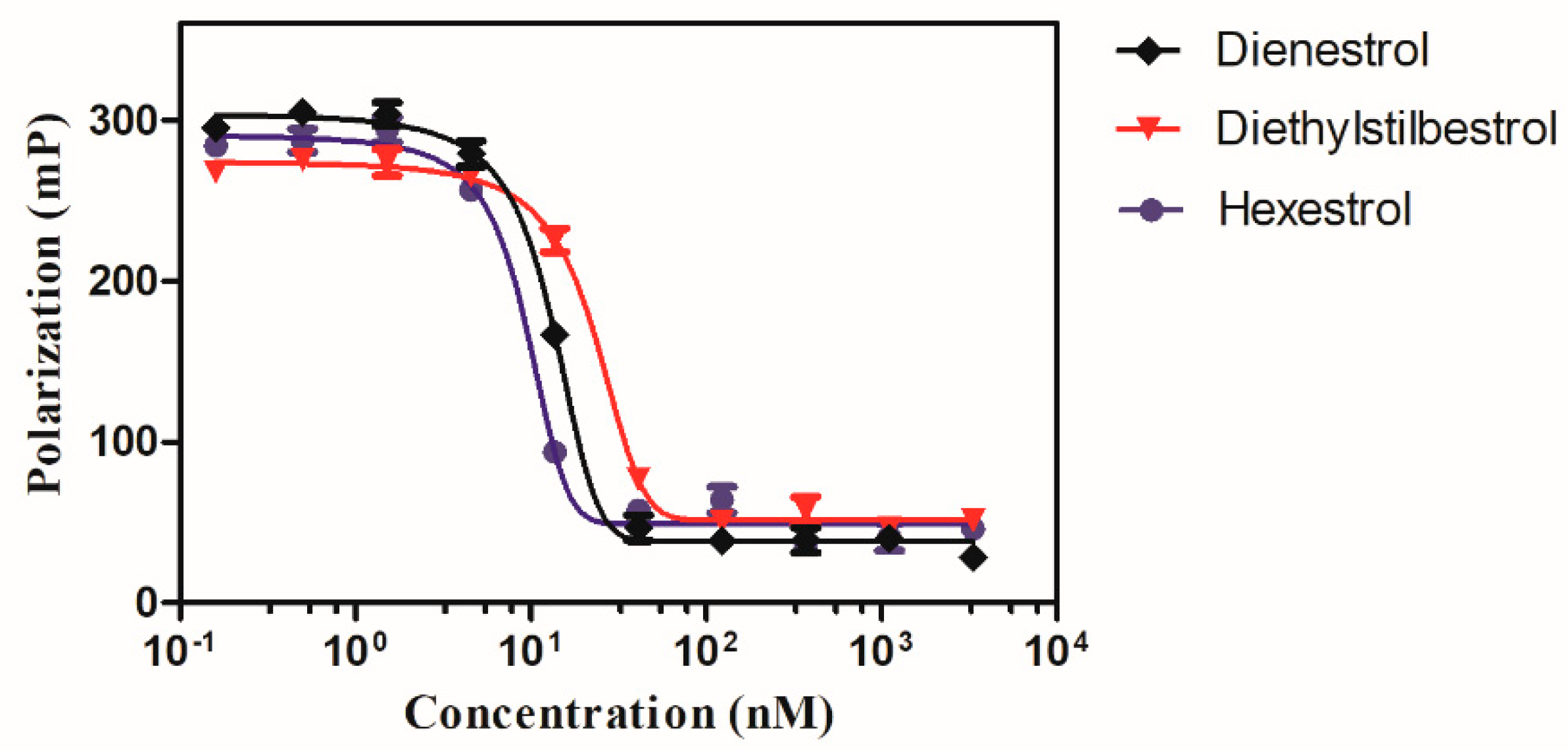
2.1. Competitive Binding Assay
2.2. Determination of Analytical Parameters
2.3. Analysis of Spiked Milk Samples
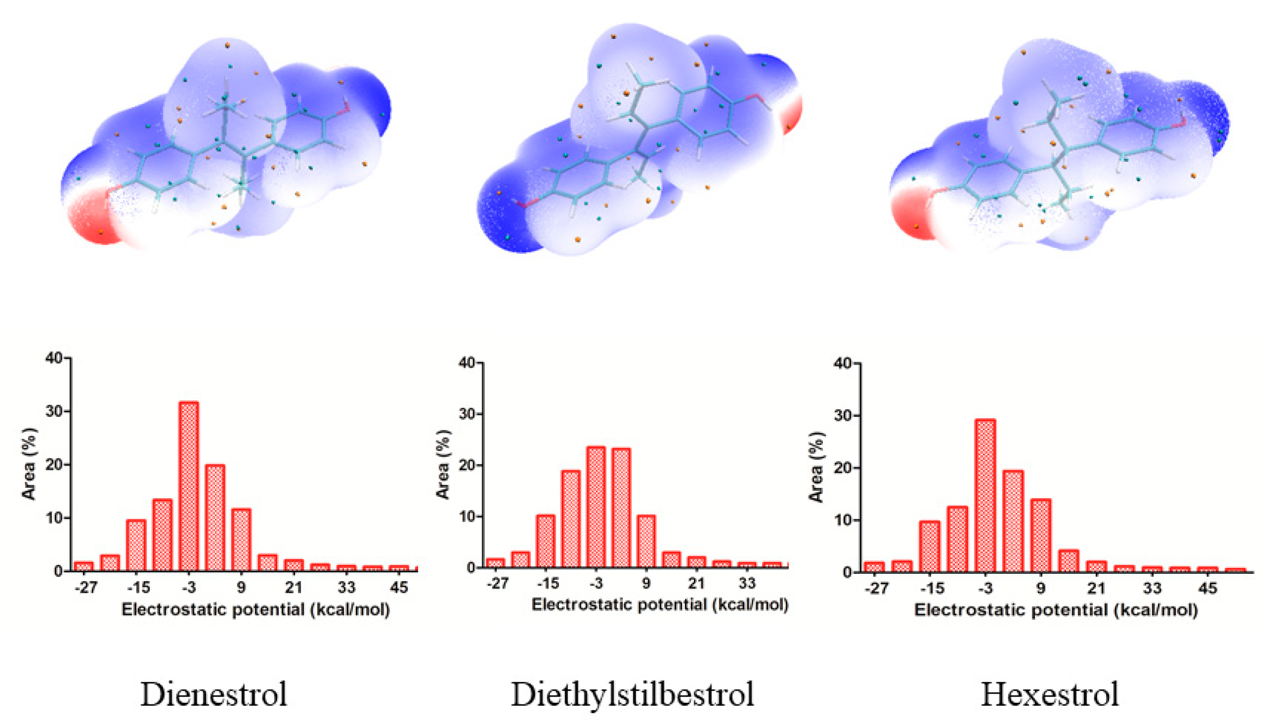
2.4. Topology Analyses
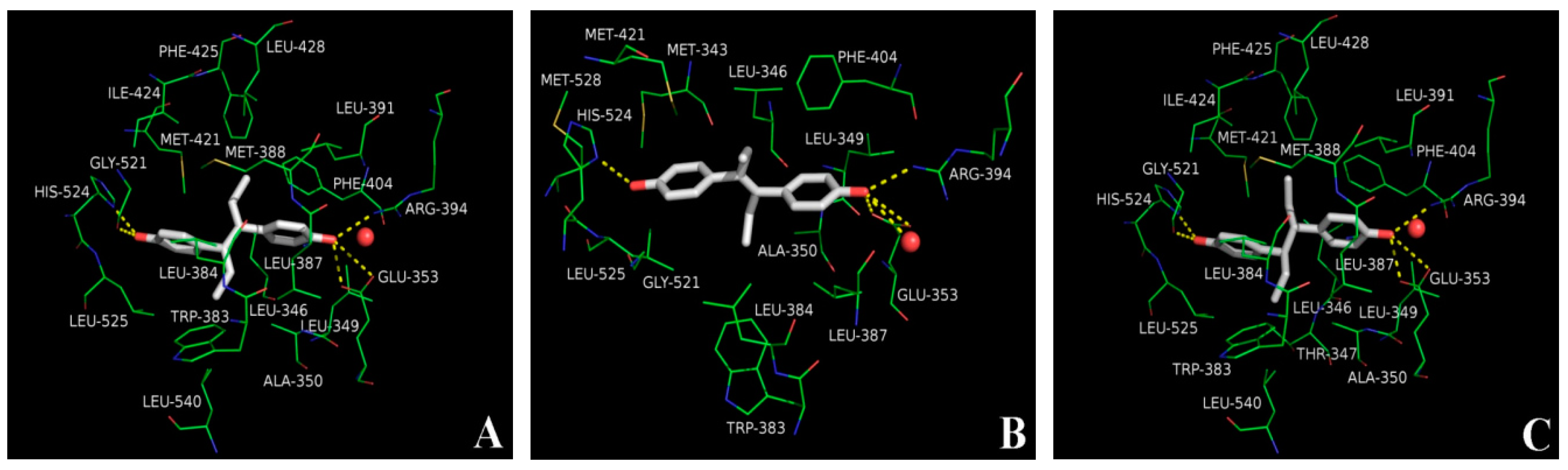
2.5. Molecular Docking Analysis
3. Discussion
4. Materials and Methods
4.1. Materials
4.2. Development of Fluorescence Polarization (FP) Assay
4.3. Determination of Analytical Parameters
4.4. Sample Treatment
4.5. Electrostatic Properties
4.6. Automated Docking Procedure
Author Contributions
Funding
Acknowledgments
Conflicts of Interest
Abbreviations
| FP | fluorescence polarization |
| ER-LBD | estrogen receptor α ligand binding domain |
| IC50 | half maximal inhibitory concentrations |
| LOD | limits of detection |
| CV | coefficients of variation |
| ESP | electrostatic potential |
| GST | glutathione S-transferase |
| CS | coumestrol |
| CR | cross-reactivity |
References
- Abafe, O.A.; Gatyeni, P.M.; Chokwe, T. Development and validation of a confirmatory method for the determination of stilbene estrogens in ostrich serum. Food Addit. Contam. Part A 2018, 35, 458–466. [Google Scholar] [CrossRef] [PubMed]
- Welshons, W.V.; Rottinghaus, G.E.; Nonneman, D.J.; Dolan-Timpe, M.; Ross, P.F. A sensitive bioassay for detection of dietary estrogens in animal feeds. J. Vet. Diagn. Investig. 1990, 2, 268–273. [Google Scholar] [CrossRef] [PubMed]
- Dickson, L.C.; Macneil, J.D.; Reid, J.; Fesser, A.C.E. Validation of Screening Method for Residues of Diethylstilbestrol Dienestrol, Hexestrol, and Zeranol in Bovine Urine Using Immunoaffinity Chromatography and Gas Chromatography/Mass Spectrometry. J. AOAC Int. 2003, 86, 631–639. [Google Scholar] [PubMed]
- Hu, W.Y.; Kang, X.J.; Chi, Z.; Jun, Y.; Rui, L.; Hu, E.L.; Ping, L. Packed-fiber solid-phase extraction coupled with high performance liquid chromatography-tandem mass spectrometry for determination of diethylstilbestrol, hexestrol, and dienestrol residues in milk products. J. Chrom. B 2014, 957, 7–13. [Google Scholar] [CrossRef] [PubMed]
- Hua, L.M.; Bin, Q.; Xia, J.; Lan, Z.; Xi, C.; Nan, C.G. Determination of estrogens in wastewater using three-phase hollow fiber-mediated liquid-phase microextraction followed by HPLC. J. Sep. Sci. 2008, 31, 622–628. [Google Scholar]
- Lange, I.G.; Daxenberger, A.; Schiffer, B.; Witters, H.; Ibarreta, D.; Meyer, H.H.D. Sex hormones originating from different livestock production systems fate and potential disrupting activity in the environment. Anal. Chim. Acta 2002, 473, 27–37. [Google Scholar] [CrossRef]
- Lone, K.P. Natural sex steroids and their xenobiotic analogs in animal production: Growth, carcass quality, pharmacokinetics, metabolism, mode of action, residues, methods, and epidemiology. Crit. Rev. Food Sci. 1997, 37, 93–209. [Google Scholar] [CrossRef]
- Metzler, M. Metabolism of stilbene estrogens and steroidal estrogens in relation to carcinogenicity. Arch. Toxicol. 1984, 55, 104–109. [Google Scholar] [CrossRef]
- Yu, Q.W.; Ma, Q.; Feng, Y.Q. Temperature-response polymer coating for in-tube solid-phase microextraction coupled to high-performance liquid chromatography. Talanta 2011, 84, 1019–1025. [Google Scholar] [CrossRef]
- Fuh, M.R.; Huang, S.Y.; Lin, T.Y. Determination of residual anabolic steroid in meat by gas chromatography-ion trap-mass spectrometer. Talanta 2004, 64, 408–414. [Google Scholar] [CrossRef]
- Marcos, V.; Perogordo, E.; Espinosa, P.; de Pozuelo, M.M.; Hooghuis, H. Multiresidue analysis of anabolic compounds in bovine hair by gas chromatography–tandem mass spectrometry. Anal. Chim. Acta 2004, 507, 219–227. [Google Scholar] [CrossRef]
- Xu, Q.; Wang, M.; Yu, S.; Tao, Q.; Tang, M. Trace analysis of diethylstilbestrol, dienestrol and hexestrol in environmental water by Nylon 6 nanofibers mat-based solid-phase extraction coupled with liquid chromatography-mass spectrometry. Analyst 2011, 136, 5030–5037. [Google Scholar] [CrossRef] [PubMed]
- Malone, E.M.; Elliott, C.T.; Kennedy, D.G.; Regan, L. Rapid confirmatory method for the determination of sixteen synthetic growth promoters and bisphenol A in bovine milk using dispersive solid-phase extraction and liquid chromatography-tandem mass spectrometry. J. Chromatogr. B 2010, 878, 1077–1084. [Google Scholar] [CrossRef] [PubMed]
- Jia, M.; Dahlman-Wright, K.; Gustafsson, J.A. Estrogen receptor alpha and beta in health and disease. Best Pract. Res. Clin. Endocrinol. Metab. 2015, 29, 557–568. [Google Scholar] [CrossRef] [PubMed]
- Gao, Y.; Li, X.X.; Guo, L.H. Assessment of estrogenic activity of perfluoroalkyl acids based on ligand-induced conformation state of human estrogen receptor. Environ. Sci. Technol. 2013, 47, 634–641. [Google Scholar] [CrossRef]
- Bardak, F. Experimental and DFT analysis of structural and spectroscopic features of nitroterephthalic acid, and computational insights into its molecular interactions with hER-α via molecular docking. J. Mol. Struct. 2019, 1175, 458–470. [Google Scholar] [CrossRef]
- VanderKuur, J.A.; Wiese, T.; Brooks, S.C. Influence of Estrogen Structure on Nuclear Binding and Progesterone Receptor Induction by the Receptor Complex. Biochemistry 1993, 32, 7002–7008. [Google Scholar] [CrossRef]
- Zhurova, E.A.; Zhurov, V.V.; Kumaradhas, P.; Cenedese, S.; Pinkerton, A.A. Charge Density and Electrostatic Potential Study of 16α,17β-Estriol and the Binding of Estrogen Molecules to the Estrogen Receptors ERα and ERβ. J. Phys. Chem. B 2016, 120, 8882–8891. [Google Scholar] [CrossRef]
- Guan, T.Z.; Sun, Y.H.; Yu, H.S.; Li, T.Z.; Zhang, J.; Zhang, T.H. A fluorescence polarization assay for bisphenol analogs in soybean oil using glucocorticoid receptor. Eur. J. Lipid Sci. Technol. 2017, 119, 1700042. [Google Scholar] [CrossRef]
- Platt, D.E.; Silverman, B.D. Registration, orientation, and similarity of molecular electrostatic potentials through multipole matching. J. Comput. Chem. 1996, 17, 358–366. [Google Scholar] [CrossRef]
- Qi, Y.J.; Zhao, Y.M.; Lu, H.N.; Wang, X.E.; Jin, N.Z. Exploring molecular flexibility and the interactions of Quercetin derivatives in the active site of α-glucosidase using molecular docking and charge density analysis. Comput. Theor. Chem. 2016, 1094, 55–68. [Google Scholar] [CrossRef]
- Politzer, P.; Laurence, P.R.; Jayasuriya, K. Molecular Electrostatic Potentials an Effective Tool for the Elucidation of Biochemical Phenomena. Environ. Health Perspect. 1985, 61, 191–202. [Google Scholar] [CrossRef] [PubMed]
- Rizwana, B.F.; Muthu, S.; Prasana, J.C.; Abraham, C.S.; Raja, M. Spectroscopic (FT-IR, FT-Raman) investigation, topology (ESP, ELF, LOL) analyses, charge transfer excitation and molecular docking (dengue, HCV) studies on ribavirin. Chem. Data Collect. 2018, 17–18, 236–250. [Google Scholar] [CrossRef]
- Shiau, A.K.; Barstad, D.; Loria, P.M.; Cheng, L.; Kushner, P.J.; Agard, D.A.; Greene, G.L. The Structural Basis of Estrogen Receptor/Coactivator Recognition and the Antagonism of This Interaction by Tamoxifen. Cell 1998, 95, 927–937. [Google Scholar] [CrossRef]
- Grande, F.; Rizzuti, B.; Occhiuzzi, M.A.; Ioele, G.; Casacchia, T.; Gelmini, F.; Guzzi, R.; Garofalo, A.; Statti, G. Identification by Molecular Docking ofHomoisoflavones from Leopoldia comosa as Ligands of Estrogen Receptors. Molecules 2018, 23, 894. [Google Scholar] [CrossRef] [PubMed]
- Zhang, F.; Liu, B.; Liu, G.; Zhang, Y.; Wang, J.; Wang, S. Substructure-activity relationship studies on antibody recognition for phenylurea compounds using competitive immunoassay and computational chemistry. Sci. Rep. 2018, 8, 3131. [Google Scholar] [CrossRef] [PubMed]
- Sakkiah, S.; Arooj, M.; Kumar, M.R.; Eom, S.H.; Lee, K.W. Identification of Inhibitor Binding Site in Human Sirtuin 2 Using Molecular Docking and Dynamics Simulations. PLoS ONE. 2013, 8, 1. [Google Scholar] [CrossRef] [PubMed]
- Zhang, J.; Wu, W.F.; Wang, Y.J.; Xing, X.J.; Zhong, S.N.; Guan, T.Z.; Zhang, T.H.; Hou, L.G.; Li, T.Z. Estrogen receptor-based fluorescence polarization assay for bisphenol analogues and molecular modeling study of their complexation mechanism. Anal. Chim. Acta 2018, 1032, 107–113. [Google Scholar] [CrossRef] [PubMed]
- Guan, T.Z.; Sun, Y.H.; Li, T.Z.; Hou, L.G.; Zhang, J.; Wang, Y.Z. Estrogen receptor-based multi-residue screening of bisphenol compounds in urine. Biotechnol. Appl. Biochem. 2018. [Google Scholar] [CrossRef] [PubMed]
- Weinstein, H.; Osman, R.; Green, J.P.; Topiol, S. Electrostatic Potentials as Descriptors of Molecular Reactivity the Basis for Some Successful Predictions of Biological Activity. In Chemical Applications of Atomic and Molecular Electrostatic Potentials; Springer: Boston, MA, USA, 1981. [Google Scholar]
- Lu, T.; Chen, F. Multiwfn: A multifunctional wavefunction analyzer. J. Comput. Chem. 2012, 33, 580–592. [Google Scholar] [CrossRef] [PubMed]
- Frisch, M.J.; Trucks, G.W.; Schlegel, H.B.; Scuseria, G.E.; Robb, M.A.; Cheeseman, J.R.; Scalmani, G.; Barone, V.; Petersson, G.A.; Nakatsuji, H.; et al. Gaussian 16 Rev. B.01; Gaussian Inc.: Wallingford, CT, USA, 2016. [Google Scholar]
- Morris, G.M.; Huey, R.; Lindstrom, W.; Sanner, M.F.; Belew, R.K.; Goodsell, D.S.; Olson, A.J. AutoDock4 and AutoDockTools4: Automated docking with selective receptor flexibility. J. Comput. Chem. 2009, 30, 2785–2791. [Google Scholar] [CrossRef] [PubMed]
- Schrödinger LLC. The PyMOL Molecular Graphics System; Version 1.8; Schrödinger LLC: New York, NY, USA, 2015. [Google Scholar]


| Compound | IC50 (nM) | LOD (nM) | IC20–IC80 (nM) | CR (%) |
|---|---|---|---|---|
| Dienestrol | 12.94 ± 0.71 | 2.89 ± 0.18 | 6.60 ± 1.07–19.28 ± 0.15 | 100.00 |
| Diethylstilbestrol | 22.38 ±0.81 | 3.12 ± 0.29 | 10.23 ± 0.89–34.53 ± 1.79 | 57.82 |
| Hexestrol | 9.27 ± 0.65 | 2.94 ± 0.13 | 5.27 ± 1.03–13.27 ± 0.57 | 139.59 |
| Compound | Spiked Level (nM) | Recovery (%, n = 9) | CV (%) |
|---|---|---|---|
| Dienestrol | 8.00 | 101.39 ± 0.40 | 10.76 |
| 12.00 | 106.30 ± 0.70 | 7.87 | |
| 16.00 | 95.76 ± 1.00 | 5.10 | |
| Diethylstilbestrol | 15.00 | 98.15 ± 0.30 | 7.58 |
| 20.00 | 102.61 ± 0.30 | 10.93 | |
| 30.00 | 104.56 ± 0.30 | 9.27 | |
| Hexestrol | 6.00 | 106.30 ± 0.30 | 11.86 |
| 10.00 | 112.78 ± 0.40 | 9.53 | |
| 14.00 | 96.27 ± 0.40 | 9.83 |
| Drug Molecule | Structure | Hydrogen Bonds | Hydrophobic Contacts | Binding Energy (kcal/mol) |
|---|---|---|---|---|
| Dienestrol | 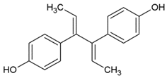 | GLU353, ARG394, GLY521, HIS524 | LEU346, LEU349, ALA350, TRP383, LEU384, LEU387, MET388, LEU391, PHE404, MET421, ILE424, PHE425, LEU428, LEU525, LEU540 | −8.99 |
| Diethylstilbestrol | 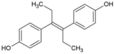 | GLU353, ARG394, HIS524, H2O | MET343, LEU346, LEU349, ALA350, TRP383, LEU384, LEU387, PHE404, MET421, LEU525, MET528 | −9.13 |
| Hexestrol | 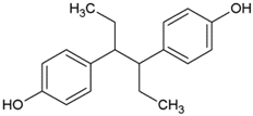 | GLU353, ARG394, GLY521, HIS524 | LEU346, LEU349, ALA350, TRP383, LEU384, LEU387, MET388, LEU391, PHE404, MET421, ILE424, PHE425, LEU428, LEU525, LEU540 | −8.57 |
© 2019 by the authors. Licensee MDPI, Basel, Switzerland. This article is an open access article distributed under the terms and conditions of the Creative Commons Attribution (CC BY) license (http://creativecommons.org/licenses/by/4.0/).
Share and Cite
Guan, T.; Sun, Y.; Wang, Y.; Li, Z.; Li, T.; Hou, L. Multi-Residue Method for the Analysis of Stilbene Estrogens in Milk. Int. J. Mol. Sci. 2019, 20, 744. https://doi.org/10.3390/ijms20030744
Guan T, Sun Y, Wang Y, Li Z, Li T, Hou L. Multi-Residue Method for the Analysis of Stilbene Estrogens in Milk. International Journal of Molecular Sciences. 2019; 20(3):744. https://doi.org/10.3390/ijms20030744
Chicago/Turabian StyleGuan, Tianzhu, Yonghai Sun, Yongjun Wang, Zhuolin Li, Tiezhu Li, and Ligang Hou. 2019. "Multi-Residue Method for the Analysis of Stilbene Estrogens in Milk" International Journal of Molecular Sciences 20, no. 3: 744. https://doi.org/10.3390/ijms20030744
APA StyleGuan, T., Sun, Y., Wang, Y., Li, Z., Li, T., & Hou, L. (2019). Multi-Residue Method for the Analysis of Stilbene Estrogens in Milk. International Journal of Molecular Sciences, 20(3), 744. https://doi.org/10.3390/ijms20030744