Communication

Conditional Deletion of Dicer in Adult Mice Impairs Skeletal Muscle Regeneration

Faculty of Sport Sciences, Waseda University, Saitama 359-1192, Japan
*
Author to whom correspondence should be addressed.
Int. J. Mol. Sci. 2019, 20(22), 5686; https://doi.org/10.3390/ijms20225686
Submission received: 20 September 2019 / Revised: 8 November 2019 / Accepted: 12 November 2019 / Published: 13 November 2019
(This article belongs to the Special Issue Muscular Dystrophy: From Molecular Basis to Therapies)

Abstract

:
Skeletal muscle has a remarkable regenerative capacity, which is orchestrated by multiple processes, including the proliferation, fusion, and differentiation of the resident stem cells in muscle. MicroRNAs (miRNAs) are small noncoding RNAs that mediate the translational repression or degradation of mRNA to regulate diverse biological functions. Previous studies have suggested that several miRNAs play important roles in myoblast proliferation and differentiation in vitro. However, their potential roles in skeletal muscle regeneration in vivo have not been fully established. In this study, we generated a mouse in which the Dicer gene, which encodes an enzyme essential in miRNA processing, was knocked out in a tamoxifen-inducible way (iDicer KO mouse) and determined its regenerative potential after cardiotoxin-induced acute muscle injury. Dicer mRNA expression was significantly reduced in the tibialis anterior muscle of the iDicer KO mice, whereas the expression of muscle-enriched miRNAs was only slightly reduced in the Dicer-deficient muscles. After cardiotoxin injection, the iDicer KO mice showed impaired muscle regeneration. We also demonstrated that the number of PAX7+ cells, cell proliferation, and the myogenic differentiation capacity of the primary myoblasts did not differ between the wild-type and the iDicer KO mice. Taken together, these data demonstrate that Dicer is a critical factor for muscle regeneration in vivo.

1. Introduction

Adult skeletal muscle has a remarkable regenerative capacity. After muscle injury, the resident muscle stem cells leave their quiescent state and begin to proliferate. These cells ultimately differentiate into multinucleated myotubes, which fuse with existing damaged myofibers [1]. Muscle regeneration is impaired by treatment with a mitosis inhibitor, colchicine, which inhibits cellular proliferation during regeneration [2]. Primary satellite cells (SCs) derived from mutant mice lacking PAX7, a marker of SCs, showed reduced proliferation and fewer myosin heavy chain (MyHC)-positive myotubes [3,4]. The adult Pax7-mutant mice also showed impaired injury-induced muscle regeneration [3]. These findings indicate that the proliferation and myogenic differentiation of resident muscle stem cells are necessary for muscle regeneration.
MicroRNAs (miRNAs) are small noncoding RNAs that repress the expression of their target genes at the posttranscriptional level to control diverse biological functions [5]. These small RNAs can bind to a complementary site, called “seed sequences”, in the 3′ untranslated region (UTR) of a target mRNA, resulting in the degradation or translational repression of that mRNA [5]. miRNAs are transcribed as long primary transcripts, called “primary miRNAs”, by an RNA polymerase II. The transcripts are cleaved by a nuclear ribonuclease III, Drosha, and then exported to the cytoplasm and further cleaved by a cytoplasmic ribonuclease III, Dicer, into double-stranded RNA. This miRNA duplex is loaded onto an Argonaute protein to form a ribonucleoprotein complex called the “RNA-induced silencing complex” [6].
Growing evidence indicates that miRNA-mediated gene silencing plays an important role in skeletal muscle cell growth and myogenic differentiation [7,8,9,10,11]. Mutant mice in which Dicer is inactivated during embryonic myogenesis show a variety of abnormal muscle phenotypes, suggesting that Dicer-mediated miRNA processing plays an essential role in muscle development [12]. An expression profiling analysis showed that three miRNAs, miR-1, miR-133, and miR-206, are abundantly expressed in skeletal muscles [13]. These three miRNAs are transcriptionally regulated by myogenic regulatory factors, which are master transcriptional factors for skeletal muscle cell-fate determination and development, and are upregulated during muscle differentiation [14,15,16,17]. It has been shown that miR-1 and miR-206, which have identical seed sequences, promote myogenesis in vitro, whereas miR-133 promotes myoblast proliferation [7,8]. A previous study also demonstrated that miR-1 and miR-206 regulate apoptosis by repressing Pax3 gene expression in Myod-KO myoblasts [18]. Although these data indicate that miRNAs are important regulators of the proliferation and differentiation of muscle cells in vitro, their potential roles in muscle regeneration in vivo have not been fully established.
In this study, we generated a tamoxifen-inducible conditional Dicer-KO (iDicer KO) mouse to deplete all mature miRNAs and analyzed its regenerative capacity during cardiotoxin-induced muscle regeneration. Our data suggest that Dicer-mediated miRNA processing is necessary for skeletal muscle regeneration in vivo.

2. Results

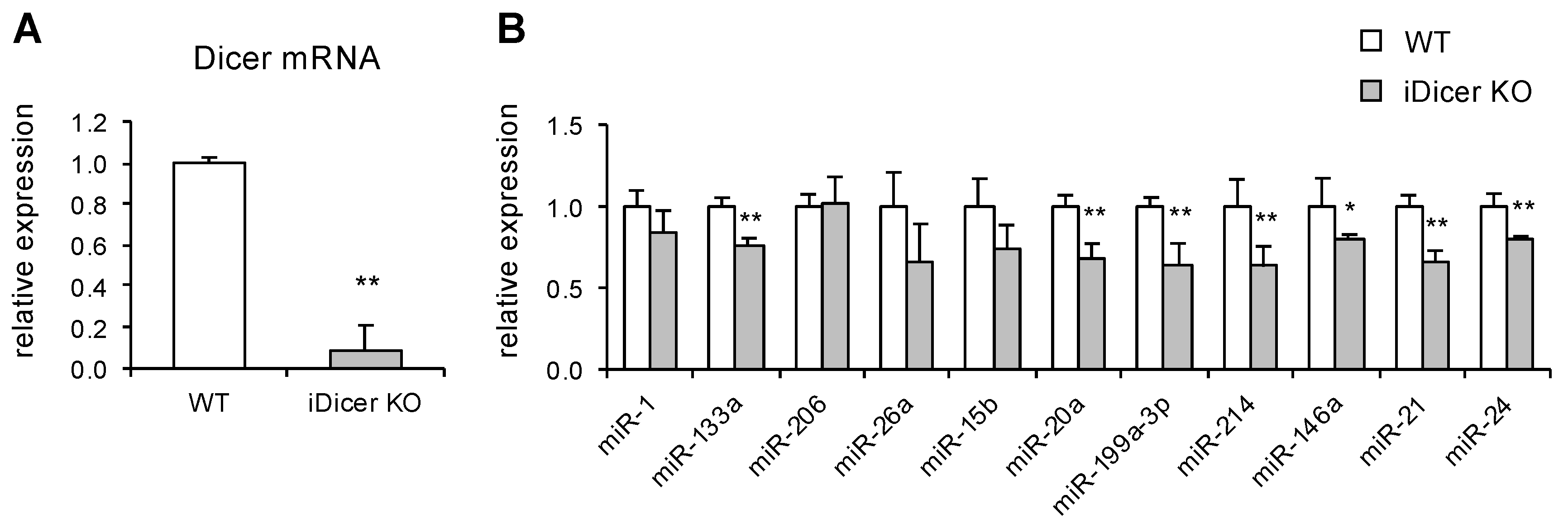
2.1. Expression of Dicer and miRNAs in iDicer KO Mice

To investigate the role of miRNAs in muscle regeneration, we first generated a mutant mouse with the tamoxifen-inducible disruption of the Dicer gene (iDicer KO). Consistent with our previous data [19], a real-time PCR analysis confirmed the significant reduction in Dicer mRNA expression in the tibialis anterior (TA) muscles of the iDicer KO mice (Figure 1A), whereas the expression levels of the muscle-enriched miRNAs miR-1, miR-133a and miR-26a, and other miRNAs (miR-15b, miR-20a, miR-199a-3p, miR-214, miR-146a, miR-21 and miR-24) were modestly reduced in the iDicer KO mice (Figure 1B).

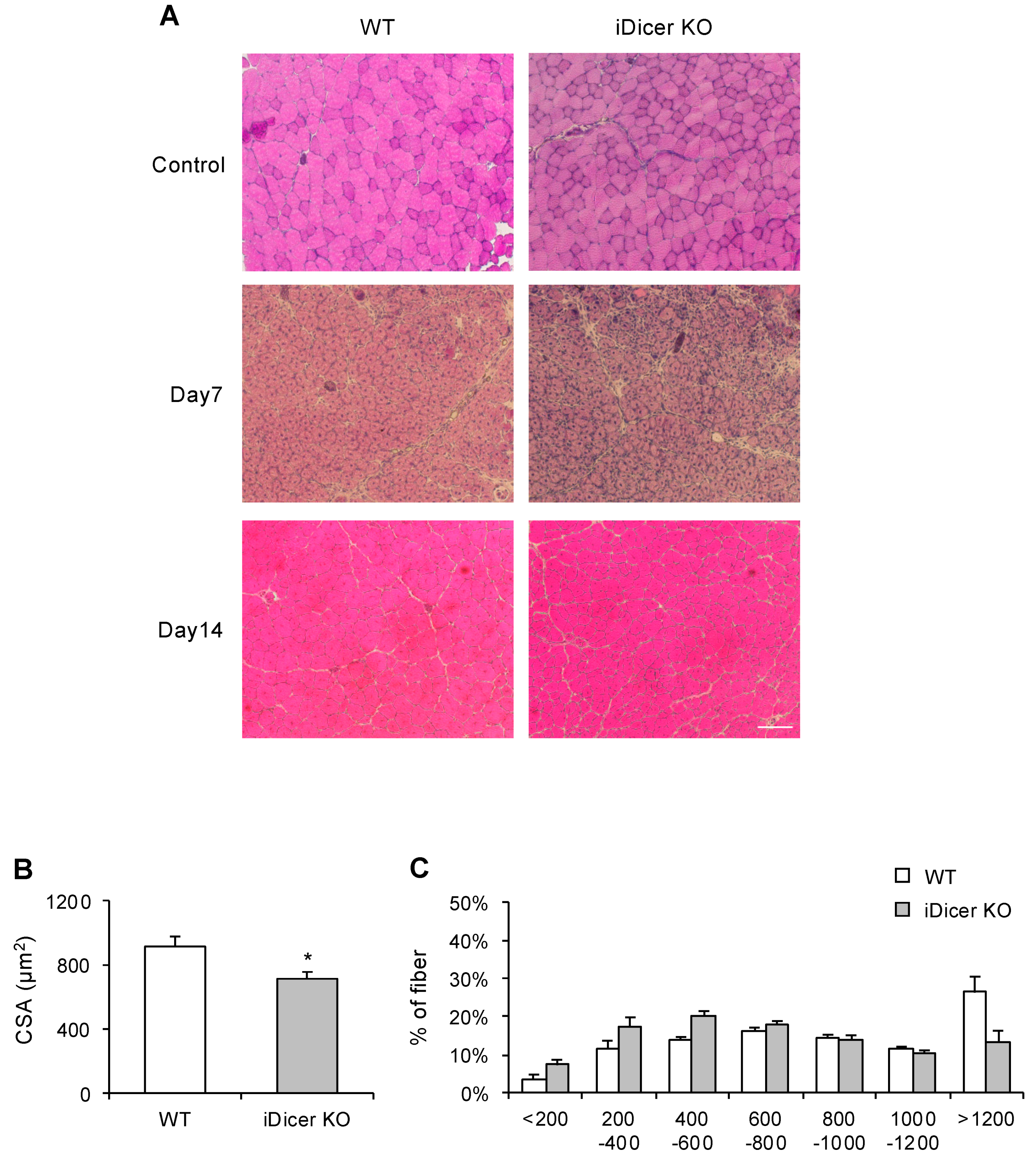
2.2. Skeletal Muscle Regeneration Is Impaired in iDicer KO Mice

We next determined the regenerative potential of the iDicer KO mice during skeletal muscle regeneration. Wild-type (WT) and iDicer KO mice were injected intramuscularly with cardiotoxin (CTX) to induce muscle injury, and the cross-sectional area (CSA) of the regenerating myofibers was analyzed with hematoxylin–eosin (H&E) staining. Fourteen days after CTX injection, the mean CSA of the regenerating myofibers with central nuclei in the iDicer KO mice was smaller than that in the WT mice (Figure 2A–C).

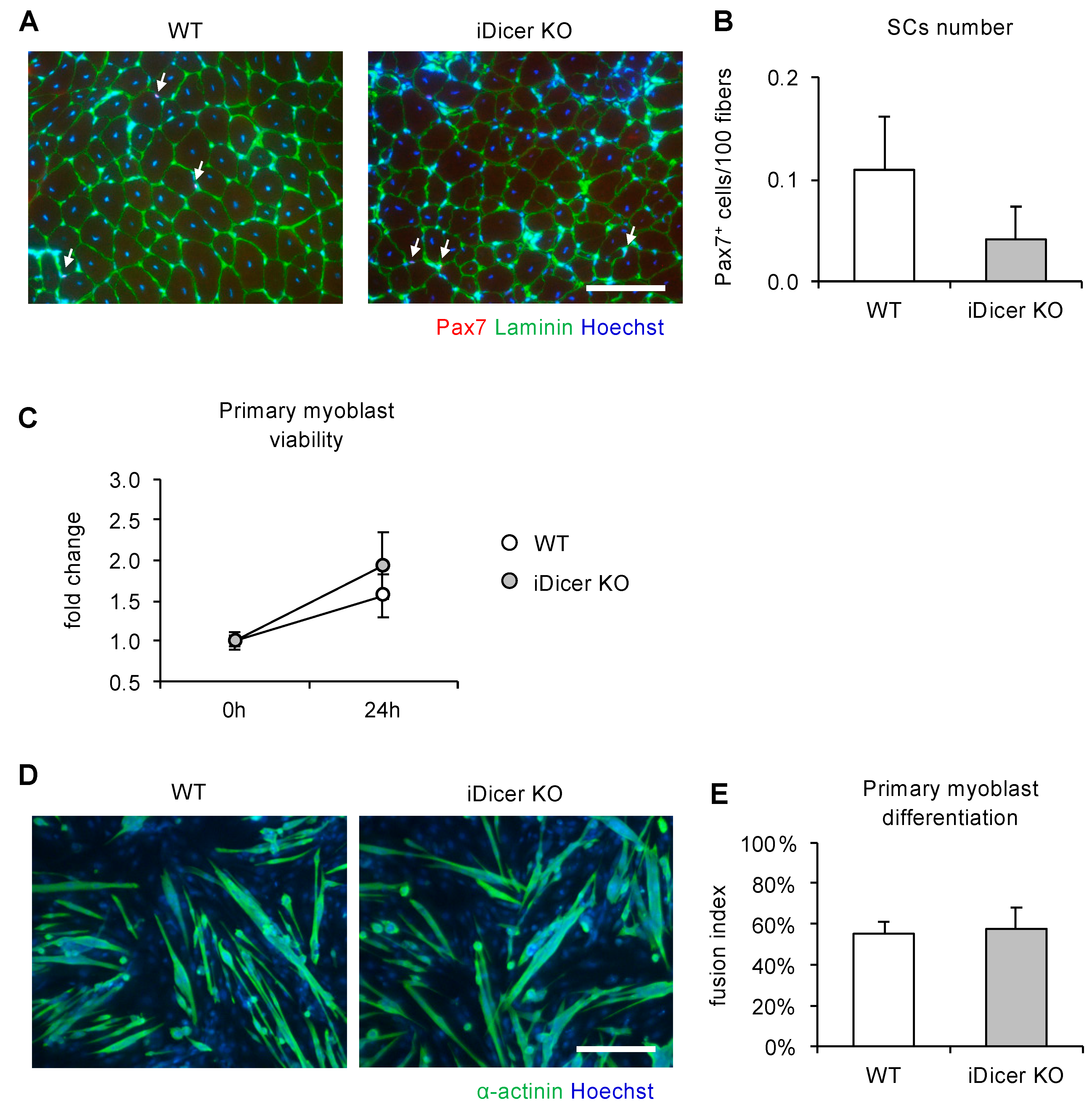
2.3. Inducible Knockout of Dicer Does Not Affect Cell Proliferation or Differentiation of Primary Myoblasts

Because the regenerative capacity of adult skeletal muscle largely depends on the functions of the resident muscle stem cells, such as muscle SCs, we investigated their numbers and the myogenic differentiation potential of primary myoblasts isolated from iDicer KO mice. Fourteen days after CTX injection, the number of PAX7+ cells on the cryosections did not differ in the WT and iDicer KO mice (Figure 3A,B). Furthermore, the cell viability and fusion index of the primary myoblasts isolated from the iDicer KO mice with tamoxifen injection were similar to those of the WT mice (Figure 3C–E).

3. Discussion

The tamoxifen-inducible knockout of Dicer in adult mice impaired the skeletal muscle regeneration that occurred in response to CTX injury (Figure 2). However, we found no reductions in the PAX7+ cell numbers, cell viability, or the myogenic differentiation potential of the primary myoblasts isolated from the iDicer KO mice (Figure 3). These data suggest that Dicer plays a prominent role in skeletal muscle regeneration in vivo. However, the molecular mechanism by which Dicer regulates skeletal muscle regeneration in this model is still unclear.
Recent studies have demonstrated that multiple miRNAs act as key regulators of skeletal muscle regeneration in adult mice [20,21,22,23,24]. For example, miR-26a, which is specifically expressed in skeletal muscle, is upregulated during muscle differentiation in vitro and muscle regeneration in vivo [10,21]. The inhibition of miR-26a with a miRNA decoy in vivo delayed muscle regeneration, indicating that miR-26a promotes muscle differentiation and regeneration [21]. It has also been shown that SC-specific Dicer knockout causes the apoptosis of SCs because they spontaneously leave the quiescent state, which results in severely impaired skeletal muscle regeneration after injury [25]. We also demonstrated that the CSA of regenerating fibers was reduced in the iDicer KO mice (Figure 2). Taken together, our data demonstrate the importance of miRNAs in skeletal muscle regeneration in mice.
Because the proliferation and differentiation of myoblasts are essential for skeletal muscle regeneration, we isolated the primary myoblasts from the skeletal muscles of iDicer KO mice, and determined the proliferation and myogenic differentiation potential of these cells. Surprisingly, there were no clear differences in cell viability or differentiation capacity of the primary myoblasts isolated from WT and iDicer KO mice (Figure 3C,D). Although the proliferation and differentiation of SCs are predominant factors in muscle repair, other cell types also affect the regeneration process [26]. Joe et al., [27] and Uezumi et al., [28] identified mesenchymal progenitors, called “fibro-adipogenic progenitors”, that enhance the myogenic differentiation of muscle stem cells and muscle regeneration [27,29]. Another recent work demonstrated that an interaction between capillary endothelial cells and SCs controls SC functions through a Notch-signaling-mediated direct cell–cell interaction [30]. Future research with mice in which individual miRNAs are cell-type-specifically knocked out may provide greater insight into the functions and regulatory mechanisms of miRNAs in muscle regeneration.
We found that muscle regeneration was disrupted in the iDicer KO mice, whereas expressions of muscle-enriched miRNAs (miR-1, miR-133a, miR-206 and miR-26a) and other miRNAs (miR-15b, miR-20a, miR-119a-3p, miR-214, miR-146a, miR-21 and miR-24) that were reported to be upregulated during muscle regeneration [31], were reduced only ~20−40% in the TA muscle of the iDicer KO mice compared with those in the WT mice (Figure 1B). It is not clear how the slight changes in miRNAs could contribute to the delayed regeneration in the iDicer KO mice. It may be possible that the global reduction in miRNAs is enough to disrupt a regulatory network of miRNAs and to delay the myofiber regeneration in vivo.
Our data showed that the muscle-enriched miRNAs were highly stable in the TA muscle of the iDicer KO mice (Figure 1B), which is consistent with our previous report [19]. Similarly, recent data on tamoxifen-inducible, skeletal muscle-specific Dicer knockout (HSA-MCM; Dicerfl/fl) mice showed that the expression levels of miRNAs including miR-1, miR-133a and miR-206 were slightly affected by the Dicer depletion [32]. Several lines of evidence indicate that the mature miRNAs expressed in liver, heart, and neuron are also stable in vivo [33,34,35]. A pulse-chase approach and high-throughput sequencing of miRNAs revealed the production and turnover rates of miRNAs in vitro [36,37], whereas miRNA turnover in vivo and its regulatory mechanism are less well understood. It will be of interest in future works to investigate how miRNA turnover is regulated in vivo.

4. Materials and Methods

4.1. Animal Experiments

All mice were maintained in temperature-controlled quarters (21 °C) under a 12 h light–dark cycle and provided with a standard chow diet. The generation of tamoxifen-inducible Dicer knockout (CAG-CreERT2, Dicerfl/fl) mice has been described previously [19]. The mice were maintained in a B6 background and intraperitoneally injected with 1 mg of tamoxifen at 8 weeks old for 5 consecutive days. The tibialis anterior (TA) muscles of 9-week-old wild-type (WT) and iDicer KO mice were injected with cardiotoxin (CTX) from Naja pallida (Latoxan, Portes-lès-Valence, France) with a 27-gauge needle. The TA muscles were harvested 7 and 14 days after CTX injury, frozen in liquid nitrogen, and stored at −80 °C. The animal protocols were approved by the Animal Care and Use Committees of Waseda University, Japan (numbers: 2017-A103a, 2018-A122, 2019-108).

4.2. qPCR

Total RNA was extracted with Isogen II (Wako Chemicals, Osaka, Japan), and 1 µg of total RNA was converted to cDNA with ReverTra Ace reverse transcriptase (Toyobo, Osaka, Japan). Real-time PCR was performed with Thunderbird® SYBR® qPCR Mix (Toyobo), with gene-specific primers. The following primers were used for real-time PCR: Dicer, 5′-CACACGCCTCCTACCACTACAACAC-3′ and 5′-CCGTGGGTCTTCATAAAGGT-3′; glyceraldehyde 3-phosphate dehydrogenase (GAPDH), 5′-AAATGGTGAAGGTCGGTGTG-3′ and 5′-TGAAGGGGTCGTTGATGG-3′. For the real-time PCR analysis of mature miRNA expression, the TaqMan® MicroRNA Reverse Transcription Kit and TaqMan® MicroRNA Assays (Applied Biosystems, Foster City, CA, USA) were used, according to the manufacturer’s protocols [38,39].

4.3. Histological Analysis

Cross-sections of frozen TA muscle were stained with H&E and immunofluorescence. The cross-sections were incubated with the following antibodies: anti-PAX7 (DSHB; University of Iowa, Iowa City, IA), anti-laminin (10765; Cappel Reseach Reagent, Turnholt, Belgium), and Hoechst 33342 (Invitrogen, Carlsbad, CA). The secondary antibodies were Alexa-Fluor-594-conjugated anti-mouse IgG antibody for PAX7 and Alexa-Fluor-488-conjugated anti-rabbit IgG antibody for laminin (Jackson ImmunoResearch Laboratories, West Grove, PA, USA). The cross-sectional area (CSA) was quantified using ImageJ software (National Institutes of Health, Bethesda, MD, USA).

4.4. Isolation and Culture of Primary Myoblasts

Primary myoblasts were isolated as previously described [40,41]. Briefly, after tamoxifen injection, the gastrocnemius, TA, and quadriceps muscles were collected and digested with 0.2% collagenase type II (Worthington Biochemical Corporation, Freehold, NJ, USA) at 37 °C for 30 min. After incubation, the muscle slurries were triturated with an 18-gauge needle and then incubated again at 37 °C for 15 min. The muscle mixtures were filtered sequentially through 100 μm and 40 μm filters (Falcon, Sunnyvale, CA, USA), and then centrifuged at 200× g for 5 min. The cells were resuspended in growth medium containing Ham’s F-10 Nutrient Mix (Thermo Fisher Scientific, Waltham, MA, USA) supplemented with 20% fetal bovine serum, 1% penicillin–streptomycin, and 2.5 ng/mL basic fibroblast growth factor (G5071; Promega, Madison, WI, USA), and seeded in gelatin-coated dishes. To evaluate cell viability, 3000 cells/well were seeded in 96-well plates and a CellTiter-Glo® 2.0 Cell Viability Assay (Promega) was performed, according to the manufacturer’s protocol. To induce cell differentiation, the myoblasts in the 96-well plate at 80–90% confluence were switched to differentiation medium (DM; DMEM supplemented with 2% horse serum). The DM was changed daily for 5 days. After differentiation, the cells were immunofluorescently stained with Hoechst 33342 and anti-α-actinin antibody (A7732; Sigma-Aldrich, St Louis, MO, USA) to identify nuclei and differentiated myotubes, respectively. The secondary antibody for anti-α-actinin was an Alexa-Fluor-488-conjugated goat anti-mouse IgG1 antibody (Jackson ImmunoResearch Laboratories). The fusion index was calculated manually as the ratio of the number of nuclei within the α-actinin-positive myotubes with more than two nuclei to the total number of nuclei, using ImageJ software (LOCI, University of Wisconsin, Madison, WI, USA).

Author Contributions

S.O. and T.A. conceived and designed the research; S.O. and M.L. performed experiments; S.O. and M.L. analyzed the data; S.O., M.L., and T.A. interpreted the results of the experiments; S.O. prepared the figures; S.O. and T.A. drafted the manuscript.

Funding

This research was supported, in part, by Grants-in-Aid for Young Investigators (A) (18680047 and 21680049 to T.A.) from the Ministry of Education, Culture, Sports, Science, and Technology, Japan. S. Oikawa was supported by the Japan Society for the Promotion of Science.

Acknowledgments

We thank Brian Harfe (University of Florida) for providing the Dicer1-floxed mice.

Conflicts of Interest

The authors declare that they have no conflict of interest.

Abbreviations

SCSatellite cell
miRNAmicroRNA
TATibialis anterior
WTWild-type
CSACross-sectional area
CTXCardiotoxin

References

  1. Chargé, S.B.; Rudnicki, M.A. Cellular and molecular regulation of muscle regeneration. Physiol. Rev. 2004, 84, 209–238. [Google Scholar] [CrossRef] [PubMed]
  2. Pietsch, P. The effects of colchicine on regeneration of mouse skeletal muscle. Anat. Rec. 1961, 139, 167–172. [Google Scholar] [CrossRef]
  3. Oustanina, S.; Hause, G.; Braun, T. Pax7 directs postnatal renewal and propagation of myogenic satellite cells but not their specification. Embo. J. 2004, 23, 3430–3439. [Google Scholar] [CrossRef] [PubMed]
  4. Seale, P.; Sabourin, L.A.; Girgis-Gabardo, A.; Mansouri, A.; Gruss, P.; Rudnicki, M.A. Pax7 is Required for the Specification of Myogenic Satellite Cells. Cell 2000, 102, 777–786. [Google Scholar] [CrossRef]
  5. Bartel, D.P. MicroRNAs Genomics, Biogenesis, Mechanism, and Function. Cell 2004, 116, 281–297. [Google Scholar] [CrossRef]
  6. Ha, M.; Kim, N.V. Regulation of microRNA biogenesis. Nat. Rev. Mol. Cell Biol. 2014, 15, 509–524. [Google Scholar] [CrossRef]
  7. Chen, J.-F.; Mandel, E.M.; Thomson, M.J.; Wu, Q.; Callis, T.E.; Hammond, S.M.; Conlon, F.L.; Wang, D.-Z. The role of microRNA-1 and microRNA-133 in skeletal muscle proliferation and differentiation. Nat. Genet. 2005, 38, 228–233. [Google Scholar] [CrossRef]
  8. Kim, H.; Lee, Y.; Sivaprasad, U.; Malhotra, A.; Dutta, A. Muscle-specific microRNA miR-206 promotes muscle differentiation. J. Cell Biol. 2006, 174, 677–687. [Google Scholar] [CrossRef]
  9. Chen, J.-F.; Tao, Y.; Li, J.; Deng, Z.; Yan, Z.; Xiao, X.; Wang, D.-Z. microRNA-1 and microRNA-206 regulate skeletal muscle satellite cell proliferation and differentiation by repressing Pax7. J. Cell Biol. 2010, 190, 867–879. [Google Scholar] [CrossRef]
  10. Wong, C.; Tellam, R.L. MicroRNA-26a Targets the Histone Methyltransferase Enhancer of Zeste homolog 2 during Myogenesis. J. Biol. Chem. 2008, 283, 9836–9843. [Google Scholar] [CrossRef]
  11. Silva, W.; Graça, F.; Cruz, A.; Silvestre, J.; Labeit, S.; Miyabara, E.; Yan, C.; Wang, D.; Moriscot, A. miR-29c improves skeletal muscle mass and function throughout myocyte proliferation and differentiation and by repressing atrophy-related genes. Acta Physiol. 2019, 226, e13278. [Google Scholar] [CrossRef] [PubMed]
  12. O’Rourke, J.; Georges, S.; Seay, H.; Tapscott, S.; McManus, M.; Goldhamer, D.; Swanson, M.; Harfe, B. Essential role for Dicer during skeletal muscle development. Dev. Biol. 2007, 311, 359–368. [Google Scholar] [CrossRef] [PubMed]
  13. Sempere, L.F.; Freemantle, S.; Pitha-Rowe, I.; Moss, E.; Dmitrovsky, E.; Ambros, V. Expression profiling of mammalian microRNAs uncovers a subset of brain-expressed microRNAs with possible roles in murine and human neuronal differentiation. Genome Biol. 2004, 5, R13. [Google Scholar] [CrossRef] [PubMed]
  14. Liu, N.; Williams, A.H.; Kim, Y.; McAnally, J.; Bezprozvannaya, S.; Sutherland, L.B.; Richardson, J.A.; Bassel-Duby, R.; Olson, E.N. An intragenic MEF2-dependent enhancer directs muscle-specific expression of microRNAs 1 and 133. Proc. Natl. Acad. Sci. USA 2007, 104, 20844–20849. [Google Scholar] [CrossRef]
  15. Rao, P.K.; Kumar, R.M.; Farkhondeh, M.; Baskerville, S.; Lodish, H.F. Myogenic factors that regulate expression of muscle-specific microRNAs. Proc. Natl. Acad. Sci. USA 2006, 103, 8721–8726. [Google Scholar] [CrossRef]
  16. Sweetman, D.; Goljanek, K.; Rathjen, T.; Oustanina, S.; Braun, T.; Dalmay, T.; Munsterberg, A. Specific requirements of MRFs for the expression of muscle specific microRNAs, miR-1, miR-206 and miR-133. Dev. Biol. 2008, 321, 491–499. [Google Scholar] [CrossRef]
  17. Koutsoulidou, A.; Mastroyiannopoulos, N.P.; Furling, D.; Uney, J.B.; Phylactou, L.A. Expression of miR-1, miR-133a, miR-133b and miR-206 increases during development of human skeletal muscle. BMC Dev. Biol. 2011, 11, 34. [Google Scholar] [CrossRef]
  18. Hirai, H.; Verma, M.; Watanabe, S.; Tastad, C.; Asakura, Y.; Asakura, A. MyoD regulates apoptosis of myoblasts through microRNA-mediated down-regulation of Pax3. J. Cell Biol. 2010, 191, 347–365. [Google Scholar] [CrossRef]
  19. Oikawa, S.; Lee, M.; Motohashi, N.; Maeda, S.; Akimoto, T. An inducible knockout of Dicer in adult mice does not affect endurance exercise-induced muscle adaptation. Am. J. Physiol. Cell Physiol. 2019, 316, C285–C292. [Google Scholar] [CrossRef]
  20. Snyder, C.M.; Rice, A.L.; Estrella, N.L.; Held, A.; Kandarian, S.C.; Naya, F.J. MEF2A regulates the Gtl2-Dio3 microRNA mega-cluster to modulate WNT signaling in skeletal muscle regeneration. Development 2013, 140, 31–42. [Google Scholar] [CrossRef]
  21. Dey, B.K.; Gagan, J.; Yan, Z.; Dutta, A. miR-26a is required for skeletal muscle differentiation and regeneration in mice. Genes Dev. 2012, 26, 2180–2191. [Google Scholar] [CrossRef] [PubMed]
  22. Lee, K.-P.; Shin, Y.; Panda, A.C.; Abdelmohsen, K.; Kim, J.; Lee, S.-M.; Bahn, Y.; Choi, J.; Kwon, E.-S.; Baek, S.-J.; et al. miR-431 promotes differentiation and regeneration of old skeletal muscle by targeting Smad4. Genes Dev. 2015, 29, 1605–1617. [Google Scholar] [CrossRef] [PubMed]
  23. Bronisz-Budzyńska, I.; Chwalenia, K.; Mucha, O.; Podkalicka, P.; Karolina, B.-S.; Józkowicz, A.; Łoboda, A.; Kozakowska, M.; Dulak, J. miR-146a deficiency does not aggravate muscular dystrophy in mdx mice. Skelet. Muscle 2019, 9, 22. [Google Scholar] [CrossRef] [PubMed]
  24. Qiu, H.; Liu, N.; Luo, L.; Zhong, J.; Tang, Z.; Kang, K.; Qu, J.; Peng, W.; Liu, L.; Li, L.; et al. MicroRNA-17-92 regulates myoblast proliferation and differentiation by targeting the ENH1/Id1 signaling axis. Cell Death Differ. 2016, 23, 1658–1669. [Google Scholar] [CrossRef] [PubMed]
  25. Cheung, T.H.; Quach, N.L.; Charville, G.W.; Liu, L.; Park, L.; Edalati, A.; Yoo, B.; Hoang, P.; Rando, T.A. Maintenance of muscle stem-cell quiescence by microRNA-489. Nature 2012, 482, 524–528. [Google Scholar] [CrossRef] [PubMed]
  26. Baghdadi, M.B.; Tajbakhsh, S. Regulation and phylogeny of skeletal muscle regeneration. Dev. Biol. 2018, 433, 200–209. [Google Scholar] [CrossRef]
  27. Joe, A.W.; Yi, L.; Natarajan, A.; Grand, F.; So, L.; Wang, J.; Rudnicki, M.A.; Rossi, F.M. Muscle injury activates resident fibro/adipogenic progenitors that facilitate myogenesis. Nat. Cell Biol. 2010, 12, 153–163. [Google Scholar] [CrossRef]
  28. Uezumi, A.; Fukada, S.; Yamamoto, N.; Takeda, S.; Tsuchida, K. Mesenchymal progenitors distinct from satellite cells contribute to ectopic fat cell formation in skeletal muscle. Nat. Cell Biol. 2010, 12, 143–152. [Google Scholar] [CrossRef]
  29. Wosczyna, M.N.; Konishi, C.T.; Carbajal, E.E.; Wang, T.T.; Walsh, R.A.; Gan, Q.; Wagner, M.W.; Rando, T.A. Mesenchymal Stromal Cells Are Required for Regeneration and Homeostatic Maintenance of Skeletal Muscle. Cell Rep. 2019, 27, 2029–2035. [Google Scholar] [CrossRef]
  30. Verma, M.; Asakura, Y.; Murakonda, B.; Pengo, T.; Latroche, C.; Chazaud, B.; McLoon, L.K.; Asakura, A. Muscle Satellite Cell Cross-Talk with a Vascular Niche Maintains Quiescence via VEGF and Notch Signaling. Cell Stem Cell 2018, 23, 530–543. [Google Scholar] [CrossRef]
  31. Liu, N.; Williams, A.H.; Maxeiner, J.M.; Bezprozvannaya, S.; elton, J.; Richardson, J.A.; Bassel-Duby, R.; Olson, E.N. microRNA-206 promotes skeletal muscle regeneration and delays progression of Duchenne muscular dystrophy in mice. J. Clin. Investig. 2012, 122, 2054–2065. [Google Scholar] [CrossRef] [PubMed] [Green Version]
  32. Vechetti, I.J.; Wen, Y.; Chaillou, T.; Murach, K.A.; Alimov, A.P.; Figueiredo, V.C.; Dal-Pai-Silva, M.; McCarthy, J.J. Life-long reduction in myomiR expression does not adversely affect skeletal muscle morphology. Sci. Rep. UK 2019, 9, 5483. [Google Scholar] [CrossRef] [PubMed] [Green Version]
  33. Gatfield, D.; Martelot, L.G.; Vejnar, C.; Gerlach, D.; Schaad, O.; Fleury-Olela, F.; Ruskeepaa, A.; Oresic, M.; Esau, C.; Zdobnov, E.; et al. Integration of microRNA miR-122 in hepatic circadian gene expression. Genes Dev. 2009, 23, 1313–1326. [Google Scholar] [CrossRef] [PubMed] [Green Version]
  34. Van Rooij, E.; Sutherland, L.; Qi, X.; Richardson, J.; Hill, J.; Olson, E. Control of stress-dependent cardiac growth and gene expression by a microRNA. Science 2007, 316, 575–579. [Google Scholar] [CrossRef] [PubMed] [Green Version]
  35. Schaefer, A.; O’Carroll, D.; Tan, C.; Hillman, D.; Sugimori, M.; Llinas, R.; Greengard, P. Cerebellar neurodegeneration in the absence of microRNAs. J. Exp. Med. 2007, 204, 1553–1558. [Google Scholar] [CrossRef] [Green Version]
  36. Reichholf, B.; Herzog, V.A.; Fasching, N.; Manzenreither, R.A.; Sowemimo, I.; Ameres, S.L. Time-Resolved Small RNA Sequencing Unravels the Molecular Principles of MicroRNA Homeostasis. Mol. Cell 2019, 75, 756–768. [Google Scholar] [CrossRef]
  37. Marzi, M.J.; Ghini, F.; Cerruti, B.; de Pretis, S.; Bonetti, P.; Giacomelli, C.; Gorski, M.M.; Kress, T.; Pelizzola, M.; Muller, H.; et al. Degradation dynamics of microRNAs revealed by a novel pulse-chase approach. Genome Res. 2016, 26, 554–565. [Google Scholar] [CrossRef] [Green Version]
  38. Oikawa, S.; Wada, S.; Lee, M.; Maeda, S.; Akimoto, T. Role of endothelial microRNA-23 clusters in angiogenesis in vivo. Am. J. Physiol. Heart C 2018, 315, H838–H846. [Google Scholar] [CrossRef]
  39. Wada, S.; Kato, Y.; Okutsu, M.; Miyaki, S.; Suzuki, K.; Yan, Z.; Schiaffino, S.; Asahara, H.; Ushida, T.; Akimoto, T. Translational suppression of atrophic regulators by microRNA-23a integrates resistance to skeletal muscle atrophy. J. Biol. Chem. 2011, 286, 38456–38465. [Google Scholar] [CrossRef] [Green Version]
  40. Rando, T.; Blau, H. Primary mouse myoblast purification, characterization, and transplantation for cell-mediated gene therapy. J. Cell Biol. 1994, 125, 1275–1287. [Google Scholar] [CrossRef] [Green Version]
  41. Hindi, L.; McMillan, J.; Afroze, D.; Hindi, S.; Kumar, A. Isolation, Culturing, and Differentiation of Primary Myoblasts from Skeletal Muscle of Adult Mice. Bio. Protoc. 2017, 7, 7. [Google Scholar] [CrossRef] [PubMed] [Green Version]
Figure 1. Expression levels of Dicer mRNA and muscle-enriched miRNAs in tibialis anterior (TA) muscles of tamoxifen-induced Dicer knock-out (iDicer KO) mice. (A) Tamoxifen induced a large reduction in Dicer mRNA expression in the iDicer KO mice (n = 5). (B) Expression levels of muscle-enriched miR-1, miR-133a and miR-26a and other miRNAs (miR-15b, miR-20a, miR-199a-3p, miR-214, miR-146a, miR-21 and miR-24) were slightly reduced in TA muscle of the iDicer KO mice by tamoxifen injection (n = 3–5). Data are means ± SE, * p < 0.05, ** p < 0.01.
Ijms 20 05686 g001
Figure 2. Skeletal muscle regeneration in the iDicer KO mice. (A) Representative image of sections of TA muscle stained with hematoxylin–eosin (H&E). Scale bar = 100 µm. (B,C) Mean cross-sectional area (CSA) of regenerating muscle fibers with central nuclei in the iDicer KO mice was significantly smaller than that in the WT mice (n = 7). Data are means ± SE, * p < 0.05.
Ijms 20 05686 g002
Figure 3. Satellite cell (SC) numbers and myogenic differentiation potential in primary myoblasts isolated from WT and iDicer KO mice (A,B). Immunofluorescence analysis revealed that the number of PAX7+ SCs in the iDicer KO mice was similar to that in the WT mice (n = 6–8) (CE). There was no difference in the cell viability of the primary myoblasts (C) or their fusion indices (D,E) in the WT and iDicer KO mice (n = 3–4). Data are means ± SE. Scale bar = 100 µm.
Ijms 20 05686 g003

