Article

Expression Analysis of mRNA Decay of Maternal Genes during Bombyx mori Maternal-to-Zygotic Transition

1
School of Biotechnology, Jiangsu University of Science and Technology, Sibaidu Rd, Zhenjiang 212018, China
2
Sericulture Research Institute, Chinese Academy of Agricultural Sciences, Sibaidu Rd, Zhenjiang 212018, China
*
Authors to whom correspondence should be addressed.
These authors contributed equally to this work.
Int. J. Mol. Sci. 2019, 20(22), 5651; https://doi.org/10.3390/ijms20225651
Submission received: 31 August 2019 / Revised: 6 November 2019 / Accepted: 6 November 2019 / Published: 12 November 2019
(This article belongs to the Special Issue Regulation of Gene Expression During Embryonic Development)

Abstract

:
Maternal genes play an important role in the early embryonic development of the silkworm. Early embryonic development without new transcription depends on maternal components stored in the egg during oocyte maturation. The maternal-to-zygotic transition (MZT) is a tightly regulated process that includes maternal mRNAs elimination and zygotic transcription initiation. This process has been extensively studied within model species. Each model organism has a unique pattern of maternal transcriptional clearance classes in MZT. In this study, we identified 66 maternal genes through bioinformatics analysis and expression analysis in the eggs of silkworm virgin moths (Bombyx mori). All 66 maternal genes were expressed in vitellogenesis in day eight female pupae. During MZT, the degradation of maternal gene mRNAs could be divided into three clusters. We found that eight maternal genes of cluster 1 remained stable from 0 to 3.0 h, 17 maternal genes of cluster 2 were significantly decayed from 0.5 to 1.0 h and 41 maternal genes of cluster 3 were significantly decayed after 1.5 h. Therefore, the initial time-point of degradation of cluster 2 was earlier than that of cluster 3. The maternal gene mRNAs decay of clusters 2 and 3 is first initiated by maternal degradation activity. Our study expands upon the identification of silkworm maternal genes and provides a perspective for further research of the embryo development in Bombyx mori.

1. Introduction

The transition from the oocyte depending on maternally supplied RNA and protein complements the commencement of zygotic transcription is a key process in the earliest stages of early embryonic development [1,2,3]. Early embryonic development is maternally regulated. Maternal mRNAs and proteins stored in oocytes are activated to initiate and regulate embryonic development. Following the period of maternal transcriptional silence, the embryonic zygote’s own genome starts transcription and plays a role in the development of embryos [4]. The transition from the maternal to the zygotic genome is a key process in the final transformation of the zygotic regulation of individual development [2]. Following the maternal-to-zygotic transition (MZT) period, the maternal control of development begins to decline and maternal mRNAs begin to degrade [5,6]. Therefore, later developmental control is exhibited via a combination of the maternal RNAs and proteins being eliminated and the zygotic genome becoming transcriptionally active [7].
The elimination of these maternal mRNAs is completed through two kinds of activities: Maternal-source encoding (maternal degradation activity) and zygotic transcription (zygotic degradation activity) [7,8]. Some molecular mechanisms regulating maternal mRNA clearance have been previously demonstrated. RNA-binding proteins (RBPs) play an important role in directing the decay of maternal mRNAs in Drosophila. Smaug (SMG) RBP participates in clearance of maternal mRNAs via binding maternal transcripts that contain SMG recognition cis-elements (SREs) [2,9,10,11,12,13]. An additional cis-element, such as Pumilio-like binding element (PBE), was also identified and is bound by Pumilio (PUM) RBP, a post-transcriptional regulator implicated in both translational repression and the destabilization of a specific subset of maternal mRNAs [12,14,15,16,17,18]. PUM has been shown to interact with brain tumor (BRAT) RBP [19]. BRAT can directly bind to RNA and mediate the decay of maternal mRNAs [20,21]. BRAT and SMG can recruit and/or stabilize ME31B (RNA-binding protein) on their targets in maternal mRNA clearance [22]. ME31B exists in complexes that also contain eIF4E (binding 5’ cap), Cup, Trailer Hitch (TRAL), and polyadenylate binding protein (PABP) (binding 3’ poly (A) tail) [22,23]. PIWI-associated RNAs (piRNAs) and their associated proteins act together with SMG to recruit the deadenylase CCR4 deadenylation complex to Nanos maternal mRNA, thus promoting its decay during early embryogenesis in Drosophila [24,25]. In Drosophila, the RNA-binding proteins of SMG, BRAT, and PUM bind to and direct the degradation of largely distinct subsets of maternal mRNAs in both maternal and zygotic degradation activities [17,21,22,26]. SMG is also essential for the synthesis of microRNAs (miRNAs) during the Drosophila maternal-to-zygotic transition [26,27,28]. miRNAs have important functions during early embryonic development in metazoans [29,30]. miRNAs facilitate the transition from an oocyte-inherited to an embryonic transcriptome by eliminating maternal mRNAs during MZT in Drosophila, zebrafish (Danio rerio), and Xenopus [5,31,32,33]. Codon identity regulates the maternal program of mRNA decay, and codon composition shapes maternal mRNA clearance during the maternal-to-zygotic transition in zebrafish, Xenopus, mouse (Mus musculus), and Drosophila [34,35,36]. Codon-mediated decay and miRNAs induced decay evolutionarily conserved mechanisms for modulating mRNA stability in metazoans [30,31,34,35].
The joint action of maternal and zygotic degradation signaling pathways triggers the clearance mechanism of maternal components both in temporal and spatial axes [8]. The biological functions of maternal mRNA elimination during MZT remain unclear thus far. However, the potential functions of this process can be hypothesized [2,7]. In Drosophila, maternal mRNA degradation starts soon after egg activation and is largely complete by the third hour of embryogenesis [8,11,12]. During the early embryo stage, maternal transcript clearance may play a passive role [7]. Permissive functions may be necessary to allow newly synthesized zygotic transcripts to exert their functions [18,37,38,39,40], whereas instructive functions regulate developmental progress [41,42].
The embryonic development of Bombyx mori is significantly different from that of Drosophila. The progress of egg formation in different positions of the ovariole is inconsistent. According to various morphological criteria, the development of the follicles is divided into 12 different stages [43]. During vitellogenesis (stages 4–10), the oocyte increases gradually in volume and is filled with yolk spheres, lipid droplets, and glycogen granules. At the end of this period, degenerated nurse cells are devoured by follicular epithelial cells [43]. In the choriogenesis period (stages 11, 12), different types of eggshell proteins are synthesized and secreted successively to construct the eggshell. The developmental stages of each ovariole are opportune, found in vitellogenesis, choriogenesis, and mature eggs from day 8 pupae [43,44]. Following the choriogenesis period, egg maturation occurs [43,44]. The time of sperm entering the egg occurs a few seconds before the egg leaves the mother. The union of sperm and egg pronuclei occurs at about two hours after silkworm eggs are laid [45,46,47].
In our previous study, we identified 76 potential maternal genes in silkworm via orthologous comparison [48]. In this study, further sequence alignment analysis and the identification of these potential maternal genes were performed. Expression patterns were analyzed in eggs of virgin moths to identify the maternal genes. In this study, the expression of the 66 successfully identified maternal genes was analyzed in the developing oocytes from day eight female pupae, and during the MZT period in silkworm.

2. Results

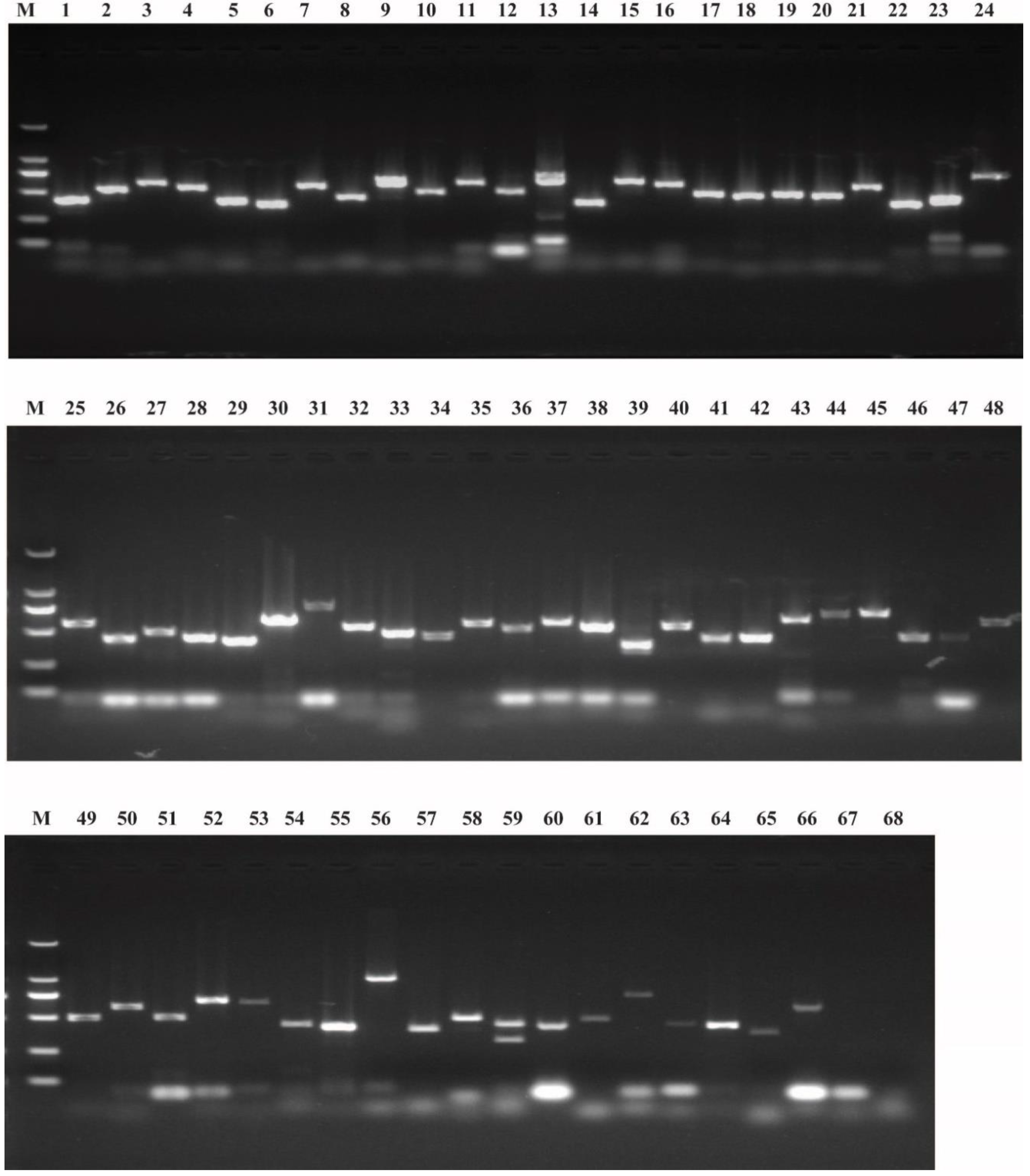
2.1. Identification of Potential Maternal Genes

In our previous study, we obtained 76 potential maternal genes in the B. mori genome [48]. In this study, we blasted the NCBI database and the newly assembled silkBase by the sequence of each gene that was obtained from the silkworm database (SilkDB) [49]. BGIBMGA012517 and BGIBMGA012518 are orthologous genes to MAMO in Drosophila melanogaster [48]. The sequences of BGIBMGA012517 and BGIBMGA012518 were found to be part of the KWMTBOMO05086 gene that was annotated in silkBase (Table 1). Similarly, BGIBMGA002518and 002519 were found to be part of KWMTBOMO005319; BGIBMGA000972, 000973, and 000974 were part of KWMTBOMO007913; BGIBMGA004415 and 004416 were part of XM_012695102; and BGIBMGA013473 and 013474 were part of XM_012690736. The BLAST results of BGIBMGA007314 and BGIBMGA001094 were very poor in the NCBI database and silkBase. Therefore, 68 preliminary potential maternal genes were identified in the B. mori genome. The mRNAs of maternal genes are produced by the females and stored in embryos [2,6]. Thus, undetectable expression in the embryo can be considered a non-maternal gene. The results of the transcriptional analysis of the 68 potential maternal genes in eggs of virgin moths by reverse transcription-PCR (RT-PCR) showed that for 66 genes, transcriptional signals were detected, whereas two had no transcriptional signals (Figure 1), BGIBMGA003296 and BGIBMGA002069 had no transcriptional signals (Figure 1). The specific primers for each gene were used in RT-PCR, as shown in Table S1. We finally identified 66 maternal genes in the silkworm genome, and information, including amino-acid length, chromosomal distribution, signal peptide, and gene name, was collected for each (Table 1).

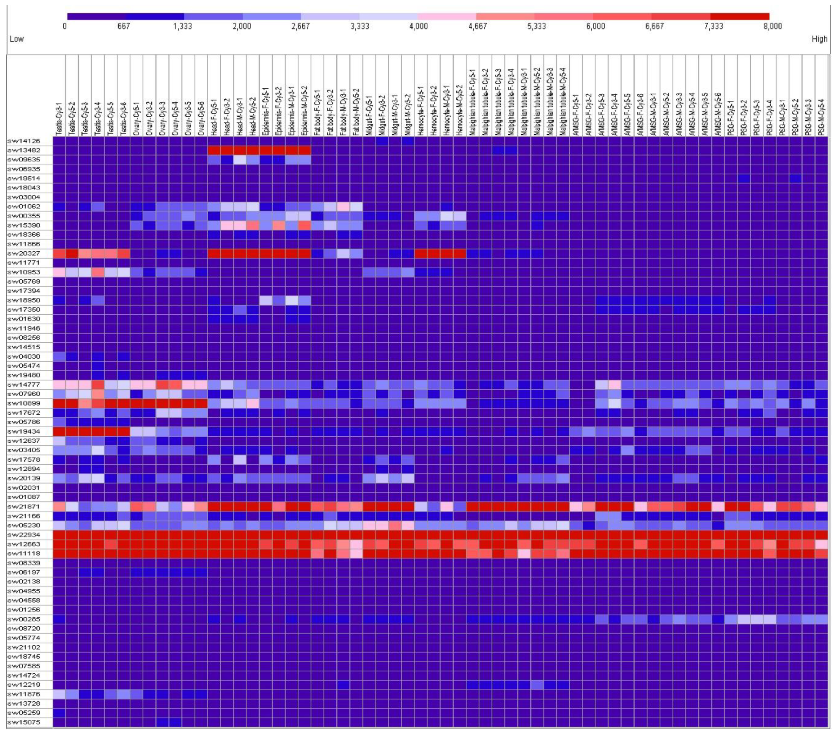
2.2. Tissue Expression Patterns on Day 3 of the Fifth Instar

The silkworm feeds and grows quickly in the fifth larval period. Day 3 of the fifth instar is typical for larval development with more active biological processes [50]. Therefore, studying this time point will enrich the expression patterns and help with further understanding of the functions of maternal genes in different developmental stages. The microarray data of 10 silkworm tissues on day 3 of the fifth instar were downloaded from the SilkMDB [50]. The probes of SPE, BAEE and Pabn2 were not found in SilkMDB from the attached BLAST search (Table 1). The microarray data of the other 63 maternal genes are provided in Table S2. The expression patterns of these 63 maternal genes are listed as found in various tissues and both sexes of silkworm in Figure 2. The expressed genes are defined as previously described [51]. Most of the maternal genes usually showed very low expression levels overall in the tissues and sex. The expression levels of sw10899 (aub) and sw14777 (me31B) were higher in the ovary and testis than in other tissues overall, sw19434 (Nelf-E) was only higher in the testis. The expression level of sw20327 (proPPAE) was higher in the testis, head, epidermis, and hemocyte, and sw13482 (Th) was higher in the head and epidermis. The expression levels of sw22934 (Eif-4a), sw12663 (eIF4AIII), sw1118 (Bin1), and sw21871 (Sod2) were higher overall. Most maternal genes showed low expression levels in multiple silkworm larval tissues on day 3 of the fifth instar. This is contrary to the abundant expression in the eggs of virgin moth (Figure 1).

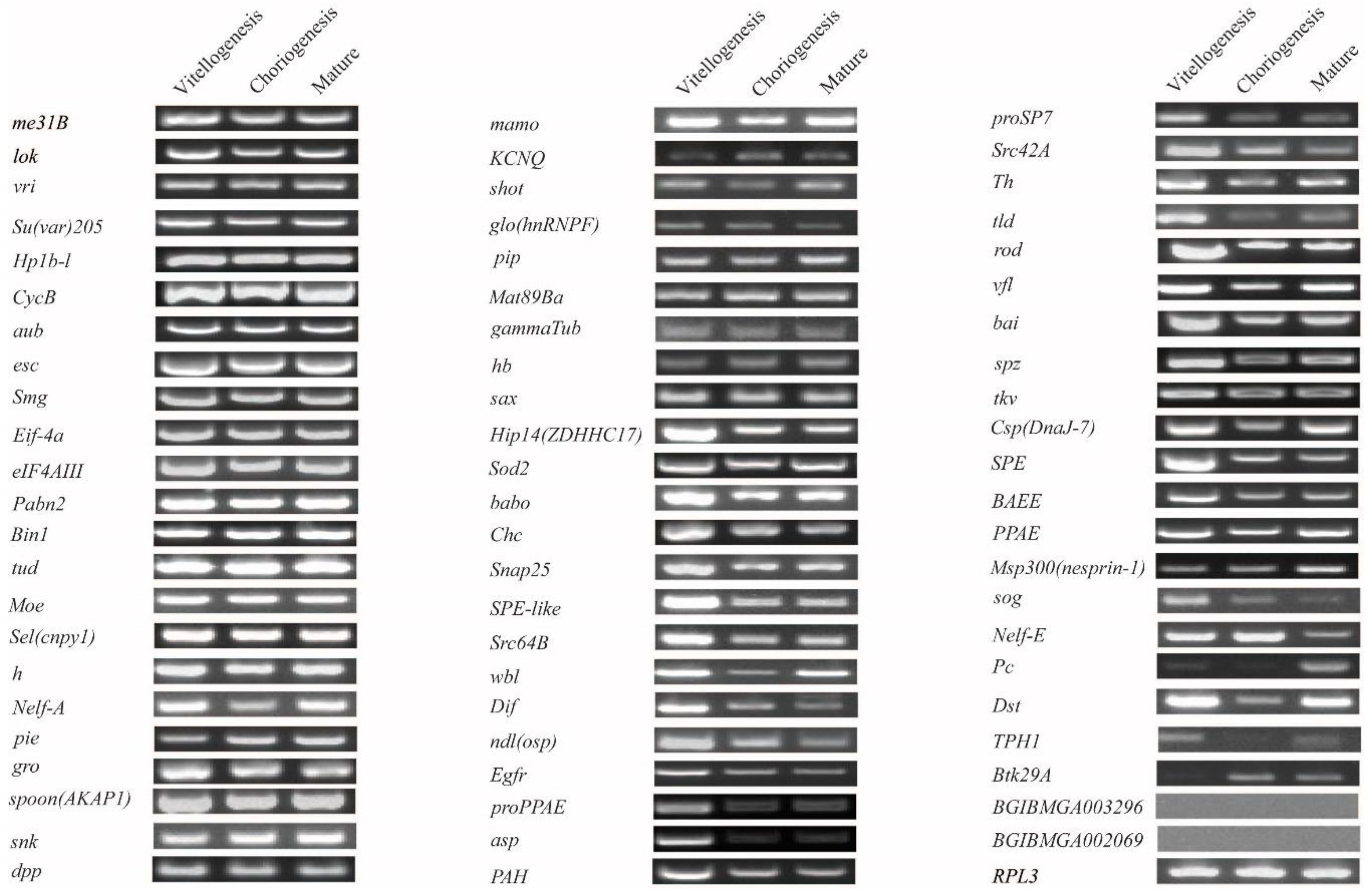
2.3. Expression Analysis in Developing Oocytes in Day 8 Pupae

The silkworm has a pair of ovaries, each of which is composed of four ovarioles. The developmental stage of each ovariole is opportune, being found in vitellogenesis, choriogenesis, and mature eggs from day 8 pupae [43,44]. A large amount of yolk proteins and no chorion proteins exist in oocytes during vitellogenesis. Chorion proteins appear just after vitellogenesis and continue throughout the whole of choriogenesis and until the formation of the eggshell of mature eggs [43,44,52,53,54]. The expression of the 68 potential maternal genes in the vitellogenesis, choriogenesis, and mature eggs in day 8 pupae was analyzed by RT-PCR. The result showed that for 66 genes, transcriptional signals were detected, whereas BGIBMGA003296 and BGIBMGA002069 also had no transcriptional signals (Figure 3). Me31B and the other 31 genes (in total 32) showed consistent expression levels in the vitellogenesis, choriogenesis, and mature eggs in day 8 pupae (Figure 3). Hip14 (ZDHHC17) and the other 33 genes (in total 34) had transcriptional signals and presented different trends in expression in the vitellogenesis, choriogenesis, and mature eggs in day 8 pupae (Figure 3).

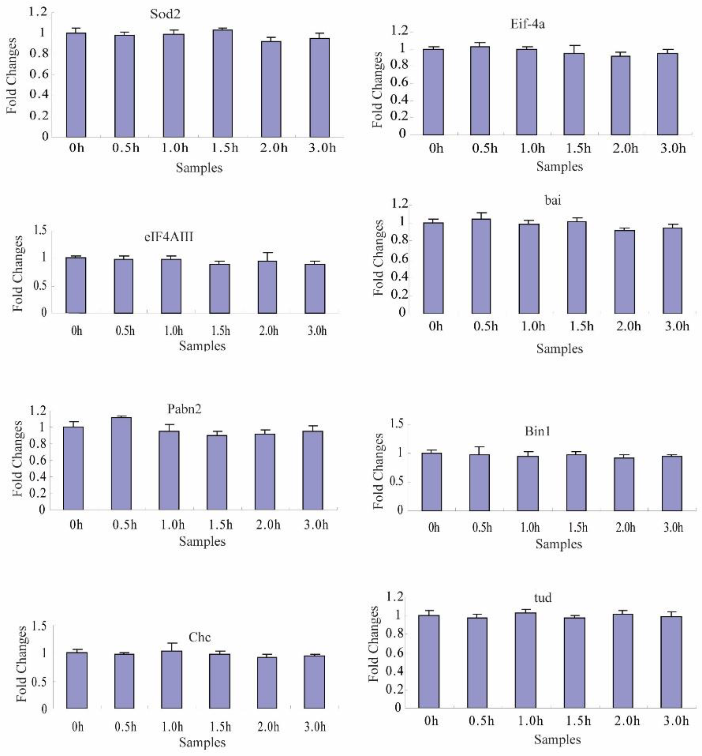
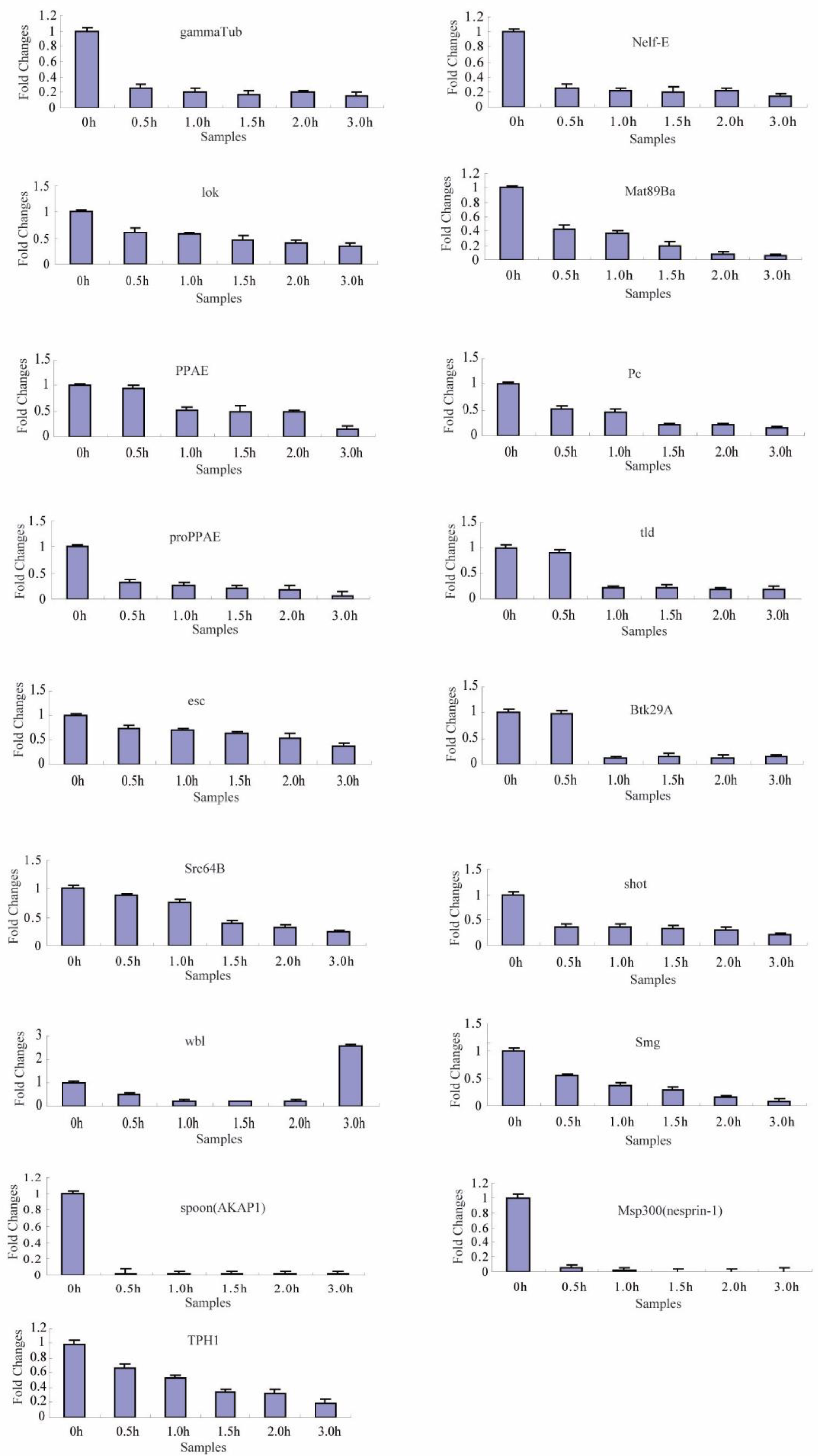
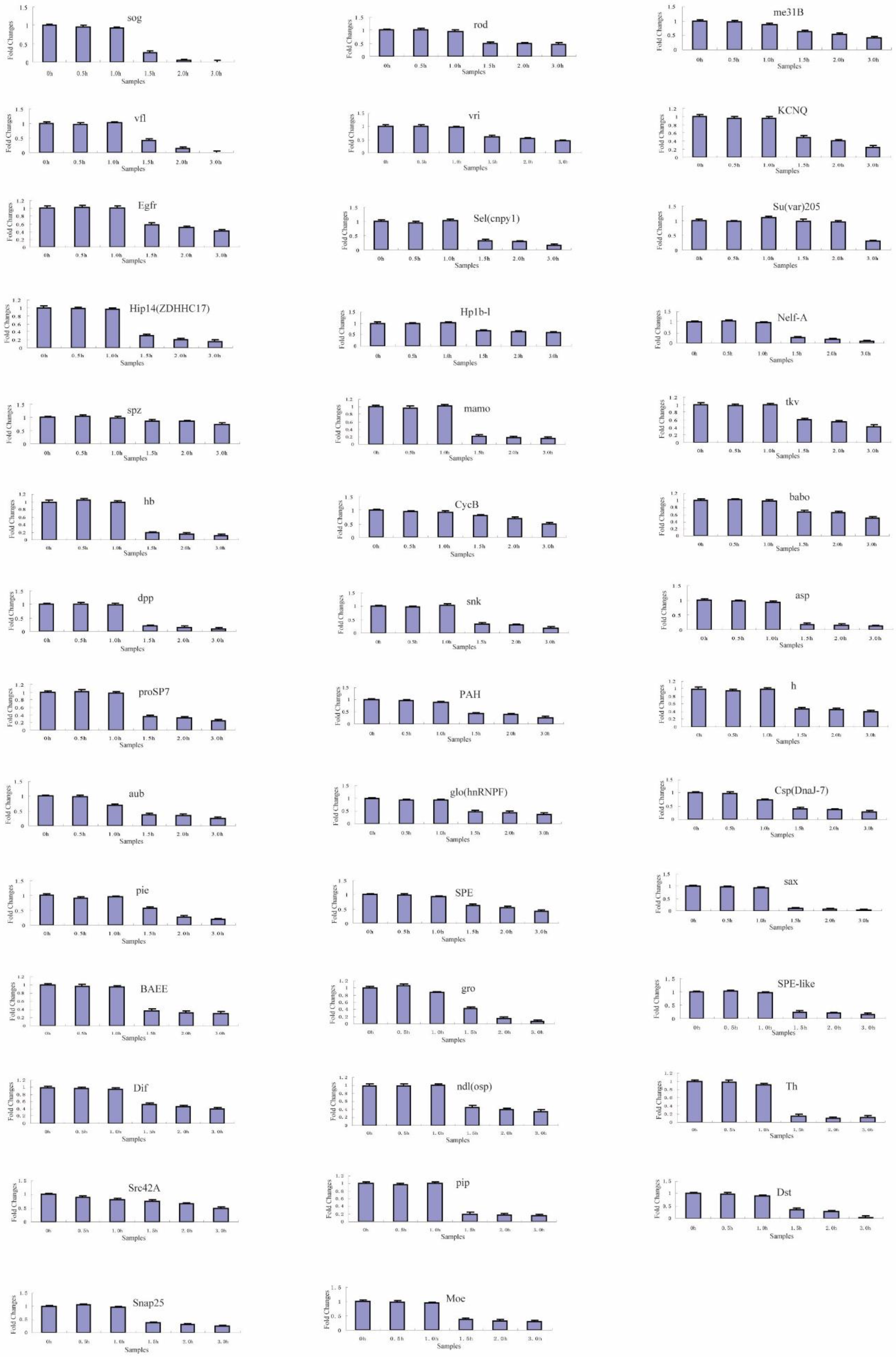
2.4. Transcriptional Degradation during the Maternal-to-Zygotic Transition

To identify the transcriptional degradation patterns of the 66 maternal genes during different developmental stage embryos, six time-series samples were collected at 0, 0.5, 1.0, 1.5, 2.0, and 3.0 h after fertilized embryo spawning, and were analyzed by reverse transcription-quantitative PCR (RT-qPCR). The specific primers for each gene that was subjected to RT-qPCR are shown in Table S1. In total, temporal control of their transcript clearance presented three different maternal transcript clusters during the maternal-to-zygotic transition (Figure 4, Figure 5 and Figure 6, Table 2).
In cluster 1 (Table 2), the transcript levels of 8 maternal genes (Sod2, Eif-4a, eIF4AIII, bai, Pabn2, Bin1, Chc and tud) showed no change from 0 to 3 h (Figure 4). The tissue expression levels of Eif-4a (sw22934), eIF4AIII (sw12663), Bin1 (sw1118), and Sod2 (sw21871) were high and uniform overall in 10 tissues at day 3 of the silkworm fifth instar (Figure 2). For a closer examination, we used RT-PCR to investigate these eight maternal genes and their transcript temporal control from 0 to 18 h after fertilized embryo spawning (Figure S1). The transcripts of Sod2, Eif-4a, eIF4AIII, Bin1, Chc, and tud kept consistent levels from 0 to 18 h after fertilized embryo spawning, respectively (Figure S1). Pabn2 and bai presented changing trends in expression at the transcriptional level (Figure S1).
Regarding the other 58 maternal genes, the RT-qPCR results showed that their transcripts significantly changed with two main characteristics during the maternal-to-zygotic transition (MZT). In cluster 2 (Table 2), the transcripts of 17 genes were significantly decreased from 0.5 to 3.0 h (Figure 5). In cluster 3 (Table 2), the transcripts of 41 genes were decreased significantly after 1.5 h (Figure 6). This indicates that the maternally supplied mRNAs of most maternal genes were universally degraded during MZT. Unlike other genes, the transcript of the wbl gene was decreased significantly from 0.5 to 2.0 h, and increased sharply at 3.0 h. This transcript belongs to cluster 2 and is an exception.

3. Discussion

In our previous study, 76 potential silkworm maternal genes were identified by orthologous comparison [48]. In this study, 68 of the 76 potential silkworm maternal genes were initially identified through further sequence alignment analysis, and 2 of the 68 maternal genes were not expressed in the silkworm eggs of virgin moths. The mRNAs of maternal genes are produced by females and loaded into the embryos [2,6]. Thus, the expression of a gene was not detected in embryos that can be identified as a non-maternal gene. A total of 66 maternal genes were finally identified in silkworm.
The embryonic development of B. mori is significantly different from that of Drosophila. The silkworm has a pair of ovaries each composed of four ovarioles, each of which contains a chain of follicles [43,52]. The previous research on in vitro culturing of B. mori ovarian follicles showed that follicle development starts from middle vitellogenesis to late choriogenesis [55]. The follicles develop depending on an endogenous developmental program that does not require the presence of additional factors from tissues outside the ovariole [43,52]. Each follicle is composed of an oocyte and seven nurse cells surrounded by a single layer of the follicular epithelium [43]. The degenerated nurse cells are devoured by follicular epithelial cells at the end of vitellogenesis [43]. The 66 maternal genes were expressed in vitellogenesis on day 8 female pupae, which suggests that maternal mRNA is derived from the nurse cells.
The development of silkworm follicles is divided into 12 different stages [43]. The developmental stage of each ovariole is opportune, being found in vitellogenesis, choriogenesis, and mature eggs from day 8 pupae [43,44]. The rate of progression of vitellogenesis toward choriogenesis is estimated to be 2–2.5 h per follicle [56,57]. The eggs are considered mature upon finishing the formation of the eggshell in the choriogenesis period, and the mature eggs first appear in each ovariole proximal oviduct in day 8 female pupae [43,44,52]. The maternal genes of Hip14 (ZDHHC17) and the other 26 genes (27 in total) had higher expression levels in vitellogenesis than in choriogenesis and mature eggs, whereas their expression levels were similar in choriogenesis and mature eggs on day 8 female pupae. These 27 maternal genes may have biological functions in the developmental process from vitellogenesis to choriogenesis in B. mori.
The MZT is a tightly regulated process that is identified by the elimination of maternal mRNAs and the initiation of zygotic transcription. This process has been extensively studied within model species. Each model organism has a unique pattern of maternal transcriptional clearance classes during the MZT. Four subsets of transcripts were characterized in Drosophila: Stable mRNAs, mRNAs targeted solely by the maternal or the zygotic degradation pathway, and those targeted by both pathways [7,12,31,58]. In activated, unfertilized eggs of Drosophila, maternal decay activity is present but zygotic activity is absent because no zygotic genome activation (ZGA) occurs. Thus, the degradation rate is significantly reduced compared with zygotic activity [7,31,59,60,61]. For maternal transcripts degradation during the development of zebrafish, a subclass of the cleared maternal mRNAs begins at fertilization, whereas others are mainly degraded after ZGA [62,63]. In Xenopus laevis, fertilization-induced deadenylation does not trigger decay immediately, but only after ZGA causing their deadenylation and degradation [40,64,65]. In the mouse, maternal mRNAs are degraded by both the maternal and the zygotic degradation pathways [41,66]. These are evolutionarily conserved mechanisms through which the mother provides gene products to the egg to drive the earliest stages of development.
Silkworms, like other insects such as Lepidoptera and Coleoptera, undergo superficial cleavage. In silkworm, the degradation of maternal gene mRNAs can be divided into three clusters during the MZT. Cluster 1 is stable mRNAs. In cluster 1, the mRNAs level of Tud is stable from zero to three hours. Tudor is a stress granule (SG) member that is activated upon various environmental stresses. Tudor (Tud) participates in posttranscriptional regulation in B. mori [67]. Silkworm Tudor depletion increases the levels of PIWI-interacting RNAs (piRNAs), which associate with PIWI proteins to protect genome integrity by silencing transposons in the germline [68]. Thus, cluster 1 includes stable mRNAs that perform essential housekeeping functions required during the MZT. The union of sperm and egg pronuclei occurs about two hours after silkworm eggs are laid. From about 2.0 to 2.5 h, the zygote divides repeatedly by mitosis and forms many cleavage nuclei [45,46,47]. The maternal gene mRNAs decay in clusters 2 and 3 is firstly initiated by maternal degradation activity. The initial time-point of degradation of cluster 2 is earlier than that of cluster 3. In unfertilized silkworm eggs, maternal decay activity is present, but zygotic activity is absent. Because no ZGA occurs, the degradation rate is significantly reduced in unfertilized eggs compared to that of fertilized eggs [48].
The study of transcriptional regulation has produced many discoveries that have improved our understanding of development. Understanding the post-transcriptional regulation of maternal mRNA is crucial to uncover the mechanisms that control the coordinated changes in zygotic transcription initiation [6]. The MZT represents an extreme scenario involving these mechanisms. In silkworm, according to the requirements of natural and programmed embryonic development [43,48,69], studying the establishment of transcriptional quiescence during oogenesis and identifying the first genes to be expressed during embryo (mature eggs) formation will continue to improve our understanding of transcriptional regulation during MZT.

4. Materials and Methods

4.1. Insects and Collection of Samples

B. mori (Dazao) larvae were reared under standard conditions (25 °C and 70% humidity). The larvae–pupae, pupae, moths, and eggs were maintained under a 12 h light/12 h dark photoperiod at 25 °C and 70% humidity. The developing oocytes (eggs) undergoing vitellogenesis, choriogenesis, and mature eggs were separately collected from ovarioles that were dissected from pharate adults eight days after larval–pupal ecdysis, according to previous studies [43,44]. The eggs of virgin moths were collected from ovarioles that were dissected from the freshly hatched female moths. Freshly hatched moths were immediately mated for 3 h, and the female moths were subsequently gathered for spawning for 15 min. Zero h is defined as the 15th minute after most female moths spawn. Then, the eggs were collected at specific points-in-time (0, 0.5, 1.0, 1.5, 2.0, 3.0, 6, 12, and 18 h) under the same conditions as previously described [48].

4.2. Identification of B. mori Maternal Genes

In our earlier study, we obtained 76 potential maternal genes in the B. mori genome [48]. We obtained their sequences from the silkworm database (SilkDB), which were used to search the NCBI database and silkBase [49]. As for the maternal genes that were similar or overlapping between the NCBI database and silkBase, the longer of the two was selected. The newly annotated protein sequences were obtained and applied to predict signal peptides by SignalP 4.1 Server.

4.3. Transcript Detection Reverse Transcription-PCR

Reverse transcription-PCR (RT-PCR) was used to analyze the expression patterns of maternal genes. Total RNA was extracted using TRIzol reagent (Invitrogen, Carlsbad, CA, USA) from the samples including developing oocytes (eggs) of vitellogenesis, choriogenesis, and mature eggs in day 8 pupae, the eggs of virgin moths, and after spawning at specific points-in-time (0, 0.5, 1.0, 1.5, 2.0, 3.0, 6, 12, and 18 h). Total RNA concentrations were quantified, and single-stranded cDNA was synthesized by using a PrimeScript™ RT kit (TaKaRa, Dalian, China) according to the manufacturer’s instructions. A 25 μL PCR reaction system was established by initial denaturing at 94 °C for 5 min, 35 cycles of denaturing at 94 °C for 30 s, annealing at 58 °C for 30 s, and extension at 72 °C for 30 s. This was followed by a final extension at 72 °C for 10 min before storing at 12 °C. BmRPL3 was used as an internal control [70]. A pair of specific primers for each gene was used in RT-PCR, as shown in Table S1. The RT-PCR product of each gene was separated by 1.2% agarose gel electrophoresis.

4.4. Tissue Expression Patterns Based on Microarray Database

We downloaded the microarray data from the SilkMDB to analyze tissue expression patterns of the maternal genes in 10 silkworm tissues on day 3 of the fifth instar [50]. A genome-wide microarray with 22,987 probes was designed and constructed in the silkworm genome, and each probe is also provided in this database [50]. The probes of SPE, BAEE, and Pabn2 were not found in the database, as shown by the attached BLAST search. The microarray data of the other 63 maternal genes are provided in Table S2. The expressed genes are defined as previously described [51]. GeneCluster 2.0 software was used to visualize the expression levels [71].

4.5. Transcript DecayDetection by Reverse Transcription-Quantitative PCR

Total RNA was extracted using TRIzol reagent (Invitrogen, Carlsbad, CA, USA) from the eggs collected at specific points-in-time (0, 0.5, 1.0, 1.5, 2.0, and 3.0 h). A fraction of the RNA was treated with DNase. After verifying the quality, the RNA was used to synthesize the first-strand cDNA using the PrimeScript™ RT Master Mix (Perfect Real Time; TaKaRa, Dalian, China) according to the manufacturer’s instructions. Reverse transcription-quantitative PCR (RT-qPCR) was performed as previously described [48]. A pair of specific primers for each gene was used in RT-qPCR, as shown in Table S1.

5. Conclusions

In the current work, 66 maternal genes in silkworm were characterized through bioinformatics analysis and expression detection. The expression of these genes in vitellogenesis, choriogenesis, and mature eggs in day 8 pupae was analyzed using RT-PCR. We analyzed the maternal gene mRNAs decay in fertilized eggs in B. mori from six points-in-time by RT-qPCR. The 66 maternal genes formed three clusters of degradation patterns during the MZT. The maternal gene mRNAs of cluster 1 were stable. The initial time-point of degradation of cluster 2 was earlier than that of cluster 3. The maternal gene mRNAs decay of clusters 2 and 3 was firstly initiated by maternal degradation activity. Our findings expand upon the identification of silkworm maternal genes and provide a perspective for the embryo development in B. mori.

Supplementary Materials

Supplementary materials can be found at https://www.mdpi.com/1422-0067/20/22/5651/s1.

Author Contributions

M.Z. performed the literature review and wrote the manuscript; P.X. performed the experiment and analyzed the data; H.P. prepared the illustrations; T.C. and G.Z. designed the study and suggested important research points. All authors have read and approved the final version of the manuscript.

Acknowledgments

This research was financially supported by the National Natural Science Foundation of China (Grant no. 31302035) and the Jiangsu Provincial Natural Science Foundation of China (Grant no. BK2012273).

Conflicts of Interest

The authors declare no conflict of interest.

References

  1. Horner, V.L.; Wolfner, M.F. Mechanical stimulation by osmotic and hydrostatic pressure activates Drosophila oocytes in vitro in a calcium-dependent manner. Dev. Biol. 2008, 316, 100–109. [Google Scholar] [CrossRef] [PubMed]
  2. Tadros, W.; Lipshitz, H.D. The maternal-to-zygotic transition: A play in two acts. Development 2009, 136, 3033–3042. [Google Scholar] [CrossRef] [PubMed]
  3. Marlow, F.L. Maternal Control of Development in Vertebrates: My Mother Made Me Do It; Morgan & Claypool Life Sciences: San Rafael, CA, USA, 2010. [Google Scholar]
  4. Yartseva, V.; Giraldez, A.J. The Maternal-to-Zygotic Transition During Vertebrate Development: A Model for Reprogramming. Curr. Top. Dev. Biol. 2015, 113, 191–232. [Google Scholar] [PubMed]
  5. Giraldez, A.J.; Mishima, Y.; Rihel, J.; Grocock, R.J.; Van Dongen, S.; Inoue, K.; Enright, A.J.; Schier, A.F. Zebrafish MiR-430 promotes deadenylation and clearance of maternal mRNAs. Science 2006, 312, 75–79. [Google Scholar] [CrossRef] [PubMed]
  6. Langley, A.R.; Smith, J.C.; Stemple, D.L.; Harvey, S.A. New insights into the maternal to zygotic transition. Development 2014, 141, 3834–3841. [Google Scholar] [CrossRef] [PubMed]
  7. Walser, C.B.; Lipshitz, H.D. Transcript clearance during the maternal-to-zygotic transition. Curr. Opin. Genet. Dev. 2011, 21, 431–443. [Google Scholar] [CrossRef] [PubMed]
  8. Bashirullah, A.; Halsell, S.R.; Cooperstock, R.L.; Kloc, M.; Karaiskakis, A.; Fisher, W.W.; Fu, W.; Hamilton, J.K.; Etkin, L.D.; Lipshitz, H.D. Joint action of two RNA degradation pathways controls the timing of maternal transcript elimination at the midblastula transition in Drosophila melanogaster. EMBO J. 1999, 18, 2610–2620. [Google Scholar] [CrossRef] [PubMed]
  9. Smibert, C.A.; Wilson, J.E.; Kerr, K.; Macdonald, P.M. smaug protein represses translation of unlocalized nanos mRNA in the Drosophila embryo. Genes Dev. 1996, 10, 2600–2609. [Google Scholar] [CrossRef] [PubMed]
  10. Semotok, J.L.; Cooperstock, R.L.; Pinder, B.D.; Vari, H.K.; Lipshitz, H.D.; Smibert, C.A. Smaug recruits the CCR4/POP2/NOT deadenylase complex to trigger maternal transcript localization in the early Drosophila embryo. Curr. Biol. 2005, 15, 284–294. [Google Scholar] [CrossRef] [PubMed]
  11. Tadros, W.; Goldman, A.L.; Babak, T.; Menzies, F.; Vardy, L.; Orr-Weaver, T.; Hughes, T.R.; Westwood, J.T.; Smilbert, C.A.; Lipshitz, H.D. SMAUG is a major regulator of maternal mRNA destabilization in Drosophila and its translation is activated by the PAN GU kinase. Dev. Cell 2007, 12, 143–155. [Google Scholar] [CrossRef] [PubMed]
  12. Thomsen, S.; Anders, S.; Janga, S.C.; Huber, W.; Alonso, C.R. Genome-wide analysis of mRNA decay patterns during early Drosophila development. Genome Biol. 2010, 11, R93. [Google Scholar] [CrossRef] [PubMed]
  13. Jeske, M.; Moritz, B.; Anders, A.; Wahle, E. Smaug assembles an ATP-dependent stable complex repressing nanos mRNA translation at multiple levels. Embo J. 2011, 30, 90–103. [Google Scholar] [CrossRef] [PubMed]
  14. Macdonald, P.M. The Drosophila pumilio gene: An unusually long transcription unit and an unusual protein. Development 1992, 114, 221–232. [Google Scholar] [PubMed]
  15. Zamore, P.D.; Williamson, J.R.; Lehmann, R. The Pumilio protein binds RNA through a conserved domain that defines a new class of RNA-binding proteins. RNA 1997, 3, 1421–1433. [Google Scholar] [PubMed]
  16. Gamberi, C.; Peterson, D.S.; He, L.; Gottlieb, E. An anterior function for the Drosophila posterior determinant Pumilio. Development 2002, 129, 2699–2710. [Google Scholar] [PubMed]
  17. Gerber, A.P.; Luschnig, S.; Krasnow, M.A.; Brown, P.O.; Herschlag, D. Genome-wide identification of mRNAs associated with the translational regulator PUMILIO in Drosophila melanogaster. Proc. Natl. Acad. Sci. USA 2006, 103, 4487–4492. [Google Scholar] [CrossRef] [PubMed]
  18. De Renzis, S.; Elemento, O.; Tavazoie, S.; Wieschaus, E.F. Unmasking activation of the zygotic genome using chromosomal deletions in the Drosophila embryo. PLoS Biol. 2007, 5, 1036–1051. [Google Scholar] [CrossRef]
  19. Sonoda, J.; Wharton, R.P. Drosophila Brain Tumor is a translational repressor. Genes Dev. 2001, 15, 762–773. [Google Scholar] [CrossRef] [PubMed]
  20. Loedige, I.; Stotz, M.; Qamar, S.; Kramer, K.; Hennig, J.; Schubert, T.; Loffler, P.; Langst, G.; Merkl, R.; Urlaub, H.; et al. The NHL domain of BRAT is an RNA-binding domain that directly contacts the hunchback mRNA for regulation. Gene Dev. 2014, 28, 749–764. [Google Scholar] [CrossRef] [PubMed]
  21. Laver, J.D.; Li, X.; Ray, D.; Cook, K.B.; Hahn, N.A.; Nabeel-Shah, S.; Kekis, M.; Luo, H.; Marsolais, A.J.; Fung, K.Y.Y.; et al. Brain tumor is a sequence-specific RNA-binding protein that directs maternal mRNA clearance during the Drosophila maternal-to-zygotic transition. Genome Biol. 2015, 16, 94. [Google Scholar] [CrossRef] [PubMed]
  22. Wang, M.; Ly, M.; Lugowski, A.; Laver, J.D.; Lipshitz, H.D.; Smibert, C.A.; Rissland, O.S. ME31B globally represses maternal mRNAs by two distinct mechanisms during the Drosophila maternal-to-zygotic transition. Elife 2017, 6, e27891. [Google Scholar] [CrossRef] [PubMed] [Green Version]
  23. Gallie, D.R. The cap and poly(A) tail function synergistically to regulate mRNA translational efficiency. Genes Dev. 1991, 5, 2108–2116. [Google Scholar] [CrossRef] [PubMed] [Green Version]
  24. Rouget, C.; Papin, C.; Boureux, A.; Meunier, A.C.; Franco, B.; Robine, N.; Lai, E.C.; Pelisson, A.; Simonelig, M. Maternal mRNA deadenylation and decay by the piRNA pathway in the early Drosophila embryo. Nature 2010, 467, 1128–1132. [Google Scholar] [CrossRef] [PubMed] [Green Version]
  25. Barckmann, B.; Pierson, S.; Dufourt, J.; Papin, C.; Armenise, C.; Port, F.; Grentzinger, T.; Chambeyron, S.; Baronian, G.; Desvignes, J.P.; et al. Aubergine iCLIP Reveals piRNA-Dependent Decay of mRNAs Involved in Germ Cell Development in the Early Embryo. Cell Rep. 2015, 12, 1205–1216. [Google Scholar] [CrossRef] [PubMed] [Green Version]
  26. Luo, H.; Li, X.; Claycomb, J.M.; Lipshitz, H.D. The Smaug RNA-Binding Protein Is Essential for microRNA Synthesis During the Drosophila Maternal-to-Zygotic Transition. G3 Genes Genomes Genet. 2016, 6, 3541–3551. [Google Scholar] [CrossRef] [PubMed] [Green Version]
  27. Laver, J.D.; Marsolais, A.J.; Smibert, C.A.; Lipshitz, H.D. Regulation and Function of Maternal Gene Products During the Maternal-to-Zygotic Transition in Drosophila. Curr. Top. Dev. Biol. 2015, 113, 43–84. [Google Scholar] [PubMed]
  28. Niinuma, S.; Tomari, Y. ATP is dispensable for both miRNA- and Smaug-mediated deadenylation reactions. RNA 2017, 23, 866–871. [Google Scholar] [CrossRef] [PubMed]
  29. Shukla, G.C.; Singh, J.; Barik, S. MicroRNAs: Processing, Maturation, Target Recognition and Regulatory Functions. Mol. Cell Pharmacol. 2011, 3, 83–92. [Google Scholar] [PubMed]
  30. Bartel, D.P. MicroRNAs: Target recognition and regulatory functions. Cell 2009, 136, 215–233. [Google Scholar] [CrossRef] [PubMed] [Green Version]
  31. Bushati, N.; Stark, A.; Brennecke, J.; Cohen, S.M. Temporal reciprocity of miRNAs and their targets during the maternal-to-zygotic transition in Drosophila. Curr. Biol. 2008, 18, 501–506. [Google Scholar] [CrossRef] [PubMed] [Green Version]
  32. Lund, E.; Liu, M.; Hartley, R.S.; Sheets, M.D.; Dahlberg, J.E. Deadenylation of maternal mRNAs mediated by miR-427 in Xenopus laevis embryos. RNA 2009, 15, 2351–2363. [Google Scholar] [CrossRef] [PubMed] [Green Version]
  33. Laue, K.; Rajshekar, S.; Courtney, A.J.; Lewis, Z.A.; Goll, M.G. The maternal to zygotic transition regulates genome-wide heterochromatin establishment in the zebrafish embryo. Nat. Commun. 2019, 10, 1551. [Google Scholar] [CrossRef] [PubMed] [Green Version]
  34. Bazzini, A.A.; del Viso, F.; Moreno-Mateos, M.A.; Johnstone, T.G.; Vejnar, C.E.; Qin, Y.D.; Yao, J.; Khokha, M.K.; Giraldez, A.J. Codon identity regulates mRNA stability and translation efficiency during the maternal-to-zygotic transition. EMBO J. 2016, 35, 2087–2103. [Google Scholar] [CrossRef] [PubMed]
  35. Chen, Y.H.; Coller, J. A Universal Code for mRNA Stability? Trends Genet. 2016, 32, 687–688. [Google Scholar] [CrossRef] [PubMed] [Green Version]
  36. Mishima, Y.; Tomari, Y. Codon Usage and 30 UTR Length Determine Maternal mRNA Stability in Zebrafish. Mol. Cell 2016, 61, 874–885. [Google Scholar] [CrossRef] [PubMed] [Green Version]
  37. Edgar, B.A.; O’Farrell, P.H. The three postblastoderm cell cycles of Drosophila embryogenesis are regulated in G2 by string. Cell 1990, 62, 469–480. [Google Scholar] [CrossRef] [Green Version]
  38. Edgar, B.A.; Datar, S.A. Zygotic degradation of two maternal Cdc25 mRNAs terminates Drosophila’s early cell cycle program. Genes Dev. 1996, 10, 1966–1977. [Google Scholar] [CrossRef] [PubMed] [Green Version]
  39. Benoit, B.; He, C.H.; Zhang, F.; Votruba, S.M.; Tadros, W.; Westwood, J.T.; Smibert, C.A.; Lipshitz, H.D.; Theurkauf, W.E. An essential role for the RNA-binding protein Smaug during the Drosophila maternal-to-zygotic transition. Development 2009, 136, 923–932. [Google Scholar] [CrossRef] [PubMed] [Green Version]
  40. Lu, X.M.; Li, J.M.; Elemento, O.; Tavazoie, S.; Wieschaus, E.F. Coupling of zygotic transcription to mitotic control at the Drosophila mid-blastula transition. Development 2009, 136, 2101–2110. [Google Scholar] [CrossRef] [PubMed] [Green Version]
  41. Zeng, F.; Baldwin, D.A.; Schultz, R.M. Transcript profiling during preimplantation mouse development. Dev. Biol. 2004, 272, 483–496. [Google Scholar] [CrossRef] [PubMed] [Green Version]
  42. Chen, L.; Dumelie, J.G.; Li, X.; Cheng, M.H.; Yang, Z.; Laver, J.D.; Siddiqui, N.U.; Westwood, J.T.; Morris, Q.; Lipshitz, H.D.; et al. Global regulation of mRNA translation and stability in the early Drosophila embryo by the Smaug RNA-binding protein. Genome Biol. 2014, 15, R4. [Google Scholar] [CrossRef] [PubMed] [Green Version]
  43. Yamauchi, H.; Yoshitake, N. Developmental stages of ovarian follicles of the silkworm, Bombyx mori L. J. Morphol. 1984, 179, 21–31. [Google Scholar] [CrossRef] [PubMed]
  44. Chen, Z.; Nohata, J.; Guo, H.; Li, S.; Liu, J.; Guo, Y.; Yamamoto, K.; Kadono-Okuda, K.; Liu, C.; Arunkumar, K.P.; et al. A comprehensive analysis of the chorion locus in silkmoth. Sci. Rep. 2015, 5, 16424. [Google Scholar] [CrossRef] [PubMed]
  45. Rasmussen, S.W. The transformation of the Synaptonemal Complex into the ‘elimination chromatin’ in Bombyx mori oocytes. Chromosoma 1977, 60, 205–221. [Google Scholar] [CrossRef] [PubMed]
  46. Kawamura, N. The early embryonic mitosis in normal and cooled eggs of the silkworm, Bombyx mori. J. Morphol. 1978, 158, 57–71. [Google Scholar] [CrossRef] [PubMed]
  47. Kawamura, N. Fertilization and the first cleavage mitosis in insects. Dev. Growth Differ. 2001, 43, 343–349. [Google Scholar] [CrossRef] [PubMed] [Green Version]
  48. Zhang, M.R.; Qin, S.; Xu, P.Z.; Zhang, G.Z. Identifying potential maternal genes of Bombyx mori using digital gene expression profiling. PLoS ONE 2018, 13, e0192745. [Google Scholar] [CrossRef] [PubMed]
  49. Kawamoto, M.; Jouraku, A.; Toyoda, A.; Yokoi, K.; Minakuchi, Y.; Katsuma, S.; Fujiyama, A.; Kiuchi, T.; Yamamoto, K.; Shimada, T. High-quality genome assembly of the silkworm, Bombyx mori. Insect Biochem. Mol. Biol. 2019, 107, 53–62. [Google Scholar] [CrossRef] [PubMed]
  50. Xia, Q.; Cheng, D.; Duan, J.; Wang, G.; Cheng, T.; Zha, X.; Liu, C.; Zhao, P.; Dai, F.; Zhang, Z.; et al. Microarray-based gene expression profiles in multiple tissues of the domesticated silkworm, Bombyx mori. Genome Biol. 2007, 8, R162. [Google Scholar] [CrossRef] [PubMed] [Green Version]
  51. Zhao, P.; Dong, Z.; Duan, J.; Wang, G.; Wang, L.; Li, Y.; Xiang, Z.; Xia, Q. Genome-wide identification and immune response analysis of serine protease inhibitor genes in the silkworm, Bombyx mori. PLoS ONE 2012, 7, e31168. [Google Scholar] [CrossRef] [PubMed]
  52. Paul, M.; Goldsmith, M.R.; Hunsley, J.R.; Kafatos, F.C. Specific protein synthesis in cellular differentiation. Production of eggshell proteins by silkmoth follicular cells. J. Cell Biol. 1972, 55, 653–680. [Google Scholar] [CrossRef] [PubMed] [Green Version]
  53. Blau, H.M.; Kafatos, F.C. Secretory kinetics in the follicular cells of silkmoths during eggshell formation. J. Cell Biol. 1978, 78, 131–151. [Google Scholar] [CrossRef] [PubMed] [Green Version]
  54. Nadel, M.R.; Goldsmith, M.R.; Goplerud, J.; Kafatos, F.C. Specific protein synthesis in cellular differentiation. V. A secretory defect of chorion formation in the Grcol mutant of Bombyx mori. Dev. Biol. 1980, 75, 41–58. [Google Scholar] [CrossRef]
  55. Machado, E.; Swevers, L.; Sdralia, N.; Medeiros, M.N.; Mello, F.G.; Iatrou, K. Prostaglandin signaling and ovarian follicle development in the silkmoth, Bombyx mori. Insect Biochem. Mol. Biol. 2007, 37, 876–885. [Google Scholar] [CrossRef] [PubMed]
  56. Nadel, M.R.; Kafatos, F.C. Specific protein synthesis in cellular differentiation. IV. The chorion proteins of Bombyx mori and their program of synthesis. Dev. Biol. 1980, 75, 26–40. [Google Scholar] [CrossRef]
  57. Swevers, L.; Iatrou, K. Early establishment and autonomous implementation of a developmental program controlling silkmoth chorion gene expression. Dev. Biol. 1992, 150, 12–22. [Google Scholar] [CrossRef]
  58. Surdej, P.; Jacobs-Lorena, M. Developmental regulation of bicoid mRNA stability is mediated by the first 43 nucleotides of the 3’ untranslated region. Mol. Cell Biol. 1998, 18, 2892–2900. [Google Scholar] [CrossRef] [PubMed] [Green Version]
  59. Bashirullah, A.; Cooperstock, R.L.; Lipshitz, H.D. Spatial and temporal control of RNA stability. Proc. Natl. Acad. Sci. USA 2001, 98, 7025–7028. [Google Scholar] [CrossRef] [PubMed] [Green Version]
  60. Tadros, W.; Houston, S.A.; Bashirullah, A.; Cooperstock, R.L.; Semotok, J.L.; Reed, B.H.; Lipshitz, H.D. Regulation of maternal transcript destabilization during egg activation in Drosophila. Genetics 2003, 164, 989–1001. [Google Scholar] [PubMed]
  61. Tadros, W.; Lipshitz, H.D. Setting the stage for development: mRNA translation and stability during oocyte maturation and egg activation in Drosophila. Dev. Dyn. 2005, 232, 593–608. [Google Scholar] [CrossRef] [PubMed]
  62. Rauwerda, H.; Pagano, J.F.; de Leeuw, W.C.; Ensink, W.; Nehrdich, U.; de Jong, M.; Jonker, M.; Spaink, H.P.; Breit, T.M. Transcriptome dynamics in early zebrafish embryogenesis determined by high-resolution time course analysis of 180 successive, individual zebrafish embryos. BMC Genom. 2017, 18, 287. [Google Scholar] [CrossRef] [PubMed] [Green Version]
  63. Mathavan, S.; Lee, S.G.; Mak, A.; Miller, L.D.; Murthy, K.R.; Govindarajan, K.R.; Tong, Y.; Wu, Y.L.; Lam, S.H.; Yang, H.; et al. Transcriptome analysis of zebrafish embryogenesis using microarrays. PLoS Genet. 2005, 1, 260–276. [Google Scholar] [CrossRef] [PubMed] [Green Version]
  64. Audic, Y.; Omilli, F.; Osborne, H.B. Postfertilization deadenylation of mRNAs in Xenopus laevis embryos is sufficient to cause their degradation at the blastula stage. Mol. Cell Biol. 1997, 17, 209–218. [Google Scholar] [CrossRef] [PubMed] [Green Version]
  65. Voeltz, G.K.; Steitz, J.A. AUUUA sequences direct mRNA deadenylation uncoupled from decay during Xenopus early development. Mol. Cell Biol. 1998, 18, 7537–7545. [Google Scholar] [CrossRef] [PubMed] [Green Version]
  66. Hamatani, T.; Carter, M.G.; Sharov, A.A.; Ko, M.S. Dynamics of global gene expression changes during mouse preimplantation development. Dev. Cell 2004, 6, 117–131. [Google Scholar] [CrossRef] [Green Version]
  67. Zhu, L.; Tatsuke, T.; Mon, H.; Li, Z.; Xu, J.; Lee, J.M.; Kusakabe, T. Characterization of Tudor-sn-containing granules in the silkworm, Bombyx mori. Insect Biochem. Mol. Biol. 2013, 43, 664–674. [Google Scholar] [CrossRef] [PubMed]
  68. Honda, S.; Kirino, Y.; Maragkakis, M.; Alexiou, P.; Ohtaki, A.; Murali, R.; Mourelatos, Z.; Kirino, Y. Mitochondrial protein BmPAPI modulates the length of mature piRNAs. Rna Publ. Rna Soc. 2013, 19, 1405–1418. [Google Scholar] [CrossRef] [PubMed] [Green Version]
  69. Yamashiki, N.; Kawamura, N. Behaviors of nucleus, basal bodies and microtubules during eupyrene and apyrene spermiogenesis in the silkworm, Bombyx mori (Lepidoptera). Dev. Growth Differ. 1997, 39, 715–722. [Google Scholar] [CrossRef] [PubMed]
  70. Li, W.; Cheng, T.; Hu, W.; Peng, Z.; Liu, C.; Xia, Q. Genome-wide identification and analysis of JHBP-domain family members in the silkworm Bombyx mori. Mol. Genet. Genom. 2016, 291, 2159–2171. [Google Scholar] [CrossRef] [PubMed]
  71. Golub, T.R.; Slonim, D.K.; Tamayo, P.; Huard, C.; Gaasenbeek, M.; Mesirov, J.P.; Coller, H.; Loh, M.L.; Downing, J.R.; Caligiuri, M.A.; et al. Molecular classification of cancer: Class discovery and class prediction by gene expression monitoring. Science 1999, 286, 531–537. [Google Scholar] [CrossRef] [PubMed] [Green Version]
Figure 1. Transcriptional detection of maternal genes in eggs of virgin moths by reverse transcription (RT)-PCR. M: DL2000 DNA Maker; numbers 1 to 68 indicate me31B, lok, vri, Egfr, Su (var) 205, Hp1b-l, spz, tkv, CycB, proPPAE, asp, PAH, aub, Csp (DnaJ-7), SPE, BAEE, PPAE, Sod2, esc, Src42A, Smg, Eif-4a, eIF4AIII, rod, vfl, bai, Nelf-E, Pabn2, Bin1, tud, Moe, Sel (cnpy1), Hip14 (ZDHHC17), mamo, sax, babo, h, Chc, Snap25, SPE-like, Src64B, wbl, Mat89Ba, Dif, ndl (osp), Nelf-A, tld, proSP7, gammaTub, Th, pie, gro, hb, pip, spoon (AKAP1), snk, Btk29A, dpp, Msp300 (nesprin-1), KCNQ, shot, sog, Pc, Dst, TPH1, glo (hnRNPF), BGIBMGA003296 and BGIBMGA002069, respectively.
Ijms 20 05651 g001
Figure 2. Tissue expression profile of maternal genes in larvae on day 3 of the silkworm fifth instar. The columns represent ten different tissues with both sexes: Testis, ovary, head, epidermis, fat body, midgut, hemocyte, Malpighian tubule, anterior/median silk gland (A/MSG), posterior silk gland (PSG), female (F), and male (M). Gene expression levels are represented by red (higher expression) and blue (lower expression) boxes.
Ijms 20 05651 g002
Figure 3. Expression patterns of maternal genes in the developing oocytes of vitellogenesis, choriogenesis, and mature eggs from day 8 female papae. Reverse transcription (RT)-PCR was performed and the RPL3 gene was used as internal control.
Ijms 20 05651 g003
Figure 4. The expression profiles of cluster 1 maternal genes by RT-qPCR during the maternal-to-zygotic transition (MZT). Each time-point was replicated three times using independently collected samples. The data are the means ± SD of three independent experiments.
Ijms 20 05651 g004
Figure 5. The expression profiles of cluster 2 maternal genes by RT-qPCR during the maternal-to-zygotic transition (MZT). Each time-point was replicated three times using independently collected samples. The data are the means ± SD of three independent experiments.
Ijms 20 05651 g005
Figure 6. The expression profiles of cluster 3 maternal genes by RT-qPCR during the maternal-to-zygotic transition (MZT). Each time-point was replicated three times using independently collected samples. The data are the means ± SD of three independent experiments.
Ijms 20 05651 g006
Table 1. Maternal genes in the silkworm Bombyx mori.
Gene NameAccession NumberProbeDescriptionProtein Length (Amino Acids)Location (Chr.)Signal PeptideNCBI Reference Sequence
me31BBGIBMGA010673sw14777ATP-dependent RNA helicase me31b44012AK383517
lokBGIBMGA005370sw11876cell cycle checkpoint kinase 25288AK382539
vriBGIBMGA013421sw05474nuclear factor interleukin-3-regulated protein36427AK388388
EgfrBGIBMGA000602sw11771epidermal growth factor receptor144911XM_004929742
Su (var) 205BGIBMGA006109 sw05786chromobox-like protein 51914AK385880
Hp1b-lBGIBMGA012860sw21166heterochromatin protein17916XM_012692600
spzBGIBMGA002397sw08256spatzle27791NM_001114594
tkvBGIBMGA007355sw21102bone morphogenetic protein receptor type-1B4853AK385287
CycBBGIBMGA003747sw04030cyclin B homolog52551AK382330
proPPAEBGIBMGA013746sw20327prophenoloxidase activating enzyme precursor441281AK383056
aspBGIBMGA005594sw10953protein abnormal spindle230917XM_004921876
PAHBGIBMGA003866sw01062phenylalanine hydroxylase4561NM_001287837
aubBGIBMGA010644sw10899aubergine protein 89912EU143547
Csp (DnaJ-7)BGIBMGA007808sw01087dnaJ (Hsp40) homolog 720315XM_012692267
SPEBGIBMGA005172serine protease easter430251XM_012689474
BAEEBGIBMGA005173BzArgOEtase369251NM_001043379
PPAEBGIBMGA010546sw15390prophenoloxidase activating enzyme382121AK383498
Sod2BGIBMGA007453sw21871Mn superoxide dismutase2213XM_012690443
escBGIBMGA006325sw12637extra sex combs4116AK385410
Src42ABGIBMGA004089 sw11866tyrosine-protein kinase Src42A-like92219XM_012693691
Smg BGIBMGA008249sw17394Bombyx mori protein Smaug59918AK385418
Eif-4aBGIBMGA003186sw22934eukaryotic translation initiation factor 4A4204AK383662
eIF4AIIIBGIBMGA004822sw12663eukaryotic initiation factor 4A-III40525AK386335
rodBGIBMGA002655sw06197rough deal protein181728XM_004932260
vflBGIBMGA012283sw05259zinc finger protein10641XM_004933146
baiBGIBMGA004891sw00285transmembrane trafficking protein20525AK385774
Nelf-EBGIBMGA003207sw19434negative elongation factor E26413AK385219
Pabn2BGIBMGA001950polyadenylate binding protein 222519XM_012696483
Bin1BGIBMGA011014sw11118histone deacetylase complex subunit SAP1815923AK384481
tudBGIBMGA011857sw17672maternal protein tudor183911XM_012695006
MoeBGIBMGA002544sw02031moesin/ezrin/radixin homolog 15749AK383231
Sel (cnpy1)BGIBMGA003267sw18745protein canopy homolog 12422AK385660
Hip14 (ZDHHC17)BGIBMGA001083sw14724palmitoyltransferase ZDHHC17 59113XM_004927675
mamoBGIBMGA012517sw18043zinc finger protein7989XM_012688563
saxBGIBMGA009134sw06935activin receptor type-1566201XM_004925975
baboBGIBMGA000601sw20139TGF-beta receptor type-15031XM_012693543
hBGIBMGA005390sw08720protein hairy isoform2618XM_004932202
ChcBGIBMGA012935sw07960Bombyx mori clathrin heavy chain168116AK378376
Snap25BGIBMGA005176sw12219synaptosomal-associated protein 2521125AK383225
SPE-likeBGIBMGA013797sw18366serine protease easter-like431281AK386026
Src64BBGIBMGA012094 sw07585tyrosine-protein kinase Src64B52111AK378283
wblBGIBMGA012931sw05230Bombyx mori protein windbeutel254161AK381984
Mat89BaBGIBMGA007162sw18950nucleolar protein 6112021AK385389
DifBGIBMGA010496sw17578embryonic polarity protein dorsal isoform52912AK386522
ndl (osp)BGIBMGA014089sw15075ovarian serine protease19209XM_012691651
Nelf-ABGIBMGA002236sw03405negative elongation factor A58126XM_012691503
tldBGIBMGA002518sw11946tolloid-like protein 1134991XM_012694771
proSP7BGIBMGA012427sw09635serine protease 7 precursor397211AK386200
gammaTubBGIBMGA013500sw02138tubulin gamma-145615AK377270
ThBGIBMGA000563sw13482tyrosine hydroxylase5611AK383721
pieBGIBMGA001789sw19480G2/M phase-specific E3 ubiquitin-protein ligase75711XM_004922174
groBGIBMGA012449sw19514groucho-like isoform X167921AK382427
hbBGIBMGA003334sw12894protein hunchback62115AK385224
pipBGIBMGA011817sw14126heparan sulfate 2-O-sulfotransferase pipe43611XM_004931477
spoon (AKAP1)BGIBMGA006841sw04955A-kinase anchor protein 1360110XM_004924760
snkBGIBMGA001745sw01630venom protease-like401111XM_004922131
Btk29ABGIBMGA000972sw08339tyrosine-protein kinase Btk29A61013XM_012691697
dppBGIBMGA010384sw00355decapentaplegic369121XM_012693077
Msp300 (nesprin-1)BGIBMGA010471sw17350nesprin-1851412XM_012693124
KCNQBGIBMGA003731sw13728potassium voltage-gated channel subfamily KQT member 57519XM_012693718
shotBGIBMGA004414sw05774Bombyx mori plectin-like132520XM_012695224
sogBGIBMGA005348sw05769dorsal-ventral patterning protein Sog92781XM_012695533
PcBGIBMGA006904sw14515polycomb28110AK383962
DstBGIBMGA004415sw01256Bombyx mori dystonin-like481120XM_012695102
TPH1BGIBMGA000642sw03004tryptophan 5-hydroxylase 15431NM_001309589
glo (hnRNPF)BGIBMGA013473sw04558heterogeneous nuclear ribonucleoprotein F3366XM_012690736
“–” indicates that no signal peptide was predicted and no probe number was found.
Table 2. The characteristics of maternal genes mRNA decay.
ClusterNo. of Maternal GenesName of Maternal Genes
18Sod2, Pabn2, Eif-4a, Bin1, eIF4AIII, Chc, bai, tud
217gammaTub, Nelf-E, lok, Mat89Ba, PPAE, Pc, proPPAE, tld, esc, Btk29A, Src64B, shot, wbl, Smg, spoon(AKAP1), Msp300(nesprin-1), TPH1
341sog, rod, me31B, vfl, vri, KCNQ, Egfr, Sel(cnpy1), Su(var)205, Hip14(ZDHHC17), Hp1b-l, Nelf-A, spz, mamo, tkv, hb, CycB, babo, dpp, snk, asp, proSP7, PAH, h, aub, glo(hnRNPF), Csp(DnaJ-7), pie, SPE, sax, BAEE, gro, SPE-like, Dif, ndl(osp), Th, Src42A, pip, Dst, Snap25, Moe

