Article

Comprehensive Analysis of the Chitinase Gene Family in Cucumber (Cucumis sativus L.): From Gene Identification and Evolution to Expression in Response to Fusarium oxysporum

1
Beijing Key Laboratory of Growth and Developmental Regulation for Protected Vegetable Crops, College of Horticulture, China Agricultural University, Beijing 100193, China
2
Changping Agricultural Technology Service Center, Beijing 102200, China
*
Author to whom correspondence should be addressed.
Int. J. Mol. Sci. 2019, 20(21), 5309; https://doi.org/10.3390/ijms20215309
Submission received: 20 September 2019 / Revised: 15 October 2019 / Accepted: 17 October 2019 / Published: 25 October 2019
(This article belongs to the Section Molecular Plant Sciences)

Abstract

:
Chitinases, a subgroup of pathogenesis-related proteins, are responsible for catalyzing the hydrolysis of chitin. Accumulating reports indicate that chitinases play a key role in plant defense against chitin-containing pathogens and are therefore good targets for defense response studies. Here, we undertook an integrated bioinformatic and expression analysis of the cucumber chitinases gene family to identify its role in defense against Fusarium oxysporum f. sp. cucumerinum. A total of 28 putative chitinase genes were identified in the cucumber genome and classified into five classes based on their conserved catalytic and binding domains. The expansion of the chitinase gene family was due mainly to tandem duplication events. The expression pattern of chitinase genes was organ-specific and 14 genes were differentially expressed in response to F. oxysporum challenge of fusarium wilt-susceptible and resistant lines. Furthermore, a class I chitinase, CsChi23, was constitutively expressed at high levels in the resistant line and may play a crucial role in building a basal defense and activating a rapid immune response against F. oxysporum. Whole-genome re-sequencing of both lines provided clues for the diverse expression patterns observed. Collectively, these results provide useful genetic resource and offer insights into the role of chitinases in cucumber-F. oxysporum interaction.

1. Introduction

Chitinases (EC 3.2.1.14) are glycosyl hydrolases (GH) that catalytic mechanism involves the hydrolysis of the β-1-4-linkage in the N-acetyl-D-glucosamine polymer of chitin, a major structural component of chitin-containing pathogens [1,2,3]. Chitinases are divided into two groups: endochitinase and exo-chitinase based on their cleavage and hydrolysis mechanisms [4,5]. Plants are devoid of chitin and it has been proposed that plant chitinases may play a role in defense response by degrading the chitin in the cell walls of invading pathogens [4,5,6]. Chitinases isolated from plants were reported to inhibit the growth of chitin-containing fungi, both in vitro [6,7] and in vivo [8,9], and over-expressed chitinases in plants confer resistance against several fungal pathogens [10,11,12,13,14]. Chitinases contains catalytic domains defining the two major GH families (GH18 and GH19) and are classified into seven classes (Class I–VII) [15,16]. GH18 chitinases (class III and V) are widely distributed in a variety of organisms, while GH19 chitinases (class I, II, IV, VI, VII) are found mainly in higher plants [17], and are responsible for the majority of chitinolytic activity within plants [4]. Despite shared chitinolytic activity, the GH18 and GH19 families do not share sequence similarity. The two families are clearly distinguished by their sequences and three-dimensional structures, indicating they are derived from different ancestral genes [4,18].
Cucumber is an economically important vegetable crop cultivated globally [19,20], which production is constrained by several biotic and abiotic stresses. Among biotic stresses, fusarium wilt caused by the soil-borne fungus, Fusarium oxysporum f. sp. cucumerinum J.H Owen, is a major threat to cucumber production worldwide [20,21,22,23]. Plants are equipped with a variety of defense mechanisms to protect themselves against pathogens, including the synthesis of pathogenesis-related (PR) proteins [24,25,26]. Chitinases, which comprise four of the PR protein families (PR-3/4/8/11), are either induced in direct response to pathogen elicitors or are constitutively expressed in tissues vulnerable to attack [4,27,28]. Davis et al. [29] reported that class I and class IV chitinases were induced after fusarium inoculation in Ananas comosus. Similarly, class IV and VII chitinase genes were up-regulated after F. graminearum infection in wheat [30]. Therefore, chitinases are good targets for defense response studies against fusarium species [18,31]. However, to date, the cucumber chitinase gene family has not been identified and systematically analyzed.
As a large gene family, it is important to elucidate the multiple functions of chitinase genes and the mechanisms responsible for the expansion of the gene family [15,32,33]. The availability of the draft cucumber genome [19] and transcriptome data [34], has promoted genome-wide identification of gene families and facilitated functional analysis studies. In this study, we reported a comprehensive genome-wide identification and analysis of the cucumber chitinase gene family. The response of chitinase genes to F. oxysporum infection was investigated in resistant and susceptible lines. We identified genome-wide nucleotide polymorphisms between resistant and susceptible lines, and these variations were functionally annotated in regions of putative chitinase genes. Our findings might provide effective gene resources for use in genetic analyses and improving F. oxysporum resistance in future cucumber-breeding programs.

2. Results

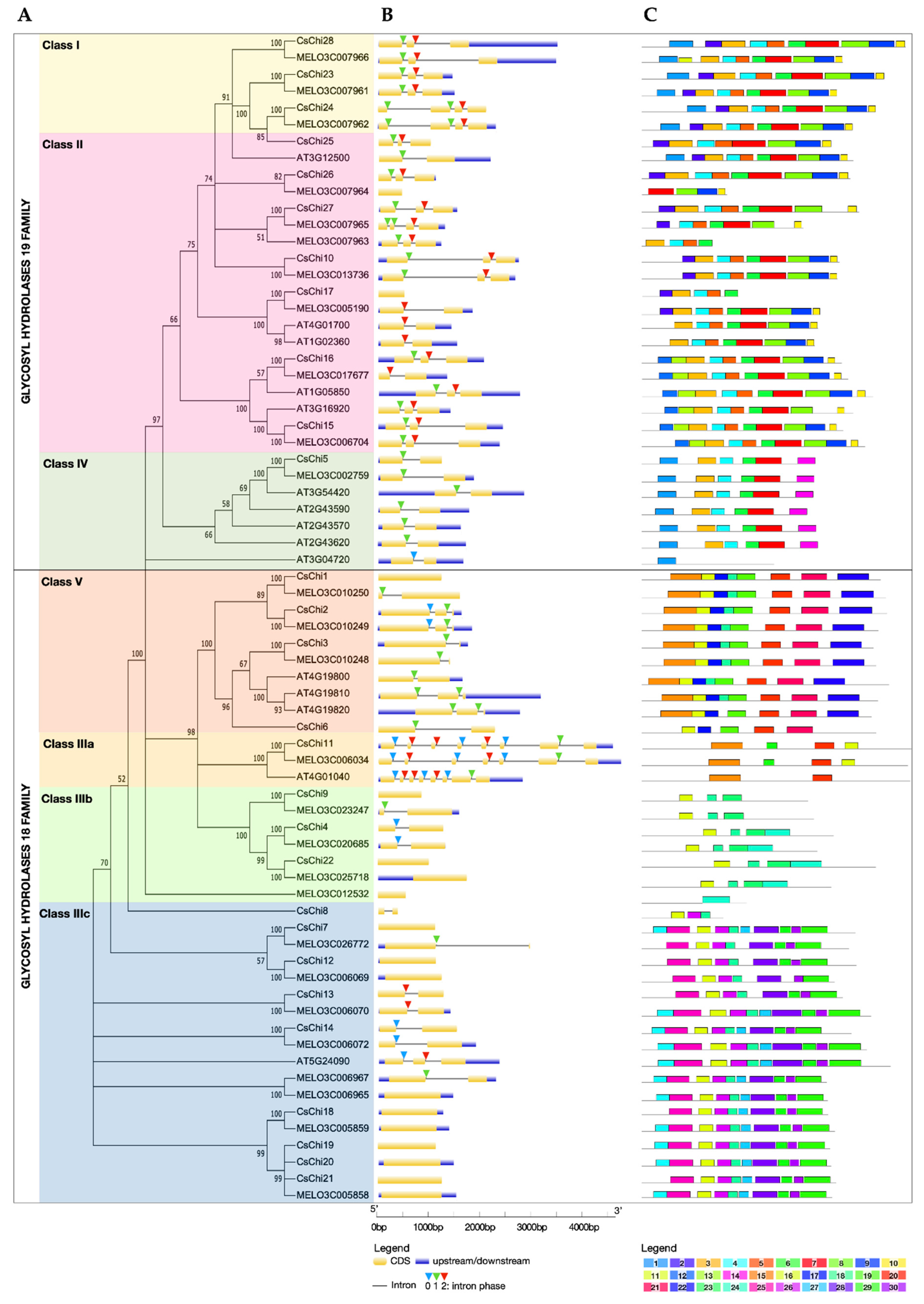
2.1. Genome-Wide Identification and Phylogenetic Analysis of Cucumber Chitinase Genes

In this study, the draft genome of C. sativus L. var. sativus cv. 9930 (ver. 2.0) [19] was used for genome-wide exploration of chitinase genes. A total of 28 putative chitinase genes were identified; each gene was annotated as CsChiXX, where Cs is the genus and species initials (Cucumis sativus) and XX is the sequential number of a gene in the genome. Biological characteristics of CsChi genes are listed in Table 1. Sequence analysis revealed that the lengths of the deduced chitinases vary from 132 (CsChi8) to 429 amino acids (CsChi11), with an average of 307 amino acids. The predicted molecular weights (MW) and the theoretical isoelectric points (pI) range from 14.80 kDa (CsChi8) to 48.53 kDa (CsChi11) and from 4.6 (CsChi9) to 9.53 (CsChi18), respectively. Of the 28 chitinases, 21 were predicted to localized in the secretory pathway, with 4 localizing in the chloroplast and 3 in the nucleus.
A total of 70 full-length chitinases sequences from C. sativus, C. melo and Arabidopsis thaliana (http://cucurbitgenomics.org; https://www.arabidopsis.org) were used to construct a neighbor-joining phylogenetic tree (Figure 1A). On the basic of phylogeny and sequence homology, all 70 putative chitinase genes were grouped into two families, one belonging to GH19: class I, class II, class IV, and the other belonging to GH18: Class III and class V. Chitinases within the GH19 family were grouped into three distinct clades representing class I, class II and class IV. Whereas, chitinase within the GH18 family resolved into four distinct, well-supported clades. Class V chitinases were grouped into a single clade that shares little sequence similarity with the class III proteins. Class III chitinases were divided into three clades with distinct sequences. Class IIIa clade contained homologs of the human stabilin-1 interacting chitinase-like proteins (SI-CLPs). Class IIIb clade contained narbonin and nodulin-like proteins, which are globulin protein that lacks conserved chitinase catalytic residues and enzymatic activity [4], while class IIIc clade contained acidic endochitinase-like (AMCase-like) proteins.

2.2. Gene Structure and Conserved Motifs Analyses of Chitinase Genes

To study the structural conservation and diversity of chitinase genes, the exon-intron architecture and conserved motifs distribution was investigated. Most GH18 chitinase genes contained 1 or 2 exons, except for chitinases in the Class IIIa clade, which are SI-CLPs known for their large size and many exons (Figure 1B) [35]. Whereas, most GH19 chitinase genes had 3 exons. Closely related genes share high similarity in terms of the length, number of exons/introns and intron phases, and these features are highly conserved. Only 8 of the 17 CsChi genes belonging to the GH18 family contained introns, compared to 10 of 11 CsChi genes from the GH19 family. The absence of an intron region was most prevalent for chitinases in the Class IIIc clade. Most class I and II CsChi genes had 2 introns and uniform intron phases, while members of class IV had 1 intron. In addition, some intron loss or gain occurred within several evolutionary branches.
A total of 30 distinct motifs were identified and their composition were consistent with the results from the phylogenetic tree (Figure 1C). As expected, closely related members shared a common motif composition. Motifs 3–10 were displayed in the same order in most class I and II chitinases, with variations only at the N-terminal chitin-binding domain, which is lacking in class II chitinases. Members of class IV had motifs 1, 3, 4, 6, 7 and 14, except for AT3G04720 which contained motif 1 only. Motifs 15–22 were present in class V chitinase, however CsChi6 lacked motifs 15, 18 and 22. The motifs composition of class III chitinases were divided into distinct three clades, which supports the results of the phylogenetic analyses, but also implied functional relevance. Furthermore, several distinct motifs were found in specific GH family, which supports the different origin of the GH18 and GH19 families.

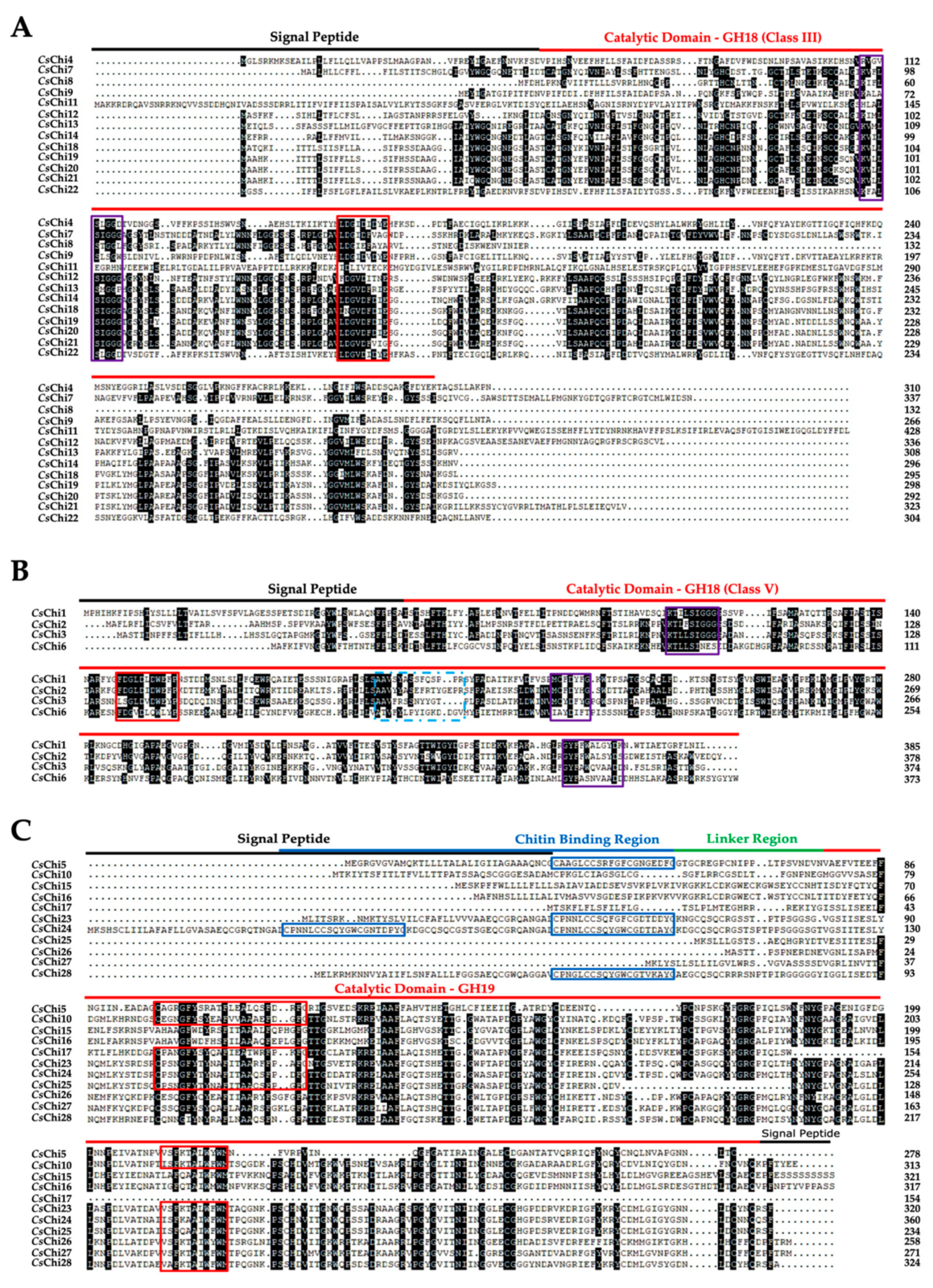
2.3. Conserved Domains and Active Site Analysis of CsChi Genes

Cucumber chitinase sequences contained conserved domain structures in accordance with previously described plant chitinases [17,36]. Analysis of the amino acid sequences reveal the presence of the GH18 catalytic domain (PF00704) with the CHITINASE_18 (PS01095) active site signature in classes III and V chitinases (Figure 2A,B). Class V chitinases also possess additional signatures and were on average longer than the members of the other classes (Figure 2B). Analysis of the amino acid sequences of classes I, II and IV reveal the presence of the GH19 catalytic domain (PF00182) with one or two active sites signatures: CHITINASE_19_1 (PS00773) and/or CHITINASE_19_2 (PS00774). Additional, class I and class IV chitinases contained a chitin-binding domain with the CHIT_BIND_I_1 (PS00026) signature at the N- terminal (Figure 2C). Interestingly, we identified a class I-like gene, CsChi24, with an unusual structure containing two chitin-binding domains. Functional site analysis using ExPASy prosite [37], predicted that only 18 of the 28 cucumber chitinase proteins contained the active sites in their catalytic domain required for chitinolytic activity. The remaining 10 genes had mismatched amino acids in their active sites and were classified as putative chitinase-like proteins (CLP).

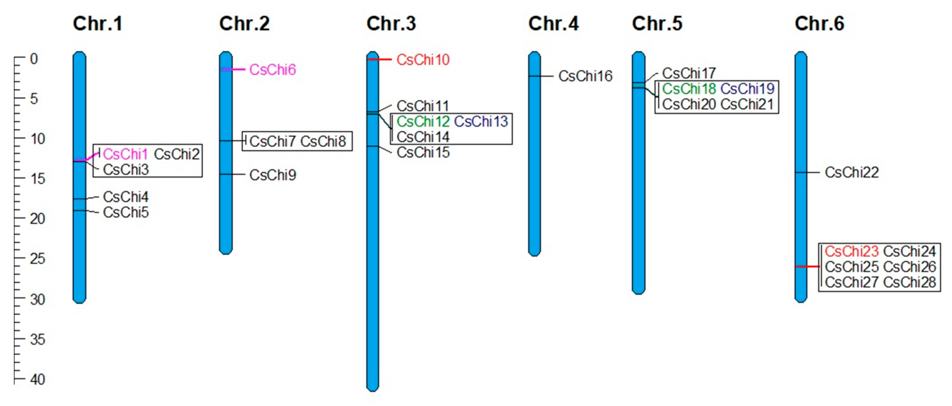
2.4. Chromosomal Distribution and Gene Duplication Analysis of CsChi Genes

To investigate the relationship between the genetic divergence and gene duplication of the cucumber chitinase gene family, their chromosomal locations were determined. The 28 CsChi genes were matched to chromosomes 1–6, with none identified on chromosome 7; the distribution of chitinase genes on each chromosome was uneven (Figure 3). Genes belonging to the same class were generally found in clusters. The C. sativus genome contained 25 genes that were orthologous to 7 Arabidopsis chitinases, indicating that most cucumber chitinases were duplicates (Figure S1). These duplicated genes were mainly found within tandemly arrayed gene clusters. Specifically, 5 clusters containing 20 cucumber chitinase genes belonging to classes I, II, III, and V were orthologous to just 3 Arabidopsis genes: AtChiA (AT5G24090), AtChiB (AT3G12500), and AtChiC (AT4G19800) (Table S1). Furthermore, 4 pairs of paralogous chitinase genes (CsChi1/CsChi6, CsChi12/CsChi18, CsChi13/CsChi19 and CsChi10/CsChi23) were located in region of segmental duplication (Figure 3).

2.5. Cis-Regulatory Elements in the Promoter of CsChi Genes

Chitinase genes have been reported to be transcriptionally regulated by several phytohormones and pathogen attack [18,38]. To elucidate the possible regulation mechanisms of CsChi genes, the cis-regulatory elements in the promoter sequence were predicted using PlantCARE [39] and PlantPAN 2.0 [40]. Twenty types of hormone and stress related cis-regulatory elements were detected (Table S2): 10 hormone- responsive elements (ABRE, ABRE3a, ABRE4, AuxRR-core, ERE, GARE-motif, P-box, TCA-element, TGACG-motif/AS-1, and TGA-element) and 10 stress- responsive elements (DRE1, HD-ZIP I, HD-ZIP III, LTR, MBS, STRE, TC-rich repeats, VOZ1, W box, and WUN-motif). Twenty-two cucumber chitinase genes contained the hormone responsive ERE element (ethylene responsive), the ABRE element (abscisic acid responsive) was detected in 20, and the TCA-element (salicylic acid responsive) in 15. In addition, the stress responsive elements W-box (wounding and pathogen responsive) and STRE (stress responsive) was predicted in 17 chitinases each and the WUN-motif (wound responsive) in 16.

2.6. Spatial Expression Profiles of CsChi Genes

To gain insight into the functions of CsChi genes, RNA-seq data (NCBI accession number: PRJNA80169) from C. sativus L. var. sativus cv. 9930 were used to quantify the expression levels of chitinase genes in various tissues (leaf, stem, female flower, male flower, ovary, root, and tendril) [34]. The expression profiles of all 28 putative CsChi genes were visualized using heat maps (Figure 4). Four of the 28 genes were highly expressed in all tissues (CsChi10/11/15/16). Most chitinase genes exhibited consistent expression levels across all tissues with some genes not expressed or only weakly expressed in female flowers, ovaries, and stems. In contrast, several chitinase genes exhibited higher expression levels in their roots compared with the other tissues (CsChi2/15/10/20/23/27/28), suggesting that they may play a role against soil-borne pathogens.

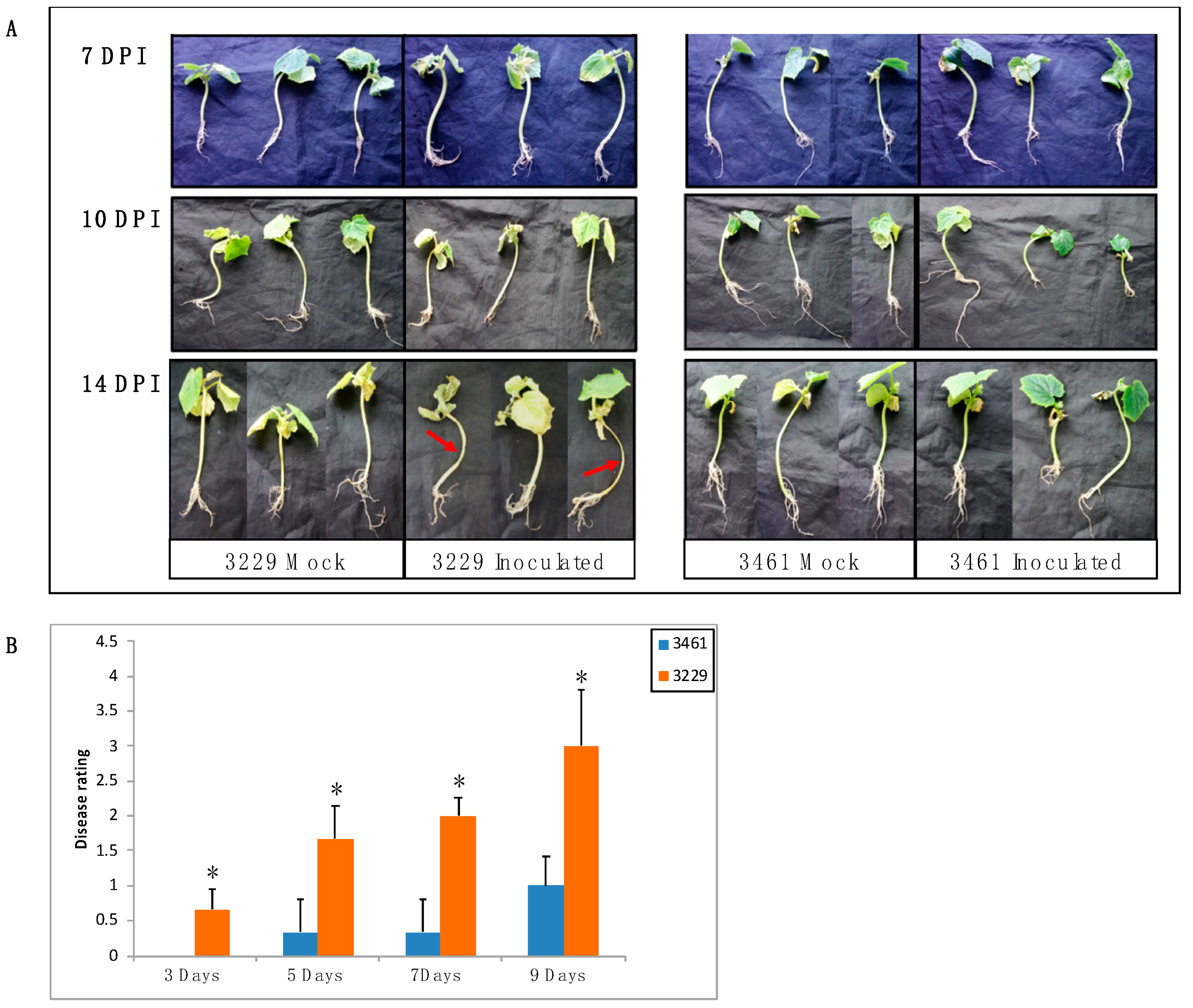
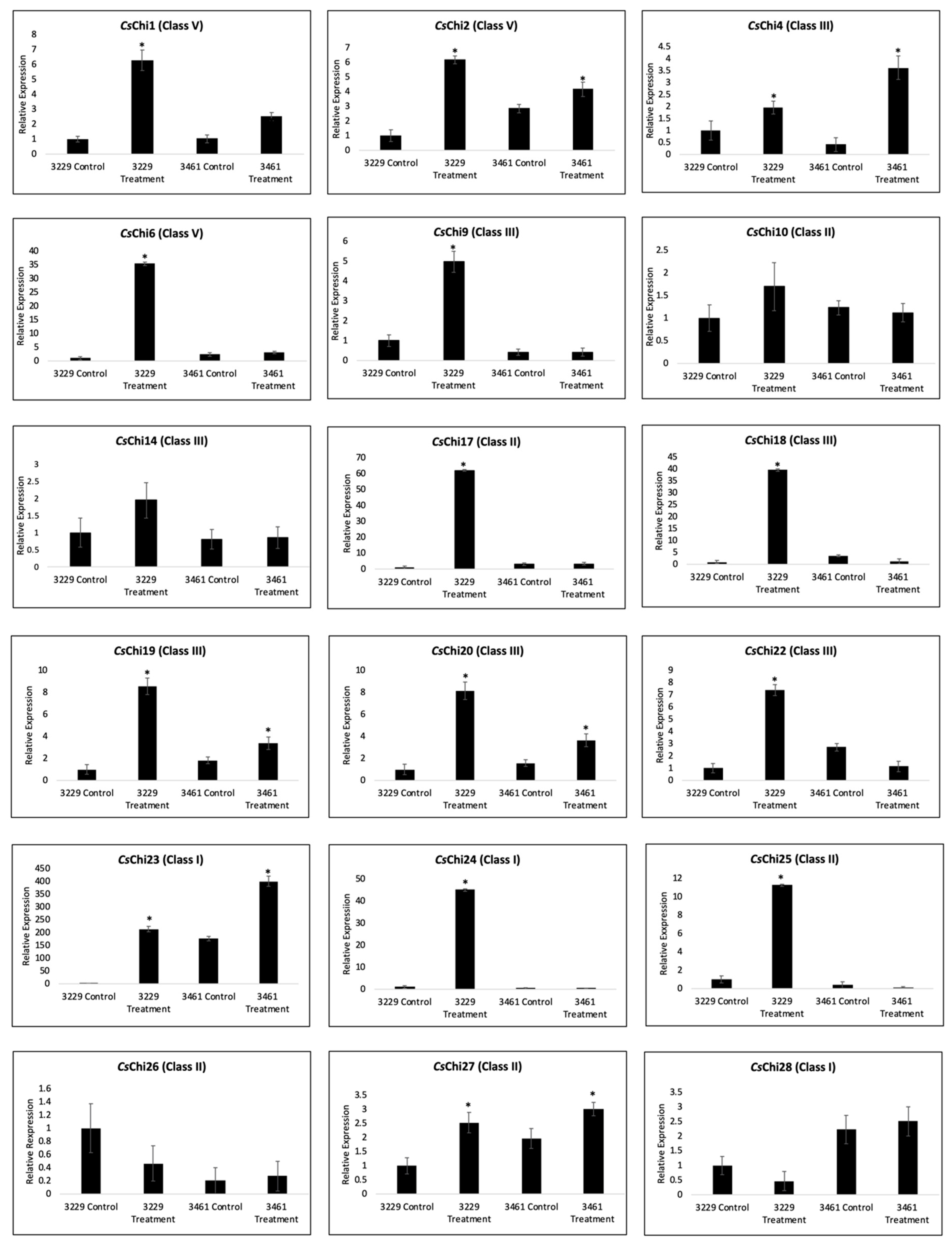
2.7. Expression Analysis of CsChi Genes in Response to F. oxysporum

The expression patterns of chitinase genes have been shown to vary between resistant and susceptible plants in response to fungal infection [38,41]. To test whether the expression of CsChi genes respond to F. oxysporum infection and to gain further insight into the differential response of resistant and susceptible lines, we selected 18 putative enzymatically active CsChi genes for expression analysis by qRT-PCR. Two cucumber inbred lines (3461 = resistant; 3229 = susceptible), with different level of resistance to F. oxysporum were used (Figure 5A). Two weeks after inoculation the fusarium wilt disease symptoms was assessed (Figure 5B). No major wilt symptoms were observed in the resistant line 3461, but severe wilt symptoms were detected in the stems and leaves of susceptible line 3229.
The expression pattern of each CsChi gene was checked 3dpi, as it has been reported that genes encoding several PR proteins showed a peak in expression 1-3dpi when distressed with hemibiotrophic pathogens [42,43,44,45]. In line 3229, expression analysis revealed that most chitinase genes responded significantly to fungal infection (Figure 6). Specifically, 14 genes were significantly up-regulated after infection, while 2 genes (CsChi26 and CsChi28) were down-regulated. In line 3461, the expression levels varied and 7 genes were up-regulated following fungal infection, 2 genes were down-regulated and no significant differences were observed in 9 genes. The expression of class I chitinase genes (CsChi23 and CsChi28) were generally higher in resistant lines. Interestingly, CsChi23 was constitutively expressed about 200-folds higher in line 3461 compared to line 3229 in mock infected plants.

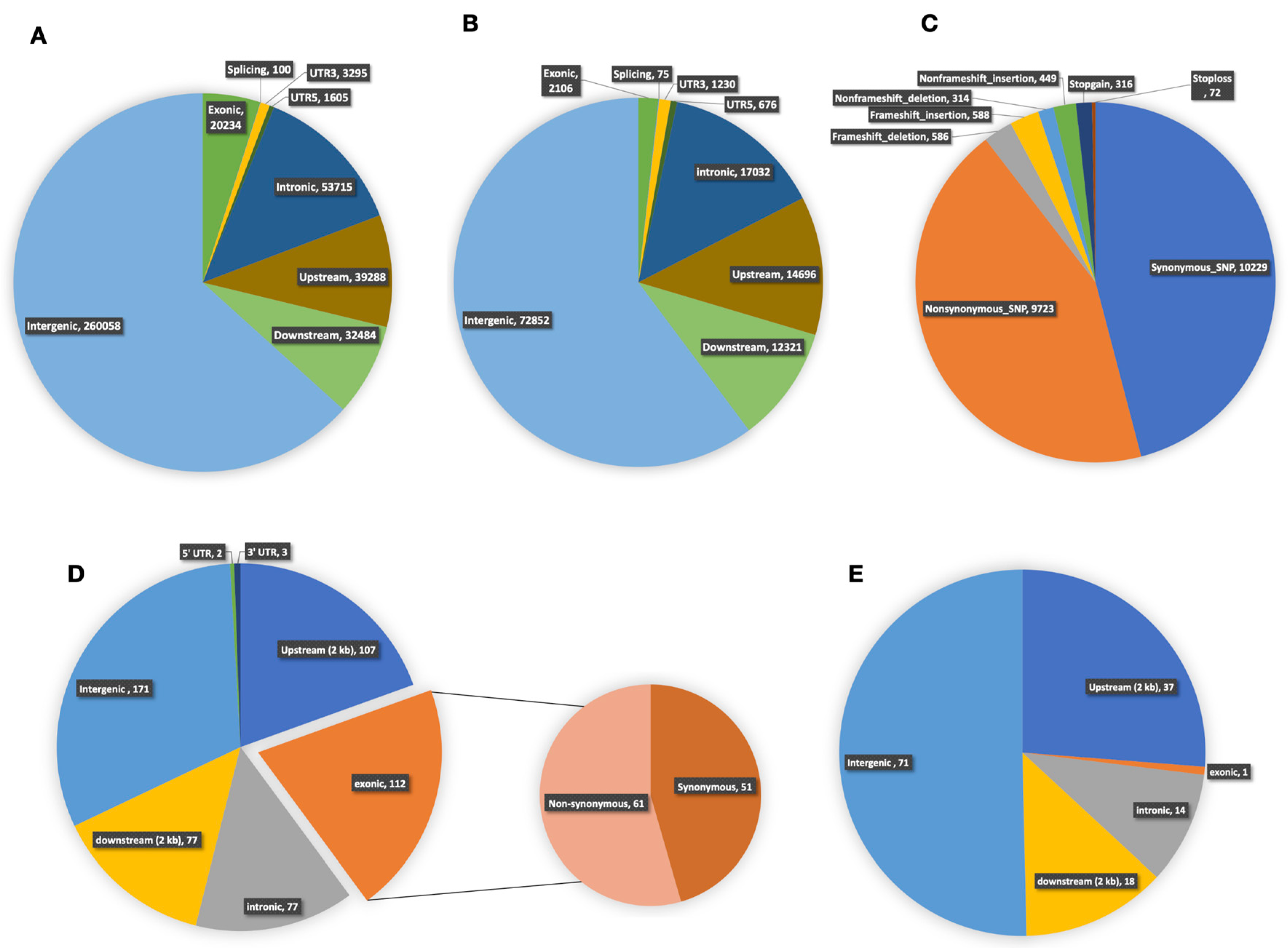
2.8. Whole-Genome Re-Sequencing and Variation Analysis between Lines 3229 and 3461

Whole-genome re-sequencing of cucumber inbred lines 3229 and 3461 generated an average of 99,446,500 and 80,794,000 raw sequence reads, respectively. About 93% of these were clean reads, with about 91% of the bases being high-quality bases (quality score ≧ Q30). Overall, an average of 72,481,500 (78.95%) reads from line 3229 and 64,168,00 (84.94%) from line 3461 were uniquely mapped to the C. sativus L. (9930) reference genome at an average read depth of 31.7X and 31.35X, respectively (Table S3). After single nucleotide polymorphism (SNP) and (insertion/deletion) InDel calling, 410,779 SNPs and 120,988 InDels were detected directly between both lines (Tables S4,S5). Overall, 81% SNPs and 83% InDels were distributed in the intergenic regions (intergenic, upstream, and downstream), while about 19% SNPs and 17% InDels were observed in the genic regions (exonic, intronic and UTRs (Figure 7A,B). Furthermore, several large- effect variations that may have severe impacts on the integrity of encoded products and gene functions, such as, SNPs and InDels distributed in the initiation/termination codon and the InDels which caused frameshift were detected (Figure 7C).

2.9. DNA Polymorphisms in Region of Chitinase Genes

To understand the diverse expression patterns of chitinase genes observed between lines 3229 and 3461, we annotated all genetic variations found in the intergenic and genic regions of putative chitinase genes (Table S6). A total of 690 genetic variations were identified between both lines, with about 80% (549) being SNPs and 20% (141) were InDels (Table S7). In general, SNPs and InDels occurred more frequently in noncoding regions than in the coding regions. Of the total SNPs discovered, ∼35% were in genic regions with ∼20% being in exonic regions, while ∼11% of InDels were found in the genic regions with >1% being in exonic (Figure 7D,E). Interestingly, about 56% of all the genetic variations in chitinase genes was found in a gene cluster on chromosome 6 (CsChi23/24/25/26/27/28) which contains classes I and II chitinases (Table S7). Furthermore, several large-effect variants in line 3229, including a splicing mutation and a frameshift-insertion was detected in CsChi25 (Chr6: 26090784/ 26090779), while a stop-gain mutation was detected in CsChi19 (Chr5: 3712267; Table S6).

3. Discussion

Due to the constant threat of pathogens, plants have evolved sophisticated detection and response systems that decipher pathogen signals and induce appropriate defenses [24]. Inducible defense responses involve a variety of defense mechanisms including the production and secretion of antimicrobial compounds such as PR proteins and phytoalexins, cell wall modification, generation of reactive oxygen species, and programmed cell death or hypersensitive response at the site of infection to limit pathogen progression [24,25,26]. Chitinases, which comprise four of the PR protein families (PR-3/4/8/11), can inhibit fungal growth by catalyzing the hydrolysis of chitin, the main component of the pathogen cell wall, and are therefore good targets for defense response studies [18,31]. Most research on the identification and analysis of chitinase gene family have focused on A. thaliana, Nicotiana benthamiana, Oryza sativa and Zea mays, whereas studies are limited on non-model plants like C. sativus [46]. A total of 28 putative chitinase genes were identified in the cucumber genome. Based on their protein sequences, all cucumber chitinases are endochitinases (EC 3.2.1.14) and are classified into 5 different classes (I–V). Here, we undertook detailed bioinformatic and expression analysis of the cucumber chitinases genes to understand their roles in cucumber- F. oxysporum interaction
Due to the great diversity of enzymes that hydrolyze or bind chitin, chitinases have been proposed as good examples of gene evolution related to gene duplication [47]. Chromosomal localization and gene duplication studies indicated that the cucumber genome contains 5 tandemly arrayed gene clusters with 20 chitinase genes orthologous to 3 Arabidopsis genes (Figure S1). Therefore, >71% of cucumber chitinase genes originated from tandem duplication (Table S1). Over the process of biological evolution, duplicated genes may acquire new structures and functions, resulting in chitinase with diverse binding, substrate, and pathogen specificity that are uniquely expressed in different tissues or under different stresses [48,49]. Interestingly, we identified a novel class I-like chitinase gene (CsChi24) that had an unusual structure containing two chitin-binding domains (Figure 2C). CsChi24 shared a close relationship with CsChi23 (class I) and CsChi25 (class II), which contained one and zero chitin-binding domain, respectively. Hardt and Laine [50], indicated that mutations of active site residues in the chitin-binding domain, caused by unequal crossing-over, can have profound effects on the expression and substrate specificity of chitinase genes. We hypothesize that structural differences in the chitin-binding region of CsChi24 may affect its binding specificity leading to functional diversification. Therefore, functional differentiation may be the mechanism driving the evolution and expansion of the cucumber chitinase gene families.
Gene structure and conserved motif distribution can provide further evidences for the evolutionary relationships of gene families. Intron positions are highly conserved in orthologous genes across species, and intron gain and loss can increase the complexity of gene organization [51]. Most cucumber chitinase genes within the same class had uniform exon-intron organization and intron phases. However, some intron loss or gain occurred within several evolutionary branches (Figure 1B). The exon-intron architecture of chitinase genes is also known to mediate the stress responses. Generally, the structures of stress-related genes contain few introns [51]. In this study, 25 of the identified 28 CsChi genes had two or fewer introns (Figure 1B). This adds to the conception that longer and several introns can prolong transcription, whereas fewer introns are present in genes that are rapidly regulated during stress [51]. The absence of an intron region was most prevalent in class IIIb and class IIIc chitinases gene. Similar chitinase genes structures were observed in B. rapa and found in other stress-related gene families [52,53]. Moreover, a similar motif composition was found in each group, suggesting functional relevance, while diverse motif composition implies functional diversification among different groups (Figure 1C).
Although plants have no chitin in their cell walls, they produce many forms of chitinases, including cellular and secretory chitinases, that play key roles in multiple physiological processes. Most cucumber chitinases were predicted to localize in the apoplast and involved in the secretory pathway (Table 1). The plant apoplast is integral for intercellular signaling, transport and plant-pathogen interactions [54]. Apoplastic chitinases are usually induced immediately after pathogen infection and can directly block the growth of the hyphae at the intercellular space [55,56]. Also, apoplastic chitinases are known to act directly or indirectly with pathogen elicitors, which triggers downstream defense pathways [57]. These results indicate that apoplastic CsChi genes may participate in chitinase-pathogen interaction.
Chitinases are transcriptionally regulated by several cis-regulatory elements, including hormone and stress cis-regulatory elements [18,39,58]. The hormone-responsive element ERE (ethylene-responsive) was found in the promoter region of 22 CsChi genes, indicating that the expression of most chitinase genes might be induced by ethylene (Table S2). Several studies also identified ethylene as a key hormone involved in chitinase gene induction [59,60]. Other hormone responsive elements (ABRE, TCA, and TGACG-motif) predicted to regulate transcription of chitinase genes [61,62,63] were also identified in the promoter of most CsChi genes (Table S2). Likewise, several known stress responsive elements (W-box, STRE, and WUN-motif) predicted to mediate pathogen- and/or elicitor-inducible transcription of chitinases [39,58,64,65,66] were also identified. These results indicate that CsChi genes may be transcriptionally regulated by hormone and stress cis-regulatory elements and may be induced by pathogen elicitors.
Cucumber chitinase gene showed differential expression patterns in the seven different cucumber tissues. Several genes were highly expressed in all tissues, these include developmentally-regulated CLPs: CsChi16 (CLP-1), CsChi15 (CLP-2) and CsChi11 (SI-CLP), and CsChi10, an ortholog of AT4G01700 known to be constitutively expressed in all tissues in Arabidopsis [18,55]. Chitinase genes were reported to be highly expressed in roots and flowers in several plant species [18,67]. This study showed similar expression patterns, with several chitinase genes being highly expressed in the roots and male flowers (Figure 5). In the roots, GH19 CsChi genes exhibited higher expression levels indicating that they may be involved in plant defense against soil-borne pathogens such as F. oxysporum. Samac et al. [55] also reported that the GH19 family of chitinases in Arabidopsis were predominantly expressed in roots and may play a role in stress response.
PR proteins, including chitinases are either silent or constitutively expressed at low levels in plants during the absence of pathogens, but are significantly induced in response to pathogens infections [68,69,70,71,72]. In agreement with previous reports, the expressions of CsChi genes showed both constitutive and induced expression patterns. Here, at least 14 CsChi genes were induced after F. oxysporum inoculation (Figure 6). Class I chitinase genes were highly expressed in resistant line 3461, whereas class III, IV and V chitinases were upregulated in line 3229. Similar gene expressions were also reported by Adhikari et al. [43] and Xin et al. [73] in wheat host following Mycosphaerella graminicola and powdery mildew infection. The chitinase gene family has many diverse functions that are regulated by numerous stimuli such as abiotic and biotic stresses as well as various phytohormones. Polymorphisms in chitinase genes and regulatory regions are also known to affect chitinase activity and expression [55,64,65]. We suspect that the varying expression patterns observed between lines 3229 and 3461 may be related to the functional diversity of cucumber chitinase genes and evolutionary differences of both lines. Several class I chitinases (CsChi23 and CsChi28) were induced in resistant line 3461. These are PR-3 proteins that are known to deter fungal pathogens by acting on chitin in their cell walls [55]. Interestingly, many chitinase genes were not induced upon F. oxysporum inoculation in line 3461. These include chitinase isoforms: CsChi5 (class IV, a homolog of carrot EP3), and CsChi4, CsChi9 and CsChi22 (class III, 2S globulin/ Narbonin homologs) that are known for their roles in plant embryogenesis and as storage protein found in seeds [4,18]. These findings further confirm that cucumber chitinase genes have diverse functions.
Xin et al. [73] suggests that some genes with pathogen-defense functions are constitutively expressed at high levels in resistant hosts and may act as a constant barrier to pathogen attacks. In this study, the expression patterns of CsChi genes in both resistant and susceptible lines were similar under control conditions, except for CsChi23, which was constitutively expressed about 200-folds higher in line 3461 (Figure 6). Takenaka et al. [74] reported that Arabidopsis class I chitinase gene AtchiB, a homolog of CsChi23, was also constitutively expressed at a high level. Class I chitinases have been extensively studied for its defensive roles and are known to inhibit hyphal growth of several fungal species and degrades chitin more rapidly than other classes of chitinase [75,76,77,78]. The constitutive expression of rice class I chitinase, OsChia1b, in several commercial crops also resulted in enhanced resistance to fungal diseases [79,80]. After F. oxysporum infection, CsChi23 was highly upregulated in both lines. This indicates that CsChi23 expression is induced by F. oxysporum. We speculate that the high constitutive expression of CsChi23 in resistant cucumber lines may play a critical role in building a basal defense and a rapid immune response against fungal attacks. However, further research is needed to validate the antifungal role of CsChi23 in C. sativus-F. oxysporum interaction.
The location and type of DNA polymorphism have a major influence on gene expression [81]. A large proportion of DNA polymorphisms are in the intronic and intergenic regions, which hinders their functional evaluation. The promoter region located upstream of the initiation site encompasses cis-regulatory sites that play an important role in regulating gene expression. Polymorphisms in the promoter region may alter the transcription factor binding sites, thus affecting gene expression [82]. In this study, about 84% of the SNPs and InDels observed in putative CsChi genes between lines 3229 and 3461 occurred in the non-coding regions, with about 21% occurring in the upstream (2 kb) promoter region (Figure 7D,E). Chorley et al. [83] suggests that in some cases, a natural binding site created or abolished by a regulatory polymorphism can account for observed differences in gene expression. We speculate that the diverse expression of CsChi genes between lines 3229 and 3461 may be caused by such polymorphisms in the promoter region. Interestingly, about 56% of all genetic variations in chitinase genes were found in a gene cluster on chromosome 6 (CsChi23/24/25/26/27/28) which contains classes I and II chitinases (Table S7). These polymorphisms may have a major effect on gene structures, functions, and expression. About 20% of the SNPs occurred in the coding region and several large-effect variants were detected which may affect chitinase activity. Furthermore, a large proportion of polymorphisms was found in the promoter region of CsChi23. To validate these polymorphisms, the putative CsChi23 promoter fragment (from 2 kb upstream of the ATG initiation codon) of line 3461 and 3229 was cloned and sequenced (Figure S2). Sequence analysis revealed multiple sequence differences, including 21 SNPs and 3 InDels, that abolish the HD-Zip III transcription factor motif, the P-box motif (gibberellin-responsive element) and the TGA motif (early auxin-responsive element) in line 3229. These polymorphisms might explain the constitutive expression of CsChi23 in line 3461. However, further promoter analysis of CsChi23 between lines 3229 and 3461 is needed to determine the effect of DNA polymorphisms.

4. Materials and Methods

4.1. Sequence Acquisition and Identification of Cucumber Chitinase Genes

To identify chitinase genes, amino acid sequences from Arabidopsis and melon chitinases were BLASTP (https://blast.ncbi.nlm.nih.gov/Blast.cgi) queried against cucumber. NCBI-CDD [84] and HMMER web server: 2018 update [85] were used to analyze the conserved domains of all non-redundant sequences. Hidden Markov models (HMMs) profiles of the chitinase catalytic domains from GH18 (PF00704) and GH19 (PF00182) were obtained from the Pfam database (Pfam 32.0) [86]. EMBL-EBI InterPro [87] was used to validate the identified genes. ExPASy prosite [37] was used to analyze all putative chitinase genes for the presence of the catalytic residues required for chitinolytic activity. The gene and coding sequence (CDS) (bp), number of amino acids and chromosome location of cucumber chitinase genes were obtained from Cucurbit Genomics Database (http://cucurbitgenomics.org). The molecular weight (kDa) and isoelectric point (pI) of each chitinase protein were analyzed using ExPASy’s Compute pI/Mw tool [88]. Subcellular localization was predicted using BaCello [89] and signal peptide cleavage sites were predicted using SignalP 4.1 [90].

4.2. Phylogenetic Tree, Gene Structures and Conserved Motifs Analyses of Chitinase Genes

To study evolutionary relationships, full-length amino acid sequences of 70 chitinase proteins, from C. sativus, C. melo and A. thaliana were first aligned using ClustalW [91] with default settings to construct an unrooted neighbor-joining (NJ) phylogenetic tree with Poisson correction using MEGA X [92]. Bootstrap analysis was employed using 1000 replicates. The pair-wise gap deletion mode was used to ensure that the more divergent C-terminal domains could contribute to the topology of the NJ tree. The CDS and corresponding genomic sequence of each chitinase genes were used to predict the exon–intron structures using GSDS v2.0 [93]. The MEME software v. 5.0.5 [94] was used to identify conserved motifs in each chitinase protein sequences according to the following parameters command: -protein -oc -nostatus -mod zoops -nmotifs 30 -minw 6 -maxw 50 -objfun classic. Multiple sequence alignment of cucumber chitinases amino acid sequences from was conducted using DNAMAN software v6.0 (Lynnon BioSoft, Quebec, Canada).

4.3. Chromosomal Location and Identification of Homologous A. thaliana Members

All identified cucumber chitinase genes were mapped to six chromosomes and visualized by Mapchart v. 2.32 [95] using base pair start position information from the cucurbit genomics database. Clusters of chitinase genes were determined based on previously defined criteria by Christie et al. [96]. Duplications between the CsChi genes were identified using the cucurbit genomics database. To identify orthologous cucumber chitinase genes in Arabidopsis, a phylogeny-based method was used and a BLAST-based reciprocal best hit method was used to verify the putative orthologous genes. The Circoletto tool [97] was used to plot sequence similarity using the pairwise alignment of cucumber and Arabidopsis chitinases.

4.4. Cis-Regulatory Elements Analysis

To predict the cis-regulatory elements of each cucumber chitinase genes, we obtained the nucleotide sequences of the promoter regions (1.5 kb upstream of the initiation codon) from the draft genome of C. sativus L. var. sativus cv. 9930 (ver. 2.0). PlantCARE [39] and PlantPAN 2.0 [40] were used to predict cis-regulatory elements.

4.5. Gene Expression Analysis

To determine the expression patterns of cucumber chitinase genes in various tissues (leaf, stem, female flower, male flower, ovary, root, and tendril) we obtained RNA-Seq data (NCBI accession number: PRJNA80169) [34]. Tissues were obtained from C. sativus var. sativus line 9930. The sequencing data is obtainable with the accession number SRA046916 in the Sequence Read Archive (SRA) at NCBI. The absolute transcript abundance values obtained for all chitinase genes were derived based on the alignments using Bowtie 2 [98] and HISAT2 [99] and then normalized using EXPANDER7 [100] to RPKM (reads per kilobase of exon per million mapped fragments).

4.6. Plant Material and Fusarium Infection Assay

Two cucumber inbred lines, 3461 and 3229, which exhibited resistance and susceptibility to F. oxysporum, respectively, were used in the present study. Seeds were surface sterilized and sown in autoclaved soil (50/50 nutritive soil/vermiculite). All plants were grown at 28 °C/22 °C under 16-h light/8-h dark conditions. The fungus, F. oxysporum f. sp. cucumerinum race 3, obtained from China Agricultural University, Beijing, P. R. China, was used as inoculating pathogen. For infection assays, the inoculum was prepared as described by Wang et al. [22], with some modifications. Cucumber seedlings at two-true leaves stage were root dip-inoculated with a conidial suspension (5 × 10⁵ spores/mL), or mock-inoculated with sterile water, after which seedlings were transplanted into 250mL plastic pots containing autoclaved soil. Development of disease symptoms was followed over time. Disease severity for each plant was scored on a scale of 0–4, only plants with an average score of 1 were considered resistant. The experiment included 6 plants per replicate with two replications. Samples were collected at 3 days post inoculation (dpi) for expression analysis and the severity of the disease was assessed 14 dpi [20]. All tissues were immediately frozen in liquid nitrogen and stored at −80 °C.

4.7. RNA Extraction and Quantitative Real-Time Reverse Transcription PCR (qRT-PCR) Analysis

Total RNA was extracted using the Quick RNA plant isolation kit (Beijing Yueyang Biotechnology Ltd., Beijing, China), and cDNA was synthesized using the OneScript® cDNA Synthesis kit with AccuRT Genomic DNA Removal (Applied Biological Materials Inc, Vancouver, Canada). qRT-PCR was performed in 96-well plates with an Applied Biosystems 7500 real-time PCR system (Applied Biosystems, Foster city, CA, USA) using the One-Step BrightGreen qRT-PCR-Low ROX kit (Applied Biological Materials Inc, Vancouver, Canada). Primers were designed using Primer3Plus, and primer specificity was evaluated with NCBI primer-BLAST against the cucumber genome. The specificity of the amplified product was confirmed by a melting curve. Three biological replicates and 3 technical replicates were performed for each combination of cDNA samples and primer pairs. The cucumber β-actin served as the internal control gene [101]. The relative expression of target genes was analyzed by comparative CT method [102]. The cucumber chitinase qRT-PCR primers are listed in Table S8.

4.8. DNA Extraction, Whole-Genome Resequencing and Variation Analysis

Genomic DNA from young leaves was extracted using the Plant Genomic DNA Rapid Extraction kit (Aidlab Biotechnologies, Beijing, China). The concentration and quality of DNA were determined using a NanoDrop2000 Spectrophotometer (Thermo Fisher Scientific, Waltham, Massachusetts, USA). DNA libraries for Illumina sequencing were constructed according to the manufacturer’s specifications (Illumina, San Diego, CA, USA). Paired-end sequencing libraries with an insert size of 300-400 bp were sequenced on an Illumina HiSeq 2500 sequencer by a commercial service (ORI-GENE, Beijing, China). Paired-end resequencing reads from lines 3229 and 3461 were aligned against the C. sativus L. (9930) reference genome using the MEM algorithm of Burrows–Wheeler Aligner [103] (BWA v0.7.5a-r405), after trimming of low-quality reads by using Trimmomatic [104]. Picard (release 2.0.1, https://broadinstitute.github.io/picard/) was used to eliminate PCR duplicates and to realign InDel regions. SNP and InDel calling for each sample were performed using Genome Analysis Toolkit (GATK) v4.1.2 (https://software.broadinstitute.org/gatk/). The variation detection followed the best practice workflow recommended by GATK [105]. Initially, variants from lines 3229 and 3461 relative to the reference genome were called separately, then merged and compared to each other. Variants located in the regions of chitinase genes were then identified and analyzed.

4.9. Validation of Variations

The sequences of selected variations located in the regions of chitinase genes were used as template for primer design (Table S8). The variation regions were amplified by PCR using genomic DNA extracted from lines 3229 and 3461. The sequences were confirmed by repeated sequencing by Sanger sequencing method. The assembled sequences were aligned to the reference genome sequences to validate the variations in all the selected regions using ClustalW [91].

5. Conclusions

In summary, we performed a complete genome-wide identification and analysis of the chitinase gene family in cucumber and generated expression data of both resistant and susceptible lines after F. oxysporum inoculation. Twenty-eight chitinases were identified in the C. sativus genome and their key structural features were examined. This provides valuable information for further elucidation of the evolution and expansion of chitinase gene family. The high constitutive expression of a class I chitinase in resistant line 3461, CsChi23, may play a key role in enhancing fungal resistance. Moreover, whole-genome re-sequencing of resistant and susceptible lines provided clues for the diverse expression patterns observed. The information obtained from this study provides new insights into potentials roles of cucumber chitinases in plant defense and gives valuable gene resources for improving F. oxysporum resistance in future cucumber-breeding programs.

Supplementary Materials

Supplementary materials can be found at https://www.mdpi.com/1422-0067/20/21/5309/s1.

Author Contributions

Conceptualization, E.S.B., H.Z. and X.L.; validation, E.S.B., K.B., L.B., Z.F. and W.L.; formal analysis, E.S.B.; investigation, E.S.B.; resources, E.S.B., N.Z., N.S., C.Q., X.Z. and L.W.; writing—original draft preparation, E.S.B.; visualization, E.S.B.; supervision, H.R. and X.L.; project administration, H.R.; funding acquisition, H.R., X.L.

Funding

This research was funded by the National Key Research and Development Program of China (Grant number: 2018YFD1000800), Beijing Municipal Natural Science Foundation (Grant number: 6184043), the Construction of Beijing Science and Technology Innovation and Service Capacity in Top Subjects (Grant number: CEFF-PXM2019_014207_000032) and the Project of Beijing Agricultural Innovation Consortium (Grant number: BAIC01-2016).

Acknowledgments

We thank the members of Ren’s lab for helpful discussions for this research.

Conflicts of Interest

The authors declare no conflict of interest.

Abbreviations

CDSCoding sequence
CLPChitinase-like protein
GHGlycosyl Hydrolases
InDelInsertion and deletion
MWMolecular weight
pIIsoelectric point
PRPathogenesis-related
qRT-PCRQuantitative real-time reverse transcription PCR
SI-CLPsStabilin-1 interacting chitinase-like proteins
SNPSingle-nucleotide polymorphism
UTRUntranslated region

References

  1. Renner, T.; Specht, C.D. Molecular Evolution and Functional Evolution of Class I Chitinases for Plant Carnivory in the Caryophyllales. Mol. Biol. Evol. 2012, 29, 2971–2985. [Google Scholar] [CrossRef] [PubMed]
  2. Lombard, V.; Golaconda Ramulu, H.; Drula, E.; Coutinho, P.M.; Henrissat, B. The Carbohydrate-Active Enzymes Database (CAZy) in 2013. Nucleic Acids Res. 2014, 42, D490–D495. [Google Scholar] [CrossRef] [PubMed]
  3. Jain, K.K.; Kumar, A.; Shankar, A.; Pandey, D.; Chaudhary, B.; Sharma, K.K. De Novo Transcriptome Assembly and Protein Profiling of Copper-Induced Lignocellulolytic Fungus Ganoderma Lucidum MDU-7 Reveals Genes Involved in Lignocellulose Degradation and Terpenoid Biosynthetic Pathways. Genomics 2019. [Google Scholar] [CrossRef] [PubMed]
  4. Tyler, L.; Bragg, J.N.; Wu, J.; Yang, X.; Tuskan, G.A.; Vogel, J.P. Annotation and Comparative Analysis of the Glycoside Hydrolase Genes in Brachypodium Distachyon. BMC Genom. 2010, 11, 600. [Google Scholar] [CrossRef]
  5. Wang, S.; Ye, X.; Chen, J.; Rao, P. A Novel Chitinase Isolated from Vicia Faba and Its Antifungal Activity. Food Res. Int. 2012, 45, 116–122. [Google Scholar] [CrossRef]
  6. Wang, S.; Shao, B.; Fu, H.; Rao, P. Isolation of a Thermostable Legume Chitinase and Study on the Antifungal Activity. Appl. Microbiol. Biotechnol. 2009, 85, 313–321. [Google Scholar] [CrossRef]
  7. Li, C.; Li, X.; Bai, C.; Zhang, Y.; Wang, Z. A Chitinase with Antifungal Activity from Naked Oat (Avena Chinensis) Seeds. J. Food Biochem. 2019, 43, e12713. [Google Scholar] [CrossRef]
  8. Liu, J.J.; Ekramoddoullah, A.K.M.; Zamani, A. A Class IV Chitinase Is Up-Regulated by Fungal Infection and Abiotic Stresses and Associated with Slow-Canker-Growth Resistance to Cronartium Ribicola in Western White Pine (Pinus Monticola). Phytopathology 2005, 95, 284–291. [Google Scholar] [CrossRef]
  9. Xiao, Y.H.; Li, X.B.; Yang, X.Y.; Luo, M.; Hou, L.; Guo, S.H.; Luo, X.Y.; Pei, Y. Cloning and Characterization of a Balsam Pear Class I Chitinase Gene (Mcchit1) and Its Ectopic Expression Enhances Fungal Resistance in Transgenic Plants. Biosci. Biotechnol. Biochem. 2007, 71, 1211–1219. [Google Scholar] [CrossRef]
  10. Maximova, S.N.; Marelli, J.P.; Young, A.; Pishak, S.; Verica, J.A.; Guiltinan, M.J. Over-Expression of a Cacao Class I Chitinase Gene in Theobroma Cacao, L. Enhances Resistance against the Pathogen, Colletotrichum Gloeosporioides. Planta 2006, 224, 740–749. [Google Scholar] [CrossRef]
  11. Dehestani, A.; Kazemitabar, K.; Ahmadian, G.; Jelodar, N.B.; Salmanian, A.H.; Seyedi, M.; Rahimian, H.; Ghasemi, S. Chitinolytic and Antifungal Activity of a Bacillus Pumilus Chitinase Expressed in Arabidopsis. Biotechnol. Lett. 2010, 32, 539–546. [Google Scholar] [CrossRef] [PubMed]
  12. Huang, Y.; Liu, H.; Jia, Z.; Fang, Q.; Luo, K. Combined Expression of Antimicrobial Genes (Bbchit1 and LJAMP2) in Transgenic Poplar Enhances Resistance to Fungal Pathogens. Tree Physiol. 2012, 32, 1313–1320. [Google Scholar] [CrossRef] [PubMed]
  13. Sharma, N.; Sharma, K.P.; Gaur, R.K.; Gupta, V.K. Role of Chitinase in Plant Defense. Asian J. Biochem. 2011, 6, 29–37. [Google Scholar] [CrossRef]
  14. Ganesan, M.; Bhanumathi, P.; Ganesh Kumari, K.; Lakshmi Prabha, A.; Song, P.S.; Jayabalan, N. Transgenic Indian Cotton (Gossypium Hirsutum) Harboring Rice Chitinase Gene (Chi II) Confers Resistance to Two Fungal Pathogens. Am. J. Biochem. Biotechnol. 2009, 5, 63–74. [Google Scholar] [CrossRef]
  15. Xu, J.; Xu, X.; Tian, L.; Wang, G.; Zhang, X.; Wang, X.; Guo, W. Discovery and Identification of Candidate Genes from the Chitinase Gene Family for Verticillium Dahliae Resistance in Cotton. Sci. Rep. 2016, 6. [Google Scholar] [CrossRef]
  16. Arakane, Y.; Muthukrishnan, S. Insect Chitinase and Chitinase-like Proteins. Cell. Mol. Life Sci. 2010, 67, 201–216. [Google Scholar] [CrossRef]
  17. Passarinho, P.A.; de Vries, S.C. Arabidopsis Chitinases: A Genomic Survey. Arab. B. 2002, 1, e0023. [Google Scholar] [CrossRef]
  18. Grover, A. Plant Chitinases: Genetic Diversity and Physiological Roles. CRC. Crit. Rev. Plant. Sci. 2012, 31, 57–73. [Google Scholar] [CrossRef]
  19. Huang, S.; Li, R.; Zhang, Z.; Li, L.; Gu, X.; Fan, W.; Lucas, W.J.; Wang, X.; Xie, B.; Ni, P.; et al. The Genome of the Cucumber, Cucumis Sativus L. Nat. Genet. 2009, 41, 1275–1281. [Google Scholar] [CrossRef]
  20. Zhang, D.; Meng, K.X.; Hao, Y.H.; Fan, H.Y.; Cui, N.; Wang, S.S.; Song, T.F. Comparative Proteomic Analysis of Cucumber Roots Infected by Fusarium Oxysporum f. Sp. Cucumerium Owen. Physiol. Mol. Plant. Pathol. 2016, 96, 77–84. [Google Scholar] [CrossRef]
  21. Hu, J.L.; Lin, X.G.; Wang, J.H.; Shen, W.S.; Wu, S.; Peng, S.P.; Mao, T.T. Arbuscular Mycorrhizal Fungal Inoculation Enhances Suppression of Cucumber Fusarium Wilt in Greenhouse Soils. Pedosphere 2010, 20, 586–593. [Google Scholar] [CrossRef]
  22. Wang, M.; Ling, N.; Dong, X.; Zhu, Y.; Shen, Q.; Guo, S. Thermographic Visualization of Leaf Response in Cucumber Plants Infected with the Soil-Borne Pathogen Fusarium Oxysporum f. Sp. Cucumerinum. Plant. Physiol. Biochem. 2012, 61, 153–161. [Google Scholar] [CrossRef] [PubMed]
  23. Martínez, R.; Aguilar, M.I.; Guirado, M.L.; Álvarez, A.; Gómez, J. First Report of Fusarium Wilt of Cucumber Caused by Fusarium Oxysporum in Spain. Plant. Pathol. 2003, 52, 410. [Google Scholar] [CrossRef]
  24. Hammerschmidt, R. Phytoalexins: What Have We Learned After 60 Years? Annu. Rev. Phytopathol. 1999, 37, 285–306. [Google Scholar] [CrossRef]
  25. Misra, A.K.; Gupta, V. Trichoderma: Biology, Biodiversity and Biotechnology. J. Eco-Friendly Agric. 2009, 4, 99–117. [Google Scholar]
  26. Gupta, V.; Misra, A.; Gupta, A.; Pandey, B.; Gaur, R. Rapd-Pcr of Trichoderma Isolates and In Vitro Antagonism Against Fusarium Wilt Pathogens of Psidium Guajaval. J. Plant. Prot. Res. 2010, 50, 256–262. [Google Scholar] [CrossRef]
  27. Minic, Z. Physiological Roles of Plant Glycoside Hydrolases. Planta 2008, 227, 723–740. [Google Scholar] [CrossRef]
  28. Sasaki, C.; Varum, K.M.; Itoh, Y.; Tamoi, M.; Fukamizo, T. Rice Chitinases: Sugar Recognition Specificities of the Individual Subsites. Glycobiology 2006, 16, 1242–1250. [Google Scholar] [CrossRef]
  29. Davis, J.M.; Wu, H.; Cooke, J.E.K.; Reed, J.M.; Luce, K.S.; Michler, C.H. Pathogen Challenge, Salicylic Acid, and Jasmonic Acid Regulate Expression of Chitinase Gene Homologs in Pine. Mol. Plant. Microbe Interact. 2002, 15, 380–387. [Google Scholar] [CrossRef]
  30. Singh, A.; Isaac Kirubakaran, S.; Sakthivel, N. Heterologous Expression of New Antifungal Chitinase from Wheat. Protein Expr. Purif. 2007, 56, 100–109. [Google Scholar] [CrossRef]
  31. Edreva, A. Pathogenesis-Related Proteins: Research Progress in the Last 15 Years. Gen. Appl. Plant. Physiol. 2005, 31, 105–124. [Google Scholar]
  32. Passarinho, P.A.; Van Hengel, A.J.; Fransz, P.F.; De Vries, S.C. Expression Pattern of the Arabidopsis Thaliana AtEP3/AtchitIV Endochitinase Gene. Planta 2001, 212, 556–567. [Google Scholar] [CrossRef] [PubMed]
  33. Patil, N.S.; Jadhav, J.P. Significance of Penicillium Ochrochloron Chitinase as a Biocontrol Agent against Pest Helicoverpa Armigera. Chemosphere 2015, 128, 231–235. [Google Scholar] [CrossRef] [PubMed]
  34. Li, Z.; Zhang, Z.; Yan, P.; Huang, S.; Fei, Z.; Lin, K. RNA-Seq Improves Annotation of Protein-Coding Genes in the Cucumber Genome. BMC Genom. 2011, 12. [Google Scholar] [CrossRef]
  35. Di Rosa, M.; Distefano, G.; Zorena, K.; Malaguarnera, L. Chitinases and immunity: Ancestral molecules with new functions. Immunobiology 2016, 221, 399–411. [Google Scholar] [CrossRef]
  36. Tobias, P.A.; Christie, N.; Naidoo, S.; Guest, D.I.; Külheim, C. Identification of the Eucalyptus Grandis Chitinase Gene Family and Expression Characterization under Different Biotic Stress Challenges. Tree Physiol. 2017, 37, 565–582. [Google Scholar] [CrossRef]
  37. Sigrist, C.J.A.; De Castro, E.; Cerutti, L.; Atrice, B.; Cuche, A.; Hulo, N.; Bridge, A.; Bougueleret, L.; Xenarios, I. New and Continuing Developments at PROSITE. Nucleic Acid Res. 2012, 41. [Google Scholar] [CrossRef]
  38. Chen, J.; Piao, Y.; Liu, Y.; Li, X.; Piao, Z. Genome-Wide Identification and Expression Analysis of Chitinase Gene Family in Brassica Rapa Reveals Its Role in Clubroot Resistance. Plant. Sci. 2018, 270, 257–267. [Google Scholar] [CrossRef]
  39. Lescot, M. PlantCARE, a Database of Plant Cis-Acting Regulatory Elements and a Portal to Tools for in Silico Analysis of Promoter Sequences. Nucleic Acids Res. 2002, 30, 325–327. [Google Scholar] [CrossRef]
  40. Chow, C.N.; Zheng, H.Q.; Wu, N.Y.; Chien, C.H.; Huang, H.D.; Lee, T.Y.; Chiang-Hsieh, Y.F.; Hou, P.F.; Yang, T.Y.; Chang, W.C. PlantPAN 2.0: An Update of Plant Promoter Analysis Navigator for Reconstructing Transcriptional Regulatory Networks in Plants. Nucleic Acids Res. 2016, 44, D1154–D1164. [Google Scholar] [CrossRef]
  41. Richa, K.; Tiwari, I.M.; Kumari, M.; Devanna, B.N.; Sonah, H.; Kumari, A.; Nagar, R.; Sharma, V.; Botella, J.R.; Sharma, T.R. Functional Characterization of Novel Chitinase Genes Present in the Sheath Blight Resistance QTL: QSBR11-1 in Rice Line Tetep. Front. Plant. Sci. 2016, 7. [Google Scholar] [CrossRef] [PubMed]
  42. Sugui, J.A.; Deising, H.B. Isolation of Infection-Specific Sequence Tags Expressed during Early Stages of Maize Anthracnose Disease Development. Mol. Plant. Pathol. Pathol. 2002, 3, 197–203. [Google Scholar] [CrossRef] [PubMed]
  43. Caldo, R.A.; Nettleton, D.; Peng, J.; Wise, R.P. Stage-Specific Suppression of Basal Defense Discriminates Barley Plants Containing Fast-and Delayed-Acting Mla Powdery Mildew Resistance Alleles. Mol. Plant. Microbe Interact. 2006, 19, 939–947. [Google Scholar] [CrossRef] [PubMed]
  44. Adhikari, T.B.; Balaji, B.; Breeden, J.; Goodwin, S.B. Resistance of Wheat to Mycosphaerella Graminicola Involves Early and Late Peaks of Gene Expression. Physiol. Mol. Plant. Pathol. 2007, 71, 55–68. [Google Scholar] [CrossRef]
  45. Takahara, H.; Dolf, A.; Endl, E.; O’Connell, R. Flow Cytometric Purification of Colletotrichum Higginsianum Biotrophic Hyphae from Arabidopsis Leaves for Stage-Specific Transcriptome Analysis. Plant. J. 2009, 59, 672–683. [Google Scholar] [CrossRef] [PubMed]
  46. Kang, J.; Park, J.; Choi, H.; Burla, B.; Kretzschmar, T.; Lee, Y.; Martinoia, E. Plant ABC Transporters. Arab. B. 2011, 9, e0153. [Google Scholar] [CrossRef] [Green Version]
  47. Adrangi, S.; Faramarzi, M.A. From Bacteria to Human: A Journey into the World of Chitinases. Biotechnol. Adv. 2013, 31, 1786–1795. [Google Scholar] [CrossRef]
  48. Casneuf, T.; De Bodt, S.; Raes, J.; Maere, S.; Van de Peer, Y. Nonrandom Divergence of Gene Expression Following Gene and Genome Duplications in the Flowering Plant Arabidopsis Thaliana. Genome Biol. 2006, 7, R13. [Google Scholar] [CrossRef]
  49. Ganko, E.W.; Meyers, B.C.; Vision, T.J. Divergence in Expression between Duplicated Genes in Arabidopsis. Mol. Biol. Evol. 2007, 24, 2298–2309. [Google Scholar] [CrossRef] [Green Version]
  50. Hardt, M.; Laine, R.A. Mutation of Active Site Residues in the Chitin-Binding Domain ChBD ChiA1 from Chitinase A1 of Bacillus Circulans Alters Substrate Specificity: Use of a Green Fluorescent Protein Binding Assay. Arch. Biochem. Biophys. 2004, 426, 286–297. [Google Scholar] [CrossRef]
  51. Jeffares, D.; Penkett, C.J.; Bahler, P.J. Rapidly Regulated Genes Are Intron Poor. Trends Genet. 2008, 24, 375–378. [Google Scholar] [CrossRef]
  52. Zhou, F.; Guo, Y.; Qiu, L.J. Genome-Wide Identification and Evolutionary Analysis of Leucine-Rich Repeat Receptor-like Protein Kinase Genes in Soybean. BMC Plant. Biol. 2016, 16, 58. [Google Scholar] [CrossRef] [PubMed]
  53. Xie, D.W.; Wang, X.N.; Fu, L.S.; Sun, J.; Zheng, W.; Li, Z.F.; College, A. Identification of the Trehalose-6-Phosphate Synthase Gene Family in Winter Wheat and Expression Analysis under Conditions of Freezing Stress. J. Genet. 2015, 94, 55–65. [Google Scholar] [CrossRef] [PubMed]
  54. Sattelmacher, B. The Apoplast and Its Significance for Plant Mineral Nutrition. New Phytol. 2001, 149, 167–192. [Google Scholar] [CrossRef]
  55. Samac, D.A.; Hironaka, C.M.; Yallaly, P.E.; Shah, D.M. Isolation and Characterization of the Genes Encoding Basic and Acidic Chitinase in Arabidopsis Thaliana. Plant. Physiol. 1990, 93, 907–914. [Google Scholar] [CrossRef]
  56. De Gerhardt, L.B.A.; Sachetto-Martins, G.; Contarini, M.G.; Sandroni, M.; de, P.; Ferreira, R.; de Lima, V.M.; Cordeiro, M.C.; de Oliveira, D.E.; Margis-Pinheiro, M. Arabidopsis Thaliana Class IV Chitinase Is Early Induced during the Interaction with Xanthomonas Campestris. FEBS Lett. 1997, 419, 69–75. [Google Scholar] [CrossRef]
  57. Li, P.; Pei, Y.; Sang, X.; Ling, Y.; Yang, Z.; He, G. Transgenic Indica Rice Expressing a Bitter Melon (Momordica Charantia) Class I Chitinase Gene (McCHIT1) Confers Enhanced Resistance to Magnaporthe Grisea and Rhizoctonia Solani. Eur. J. Plant. Pathol. 2009, 125, 533–543. [Google Scholar] [CrossRef]
  58. Gao, Y.; Zan, X.; Wu, X.; Yao, L.; Chen, Y.; Jia, S.; Zhao, K. Identification of Fungus-Responsive Cis -Acting Element in the Promoter of Brassica Juncea Chitinase Gene, BjCHI1. Plant. Sci. 2014, 215–216, 190–198. [Google Scholar] [CrossRef]
  59. Kasprzewska, A. Plant Chitinases—Regulation and Function. Cell. Mol. Biol. Lett. 2003, 8, 809–824. [Google Scholar]
  60. Taira, T.; Toma, N.; Ichi, M.; Takeuchi, M.; Ishihara, M. Tissue Distribution, Synthesis Stage, and Ethylene Induction of Pineapple (Ananas Comosus) Chitinases. Biosci. Biotechnol. Biochem. 2005, 69, 852–854. [Google Scholar] [CrossRef]
  61. Yamamoto, S.; Nakano, T.; Suzuki, K.; Shinshi, H. Elicitor-Induced Activation of Transcription via W Box-Related Cis-Acting Elements from a Basic Chitinase Gene by WRKY Transcription Factors in Tobacco. Biochim. Biophys. Acta-Gene Struct. Expr. 2004, 1679, 279–287. [Google Scholar] [CrossRef] [PubMed]
  62. Rakwal, R.; Yang, G.; Komatsu, S. Chitinase Induced by Jasmonic Acid, Methyl Jasmonate, Ethylene and Protein Phosphatase Inhibitors in Rice. Mol. Biol. Rep. 2004, 31, 113–119. [Google Scholar] [CrossRef] [PubMed]
  63. van Loon, L.C.; Rep, M.; Pieterse, C.M.J. Significance of Inducible Defense-Related Proteins in Infected Plants. Annu. Rev. Phytopathol. 2006, 44, 135–162. [Google Scholar] [CrossRef] [PubMed]
  64. Kirsch, C.; Logemann, E.; Lippok, B.; Schmelzer, E.; Hahlbrock, K. A Highly Specific Pathogen-Responsive Promoter Element from the Immediate-Early Activated CMPG1 Gene in Petroselinum Crispum. Plant. J. 2001, 26, 217–227. [Google Scholar] [CrossRef]
  65. Rushton, P.J. Synthetic Plant Promoters Containing Defined Regulatory Elements Provide Novel Insights into Pathogen- and Wound-Induced Signaling. Plant. Cell Online 2002, 14, 749–762. [Google Scholar] [CrossRef] [Green Version]
  66. de las Mercedes Dana, M.; Pintor-Toro, J.A.; Cubero, B. Transgenic Tobacco Plants Overexpressing Chitinases of Fungal Origin Show Enhanced Resistance to Biotic and Abiotic Stress Agents. Plant. Physiol. 2006, 142, 722–730. [Google Scholar] [CrossRef] [Green Version]
  67. Zhong, R.; Kays, S.J.; Schroeder, B.P.; Ye, Z.H. Mutation of a Chitinase-Like Gene Causes Ectopic Deposition of Lignin, Aberrant Cell Shapes, and Overproduction of Ethylene. Plant. Cell Online 2002, 14, 165–179. [Google Scholar] [CrossRef] [Green Version]
  68. Leubner-Metzger, G.; Meins, F.J. Functions and Regulation of Plant β-1,3-Glucanases (PR-2). In Pathogenesis-Related Proteins in Plants; Datta, S.K., Muthukrishnan, S., Eds.; CRC Press: Boca Raton, FL, USA, 1999; pp. 49–76. [Google Scholar]
  69. Neuhaus, J.M. Plant chitinases (PR-3, PR-4, PR-8, PR-11). In Pathogenesis-Related Proteins in Plants; Datta, S.K., Muthukrishnan, S., Eds.; CRC Press: Boca Raton, FL, USA, 1999; pp. 77–105. [Google Scholar]
  70. Wu, C.T.; Bradford, K.J. Class I Chitinase and 1,3-Glucanase are Differentially Regulated by Wounding, Methyl Jasmonate, Ethylene, and Gibberellin in Tomato Seeds and Leaves. Plant. Physiol. 2003, 133, 263–273. [Google Scholar] [CrossRef]
  71. Datta, K.; Tu, J.; Oliva, N.; Ona, I.; Velazhahan, R.; Mew, T.W.; Muthukrishnan, S.; Datta, S.K. Enhanced Resistance to Sheath Blight by Constitutive Expression of Infection-Related Rice Chitinase in Transgenic Elite Indica Rice Cultivars. Plant. Sci. 2001, 160, 405–414. [Google Scholar] [CrossRef]
  72. Javed, S.; Ahmad, M.; Ahmad, M.; Abdin, M.; Hamid, R.; Khan, M.; Musarrat, J. Chitinases: An Update. J. Pharm. Bioallied Sci. 2013, 5, 21. [Google Scholar] [CrossRef]
  73. Xin, M.; Wang, X.; Peng, H.; Yao, Y.; Xie, C.; Han, Y.; Ni, Z.; Sun, Q. Transcriptome Comparison of Susceptible and Resistant Wheat in Response to Powdery Mildew Infection. Genomics, Proteomics Bioinforma. 2012, 10, 94–106. [Google Scholar] [CrossRef] [PubMed] [Green Version]
  74. Takenaka, Y.; Nakano, S.; Tamoi, M.; Sakuda, S.; Fukamizo, T. Chitinase Gene Expression in Response to Environmental Stresses in Arabidopsis Thaliana: Chitinase Inhibitor Allosamidin Enhances Stress Tolerance. Biosci. Biotechnol. Biochem. 2009, 73, 1066–1071. [Google Scholar] [CrossRef] [PubMed]
  75. Brunner, F.; Stintzi, A.; Fritig, B.; Legrand, M. Substrate Specificities of Tobacco Chitinases. Plant. J. 1998, 14, 225–234. [Google Scholar] [CrossRef] [PubMed]
  76. Taira, T.; Ohnuma, T.; Yamagami, T.; Aso, Y.; Ishiguro, M.; Ishihara, M. Antifungal Activity of Rye (Secale Cereale) Seed Chitinases: The Different Binding Manner of Class I and Class II Chitinases to the Fungal Cell Walls. Biosci. Biotechnol. Biochem. 2002, 66, 970–977. [Google Scholar] [CrossRef]
  77. Taira, T.; Mahoe, Y.; Kawamoto, N.; Onaga, S.; Iwasaki, H.; Ohnuma, T.; Fukamizo, T. Cloning and Characterization of a Small Family 19 Chitinase from Moss (Bryum Coronatum). Glycobiology 2011, 21, 644–654. [Google Scholar] [CrossRef]
  78. Oyeleye, A.; Normi, Y.M. Chitinase: Diversity, Limitations, and Trends in Engineering for Suitable Applications. Biosci. Rep. 2018, 38, BSR2018032300. [Google Scholar] [CrossRef]
  79. Yamamoto, T.; Iketani, H.; Ieki, H.; Nishizawa, Y.; Notsuka, K.; Hibi, T.; Hayashi, T.; Matsuta, N. Transgenic Grapevine Plants Expressing a Rice Chitinase with Enhanced Resistance to Fungal Pathogens. Plant. Cell Rep. 2000, 19, 639–646. [Google Scholar] [CrossRef]
  80. Takahashi, W.; Fujimori, M.; Miura, Y.; Komatsu, T.; Nishizawa, Y.; Hibi, T.; Takamizo, T. Increased Resistance to Crown Rust Disease in Transgenic Italian Ryegrass (Lolium Multiflorum Lam.) Expressing the Rice Chitinase Gene. Plant. Cell Rep. 2005, 23, 811–818. [Google Scholar] [CrossRef]
  81. Chai, C.; Shankar, R.; Jain, M.; Subudhi, P.K. Genome-Wide Discovery of DNA Polymorphisms by Whole Genome Sequencing Differentiates Weedy and Cultivated Rice. Sci. Rep. 2018, 8, 14218. [Google Scholar] [CrossRef]
  82. Hudson, T. Wanted: Regulatory SNPs. Nat. Genet. 2003, 33, 439–440. [Google Scholar] [CrossRef]
  83. Chorley, B.N.; Wang, X.; Campbell, M.R.; Pittman, G.S.; Noureddine, M.A.; Bell, D.A. Discovery and Verification of Functional Single Nucleotide Polymorphisms in Regulatory Genomic Regions: Current and Developing Technologies. Mutat. Res. - Rev. Mutat. Res. 2008, 659, 147–157. [Google Scholar] [CrossRef] [PubMed]
  84. Marchler-Bauer, A.; Bo, Y.; Han, L.; He, J.; Lanczycki, C.J.; Lu, S.; Chitsaz, F.; Derbyshire, M.K.; Geer, R.C.; Gonzales, N.R.; et al. CDD/SPARCLE: Functional Classification of Proteins via Subfamily Domain Architectures. Nucleic Acids Res. 2017, 45, D200–D203. [Google Scholar] [CrossRef] [PubMed]
  85. Potter, S.C.; Luciani, A.; Eddy, S.R.; Park, Y.; Lopez, R.; Finn, R.D. HMMER Web Server: 2018 Update. Nucleic Acids Res. 2018, 46, W200–W204. [Google Scholar] [CrossRef] [PubMed]
  86. El-Gebali, S.; Mistry, J.; Bateman, A.; Eddy, S.R.; Luciani, A.; Potter, S.C.; Qureshi, M.; Richardson, L.J.; Salazar, G.A.; Smart, A.; et al. The Pfam Protein Families Database in 2019. Nucleic Acids Res. 2018. [Google Scholar] [CrossRef] [PubMed]
  87. Mitchell, A.L.; Attwood, T.K.; Babbitt, P.C.; Blum, M.; Bork, P.; Bridge, A.; Brown, S.D.; Chang, H.Y.; El-Gebali, S.; Fraser, M.I.; et al. InterPro in 2019: Improving Coverage, Classification and Access to Protein Sequence Annotations. Nucleic Acids Res. 2018. [Google Scholar] [CrossRef] [PubMed]
  88. Gasteiger, E.; Hoogland, C.; Gattiker, A.; Duvaud, S.; Wilkins, M.R.; Appel, R.D.; Bairoch, A. Protein Identification and Analysis Tools on the ExPASy Server. In The Proteomics Protocols Handbook; Walker, J.M., Ed.; Humana Press: Totowa, NJ, USA, 2009; pp. 571–607. [Google Scholar]
  89. Pierleoni, A.; Martelli, P.L.; Fariselli, P.; Casadio, R. BaCelLo: A Balanced Subcellular Localization Predictor. Bioinformatics 2006, 22. [Google Scholar] [CrossRef]
  90. Nielsen, H. Predicting Secretory Proteins with SignalP. In Protein Function Prediction. Methods in Molecular Biology; Kihara, D., Ed.; Humana Press: Totowa, NJ, USA, 2017; Volume 1611, pp. 59–73. [Google Scholar]
  91. Thompson, J.D.; Higgins, D.G.; Gibson, T.J. CLUSTALW: Improving the Sensitivity of Progressive Multiple Sequence Alignment through Sequence Weighting, Position-Specific Gap Penalties and Weight Matrix Choice. Nucleic Acids Res. 1994, 22, 4673–4680. [Google Scholar] [CrossRef]
  92. Kumar, S.; Stecher, G.; Li, M.; Knyaz, C.; Tamura, K. MEGA X: Molecular Evolutionary Genetics Analysis across Computing Platforms. Mol. Biol. Evol. 2018, 35, 1547–1549. [Google Scholar] [CrossRef]
  93. Hu, B.; Jin, J.; Guo, A.Y.; Zhang, H.; Luo, J.; Gao, G. GSDS 2.0: An Upgraded Gene Feature Visualization Server. Bioinformatics 2015, 31, 1296–1297. [Google Scholar] [CrossRef]
  94. Bailey, T.L.; Boden, M.; Buske, F.A.; Frith, M.; Grant, C.E.; Clementi, L.; Ren, J.; Li, W.W.; Noble, W.S. MEME Suite: Tools for Motif Discovery and Searching. Nucleic Acids Res. 2009, 37, W202–W208. [Google Scholar] [CrossRef]
  95. Voorrips, R.E. MapChart: Software for the Graphical Presentation of Linkage Maps and QTLs. J. Hered. 2002, 93, 77–78. [Google Scholar] [CrossRef] [PubMed] [Green Version]
  96. Christie, N.; Tobias, P.A.; Naidoo, S.; Külheim, C. The Eucalyptus Grandis NBS-LRR Gene Family: Physical Clustering and Expression Hotspots. Front. Plant. Sci. 2016, 6. [Google Scholar] [CrossRef]
  97. Darzentas, N. Circoletto: Visualizing Sequence Similarity with Circos. Bioinformatics 2010, 26, 2620–2621. [Google Scholar] [CrossRef] [PubMed]
  98. Langmead, B.; Salzberg, S.L. Fast Gapped-Read Alignment with Bowtie 2. Nat. Methods 2013, 9, 357–359. [Google Scholar] [CrossRef] [PubMed]
  99. Kim, D.; Langmead, B.; Salzberg, S.L. HISAT: A Fast spliced aligner with low memory requirements. Nat. Methods 2015, 12, 357–360. [Google Scholar] [CrossRef] [PubMed]
  100. Ulitsky, I.; Maron-Katz, A.; Shavit, S.; Sagir, D.; Linhart, C.; Elkon, R.; Tanay, A.; Sharan, R.; Shiloh, Y.; Shamir, R. Expander: From Expression Microarrays to Networks and Functions. Nat. Protoc. 2010, 5, 303–322. [Google Scholar] [CrossRef]
  101. Zhang, Y.; Zhang, X.; Liu, B.; Wang, W.; Liu, X.; Chen, C.; Liu, X.; Yang, S.; Ren, H. A GAMYB Homologue CsGAMYB1 Regulates Sex Expression of Cucumber via an Ethylene-Independent Pathway. J. Exp. Bot. 2014, 65, 3201–3213. [Google Scholar] [CrossRef]
  102. Schmittgen, T.D.; Livak, K.J. Analyzing Real-Time PCR Data by the Comparative CT Method. Nat. Protoc. 2008, 3, 1101. [Google Scholar] [CrossRef]
  103. Li, H.; Durbin, R. Fast and Accurate Short Read Alignment with Burrows-Wheeler Transform. Bioinformatics 2009, 25, 1754–1760. [Google Scholar] [CrossRef]
  104. Bolger, A.M.; Lohse, M.; Usadel, B. Trimmomatic: A Flexible Trimmer for Illumina Sequence Data. Bioinformatics 2014, 30, 2114–2120. [Google Scholar] [CrossRef]
  105. McKenna, A.; Hanna, M.; Banks, E.; Sivachenko, A.; Cibulskis, K.; Kernytsky, A.; Garimella, K.; Altshuler, D.; Gabriel, S.; Daly, M.; et al. The Genome Analysis Toolkit: A MapReduce Framework for Analyzing Next-Generation DNA Sequencing Data. Genome Res. 2010, 20, 1297–1303. [Google Scholar] [CrossRef] [PubMed]
Figure 1. Phylogenetic and structure analysis of chitinase genes from C. sativus, C. melo and A. thaliana. (A) Phylogenetic tree built using the neighbor-joining (NJ) method in MEGA X. Boot strap values are from 1000 replications. The roman numerals (I–V) representing each chitinase gene class. The numbers at the nodes represent bootstrap percentage values. (B) Schematic representation of exon-intron structure of chitinase genes built using GSDS 2.0. Lengths of exons and introns of each gene are exhibited proportionally. For all genes, black lines represent introns, yellow boxes represent exons and purple boxes represent untranslated regions (UTRs). Intron phases 0, 1 and 2 are represented by blue, green and red triangles, respectively. (C) Schematic representation of conserved motifs of chitinase genes from C. sativus, C. melo and A. thaliana. The conserved motifs in chitinase proteins were identified by MEME software. Grey lines represent the non-conserved sequences, and each motif is indicated by a colored box numbered on the bottom of the figure. The length of motifs in each protein is presented proportionally.
Ijms 20 05309 g001
Figure 2. Multiple sequence alignment of the conserved domains of cucumber chitinase. (A) Sequence alignment of GH18 class III chitinases. Shaded amino acids are 50–100% homologous. (B) Sequence alignment of GH18 class V chitinases. Shaded amino acids are 75–100% homologous. (C) Sequence alignment of GH19 (class I, II and IV) chitinases. Shaded amino acids are 75–100% homologous. Amino acid sequences were aligned using DNAMAN software (ver. 6.0). Black and red lines over sequences indicate signal peptides and catalytic domain. While chitin-binding and linker regions are indicated by blue and green line over sequence. Purple boxed residues are conserved. Blue box (dash line) = PS00225, red box = catalytic domain and blue boxes = chitin-binding domain.
Ijms 20 05309 g002
Figure 3. Chromosomal location and duplication of chitinase genes in cucumber. The chromosome size is indicated by its relative length using the information from the cucumber 9930 draft genome ver. 2.0 [19]. Black boxes indicated tandem duplication genes. The paralogous genes were written with same colors. Locations are mapped according to base pair start positions. The chromosome number is indicated above each bar and the scale bar on the left is in megabase (Mb).
Ijms 20 05309 g003
Figure 4. Heat map representation of chitinase genes expression in different cucumber tissues. The expression data was converted with Log2 (RPKM) to calculate gene expression levels. Gene expression differences are shown in the colors indicated in the scale. The RNA-Seq data used here could be downloaded from http://www.ncbi.nlm.nih.gov/bioproject/PRJNA80169/.
Ijms 20 05309 g004
Figure 5. Fusarium wilt infection assay. (A) Disease symptoms of fusarium wilt in cucumber inbred lines 3229 and line 3461. The roots were inoculated with F. oxysporum f. sp. Cucumerinum Owen race 3. Mock plants were treated with distilled water. Red arrows show severe wilting in the stem. (B) Average disease symptom rating of both lines was scored using a disease rating scale (0–4). Values are shown as means ± SD (n = 6). (*, p < 0.05; Student’s t-test).
Ijms 20 05309 g005
Figure 6. Expression patterns of 18 chitinase genes in C. sativus lines 3229 (susceptible) and 3461 (resistant) after infection with F. oxysporum f. sp. cucumerinum Owen race 3. Data were normalized to the expression level of β-actin. All data points are the means ± SE (n = 3). (*, p < 0.05; Student’s t-test).
Ijms 20 05309 g006
Figure 7. Annotation and distribution of single-nucleotide polymorphisms (SNPs) and insertions and deletions (InDels). Distribution of (A) SNPs; and (B) InDels in different genomic regions. (C) Annotation of total large-effect SNPs and InDels in different genic regions. Distribution of (D) SNPs; and (E) InDels in different intergenic and genic regions of chitinase genes from C. sativus lines 3229 vs 3461. The number of synonymous and non-synonymous SNPs detected within the CDS region has also been shown. Distribution of SNPs and InDels were 3229 vs 3461.
Ijms 20 05309 g007
Table 1. Detailed information about 28 predicted cucumber chitinase genes.
Gene NameGene IDClassGene PositionCDS (bp)Size (aa)MW (kDa)pILocalization
StartEnd (+/−)Signal PBaCello
CsChi1Csa1G267220V1294637412947531 (+)115838542.117244.95S.PS.P
CsChi2Csa1G267230V1294909312950626 (−)113737842.4997.73S.PS.P
CsChi3Csa1G267240V1295474212956249 (−)112537440.433338.86S.PS.P
CsChi4Csa1G496300III1755520417556387 (+)93331034.623236.14 C
CsChi5Csa1G534750IV1914745119148640 (+)83727830.032724.79S.PS.P
CsChi6Csa2G008760V15143221516483 (−)112537442.666346.87 C
CsChi7Csa2G193360III1035647410357487 (−)101433736.819495.09S.PS.P
CsChi8Csa2G193370III1036106610361589 (−)39913214.803928.84S.PS.P
CsChi9Csa2G302200III1458679014587590 (+)80126629.799554.6 N
CsChi10Csa3G002420II304482307249 (+)94231333.648595.14S.PS.P
CsChi11Csa3G119680III68263816830851 (+)129042948.535366.91 C
CsChi12Csa3G120470III69935456994578 (+)101133637.344215.85S.PS.P
CsChi13Csa3G120480III69965576997717 (+)92730834.251928.54S.PS.P
CsChi14Csa3G120500III70062037007553 (+)89129631.945498.69S.PS.P
CsChi15Csa3G166220II1098069010983104 (−)96932235.638446.2S.PS.P
CsChi16Csa4G017110II22834132285478 (−)95731835.413386.23S.PS.P
CsChi17Csa5G128240II31401613140625 (+)40515417.336858.61S.PS.P
CsChi18Csa5G139730III37090133710042 (+)88829531.129399.53S.PC
CsChi19Csa5G139740III37115633712459 (−)89729831.923644.12S.PS.P
CsChi20Csa5G139760III37150293716187 (−)87929230.765424.38S.PS.P
CsChi21Csa5G139770III37186393719610 (−)97232334.478145.52S.PS.P
CsChi22Csa6G301040III1433372714334641 (−)91530434.109215.61S.PS.P
CsChi23Csa6G507520I2607790326079303 (−)96332034.8298.4S.PS.P
CsChi24Csa6G508020I2608216026084273 (−)108336038.735015.71S.PS.P
CsChi25Csa6G508520II2609019226091124 (−)70523425.793735.78 N
CsChi26Csa6G509020II2609568326096793 (+)77725829.193418.78 N
CsChi27Csa6G509030II2609800526099490 (+)81827129.964529.44S.PS.P
CsChi28Csa6G509040I2610092426104416 (+)97532435.86486.31S.PS.P
S.P, Secretory pathway; C, Chloroplast; N, Nucleus; CDS, the coding sequence of a gene; aa, amino acid; +/−, the positive (sense) and negative (antisense) strand of DNA.

