Article

A Novel Prognostic DNA Methylation Panel for Colorectal Cancer

1
Graduate Institute of Medical Sciences, National Defense Medical Center, No. 161, Sec. 6, Minquan East Rd., Neihu District, Taipei 11490, Taiwan
2
Teaching and Research Office, Tri-Service General Hospital Songshan Branch, No. 131, Jiankang Rd., Songshan District, Taipei 10581, Taiwan
3
Division of Colorectal Surgery, Department of Surgery, Tri-Service General Hospital, National Defense Medical Center, No. 325, Sec. 2, Chenggong Rd., Neihu District, Taipei 11490, Taiwan
4
Adjunct Instructor, School of Medicine, National Defense Medical Center, No. 161, Sec. 6, Minquan East Rd., Neihu District, Taipei 11490, Taiwan
5
Teaching Office, Tri-Service General Hospital, No. 325, Sec. 2, Chenggong Rd., Neihu District, Taipei 11490, Taiwan
6
School of Public Health, National Defense Medical Center, No. 161, Sec. 6, Minquan East Rd., Neihu District, Taipei 11490, Taiwan
7
Department and Graduate Institute of Microbiology and Immunology, National Defense Medical Center, No. 161, Sec. 6, Minquan East Rd., Neihu District, Taipei 11490, Taiwan
8
Graduate Institute of Life Sciences, National Defense Medical Center, No. 161, Sec. 6, Minquan East Rd., Neihu District, Taipei 11490, Taiwan
9
Division of Gastroenterology, Department of Internal Medicine, Tri-Service General Hospital, National Defense Medical Center, No. 325, Sec. 2, Chenggong Rd., Neihu District, Taipei 11490, Taiwan
*
Author to whom correspondence should be addressed.
Int. J. Mol. Sci. 2019, 20(19), 4672; https://doi.org/10.3390/ijms20194672
Submission received: 21 August 2019 / Revised: 16 September 2019 / Accepted: 17 September 2019 / Published: 20 September 2019
(This article belongs to the Special Issue Epigenetics of Non-transmissible Diseases)

Abstract

:
Colorectal cancer (CRC) is one of the most common cancers and the second leading cause of cancer-related deaths. Discrepancies in clinical outcomes are observed even among patients with same-stage CRC due to molecular heterogeneity. Thus, biomarkers for predicting prognosis in CRC patients are urgently needed. We previously demonstrated that stage II CRC patients with NKX6.1 methylation had poor 5-year overall survival. However, the methylation frequency of NKX6.1 was only 23% in 151 pairs of CRC tissues. Thus, we aimed to develop a more robust prognostic panel for CRC using NKX6.1 in combination with three genes: LIM homeobox transcription factor 1α (LMX1A), sex-determining region Y-box 1 (SOX1), and zinc finger protein 177 (ZNF177). Through quantitative methylation analysis, we found that LMX1A, SOX1, and ZNF177 were hypermethylated in CRC tissues. LMX1A methylation was significantly associated with poor 5-year overall, and disease-free survivals in stage I and II CRC patients. Sensitivity and specificity analyses of the four-gene combination revealed the best sensitivity and optimal specificity. Moreover, patients with the four-gene methylation profile exhibited poorer disease-free survival than those without methylation. A significant effect of the four-gene methylation status on overall survival and disease-free survival was observed in early stage I and II CRC patients (p = 0.0016 and p = 0.0230, respectively). Taken together, these results demonstrate that the combination of the methylation statuses of NKX6.1, LMX1A, SOX1, and ZNF177 creates a novel prognostic panel that could be considered a molecular marker for outcomes in CRC patients.

1. Introduction

Colorectal cancer (CRC) is the second leading cause of cancer-related deaths globally, following lung cancer, and was responsible for an estimated 862,000 deaths in 2018 [1]. With advances in strategies for early detection and treatment, the survival of CRC patients has improved over the last two decades. However, the 5-year survival rate of CRC patients is only 65%, due to the high rates of recurrence and metastasis [2].
The outcomes of CRC patients are closely related to the tumor stages at diagnosis [3]. The 5-year relative survival rate of patients with localized, i.e., stage I, CRC is 90%, whereas that of patients with cancer spreading to the surrounding tissues or regional lymph nodes (stage II and III) is approximately 70%. In addition, the 5-year survival rate of CRC patients in whom the cancer has spread to distant sites (stage IV) is unfortunately <15%. While the data indicate that most stage II or III CRC patients might have good outcomes, a small percentage of these patients remains at risk of recurrence and death due to metastatic disease. Therefore, the identification of molecular markers, other than the cancer stage, is necessary for predicting prognosis; moreover, it may lead to the development of effective diagnostic, therapeutic, and preventive strategies for CRC.
DNA methylation is an important epigenetic modification of cancer cells, which is considered to be one of the major mechanisms of the inactivation of tumor-related suppressor genes that ultimately contribute to carcinogenesis [4,5]. In addition to its role in repressing gene expression by influencing the chromatin structure and interfering with transcription initiation, many studies demonstrated the clinical utility of DNA methylation biomarkers in various cancers, including CRC [6,7]. For CRC screening, a stool-based screening test for N-Myc downstream-regulated gene 4 (NDRG4) and bone morphogenic protein 3 (BMP3) methylation, as well as a blood-based assay for septin 9 (SEPT9) methylation, were approved by the United States Food and Drug Administration [8,9,10,11]. However, markers based on abnormal DNA methylation patterns remain poor choices for diagnosis, prognostic prediction, and treatment selection; and the identification of novel genes in CRC is highly desired.
We recently demonstrated the abnormal methylation of NK6 homeobox 1 (NKX6.1) in CRC and found that NKX6.1 methylation was an independent indicator of 5-year disease-free survival in stage II CRC patients receiving adjuvant chemotherapy [12]. However, the methylation frequency of NKX6.1 was only 23% in our cohort of 151 pairs of CRC tissues. Therefore, a DNA methylation panel might potentially improve the sensitivity of detection. Several genes, including LIM homeobox transcription factor 1α (LMX1A), sex-determining region Y-box 1 (SOX1), and zinc finger protein 177 (ZNF177) were previously identified to be methylated in a high fraction of cancers, and the methylations of genes alone or in combination were shown to be potential biomarkers for cancers [13,14,15,16,17,18,19,20,21,22,23,24]. However, the methylation frequencies of LMX1A, SOX1, and ZNF177, as well as the potential of their combined methylation status to be a prognostic factor of CRC, are unknown. In the current study, we aimed to determine the methylation frequencies of LMX1A, SOX1, and ZNF177 in CRC and to elucidate the prognostic utility and efficacy of their methylation status in combination with the NKX6.1 methylation status in CRC.
Our analysis of the methylation levels of LMX1A, SOX1, and ZN177 in CRC revealed that the combination of the methylation statuses of NKX6.1, LMX1A, SOX1, and ZNF177 was an independent indicator of the outcomes of CRC patients. These results indicate that the combination of four-gene methylation statuses could be a novel prognostic DNA methylation marker for CRC.

2. Results

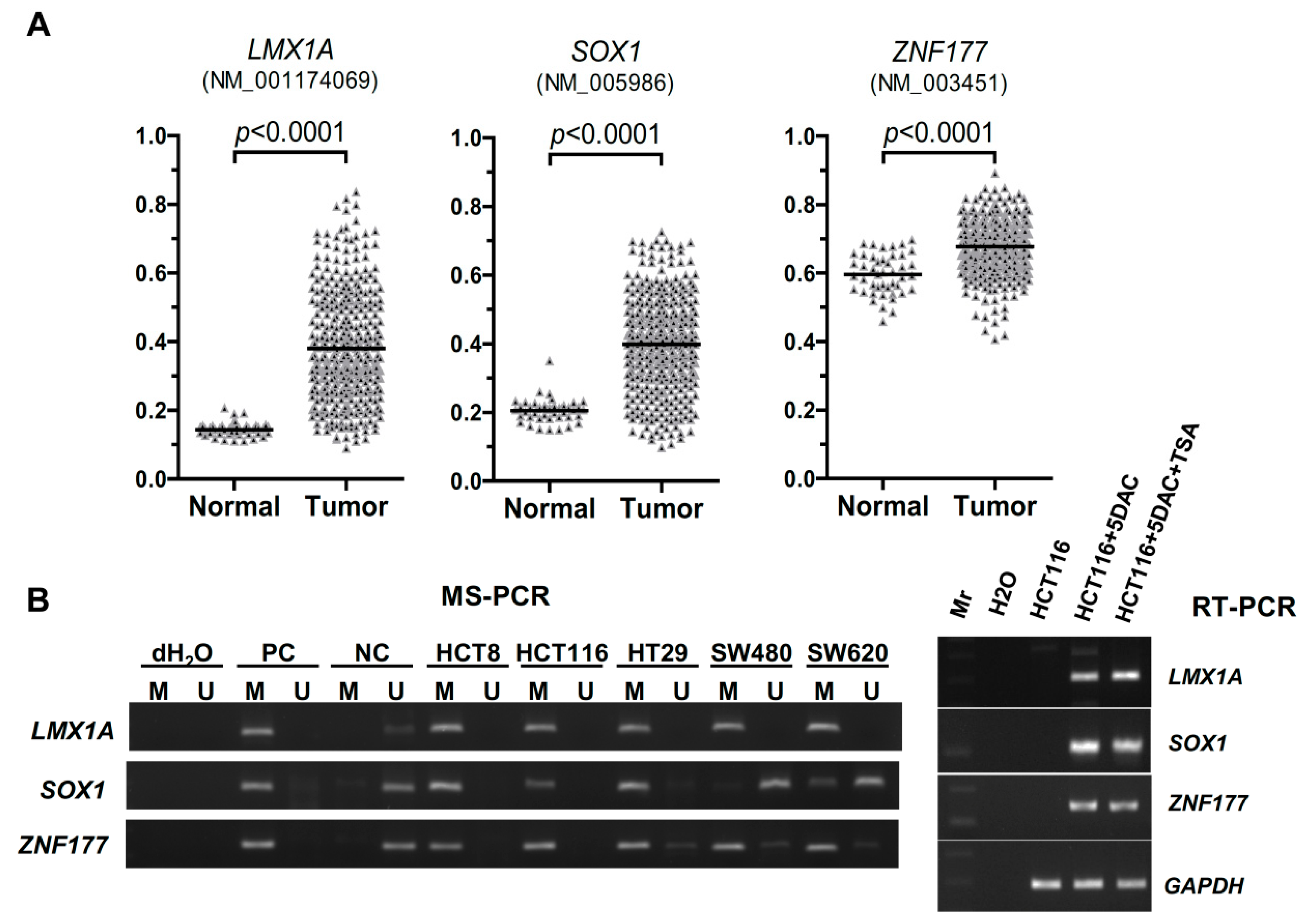
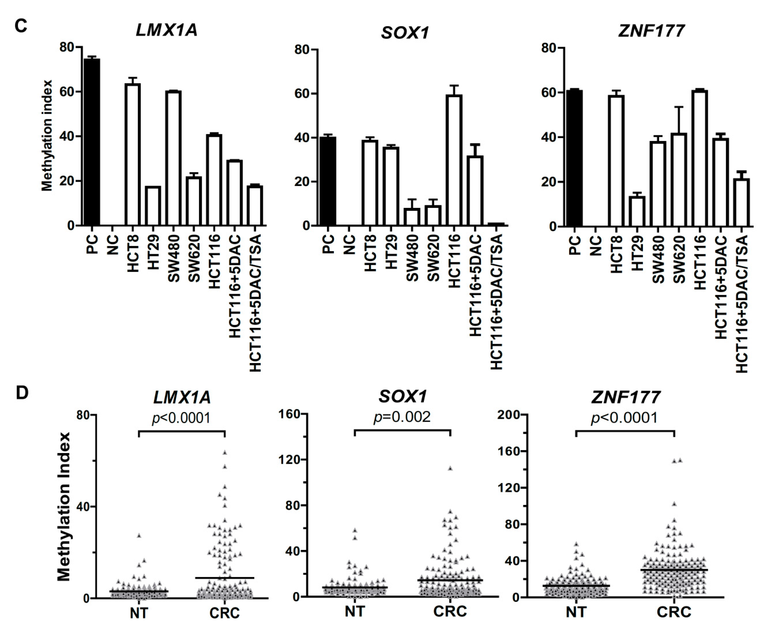
2.1. Abnormal Methylation of LMX1A, SOX1, and ZNF177 in CRC

To investigate whether LMX1A, SOX1, and ZNF177 were aberrantly methylated in CRC, we used the data of the Infinium Human Methylation 450K BeadChips from the MethHC database to analyze the methylation levels of LMX1A (NM_001174069), SOX1 (NM_005986), and ZNF177 (NM_003451) in 369 tissue samples from colon and rectal adenocarcinoma patients (Figure 1A). The analyses showed that the average β values were significantly higher in the tumor group than in the normal control group for all three genes (p < 0.0001). To further confirm the abnormal methylation phenotype, we examined the methylation status of LMX1A, SOX1, and ZNF177 in five CRC cell lines (HCT8, HCT116, HT29, SW480, and SW620) using the MSP assay (Figure 1B) and quantified the methylation levels using the Q-MSP assay (Figure 1C). The results of both the gel-based MSP assay and the Q-MPS assay revealed that LMX1A, SOX1, and ZNF177 were heavily methylated in over half of the cell lines. To analyze the correlation between LMX1A, SOX1, and ZNF177 and their gene expressions, we treated HCT116 cells with a DNA methylation inhibitor (5′-aza-2′-deoxycytidine; DAC) alone or in combination with an HDCA (histone deacetylase) inhibitor (Trichostatin A; TSA). The reverse transcription polymerase chain reaction (RT-PCR) data showed that the expressions of LMX1A, SOX1, and ZNF177 were restored in HCT116 cells after the cells were treated with the two drugs (Figure 1B, right panel). The Q-MSP results showed that the DNA methylation levels (methylation indexes) of LMX1A, SOX1, and ZNF177 were decreased after the HCT116 cells were treated with DAC only or the combination of DAC and TSA (Figure 1C). Then we quantified the methylation levels of LMX1A, SOX1, and ZNF177 in 151 pairs of CRC tissues and found that the DNA methylation levels of LMX1A, SOX1, and ZNF177 in 151 CRC tissue samples were significantly higher than those in the corresponding nontumor tissue samples (Figure 1D). Furthermore, we performed a receiver operating characteristic (ROC) curve analysis to discriminate between the CRC tissue samples and their nontumor counterparts (supporting information, Figure S1) and determined that the methylation frequencies of LMX1A, SOX1, and ZNF177 were 22.5%, 13.9%, and 12.6%, respectively, under the best cut-off values (Table 1).

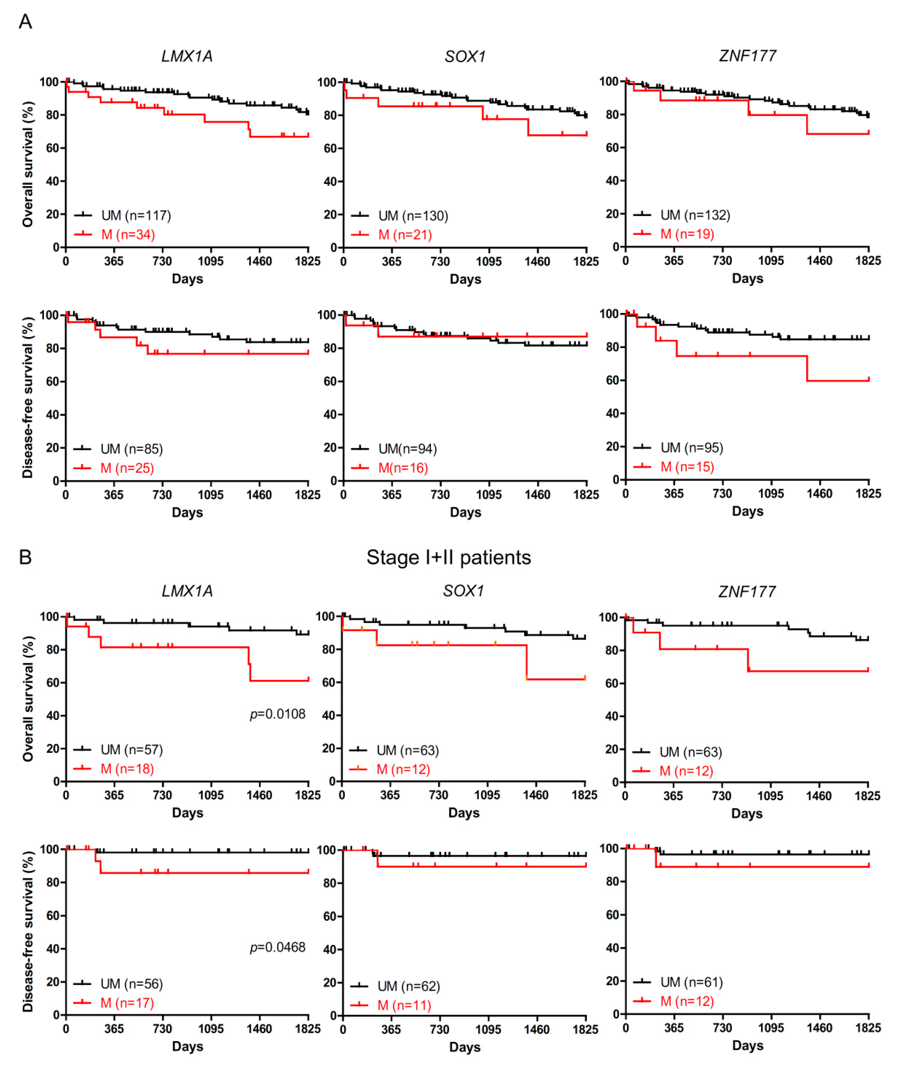
2.2. The Association of LMX1A Methylation and Patient Survival

To elucidate whether LMX1A, SOX1, and ZNF177 methylation could have a potential use in clinical practice, we explored the association of the methylation of LMX1A, SOX1, and ZNF177 and the clinical characteristics of 151 CRC patients (Table 2). We found a statistically significant association between LMX1A methylation and tumor size (p = 0.0048) but did not observe significant associations between SOX1 or ZNF177 methylation and any of the clinicopathological parameters. To further investigate the relevance of gene methylation with survival, we next employed the Kaplan-Meier method with the log-rank test for each gene methylation. As shown in Figure 2A, the methylation of neither LMX1A, SOX1, nor ZNF177 had any effect on the overall 5-year overall survival or disease-free survival of CRC patients. However, we found that early-stage (stage I or II) CRC patients with LMX1A methylation exhibited a poorer 5-year overall survival (p = 0.0108) and disease-free survival (p = 0.0468) than those without LMX1A methylation (Figure 2B).

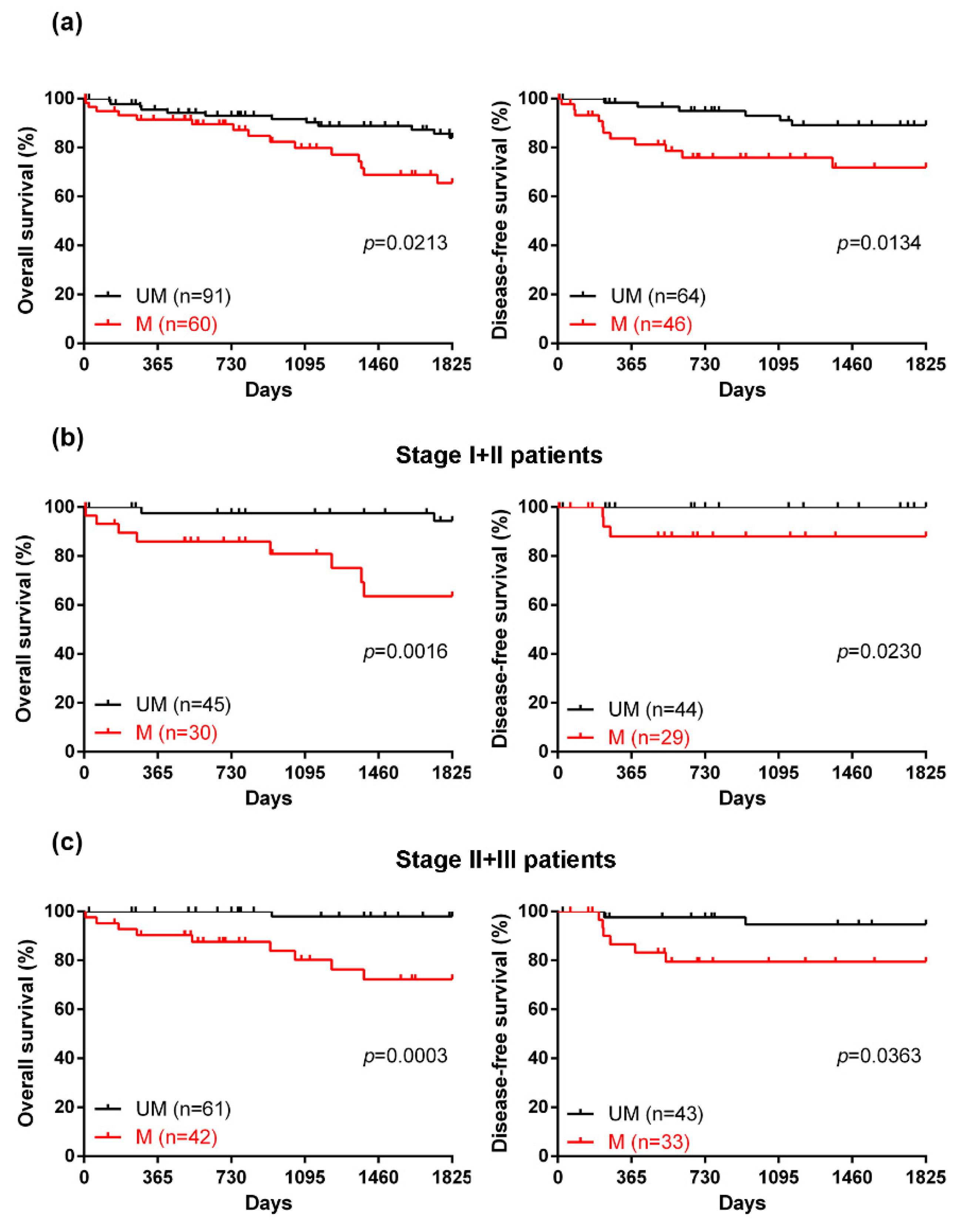
2.3. The Efficacy of a Novel DNA Methylation Panel for Predicting the Prognosis of CRC

We previously demonstrated that NKX6.1 was a novel prognostic biomarker for CRC and that the frequency of NKX6.1 methylation was 23% in a cohort of 151 CRC tissues [12]. Here, we determined whether the methylation status of NKX6.1, in combination with those of LMX1A, SOX1, and ZNF177, had a greater power in detecting CRC. The sensitivity and specificity of two and three-gene combinations ranged from 21.9% to 37.7% and 97.4% to 98.7%, respectively, and the four-gene panel provided the best sensitivity, 39.7%, and the optimal specificity, 97.4% (Table 1).
To further evaluate the utility of the four-gene methylation panel, we first analyzed its association with the clinicopathological characteristics. As shown in Table 3, the four-gene methylation panel was significantly associated with age (p = 0.0083), tumor size (p = 0.0323), and survival (p = 0.0297). To determine its prognostic utility, we next used the Cox proportional hazards model to identify independent factors associated with survival (Table 4). Univariate analysis revealed that the four-gene methylation panel was indeed a statistically significant indicator of overall survival (hazard ratio (HR), 2.95; 95% confidence interval (CI), 1.36–6.39; p < 0.01). In the multivariate analysis, gene methylation and chemotherapy were confirmed as independent factors for overall survival (HR for gene methylation, 3.43; 95%CI, 1.37–8.57; p < 0.01) (HR for chemotherapy, 0.32; 95%CI, 0.12–0.85; p < 0.05). Furthermore, the Kaplan–Meier survival analysis determined the effect of the four-gene methylation panel on survival, revealing that patients who exhibited methylation for all four genes had a worse 5-year overall survival (p = 0.0213) and disease-free survival (p = 0.0134) than those without methylation of any of the four genes (Figure 3a). Finally, the four-gene methylation status was associated with 5-year overall survival and disease-free survival in early-stage patients (Figure 3b; p = 0.0016 and p = 0.0230), and in stage II and III CRC patients (Figure 3c; p = 0.0009 and p = 0.0363).

3. Discussion

The tumor-node-metastasis (TNM) staging is a key determinant of the therapy selection and prognosis of CRC patients. However, beyond creating a more complex tumor classification, the TNM staging system has not provided clinicians with the optimal treatment and management strategies that it was originally designed for. Recent advances in the determination of the molecular CRC subtypes have led to the identification of several prognostic biomarkers, based on molecular heterogeneity, which might contribute to the risk stratification of patients with the same disease stages and who are receiving the same treatment [25,26]. In the current study, we analyzed the methylation levels of LMX1A, SOX1, and ZNF177 to elucidate novel changes in the methylation levels of genes in CRC and determined that the novel four-gene methylation panel, which includes LMX1A, SOX1, ZNF177, and NKX6.1, could discriminate the outcomes of CRC patients.
As in other cancers, CRC encompasses multiple molecular subtypes with specific characteristics. The CpG island methylator phenotype (CIMP) is one famous CRC subtypes, including tumors with a high frequency of hypermethylation at CpG islands [27]. CIMP-associated CRC has distinct epidemiology, histology, precursor lesions, and molecular features (e.g., BRAF and KRAS mutations) [28,29].
In addition, recent studies have reported that the prognosis was significantly poor in CIMP-high patients among the microsatellite stability (MSS) subgroup, although there was a trend of increased cancer-specific survival in CIMP-positive CRC patients [30,31,32,33,34,35,36]. However, the association of CIMP with the prognosis and survival of CRC subtypes remains unknown.
We previously reported several DNA methylation markers in various cancers, including cervical cancer, ovarian cancer, and hepatocellular carcinoma [13,16,19]. However, the utility of these markers or marker panels for CRC is unknown. Therefore, we designed a strategy to first verify the DNA methylation levels of specific markers that we identified in colon and rectal adenocarcinomas using array data from existing databases and then validated these results by a quantitative DNA methylation analysis of potential genes in the study cohort using Q-MSP. With this strategy, we recently reported NKX6.1 hypermethylation as a new biomarker for the prediction of outcomes and therapy selection in stage II CRC patients [12]. In the current study, we not only showed that LMX1A hypermethylation was significantly associated with tumor size and poor 5-year overall survival rates and disease-free survival rates in early-stage CRC patients, but also demonstrated that the four-gene methylation panel, including NKX6.1, LMX1A, SOX1, and ZNF177, was a novel and independent prognostic tool for stage I and II, or stage II and III CRCs.
The four-gene methylation status was decreased and the expression level was restored in HCT116 cells after the cells were treated with DAC alone or with the combination of DAC/TSA. Moreover, the correlation analysis of the differential methylation and expression levels of LMX1A, SOX1, ZNF177 and NKX6.1 in colon adenocarcinoma patients was based on data derived from the MethHC database (supporting information in Figure S2). The inverse correlation between the gene expression and DNA methylation of SOX1 (correlation = −0.2971, p < 0.0001) was statistically significant. There was no correlation between the methylation and gene expression of LMX1A (correlation = −0.00675) and NKX6.1 (correlation = −0.1046) from the MethHC database. However, a positive correlation was observed between the gene expression level and the methylation status of ZNF177 (correlation = 0.3487, p < 0.0001) (Figure S2). There are two ZNF177 variants reported at the NCBI, but the probe was designed only for the variant NM_003451′s methylation analysis from MethHC. These controversial data may be due to the different variants of ZNF177 and the heterogeneity of the clinical samples. In summary, there was an inverse correlation between the methylation and expression of the four-gene panel in HCT116 cells. Among the markers, LMX1A is a LIM homeobox-containing gene, with roles in a wide variety of developmental contexts [37]; SOX1 encodes a protein that plays a key regulatory role in neural cell fate determination and differentiation [38,39]; and ZNF177 belongs to a zinc finger gene family encoding a large number of common transcription factors [40,41]. Previously, in addition to identifying DNA methylation biomarkers, we also revealed that LMX1A and SOX1 were tumor suppressor genes in cervical cancer and hepatocellular carcinoma [42,43,44]. Additionally, the promoter-hypermethylations of LMX1A, SOX1, and ZNF177 were previously reported in lung, stomach, and bladder cancers, respectively, by other groups [14,17,18]. In the two most recent reports, SOX1 hypermethylation was also identified in colon cancer and CRC [45,46]. Overall, the data from previous studies, as well as the data from the current report, indicate promoter hypermethylation of these genes are common epigenetic events in multiple cancer types. The biological function of these four genes in CRC needs further investigation.
Despite the promising results, the current study does have limitations. The cutoff value for each gene was based on a hospital-based, retrospective case-control study from a research platform and may not be applied directly to clinical settings with larger populations. To determine its prognostic utility, we used the Cox proportional hazards model to identify independent factors associated with survival (Tables S1–S4). The univariate analysis revealed that LMX1A and NKX6.1 methylation was indeed a statistically significant indicator of overall survival (hazard ratio (HR) for LMX1A methylation, 2.58; p < 0.05) (HR for NKX6.1 methylation, 2.60; p < 0.01). In the multivariate analysis, NKX6.1 methylation, stage and chemotherapy were confirmed as independent factors for overall survival (HR for NKX6.1 methylation, 6.06; p < 0.01) (HR for stage, 3.77; p < 0.05) (HR for chemotherapy, 0.26; p < 0.05). LMX1A methylation and chemotherapy were confirmed as independent factors for overall survival (HR for LMX1A methylation; p < 0.05) (HR for chemotherapy, 0.33; p < 0.05). However, our previous study demonstrated that the methylation frequency of NKX6.1 was 23% in our cohort of 151 pairs of CRC tissues. In this study, we aimed to develop a DNA methylation panel that might potentially improve the sensitivity of the application. While the methylation of SOX1 and ZNF177 was not a statistically significant indicator of overall survival, the combination of these two genes could improve the sensitivity (23% to 39.7%) when discriminating 151 CRC tissues from nontumor tissues. Therefore, we used a four-gene methylation panel to elucidate its prognostic utility for CRC. As there was no patient survival information in the MethHC database, we could not determine whether the four-gene set had a prognostic value in these cohorts. In this study, we did not have any other available cohort to further validate the prognostic value. However, we analyzed data from PRECOG (https://precog.stanford.edu), a database for querying associations between gene expressions and clinical outcomes, to examine the association between the expression of the four-gene panel and the survival of CRC patients. A GEO dataset (GSE12945) showed that patients with a low NKX6.1 expression had a poorer survival rate, compared with those with a high NKX6.1 expression (p = 0.0159; Figure S3). For the other three genes, LMX1A, SOX1, and ZNF177, there was no significant correlation between the gene expression and patient survival. The reason for these results was due to the limited patient survival information and the heterogeneity of the clinical samples. In summary, this is a pilot study that indicates that the abnormal DNA methylation panel is a potential prognostic biomarker of CRC. Nonetheless, the prognostic value of the four-gene panel needs another independent cohort to further validate these results, and an in vitro validation of the biological function of these genes will provide a better understanding of the findings.
The molecular heterogeneity of CRC may aid in determining clinical survival of CRC patients. The current study examined the prognostic value of the methylation levels of LMX1A, SOX1, and ZNF177 in CRC patients. The results suggest that the combination of NKX6.1, LMX1A, SOX1, and ZNF177 promoter methylation statuses is a potentially novel stage-independent prognostic marker.

4. Materials and Methods

4.1. MethHC Database

MethHC (http://MethHC.mbc.nctu.edu.tw) is a web resource specifically focused on the DNA methylation of human cancers [13]. In the current study, we used DNA methylation data in the MethHC database for preliminary analysis of the methylation levels of LMX1A, SOX1, and ZNF177 in 46 tissue samples from normal individuals and 369 tissue samples from colon or rectal adenocarcinoma patients.

4.2. Clinical Samples and Cell Lines

Our cohort included 151 paired tumor tissue and adjacent nontumor tissue specimens that were obtained from the Tri-Service General Hospital. The use of these samples was approved by our Institutional Review Board (TSGHIRB number: 098-05-292). The clinicopathologic characteristics of the patients were summarized previously [12]. Additionally, five CRC cell lines (HCT8, HCT116, HT29, SW480, and SW620) were used in the current study. HT29 cells were purchased from the American Type Culture Collection. HCT8, HCT116, SW480, and SW620 cells were purchased from the Food Industry Research and Development Institute (Taiwan).

4.3. DNA Methylation and Gene Expression Analysis

For DNA methylation analysis, the genomic DNA of clinical samples and cell lines were extracted and bisulfite-converted as previously described [12]. CpG methylated human genomic DNA (Thermo Fisher Scientific, San Diego, CA, USA) and DNA extracted from normal peripheral blood lymphocytes were modified by sodium bisulfite to generate positive and negative controls, respectively. MS-PCR and Q-MSP were performed as previously described. For Q-MSP, the DNA methylation levels were assessed by determining the methylation index (MI) using the following formula: 100 × 2−((Cp of Gene)−(Cp of COL2A)). The Q-MSP primer sequences used in the current study were as follows: LMX1A-F tgggacgcgggattgtaaattttat, LMX1A-R aaaccctcgaaacgtctctacaaaa, SOX1-F ggttgtatcgtaatcgttttttgtaggtt, SOX1-R cctccaactcgaaaactacaacttct, ZNF177-F ggaagtgggcgttcgtcgtttc, ZNF177-R cccttccctcccgattccg, COL2A-F gggaagatgggatagaagggaatat, and COL2A-R tctaacaattataaactccaaccaccaa. For gene expression analysis, reverse transcription polymerase chain reaction (RT-PCR) was conducted as previously described [12]. The RT-PCR primer sequences used in the current study are shown in the Table S5.

4.4. Statistical Analysis

Statistical analyses were performed using GraphPad Prism software (version 4.03; GraphPad Software, La Jolla, CA, USA) and SPSS software (IBM SPSS Statistics 21; Asia Analytics Taiwan, Taipei, Taiwan). The Mann–Whitney U and Wilcoxon signed-rank tests were used to determine differences between gene methylation levels and disease status. Receiver operating characteristic (ROC) curves were generated to calculate the cut-off values for LMX1A, SOX1, and ZNF177 methylation for discriminating tumors from nontumor tissues. χ2 test and an χ2 test for trend were used to calculate the associations between gene methylations and clinical parameters. Kaplan–Meier curves were calculated to estimate survival rates at five years after treatment. The log-rank test was used to compare the association between survival and gene methylation. The Cox proportional hazards model was used to identify factors for overall survival.

5. Conclusions

In summary, our results demonstrate that the combination of the methylation statuses of NKX6.1, LMX1A, SOX1, and ZNF177 is a novel prognostic panel that could be considered a molecular marker for outcomes in CRC patients. This is a pilot study that indicates that the abnormal DNA methylation panel is a potential prognostic biomarker of CRC. Nonetheless, the prognostic value of the four-gene panel needs another independent cohort to further validate these results.

Supplementary Materials

Supplementary materials can be found at https://www.mdpi.com/1422-0067/20/19/4672/s1. Figure S1. ROC curves of LMX1A, SOX1, and ZNF177 in CRC tissues. Figure S2. Correlation between gene expression level and methylation status of LMX1A, SOX1, ZNF177, and NKX6.1 in CRC tissues. Figure S3. Kaplan–Meier curves for survival analysis in 62 CRC patients. Table S1. Univariate and multivariate of overall survival analysis using clinical characteristics and the LMX1A methylation panel in 151 CRC patients. Table S2. Univariate and multivariate of overall survival analysis using clinical characteristics and the SOX1 methylation panel in 151 CRC patients. Table S3. Univariate and multivariate of overall survival analysis using clinical characteristics and the ZNF177 methylation panel in 151 CRC patients. Table S4. Univariate and multivariate of overall survival analysis using clinical characteristics and the NKX6.1 methylation panel in 151 CRC patients. Table S5. Primer sequences used for RT-PCR.

Author Contributions

H.-H.C. and C.-C.K. designed the study. Y.-W.L., Y.-L.S., Y.-C.C., C.-W.H., C.-Y.C., J.-M.H., and C.-H.H. provided experimental materials. H.-H.C. cultured the CRC cell lines. H.-H.C. and C.-C.K. performed genomic DNA extraction, DNA bisulfite-conversion, MSP, and the Q-MSP experiments. H.-H.C. and C.-C.K. analyzed the results and prepared the figures and tables. H.-H.C. wrote the paper. All authors discussed the results and commented on the manuscript. All authors have read and approved the final manuscript.

Funding

This work was supported in part by the following grants: MOST 105-2314-B-016-049, MOST 107-2314-B-016-012, and MOST 105-2320-B-016-017-MY3 from the Ministry of Science and Technology, Taiwan, Republic of China; MAB-106-045, MAB-106-046, MAB-106-047, MAB-107-026, MAB-107-027, and MAB-107-028 from the Ministry of National Defense, Taiwan, Republic of China; and TSGH-C107-049 and TSGH-C108-072 from Tri-Service General Hospital, Taiwan.

Acknowledgments

We thank Ming-De Yan for advice on paper-writing, and Chia-Hsin Lin and Shin-Ping Lin for technical assistance.

Conflicts of Interest

The authors declare that they have no competing interests.

References

  1. Bray, F.; Ferlay, J.; Soerjomataram, I.; Siegel, R.L.; Torre, L.A.; Jemal, A. Global cancer statistics 2018: GLOBOCAN estimates of incidence and mortality worldwide for 36 cancers in 185 countries. CA-Cancer J. Clin. 2018, 68, 394–424. [Google Scholar] [CrossRef] [PubMed] [Green Version]
  2. Brenner, H.; Kloor, M.; Pox, C.P. Colorectal cancer. Lancet 2014, 383, 1490–1502. [Google Scholar] [CrossRef]
  3. Wu, X.; Zhang, J.; He, X.; Wang, C.; Lian, L.; Liu, H.; Wang, J.; Lan, P. Postoperative adjuvant chemotherapy for stage II colorectal cancer: A systematic review of 12 randomized controlled trials. J. Gastrointest. Surg. Off. J. Soc. Surg. Aliment. Tract 2012, 16, 646–655. [Google Scholar] [CrossRef] [PubMed]
  4. Robertson, K.D. DNA methylation and human disease. Nat. Rev. Genet. 2005, 6, 597–610. [Google Scholar] [CrossRef] [PubMed]
  5. Fakhr, M.G.; Hagh, M.F.; Shanehbandi, D.; Baradaran, B. DNA methylation pattern as important epigenetic criterion in cancer. Genet. Res. Int. 2013, 2013, 317569. [Google Scholar] [CrossRef]
  6. Silva, T.D.; Vidigal, V.M.; Felipe, A.V.; De Lima, J.M.; Neto, R.A.; Saad, S.S.; Forones, N.M. DNA methylation as an epigenetic biomarker in colorectal cancer. Oncol. Lett. 2013, 6, 1687–1692. [Google Scholar] [CrossRef] [PubMed] [Green Version]
  7. Lam, K.; Pan, K.; Linnekamp, J.F.; Medema, J.P.; Kandimalla, R. DNA methylation based biomarkers in colorectal cancer: A systematic review. Biochim. Biophys. Acta 2016, 1866, 106–120. [Google Scholar] [CrossRef] [PubMed]
  8. Imperiale, T.F.; Ransohoff, D.F.; Itzkowitz, S.H. Multitarget stool DNA testing for colorectal-cancer screening. N. Engl. J. Med. 2014, 371, 187–188. [Google Scholar] [CrossRef] [PubMed]
  9. Toth, K.; Wasserkort, R.; Sipos, F.; Kalmar, A.; Wichmann, B.; Leiszter, K.; Valcz, G.; Juhasz, M.; Miheller, P.; Patai, A.V.; et al. Detection of methylated septin 9 in tissue and plasma of colorectal patients with neoplasia and the relationship to the amount of circulating cell-free DNA. PLoS ONE 2014, 9, e115415. [Google Scholar] [CrossRef]
  10. Wasserkort, R.; Kalmar, A.; Valcz, G.; Spisak, S.; Krispin, M.; Toth, K.; Tulassay, Z.; Sledziewski, A.Z.; Molnar, B. Aberrant septin 9 DNA methylation in colorectal cancer is restricted to a single CpG island. BMC Cancer 2013, 13, 398. [Google Scholar] [CrossRef]
  11. Molnar, B.; Toth, K.; Bartak, B.K.; Tulassay, Z. Plasma methylated septin 9: A colorectal cancer screening marker. Expert Rev. Mol. Diagn. 2015, 15, 171–184. [Google Scholar] [CrossRef] [PubMed]
  12. Chang, S.Y.; Kuo, C.C.; Wu, C.C.; Hsiao, C.W.; Hu, J.M.; Hsu, C.H.; Chou, Y.C.; Shih, Y.L.; Lin, Y.W. NKX6.1 hypermethylation predicts the outcome of stage II colorectal cancer patients undergoing chemotherapy. Genes Chromosom. Cancer 2018, 57, 268–277. [Google Scholar] [CrossRef] [PubMed]
  13. Kuo, C.C.; Lin, C.Y.; Shih, Y.L.; Hsieh, C.B.; Lin, P.Y.; Guan, S.B.; Hsieh, M.S.; Lai, H.C.; Chen, C.J.; Lin, Y.W. Frequent methylation of HOXA9 gene in tumor tissues and plasma samples from human hepatocellular carcinomas. Clin. Chem. Lab. Med. 2014, 52, 1235–1245. [Google Scholar] [CrossRef] [PubMed]
  14. Diaz-Lagares, A.; Mendez-Gonzalez, J.; Hervas, D.; Saigi, M.; Pajares, M.J.; Garcia, D.; Crujerias, A.B.; Pio, R.; Montuenga, L.M.; Zulueta, J.; et al. A Novel Epigenetic Signature for Early Diagnosis in Lung Cancer. Clin. Cancer Res. Off. J. Am. Assoc. Cancer Res. 2016, 22, 3361–3371. [Google Scholar] [CrossRef] [PubMed] [Green Version]
  15. Chen, Y.C.; Tsao, C.M.; Kuo, C.C.; Yu, M.H.; Lin, Y.W.; Yang, C.Y.; Li, H.J.; Yan, M.D.; Wang, T.J.; Chou, Y.C.; et al. Quantitative DNA methylation analysis of selected genes in endometrial carcinogenesis. Taiwan. J. Obstet. Gynecol. 2015, 54, 572–579. [Google Scholar] [CrossRef] [PubMed] [Green Version]
  16. Lai, H.C.; Lin, Y.W.; Huang, T.H.; Yan, P.; Huang, R.L.; Wang, H.C.; Liu, J.; Chan, M.W.; Chu, T.Y.; Sun, C.A.; et al. Identification of novel DNA methylation markers in cervical cancer. Int. J. Cancer 2008, 123, 161–167. [Google Scholar] [CrossRef] [PubMed]
  17. Dong, W.; Feng, L.; Xie, Y.; Zhang, H.; Wu, Y. Hypermethylation-mediated reduction of LMX1A expression in gastric cancer. Cancer Sci. 2011, 102, 361–366. [Google Scholar] [CrossRef]
  18. Zhao, Y.; Guo, S.; Sun, J.; Huang, Z.; Zhu, T.; Zhang, H.; Gu, J.; He, Y.; Wang, W.; Ma, K.; et al. Methylcap-seq reveals novel DNA methylation markers for the diagnosis and recurrence prediction of bladder cancer in a Chinese population. PLoS ONE 2012, 7, e35175. [Google Scholar] [CrossRef]
  19. Su, H.Y.; Lai, H.C.; Lin, Y.W.; Chou, Y.C.; Liu, C.Y.; Yu, M.H. An epigenetic marker panel for screening and prognostic prediction of ovarian cancer. Int. J. Cancer 2009, 124, 387–393. [Google Scholar] [CrossRef]
  20. Shih, Y.L.; Hsieh, C.B.; Yan, M.D.; Tsao, C.M.; Hsieh, T.Y.; Liu, C.H.; Lin, Y.W. Frequent concomitant epigenetic silencing of SOX1 and secreted frizzled-related proteins (SFRPs) in human hepatocellular carcinoma. J. Gastroenterol. Hepatol. 2013, 28, 551–559. [Google Scholar] [CrossRef]
  21. Zhao, Y.; Zhou, H.; Ma, K.; Sun, J.; Feng, X.; Geng, J.; Gu, J.; Wang, W.; Zhang, H.; He, Y.; et al. Abnormal methylation of seven genes and their associations with clinical characteristics in early stage non-small cell lung cancer. Oncol. Lett. 2013, 5, 1211–1218. [Google Scholar] [CrossRef] [PubMed] [Green Version]
  22. Kuo, I.Y.; Chang, J.M.; Jiang, S.S.; Chen, C.H.; Chang, I.S.; Sheu, B.S.; Lu, P.J.; Chang, W.L.; Lai, W.W.; Wang, Y.C. Prognostic CpG methylation biomarkers identified by methylation array in esophageal squamous cell carcinoma patients. Int. J. Med. Sci. 2014, 11, 779–787. [Google Scholar] [CrossRef] [PubMed]
  23. Li, N.; Li, S. Epigenetic inactivation of SOX1 promotes cell migration in lung cancer. Tumour Biol. J. Int. Soc. Oncodev. Biol. Med. 2015, 36, 4603–4610. [Google Scholar] [CrossRef] [PubMed]
  24. Lopez, J.I.; Angulo, J.C.; Martin, A.; Sanchez-Chapado, M.; Gonzalez-Corpas, A.; Colas, B.; Ropero, S. A DNA hypermethylation profile reveals new potential biomarkers for the evaluation of prognosis in urothelial bladder cancer. Acta Pathol. Microbiol. Immunol. Scand. 2017, 125, 787–796. [Google Scholar] [CrossRef] [PubMed]
  25. Herzig, D.O.; Tsikitis, V.L. Molecular markers for colon diagnosis, prognosis and targeted therapy. J. Surg. Oncol. 2015, 111, 96–102. [Google Scholar] [CrossRef] [PubMed]
  26. Mahasneh, A.; Al-Shaheri, F.; Jamal, E. Molecular biomarkers for an early diagnosis, effective treatment and prognosis of colorectal cancer: Current updates. Exp. Mol. Pathol. 2017, 102, 475–483. [Google Scholar] [CrossRef] [PubMed]
  27. Toyota, M.; Ahuja, N.; Ohe-Toyota, M.; Herman, J.G.; Baylin, S.B.; Issa, J.P. CpG island methylator phenotype in colorectal cancer. Proc. Natl. Acad. Sci. USA 1999, 96, 8681–8686. [Google Scholar] [CrossRef] [PubMed] [Green Version]
  28. Shen, L.; Toyota, M.; Kondo, Y.; Lin, E.; Zhang, L.; Guo, Y.; Hernandez, N.S.; Chen, X.; Ahmed, S.; Konishi, K.; et al. Integrated genetic and epigenetic analysis identifies three different subclasses of colon cancer. Proc. Natl. Acad. Sci. USA 2007, 104, 18654–18659. [Google Scholar] [CrossRef] [Green Version]
  29. Ang, P.W.; Loh, M.; Liem, N.; Lim, P.L.; Grieu, F.; Vaithilingam, A.; Platell, C.; Yong, W.P.; Iacopetta, B.; Soong, R. Comprehensive profiling of DNA methylation in colorectal cancer reveals subgroups with distinct clinicopathological and molecular features. BMC Cancer 2010, 10, 227. [Google Scholar] [CrossRef]
  30. Lee, S.; Cho, N.Y.; Yoo, E.J.; Kim, J.H.; Kang, G.H. CpG island methylator phenotype in colorectal cancers: Comparison of the new and classic CpG island methylator phenotype marker panels. Arch. Pathol. Lab. Med. 2008, 132, 1657–1665. [Google Scholar] [CrossRef]
  31. Barault, L.; Charon-Barra, C.; Jooste, V.; de la Vega, M.F.; Martin, L.; Roignot, P.; Rat, P.; Bouvier, A.M.; Laurent-Puig, P.; Faivre, J.; et al. Hypermethylator phenotype in sporadic colon cancer: Study on a population-based series of 582 cases. Cancer Res. 2008, 68, 8541–8546. [Google Scholar] [CrossRef] [PubMed]
  32. Shen, L.; Catalano, P.J.; Benson, A.B., 3rd; O’Dwyer, P.; Hamilton, S.R.; Issa, J.P. Association between DNA methylation and shortened survival in patients with advanced colorectal cancer treated with 5-fluorouracil based chemotherapy. Clin. Cancer Res. Off. J. Am. Assoc. Cancer Res. 2007, 13, 6093–6098. [Google Scholar] [CrossRef] [PubMed]
  33. Lee, S.; Cho, N.Y.; Choi, M.; Yoo, E.J.; Kim, J.H.; Kang, G.H. Clinicopathological features of CpG island methylator phenotype-positive colorectal cancer and its adverse prognosis in relation to KRAS/BRAF mutation. Pathol. Int. 2008, 58, 104–113. [Google Scholar] [CrossRef] [PubMed]
  34. Kim, J.H.; Shin, S.H.; Kwon, H.J.; Cho, N.Y.; Kang, G.H. Prognostic implications of CpG island hypermethylator phenotype in colorectal cancers. Virchows Arch. Int. J. Pathol. 2009, 455, 485–494. [Google Scholar] [CrossRef]
  35. Sanchez, J.A.; Krumroy, L.; Plummer, S.; Aung, P.; Merkulova, A.; Skacel, M.; DeJulius, K.L.; Manilich, E.; Church, J.M.; Casey, G.; et al. Genetic and epigenetic classifications define clinical phenotypes and determine patient outcomes in colorectal cancer. Br. J. Surg. 2009, 96, 1196–1204. [Google Scholar] [CrossRef] [PubMed]
  36. Ward, R.L.; Cheong, K.; Ku, S.L.; Meagher, A.; O’Connor, T.; Hawkins, N.J. Adverse prognostic effect of methylation in colorectal cancer is reversed by microsatellite instability. J. Clin. Oncol. Off. J. Am. Soc. Clin. Oncol. 2003, 21, 3729–3736. [Google Scholar] [CrossRef] [PubMed]
  37. Hobert, O.; Westphal, H. Functions of LIM-homeobox genes. Trends Genet. 2000, 16, 75–83. [Google Scholar] [CrossRef]
  38. Buescher, M.; Hing, F.S.; Chia, W. Formation of neuroblasts in the embryonic central nervous system of Drosophila melanogaster is controlled by SoxNeuro. Development 2002, 129, 4193–4203. [Google Scholar] [PubMed]
  39. Kan, L.; Israsena, N.; Zhang, Z.; Hu, M.; Zhao, L.R.; Jalali, A.; Sahni, V.; Kessler, J.A. Sox1 acts through multiple independent pathways to promote neurogenesis. Dev. Biol. 2004, 269, 580–594. [Google Scholar] [CrossRef] [PubMed] [Green Version]
  40. Laity, J.H.; Lee, B.M.; Wright, P.E. Zinc finger proteins: New insights into structural and functional diversity. Curr. Opin. Struct. Biol. 2001, 11, 39–46. [Google Scholar] [CrossRef]
  41. Cassandri, M.; Smirnov, A.; Novelli, F.; Pitolli, C.; Agostini, M.; Malewicz, M.; Melino, G.; Raschella, G. Zinc-finger proteins in health and disease. Cell Death Discov. 2017, 3, 17071. [Google Scholar] [CrossRef] [PubMed]
  42. Liu, C.Y.; Chao, T.K.; Su, P.H.; Lee, H.Y.; Shih, Y.L.; Su, H.Y.; Chu, T.Y.; Yu, M.H.; Lin, Y.W.; Lai, H.C. Characterization of LMX-1A as a metastasis suppressor in cervical cancer. J. Pathol. 2009, 219, 222–231. [Google Scholar] [CrossRef] [PubMed]
  43. Tsao, C.M.; Yan, M.D.; Shih, Y.L.; Yu, P.N.; Kuo, C.C.; Lin, W.C.; Li, H.J.; Lin, Y.W. SOX1 functions as a tumor suppressor by antagonizing the WNT/beta-catenin signaling pathway in hepatocellular carcinoma. Hepatology 2012, 56, 2277–2287. [Google Scholar] [CrossRef] [PubMed]
  44. Lin, Y.W.; Tsao, C.M.; Yu, P.N.; Shih, Y.L.; Lin, C.H.; Yan, M.D. SOX1 suppresses cell growth and invasion in cervical cancer. Gynecol. Oncol. 2013, 131, 174–181. [Google Scholar] [CrossRef] [PubMed]
  45. Huang, J.; Tan, Z.R.; Yu, J.; Li, H.; Lv, Q.L.; Shao, Y.Y.; Zhou, H.H. DNA hypermethylated status and gene expression of PAX1/SOX1 in patients with colorectal carcinoma. OncoTargets Ther. 2017, 10, 4739–4751. [Google Scholar] [CrossRef] [PubMed]
  46. Molnar, B.; Galamb, O.; Peterfia, B.; Wichmann, B.; Csabai, I.; Bodor, A.; Kalmar, A.; Szigeti, K.A.; Bartak, B.K.; Nagy, Z.B.; et al. Gene promoter and exon DNA methylation changes in colon cancer development—mRNA expression and tumor mutation alterations. BMC Cancer 2018, 18, 695. [Google Scholar] [CrossRef] [PubMed]
Figure 1. DNA methylation levels of LIM homeobox transcription factor 1α (LMX1A), sex-determining region Y-box 1 (SOX1), and zinc finger protein 177 (ZNF177) in colorectal carcinoma (CRC). (A) DNA methylation array data for LMX1A, SOX1, and ZNF177 in 45 tissue samples from normal individuals and 369 tissue samples from colon and rectal adenocarcinoma patients from the MethHC database are shown. The results are represented as average (AVG) β values for the probes. Black lines indicate the mean of AVG β value. The p-values for LMX1A, SOX1, and ZNF177 methylation levels among the groups (normal versus tumor) were determined by the Mann–Whitney U test. (B) The promoter methylation statuses of LMX1A, SOX1, and ZNF177 in five CRC cell lines were analyzed by MSP with methylated and unmethylated-specific primers. PC: positive control; NC: negative control. Gene expression levels of three genes and the internal reference GAPDH in HCT116 cells treated with 5 µM DAC or 5 µM DAC combined with 300 nM TSA (5DAC/TSA), or left untreated, were analyzed by reverse transcription polymerase chain reaction (RT-PCR). Mr: molecular marker, or DNA ladder. (C) Quantitative DNA methylation levels of LMX1A, SOX1, and ZNF177 in five CRC cell lines were analyzed by Q-MSP. Quantitative DNA methylation levels of LMX1A, SOX1, and ZNF177 in HCT116 cells treated with 5 µM DAC or 5 µM DAC combined with 300 nM TSA (5DAC/TSA), or left untreated, were determined by Q-MSP. The results are represented as differences in the methylation index (MI). (D) Quantitative DNA methylation levels of LMX1A, SOX1, and ZNF177 were determined in 151 paired CRC tissue samples and the adjacent nontumor tissue samples (NT) by Q-MSP. The results are represented as differences in the methylation index. Black lines indicate mean methylation index. p values for methylation levels among the groups were determined by the Wilcoxon signed-rank test.
Ijms 20 04672 g001aIjms 20 04672 g001b
Figure 2. Correlation analysis between LMX1A, SOX1, and ZNF177 methylation levels and survival in CRC patients. (A) The 5-year overall survival and disease-free survival rates of CRC patients with different LMX1A, SOX1, and ZNF177 methylation statuses are presented. Red lines indicate cases with LMX1A methylation (methylation index (MI) > 16.84), SOX1 methylation (MI > 30.26), and ZNF177 methylation (MI > 51.14). Black lines indicate cases without LMX1A methylation (MI ≤ 16.84), SOX1 methylation (MI ≤ 30.26), and ZNF177 methylation (MI ≤ 51.14). (B) The 5-year overall survival and disease-free survival rates of stage I and II CRC patients.
Ijms 20 04672 g002
Figure 3. Correlation analysis between the four-gene methylation panel and the survival of CRC patients. (a) The 5-year overall survival and disease-free survival rates of CRC patients according to the methylation status are presented. Red line: methylated cases discriminated by the four-gene methylation panel; black line: unmethylated cases. (b) The 5-year overall survival and disease-free survival rates of stage I and II CRC patients. (c) The 5-year overall survival and disease-free survival rates of stage II and III CRC patients.
Ijms 20 04672 g003
Table 1. Sensitivity and specificity of candidate gene sets for discriminating 151 CRC from nontumor tissues.
Gene SetBest Cut-Off ValueSensitivitySpecificity
NKX6.1MI > 18.4223.2%99.3%
LMX1AMI > 16.8422.5%99.3%
SOX1MI > 30.2613.9%98.7%
ZNF177MI > 51.1412.6%99.3%
NKX6.1 or ZNF177 33.1%98.7%
NKX6.1 or LMX1A 31.1%98.7%
LMX1A or ZNF177 30.5%98.7%
NKX6.1 or SOX1 29.1%98.0%
LMX1A or SOX1 27.2%98.0%
SOX1 or ZNF177 21.9%98.7%
NKX6.1 or LMX1A or ZNF177 37.7%98.0%
NKX6.1 or SOX1 or ZNF177 36.4%98.0%
LMX1A or SOX1 or ZNF177 33.8%98.0%
NKX6.1 or LMX1A or SOX1 33.8%97.4%
NKX6.1 or LMX1A or SOX1 or ZNF177 39.7%97.4%
The methylation level of each gene was assessed as a methylation index (MI).
Table 2. Association between gene methylation and clinicopathological characteristics in 151 CRC patients.
SymbolLMX1ASOX1ZNF177
(MI > 16.84)(MI > 30.26)(MI > 51.14)
UMMpUMMpUMMp
Age (63.44 ± 14.46)<64 years62120.050867100.81626980.4673
≥64 years5522 6311 6311
SexFemale61181.000066130.35977090.8067
Male5616 648 6210
StageІ2020.28852200.05101840.5910
ІІ3815 4112 458
ІІІ4010 437 464
ІV197 242 233
Tumor gradeWell differentiated400.5387400.6122400.1327
Moderately differentiated9527 10418 10418
Poorly or undifferentiated155 182 200
Missing data41 41 41
Tumor size≤5 cm72100.004872100.61517480.5894
>5 cm3718 469 487
Missing data95 122 104
No. of lymph node≥1295220.3755101161.0000105120.2676
0–11156 183 174
Missing data85 112 103
ChemotherapyNo3181.00003270.41413450.7729
Yes7920 8712 8811
Missing data85 112 103
RecurrenceNo74190.548379140.809582110.8025
Yes4315 517 508
SurvivalAlive100240.0722108160.5381109150.7494
Dead1710 225 234
Unmethylation (UM) of each gene is represented as MI ≤ the best cut-off value; methylation (M) of each gene is represented as MI > the best cut-off value.
Table 3. The associations between the four-gene methylation panel and clinicopathological characteristics in 151 CRC patients.
SymbolNKX6.1 or LMX1A or SOX1 or ZNF177
UMMp
Age (63.44 ± 14.46)<64 years52210.0083
≥64 years3939
SexFemale48311.0000
Male4329
StageІ1660.3678
ІІ3023
ІІІ3218
ІV1313
Tumor gradeWell differentiated400.2355
Moderately differentiated7250
Poorly or undifferentiated137
Missing data41
Tumor size≤5 cm57250.0323
>5 cm2827
Missing data77
No. of lymph node≥1274430.4653
0–111110
Missing data76
ChemotherapyNo24151.0000
Yes6138
Missing data76
RecurrenceNo59340.3928
Yes3226
SurvivalAlive80440.0297
Dead1116
Unmethylation (UM) of each gene is represented as MI ≤ the best cut-off value; methylation (M) of each gene is represented as MI > the best cut-off value.
Table 4. Univariate and multivariate analysis of overall survival using clinical characteristics and the the four-gene methylation panel in 151 CRC patients.
VariableUnivariate Analysis Hazard RatioMultivariate Analysis Hazard Ratio
(95% Confidence Interval)(95% Confidence Interval)
Age (years)0.99 (0.96–1.02)0.98 (0.95–1.01)
Sex (female versus male)1.05 (0.49–2.22)1.35 (0.57–3.21)
Gene methylation
 UnmethylationReferenceReference
 Methylation2.95 (1.36–6.39) **3.43 (1.37–8.57) **
Stage
 I + IIReferenceReference
 III + IV1.77 (0.81–3.87)2.66 (0.98–7.26)
Tumor grade
 Well + moderatelyReferenceReference
 Poorly or undifferentiated0.67 (0.16–2.86)0.74 (0.16–3.36)
Tumor size
 ≤5 cmReferenceReference
 >5 cm0.97 (0.42–2.21)0.63 (0.25–1.61)
No. of lymph node
 ≥12ReferenceReference
 0–110.42 (0.17–1.07)0.49 (0.17–1.40)
Chemotherapy
 NoReferenceReference
 Yes0.51 (0.23–1.13)0.32 (0.12–0.85) *
* p < 0.05; ** p < 0.01.

