Combinatorial Epigenetics Impact of Polyphenols and Phytochemicals in Cancer Prevention and Therapy


Abstract
1. Introduction
2. Dietary-Based Polyphenols: Role in Cancer Prevention and Therapy
3. Bioavailability of Polyphenols
4. Epigenetics Mechanisms and Cancer
4.1. DNA Methylation
4.2. Histone Modifications
4.3. Noncoding RNAs
5. Combinatorial Effect of Polyphenols on Cancer Prevention and Therapy
5.1. Combinatorial Effect of Apigenin with Other Polyphenols and Anti-Cancer Drugs
5.2. Combinatorial Effect of Curcumin with Other Polyphenols and Anti-Cancer Drugs
5.3. Combinatorial Effect of Genistein with Other Polyphenols and Anti-Cancer Drugs
5.4. Combinatorial Effect of Resveratrol with Other Polyphenols and Anti-Cancer Drugs
5.5. Combinatorial Effect of Epigallocatechin Gallate (EGCG) with Other Polyphenols and Anti-Cancer Drugs
5.6. Combinatorial Effect of Sulforaphane with Other Polyphenols and Anti-Cancer Drugs
6. Conclusions
Author Contributions
Funding
Conflicts of Interest
Abbreviations
| AOH | Alternariol |
| AZ | Acetazolamide |
| Bp | Base pair |
| CUR | Curcumin |
| DMH | 1, 2-dimethylhydrazine |
| DOX | Doxorubicin |
| DNMTs | DNA methyltransferases |
| DNMT1 | DNA methyltransferase 1 |
| DNMT3a | DNA methyltransferase 3 Alpha |
| DNMT3b | DNA methyltransferase 3 Beta |
| EGFR | Epidermal growth factors |
| EGCG | Epigallocatechin-3-gallate |
| GTPs | Green tea polyphenols |
| HAT | Histone acetyltransferase |
| HDAC | Histone deacetylase |
| HMT | Histone methyltransferase |
| HDM | Histone demethylase |
| H3-K27 | Histone H3 on lysine 27 |
| H3-K9 | Histone H3 on lysine 9 |
| IFNγ | Interferon γ |
| miRNA | microRNA |
| NSCLC | Non-small cell lung cancer |
| PI | Proliferation index |
| PRSE | Polyphenol- rich Strawberry extract |
| ROS | Reactive oxygen species |
| SAM | S-adenosyl-L-methionine |
| SNP | Single Nucleotide Polymorphism |
| SFN | Sulforaphane |
| miRNAs | microRNAs |
| NAC | N-acetylcysteine |
| NQO1 | Quinone reductase type 1 |
| PTMs | Post-translational modifications |
| TSGs | Tumor suppressor genes |
| TRAIL | Tumor necrosis factor genes |
| YMAC | Young adult mouse colonocytes cells |
References
- Siegel, R.L.; Miller, K.D.; Jemal, A. Cancer statistics, 2019. Cancer J. Clin. 2019, 69, 7–34. [Google Scholar] [CrossRef] [PubMed]
- Seyfried, T.N.; Huysentruyt, L.C. On the origin of cancer metastasis. Crit. Rev. Oncog. 2013, 18, 43. [Google Scholar] [CrossRef] [PubMed]
- Siegel, R.L.; Miller, K.D.; Jemal, A. Cancer statistics, 2019. CA, 2019; 69, 7–43. [Google Scholar]
- Bray, F.; Ferlay, J.; Soerjomataram, I.; Siegel, R.L.; Torre, L.A.; Jemal, A. Global cancer statistics 2018: GLOBOCAN estimates of incidence and mortality worldwide for 36 cancers in 185 countries. Cancer J. Clin. 2018, 68, 394–424. [Google Scholar] [CrossRef] [PubMed]
- Ferlay, J.; Shin, H.R.; Bray, F.; Forman, D.; Mathers, C.; Parkin, D.M. Estimates of worldwide burden of cancer in 2008: GLOBOCAN 2008. Int. J. Cancer 2010, 127, 2893–2917. [Google Scholar] [CrossRef] [PubMed]
- Chaffer, C.L.; Weinberg, R.A. A perspective on cancer cell metastasis. Science 2011, 331, 1559–1564. [Google Scholar] [CrossRef] [PubMed]
- Jochem, C.; Wallmann-Sperlich, B.; Leitzmann, M.F. The Influence of Sedentary Behavior on Cancer Risk: Epidemiologic Evidence and Potential Molecular Mechanisms. Curr. Nutr. Rep. 2019, 1–8. [Google Scholar] [CrossRef] [PubMed]
- Wu, S.; Powers, S.; Zhu, W.; Hannun, Y.A. Substantial contribution of extrinsic risk factors to cancer development. Nature 2016, 529, 43. [Google Scholar] [CrossRef] [PubMed]
- Zitvogel, L.; Pietrocola, F.; Kroemer, G. Nutrition, inflammation and cancer. Nat. Immunol. 2017, 18, 843. [Google Scholar] [CrossRef] [PubMed]
- Sever, R.; Brugge, J.S. Signal transduction in cancer. Cold Spring Harb. Perspect. Med. 2015, 5, a006098. [Google Scholar] [CrossRef]
- Basak, P.; Sadhukhan, P.; Sarkar, P.; Sil, P.C. Perspectives of the Nrf-2 signaling pathway in cancer progression and therapy. Toxicol. Rep. 2017, 4, 306–318. [Google Scholar] [CrossRef] [PubMed]
- Fallah, Y.; Brundage, J.; Allegakoen, P.; Shajahan-Haq, A.N. MYC-driven pathways in breast cancer subtypes. Biomolecules 2017, 7, 53. [Google Scholar] [CrossRef] [PubMed]
- Fruman, D.A.; Chiu, H.; Hopkins, B.D.; Bagrodia, S.; Cantley, L.C.; Abraham, R.T. The PI3K pathway in human disease. Cell 2017, 170, 605–635. [Google Scholar] [CrossRef] [PubMed]
- Joerger, A.C.; Fersht, A.R. The p53 pathway: origins, inactivation in cancer, and emerging therapeutic approaches. Annu. Rev. Biochem. 2016, 85, 375–404. [Google Scholar] [CrossRef] [PubMed]
- Otto, T.; Sicinski, P. Cell cycle proteins as promising targets in cancer therapy. Nat. Rev. Cancer 2017, 17, 93. [Google Scholar] [CrossRef] [PubMed]
- Simón-Carrasco, L.; Jiménez, G.; Barbacid, M.; Drosten, M. The Capicua tumor suppressor: a gatekeeper of Ras signaling in development and cancer. Cell Cycle 2018, 17, 702–711. [Google Scholar] [CrossRef]
- Esteller, M. Epigenetics in cancer. New Engl. J. Med. 2008, 358, 1148–1159. [Google Scholar] [CrossRef] [PubMed]
- Aggarwal, R.; Jha, M.; Shrivastava, A.; Jha, A.K. Natural compounds: role in reversal of epigenetic changes. Biochemistry 2015, 80, 972–989. [Google Scholar] [CrossRef]
- Audia, J.E.; Campbell, R.M. Histone modifications and cancer. Cold Spring Harb. Perspect. Biol. 2016, 8, a019521. [Google Scholar] [CrossRef]
- Berghe, W.V.; Ndlovu, M.N.; Hoya-Arias, R.; Dijsselbloem, N.; Gerlo, S.; Haegeman, G. Keeping up NF-κB appearances: Epigenetic control of immunity or inflammation-triggered epigenetics. Biochem. Pharmacol. 2006, 72, 1114–1131. [Google Scholar] [CrossRef]
- Lee, B.M.; Mahadevan, L.C. Stability of histone modifications across mammalian genomes: implications for ‘epigenetic’marking. J. Cell. Biochem. 2009, 108, 22–34. [Google Scholar] [CrossRef]
- Lin, S.; Gregory, R.I. MicroRNA biogenesis pathways in cancer. Nat. Rev. Cancer 2015, 15, 321. [Google Scholar] [CrossRef] [PubMed]
- Moolgavkar, S.H.; Knudson, A.G. Mutation and cancer: a model for human carcinogenesis. Jnci: J. Natl. Cancer Inst. 1981, 66, 1037–1052. [Google Scholar] [CrossRef] [PubMed]
- Sager, R. Expression genetics in cancer: shifting the focus from DNA to RNA. Proc. Natl. Acad. Sci. USA 1997, 94, 952–955. [Google Scholar] [CrossRef] [PubMed]
- Hellebrekers, D.M.; Griffioen, A.W.; van Engeland, M. Dual targeting of epigenetic therapy in cancer. Biochim. Et Biophys. Acta (Bba)-Rev. Cancer 2007, 1775, 76–91. [Google Scholar] [CrossRef] [PubMed]
- Berghe, W.V. Epigenetic impact of dietary polyphenols in cancer chemoprevention: lifelong remodeling of our epigenomes. Pharmacol. Res. 2012, 65, 565–576. [Google Scholar] [CrossRef]
- Li, X.; Oprea-Ilies, G.M.; Krishnamurti, U. New developments in breast cancer and their impact on daily practice in pathology. Arch. Pathol. Lab. Med. 2017, 141, 490–498. [Google Scholar] [CrossRef]
- Kawada, M.; Atsumi, S.; Wada, S.-I.; Sakamoto, S. Novel approaches for identification of anti-tumor drugs and new bioactive compounds. J. Antibiot. 2018, 71, 39. [Google Scholar] [CrossRef]
- Ruiz-Torres, V.; Encinar, J.; Herranz-López, M.; Pérez-Sánchez, A.; Galiano, V.; Barrajón-Catalán, E.; Micol, V. An updated review on marine anticancer compounds: The use of virtual screening for the discovery of small-molecule cancer drugs. Molecules 2017, 22, 1037. [Google Scholar] [CrossRef]
- Chun, S.; Cha, B.; Ko, K.; Ryu, A.; Kim, Y.; Kim, S. PO227 gender differences of diabetic peripheral neuropathy in korea. Diabetes Res. Clin. Pract. 2014, 106, S164–S165. [Google Scholar] [CrossRef]
- Jagtap, S.; Meganathan, K.; Wagh, V.; Winkler, J.; Hescheler, J.; Sachinidis, A. Chemoprotective mechanism of the natural compounds, epigallocatechin-3-O-gallate, quercetin and curcumin against cancer and cardiovascular diseases. Curr. Med. Chem. 2009, 16, 1451–1462. [Google Scholar] [CrossRef]
- Pan, M.H.; Lai, C.S.; Tsai, M.L.; Ho, C.T. Chemoprevention of nonalcoholic fatty liver disease by dietary natural compounds. Mol. Nutr. Food Res. 2014, 58, 147–171. [Google Scholar] [CrossRef] [PubMed]
- Chen, S. Natural products triggering biological targets-a review of the anti-inflammatory phytochemicals targeting the arachidonic acid pathway in allergy asthma and rheumatoid arthritis. Curr. Drug Targets 2011, 12, 288–301. [Google Scholar] [CrossRef] [PubMed]
- Albarracin, S.L.; Stab, B.; Casas, Z.; Sutachan, J.J.; Samudio, I.; Gonzalez, J.; Gonzalo, L.; Capani, F.; Morales, L.; Barreto, G.E. Effects of natural antioxidants in neurodegenerative disease. Nutr. Neurosci. 2012, 15, 1–9. [Google Scholar] [CrossRef]
- An, J.; Yang, H.; Zhang, Q.; Liu, C.; Zhao, J.; Zhang, L.; Chen, B. Natural products for treatment of osteoporosis: The effects and mechanisms on promoting osteoblast-mediated bone formation. Life Sci. 2016, 147, 46–58. [Google Scholar] [CrossRef] [PubMed]
- Huang, W.-Y.; Cai, Y.-Z.; Zhang, Y. Natural phenolic compounds from medicinal herbs and dietary plants: potential use for cancer prevention. Nutr. Cancer 2009, 62, 1–20. [Google Scholar] [CrossRef] [PubMed]
- Gericke, N.P.; Van Wyk, B.-E. Pharmaceutical compositions containing mesembrine and related compounds. Google Patents WO1997046234A1, 2001. [Google Scholar]
- Grienke, U.; Schmidtke, M.; von Grafenstein, S.; Kirchmair, J.; Liedl, K.R.; Rollinger, J.M. Influenza neuraminidase: a druggable target for natural products. Nat. Prod. Rep. 2012, 29, 11–36. [Google Scholar] [CrossRef] [PubMed]
- Carocho, M.; Ferreira, I.C. A review on antioxidants, prooxidants and related controversy: natural and synthetic compounds, screening and analysis methodologies and future perspectives. Food Chem. Toxicol. 2013, 51, 15–25. [Google Scholar] [CrossRef] [PubMed]
- Maqbool, F.; Mostafalou, S.; Bahadar, H.; Abdollahi, M. Review of endocrine disorders associated with environmental toxicants and possible involved mechanisms. Life Sci. 2016, 145, 265–273. [Google Scholar] [CrossRef] [PubMed]
- Tőzsér, J.; Benkő, S. Natural compounds as regulators of NLRP3 inflammasome-mediated IL-1β production. Mediat. Inflamm. 2016, 2016, 5460302. [Google Scholar] [CrossRef]
- Souza, A.B.; De Souza, M.G.; Moreira, M.A.; Moreira, M.R.; Furtado, N.A.; Martins, C.H.; Bastos, J.K.; Santos, R.A.; Heleno, V.C.; Ambrosio, S.R. Antimicrobial evaluation of diterpenes from Copaifera langsdorffii oleoresin against periodontal anaerobic bacteria. Molecules 2011, 16, 9611–9619. [Google Scholar] [CrossRef] [PubMed]
- Balasuriya, B.N.; Rupasinghe, H.V. Plant flavonoids as angiotensin converting enzyme inhibitors in regulation of hypertension. Funct. Foods Health Dis. 2011, 1, 172–188. [Google Scholar]
- Działo, M.; Mierziak, J.; Korzun, U.; Preisner, M.; Szopa, J.; Kulma, A. The potential of plant phenolics in prevention and therapy of skin disorders. Int. J. Mol. Sci. 2016, 17, 160. [Google Scholar] [CrossRef] [PubMed]
- Puertollano, M.; Puertollano, E.; Alvarez de Cienfuegos, G. Dietary antioxidants: immunity and host defense. Curr. Top. Med. Chem. 2011, 11, 1752–1766. [Google Scholar] [CrossRef] [PubMed]
- Jakobek, L. Interactions of polyphenols with carbohydrates, lipids and proteins. Food Chem. 2015, 175, 556–567. [Google Scholar] [CrossRef] [PubMed]
- Ramos, S. Cancer chemoprevention and chemotherapy: dietary polyphenols and signalling pathways. Mol. Nutr. Food Res. 2008, 52, 507–526. [Google Scholar] [CrossRef]
- Sun, X.; Lin, D.; Wu, W.; Lv, Z. Translational Chinese medicine: A way for development of Traditional Chinese Medicine. Chin. Med. 2011, 2, 186. [Google Scholar] [CrossRef]
- Meeran, S.M.; Ahmed, A.; Tollefsbol, T.O. Epigenetic targets of bioactive dietary components for cancer prevention and therapy. Clin. Epigenetics 2010, 1, 101. [Google Scholar] [CrossRef]
- Fantini, M.; Benvenuto, M.; Masuelli, L.; Frajese, G.; Tresoldi, I.; Modesti, A.; Bei, R. In vitro and in vivo antitumoral effects of combinations of polyphenols, or polyphenols and anticancer drugs: Perspectives on cancer treatment. Int. J. Mol. Sci. 2015, 16, 9236–9282. [Google Scholar] [CrossRef]
- Scalbert, A.; Manach, C.; Morand, C.; Rémésy, C.; Jiménez, L. Dietary polyphenols and the prevention of diseases. Crit. Rev. Food Sci. Nutr. 2005, 45, 287–306. [Google Scholar] [CrossRef]
- Tsao, R. Chemistry and biochemistry of dietary polyphenols. Nutrients 2010, 2, 1231–1246. [Google Scholar] [CrossRef]
- Manach, C.; Scalbert, A.; Morand, C.; Rémésy, C.; Jiménez, L. Polyphenols: food sources and bioavailability. Am. J. Clin. Nutr. 2004, 79, 727–747. [Google Scholar] [CrossRef] [PubMed]
- Yao, L.H.; Jiang, Y.; Shi, J.; Tomas-Barberan, F.; Datta, N.; Singanusong, R.; Chen, S. Flavonoids in food and their health benefits. Plant Foods Hum. Nutr. 2004, 59, 113–122. [Google Scholar] [CrossRef] [PubMed]
- González-Vallinas, M.; González-Castejón, M.; Rodríguez-Casado, A.; Ramírez de Molina, A. Dietary phytochemicals in cancer prevention and therapy: A complementary approach with promising perspectives. Nutr. Rev. 2013, 71, 585–599. [Google Scholar] [CrossRef] [PubMed]
- Beecher, G.R. Overview of dietary flavonoids: nomenclature, occurrence and intake. J. Nutr. 2003, 133, 3248S–3254S. [Google Scholar] [CrossRef] [PubMed]
- Kao, Y.-H.; Hiipakka, R.A.; Liao, S. Modulation of endocrine systems and food intake by green tea epigallocatechin gallate. Endocrinology 2000, 141, 980–987. [Google Scholar] [CrossRef] [PubMed]
- Sharma, R.A.; McLelland, H.R.; Hill, K.A.; Ireson, C.R.; Euden, S.A.; Manson, M.M.; Pirmohamed, M.; Marnett, L.J.; Gescher, A.J.; Steward, W.P. Pharmacodynamic and pharmacokinetic study of oral Curcuma extract in patients with colorectal cancer. Clin. Cancer Res. 2001, 7, 1894–1900. [Google Scholar] [PubMed]
- Tang, S.-N.; Singh, C.; Nall, D.; Meeker, D.; Shankar, S.; Srivastava, R.K. The dietary bioflavonoid quercetin synergizes with epigallocathechin gallate (EGCG) to inhibit prostate cancer stem cell characteristics, invasion, migration and epithelial-mesenchymal transition. J. Mol. Signal. 2010, 5, 14. [Google Scholar] [CrossRef] [PubMed]
- Havsteen, B.H. The biochemistry and medical significance of the flavonoids. Pharmacol. Ther. 2002, 96, 67–202. [Google Scholar] [CrossRef]
- Crozier, A.; Jaganath, I.B.; Clifford, M.N. Dietary phenolics: chemistry, bioavailability and effects on health. Nat. Prod. Rep. 2009, 26, 1001–1043. [Google Scholar] [CrossRef] [PubMed]
- Koo, J.; Cabarcas-Petroski, S.; Petrie, J.L.; Diette, N.; White, R.J.; Schramm, L. Induction of proto-oncogene BRF2 in breast cancer cells by the dietary soybean isoflavone daidzein. BMC Cancer 2015, 15, 905. [Google Scholar] [CrossRef]
- Shukla, S.; Gupta, S. Apigenin: a promising molecule for cancer prevention. Pharm. Res. 2010, 27, 962–978. [Google Scholar] [CrossRef] [PubMed]
- Hollands, A.; Corriden, R.; Gysler, G.; Dahesh, S.; Olson, J.; Ali, S.R.; Kunkel, M.T.; Lin, A.E.; Forli, S.; Newton, A.C. Natural product anacardic acid from cashew nut shells stimulates neutrophil extracellular trap production and bactericidal activity. J. Biol. Chem. 2016, 291, 13964–13973. [Google Scholar] [CrossRef] [PubMed]
- Moon, Y.J.; Shin, B.S.; An, G.; Morris, M.E. Biochanin A inhibits breast cancer tumor growth in a murine xenograft model. Pharm. Res. 2008, 25, 2158. [Google Scholar] [CrossRef] [PubMed]
- Szliszka, E.; Czuba, Z.P.; Mertas, A.; Paradysz, A.; Krol, W. Urologic Oncology: Seminars and Original Investigations, 2013. In The Dietary Isoflavone Biochanin-A Sensitizes Prostate Cancer Cells to TRAIL-Induced Apoptosis; Elsevier: Katowice, Poland, 2013; Volume 31, pp. 331–342. [Google Scholar]
- Ullah, M.F.; Shamim, U.; Hanif, S.; Azmi, A.S.; Hadi, S.M. Cellular DNA breakage by soy isoflavone genistein and its methylated structural analogue biochanin A. Mol. Nutr. Food Res. 2009, 53, 1376–1385. [Google Scholar] [CrossRef] [PubMed]
- Cai, Y.; Luo, Q.; Sun, M.; Corke, H. Antioxidant activity and phenolic compounds of 112 traditional Chinese medicinal plants associated with anticancer. Life Sci. 2004, 74, 2157–2184. [Google Scholar] [CrossRef] [PubMed]
- Cai, Y.-Z.; Sun, M.; Xing, J.; Luo, Q.; Corke, H. Structure–radical scavenging activity relationships of phenolic compounds from traditional Chinese medicinal plants. Life Sci. 2006, 78, 2872–2888. [Google Scholar] [CrossRef] [PubMed]
- Clarke, K.A.; Dew, T.P.; Watson, R.E.; Farrar, M.D.; Osman, J.E.; Nicolaou, A.; Rhodes, L.E.; Williamson, G. Green tea catechins and their metabolites in human skin before and after exposure to ultraviolet radiation. J. Nutr. Biochem. 2016, 27, 203–210. [Google Scholar] [CrossRef] [PubMed]
- Khoo, H.E.; Azlan, A.; Tang, S.T.; Lim, S.M. Anthocyanidins and anthocyanins: colored pigments as food, pharmaceutical ingredients, and the potential health benefits. Food Nutr. Res. 2017, 61, 1361779. [Google Scholar] [CrossRef]
- Byun, S.-Y.; Kim, D.-B.; Kim, E. Curcumin ameliorates the tumor-enhancing effects of a high-protein diet in an azoxymethane-induced mouse model of colon carcinogenesis. Nutr. Res. 2015, 35, 726–735. [Google Scholar] [CrossRef]
- Etherton, A.K.; Omaye, S.T. Oxidation of olive oil fortified with quercetin, caffeic acid, tyrosol and hydroxytyrosol. Nutr. Food Sci. 2015, 45, 493–508. [Google Scholar] [CrossRef]
- Hmid, I.; Elothmani, D.; Hanine, H.; Oukabli, A.; Mehinagic, E. Comparative study of phenolic compounds and their antioxidant attributes of eighteen pomegranate (Punica granatum L.) cultivars grown in Morocco. Arab. J. Chem. 2017, 10, S2675–S2684. [Google Scholar] [CrossRef]
- Hsia, S.-M.; Lee, W.-H.; Yen, G.-C.; Wu, C.-H. Capsaicin, an active ingredient from chilli peppers, attenuates glycative stress and restores sRAGE levels in diabetic rats. J. Funct. Foods 2016, 21, 406–417. [Google Scholar] [CrossRef]
- Barnes, S.; Prasain, J.; D’Alessandro, T.; Arabshahi, A.; Botting, N.; Lila, M.A.; Jackson, G.; Janle, E.M.; Weaver, C.M. The metabolism and analysis of isoflavones and other dietary polyphenols in foods and biological systems. Food Funct. 2011, 2, 235–244. [Google Scholar] [CrossRef] [PubMed]
- Zhang, Y.; Wang, G.-J.; Song, T.T.; Murphy, P.A.; Hendrich, S. Urinary disposition of the soybean isoflavones daidzein, genistein and glycitein differs among humans with moderate fecal isoflavone degradation activity. J. Nutr. 1999, 129, 957–962. [Google Scholar] [CrossRef] [PubMed]
- Diniz, C.; Suliburska, J.; Ferreira, I.M. New insights into the antiangiogenic and proangiogenic properties of dietary polyphenols. Mol. Nutr. Food Res. 2017, 61, 1600912. [Google Scholar] [CrossRef]
- Polissiou, M.; Daferera, D. Major Dietary Antioxidants and Their Food Sources. In Antioxidants in Health and Disease; CRC Press: Boca Raton, FL, USA, 2015; pp. 42–63. [Google Scholar]
- González-Sarrías, A.; García-Villalba, R.; Núñez-Sánchez, M.Á.; Tomé-Carneiro, J.; Zafrilla, P.; Mulero, J.; Tomás-Barberán, F.A.; Espín, J.C. Identifying the limits for ellagic acid bioavailability: A crossover pharmacokinetic study in healthy volunteers after consumption of pomegranate extracts. J. Funct. Foods 2015, 19, 225–235. [Google Scholar] [CrossRef]
- Gadkari, P.V.; Balaraman, M. Catechins: Sources, extraction and encapsulation: A review. Food Bioprod. Process. 2015, 93, 122–138. [Google Scholar] [CrossRef]
- Yang, X.; Tomás-Barberán, F.A. Tea is a significant dietary source of ellagitannins and ellagic acid. J. Agric. Food Chem. 2019, 67, 5394–5404. [Google Scholar] [CrossRef]
- Krenn, L.; Unterrieder, I.; Ruprechter, R. Quantification of isoflavones in red clover by high-performance liquid chromatography. J. Chromatogr. B 2002, 777, 123–128. [Google Scholar] [CrossRef]
- Umphress, S.T.; Murphy, S.P.; Franke, A.A.; Custer, L.J.; Blitz, C.L. Isoflavone content of foods with soy additives. J. Food Compos. Anal. 2005, 18, 533–550. [Google Scholar] [CrossRef]
- Remsberg, C.M.; Martinez, S.E.; Akinwumi, B.C.; Anderson, H.D.; Takemoto, J.K.; Sayre, C.L.; Davies, N.M. Preclinical pharmacokinetics and pharmacodynamics and content analysis of gnetol in foodstuffs. Phytother. Res. 2015, 29, 1168–1179. [Google Scholar] [CrossRef] [PubMed]
- Simitzis, P.; Charismiadou, M.; Goliomytis, M.; Charalambous, A.; Detska, I.; Deligeorgis, S. The effects of dietary hesperidin and naringin supplementation on lamb performance and meat characteristics. Animal Sci. J. 2016. [Google Scholar] [CrossRef]
- Lee, Y.-I.; Lee, Y.; Kim, H.; Ramalingam, M. Phytochemical and pharmacological role of liquiritigenin and isoliquiritigenin from Radix Glycyrrhizae in human health and disease models. Front. Aging Neurosci. 2018, 10, 348. [Google Scholar]
- Venturelli, S.; Leischner, C.; Burkard, M. Natural Polyphenol Kaempferol and Its Epigenetic Impact on Histone Deacetylases: Focus on Human Liver Cells. Handb. Nutr. Dietand Epigenetics 2017, 1–17. [Google Scholar]
- Gonçalves, S.; Romano, A. Inhibitory properties of phenolic compounds against enzymes linked with human diseases. In Phenolic Compounds. Biological Activity; Soto-Hernández, M., Palma-Tenango, M., García-Mateos, M.R., Eds.; InTech: London, UK, 2017; pp. 99–118. [Google Scholar]
- López-Lázaro, M. Distribution and biological activities of the flavonoid luteolin. Mini Rev. Med. Chem. 2009, 9, 31–59. [Google Scholar] [CrossRef] [PubMed]
- Shimoi, K.; Okada, H.; Furugori, M.; Goda, T.; Takase, S.; Suzuki, M.; Hara, Y.; Yamamoto, H.; Kinae, N. Intestinal absorption of luteolin and luteolin 7-O-β-glucoside in rats and humans. Febs Lett. 1998, 438, 220–224. [Google Scholar] [CrossRef]
- Khan, M.T.H.; Orhan, I.; Şenol, F.; Kartal, M.; Şener, B.; Dvorská, M.; Šmejkal, K.; Šlapetová, T. Cholinesterase inhibitory activities of some flavonoid derivatives and chosen xanthone and their molecular docking studies. Chem. -Biol. Interact. 2009, 181, 383–389. [Google Scholar] [CrossRef] [PubMed]
- Bresciani, L.; Calani, L.; Cossu, M.; Mena, P.; Sayegh, M.; Ray, S.; Del Rio, D. (Poly) phenolic characterization of three food supplements containing 36 different fruits, vegetables and berries. PharmaNutrition 2015, 3, 11–19. [Google Scholar] [CrossRef]
- Felgines, C.; Texier, O.; Morand, C.; Manach, C.; Scalbert, A.; Régerat, F.; Rémésy, C. Bioavailability of the flavanone naringenin and its glycosides in rats. Am. J. Physiol. -Gastrointest. Liver Physiol. 2000, 279, G1148–G1154. [Google Scholar] [CrossRef] [PubMed]
- Chen, W.; Yeo, S.C.M.; Elhennawy, M.G.A.A.; Lin, H.-S. Oxyresveratrol: A bioavailable dietary polyphenol. J. Funct. Foods 2016, 22, 122–131. [Google Scholar] [CrossRef]
- Truong, V.-D.; Deighton, N.; Thompson, R.T.; McFeeters, R.F.; Dean, L.O.; Pecota, K.V.; Yencho, G.C. Characterization of anthocyanins and anthocyanidins in purple-fleshed sweetpotatoes by HPLC-DAD/ESI-MS/MS. J. Agric. Food Chem. 2009, 58, 404–410. [Google Scholar] [CrossRef] [PubMed]
- Kershaw, J.; Kim, K.-H. The therapeutic potential of piceatannol, a natural stilbene, in metabolic diseases: a review. J. Med. Food 2017, 20, 427–438. [Google Scholar] [CrossRef] [PubMed]
- Tsai, H.-Y.; Ho, C.-T.; Chen, Y.-K. Biological actions and molecular effects of resveratrol, pterostilbene, and 3′-hydroxypterostilbene. J. Food Drug Anal. 2017, 25, 134–147. [Google Scholar] [CrossRef] [PubMed]
- Hertog, M.G.; Hollman, P.C.; Van de Putte, B. Content of potentially anticarcinogenic flavonoids of tea infusions, wines, and fruit juices. J. Agric. Food Chem. 1993, 41, 1242–1246. [Google Scholar] [CrossRef]
- Justesen, U.; Knuthsen, P. Composition of flavonoids in fresh herbs and calculation of flavonoid intake by use of herbs in traditional Danish dishes. Food Chem. 2001, 73, 245–250. [Google Scholar] [CrossRef]
- Vlavcheski, F.; Naimi, M.; Murphy, B.; Hudlicky, T.; Tsiani, E. Rosmarinic acid, a rosemary extract polyphenol, increases skeletal muscle cell glucose uptake and activates AMPK. Molecules 2017, 22, 1669. [Google Scholar] [CrossRef] [PubMed]
- Atanassova, M.; Bagdassarian, V. Rutin content in plant products. J. Univ. Chem. Technol. Metall. 2009, 44, 201–203. [Google Scholar]
- Chang, S.; Tan, C.; Frankel, E.N.; Barrett, D.M. Low-density lipoprotein antioxidant activity of phenolic compounds and polyphenol oxidase activity in selected clingstone peach cultivars. J. Agric. Food Chem. 2000, 48, 147–151. [Google Scholar] [CrossRef]
- Gálvez, M.C.; Barroso, C.G.; Pérez-Bustamante, J.A. Analysis of polyphenolic compounds of different vinegar samples. Z. Für Lebensm. -Unters. Und Forsch. 1994, 199, 29–31. [Google Scholar] [CrossRef]
- Pereira, C.; Barros, L.; Alves, M.J.; Santos-Buelga, C.; Ferreira, I.C. Artichoke and milk thistle pills and syrups as sources of phenolic compounds with antimicrobial activity. Food Funct. 2016, 7, 3083–3090. [Google Scholar] [CrossRef] [PubMed]
- Suvarna, V.; Chaubey, P.; Sangave, P.C.; Singh, A.K. An Insight of Polyphenols in Lung Cancer Chemoprevention. In Polyphenols: Prevention and Treatment of Human Disease; Elsevier: Amsterdam, The Netherlands, 2018; pp. 125–136. [Google Scholar]
- Arivazhagan, L.; Subramanian, S.P. Tangeretin, a citrus flavonoid attenuates oxidative stress and protects hepatocellular architecture in rats with 7, 12-dimethylbenz (a) anthracene induced experimental mammary carcinoma. J. Funct. Foods 2015, 15, 339–353. [Google Scholar] [CrossRef]
- Cerezo, A.B.; Tesfaye, W.; Soria-Díaz, M.; Torija, M.J.; Mateo, E.; Garcia-Parrilla, M.C.; Troncoso, A.M. Effect of wood on the phenolic profile and sensory properties of wine vinegars during ageing. J. Food Compos. Anal. 2010, 23, 175–184. [Google Scholar] [CrossRef]
- Leung, L.K.; Su, Y.; Chen, R.; Zhang, Z.; Huang, Y.; Chen, Z.-Y. Theaflavins in black tea and catechins in green tea are equally effective antioxidants. J. Nutr. 2001, 131, 2248–2251. [Google Scholar] [CrossRef] [PubMed]
- Alves, V.G.; Souza, A.G.; Chiavelli, L.U.; Ruiz, A.L.; Carvalho, J.E.; Pomini, A.M.; Silva, C.C. Phenolic compounds and anticancer activity of commercial sugarcane cultivated in Brazil. An. Da Acad. Bras. De Ciências 2016, 88, 1201–1209. [Google Scholar] [CrossRef] [PubMed]
- Takahashi, K.; Osada, K. Effect of dietary purified xanthohumol from hop (Humulus lupulus L.) pomace on adipose tissue mass, fasting blood glucose level, and lipid metabolism in KK-Ay mice. J. Oleo Sci. 2017, 66, 531–541. [Google Scholar] [CrossRef] [PubMed]
- Bhat, K.P.; Pezzuto, J.M. Cancer chemopreventive activity of resveratrol. Ann. N. Y. Acad. Sci. USA 2002, 957, 210–229. [Google Scholar] [CrossRef]
- Lall, R.; Syed, D.; Adhami, V.; Khan, M.; Mukhtar, H. Dietary polyphenols in prevention and treatment of prostate cancer. Int. J. Mol. Sci. 2015, 16, 3350–3376. [Google Scholar] [CrossRef]
- Yang, C.S.; Landau, J.M.; Huang, M.-T.; Newmark, H.L. Inhibition of carcinogenesis by dietary polyphenolic compounds. Annu. Rev. Nutr. 2001, 21, 381–406. [Google Scholar] [CrossRef]
- Scalbert, A.; Williamson, G. Dietary intake and bioavailability of polyphenols. J. Nutr. 2000, 130, 2073S–2085S. [Google Scholar] [CrossRef]
- Bogaards, J.; Van Ommen, B.; Falke, H.; Willems, M.; Van Bladeren, P. Glutathione S-transferase subunit induction patterns of Brussels sprouts, allyl isothiocyanate and goitrin in rat liver and small intestinal mucosa: a new approach for the identification of inducing xenobiotics. Food Chem. Toxicol. 1990, 28, 81–88. [Google Scholar] [CrossRef]
- Shaikh, J.; Ankola, D.; Beniwal, V.; Singh, D.; Kumar, M.R. Nanoparticle encapsulation improves oral bioavailability of curcumin by at least 9-fold when compared to curcumin administered with piperine as absorption enhancer. Eur. J. Pharm. Sci. 2009, 37, 223–230. [Google Scholar] [CrossRef] [PubMed]
- Barras, A.; Mezzetti, A.; Richard, A.; Lazzaroni, S.; Roux, S.; Melnyk, P.; Betbeder, D.; Monfilliette-Dupont, N. Formulation and characterization of polyphenol-loaded lipid nanocapsules. Int. J. Pharm. 2009, 379, 270–277. [Google Scholar] [CrossRef] [PubMed]
- Siddiqui, I.A.; Adhami, V.M.; Bharali, D.J.; Hafeez, B.B.; Asim, M.; Khwaja, S.I.; Ahmad, N.; Cui, H.; Mousa, S.A.; Mukhtar, H. Introducing nanochemoprevention as a novel approach for cancer control: proof of principle with green tea polyphenol epigallocatechin-3-gallate. Cancer Res. 2009, 69, 1712–1716. [Google Scholar] [CrossRef] [PubMed]
- Zhou, X.J.; Hu, X.M.; Yi, Y.M.; Wan, J. Preparation and body distribution of freeze-dried powder of ursolic acid phospholipid nanoparticles. Drug Dev. Ind. Pharm. 2009, 35, 305–310. [Google Scholar] [CrossRef] [PubMed]
- Williamson, G.; Manach, C. Bioavailability and bioefficacy of polyphenols in humans. II. Review of 93 intervention studies. Am. J. Clin. Nutr. 2005, 81, 243S–255S. [Google Scholar] [CrossRef] [PubMed]
- Berdasco, M.; Esteller, M. Aberrant epigenetic landscape in cancer: how cellular identity goes awry. Dev. Cell 2010, 19, 698–711. [Google Scholar] [CrossRef] [PubMed]
- Esteller, M. Aberrant DNA methylation as a cancer-inducing mechanism. Annu. Rev. Pharmacol. Toxicol. 2005, 45, 629–656. [Google Scholar] [CrossRef] [PubMed]
- Zaidi, S.K.; Van Wijnen, A.J.; Lian, J.B.; Stein, J.L.; Stein, G.S. Targeting deregulated epigenetic control in cancer. J. Cell. Physiol. 2013, 228, 2103–2108. [Google Scholar] [CrossRef] [PubMed]
- Asif, D.; Naveed, M.; Rashid, U. DNA Methylation in Cancer Tissues. J. Cell Sci. 2017, 8, 2. [Google Scholar]
- Goll, M.G.; Bestor, T.H. Eukaryotic cytosine methyltransferases. Annu. Rev. Biochem. 2005, 74, 481–514. [Google Scholar] [CrossRef]
- Siedlecki, P.; Zielenkiewicz, P. Mammalian DNA methyltransferases. Acta Biochim. Pol. Engl. Ed. 2006, 53, 245. [Google Scholar]
- Klutstein, M.; Nejman, D.; Greenfield, R.; Cedar, H. DNA methylation in cancer and aging. Cancer Res. 2016, 76, 3446–3450. [Google Scholar] [CrossRef] [PubMed]
- Watanabe, Y.; Maekawa, M. Methylation of DNA in cancer. In Advances in Clinical Chemistry; Elsevier: Amsterdam, The Netherlands, 2010; Volume 52, pp. 145–167. [Google Scholar]
- Drexler, H. Review of alterations of the cyclin-dependent kinase inhibitor INK4 family genes p15, p16, p18 and p19 in human leukemia–lymphoma cells. Leukemia 1998, 12, 845. [Google Scholar] [CrossRef] [PubMed]
- Das, P.M.; Singal, R. DNA methylation and cancer. J. Clin. Oncol. 2004, 22, 4632–4642. [Google Scholar] [CrossRef] [PubMed]
- Akhavan-Niaki, H.; Samadani, A.A. DNA methylation and cancer development: molecular mechanism. Cell Biochem. Biophys. 2013, 67, 501–513. [Google Scholar] [CrossRef] [PubMed]
- Carlos-Reyes, A.; López-González, J.S.; Meneses-Flores, M.; Gallardo-Rincón, D.; Ruíz-García, E.; Marchat, L.A.; Astudillo de la Vega, H.; Hernández de la Cruz, O.N.; López-Camarillo, C. Dietary Compounds as Epigenetic Modulating Agents in Cancer. Front. Genet. 2019, 10, 79. [Google Scholar] [CrossRef] [PubMed]
- Jones, P.A.; Issa, J.-P.J.; Baylin, S. Targeting the cancer epigenome for therapy. Nat. Rev. Genet. 2016, 17, 630. [Google Scholar] [CrossRef]
- Sato, T.; Issa, J.-P.J.; Kropf, P. DNA hypomethylating drugs in cancer therapy. Cold Spring Harb. Perspect. Med. 2017, 7, a026948. [Google Scholar] [CrossRef] [PubMed]
- Xie, Q.; Bai, Q.; Zou, L.Y.; Zhang, Q.Y.; Zhou, Y.; Chang, H.; Yi, L.; Zhu, J.D.; Mi, M.T. Genistein inhibits DNA methylation and increases expression of tumor suppressor genes in human breast cancer cells. Geneschromosomes Cancer 2014, 53, 422–431. [Google Scholar] [CrossRef]
- Kala, R.; Shah, H.N.; Martin, S.L.; Tollefsbol, T.O. Epigenetic-based combinatorial resveratrol and pterostilbene alters DNA damage response by affecting SIRT1 and DNMT enzyme expression, including SIRT1-dependent γ-H2AX and telomerase regulation in triple-negative breast cancer. BMC Cancer 2015, 15, 672. [Google Scholar] [CrossRef]
- Luger, K.; Mäder, A.W.; Richmond, R.K.; Sargent, D.F.; Richmond, T.J. Crystal structure of the nucleosome core particle at 2.8 Å resolution. Nature 1997, 389, 251. [Google Scholar] [CrossRef] [PubMed]
- Hergeth, S.P.; Schneider, R. The H1 linker histones: multifunctional proteins beyond the nucleosomal core particle. Embo Rep. 2015, 16, 1439–1453. [Google Scholar] [CrossRef] [PubMed]
- Cutter, A.R.; Hayes, J.J. A brief review of nucleosome structure. Febs Lett. 2015, 589, 2914–2922. [Google Scholar] [CrossRef] [PubMed]
- Füllgrabe, J.; Kavanagh, E.; Joseph, B. Histone onco-modifications. Oncogene 2011, 30, 3391. [Google Scholar] [CrossRef] [PubMed]
- Kouzarides, T. Chromatin modifications and their function. Cell 2007, 128, 693–705. [Google Scholar] [CrossRef] [PubMed]
- Sawan, C.; Herceg, Z. Histone modifications and cancer. In Advances in Genetics; Elsevier: Lyon, France, 2010; Volume 70, pp. 57–85. [Google Scholar]
- Taby, R.; Issa, J.P.J. Cancer epigenetics. Cancer J. Clin. 2010, 60, 376–392. [Google Scholar] [CrossRef] [PubMed]
- Utley, R.T.; Ikeda, K.; Grant, P.A.; Côté, J.; Steger, D.J.; Eberharter, A.; John, S.; Workman, J.L. Transcriptional activators direct histone acetyltransferase complexes to nucleosomes. Nature 1998, 394, 498. [Google Scholar] [CrossRef] [PubMed]
- Yang, X.J. The diverse superfamily of lysine acetyltransferases and their roles in leukemia and other diseases. Nucleic Acids Res. 2004, 32, 959–976. [Google Scholar] [CrossRef]
- Yang, X.; Seto, E. HATs and HDACs: from structure, function and regulation to novel strategies for therapy and prevention. Oncogene 2007, 26, 5310. [Google Scholar] [CrossRef]
- Albert, M.; Helin, K. Seminars in cell & developmental biology, 2010. In Histone Methyltransferases in Cancer; Elsevier: Copenhagen, Denmark, 2010; pp. 209–220. [Google Scholar]
- Liu, L.; Kimball, S.; Liu, H.; Holowatyj, A.; Yang, Z.-Q. Genetic alterations of histone lysine methyltransferases and their significance in breast cancer. Oncotarget 2015, 6, 2466. [Google Scholar] [CrossRef]
- Nair, V.S.; El Salhat, H.; Taha, R.Z.; John, A.; Ali, B.R.; Elkord, E. DNA methylation and repressive H3K9 and H3K27 trimethylation in the promoter regions of PD-1, CTLA-4, TIM-3, LAG-3, TIGIT, and PD-L1 genes in human primary breast cancer. Clin. Epigenetics 2018, 10, 78. [Google Scholar] [CrossRef] [PubMed]
- Okonkwo, A.; Mitra, J.; Johnson, G.S.; Li, L.; Dashwood, W.M.; Hegde, M.L.; Yue, C.; Dashwood, R.H.; Rajendran, P. Heterocyclic analogs of sulforaphane trigger DNA damage and impede DNA repair in colon cancer cells: interplay of HATs and HDACs. Mol. Nutr. Food Res. 2018, 62, 1800228. [Google Scholar] [CrossRef]
- Lu, J.; Getz, G.; Miska, E.A.; Alvarez-Saavedra, E.; Lamb, J.; Peck, D.; Sweet-Cordero, A.; Ebert, B.L.; Mak, R.H.; Ferrando, A.A. MicroRNA expression profiles classify human cancers. Nature 2005, 435, 834. [Google Scholar] [CrossRef] [PubMed]
- Calin, G.A.; Dumitru, C.D.; Shimizu, M.; Bichi, R.; Zupo, S.; Noch, E.; Aldler, H.; Rattan, S.; Keating, M.; Rai, K. Frequent deletions and down-regulation of micro-RNA genes miR15 and miR16 at 13q14 in chronic lymphocytic leukemia. Proc. Natl. Acad. Sci. USA 2002, 99, 15524–15529. [Google Scholar] [CrossRef] [PubMed]
- Iorio, M.V.; Piovan, C.; Croce, C.M. Interplay between microRNAs and the epigenetic machinery: An intricate network. Biochim. Et Biophys. Acta (Bba)-Gene Regul. Mech. 2010, 1799, 694–701. [Google Scholar] [CrossRef]
- Lopez-Serra, P.; Esteller, M. DNA methylation-associated silencing of tumor-suppressor microRNAs in cancer. Oncogene 2012, 31, 1609. [Google Scholar] [CrossRef] [PubMed]
- Calin, G.A.; Ferracin, M.; Cimmino, A.; Di Leva, G.; Shimizu, M.; Wojcik, S.E.; Iorio, M.V.; Visone, R.; Sever, N.I.; Fabbri, M. A MicroRNA signature associated with prognosis and progression in chronic lymphocytic leukemia. New Engl. J. Med. 2005, 353, 1793–1801. [Google Scholar] [CrossRef] [PubMed]
- Chang, T.-C.; Wentzel, E.A.; Kent, O.A.; Ramachandran, K.; Mullendore, M.; Lee, K.H.; Feldmann, G.; Yamakuchi, M.; Ferlito, M.; Lowenstein, C.J. Transactivation of miR-34a by p53 broadly influences gene expression and promotes apoptosis. Mol. Cell 2007, 26, 745–752. [Google Scholar] [CrossRef]
- Raver-Shapira, N.; Marciano, E.; Meiri, E.; Spector, Y.; Rosenfeld, N.; Moskovits, N.; Bentwich, Z.; Oren, M. Transcriptional activation of miR-34a contributes to p53-mediated apoptosis. Mol. Cell 2007, 26, 731–743. [Google Scholar] [CrossRef]
- Lehmann, U.; Hasemeier, B.; Christgen, M.; Müller, M.; Römermann, D.; Länger, F.; Kreipe, H. Epigenetic inactivation of microRNA gene hsa-mir-9-1 in human breast cancer. J. Pathol. 2008, 214, 17–24. [Google Scholar]
- Toyota, M.; Suzuki, H.; Sasaki, Y.; Maruyama, R.; Imai, K.; Shinomura, Y.; Tokino, T. Epigenetic silencing of microRNA-34b/c and B-cell translocation gene 4 is associated with CpG island methylation in colorectal cancer. Cancer Res. 2008, 68, 4123–4132. [Google Scholar] [CrossRef] [PubMed]
- Lujambio, A.; Calin, G.A.; Villanueva, A.; Ropero, S.; Sánchez-Céspedes, M.; Blanco, D.; Montuenga, L.M.; Rossi, S.; Nicoloso, M.S.; Faller, W.J. A microRNA DNA methylation signature for human cancer metastasis. Proc. Natl. Acad. Sci. USA 2008, 105, 13556–13561. [Google Scholar] [CrossRef] [PubMed]
- Weber, B.; Stresemann, C.; Brueckner, B.; Lyko, F. Methylation of human microRNA genes in normal and neoplastic cells. Cell Cycle 2007, 6, 1001–1005. [Google Scholar] [CrossRef] [PubMed]
- Chakrabarti, M.; Banik, N.L.; Ray, S.K. miR-138 overexpression is more powerful than hTERT knockdown to potentiate apigenin for apoptosis in neuroblastoma in vitro and in vivo. Exp. Cell Res. 2013, 319, 1575–1585. [Google Scholar] [CrossRef] [PubMed]
- Chunhua, L.; Donglan, L.; Xiuqiong, F.; Lihua, Z.; Qin, F.; Yawei, L.; Liang, Z.; Ge, W.; Linlin, J.; Ping, Z. Apigenin up-regulates transgelin and inhibits invasion and migration of colorectal cancer through decreased phosphorylation of AKT. J. Nutr. Biochem. 2013, 24, 1766–1775. [Google Scholar] [CrossRef]
- Fang, M.; Chen, D.; Yang, C.S. Dietary polyphenols may affect DNA methylation. J. Nutr. 2007, 137, 223S–228S. [Google Scholar] [CrossRef]
- Kuo, C.-H.; Weng, B.-C.; Wu, C.-C.; Yang, S.-F.; Wu, D.-C.; Wang, Y.-C. Apigenin has anti-atrophic gastritis and anti-gastric cancer progression effects in Helicobacter pylori-infected Mongolian gerbils. J. Ethnopharmacol. 2014, 151, 1031–1039. [Google Scholar] [CrossRef]
- Pandey, M.; Kaur, P.; Shukla, S.; Abbas, A.; Fu, P.; Gupta, S. Plant flavone apigenin inhibits HDAC and remodels chromatin to induce growth arrest and apoptosis in human prostate cancer cells: in vitro and in vivo study. Mol. Carcinog. 2012, 51, 952–962. [Google Scholar] [CrossRef]
- Paredes-Gonzalez, X.; Fuentes, F.; Su, Z.-Y.; Kong, A.-N.T. Apigenin reactivates Nrf2 anti-oxidative stress signaling in mouse skin epidermal JB6 P+ cells through epigenetics modifications. Aaps J. 2014, 16, 727–735. [Google Scholar] [CrossRef]
- Kumar, S.; Pandey, A.K. Chemistry and biological activities of flavonoids: An overview. Sci. World J. 2013, 2013. [Google Scholar] [CrossRef]
- Zheng, P.-W.; Chiang, L.-C.; Lin, C.-C. Apigenin induced apoptosis through p53-dependent pathway in human cervical carcinoma cells. Life Sci. 2005, 76, 1367–1379. [Google Scholar] [CrossRef] [PubMed]
- Du, L.; Xie, Z.; Wu, L.-C.; Chiu, M.; Lin, J.; Chan, K.K.; Liu, S.; Liu, Z. Reactivation of RASSF1A in breast cancer cells by curcumin. Nutr. Cancer 2012, 64, 1228–1235. [Google Scholar] [CrossRef] [PubMed]
- Khor, T.O.; Huang, Y.; Wu, T.-Y.; Shu, L.; Lee, J.; Kong, A.-N.T. Pharmacodynamics of curcumin as DNA hypomethylation agent in restoring the expression of Nrf2 via promoter CpGs demethylation. Biochem. Pharmacol. 2011, 82, 1073–1078. [Google Scholar] [CrossRef] [PubMed]
- Link, A.; Balaguer, F.; Shen, Y.; Lozano, J.J.; Leung, H.-C.E.; Boland, C.R.; Goel, A. Curcumin modulates DNA methylation in colorectal cancer cells. PLoS ONE 2013, 8, e57709. [Google Scholar] [CrossRef] [PubMed]
- Shu, L.; Khor, T.O.; Lee, J.-H.; Boyanapalli, S.S.; Huang, Y.; Wu, T.-Y.; Saw, C.L.-L.; Cheung, K.-L.; Kong, A.-N.T. Epigenetic CpG demethylation of the promoter and reactivation of the expression of Neurog1 by curcumin in prostate LNCaP cells. Aaps J. 2011, 13, 606–614. [Google Scholar] [CrossRef] [PubMed]
- Aygul, I.; Yaylaci Karahalil, F.; Supuran, C.T. Investigation of the inhibitory properties of some phenolic standards and bee products against human carbonic anhydrase I and II. J. Enzym. Inhib. Med. Chem. 2016, 31, 119–124. [Google Scholar] [CrossRef] [PubMed]
- Karioti, A.; Carta, F.; Supuran, C. Phenols and polyphenols as carbonic anhydrase inhibitors. Molecules 2016, 21, 1649. [Google Scholar] [CrossRef] [PubMed]
- Kim, S.W.; Cha, M.-J.; Lee, S.-K.; Song, B.-W.; Jin, X.; Lee, J.M.; Park, J.H.; Lee, J.D. Curcumin Treatment in Combination with Glucose Restriction Inhibits Intracellular Alkalinization and Tumor Growth in Hepatoma Cells. Int. J. Mol. Sci. 2019, 20, 2375. [Google Scholar] [CrossRef] [PubMed]
- Ramya, P.S.; Angapelly, S.; Angeli, A.; Digwal, C.S.; Arifuddin, M.; Babu, B.N.; Supuran, C.T.; Kamal, A. Discovery of curcumin inspired sulfonamide derivatives as a new class of carbonic anhydrase isoforms I, II, IX, and XII inhibitors. J. Enzym. Inhib. Med. Chem. 2017, 32, 1274–1281. [Google Scholar] [CrossRef]
- Şentürk, M.; Gülçin, İ.; Beydemir, Ş.; Küfrevioğlu, Ö.İ.; Supuran, C.T. In vitro inhibition of human carbonic anhydrase I and II isozymes with natural phenolic compounds. Chem. Biol. Drug Des. 2011, 77, 494–499. [Google Scholar] [CrossRef]
- Liontas, A.; Yeger, H. Curcumin and resveratrol induce apoptosis and nuclear translocation and activation of p53 in human neuroblastoma. Anticancer Res. 2004, 24, 987–998. [Google Scholar] [PubMed]
- Rasyid, A.; Rahman, A.R.A.; Jaalam, K.; Lelo, A. Effect of different curcumin dosages on human gall bladder. Asia Pac. J. Clin. Nutr. 2002, 11, 314–318. [Google Scholar] [CrossRef] [PubMed]
- Strimpakos, A.S.; Sharma, R.A. Curcumin: Preventive and therapeutic properties in laboratory studies and clinical trials. Antioxid. Redox Signal. 2008, 10, 511–546. [Google Scholar] [CrossRef] [PubMed]
- Magee, P.J.; Allsopp, P.; Samaletdin, A.; Rowland, I.R. Daidzein, R-(+) equol and S-(−) equol inhibit the invasion of MDA-MB-231 breast cancer cells potentially via the down-regulation of matrix metalloproteinase-2. Eur. J. Nutr. 2014, 53, 345–350. [Google Scholar] [CrossRef] [PubMed]
- Park, H.J.; Jeon, Y.K.; You, D.H.; Nam, M.J. Daidzein causes cytochrome c-mediated apoptosis via the Bcl-2 family in human hepatic cancer cells. Food Chem. Toxicol. 2013, 60, 542–549. [Google Scholar] [CrossRef]
- Bin Hafeez, B.; Asim, M.; Siddiqui, I.A.; Adhami, V.M.; Murtaza, I.; Mukhtar, H. Delphinidin, a dietary anthocyanidin in pigmented fruits and vegetables: a new weapon to blunt prostate cancer growth. Cell Cycle 2008, 7, 3320–3326. [Google Scholar] [CrossRef]
- Lamy, S.; Blanchette, M.; Michaud-Levesque, J.; Lafleur, R.; Durocher, Y.; Moghrabi, A.; Barrette, S.; Gingras, D.; Béliveau, R. Delphinidin, a dietary anthocyanidin, inhibits vascular endothelial growth factor receptor-2 phosphorylation. Carcinogenesis 2005, 27, 989–996. [Google Scholar] [CrossRef]
- Pal, H.C.; Sharma, S.; Strickland, L.R.; Agarwal, J.; Athar, M.; Elmets, C.A.; Afaq, F. Delphinidin reduces cell proliferation and induces apoptosis of non-small-cell lung cancer cells by targeting EGFR/VEGFR2 signaling pathways. PLoS ONE 2013, 8, e77270. [Google Scholar] [CrossRef]
- Yun, J.M.; Afaq, F.; Khan, N.; Mukhtar, H. Delphinidin, an anthocyanidin in pigmented fruits and vegetables, induces apoptosis and cell cycle arrest in human colon cancer HCT116 cells. Mol. Carcinog. Publ. Coop. Univ. Tex. Md Anderson Cancer Cent. 2009, 48, 260–270. [Google Scholar] [CrossRef]
- Feng, J.; Chen, X.; Wang, Y.; Du, Y.; Sun, Q.; Zang, W.; Zhao, G. Myricetin inhibits proliferation and induces apoptosis and cell cycle arrest in gastric cancer cells. Mol. Cell. Biochem. 2015, 408, 163–170. [Google Scholar] [CrossRef]
- Kim, M.E.; Ha, T.K.; Yoon, J.H.; Lee, J.S. Myricetin induces cell death of human colon cancer cells via BAX/BCL2-dependent pathway. Anticancer Res. 2014, 34, 701–706. [Google Scholar] [PubMed]
- Ratovitski, E.A. Anticancer natural compounds as epigenetic modulators of gene expression. Curr. Genom. 2017, 18, 175. [Google Scholar] [CrossRef] [PubMed]
- Wang, L.-S.; Kuo, C.-T.; Cho, S.-J.; Seguin, C.; Siddiqui, J.; Stoner, K.; Weng, Y.-I.; Huang, T.H.-M.; Tichelaar, J.; Yearsley, M. Black raspberry-derived anthocyanins demethylate tumor suppressor genes through the inhibition of DNMT1 and DNMT3B in colon cancer cells. Nutr. Cancer 2013, 65, 118–125. [Google Scholar] [CrossRef] [PubMed]
- Paluszczak, J.; Krajka-Kuźniak, V.; Baer-Dubowska, W. The effect of dietary polyphenols on the epigenetic regulation of gene expression in MCF7 breast cancer cells. Toxicol. Lett. 2010, 192, 119–125. [Google Scholar] [CrossRef] [PubMed]
- Kowshik, J.; Giri, H.; Kranthi Kiran Kishore, T.; Kesavan, R.; Naik Vankudavath, R.; Bhanuprakash Reddy, G.; Dixit, M.; Nagini, S. Ellagic acid inhibits VEGF/VEGFR2, PI3K/Akt and MAPK signaling cascades in the hamster cheek pouch carcinogenesis model. Anti-Cancer Agents Med. Chem. (Former. Curr. Med. Chem. -Anti-Cancer Agents) 2014, 14, 1249–1260. [Google Scholar] [CrossRef]
- Huang, S.-T.; Yang, R.-C.; Wu, H.-T.; Wang, C.-N.; Pang, J.-H.S. Zinc-chelation contributes to the anti-angiogenic effect of ellagic acid on inhibiting MMP-2 activity, cell migration and tube formation. PLoS ONE 2011, 6, e18986. [Google Scholar] [CrossRef] [PubMed][Green Version]
- Cerezo-Guisado, M.I.; Zur, R.; Lorenzo, M.J.; Risco, A.; Martín-Serrano, M.A.; Alvarez-Barrientos, A.; Cuenda, A.; Centeno, F. Implication of Akt, ERK1/2 and alternative p38MAPK signalling pathways in human colon cancer cell apoptosis induced by green tea EGCG. Food Chem. Toxicol. 2015, 84, 125–132. [Google Scholar] [CrossRef]
- Deng, Y.-T.; Lin, J.-K. EGCG inhibits the invasion of highly invasive CL1-5 lung cancer cells through suppressing MMP-2 expression via JNK signaling and induces G2/M arrest. J. Agric. Food Chem. 2011, 59, 13318–13327. [Google Scholar] [CrossRef]
- Onoda, C.; Kuribayashi, K.; Nirasawa, S.; Tsuji, N.; Tanaka, M.; Kobayashi, D.; Watanabe, N. (-)-Epigallocatechin-3-gallate induces apoptosis in gastric cancer cell lines by down-regulating survivin expression. Int. J. Oncol. 2011, 38, 1403–1408. [Google Scholar]
- Siddiqui, I.A.; Asim, M.; Hafeez, B.B.; Adhami, V.M.; Tarapore, R.S.; Mukhtar, H. Green tea polyphenol EGCG blunts androgen receptor function in prostate cancer. Faseb J. 2011, 25, 1198–1207. [Google Scholar] [CrossRef]
- Tanaka, T.; Ishii, T.; Mizuno, D.; Mori, T.; Yamaji, R.; Nakamura, Y.; Kumazawa, S.; Nakayama, T.; Akagawa, M. (−)-Epigallocatechin-3-gallate suppresses growth of AZ521 human gastric cancer cells by targeting the DEAD-box RNA helicase p68. Free Radic. Biol. Med. 2011, 50, 1324–1335. [Google Scholar] [CrossRef] [PubMed]
- Tu, S.H.; Ku, C.Y.; Ho, C.T.; Chen, C.S.; Huang, C.S.; Lee, C.H.; Chen, L.C.; Pan, M.H.; Chang, H.W.; Chang, C.H. Tea polyphenol (-)-epigallocatechin-3-gallate inhibits nicotine-and estrogen-induced α9-nicotinic acetylcholine receptor upregulation in human breast cancer cells. Mol. Nutr. Food Res. 2011, 55, 455–466. [Google Scholar] [CrossRef] [PubMed]
- Khan, M.A.; Hussain, A.; Sundaram, M.K.; Alalami, U.; Gunasekera, D.; Ramesh, L.; Hamza, A.; Quraishi, U. (-)-Epigallocatechin-3-gallate reverses the expression of various tumor-suppressor genes by inhibiting DNA methyltransferases and histone deacetylases in human cervical cancer cells. Oncol. Rep. 2015, 33, 1976–1984. [Google Scholar] [CrossRef] [PubMed]
- Saldanha, S.N.; Kala, R.; Tollefsbol, T.O. Molecular mechanisms for inhibition of colon cancer cells by combined epigenetic-modulating epigallocatechin gallate and sodium butyrate. Exp. Cell Res. 2014, 324, 40–53. [Google Scholar] [CrossRef] [PubMed]
- Ferreira, N.; Cardoso, I.; Domingues, M.R.; Vitorino, R.; Bastos, M.; Bai, G.; Saraiva, M.J.; Almeida, M.R. Binding of epigallocatechin-3-gallate to transthyretin modulates its amyloidogenicity. Febs Lett. 2009, 583, 3569–3576. [Google Scholar] [CrossRef] [PubMed]
- Guo, S.; Yang, S.; Taylor, C.; Sonenshein, G.E. Green tea polyphenol epigallocatechin-3 gallate (EGCG) affects gene expression of breast cancer cells transformed by the carcinogen 7, 12-dimethylbenz [a] anthracene. J. Nutr. 2005, 135, 2978S–2986S. [Google Scholar] [CrossRef] [PubMed]
- Balasubramanian, S.; Adhikary, G.; Eckert, R.L. The Bmi-1 polycomb protein antagonizes the (-)-epigallocatechin-3-gallate-dependent suppression of skin cancer cell survival. Carcinogenesis 2009, 31, 496–503. [Google Scholar] [CrossRef]
- Choudhury, S.R.; Balasubramanian, S.; Chew, Y.C.; Han, B.; Marquez, V.E.; Eckert, R.L. (-)-Epigallocatechin-3-gallate and DZNep reduce polycomb protein level via a proteasome-dependent mechanism in skin cancer cells. Carcinogenesis 2011, 32, 1525–1532. [Google Scholar] [CrossRef]
- Nandakumar, V.; Vaid, M.; Katiyar, S.K. (-)-Epigallocatechin-3-gallate reactivates silenced tumor suppressor genes, Cip1/p21 and p 16 INK4a, by reducing DNA methylation and increasing histones acetylation in human skin cancer cells. Carcinogenesis 2011, 32, 537–544. [Google Scholar] [CrossRef]
- Alshatwi, A.A.; Ramesh, E.; Periasamy, V.; Subash-Babu, P. The apoptotic effect of hesperetin on human cervical cancer cells is mediated through cell cycle arrest, death receptor, and mitochondrial pathways. Fundam. Clin. Pharmacol. 2013, 27, 581–592. [Google Scholar] [CrossRef]
- Aranganathan, S.; Nalini, N. Antiproliferative efficacy of hesperetin (citrus flavanoid) in 1, 2-dimethylhydrazine-induced colon cancer. Phytother. Res. 2013, 27, 999–1005. [Google Scholar] [CrossRef] [PubMed]
- Cincin, Z.; Kiran, B.; Baran, Y.; Cakmakoglu, B. Hesperidin promotes programmed cell death by downregulation of nongenomic estrogen receptor signalling pathway in endometrial cancer cells. Biomed. Pharmacother. 2018, 103, 336–345. [Google Scholar] [CrossRef] [PubMed]
- Palit, S.; Kar, S.; Sharma, G.; Das, P.K. Hesperetin induces apoptosis in breast carcinoma by triggering accumulation of ROS and activation of ASK1/JNK pathway. J. Cell. Physiol. 2015, 230, 1729–1739. [Google Scholar] [CrossRef] [PubMed]
- Sambantham, S.; Radha, M.; Paramasivam, A.; Anandan, B.; Malathi, R.; Chandra, S.R.; Jayaraman, G. Molecular mechanism underlying hesperetin-induced apoptosis by in silico analysis and in prostate cancer PC-3 cells. Asian Pac. J. Cancer Prev. 2013, 14, 4347–4352. [Google Scholar] [CrossRef] [PubMed]
- Yang, Y.; Wolfram, J.; Boom, K.; Fang, X.; Shen, H.; Ferrari, M. Hesperetin impairs glucose uptake and inhibits proliferation of breast cancer cells. Cell Biochem. Funct. 2013, 31, 374–379. [Google Scholar] [CrossRef] [PubMed]
- Zhang, J.; Wu, D.; Song, J.; Wang, J.; Yi, J.; Dong, W. Hesperetin induces the apoptosis of gastric cancer cells via activating mitochondrial pathway by increasing reactive oxygen species. Dig. Dis. Sci. 2015, 60, 2985–2995. [Google Scholar] [CrossRef] [PubMed]
- Azevedo, C.; Correia-Branco, A.; Araújo, J.R.; Guimarães, J.T.; Keating, E.; Martel, F. The chemopreventive effect of the dietary compound kaempferol on the MCF-7 human breast cancer cell line is dependent on inhibition of glucose cellular uptake. Nutr. Cancer 2015, 67, 504–513. [Google Scholar] [CrossRef] [PubMed]
- Jo, E.; Park, S.J.; Choi, Y.S.; Jeon, W.-K.; Kim, B.-C. Kaempferol suppresses transforming growth factor-β1–induced epithelial-to-mesenchymal transition and migration of A549 lung cancer cells by inhibiting Akt1-mediated phosphorylation of Smad3 at threonine-179. Neoplasia 2015, 17, 525–537. [Google Scholar] [CrossRef] [PubMed]
- Kim, S.-H.; Hwang, K.-A.; Choi, K.-C. Treatment with kaempferol suppresses breast cancer cell growth caused by estrogen and triclosan in cellular and xenograft breast cancer models. J. Nutr. Biochem. 2016, 28, 70–82. [Google Scholar] [CrossRef]
- Kim, T.W.; Lee, S.Y.; Kim, M.; Cheon, C.; Ko, S.-G. Kaempferol induces autophagic cell death via IRE1-JNK-CHOP pathway and inhibition of G9a in gastric cancer cells. Cell Death Dis. 2018, 9, 875. [Google Scholar] [CrossRef]
- Lee, H.; Cho, H.; Yu, R.; Lee, K.; Chun, H.; Park, J. Mechanisms underlying apoptosis-inducing effects of Kaempferol in HT-29 human colon cancer cells. Int. J. Mol. Sci. 2014, 15, 2722–2737. [Google Scholar] [CrossRef] [PubMed]
- Song, H.; Bao, J.; Wei, Y.; Chen, Y.; Mao, X.; Li, J.; Yang, Z.; Xue, Y. Kaempferol inhibits gastric cancer tumor growth: An in vitro and in vivo study. Oncol. Rep. 2015, 33, 868–874. [Google Scholar] [CrossRef] [PubMed]
- Cai, X.; Ye, T.; Liu, C.; Lu, W.; Lu, M.; Zhang, J.; Wang, M.; Cao, P. Luteolin induced G2 phase cell cycle arrest and apoptosis on non-small cell lung cancer cells. Toxicol. Vitro 2011, 25, 1385–1391. [Google Scholar] [CrossRef] [PubMed]
- Choi, H.-J.; Choi, H.-J.; Chung, T.-W.; Ha, K.-T. Luteolin inhibits recruitment of monocytes and migration of Lewis lung carcinoma cells by suppressing chemokine (C–C motif) ligand 2 expression in tumor-associated macrophage. Biochem. Biophys. Res. Commun. 2016, 470, 101–106. [Google Scholar] [CrossRef] [PubMed]
- Kim, Y.S.; Kim, S.-H.; Shin, J.; Harikishore, A.; Lim, J.-K.; Jung, Y.; Lyu, H.-N.; Baek, N.-I.; Choi, K.Y.; Yoon, H.S. Luteolin suppresses cancer cell proliferation by targeting vaccinia-related kinase 1. PLoS ONE 2014, 9, e109655. [Google Scholar] [CrossRef] [PubMed]
- Lu, J.; Li, G.; He, K.; Jiang, W.; Xu, C.; Li, Z.; Wang, H.; Wang, W.; Wang, H.; Teng, X. Luteolin exerts a marked antitumor effect in cMet-overexpressing patient-derived tumor xenograft models of gastric cancer. J. Transl. Med. 2015, 13, 42. [Google Scholar] [CrossRef] [PubMed]
- Ruan, J.; Zhang, L.; Yan, L.; Liu, Y.; Yue, Z.; Chen, L.; Wang, A.-Y.; Chen, W.; Zheng, S.; Wang, S. Inhibition of hypoxia-induced epithelial mesenchymal transition by luteolin in non-small cell lung cancer cells. Mol. Med. Rep. 2012, 6, 232–238. [Google Scholar] [PubMed]
- Wang, L.-M.; Xie, K.-P.; Huo, H.-N.; Shang, F.; Zou, W.; Xie, M.-J. Luteolin inhibits proliferation induced by IGF-1 pathway dependent ERα in human breast cancer MCF-7 cells. Asian Pac. J. Cancer Prev. 2012, 13, 1431–1437. [Google Scholar] [CrossRef] [PubMed]
- Wu, C.-H.; Hong, B.-H.; Ho, C.-T.; Yen, G.-C. Targeting cancer stem cells in breast cancer: potential anticancer properties of 6-shogaol and pterostilbene. J. Agric. Food Chem. 2015, 63, 2432–2441. [Google Scholar] [CrossRef]
- Su, C.-M.; Lee, W.-H.; Wu, A.T.; Lin, Y.-K.; Wang, L.-S.; Wu, C.-H.; Yeh, C.-T. Pterostilbene inhibits triple-negative breast cancer metastasis via inducing microRNA-205 expression and negatively modulates epithelial-to-mesenchymal transition. J. Nutr. Biochem. 2015, 26, 675–685. [Google Scholar] [CrossRef]
- Lin, V.C.-H.; Tsai, Y.-C.; Lin, J.-N.; Fan, L.-L.; Pan, M.-H.; Ho, C.-T.; Wu, J.-Y.; Way, T.-D. Activation of AMPK by pterostilbene suppresses lipogenesis and cell-cycle progression in p53 positive and negative human prostate cancer cells. J. Agric. Food Chem. 2012, 60, 6399–6407. [Google Scholar] [CrossRef] [PubMed]
- Kala, R.; Tollefsbol, T.O. A novel combinatorial epigenetic therapy using resveratrol and pterostilbene for restoring estrogen receptor-α (ERα) expression in ERα-negative breast cancer cells. PLoS ONE 2016, 11, e0155057. [Google Scholar] [CrossRef] [PubMed]
- Dhar, S.; Kumar, A.; Rimando, A.M.; Zhang, X.; Levenson, A.S. Resveratrol and pterostilbene epigenetically restore PTEN expression by targeting oncomiRs of the miR-17 family in prostate cancer. Oncotarget 2015, 6, 27214. [Google Scholar] [CrossRef] [PubMed]
- Amatori, S.; Mazzoni, L.; Alvarez-Suarez, J.M.; Giampieri, F.; Gasparrini, M.; Forbes-Hernandez, T.Y.; Afrin, S.; Provenzano, A.E.; Persico, G.; Mezzetti, B. Polyphenol-rich strawberry extract (PRSE) shows in vitro and in vivo biological activity against invasive breast cancer cells. Sci. Rep. 2016, 6, 30917. [Google Scholar] [CrossRef] [PubMed]
- Mahmoud, A.M.; Zhu, T.; Parray, A.; Siddique, H.R.; Yang, W.; Saleem, M.; Bosland, M.C. Differential effects of genistein on prostate cancer cells depend on mutational status of the androgen receptor. PLoS ONE 2013, 8, e78479. [Google Scholar] [CrossRef] [PubMed]
- Pan, H.; Zhou, W.; He, W.; Liu, X.; Ding, Q.; Ling, L.; Zha, X.; Wang, S. Genistein inhibits MDA-MB-231 triple-negative breast cancer cell growth by inhibiting NF-κB activity via the Notch-1 pathway. Int. J. Mol. Med. 2012, 30, 337–343. [Google Scholar] [CrossRef]
- Rigalli, J.P.; Tocchetti, G.N.; Arana, M.R.; Villanueva, S.S.M.; Catania, V.A.; Theile, D.; Ruiz, M.L.; Weiss, J. The phytoestrogen genistein enhances multidrug resistance in breast cancer cell lines by translational regulation of ABC transporters. Cancer Lett. 2016, 376, 165–172. [Google Scholar] [CrossRef]
- Tian, T.; Li, J.; Li, B.; Wang, Y.; Li, M.; Ma, D.; Wang, X. Genistein exhibits anti-cancer effects via down-regulating FoxM1 in H446 small-cell lung cancer cells. Tumor Biol. 2014, 35, 4137–4145. [Google Scholar] [CrossRef]
- Caldarelli, A.; Diel, P.; Vollmer, G. Effect of phytoestrogens on gene expression of carbonic anhydrase II in rat uterus and liver. J. Steroid Biochem. Mol. Biol. 2005, 97, 251–256. [Google Scholar] [CrossRef]
- Norrby, M.; Madej, A.; Ekstedt, E.; Holm, L. Effects of genistein on oestrogen and progesterone receptor, proliferative marker Ki-67 and carbonic anhydrase localisation in the uterus and cervix of gilts after insemination. Anim. Reprod. Sci. 2013, 138, 90–101. [Google Scholar] [CrossRef]
- Zhang, Y.; Li, Q.; Wan, H.-Y.; Helferich, W.G.; Wong, M.-S. Genistein and a soy extract differentially affect three-dimensional bone parameters and bone-specific gene expression in ovariectomized mice. J. Nutr. 2009, 139, 2230–2236. [Google Scholar] [CrossRef] [PubMed]
- Majid, S.; Dar, A.A.; Ahmad, A.E.; Hirata, H.; Kawakami, K.; Shahryari, V.; Saini, S.; Tanaka, Y.; Dahiya, A.V.; Khatri, G. BTG3 tumor suppressor gene promoter demethylation, histone modification and cell cycle arrest by genistein in renal cancer. Carcinogenesis 2009, 30, 662–670. [Google Scholar] [CrossRef] [PubMed]
- Majid, S.; Dar, A.A.; Shahryari, V.; Hirata, H.; Ahmad, A.; Saini, S.; Tanaka, Y.; Dahiya, A.V.; Dahiya, R. Genistein reverses hypermethylation and induces active histone modifications in tumor suppressor gene B-Cell translocation gene 3 in prostate cancer. Cancer: Interdiscip. Int. J. Am. Cancer Soc. 2010, 116, 66–76. [Google Scholar] [CrossRef]
- Zhao, B.; Hu, M. Gallic acid reduces cell viability, proliferation, invasion and angiogenesis in human cervical cancer cells. Oncol. Lett. 2013, 6, 1749–1755. [Google Scholar] [CrossRef] [PubMed]
- Liu, K.-C.; Huang, A.-C.; Wu, P.-P.; Lin, H.-Y.; Chueh, F.-S.; Yang, J.-S.; Lu, C.-C.; Chiang, J.-H.; Meng, M.; Chung, J.-G. Gallic acid suppresses the migration and invasion of PC-3 human prostate cancer cells via inhibition of matrix metalloproteinase-2 and-9 signaling pathways. Oncol. Rep. 2011, 26, 177–184. [Google Scholar] [PubMed]
- Liu, K.C.; Ho, H.C.; Huang, A.C.; Ji, B.C.; Lin, H.Y.; Chueh, F.S.; Yang, J.S.; Lu, C.C.; Chiang, J.H.; Meng, M. Gallic acid provokes DNA damage and suppresses DNA repair gene expression in human prostate cancer PC-3 cells. Environ. Toxicol. 2013, 28, 579–587. [Google Scholar] [CrossRef] [PubMed]
- Li, Y.; Tollefsbol, T.O. Impact on DNA methylation in cancer prevention and therapy by bioactive dietary components. Curr. Med. Chem. 2010, 17, 2141–2151. [Google Scholar] [CrossRef] [PubMed]
- Kam, A.; Li, K.M.; Razmovski-Naumovski, V.; Nammi, S.; Chan, K.; Li, G.Q. Gallic acid protects against endothelial injury by restoring the depletion of DNA methyltransferase 1 and inhibiting proteasome activities. Int. J. Cardiol. 2014, 171, 231–242. [Google Scholar] [CrossRef] [PubMed]
- Yen, H.-R.; Liu, C.-J.; Yeh, C.-C. Naringenin suppresses TPA-induced tumor invasion by suppressing multiple signal transduction pathways in human hepatocellular carcinoma cells. Chem. Biol. Interact. 2015, 235, 1–9. [Google Scholar] [CrossRef]
- Song, H.M.; Park, G.H.; Eo, H.J.; Jeong, J.B. Naringenin-mediated ATF3 expression contributes to apoptosis in human colon cancer. Biomol. Ther. 2016, 24, 140. [Google Scholar] [CrossRef] [PubMed]
- Jin, C.Y.; Park, C.; Hwang, H.J.; Kim, G.Y.; Choi, B.T.; Kim, W.J.; Choi, Y.H. Naringenin up-regulates the expression of death receptor 5 and enhances TRAIL-induced apoptosis in human lung cancer A549 cells. Mol. Nutr. Food Res. 2011, 55, 300–309. [Google Scholar] [CrossRef]
- Reinders, J.; Vivier, C.D.; Theiler, G.; Chollet, D.; Descombes, P.; Paszkowski, J. Genome-wide, high-resolution DNA methylation profiling using bisulfite-mediated cytosine conversion. Genome Res. 2008, 18, 469–476. [Google Scholar] [CrossRef] [PubMed]
- Zhang, H.; Jia, R.; Wang, C.; Hu, T.; Wang, F. Piceatannol promotes apoptosis via up-regulation of microRNA-129 expression in colorectal cancer cell lines. Biochem. Biophys. Res. Commun. 2014, 452, 775–781. [Google Scholar] [CrossRef] [PubMed]
- Kwon, G.T.; Jung, J.I.; Song, H.R.; Woo, E.Y.; Jun, J.-G.; Kim, J.-K.; Her, S.; Park, J.H.Y. Piceatannol inhibits migration and invasion of prostate cancer cells: possible mediation by decreased interleukin-6 signaling. J. Nutr. Biochem. 2012, 23, 228–238. [Google Scholar] [CrossRef] [PubMed]
- Hsieh, T.-C.; Lin, C.-Y.; Lin, H.-Y.; Wu, J.M. AKT/mTOR as novel targets of polyphenol piceatannol possibly contributing to inhibition of proliferation of cultured prostate cancer cells. Isrn Urol. 2012, 2012, 272697. [Google Scholar] [CrossRef]
- Wang, K.; Liu, R.; Li, J.; Mao, J.; Lei, Y.; Wu, J.; Zeng, J.; Zhang, T.; Wu, H.; Chen, L. Quercetin induces protective autophagy in gastric cancer cells: involvement of Akt-mTOR-and hypoxia-induced factor 1α-mediated signaling. Autophagy 2011, 7, 966–978. [Google Scholar] [CrossRef]
- Velázquez, K.T.; Enos, R.T.; Narsale, A.A.; Puppa, M.J.; Davis, J.M.; Murphy, E.A.; Carson, J.A. Quercetin supplementation attenuates the progression of cancer cachexia in ApcMin/+ mice. J. Nutr. 2014, 144, 868–875. [Google Scholar] [CrossRef]
- Dai, W.; Gao, Q.; Qiu, J.; Yuan, J.; Wu, G.; Shen, G. Quercetin induces apoptosis and enhances 5-FU therapeutic efficacy in hepatocellular carcinoma. Tumor Biol. 2016, 37, 6307–6313. [Google Scholar] [CrossRef]
- Beyza Öztürk Sarıkaya, S.; Gülçin, İ.; Supuran, C.T. Carbonic anhydrase inhibitors: Inhibition of human erythrocyte isozymes I and II with a series of phenolic acids. Chem. Biol. Drug Des. 2010, 75, 515–520. [Google Scholar] [CrossRef]
- Ekinci, D.; Karagoz, L.; Ekinci, D.; Senturk, M.; Supuran, C.T. Carbonic anhydrase inhibitors: in vitro inhibition of α isoforms (hCA I, hCA II, bCA III, hCA IV) by flavonoids. J. Enzym. Inhib. Med. Chem. 2013, 28, 283–288. [Google Scholar] [CrossRef]
- Innocenti, A.; Sarıkaya, S.B.Ö.; Gülçin, I.; Supuran, C.T. Carbonic anhydrase inhibitors. Inhibition of mammalian isoforms I–XIV with a series of natural product polyphenols and phenolic acids. Bioorganic Med. Chem. 2010, 18, 2159–2164. [Google Scholar] [CrossRef] [PubMed]
- Ma, T.; Liu, Y.; Wu, Q.; Luo, L.; Cui, Y.; Wang, X.; Chen, X.; Tan, L.; Meng, X. Quercetin-Modified Metal–Organic Frameworks for Dual Sensitization of Radiotherapy in Tumor Tissues by Inhibiting the Carbonic Anhydrase IX. Acs Nano 2019, 13, 4209–4219. [Google Scholar] [CrossRef] [PubMed]
- Abdulla, A.; Zhao, X.; Yang, F. Natural polyphenols inhibit lysine-specific demethylase-1 in vitro. J. Biochem. Pharmacol. Res. 2013, 1, 56. [Google Scholar] [PubMed]
- Yong, W.K.; Ho, Y.F.; Malek, S.N.A. Xanthohumol induces apoptosis and S phase cell cycle arrest in A549 non-small cell lung cancer cells. Pharmacogn. Mag. 2015, 11, S275. [Google Scholar] [PubMed]
- Wang, Y.; Chen, Y.; Wang, J.; Chen, J.; Aggarwal, B.; Pang, X.; Liu, M. Xanthohumol, a prenylated chalcone derived from hops, suppresses cancer cell invasion through inhibiting the expression of CXCR4 chemokine receptor. Curr. Mol. Med. 2012, 12, 153–162. [Google Scholar] [CrossRef] [PubMed]
- Venè, R.; Benelli, R.; Minghelli, S.; Astigiano, S.; Tosetti, F.; Ferrari, N. Xanthohumol impairs human prostate cancer cell growth and invasion and diminishes the incidence and progression of advanced tumors in TRAMP mice. Mol. Med. 2012, 18, 1292–1302. [Google Scholar] [CrossRef]
- Strathmann, J.; Peterssen, J.; Claus, R.; Knapp-Mohammedy, M.; Plass, C.; Gerhauser, C. Abstract B48: Xanthohumol from hops (Humulus lupulus L.) reactivates epigentically silenced genes by potential DNA methyltransferase inhibition. Am. Asso. Cancer Res. 2010. [Google Scholar] [CrossRef]
- Reikvam, H.; Olsnes, A.M.; Gjertsen, B.T.; Ersvar, E.; Bruserud, O. Nuclear factor-κB signaling: a contributor in leukemogenesis and a target for pharmacological intervention in human acute myelogenous leukemia. Crit. Rev. ™ Oncog. 2009, 15, 1–41. [Google Scholar] [CrossRef]
- Shan, Y.; Zhang, L.; Bao, Y.; Li, B.; He, C.; Gao, M.; Feng, X.; Xu, W.; Zhang, X.; Wang, S. Epithelial-mesenchymal transition, a novel target of sulforaphane via COX-2/MMP2, 9/Snail, ZEB1 and miR-200c/ZEB1 pathways in human bladder cancer cells. J. Nutr. Biochem. 2013, 24, 1062–1069. [Google Scholar] [CrossRef]
- Shan, Y.; Sun, C.; Zhao, X.; Wu, K.; Cassidy, A.; Bao, Y. Effect of sulforaphane on cell growth, G0/G1 phase cell progression and apoptosis in human bladder cancer T24 cells. Int. J. Oncol. 2006, 29, 883–888. [Google Scholar] [CrossRef]
- Lewinska, A.; Adamczyk-Grochala, J.; Deregowska, A.; Wnuk, M. Sulforaphane-induced cell cycle arrest and senescence are accompanied by DNA hypomethylation and changes in microRNA profile in breast cancer cells. Theranostics 2017, 7, 3461. [Google Scholar] [CrossRef] [PubMed]
- Atwell, L.L.; Beaver, L.M.; Shannon, J.; Williams, D.E.; Dashwood, R.H.; Ho, E. Epigenetic regulation by sulforaphane: opportunities for breast and prostate cancer chemoprevention. Curr. Pharmacol. Rep. 2015, 1, 102–111. [Google Scholar] [CrossRef] [PubMed]
- Herman-Antosiewicz, A.; Xiao, H.; Lew, K.L.; Singh, S.V. Induction of p21 protein protects against sulforaphane-induced mitotic arrest in LNCaP human prostate cancer cell line. Mol. Cancer Ther. 2007, 6, 1673–1681. [Google Scholar] [CrossRef] [PubMed]
- Jackson, S.J.; Singletary, K.W. Sulforaphane inhibits human MCF-7 mammary cancer cell mitotic progression and tubulin polymerization. J. Nutr. 2004, 134, 2229–2236. [Google Scholar] [CrossRef] [PubMed]
- Rajendran, P.; Delage, B.; Dashwood, W.M.; Yu, T.-W.; Wuth, B.; Williams, D.E.; Ho, E.; Dashwood, R.H. Histone deacetylase turnover and recovery in sulforaphane-treated colon cancer cells: competing actions of 14-3-3 and Pin1 in HDAC3/SMRT corepressor complex dissociation/reassembly. Mol. Cancer 2011, 10, 68. [Google Scholar] [CrossRef]
- Singh, S.V.; Herman-Antosiewicz, A.; Singh, A.V.; Lew, K.L.; Srivastava, S.K.; Kamath, R.; Brown, K.D.; Zhang, L.; Baskaran, R. Sulforaphane-induced G2/M phase cell cycle arrest involves checkpoint kinase 2-mediated phosphorylation of cell division cycle 25C. J. Biol. Chem. 2004, 279, 25813–25822. [Google Scholar] [CrossRef] [PubMed]
- Yang, Q.; Wang, B.; Zang, W.; Wang, X.; Liu, Z.; Li, W.; Jia, J. Resveratrol inhibits the growth of gastric cancer by inducing G1 phase arrest and senescence in a Sirt1-dependent manner. PLoS ONE 2013, 8, e70627. [Google Scholar] [CrossRef]
- Wang, Z.; Zhang, L.; Ni, Z.; Sun, J.; Gao, H.; Cheng, Z.; Xu, J.; Yin, P. Resveratrol induces AMPK-dependent MDR1 inhibition in colorectal cancer HCT116/L-OHP cells by preventing activation of NF-κB signaling and suppressing cAMP-responsive element transcriptional activity. Tumor Biol. 2015, 36, 9499–9510. [Google Scholar] [CrossRef]
- Selvaraj, S.; Sun, Y.; Sukumaran, P.; Singh, B.B. Resveratrol activates autophagic cell death in prostate cancer cells via downregulation of STIM1 and the mTOR pathway. Mol. Carcinog. 2016, 55, 818–831. [Google Scholar] [CrossRef]
- Qin, W.; Zhang, K.; Clarke, K.; Weiland, T.; Sauter, E.R. Methylation and miRNA effects of resveratrol on mammary tumors vs. normal tissue. Nutr. Cancer 2014, 66, 270–277. [Google Scholar] [CrossRef]
- Miki, H.; Uehara, N.; Kimura, A.; Sasaki, T.; Yuri, T.; Yoshizawa, K.; Tsubura, A. Resveratrol induces apoptosis via ROS-triggered autophagy in human colon cancer cells. Int. J. Oncol. 2012, 40, 1020–1028. [Google Scholar] [CrossRef] [PubMed]
- Ko, J.C.; Syu, J.J.; Chen, J.C.; Wang, T.J.; Chang, P.Y.; Chen, C.Y.; Jian, Y.T.; Jian, Y.J.; Lin, Y.W. Resveratrol Enhances Etoposide-Induced Cytotoxicity through Down-Regulating ERK 1/2 and AKT-Mediated X-ray Repair Cross-Complement Group 1 (XRCC 1) Protein Expression in Human Non-Small-Cell Lung Cancer Cells. Basic Clin. Pharmacol. Toxicol. 2015, 117, 383–391. [Google Scholar] [CrossRef] [PubMed]
- Ganapathy, S.; Chen, Q.; Singh, K.P.; Shankar, S.; Srivastava, R.K. Resveratrol enhances antitumor activity of TRAIL in prostate cancer xenografts through activation of FOXO transcription factor. PLoS ONE 2010, 5, e15627. [Google Scholar] [CrossRef] [PubMed]
- Innocenti, A.; Gülçin, I.; Scozzafava, A.; Supuran, C.T. Carbonic anhydrase inhibitors. Antioxidant polyphenols effectively inhibit mammalian isoforms I–XV. Bioorganic Med. Chem. Lett. 2010, 20, 5050–5053. [Google Scholar] [CrossRef] [PubMed]
- Hong, Y.B.; Kang, H.J.; Kim, H.J.; Rosen, E.M.; Dakshanamurthy, S.; Rondanin, R.; Baruchello, R.; Grisolia, G.; Daniele, S.; Bae, I. Inhibition of cell proliferation by a resveratrol analog in human pancreatic and breast cancer cells. Exp. Mol. Med. 2009, 41, 151. [Google Scholar] [CrossRef] [PubMed]
- Podhorecka, M.; Halicka, D.; Klimek, P.; Kowal, M.; Chocholska, S.; Dmoszynska, A. Resveratrol increases rate of apoptosis caused by purine analogues in malignant lymphocytes of chronic lymphocytic leukemia. Ann. Hematol. 2011, 90, 173–183. [Google Scholar] [CrossRef] [PubMed]
- Archivio, M.; Filesi, C.; Di Benedetto, R.; Gargiulo, R.; Giovannini, C.; Masella, R. Polyphenols, dietary sources and bioavailability. Ann. -Ist. Super. Di Sanita 2007, 43, 348. [Google Scholar]
- He, S.; Yan, X. From resveratrol to its derivatives: new sources of natural antioxidant. Curr. Med. Chem. 2013, 20, 1005–1017. [Google Scholar]
- She, Q.-B.; Bode, A.M.; Ma, W.-Y.; Chen, N.-Y.; Dong, Z. Resveratrol-induced activation of p53 and apoptosis is mediated by extracellular-signal-regulated protein kinases and p38 kinase. Cancer Res. 2001, 61, 1604–1610. [Google Scholar]
- Yang, P.-M.; Chou, C.-J.; Tseng, S.-H.; Hung, C.-F. Bioinformatics and in vitro experimental analyses identify the selective therapeutic potential of interferon gamma and apigenin against cervical squamous cell carcinoma and adenocarcinoma. Oncotarget 2017, 8, 46145. [Google Scholar] [CrossRef]
- Xu, Y.; Xin, Y.; Diao, Y.; Lu, C.; Fu, J.; Luo, L.; Yin, Z. Synergistic effects of apigenin and paclitaxel on apoptosis of cancer cells. PLoS ONE 2011, 6, e29169. [Google Scholar] [CrossRef]
- Dasari, S.; Tchounwou, P.B. Cisplatin in cancer therapy: molecular mechanisms of action. Eur. J. Pharmacol. 2014, 740, 364–378. [Google Scholar] [CrossRef] [PubMed]
- Erdogan, S.; Turkekul, K.; Serttas, R.; Erdogan, Z. The natural flavonoid apigenin sensitizes human CD44+ prostate cancer stem cells to cisplatin therapy. Biomed. Pharmacother. 2017, 88, 210–217. [Google Scholar] [CrossRef]
- Ju, S.M.; Kang, J.G.; Bae, J.S.; Pae, H.O.; Lyu, Y.S.; Jeon, B.H. The flavonoid apigenin ameliorates cisplatin-induced nephrotoxicity through reduction of p53 activation and promotion of PI3K/Akt pathway in human renal proximal tubular epithelial cells. Evid. -Based Complementary Altern. Med. 2015, 2015, 186436. [Google Scholar] [CrossRef] [PubMed]
- Mahbub, A.; Le Maitre, C.; Haywood-Small, S.; Cross, N.; Jordan-Mahy, N. Polyphenols act synergistically with doxorubicin and etoposide in leukaemia cell lines. Cell Death Discov. 2015, 1, 15043. [Google Scholar] [CrossRef]
- Yang, L.; Allred, K.F.; Dykes, L.; Allred, C.D.; Awika, J.M. Enhanced action of apigenin and naringenin combination on estrogen receptor activation in non-malignant colonocytes: Implications on sorghum-derived phytoestrogens. Food Funct. 2015, 6, 749–755. [Google Scholar] [CrossRef] [PubMed]
- Lee, S.H.; Ryu, J.K.; Lee, K.-Y.; Woo, S.M.; Park, J.K.; Yoo, J.W.; Kim, Y.-T.; Yoon, Y.B. Enhanced anti-tumor effect of combination therapy with gemcitabine and apigenin in pancreatic cancer. Cancer Lett. 2008, 259, 39–49. [Google Scholar] [CrossRef]
- Narayanan, N.K.; Nargi, D.; Randolph, C.; Narayanan, B.A. Liposome encapsulation of curcumin and resveratrol in combination reduces prostate cancer incidence in PTEN knockout mice. Int. J. Cancer 2009, 125, 1–8. [Google Scholar] [CrossRef]
- Xu, G.; Ren, G.; Xu, X.; Yuan, H.; Wang, Z.; Kang, L.; Yu, W.; Tian, K. Combination of curcumin and green tea catechins prevents dimethylhydrazine-induced colon carcinogenesis. Food Chem. Toxicol. 2010, 48, 390–395. [Google Scholar] [CrossRef]
- Zheng, J.; Zhou, Y.; Li, Y.; Xu, D.-P.; Li, S.; Li, H.-B. Spices for prevention and treatment of cancers. Nutrients 2016, 8, 495. [Google Scholar] [CrossRef]
- Kakarala, M.; Brenner, D.E.; Korkaya, H.; Cheng, C.; Tazi, K.; Ginestier, C.; Liu, S.; Dontu, G.; Wicha, M.S. Targeting breast stem cells with the cancer preventive compounds curcumin and piperine. Breast Cancer Res. Treat. 2010, 122, 777–785. [Google Scholar] [CrossRef] [PubMed]
- Kang, H.J.; Lee, S.H.; Price, J.E.; Kim, L.S. Curcumin Suppresses the Paclitaxel-Induced Nuclear Factor-κB in Breast Cancer Cells and Potentiates the Growth Inhibitory Effect of Paclitaxel in a Breast Cancer Nude Mice Model. Breast J. 2009, 15, 223–229. [Google Scholar] [CrossRef] [PubMed]
- Awale, S.; Lu, J.; Kalauni, S.K.; Kurashima, Y.; Tezuka, Y.; Kadota, S.; Esumi, H. Identification of arctigenin as an antitumor agent having the ability to eliminate the tolerance of cancer cells to nutrient starvation. Cancer Res. 2006, 66, 1751–1757. [Google Scholar] [CrossRef] [PubMed]
- Wang, P.; Wang, B.; Chung, S.; Wu, Y.; Henning, S.M.; Vadgama, J.V. Increased chemopreventive effect by combining arctigenin, green tea polyphenol and curcumin in prostate and breast cancer cells. Rsc Adv. 2014, 4, 35242–35250. [Google Scholar] [CrossRef] [PubMed]
- Zhou, D.-H.; Wang, X.; Yang, M.; Shi, X.; Huang, W.; Feng, Q. Combination of low concentration of (−)-epigallocatechin gallate (EGCG) and curcumin strongly suppresses the growth of non-small cell lung cancer in vitro and in vivo through causing cell cycle arrest. Int. J. Mol. Sci. 2013, 14, 12023–12036. [Google Scholar] [CrossRef]
- Eom, D.-W.; Lee, J.H.; Kim, Y.-J.; Hwang, G.S.; Kim, S.-N.; Kwak, J.H.; Cheon, G.J.; Kim, K.H.; Jang, H.-J.; Ham, J. Synergistic effect of curcumin on epigallocatechin gallate-induced anticancer action in PC3 prostate cancer cells. Bmb Rep. 2015, 48, 461. [Google Scholar] [CrossRef] [PubMed]
- Nautiyal, J.; Banerjee, S.; Kanwar, S.S.; Yu, Y.; Patel, B.B.; Sarkar, F.H.; Majumdar, A.P. Curcumin enhances dasatinib-induced inhibition of growth and transformation of colon cancer cells. Int. J. Cancer 2011, 128, 951–961. [Google Scholar] [CrossRef] [PubMed]
- Andrzejewski, T.; Deeb, D.; Gao, X.; Danyluk, A.; Arbab, A.S.; Dulchavsky, S.A.; Gautam, S.C. Therapeutic efficacy of curcumin/TRAIL combination regimen for hormone-refractory prostate cancer. Oncol. Res. Featur. Preclin. Clin. Cancer Ther. 2008, 17, 257–267. [Google Scholar] [CrossRef]
- Shankar, S.; Ganapathy, S.; Chen, Q.; Srivastava, R.K. Curcumin sensitizes TRAIL-resistant xenografts: molecular mechanisms of apoptosis, metastasis and angiogenesis. Mol. Cancer 2008, 7, 16. [Google Scholar] [CrossRef]
- Yoshida, K.; Toden, S.; Ravindranathan, P.; Han, H.; Goel, A. Curcumin sensitizes pancreatic cancer cells to gemcitabine by attenuating PRC2 subunit EZH2, and the lncRNA PVT1 expression. Carcinogenesis 2017, 38, 1036–1046. [Google Scholar] [CrossRef]
- Aichinger, G.; Beisl, J.; Marko, D. Genistein and delphinidin antagonize the genotoxic effects of the mycotoxin alternariol in human colon carcinoma cells. Mol. Nutr. Food Res. 2017, 61, 1600462. [Google Scholar] [CrossRef] [PubMed]
- Aichinger, G.; Pahlke, G.; Nagel, L.; Berger, W.; Marko, D. Bilberry extract, its major polyphenolic compounds, and the soy isoflavone genistein antagonize the cytostatic drug erlotinib in human epithelial cells. Food Funct. 2016, 7, 3628–3636. [Google Scholar] [CrossRef] [PubMed]
- Paul, B.; Li, Y.; Tollefsbol, T. The effects of combinatorial genistein and sulforaphane in breast tumor inhibition: Role in epigenetic regulation. Int. J. Mol. Sci. 2018, 19, 1754. [Google Scholar] [CrossRef] [PubMed]
- Pons, D.G.; Nadal-Serrano, M.; Torrens-Mas, M.; Oliver, J.; Roca, P. The phytoestrogen genistein affects breast cancer cells treatment depending on the ERα/ERβ ratio. J. Cell. Biochem. 2016, 117, 218–229. [Google Scholar] [CrossRef] [PubMed]
- Harper, C.E.; Cook, L.M.; Patel, B.B.; Wang, J.; Eltoum, I.A.; Arabshahi, A.; Shirai, T.; Lamartiniere, C.A. Genistein and resveratrol, alone and in combination, suppress prostate cancer in SV-40 tag rats. Prostate 2009, 69, 1668–1682. [Google Scholar] [CrossRef] [PubMed]
- Kumar, R.; Verma, V.; Jain, A.; Jain, R.K.; Maikhuri, J.P.; Gupta, G. Synergistic chemoprotective mechanisms of dietary phytoestrogens in a select combination against prostate cancer. J. Nutr. Biochem. 2011, 22, 723–731. [Google Scholar] [CrossRef] [PubMed]
- Hsieh, T.-C.; Wu, J.M. Targeting CWR22Rv1 prostate cancer cell proliferation and gene expression by combinations of the phytochemicals EGCG, genistein and quercetin. Anticancer Res. 2009, 29, 4025–4032. [Google Scholar]
- Alobaedi, O.H.; Talib, W.H.; Basheti, I.A. Antitumor effect of thymoquinone combined with resveratrol on mice transplanted with breast cancer. Asian Pac. J. Trop. Med. 2017, 10, 400–408. [Google Scholar] [CrossRef]
- Singh, C.K.; Ahmad, N. Resveratrol-Quercetin combination significantly inhibits prostate cancer in TRAMP mice. Am. Asso. Cancer Res. 2015, 75, 2801. [Google Scholar]
- Al Fatease, A.; Shah, V.; Nguyen, D.X.; Cote, B.; LeBlanc, N.; Rao, D.A.; Alani, A.W. Chemosensitization and mitigation of Adriamycin-induced cardiotoxicity using combinational polymeric micelles for co-delivery of quercetin/resveratrol and resveratrol/curcumin in ovarian cancer. Nanomed. Nanotechnol. Biol. Med. 2019, 19, 39–48. [Google Scholar] [CrossRef]
- Xu, J.; Liu, D.; Niu, H.; Zhu, G.; Xu, Y.; Ye, D.; Li, J.; Zhang, Q. Resveratrol reverses Doxorubicin resistance by inhibiting epithelial-mesenchymal transition (EMT) through modulating PTEN/Akt signaling pathway in gastric cancer. J. Exp. Clin. Cancer Res. 2017, 36, 19. [Google Scholar] [CrossRef] [PubMed]
- Ślusarz, A.; Shenouda, N.S.; Sakla, M.S.; Drenkhahn, S.K.; Narula, A.S.; MacDonald, R.S.; Besch-Williford, C.L.; Lubahn, D.B. Common botanical compounds inhibit the hedgehog signaling pathway in prostate cancer. Cancer Res. 2010, 70, 3382–3390. [Google Scholar] [CrossRef] [PubMed]
- Castillo-Pichardo, L.; Dharmawardhane, S.F. Grape polyphenols inhibit Akt/mammalian target of rapamycin signaling and potentiate the effects of gefitinib in breast cancer. Nutr. Cancer 2012, 64, 1058–1069. [Google Scholar] [CrossRef] [PubMed]
- Ávila-Gálvez, M.Á.; García-Villalba, R.; Martínez-Díaz, F.; Ocaña-Castillo, B.; Monedero-Saiz, T.; Torrecillas-Sánchez, A.; Abellán, B.; González-Sarrías, A.; Espín, J.C. Metabolic profiling of dietary polyphenols and methylxanthines in normal and malignant mammary tissues from breast cancer patients. Mol. Nutr. Food Res. 2019, 63, 1801239. [Google Scholar] [CrossRef] [PubMed]
- Zhou, Y.; Tang, J.; Du, Y.; Ding, J.; Liu, J.-Y. The green tea polyphenol EGCG potentiates the antiproliferative activity of sunitinib in human cancer cells. Tumor Biol. 2016, 37, 8555–8566. [Google Scholar] [CrossRef]
- Papi, A.; Farabegoli, F.; Iori, R.; Orlandi, M.; De Nicola, G.R.; Bagatta, M.; Angelino, D.; Gennari, L.; Ninfali, P. Vitexin-2-O-xyloside, raphasatin and (−)-epigallocatechin-3-gallate synergistically affect cell growth and apoptosis of colon cancer cells. Food Chem. 2013, 138, 1521–1530. [Google Scholar] [CrossRef]
- Lambert, J.D.; Sang, S.; Yang, C.S. N-Acetylcysteine enhances the lung cancer inhibitory effect of epigallocatechin-3-gallate and forms a new adduct. Free Radic. Biol. Med. 2008, 44, 1069–1074. [Google Scholar] [CrossRef]
- Kostin, S.F.; McDonald, D.E.; McFadden, D.W. Inhibitory effects of (-)-epigallocatechin-3-gallate and pterostilbene on pancreatic cancer growth in vitro. J. Off Surg. Res. 2012, 177, 255–262. [Google Scholar] [CrossRef]
- Basu, A.; Haldar, S. Combinatorial effect of epigallocatechin-3-gallate and TRAIL on pancreatic cancer cell death. Int. J. Oncol. 2009, 34, 281–286. [Google Scholar] [CrossRef]
- Toden, S.; Tran, H.-M.; Tovar-Camargo, O.A.; Okugawa, Y.; Goel, A. Epigallocatechin-3-gallate targets cancer stem-like cells and enhances 5-fluorouracil chemosensitivity in colorectal cancer. Oncotarget 2016, 7, 16158. [Google Scholar] [CrossRef]
- Sinha, S.; Shukla, S.; Khan, S.; Tollefsbol, T.O.; Meeran, S.M. Epigenetic reactivation of p21CIP1/WAF1 and KLOTHO by a combination of bioactive dietary supplements is partially ERα-dependent in ERα-negative human breast cancer cells. Mol. Cell. Endocrinol. 2015, 406, 102–114. [Google Scholar] [CrossRef] [PubMed]
- Royston, K.; Udayakumar, N.; Lewis, K.; Tollefsbol, T. A novel combination of withaferin A and sulforaphane inhibits epigenetic machinery, cellular viability and induces apoptosis of breast cancer cells. Int. J. Mol. Sci. 2017, 18, 1092. [Google Scholar] [CrossRef] [PubMed]
- Fuentes, F.; Gomez, Y.; Paredes-Gonzalez, X.; Barve, A.; Nair, S.; Yu, S.; Saw, C.L.L.; Kong, A.-N.T. Nrf2-mediated antioxidant and detoxifying enzyme induction by a combination of curcumin and sulforaphane. Gene Expr. 2016, 11, 18. [Google Scholar]
- Chen, H.; Landen, C.N.; Li, Y.; Alvarez, R.D.; Tollefsbol, T.O. Epigallocatechin gallate and sulforaphane combination treatment induce apoptosis in paclitaxel-resistant ovarian cancer cells through hTERT and Bcl-2 down-regulation. Exp. Cell Res. 2013, 319, 697–706. [Google Scholar] [CrossRef] [PubMed]
- Nair, S.; Barve, A.; Khor, T.-O.; Shen, G.-x.; Lin, W.; Chan, J.Y.; Cai, L.; Kong, A.-N. Regulation of Nrf2-and AP-1-mediated gene expression by epigallocatechin-3-gallate and sulforaphane in prostate of Nrf2-knockout or C57BL/6J mice and PC-3 AP-1 human prostate cancer cells. Acta Pharmacol. Sin. 2010, 31, 1223. [Google Scholar] [CrossRef] [PubMed]
- Nair, S.; Hebbar, V.; Shen, G.; Gopalakrishnan, A.; Khor, T.O.; Yu, S.; Xu, C.; Kong, A.-N. Synergistic effects of a combination of dietary factors sulforaphane and (-) epigallocatechin-3-gallate in HT-29 AP-1 human colon carcinoma cells. Pharm. Res. 2008, 25, 387–399. [Google Scholar] [CrossRef]
- Islam, S.; Mokhtari, R.; Akbari, P.; Hatina, J.; Yeger, H.; Farhat, W. Simultaneous targeting of bladder tumor growth, survival, and epithelial-to-mesenchymal transition with a novel therapeutic combination of acetazolamide (AZ) and sulforaphane (SFN). Target. Oncol. 2016, 11, 209–227. [Google Scholar] [CrossRef]
- Burnett, J.P.; Lim, G.; Li, Y.; Shah, R.B.; Lim, R.; Paholak, H.J.; McDermott, S.P.; Sun, L.; Tsume, Y.; Bai, S. Sulforaphane enhances the anticancer activity of taxanes against triple negative breast cancer by killing cancer stem cells. Cancer Lett. 2017, 394, 52–64. [Google Scholar] [CrossRef]


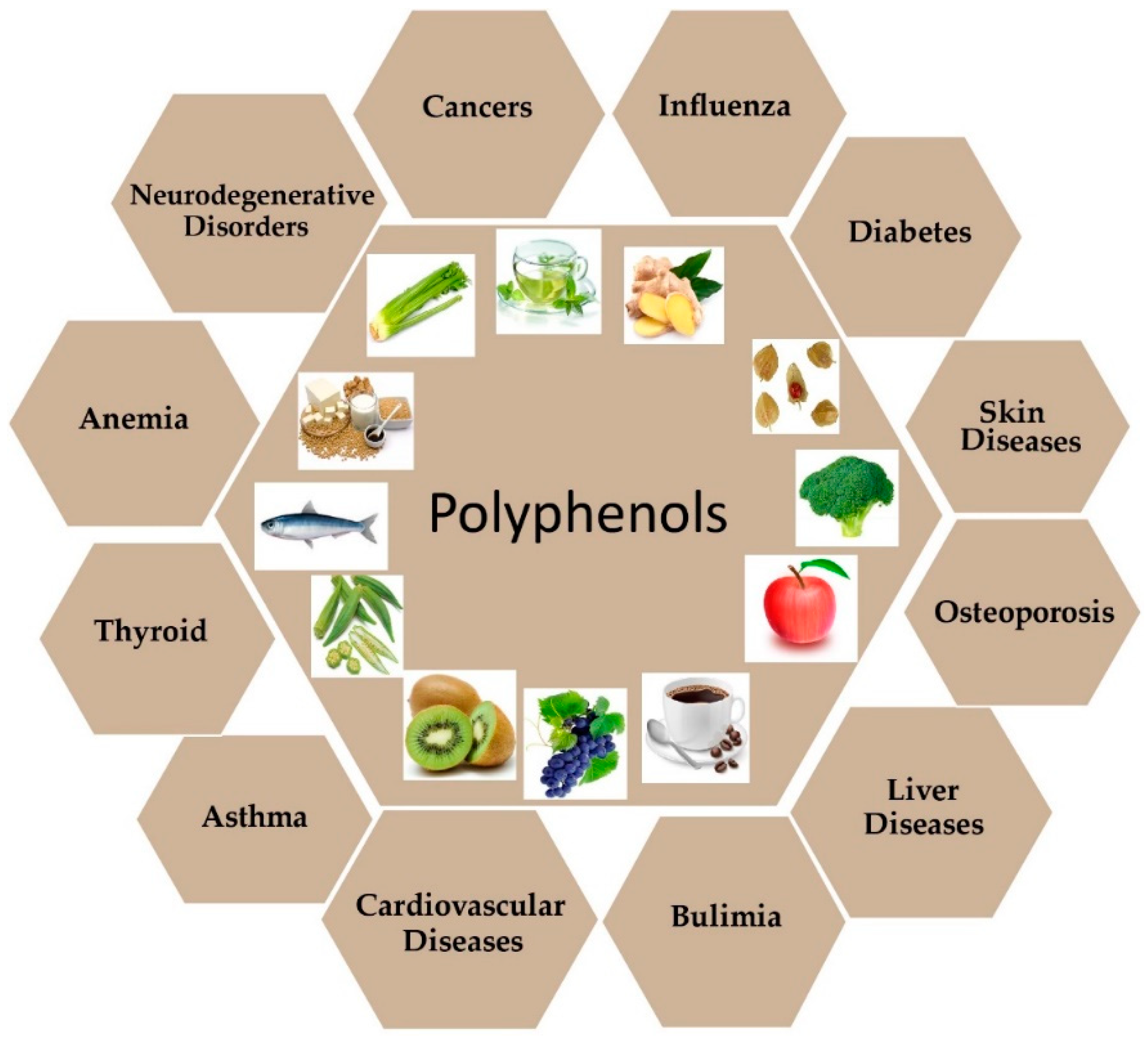
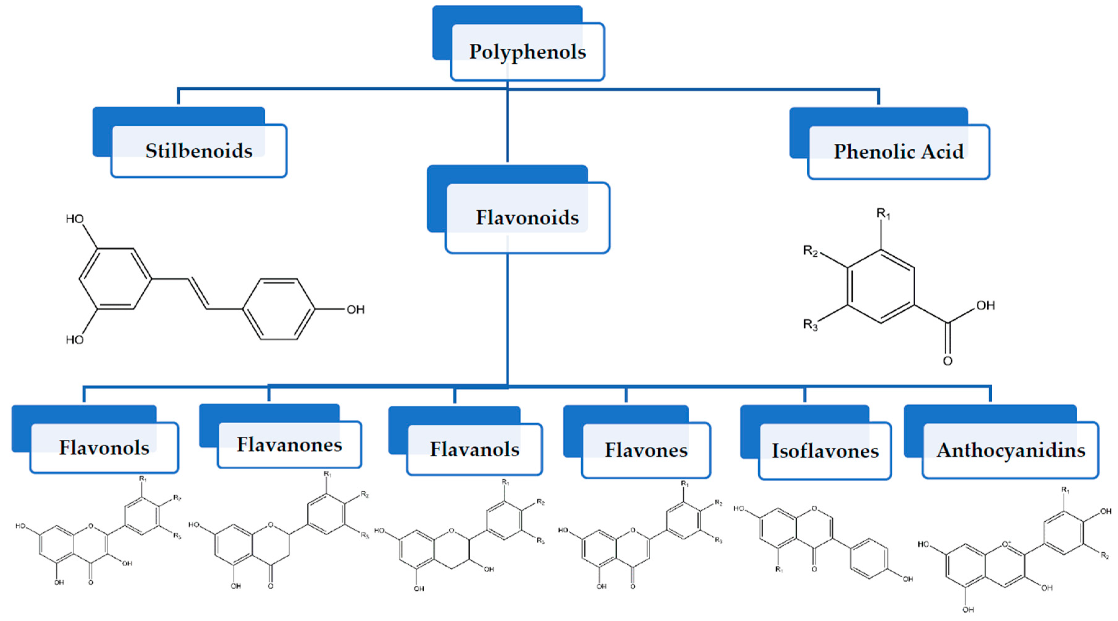
| Polyphenols | Dietary Source | * Chemical Structure | ** Molecular Formula | References |
|---|---|---|---|---|
| Apigenin | Grapefruit, parsley, onion, orange, tea and wheat sprouts |  | C15H10O5 | [63] |
| Anacardic Acid | Cashew nuts |  | C22H36O3 | [64] |
| Biochanin | Red clove, chickpea, clover sprout and kidney beans |  | C16H12O5 | [65,66,67] |
| Butein | Rhus verniciflua, Caesalpinia sappan and Carthamus tinc-torius |  | C15H12O5 | [68,69] |
| Catechin | Green tea, apples, blackberries, dark chocolate and red wine |  | C15H14O6 | [70] |
| Cyanidin | Acai berry, bilberry, blackberry, cranberry and raspberry |  | C15H11O6+ | [71] |
| Curcumin (CUR) | Turmeric |  | IC21H20O6 or C21H20O6 | [72] |
| Caffeic Acid | Coffee and olive oil |  | C9H8O4 | [73] |
| Cholorogenic Acid | Pomegranate and berries |  | C16H18O9 | [74] |
| Capsaicin | Chili peppers |  | C18H27NO3 | [75] |
| Daidzein | Soybeans and tofu |  | C15H10O4 | [76,77] |
| Delphinidin | Cereal grains |  | C15H11CIO7 | [78] |
| Diosmetin | Vetch |  | C16H12O6 | [79] |
| Ellagic Acid | Blackberries, raspberries and pomegranate |  | C14H6O8 | [80] |
| Epicatechin | Milk, chocolates, and commercial reduced fat |  | C15H14O6 | [81] |
| Epigallocatechin-3-gallate (EGCG) | Green tea |  | C22H18O11 | [70] |
| Gallic Acid | Pomegranate, nuts and green tea |  | C7H6O5 | [82] |
| Genistein | Fats, oils, beef, red clover, soybeans, and fava beans |  | C15H10O5 | [83,84] |
| Gnetol | Gnetum ula, gnetum gnemon, trees, shrubs and lianas |  | C14H12O4 | [85] |
| Hesperidin | Bitter orange, petit grains, orange, lime and lemon |  | C28H34O15 | [86] |
| Isoliquiritigenin | Rose petals |  | C15H12O4 | [87] |
| Kaempferol | Apples, grapes, tomatoes, green tea, potatoes, onions and broccoli |  | C15H10O6 | [88] |
| Licochalcone A | Cranberry |  | C21H22O4 | [89] |
| Luteolin | Celery, broccoli, green pepper, parsley, thyme, dandelion, perilla and chamomile tea |  | C15H10O6 | [90,91] |
| Macluraxanthone | Maclura tinctoria (Hedge apple) and dyer’s mulberry |  | C23H22O6 | [92] |
| Myricetin | Vegetables, fruits, nuts, berries, tea and red wine |  | C15H10O8 | [93] |
| Naringenin | Grapes |  | C15H12O5 | [94] |
| Oxyresveratrol | Morus alba and artocarpus lakoocha |  | C14H12O4 | [95] |
| Peonidin | Cranberries, blueberries, plums, cherries and sweet potatoes |  | C16H13O6+ | [96] |
| Piceatannol | Berries, grapes, rhubarb (rheum), passion fruit (passiflora) and white tea. |  | C14H12O4 | [97] |
| Pterostilbene | Blueberries and grapes |  | C16H16O3 | [98] |
| Quercetin | Vegetables, fruits and beverages, spices, soups and fruit juices |  | C15H10O7 | [99,100] |
| Resveratrol | Almonds, blueberries and grapes |  | C14H12O3 | [98] |
| Rosemarinic Acid | Rosemary |  | C18H16O8 | [101] |
| Rutin | Citrus fruits, apple, berries and peaches |  | C27H30O16 | [92,102,103] |
| Scopoletin | Vinegar, dandelion and coffee |  | C10H8O4 | [104] |
| Silibinin | Milk and artichokes |  | C25H22O10 | [105,106] |
| Tangeretin | Citrus fruits |  | C20H20O7 | [107] |
| Taxifolin | Vinegar |  | C15H12O7 | [108] |
| Theaflavin | Tea leaves, black tea and oolong tea |  | C29H24O12 | [109] |
| Tricin | Rice bran and sugarcane |  | C17H14O7 | [110] |
| Xanthohumol | Hop plants |  | C21H22O5 | [111] |
| Dietary Compounds | Epigenetic Modifications | Gene Targets | * Overall Role in Cancer Progression | Dose | In Vitro Model | In Vivo Model | References |
|---|---|---|---|---|---|---|---|
| Apigenin | DNMT1 inhibitor DNMT3a inhibitor DNMT3b inhibitor HDAC1 inhibitor HDAC3 inhibitor | NFE2L, NQO1, Nrf2, GRP78, GADD153, p21, waf1, and hTERT p53 | ↓ Viability ↓ GLI1 expression Cell cycle arrest ↑ Apoptosis ↑ Caspase 3 Activity | 20–30 μM 40–160 μM 20–50 μM 20–100 μM 20–40 μM | Pancreatic cancer Lung cancer (H460 cells) Breast cancer (BT-474 cells) Skin cancer (JB6 P+ cells) Prostate cancer (PC-3, 22Rv1 cells) | Mouse | [163,164,165,166,167,168,169,170] |
| Curcumin | DNMT inhibitor HAT inhibitor HDAC1 inhibitor Down-regulation of histone methylation | CDKN2B, NEUROG1, NFE2L2, Nrf2, Neurog1 RASSF1A, p16, SPARC, SOCS1, SOCS3, p53, p21, GAS5, HOTAIR, H19, AF086415, AK095147, RP1-17916.3, MUDENG, AK056098, AK294004 | ↓ Proliferation ↑ Apoptosis ↓ ERK, MKK4, JNK activity ↓ Bcl-2 ↓ Akt expression ↑ p38 activation ↓ Cell viability ↑ Bax Activity | 40 μM 0–100 μM 0–50 μM 2.5–160 μM 7.5–10 µM 0–50 μM | Breast cancer (MCF-7 cells) Prostate cancer (LnCap cells) Colorectal cancer (HCT116, HT29 and RKO cell lines) Gastric cancer (MiaPaCa-2, PANC-1 cells) Breast cancer (MCF-7 cells) Ovarian cancer (SKOV3 cells) | [171,172,173,174,175,176,177,178,179,180,181,182] | |
| Daidzein | DNMT inhibitor HDAC inhibitor | BRCA1, GSTP1, EPHB2, MMP-2, BRF1, BRF2, RARβ | ↓ Proliferation ↑ Apoptosis ↓ ERK, MKK4, JNK activity | 200–600 μM 20–100 μM 3–50 μM 12.8–100 μM | Colorectal cancer Breast cancer (MDA-MB-231 cells) Liver cancer (SKHEP-1 cells) Prostate cancer | [62,183,184] | |
| Delphinidin | DNMT inhibitor HAT inhibitor HDAC-3 inhibitor | p21, WAF1/Cip1, p53, p27/KIP1 | ↓ Cell Proliferation ↓ Metastasis Cell Cycle Arrest Oxidative Stress | 3–90 μM 30–240 μM 5–60 µM 100 μM | Prostate cancer (PC3 cells) Colorectal cancer (HCT116 cells) Lung cancer (NCI-H441 cells) Prostate cancer (LNCaP cells) | Athymic nude mice | [185,186,187,188] |
| Myricetin | DNMT inhibitor Increased SIRT1 activity | GSTP1, RARβ, HIN-1 | ↑ Apoptosis Autophagy | 20–40 μM 5–25 μM 100 μM | Gastric cancer (GC HGC-27, SGC7901 cells) Breast cancer Colon cancer (HCT-15 cells) | [189,190,191] | |
| Ellagic Acid | DNMT1 inhibitor DNMT3b inhibitor HDAC inhibitor | p16INK4a, RASSF1A, GSTP1, HIN1, VEGF, MMP-2, p53 | ↑ Apoptosis Cell proliferation Cell migration ↑ Caspase 3 Activity ↑ Caspase 9 Activity | 50–200 μM 10–100 μM | Colorectal cancer Prostate cancer (PC-3 cells) Breast cancer (MCF-7 cells) | [192,193,194,195] | |
| EGCG | DNMT inhibitor HAT inhibitor Down-regulation of histone methylation Effect on histone ubiquitination Upregulation of histone phosphorylation | p16INK4a; RARβ; MGMT; hMLH1; GSTP1; WIF-1; RECK, Cip1/p21 AT102202, p53, p21 | ↓ Invasiveness ↓ Proliferation ↑ Apoptosis ↑ Caspase 3 Activity ↑ Caspase 8 Activity ↑ Cytochrome c | 1–40 μM 1–50 μM 5–20 μM 20–100 μM 1–50 μM 0–20 μg/mL | Breast cancer (MCF-7 cells) Colorectal cancer (HT-29 cells) Lung cancer (CL1-5 cells) Gastric cancer (MKN-1, MKN-28, MKN-45, NUGC-3 and TMK-1) Colorectal cancer Skin cancer (A431 cells) | Xenograft mice | [47,196,197,198,199,200,201,202,203,204,205,206,207,208] |
| Hesperidin | DNMT inhibitor HDAC inhibitor | GSTP1, Akt, LAMTOR2, LAMTOR3, LAMTOR5, MAPK1, KRAS, HRAS, MAPK3 | ↓ Cell proliferation ↑ Apoptosis ↑ Glucose uptake ↑ ASK1/JNK pathway ↑ ROS production | 40–90 μM 40–200 μM 650 μM 20–50 μM 90 μM 50 μM | Breast cancer (MCF-7, MDA-MB-231 Cells) Liver cancer Cervical cancer (SiHa cells) Esophageal cancer Prostate cancer (PC-3 cells) Endometrial carcinoma (ECC-1 cells) | Xenograft mice Rats | [209,210,211,212,213,214,215] |
| Kaempferol | DNMT3a inhibitor DNMT3b inhibitor HDAC1 inhibitor | p-Akt, ERK, MSK1, CD1, p23, BTG3, BRCA1, MGMT, and hMLH1 | ↑ Apoptosis ↓ Glucose uptake Autophagy Cell cycle arrest | 100 μM 10–50 μM 0–60 μM 4 µM 50 μM 20 mg/kg | Liver cancer (SK-HEP-1 cells) Lung cancer (A549 cells) Colorectal cancer (HT-29 cells) Breast cancer (MCF-7 cells) Gastric cancer (G9a cells) Gastric cancer (MKN28, SGC7901 and GSE-1 cells) | Athymic mice Xenograft mice | [216,217,218,219,220,221] |
| Luteolin | DNMT inhibitor HDAC inhibitor | VRK1, MPK2 | ↑ Apoptosis Cell cycle arrest Cell invasion | 20–50 μM 5–50 μM 10–40 μM 20–100 μM 10 μM | Esophageal cancer Lung cancer (A549 cells) Breast cancer (MCF-7 cells) Colorectal cancer Lung cancer (A549 cells) | Xenograft Mice | [222,223,224,225,226,227] |
| Pterostilbene | DNMT inhibitor Decreased SIRT1 activity | p53, NF-κB and miRNA488 | ↑ Apoptosis Cell cycle arrest | 25–75 μM | Breast cancer (MCF-7 and MDA-MB-231 cells) | Mice | [137,228,229,230,231,232] |
| Polyphenol- rich Strawberry extract (PRSE) | Csf1, Mcam, Nr4a3, SET, Gpnmb, Itgb3, CC17, Ctsl, Cxcr4, Htatip2, Mmp-10 and Mmp3 | ↓ Cellular Viability ↓ Number of cells in S phase Accumulation of cells in G1 phase ↓ Tumor Weight ↓ Tumor Volume | 0.5–5 mg/mL | Breast Cancer (MCF-7 and A-17 cells) | Mice | [233] | |
| Genistein | DNMT1 inhibitor DNMT3a inhibitor DNMT3b inhibitor HDAC inhibitor HAT activator Decreased SIRT activity Upregulation of histone methylation | p16INK4a; RAR β; MGMT; PTEN; CYLD, MGMT, CDKN2A, BTG3, TERT, GSTP1, EZH 2, FoxM1, sFRP1, p21, p16, PTEN, CCLD, p53, FOXA3, SIRT1, BTG3, hTERT, RAR, HOTAIR | ↓ Proliferation ↓ Tumorigenesis ↑ Apoptosis ↑ mRNA expression of tumor suppressor genes ↑ H2A variant at serine 139 (γ-H2AX) | 25–75 μM 0.5–50 μM 0.5–50 μM 100 μM 20–50 μmol/L 5–100 μM | Lung cancer (H446 cells) Breast cancer (MCF-7, MDA-MB-231 cell lines) Prostate cancer (LAPC-4 cells) Cervical cancer Esophageal squamous cell carcinoma Prostate, breast cancer and renal cancer | Agouti mice Sprague-Dawley rats | [234,235,236,237,238,239,240,241,242] |
| Gallic Acid | DNMT1 inhibitor DNMT3b inhibitor | MMP-2, MMP-9, ADAM17, Erk/p-Erk, p-Akt | ↑ Apoptosis | 200 μM 80.5 μM 25–200 μM 0–40 μg/mL 50 μM | Colorectal cancer Breast cancer (MCF-7 cells) Prostate cancer (PC-3 cells) Cervical cancer (HeLa and HTB-35 cells) Oral cancer (H1299 cells) | [243,244,245,246,247] | |
| Naringenin | DNMT1 inhibitor DNMT3a inhibitor DNMT3b inhibitor HDAC1 inhibitor | ATF3, PARP, p38, MMP-9, ERK, Akt | ↑ Apoptosis Cell cycle arrest ↓ Cell growth ↓ Cell proliferation | 100 M μM 20–80 μM 20 or 50 μmol/L 25–200 μM 50–100 μM | Lung cancer (A549 cells) Gastric cancer Esophageal cancer (KYSE-510 cells) Liver cancer (HepG2, Huh-7, and HA22T cells) Colorectal cancer (HCT116, SW480, Lovo, and HT-29 cells) | Resection mice Rats | [248,249,250,251] |
| Piceatannol | DNMT3a inhibitor Decreased SIRT1 activity | RASSF1A, GSTP1, HIN1 mTOR | ↑ Apoptosis Cell cycle arrest | 30 μM 25 μM 50 μM | Colorectal cancer (HCT116 and HT29 cells) Prostate cancer (DU145 and PC-3 cells) Breast cancer (MCF-7 cells) | Mice | [193,252,253,254] |
| Quercetin | DNMT1 inhibitor HDAC inhibitor Down-regulation of histone demethylation | CDKN2A (p16INK4a), AMPK, Akt, DBH-AS1, p53 | ↓ Proliferation ↓ Akt phosphorylation ↓ Angiogenesis ↑ Caspase 3 and 7 activity ↑ Bax Activity | 1–200 μM 40–160 μM | Liver cancer (HepG2 and SMCC-7721 cells) Lung cancer (A549 cells) Gastric cancer (AGS and MKN28 cells) Colon cancer (HCT116 cells) | Mice | [47,255,256,257,258,259,260,261,262] |
| Xanthohumol | DNMT inhibitor HDAC inhibitor | CXCR4, CXCL12, p53 | ↑ Apoptosis Cell cycle arrest Cell Invasion Cell proliferation Cell migration | 14–42 μM 5–40 μM | Lung cancer (A549 cells) Liver cancer Breast cancer Prostate cancer (DU145 and PC3 cells) | Transgenic Mice | [263,264,265,266,267] |
| Sulforaphane | DNMT3a inhibitor DNMT3b inhibitor HDAC inhibitor Upregulation of histone phosphorylation | NFE2L2, TERT, Nrf2, ZEB1, COX-2/MMP-2, 9/snail, p21, p27, RBP2 | ↓ Proliferation ↑ Apoptosis Cell cycle arrest | 50 μM 5–10 µM 0–30 μM | Colorectal cancer Breast cancer (MDA-MB-231 and MCF-7 cells) Bladder cancer (T24 cells and 5637 cells) | Xenograft Mice | [268,269,270,271,272,273,274,275] |
| Resveratrol | DNMT3a inhibitor DNMT3b inhibitor Decreased SIRT1, SIRT2, and SIRT3 activity HAT inhibitor Regulation of histone phosphorylation | PTEN, XRCC1, p21 p16, MDR1, SP-1, STIM1, FOXO, PCGEM1, PRNCR1, PCAT29, AK001796, MALAT1, u-Eleanor, LINC00978, p53, p21 | ↓ Proliferation ↑ Apoptosis ↓ Metastasis ↑ Caspase 8/9 activity ↑ Bax Activity ↓Bcl-2 Activity | 50–150 μM 5–50 μM 50–200 μM 20–150 μM 25–100 μM 150–250 μM 25–100 μM | Breast cancer (MCF-7 cells) Lung cancer (H1703 and H1975 cells) Gastric cancer (Ki67 cells) Colon cancer (HT-29 cells, COLO 21 cells) Prostate cancer (PC3 and DU145 cells) Cervical cancer Liver cancer (Huh7 cells) | Xenograft Mice | [179,276,277,278,279,280,281,282,283,284,285,286,287,288] |
| Combinatorial Therapy | Organ of Study | In Vitro Model | In Vivo Model | *Epigenetic Modifications and Molecular Mechanism | References | ||
|---|---|---|---|---|---|---|---|
| Cell Lines | Dose | ||||||
| Apigenin + IFNγ | Cervical cancer | HeLa and SiHa cells | 5–15 μM + 100 ng/mL | ↓ Cell viability ↑ Apoptosis Up-regulation of DNMT1 | [289] | ||
| Apigenin + Paclitaxel | Ovarian cancer | HeLa, A549, Hep3B and HEK293A cells | 15 µM + 4 nM | Apoptosis through suppressing SOC activity ↑ ROS and caspase-2 cleavage. | [290] | ||
| Apigenin + Cisplatin | Renal cancer | Human renal proximal tubular epithelial (HK-2) cells | 5–20 µM + 40 µM | Apigenin reduced cisplatin-induced caspase-3 activity and PARP cleavage ↓ ROS production and p53 activation Akt phosphorylation | [293] | ||
| Prostate cancer | PC3 PCa cells | 15 μM + 7.5 µM | Upregulation of Caspase-8, Apaf-1 and p53 Down-regulation of Snail expression. Repressed phosphorylation of p-PI3K and p-Akt | [292] | |||
| Apigenin + doxorubicin | Lymphoid leukemia | CCRF, CEM, Jurkat and THP-1 cells | 0.01 μM + 0.4 μM | ↑ caspase-3 activity Cell cycle arrest at S and G2/M phase ↑ DNA damage | [294] | ||
| Apigenin + etoposide | ↑ caspase-3 activity ↑ caspase-8 activity ↑ caspase-9 activity | [294] | |||||
| Apigenin + Naringenin | Colon cancer | Mice | 0.1 μM + 0.05 μM | ↑ ER-mediated YAMC cell growth ↑ activation of ERβ | [295] | ||
| Apigenin + Gemcitabine | Pancreatic cancer | MiaPaca-2, AsPC-1 cell lines | 30 μM + 05–2 μM | Xenograft mice model | Down-regulation of NF-κB activity Suppression of Akt activation | [296] | |
| Curcumin + Resveratrol | Prostate cancer | PTEN-CaP8 cancer cells | B6C3F1/J mice | ↓ p-Akt, and cyclin D1 activity | [297] | ||
| Curcumin + Catechins | Colon cancer | DMH rat model | 0.1% + 0.1% | ↑ Apoptotic index ↓ Proliferation index | [298] | ||
| Curcumin + Piperine | Breast cancer | MCF-7 and Sum159 cells | 5–25 μM + 5–25 µM | Inhibit mammosphere formation ↓ stem cell self-renewal ↓ in the cell percentage expressing stem cell marker ALDH1 Inhibit Wnt Signaling | [300] | ||
| Curcumin + Paclitaxel | Breast cancer | MDA-MB-231 cells | 10 μM + 10 µM | Nude mice model | 100 mg/kg + 7 mg/kg | Inhibition of tumor cell growth ↓ Tumor size ↓ Tumor cell proliferation ↑ Expression of MMP-9 | [301] |
| Curcumin + Arctigenin + Green tea + Epigallocatechin gallate (EGCG) | Prostate cancer | LNCaP cells | 5–10 μM + 1 μM + 40 μM | ↑ Apoptosis ↑ Cell cycle arrest at G0/G1 phase ↓ Activation of NFκB, PI3K/Akt and Stat3 pathways ↓ Cell migration | [303] | ||
| Breast cancer | MCF-7 cells | 5–10 μM + 1 μM + 40 μM | |||||
| Curcumin + Epigallocatechin gallate (EGCG) | Non-small lung cancer | A549 and NCI-H460 cells | 10 μmol/L +10 μmol/L | ↓ Tumor size Cell cycle arrest at G1 and S/G2 phase Inhibition of expression of cyclin B1 and cyclin D1 Inhibition of clonal formation Down-regulation of DNMTs | [304,305] | ||
| Prostate cancer | LNCaP, DU145 and PC3 cells | 50 μM + 100 µM | ↑ p21 expression Cell cycle arrest at S, G2/M phase | ||||
| Curcumin + Dasatinib | Colon cancer | HCT-116, HT-29 and SW-620 | APCMin+/− mice | ↓ Cell proliferation ↑ Apoptosis ↑ Invasion through the extracellular matrix ↑ Tubule formation by endothelial cells | [306] | ||
| Curcumin + TRAIL | Prostate cancer | LNCaP cells | PC3 cells | ↑ Apoptosis ↑ MMP-9, MMP-2, caspase-3, and caspase-9 activity | [307,308] | ||
| Curcumin + Gemcitabine | Pancreatic cancer | BxPC3, MiaPaCa2 and Panc1 PDAC cells | 100 mg/kg + 25 mg/kg | ↓ Tumor growth ↓ NF-κB activity ↓ VFGF expression | [309] | ||
| Genistein + Delphinidin + Alternariol (AOH) | Colon cancer | HT-29 cells | 25 μM + 100 μM + 50 μM | ↑ Cytotoxic effect ↑ Genotoxicity effect ↑ Topoisomerase poisoning ↓ ROS generation | [310] | ||
| Genistein + Erlotinib | Bladder cancer | A431 cells | 100 μM + 10 nM | Inhibitor of EGFRs Overexpression of RTKs | [311] | ||
| Genistein + Sulforaphane | Breast cancer | MCF-7 and MDA-MB-231 cells | 5 µM + 10-15 µM | Cell cycle arrest at G1 and G2/M phase ↓ Cell viability ↑ Apoptosis | [312] | ||
| Genistein + Cisplatin | Breast cancer | MCF-7 and T47D cells | 1 μM + 10 μM | ↓ ROS production ↑ Cell viability ↓ Autophagy ↓ Apoptosis Cell cycle arrest at subG0/G1 phase | [313] | ||
| Genistein + Tamoxifen | 1 μM + 10 μM | ||||||
| Genistein + Paclitaxel | 1 μM + 10 μM | ||||||
| Genistein + Resveratrol | Prostate cancer | Rats | 83 mg/kg + 83 mg/kg | ↓ Tumor growth Inhibition of Growth factors | [314] | ||
| Genistein + Quercetin + Biochanin A | Prostate cancer | PC-3, LNCaP, DU-145 cells | 8.33 μM + 8.33 μM + 8.33 μM | ↑ BAX/BCL-2 activity ↑ caspase-3 activity ↑ ER-β activity ↑ p-JNK activity ↓ p-ERK activity ↓ PCNA activity ↓ Cell proliferation ↑ Apoptosis | [315] | ||
| Genistein + EGCG + quercetin | Prostate cancer | CWR22Rv1 cells | 2.5 μM + 2.5 μM + 2.5 μM | ↑ p53 activity ↓cell proliferation | [316] | ||
| Resveratrol + Thymoquinone | Breast cancer | MCF-7 and T47D cells | 10 μM + 25–300 μM | Balb/C mice | 50 mg/kg + 50 mg/kg per day | ↑ Apoptosis, ↓ Tumor growth Inhibition of angiogenesis | [317] |
| Resveratrol + Quercetin | Prostate cancer | 22Rν1, DU145 and PC3 cells | TRAMP mice model | 625 mg/kg + 60 mg/kg | ↓ Cell proliferation | [318] | |
| Resveratrol + Curcumin + ADR | Ovarian cancer | A2780 cells | 10 μM +2 μM +1 μM | Xenograft model in Athymic mice | 19.68 mg/kg + 26.06 mg/kg + 5mg/kg | ↓ Cell viability ↓ Tumor size ↑ Apoptosis | [319] |
| Resveratrol + Quercetin + ADR | Ovarian cancer | A2780 cells | 10 μM +10 μM +1 μM | Xenograft model in Athymic mice | 19.68 mg/kg + 5.2 mg/kg + 5mg/kg | ↓ Cell viability ↓ Tumor size ↑ Apoptosis | [319] |
| Resveratrol + Doxorubicin | Gastric cancer | SGC7901 and MGC803 cell lines | 50 mg/L + 0.75 mg/L | Nude xenograft mice model | 3 mg/kg + 50 mg/kg$ | ↑ expression of caspase 9 ↑ PTEN, TSC1, TSC2, and cleaved caspase 3 ↓ p-AkT, and mTOR activity | [320] |
| Resveratrol + Genistein + Quercetin + Apigenin + Baicalein + Curcumin + EGCG | Prostate cancer | PC3 and LNCaP cells | TRAMP mice model | 1 μmol/L + 20 μmol/l + 10 μmol/L + 3 μmol/L+ 3 μmol/L+ 10 μmol/L+ 10 μmol/L | ↓ GLi1 mRNA activity$↓ Tumor size | [321] | |
| Resveratrol + Pomegranate + Orange + Lemon + Olive + Cocoa + Grape seed | Breast Cancer | MCF-7 cells | 53.85 mg + 161.5 mg + 53.85mg + 53.85 mg + 161.5 mg + 161.5 mg + 53.85 mg | ↓ Anti-proliferative activity $↓ Estrogenic estrogenic/anti-esterogenic activity | [323] | ||
| EGCG + Sunitinib | Breast cancer Non-small cell lung cancer | H460, H1975, and MCF-7 cells | 50 μM + 3 μM | Xenograft mice model | 50 mg/kg + 40 mg/kg | Suppression of IRS/MAPK/p-S6K1 signaling | [324] |
| Resveratrol + Quercetin + Catechin + Gefitinib | Breast cancer | MDA-MB-231 cells | 15 μM + 15 μM+ 15 μM + 15 μM | SCID mice model | 5 mg/kg +5 mg/kg + 5 mg/kg + 5 mg/kg + 200 mg/kg | ↓Tumor size Cell cycle arrest at S phase ↓ Cell viability Inhibition of Akt/mTOR signaling | [322] |
| EGCG + Vitexin-2-O-xyloside + Glucoraphasatin | Breast cancer | MDA-MB-231 cells MCF-7 cells | (1980 ± 94) μg/mL + (1200 ± 66) μg/mL + (21 ± 6) μg/mL + (350 ± 47) μg/mL + (350 ± 48) μg/mL + (31 ± 4) μg/mL | Induction of apoptosis Cell cycle arrest at Regulation of Bcl2, Bax, cleaved caspase-9 and PARP ↑ ROS production | [325] | ||
| Colorectal cancer | Caco-2 cells LoVo cell | (21 ± 3) μg/mL + (120 ± 9) μg/mL + (16 ± 4) μg/mL + (135 ± 16) μg/mL + (158 ± 13) μg/mL + (36 ± 5) μg/mL | |||||
| EGCG + NAC | Lung cancer | H1299 cells | 100 μM + 2 mM | CL13 mice | ↑ Apoptosis ↑ ROS production | [326] | |
| EGCG + Pterostilbene | Pancreatic cancer | PANC-1 and MIA-Pa-Ca-2 cells | 20–40 μM + 30 μM | ↓ Cell proliferation Cell cycle arrest at S phase ↑ caspase-3/7 activity | [327] | ||
| EGCG + TRAIL | Pancreatic cancer | MIA-Pa-Ca-2 cells | 50 μg/mL + 5 ng/mL | ↑ Apoptosis ↑ Activation of caspase-8 and caspase-9 | [328] | ||
| EGCG + 5- Fluorouracil | Colorectal cancer | HCT-116 and SW480 cells | 25–400 μM + 2.5–40 μM | Xenograft mice model | ↓ miR-34a, miR-145, and miR-200c Cell cycle arrest ↓ Notch1, Bmi1, Suz12, and Ezh2 activity | [329] | |
| EGCG + 5- Fluorouracil | Colorectal cancer | HCT-116 and SW480 cells | 25–400 μM + 2.5–40 μM | Xenograft mice model | ↓ miR-34a, miR-145, and miR-200c ↑ spheroid formation ↓ Notch1, Bmi1, Suz12, and Ezh2 activity Cell cycle arrest at G0/G1 phase | [329] | |
| Sulforaphane + Green tea polyphenols (GTPs) | Breast cancer | MDA-MB-231 cells | 5–10 μM + 20 µg/mL | Reactivation of Tumor suppressor genes (TSGs) p21CIP1/WAF1 and KLOTHO Cell cycle arrest at G2/M phase ↓ CDK1 and CDC25C expressions Inhibition of IGF-1 pathways | [330] | ||
| Sulforaphane + Withaferin-A | Breast cancer | MCF-7 and MDA-MB-231 cells | 5 μM + 10 μM | ↓ HDAC1 Inhibition of DNMT1, DNMT3A, and DNMT3B ↑ Apoptosis ↓ BAX/BCL-2 activity | [331] | ||
| Sulforaphane + Curcumin | Liver cancer | HepG2-C8 cells | Low dose: 12.5 μM + 10 μM High dose: 50 μM + 25 μM | ↑ ARE-luciferase activity ↑ Expression of HO-1 and UGT1A1 ↑ Nrf2 mRNA levels | [332] | ||
| Sulforaphane + EGCG | Prostate cancer | PC-3-AP-1 cells | Low Dose: 25 μmol/L +20 μmol/L High Dose: 25 μmol/L +μmol/L | Nrf2-deficient mice | 45 mg/kg + 100 mg/kg | Inhibition of AP-1 activity Down-regulation of Nrf2-dependent genes | [334] |
| Ovarian cancer | SKOV-ip1 and SKOVTR-ip2 cells | 10 μM + 20 μM | ↑ Expression of hTERT, DNMT1 ↓ Cell viability Cell cycle arrest in G2/M and S phases ↑ Apoptosis | [333] | |||
| Colon cancer | HT-29 cells | Low Dose: 25 μM + 20 μM High Dose: 10 μM + 20 μM | ↓ Cell viability ↑ AP-1 activity | [335] | |||
| Sulforaphane + Acetazolamide (AZ) | Urothelial cancer | HTB-9 and RT112(H) cells | 40 μM + 40 μM | ↑ Apoptosis ↓ Ki-67, pHH3, cyclin D1 activity Cell cycle arrest Inhibition of Akt kinase activity Downregulation of p-Akt (Ser473) and p-S6 activity | [336] | ||
| Sulforaphane + Docetaxel + Paclitaxel | Breast cancer | SUM149 and SUM159 cells | 5 μM + 0–20 μM | Xenograft mice model | 50 mg/kg daily + 10 mg/kg weekly | ↓ Tumor growth Inhibition of NF-κB p65 translocation | |
© 2019 by the authors. Licensee MDPI, Basel, Switzerland. This article is an open access article distributed under the terms and conditions of the Creative Commons Attribution (CC BY) license (http://creativecommons.org/licenses/by/4.0/).
Share and Cite
Arora, I.; Sharma, M.; Tollefsbol, T.O. Combinatorial Epigenetics Impact of Polyphenols and Phytochemicals in Cancer Prevention and Therapy. Int. J. Mol. Sci. 2019, 20, 4567. https://doi.org/10.3390/ijms20184567
Arora I, Sharma M, Tollefsbol TO. Combinatorial Epigenetics Impact of Polyphenols and Phytochemicals in Cancer Prevention and Therapy. International Journal of Molecular Sciences. 2019; 20(18):4567. https://doi.org/10.3390/ijms20184567
Chicago/Turabian StyleArora, Itika, Manvi Sharma, and Trygve O. Tollefsbol. 2019. "Combinatorial Epigenetics Impact of Polyphenols and Phytochemicals in Cancer Prevention and Therapy" International Journal of Molecular Sciences 20, no. 18: 4567. https://doi.org/10.3390/ijms20184567
APA StyleArora, I., Sharma, M., & Tollefsbol, T. O. (2019). Combinatorial Epigenetics Impact of Polyphenols and Phytochemicals in Cancer Prevention and Therapy. International Journal of Molecular Sciences, 20(18), 4567. https://doi.org/10.3390/ijms20184567