Review

All-trans Retinoic Acid as a Versatile Cytosolic Signal Modulator Mediated by CRABP1

Department of Pharmacology, University of Minnesota, Minneapolis, MN 55455, USA
*
Author to whom correspondence should be addressed.
Int. J. Mol. Sci. 2019, 20(15), 3610; https://doi.org/10.3390/ijms20153610
Submission received: 30 June 2019 / Revised: 18 July 2019 / Accepted: 20 July 2019 / Published: 24 July 2019

Abstract

:
All-trans retinoic acid (AtRA), an active metabolite of vitamin A, is recognized for its classical action as an endocrine hormone that triggers genomic effects mediated through nuclear receptors RA receptors (RARs). New evidence shows that atRA-mediated cellular responses are biphasic with rapid and delayed responses. Most of these rapid atRA responses are the outcome of its binding to cellular retinoic acid binding protein 1 (CRABP1) that is predominantly localized in cytoplasm and binds to atRA with a high affinity. This review summarizes the most recent studies of such non-genomic outcomes of atRA and the role of CRABP1 in mediating such rapid effects in different cell types. In embryonic stem cells (ESCs), atRA-CRABP1 dampens growth factor sensitivity and stemness. In a hippocampal neural stem cell (NSC) population, atRA-CRABP1 negatively modulates NSC proliferation and affects learning and memory. In cardiomyocytes, atRA-CRABP1 prevents over-activation of calcium-calmodulin-dependent protein kinase II (CaMKII), protecting heart function. These are supported by the fact that CRABP1 gene knockout (KO) mice exhibit multiple phenotypes including hippocampal NSC expansion and spontaneous cardiac hypertrophy. This indicates that more potential processes/signaling pathways involving atRA-CRABP1 may exist, which remain to be identified.

Graphical Abstract

1. Introduction

Vitamins are essential micronutrients required for a myriad of biological processes. Vitamin A was first known to play an essential role in vision [1,2]. Its chemical structure was first deciphered in 1931 [3] and later crystallized in 1937 [4]. Since then, numerous studies have demonstrated the significance of vitamin A and its metabolites in various physiological processes such as fertility [5], embryonic development [6,7], and many other biological processes for survival and maintaining health [8,9,10,11,12,13,14,15]. Vitamin A must be provided by one’s diet and is metabolized into different metabolites collectively called the retinoids, including retinoic acid (RA), retinol (ROL), retinal, etc., characterized based on the functional modification concatenated to the extreme end of the core scaffold [13,16]. Amongst these retinoids, various isomeric forms of RA: atRA, 9-cis-retinoic acid, and 13-cis-retinoic acid are best studied for their activities in regulating gene expression via binding to nuclear RA receptors (RARs) or retinoid X receptors (RXRs) [17,18,19,20]. Together, these mediate the well-established, canonical action of vitamin A as an endocrine factor that mainly acts to provide a homeostatic control of gene expression to maintain health [21,22].
Interestingly, recent studies have begun to demonstrate that vitamin A, especially its most active metabolite, atRA, can also elicit specific non-canonical (RAR/RXR-independent) activities beyond regulating gene expression [23,24,25,26,27]. Using genetic tools, it has been established that most of these non-canonical activities are mediated by a specific, highly conserved, cellular binding protein for atRA named CRABP1 that serves as a cytosolic signal modulator/integrator or functions as an RA-regulated signal scaffold [27]. This review focuses specifically on these new findings and the emerging concept that atRA can exert specific, non-canonical (RAR/RXR-independent) physiological activities through CRABP1.

2. Generation of RA

Vertebrates, including humans, derive vitamin A from plant sources as α and β carotenoids (commonly called proretenoid carotenoids) [28,29], and metabolites of vitamin A from animal sources as retinyl esters (REs) or retinol (ROL) [30,31]. In humans, vitamin A is metabolized in the small intestines and stored in the liver [32,33,34]. Carotenoids are metabolized by two pathways: central and eccentric pathways. The central pathway engages β-carotene 15, 15′-Dioxygenase 1 (BCO1) enzyme to cleave the core double bond of β-carotene, resulting in two molecules of retinal (retinaldehyde) [35], which are further reduced to form ROL by ROL reductases [36] to be stored or further metabolized. The eccentric pathway can be triggered enzymatically or non-enzymatically [37] to cleave the non-central β-carotene double bond [38], generating retinoids such as RAR antagonists, β-carotenes, and β-apocarotenones [39]. ROL can be directly absorbed by the mucosal cell of the small intestine [40], while REs are further hydrolyzed for intestinal absorption by enzymes such as pancreatic triglyceride lipase and pancreatic lipase-related protein 2 [32,41]. The absorbed ROL is re-esterified by transmembrane lecithin retinol acyltransferase (LRAT) [42,43] and incorporated into chylomicrons that are transferred to hepatic stellate cells [44,45] for storage or taken up by other organs/tissues [33,46,47].
Circulating ROL is carried by ROL-binding proteins (RBPs), which are then taken up by cells via STRA6 (stimulated by retinoic acid gene 6), a cell surface receptor for holo-RBP [31,48,49,50,51]. Inside the cells, ROL is converted to retinaldehyde by ROL dehydrogenases (RDHs) or retinyl esters [31]. Retinaldehyde is then irreversibly oxidized to RA by retinaldehyde dehydrogenases (RALDHs), which are key enzymes controlling the intracellular RA concentrations [52,53]. For cells that do not generate RA, they can obtain RA from RA-producing neighboring cells as a paracrine or from circulation as an endocrine factor [54]. Inside the cells, RA is first received by one of the two cellular RA-binding proteins (CRABPs) in the cytoplasm: CRABP1 or CRABP2 [55,56]. It is believed that RA, received by CRABP1, is targeted for degradation by cytochrome (CYP26) family members [57,58,59]. Whereas, when bound to CRABP2, RA is delivered to the nucleus for RAR/RXR-mediated regulation of the expression of RA-targeted genes [60,61,62,63]. This RAR/RXR-mediated gene-regulatory activity of RA elicits the well-established canonical signaling of RA and is believed to underlie the ubiquitous importance of vitamin A as an essential nutrient and endocrine factor. Studies of canonical actions of RA have been extensively reviewed; readers are referred to several excellent reviews in the literature for more detail [18,21,60,64,65,66,67,68,69].

3. Non-Canonical Activities of atRA

Recent studies have begun to show that RA, especially atRA, can also modulate cellular growth and function without involving RAR or RXR-mediated gene regulation. Most of these activities are specific to the cytoplasm, and they are mostly mediated by CRABP1. While the physiological implications of these findings remain debatable, studies using CRABP1 gene KO mice and primary cells have presented compelling evidence supporting this emerging concept. One unique feature of these non-canonical activities is their context-dependency, as demonstrated in the context of cellular growth (for stem cells) [26,70,71] and specific cellular function (for differentiated cells such as cardiomyocyte) [72]. These non-canonical functions of atRA are reviewed in the following.

3.1. atRA-CRABP1 Cross-Talks with RAF-MEK-ERK to Dampen Stem Cell Growth

The first evidence of the non-canonical activity of atRA was presented in studies using ESC, which is used to modulate growth factor sensitivity, as reflected by the activation of extracellular signal-regulated kinases 1/2 (ERK1/2) [73,74]. ERK1/2 is a known atRA-regulated gene in ESC; its gene expression is typically up-regulated by atRA approximately 8–12 h post-RA addition. However, careful kinetic studies revealed a surprising biphasic (15 min and 8 h, respectively) activation of ERK1/2 by atRA in ESC. Intriguingly, while phase 2 activation (8 h post-treatment) is effectively blocked by RAR antagonists (indicated via the canonical activity of RA), phase 1 (15 min post-treatment) activation cannot be blocked by RAR antagonists, suggesting a non-canonical response to RA [23,26].
Studies using CRABP1 knockdown ESCs have provided unambiguous evidence that phase 1 ERK1/2 activation by atRA is CRABP1-dependent [26]. This phenomenon is also observed in cancer cells using gene knockdown approaches [71]. Mechanically, atRA elicits such a non-canonical activity via CRABP1 [75], which directly interacts with rapidly accelerated fibrosarcoma (RAF), the first kinase typically activated by growth factors. This modulates the extent of the classical activation of ERK1/2 in ESCs by growth factors, such as in the presence of epidermal growth factor (EGF) that binds to EGF receptor (EGFR). Stimulated EGFR then activates Ras GTPase, which further activates RAF kinase [76,77]. Once activated, RAF phosphorylates mitogen-activated protein kinase MAPK-ERK kinase 1/2 (MEK1/2), which then phosphorylates/activates ERK1/2 [78]. In propagating this classical growth factor signaling pathway, the Ras-binding domain (RBD) of RAF plays an important role by engaging critical protein–protein interactions that regulate RAF activity [79]. In this context, CRABP1 functions by binding to the RBD of RAF, thereby modulating the activation of cRAF. NMR studies demonstrated that CRABP1 directly interacts with the RBD of cRAF and that atRA regulates this interaction, thereby negatively modulating growth-factor-stimulated RAF activation and its downstream signaling, leading to dampened ERK1/2 activation [75]. Thus, atRA, via binding to CRABP1, provides a physiological check (or dampening signal) for growth-factor-stimulated RAF activation in stem cells, thereby reducing their sensitivity to growth factor stimulation (Figure 1). This mechanism may operate in the context of the maintenance of a healthy stem cell population, which is to avoid accidental, unwanted stimulation by growth factors.
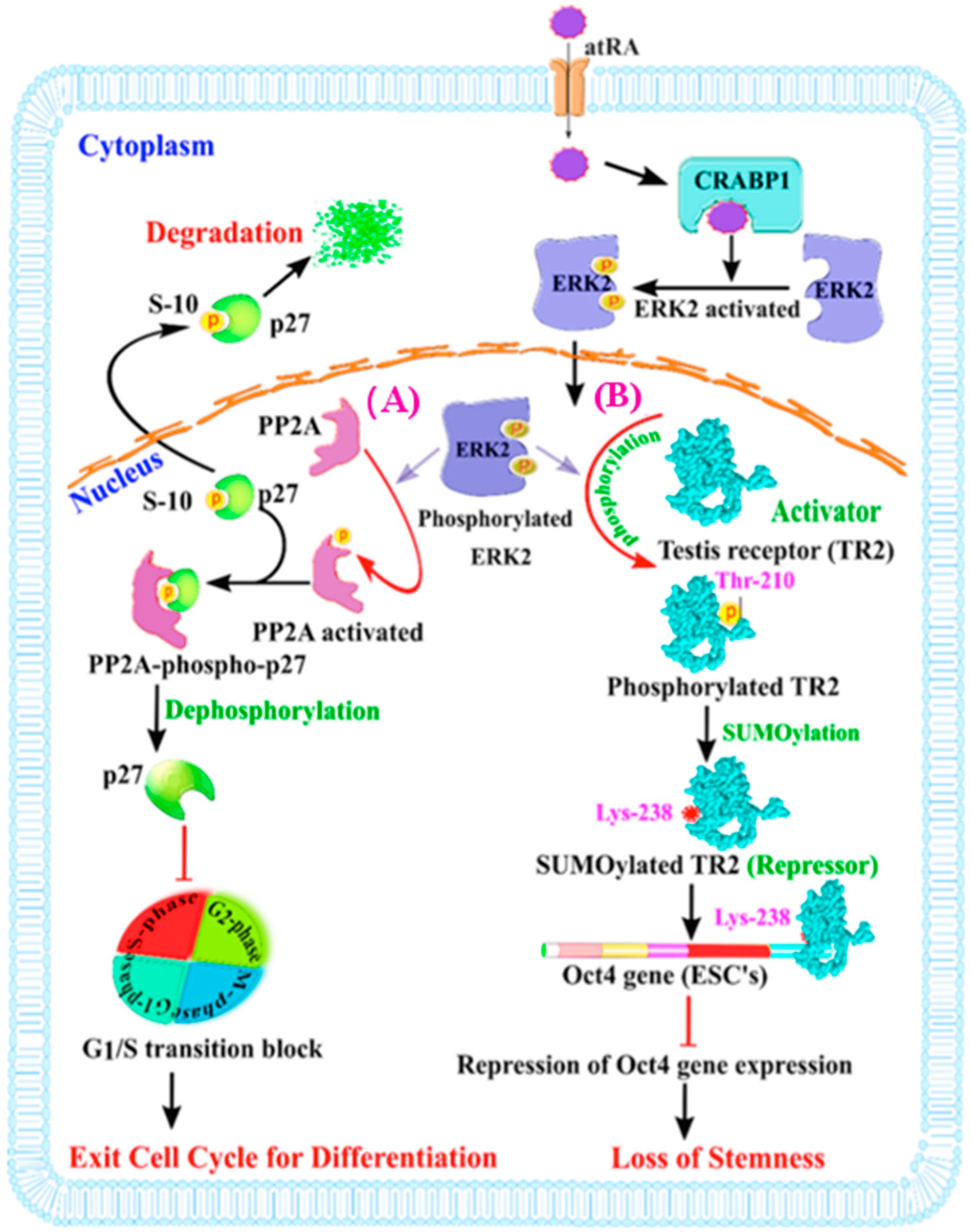
A physiological consequence of modulating ERK1/2 activation by atRA-CRABP1 in stem cells can also play out in at least two cellular processes when stem cells are meant to differentiate in an environment where the level of growth factor tends to subside. The first is related to cell cycle control via regulating p27 activity [26], and the second is to stimulate the phosphorylation (at Thr-210) of the testis receptor 2, TR2 (an orphan receptor), which triggers its SUMOylation/repression, thereby repressing the expression of octamer-binding transcription factor 4 (Oct4), the key stemness gene in maintaining a stem cell population [23,80]. ESCs have a relatively short G1 phase, which is required for their rapid entry into S phase and continuous self-renewal [81]. Molecularly, a slender G1 phase is ensured by the degradation of the p27 protein, an important regulatory protein of the cell cycle [82,83]. p27 phosphorylation/dephosphorylation status regulates its activity. The dephosphorylated p27 physically interacts with, and inhibits, G1 CDK/cyclin complexes (cyclin-dependent kinases), thus blocking the G1 to S phase transition [84]. When p27 is phosphorylated, particularly at ser-10, it is exported to cytosol for degradation [82,85]. In ESCs, when growth factors are withdrawn or reduced, atRA-CRABP1 can activate ERK1/2, which then rapidly translocates into the nucleus and phosphorylates/activates protein phosphatase 2A (PP2A) [26,71], which in turn enhances the stability of the p27 protein by dephosphorylation, thereby blocking G1 CDK/cyclin complexes and dampening cellular growth (Figure 2A, left).
The second process affected by ERK1/2 in differentiating stem cells is related to the maintenance of their stemness (Figure 2B, right). To ensure continuous stem cell proliferation, a proper amount of the stemness gene product, Oct4, is essential [80,86]. Oct4 expression is regulated mainly by positive and negative transcriptional controls, where TR2 orphan nuclear receptor’s activity is important [23,80,87]. Oct4 gene transcription is fine-tuned by the homeostasis of multiple activators and repressors. The activators include polymerase-associated factor 1 (PAF1) complex [88,89], non-POU domain-containing octamer-binding (NonO) protein [90], steroidogenic factor-1 (SF-1) [91], TR2 [80], etc. The key repressors are SUMOylated TR2 [80] and chicken ovalbumin upstream promoter transcription factors (COUP-TFs) or Pou5f1 [92]. In this context, atRA-CRABP1-stimulated ERK1/2 initiates TR2 phosphorylation at Thr-210 and its subsequent SUMOylation at Lys238, resulting in the replacement of coactivator p300/CBP associated factor (Pcaf) by corepressor RIP140, which turns TR2 from an activator to a repressor of Oct4 gene transcription [80]. Therefore, for differentiating ESCs, atRA’s non-canonical activity rapidly activates ERK1/2, via CRABP1, and contributes to the rapid repression of the Oct4 gene and timely suspension of their stemness feature, allowing differentiation to efficiently occur.
Together, for stem cells, the non-canonical effects of atRA’s, mediated by CRABP1, provide a timely and rapid control that not only desensitizes stimulation by growth factors (by intercepting the RAF/MEK/ERK axis) (Figure 1), but also stimulates their differentiation by augmenting cell cycle control and the dampening stemness feature (Figure 2B, right). This non-canonical, rapid activity of atRA may help to ensure that the genomic effects of atRA, that typically manifest later by RAR-mediated transcriptional events, can effectively take place in order to induce cell differentiation.
In a more physiologically relevant stem cell context, such as maintaining the NSC pool in the brain, the non-canonical activity of atRA via CRABP1 appears to be important in maintaining the NSC pool in the hippocampus [70]. In a CRABP1 gene KO mouse brain, the NSC pool is expanded due to the removal of CRABP1 that otherwise provides a negative modulation for NSC proliferation efficiency. As a result of NSC expansion in the hippocampus, neurogenesis in the CRABP1 gene KO mice is improved, and these CRABP1 gene KO mice indeed exhibit behavioral changes in line with improved learning and memory [70].

3.2. AtRA-CRABP1 Modulates CaMKII and Cardiomyocyte Function

AtRA-CRABP1 is also found to modulate CaMKII activation in certain differentiated cells such as cardiomyocytes [72,93]. CaMKII is critical for proper heart function, such as contraction, and also contributes to cardiomyocyte death, particularly in heart failure [94]. Targeting β-adrenergic receptors (β-AR) by agonist catecholamines, such as isoproterenol (ISO), is one of the commonly used clinical practices in treating cardiac disease [95]. However, sustained and/or excessive stimulation of CaMKII by catecholamines causes cardiac remodeling, hypertrophy, and cardiac failure [96,97]. CaMKII is a protein kinase capable of phosphorylating diverse substrates, including those leading to apoptosis and hypertrophy [94]. It appears that CRABP1 also directly interacts with CaMKIIδ (a predominant isoform of CaMKII) in the heart, thereby competing with calmodulin (CaM) to dampen Ca2+ CaM-induced CaMKII activation [72].
In mice, deleting CRABP1 leads to spontaneously developed, reduced cardiac function in older animals (such as a decrease in the ejection fraction of the left ventricle), corresponding to spontaneous over-activation of CaMKII in cardiomyocyte. Further, under acute ISO treatment, CRABP1 gene KO mice showed more severe cytotoxicity as compared to wild type (WT) mice. Under a chronic ISO challenge, CRABP1 gene KO mice exhibited a more severe cardiac phenotype, as reflected in a further enlarged heart and cardiomyocyte hypertrophy and fibrosis. Mechanically, CRABP1 can directly interact with CaMKII to inhibit its activation, and the addition of atRA further enhances this interaction. Therefore, without CRABP1, CaMKII tends to be overactivated, even under an otherwise normal stimulation such as physiological signal catecholamines. Furthermore, we have recently shown that atRA can protect ISO-induced cardiac damage in WT, but not CRABP1 gene KO mice [72]. Thus, in the physiological context of maintaining heart health, atRA-CRABP1 may also provide a mechanism to ensure the homeostasis of CaMKII activation and to protect against accidental, unwanted overactivation of CaMKII in cardiomyocyte that may lead to cardiac damage.

3.3. Other Non-Canonical Activities of RA

There are a few reports also demonstrating the non-genomic action of RA. For instance, atRA directly interacts with and activates cytosolic FABP5, a fatty acid binding protein that can shuttle between the cytoplasm and the nucleus. Activated FABP5 then activates nuclear receptors PPARβ/δ and thus enhances PPARβ/δ mediated gene expression [98]. Recently, another study showed that a pool of RARα is localized in the membrane lipid rafts along with G protein alpha Q (Gαq). Intriguingly, the efficiency of the RARα-Gαq complex prominently increased upon treatment with RA. This finding indicates that the RARα-Gαq complex can exist in the cytoplasm, which can be enhanced by cytoplasmic RA [99]. Gαq is an activator of p38 MAPK; thus, by increasing the pool of the RARα-Gαq complex, RA can enhance the activation of the p38 MAPK pathway without triggering a genomic effect [100]. However, the study did not provide compelling genetic evidence, casting doubt over the physiological relevance of this intriguing signaling pathway.
The emerging “non-canonical” signaling is also supported by additional findings about the potential signaling cascade of retinoid, the alcohol form of retinoid. It appears that the retinol-retinol binding protein (RBP) complex can bind and activate cell surface receptor signaling cascades by forming a STRA6-RBP complex to regulate the JAK-STAT signaling pathways [51,101].

4. Conclusions and Future Directions

AtRA is the principal active metabolite of vitamin A and represents one classical endocrine hormone, which mainly elicits genomic effects. This type of canonical signaling typically spans a more extended period of time and causes more permanent changes in cellular processes often associated with an alteration in gene expression. With this principal action, atRA is known primarily as a differentiation agent. Recent studies present evidence for atRA’s new activities, mediated by its cytoplasmic binding protein CRABP1, but not by its nuclear receptors RARs/RXRs, to elicit very rapid (within minutes) activities intercepting several specific cytosolic signaling pathways such as growth factor-stimulated ERK1/2 (for stem cells) and catecholamine-triggered CaMKII activation (for cardiomyocytes). While results of gene KO studies unambiguously demonstrate the consequences of deleting these specific pathways, both in animals and in primary cells, the physiological context of this non-canonical signaling remains to be further elucidated. For instance, questions remain to be answered when it comes to the integration of atRA (or vitamin A status) with other growth factors or neuropeptides in the context of whole animals. Will hypovitaminosis A also cause an abnormal NSC pool in the brain or more severe cardiac outcome? Given that CRABP1 can intercept growth factor signaling, can CRABP1 also serve as a target in cancer therapy? Classical transgenic studies of over-expressing CRABP1 demonstrate that abnormally high levels of CRABP1 can lead to abnormalities in the lung and the liver in mice [68]. Does this contribute to the pathology of hypervitaminosis A? Among all receptors and binding proteins for RA, CRABP1 is the most conserved member. This also suggests a versatile physiological role for CRABP1, which constrains the progression in its molecular divergence during evolution. Another important question is, can other forms of RA also elicit non-canonical activity via binding CRABP1? Finally, it is tempting to speculate that there remain more functions of RA-CRABP1 to be identified.
To further dissect these functions, there is an urgent need to determine other CRABP1- interacting partners/signaling networks. One such approach can be through utilizing molecular docking to identify certain atRA-like compounds that can mimic atRA’s action by binding to CRABP1. Because such compounds are less likely to produce the canonical effects of atRA (which requires binding to RARs), studies using such compounds can provide a better insight into CRABP1′s crosstalk with other signaling pathways without interference from RAR/RXR-mediated nuclear activities of RA. Further, protein-protein docking of CRABP1, followed by experimental evaluation, can reveal additional potential partner proteins of CRABP1 and their functional roles in other signaling pathways. Finally, from a therapeutic point of view, compounds specific to CRABP1 that do not bind to RARs/RXRs would be better candidates as therapeutics for targeting CRABP1 to selectively intercept certain biological processes, such as inhibiting cancer cell growth and improving survival/function.

Funding

This study was supported by NIH grants DK54733, DK60521, and the Dean’s Commitment and the Distinguished McKnight Professorship of the University of Minnesota to L.-N.W.

Conflicts of Interest

The authors declare no conflict of interest.

References

  1. Wald, G. The molecular basis of visual excitation. Nature 1968, 219, 800–807. [Google Scholar] [CrossRef] [PubMed]
  2. Wolf, G. The discovery of the visual function of vitamin A. J. Nutr. 2001, 131, 1647–1650. [Google Scholar] [CrossRef] [PubMed]
  3. Karrer, P.; Morf, R.; Schöpp, K. Zur Kenntnis des Vitamins-A aus Fischtranen. Helv. Chim. Acta 1931, 14, 1036–1040. [Google Scholar] [CrossRef]
  4. Holmes, H.N.; Corbet, R.E. The Isolation of Crystalline Vitamin A1. J. Am. Chem. Soc. 1937, 59, 2042–2047. [Google Scholar] [CrossRef]
  5. Bowles, J.; Koopman, P. Retinoic acid, meiosis and germ cell fate in mammals. Development 2007, 134, 3401–3411. [Google Scholar] [CrossRef] [Green Version]
  6. Kam, R.K.T.; Deng, Y.; Chen, Y.; Zhao, H. Retinoic acid synthesis and functions in early embryonic development. Cell Biosci. 2012, 2, 11. [Google Scholar] [CrossRef]
  7. Cañete, A.; Cano, E.; Muñoz-Chápuli, R.; Carmona, R. Role of vitamin a/retinoic acid in regulation of embryonic and adult hematopoiesis. Nutrients 2017, 9, 159. [Google Scholar] [CrossRef]
  8. Goodman, D.S. Vitamin A and Retinoids in Health and Disease. N. Engl. J. Med. 2010, 310, 1023–1031. [Google Scholar]
  9. Blaner, W.S. Vitamin A signaling and homeostasis in obesity, diabetes, and metabolic disorders. Pharmacol. Ther. 2019, 197, 153–178. [Google Scholar] [CrossRef]
  10. Pan, J.; Guleria, R.S.; Zhu, S.; Baker, K.M. Clinical Medicine Molecular Mechanisms of Retinoid Receptors in Diabetes-Induced Cardiac Remodeling. J. Clin. Med. 2014, 3, 566–594. [Google Scholar] [CrossRef]
  11. Rhee, E.J.; Nallamshetty, S.; Plutzky, J. Retinoid metabolism and its effects on the vasculature. Biochim. Biophys. Acta Mol. Cell Biol. Lipids 2012, 1821, 230–240. [Google Scholar] [CrossRef] [PubMed]
  12. Shirakami, Y.; Lee, S.A.; Clugston, R.D.; Blaner, W.S. Hepatic metabolism of retinoids and disease associations. Biochim. Biophys. Acta Mol. Cell Biol. Lipids 2012, 1821, 124–136. [Google Scholar] [CrossRef] [PubMed] [Green Version]
  13. Dollé, P.; Niederreither, K. The Retinoids: Biology, Biochemistry, and Disease; Wiley Blackwell: New York, NY, USA, 2015. [Google Scholar]
  14. Brun, P.J.; Wongsiriroj, N.; Blaner, W.S. Retinoids in the pancreas. Hepatobiliary Surg. Nutr. 2016, 5, 1–14. [Google Scholar] [PubMed]
  15. Green, A.C.; Martin, T.J.; Purton, L.E. The role of vitamin A and retinoic acid receptor signaling in post-natal maintenance of bone. J. Steroid Biochem. Mol. Biol. 2016, 155, 135–146. [Google Scholar] [CrossRef] [PubMed]
  16. Mangelsdorf, D.J.; Umesono, K.; Evans, R. The retinoid receptors. In The Retinoids: Biology, Chemistry and Medicine; Roberts, A.B., Sporn, M.B., Goodman, D.S., Eds.; Raven Press: New York, NY, USA, 1994; pp. 319–349. [Google Scholar]
  17. Mangelsdorf, D.J.; Borgmeyer, U.; Heyman, R.A.; Yang Zhou, J.; Ong, E.S.; Oro, A.E.; Kakizuka, A.; Evans, R.M. Characterization of three RXR genes that mediate the action of 9-cis retinoic acid. Genes Dev. 1992, 6, 329–344. [Google Scholar] [CrossRef]
  18. Chambon, P. A decade of molecular biology of retinoic acid receptors. FASEB J. 1996, 10, 940–954. [Google Scholar] [CrossRef] [PubMed]
  19. Blomhoff, R.; Blomhoff, H.K. Overview of retinoid metabolism and function. J. Neurobiol. 2006, 66, 606–630. [Google Scholar] [CrossRef]
  20. Larange, A.; Cheroutre, H. Retinoic Acid and Retinoic Acid Receptors as Pleiotropic Modulators of the Immune System. Annu. Rev. Immunol. 2016, 34, 369–394. [Google Scholar] [CrossRef]
  21. Duong, V.; Rochette-Egly, C. The molecular physiology of nuclear retinoic acid receptors. From health to disease. Biochim. Biophys. Acta Mol. Basis Dis. 2011, 1812, 1023–1031. [Google Scholar] [CrossRef] [Green Version]
  22. Brossaud, J.; Pallet, V.; Corcuff, J.B. Vitamin A, endocrine tissues and hormones: Interplay and interactions. Endocr. Connect. 2017, 6, R121–R130. [Google Scholar] [CrossRef]
  23. Gupta, P.; Ho, P.C.; Huq, M.M.; Ha, S.G.; Park, S.W.; Khan, A.A.; Tsai, N.P.; Wei, L.N. Retinoic acid-stimulated sequential phosphorylation, PML recruitment, and SUMOylation of nuclear receptor TR2 to suppress Oct4 expression. Proc. Natl. Acad. Sci. USA. 2008, 105, 11424–11429. [Google Scholar] [CrossRef] [PubMed]
  24. Chuang, Y.S.; Huang, W.H.; Park, S.W.; Persaud, S.D.; Hung, C.H.; Ho, P.C.; Wei, L.N. Promyelocytic leukemia protein in retinoic acid-induced chromatin remodeling of Oct4 gene promoter. Stem Cells 2011, 29, 660–669. [Google Scholar] [CrossRef] [PubMed]
  25. Wu, C.Y.; Persaud, S.D.; Wei, L.N. Retinoic Acid Induces Ubiquitination-Resistant RIP140/LSD1 Complex to Fine-Tune Pax6 Gene in Neuronal Differentiation. Stem Cells 2016, 34, 114–123. [Google Scholar] [CrossRef] [PubMed]
  26. Persaud, S.D.; Lin, Y.W.; Wu, C.Y.; Kagechika, H.; Wei, L.N. Cellular retinoic acid binding protein I mediates rapid non-canonical activation of ERK1/2 by all-trans retinoic acid. Cell. Signal. 2013, 25, 19–25. [Google Scholar] [CrossRef] [PubMed]
  27. Das, B.C.; Thapa, P.; Karki, R.; Das, S.; Mahapatra, S.; Liu, T.C.; Torregroza, I.; Wallace, D.P.; Kambhampati, S.; Van Veldhuizen, P.; et al. Retinoic acid signaling pathways in development and diseases. Bioorg. Med. Chem. 2014, 22, 673–683. [Google Scholar] [CrossRef] [PubMed]
  28. Perera, C.O.; Yen, G.M. Functional properties of carotenoids in human health. Int. J. Food Prop. 2007, 10, 201–230. [Google Scholar] [CrossRef]
  29. Chelstowska, S.; Widjaja-Adhi, M.A.K.; Silvaroli, J.A.; Golczak, M. Molecular basis for vitamin A uptake and storage in vertebrates. Nutrients 2016, 8, 676. [Google Scholar] [CrossRef] [PubMed]
  30. Harrison, E.H. Mechanisms of Digestion and Absorption of Dietary Vitamin, A. Annu. Rev. Nutr. 2005, 25, 87–103. [Google Scholar] [CrossRef]
  31. O’Byrne, S.M.; Blaner, W.S. Retinol and retinyl esters: Biochemistry and physiology Thematic Review Series: Fat-soluble vitamins: Vitamin, A. J. Lipid Res. 2013, 54, 1731–1743. [Google Scholar] [CrossRef]
  32. D’Ambrosio, D.N.; Clugston, R.D.; Blaner, W.S. Vitamin A metabolism: An update. Nutrients 2011, 3, 63–103. [Google Scholar] [CrossRef]
  33. Blomhoff, R.; Green, M.H.; Green, J.B.; Berg, T.; Norum, K.R. Vitamin A metabolism: New perspectives on absorption, transport, and storage. Physiol. Rev. 1991, 71, 951–990. [Google Scholar] [CrossRef] [PubMed]
  34. Blaner, W.S.; Li, Y. Vitamin A Metabolism, Storage and Tissue Delivery Mechanisms. In The Retinoids: Biology, Biochemistry, and Disease; Dolle, P., Niederreither, K., Eds.; Wiley-Blackwell: New York, NY, USA, 2015; pp. 1–34. ISBN 9781118628003. [Google Scholar]
  35. Paik, J.; During, A.; Harrison, E.H.; Mendelsohn, C.L.; Lai, K.; Blaner, W.S. Expression and characterization of a murine enzyme able to cleave β-carotene. The formation of retinoids. J. Biol. Chem. 2001, 276, 32160–32168. [Google Scholar] [CrossRef] [PubMed]
  36. Hong, S.H.; Kim, K.R.; Oh, D.K. Biochemical properties of retinoid-converting enzymes and biotechnological production of retinoids. Appl. Microbiol. Biotechnol. 2015, 99, 7813–7826. [Google Scholar] [CrossRef] [PubMed]
  37. Eroglu, A.; Hruszkewycz, D.P.; Dela Sena, C.; Narayanasamy, S.; Riedl, K.M.; Kopec, R.E.; Schwartz, S.J.; Curley, R.W.; Harrison, E.H. Naturally occurring eccentric cleavage products of provitamin A β-carotene function as antagonists of retinoic acid receptors. J. Biol. Chem. 2012, 287, 15886–15895. [Google Scholar] [CrossRef] [PubMed]
  38. Kiefer, C.; Hessel, S.; Lampert, J.M.; Vogt, K.; Lederer, M.O.; Breithaupt, D.E.; Von Lintig, J. Identification and Characterization of a Mammalian Enzyme Catalyzing the Asymmetric Oxidative Cleavage of Provitamin A. J. Biol. Chem. 2001, 276, 14110–14116. [Google Scholar] [CrossRef] [PubMed] [Green Version]
  39. Eroglu, A.; Harrison, E.H. Carotenoid metabolism in mammals, including man: Formation, occurrence, and function of apocarotenoids thematic review series: Fat-soluble vitamins: Vitamin A. J. Lipid Res. 2013, 54, 1719–1730. [Google Scholar] [CrossRef] [PubMed]
  40. During, A.; Harrison, E.H. Mechanisms of provitamin A (carotenoid) and vitamin A (retinol) transport into and out of intestinal Caco-2 cells. J. Lipid Res. 2007, 48, 2283–2294. [Google Scholar] [CrossRef] [Green Version]
  41. Van Bennekum, A.M.; Fisher, E.A.; Blaner, W.S.; Harrison, E.H. Hydrolysis of Retinyl Esters by Pancreatic Triglyceride Lipase. Biochemistry 2000, 39, 4900–4906. [Google Scholar] [CrossRef]
  42. Moise, A.R.; Golczak, M.; Imanishi, Y.; Palczewski, K. Topology and membrane association of lecithin: Retinol acyltransferase. J. Biol. Chem. 2007, 282, 2081–2090. [Google Scholar] [CrossRef]
  43. Sears, A.E.; Palczewski, K. Lecithin:Retinol Acyltransferase: A Key Enzyme Involved in the Retinoid (visual) Cycle. Biochemistry 2016, 55, 3082–3091. [Google Scholar] [CrossRef] [Green Version]
  44. Blaner, W.S.; O’Byrne, S.M.; Wongsiriroj, N.; Kluwe, J.; D’Ambrosio, D.M.; Jiang, H.; Schwabe, R.F.; Hillman, E.M.C.; Piantedosi, R.; Libien, J. Hepatic stellate cell lipid droplets: A specialized lipid droplet for retinoid storage. Biochim. Biophys. Acta Mol. Cell Biol. Lipids 2009, 1791, 467–473. [Google Scholar] [CrossRef] [PubMed] [Green Version]
  45. Blaner, W.S. Hepatic Stellate Cells and Retinoids: Toward A Much More Defined Relationship. Hepatology 2019, 69, 484–486. [Google Scholar] [CrossRef] [PubMed]
  46. Hussain, M.M.; Kancha, R.K.; Zhou, Z.; Luchoomun, J.; Zu, H.; Bakillah, A. Chylomicron assembly and catabolism: Role of apolipoproteins and receptors. Biochim. Biophys. Acta Lipids Lipid Metab. 1996, 1300, 151–170. [Google Scholar] [CrossRef]
  47. Li, Y.; Wongsiriroj, N.; Blaner, W.S. The multifaceted nature of retinoid transport and metabolism. Hepatobiliary Surg. Nutr. 2014, 3, 126–139. [Google Scholar] [PubMed]
  48. Kawaguchi, R.; Yu, J.; Honda, J.; Hu, J.; Whitelegge, J.; Ping, P.; Wiita, P.; Bok, D.; Sun, H. A membrane receptor for retinol binding protein mediates cellular uptake of vitamin A. Science 2007, 315, 820–825. [Google Scholar] [CrossRef] [PubMed]
  49. Sun, H.; Kawaguchi, R. The Membrane Receptor for Plasma Retinol-Binding Protein, A New Type of Cell-Surface Receptor. Int. Rev. Cell Mol. Biol. 2011, 288, 1–41. [Google Scholar] [Green Version]
  50. Chen, Y.; Clarke, O.B.; Kim, J.; Stowe, S.; Kim, Y.K.; Assur, Z.; Cavalier, M.; Godoy-Ruiz, R.; Von Alpen, D.C.; Manzini, C.; et al. Structure of the STRA6 receptor for retinol uptake. Science 2016, 353. [Google Scholar] [CrossRef]
  51. Berry, D.C.; O’Byrne, S.M.; Vreeland, A.C.; Blaner, W.S.; Noy, N. Cross Talk between Signaling and Vitamin A Transport by the Retinol-Binding Protein Receptor STRA6. Mol. Cell. Biol. 2012, 32, 3164–3175. [Google Scholar] [CrossRef] [Green Version]
  52. Kumar, S.; Sandell, L.L.; Trainor, P.A.; Koentgen, F.; Duester, G. Alcohol and aldehyde dehydrogenases: Retinoid metabolic effects in mouse knockout models. Biochim. Biophys. Acta Mol. Cell Biol. Lipids. 2012, 1821, 198–205. [Google Scholar] [CrossRef] [Green Version]
  53. Napoli, J.L. Physiological insights into all-trans-retinoic acid biosynthesis. Biochim. Biophys. Acta Mol. Cell Biol. Lipids 2012, 1821, 152–167. [Google Scholar] [CrossRef] [Green Version]
  54. Clagett-Dame, M.; Knutson, D. Vitamin A in reproduction and development. Nutrients 2011, 3, 385–428. [Google Scholar] [CrossRef] [PubMed]
  55. Napoli, J.L. Cellular retinoid binding-proteins, CRBP, CRABP, FABP5: Effects on retinoid metabolism, function and related diseases. Pharmacol. Ther. 2017, 173, 19–33. [Google Scholar] [CrossRef] [PubMed] [Green Version]
  56. Napoli, J.L. Biosynthesis and metabolism of retinoic acid: Roles of CRBP and CRABP in retinoic acid: Roles of CRBP and CRABP in retinoic acid homeostasis. J. Nutr. 1993, 123, 362–366. [Google Scholar] [CrossRef] [PubMed]
  57. Thatcher, J.E.; Isoherranen, N. The role of CYP26 enzymes in retinoic acid clearance. Expert Opin. Drug Metab. Toxicol. 2009, 5, 875–886. [Google Scholar] [CrossRef] [PubMed] [Green Version]
  58. Catharine Ross, A.; Zolfaghari, R. Cytochrome P450s in the Regulation of Cellular Retinoic Acid Metabolism. Annu. Rev. Nutr. 2011, 31, 65–87. [Google Scholar] [CrossRef] [Green Version]
  59. Napoli, J.L. Retinoic acid biosynthesis and metabolism. FASEB J. 1996, 10, 993–1001. [Google Scholar] [CrossRef] [PubMed]
  60. Wei, L.N. Retinoid receptors and their coregulators. Annu. Rev. Pharmacol. Toxicol. 2003, 43, 47–72. [Google Scholar] [CrossRef]
  61. Delva, L.; Bastie, J.N.; Rochette-Egly, C.; Kraïba, R.; Balitrand, N.; Despouy, G.; Chambon, P.; Chomienne, C. Physical and functional interactions between cellular retinoic acid binding protein II and the retinoic acid-dependent nuclear complex. Mol. Cell. Biol. 1999, 19, 7158–7167. [Google Scholar] [CrossRef]
  62. Dong, D.; Ruuska, S.E.; Levinthal, D.J.; Noy, N. Distinct roles for cellular retinoic acid-binding proteins I and II in regulating signaling by retinoic acid. J. Biol. Chem. 1999, 274, 23695–23698. [Google Scholar] [CrossRef]
  63. Budhu, A.; Gillilan, R.; Noy, N. Localization of the RAR interaction domain of cellular retinoic acid binding protein-II. J. Mol. Biol. 2001, 305, 939–949. [Google Scholar] [CrossRef]
  64. Al Tanoury, Z.; Piskunov, A.; Rochette-Egly, C. Vitamin A and retinoid signaling: Genomic and nongenomic effects. J. Lipid Res. 2013, 54, 1761–1775. [Google Scholar] [CrossRef] [PubMed]
  65. Rochette-Egly, C. Retinoic acid signaling and mouse embryonic stem cell differentiation: Cross talk between genomic and non-genomic effects of RA. Biochim. Biophys. Acta Mol. Cell Biol. Lipids 2015, 1851, 66–75. [Google Scholar] [CrossRef] [PubMed]
  66. Dawson, M.I.; Xia, Z. The retinoid X receptors and their ligands. Biochim. Biophys. Acta Mol. Cell Biol. Lipids 2012, 1821, 21–56. [Google Scholar] [CrossRef] [PubMed] [Green Version]
  67. Samarut, E.; Rochette-Egly, C. Nuclear retinoic acid receptors: Conductors of the retinoic acid symphony during development. Mol. Cell. Endocrinol. 2012, 348, 348–360. [Google Scholar] [CrossRef] [PubMed]
  68. Wei, L.N. Cellular retinoic acid binding proteins: Genomic and non-genomic functions and their regulation. In The Physiology of Vitamin A—Uptake, Transport, Metabolism and Signaling; Asson-Batres, M.A., Rochette-Egly, C., Eds.; Springer: Dordrecht, The Netherlands, 2016; Volume 81, pp. 163–178. [Google Scholar]
  69. Dilworth, F.J.; Chambon, P. Nuclear receptors coordinate the activities of chromatin remodeling complexes and coactivators to facilitate initiation of transcription. Oncogene 2001, 20, 3047–3054. [Google Scholar] [CrossRef] [PubMed] [Green Version]
  70. Lin, Y.L.; Persaud, S.D.; Nhieu, J.; Wei, L.N. Cellular retinoic acid-binding protein 1 modulates stem cell proliferation to affect learning and memory in male mice. Endocrinology 2017, 158, 3004–3014. [Google Scholar] [CrossRef]
  71. Persaud, S.D.; Park, S.W.; Ishigami-Yuasa, M.; Koyano-Nakagawa, N.; Kagechika, H.; Wei, L.N. All trans-retinoic acid analogs promote cancer cell apoptosis through non-genomic Crabp1 mediating ERK1/2 phosphorylation. Sci. Rep. 2016, 6, 2–3. [Google Scholar] [CrossRef] [PubMed]
  72. Park, S.W.; Persaud, S.D.; Ogokeh, S.; Meyers, T.A.; Townsend, D.; Wei, L.N. CRABP1 protects the heart from isoproterenol-induced acute and chronic remodeling. J. Endocrinol. 2018, 236, 151–165. [Google Scholar] [CrossRef] [PubMed]
  73. Bost, F.; Caron, L.; Marchetti, I.; Dani, C.; Le Marchand-Brustel, Y.; Binétruy, B. Retinoic acid activation of the ERK pathway is required for embryonic stem cell commitment into the adipocyte lineage. Biochem. J. 2002, 361, 621–627. [Google Scholar] [CrossRef]
  74. Li, Z.; Theus, M.H.; Wei, L. Role of ERK 1/2 signaling in neuronal differentiation of cultured embryonic stem cells. Dev. Growth Differ. 2006, 48, 513–523. [Google Scholar] [CrossRef]
  75. Park, S.W.; Nhieu, J.; Persaud, S.D.; Miller, M.C.; Xia, Y.; Lin, Y.W.; Lin, Y.L.; Kagechika, H.; Mayo, K.H.; Wei, L.N. A new regulatory mechanism for Raf kinase activation, retinoic acid-bound Crabp1. Sci. Rep. 2019, in press. [Google Scholar]
  76. Kolch, W. Meaningful relationships: The regulation of the Ras/Raf/MEK/ERK pathway by protein interactions. Biochem. J. 2000, 351, 289. [Google Scholar] [CrossRef] [PubMed]
  77. Morrison, D.K.; Cutler, R.E. The complexity of Raf-1 regulation. Curr. Opin. Cell Biol. 1997, 9, 174–179. [Google Scholar] [CrossRef]
  78. Roskoski, R. ERK1/2 MAP kinases: Structure, function, and regulation. Pharmacol. Res. 2012, 66, 105–143. [Google Scholar] [CrossRef] [PubMed]
  79. Lavoie, H.; Therrien, M. Regulation of RAF protein kinases in ERK signalling. Nat. Rev. Mol. Cell Biol. 2015, 16, 281–298. [Google Scholar] [CrossRef] [PubMed]
  80. Park, S.W.; Hu, X.; Gupta, P.; Lin, Y.P.; Ha, S.G.; Wei, L.N. SUMOylation of Tr2 orphan receptor involves Pml and fine-tunes Oct4 expression in stem cells. Nat. Struct. Mol. Biol. 2007, 14, 68–75. [Google Scholar] [CrossRef] [PubMed]
  81. Coronado, D.; Godet, M.; Bourillot, P.Y.; Tapponnier, Y.; Bernat, A.; Petit, M.; Afanassieff, M.; Markossian, S.; Malashicheva, A.; Iacone, R.; et al. A short G1 phase is an intrinsic determinant of naïve embryonic stem cell pluripotency. Stem Cell Res. 2013, 10, 118–131. [Google Scholar] [CrossRef]
  82. Chu, I.M.; Hengst, L.; Slingerland, J.M. The Cdk inhibitor p27 in human cancer: Prognostic potential and relevance to anticancer therapy. Nat. Rev. Cancer 2008, 8, 253–267. [Google Scholar] [CrossRef]
  83. Starostina, N.G.; Kipreos, E.T. Multiple degradation pathways regulate versatile CIP/KIP CDK inhibitors. Trends Cell Biol. 2012, 22, 33–41. [Google Scholar] [CrossRef] [Green Version]
  84. Orford, K.W.; Scadden, D.T. Deconstructing stem cell self-renewal: Genetic insights into cell-cycle regulation. Nat. Rev. Genet. 2008, 9, 115–128. [Google Scholar] [CrossRef]
  85. Wander, S.A.; Zhao, D.; Slingerland, J.M. p27: A barometer of signaling deregulation and potential predictor of response to targeted therapies. Clin. Cancer Res. 2011, 17, 12–18. [Google Scholar] [CrossRef] [PubMed]
  86. Shi, G.; Jin, Y. Role of Oct4 in maintaining and regaining stem cell pluripotency. Stem Cell Res. Ther. 2010, 1, 39. [Google Scholar] [CrossRef] [PubMed]
  87. Chinpaisal, C.; Lee, C.H.; Wei, L.N. Mechanisms of the mouse orphan nuclear receptor TR2-11-mediated gene suppression. J. Biol. Chem. 1998, 273, 18077–18085. [Google Scholar] [CrossRef] [PubMed]
  88. Kellner, S.; Kikyo, N. Transcriptional regulation of the Oct4 gene, a master gene for pluripotency. Histol. Histopathol. 2010, 25, 405–412. [Google Scholar] [PubMed]
  89. Ding, L.; Paszkowski-Rogacz, M.; Nitzsche, A.; Slabicki, M.M.; Heninger, A.K.; de Vries, I.; Kittler, R.; Junqueira, M.; Shevchenko, A.; Schulz, H.; et al. A Genome-Scale RNAi Screen for Oct4 Modulators Defines a Role of the Paf1 Complex for Embryonic Stem Cell Identity. Cell Stem Cell 2009, 4, 403–415. [Google Scholar] [CrossRef] [PubMed] [Green Version]
  90. Park, Y.; Lee, J.M.; Hwang, M.Y.; Son, G.H.; Geum, D. NonO binds to the CpG island of oct4 promoter and functions as a transcriptional activator of oct4 gene expression. Mol. Cells 2013, 35, 61–69. [Google Scholar] [CrossRef] [PubMed]
  91. Yang, H.M.; Do, H.J.; Kim, D.K.; Park, J.K.; Chang, W.K.; Chung, H.M.; Choi, S.Y.; Kim, J.H. Transcriptional regulation of human Oct4 by steroidogenic factor-1. J. Cell. Biochem. 2007, 101, 1198–1209. [Google Scholar] [CrossRef] [PubMed]
  92. Schoorlemmer, J.; van Puijenbroek, A.; van Den Eijnden, M.; Jonk, L.; Pals, C.; Kruijer, W. Characterization of a negative retinoic acid response element in the murine Oct4 promoter. Mol. Cell. Biol. 1994, 14, 1122–1136. [Google Scholar] [CrossRef] [PubMed]
  93. Park, S.W.; Nhieu, J.; Lin, Y.W.; Wei, L.N. All-trans retinoic acid attenuates isoproterenol-induced cardiac dysfunction through Crabp1 to dampen CaMKII activation. Eur. J. Pharmacol. 2019, 172485. [Google Scholar] [CrossRef] [PubMed]
  94. Anderson, M.E.; Brown, J.H.; Bers, D.M. CaMKII in myocardial hypertrophy and heart failure. J. Mol. Cell. Cardiol. 2011, 51, 468–473. [Google Scholar] [CrossRef] [PubMed] [Green Version]
  95. Lefkowitz, R.J.; Rockman, H.A.; Koch, W.J. Catecholamines, cardiac β-adrenergic receptors, and heart failure. Circulation 2000, 101, 1634–1637. [Google Scholar] [CrossRef] [PubMed]
  96. Garg, M.; Khanna, D. Exploration of pharmacological interventions to prevent isoproterenol-induced myocardial infarction in experimental models. Ther. Adv. Cardiovasc. Dis. 2014, 8, 155–169. [Google Scholar] [CrossRef] [PubMed]
  97. Nichtova, Z.; Novotova, M.; Kralova, E.; Stankovicova, T. Morphological and functional characteristics of models of experimental myocardial injury induced by isoproterenol. Gen. Physiol. Biophys. 2012, 31, 141–151. [Google Scholar] [CrossRef] [PubMed] [Green Version]
  98. Schug, T.T.; Berry, D.C.; Shaw, N.S.; Travis, S.N.; Noy, N. Opposing Effects of Retinoic Acid on Cell Growth Result from Alternate Activation of Two Different Nuclear Receptors. Cell 2007, 129, 723–733. [Google Scholar] [CrossRef] [PubMed] [Green Version]
  99. Piskunov, A.; Rochette-Egly, C. A retinoic acid receptor RARα pool present in membrane lipid rafts forms complexes with G protein αQ to activate p38MAPK. Oncogene 2012, 31, 3333–3345. [Google Scholar] [CrossRef] [PubMed]
  100. Bruck, N.; Vitoux, D.; Ferry, C.; Duong, V.; Bauer, A.; De Thé, H.; Rochette-Egly, C. A coordinated phosphorylation cascade initiated by p38MAPK/MSK1 directs RARα to target promoters. EMBO J. 2009, 28, 34–47. [Google Scholar] [CrossRef]
  101. Berry, D.C.; Jin, H.; Majumdar, A.; Noy, N. Signaling by vitamin A and retinol-binding protein regulates gene expression to inhibit insulin responses. Proc. Natl. Acad. Sci. USA 2011, 108, 4340–4345. [Google Scholar] [CrossRef] [Green Version]
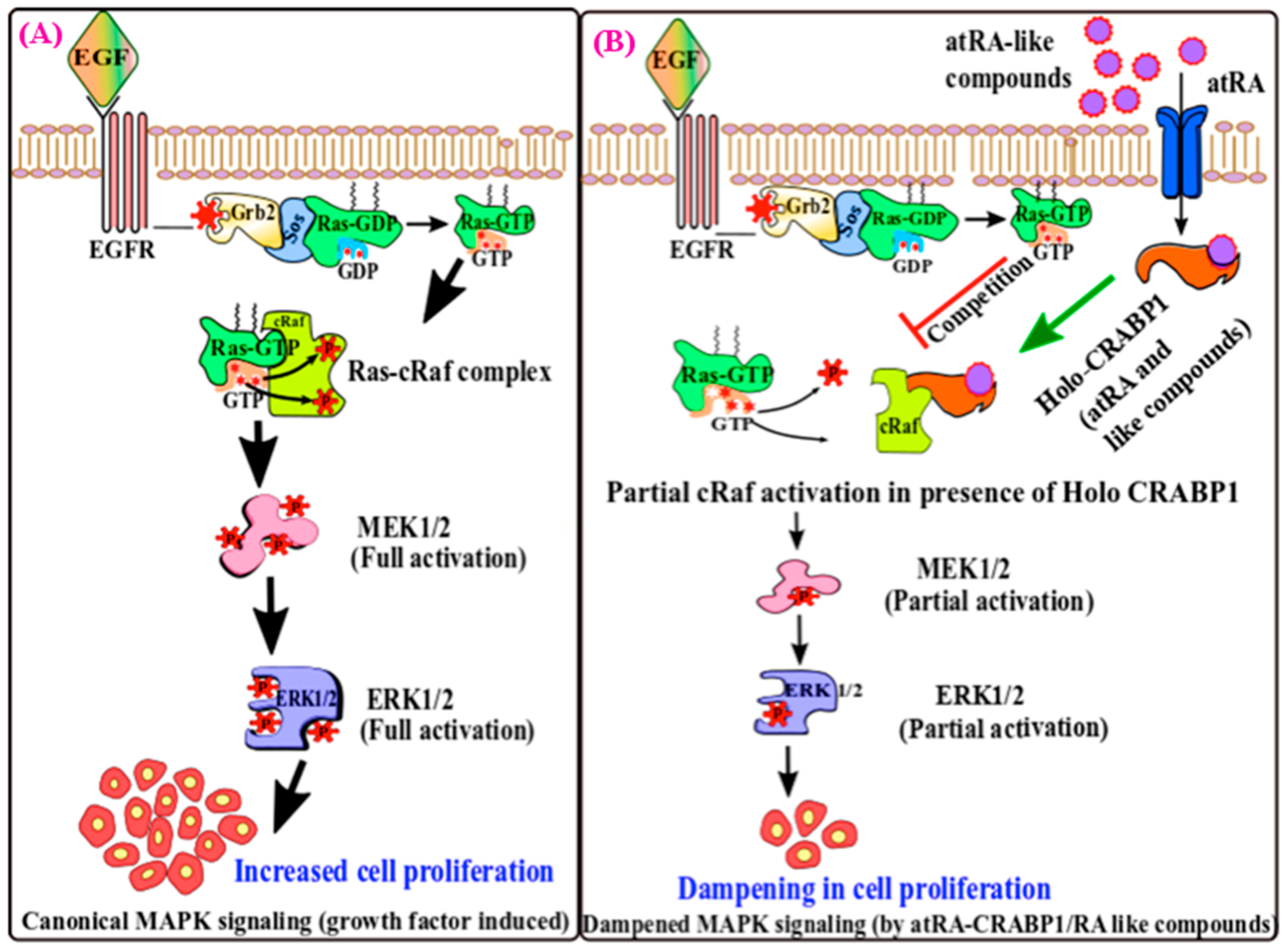
Figure 1. A model illustrating the effect of atRA-CRABP1 in blunting growth activation in stem cells. (A) In response to growth factors like EGF, robust canonical MAPK signaling is triggered involving the Ras-mediated activation of cRAF kinase by protein–protein interaction, which leads to sequential phosphorylation of MEK1/2 and ERK1/2 and enhances cell proliferation. (B) Cells co-exposed to EGF and atRA show reduced MAPK signaling by competitive binding of atRA-CRABP1 with cRAF, resulting in weak Ras-cRAF protein–protein interactions and downstream ERK1/2 phosphorylation, thus dampening cellular growth.
Ijms 20 03610 g001
Figure 2. Schematic illustration of atRA-CRABP1 that activates ERK1/2 in stem cells when the level of growth factors is lowered, which dampens stem proliferation and facilitates differentiation. In the cytoplasm, atRA binds to CRABP1 and promotes MEK/ERK1/2 complex formation and activation of ERK1/2, which then translocates into the nucleus to (A) induce dephosphorylation of p27 (stabilized) at Ser-10 by activating PP2A and impede G1/S transition and facilitate stem cells differentiation; or (B) phosphorylate TR2 at Thr-210, promoting pml (promyelocytic leukemia) recruitment and SUMOylation of TR2, which represses the transcription of Oct4 gene and induces a loss of the stemness feature of stem cells.
Ijms 20 03610 g002