Review

Cross-Talk between Inflammatory Mediators and the Epithelial Mesenchymal Transition Process in the Development of Thyroid Carcinoma

by
Giovanna Revilla
1,2,
Rosa Corcoy
3,4,5,
Antonio Moral
6,7,
Joan Carles Escolà-Gil
1,2,8,* and
Eugenia Mato
3,4,*
1
Institut d’Investigacions Biomèdiques (IIB) Sant Pau, Hospital de la Santa Creu i Sant Pau (HSCSP), 08041 Barcelona, Spain
2
Departament de Bioquímica, Biologia Molecular i Biomedicina, Universitat Autònoma de Barcelona (UAB), 08193 Barcelona, Spain
3
Department of Endocrinology, Hospital de la Santa Creu i Sant Pau (HSCSP), 08025 Barcelona, Spain
4
Centro de Investigación Biomédica en Red en Bioingeniería, Biomateriales y Nanomedicina (CIBER-BBN), 28029 Madrid, Spain
5
Departament de Medicina, Universitat Autònoma de Barcelona (UAB), 08193 Barcelona, Spain
6
Department of General Surgery—Hospital de la Santa Creu i Sant Pau (HSCSP), 08025 Barcelona, Spain
7
Departament de Cirugia, Universitat Autònoma de Barcelona (UAB), 08193 Barcelona, Spain
8
Centro de Investigación Biomédica en Red en Diabetes y Enfermedades Metabólicas Asociadas (CIBERDEM), 28029 Madrid, Spain
*
Authors to whom correspondence should be addressed.
Int. J. Mol. Sci. 2019, 20(10), 2466; https://doi.org/10.3390/ijms20102466
Submission received: 16 April 2019 / Revised: 9 May 2019 / Accepted: 16 May 2019 / Published: 18 May 2019
(This article belongs to the Special Issue Epithelial-Mesenchymal Transition (EMT))

Abstract

:
There is strong association between inflammatory processes and their main metabolic mediators, such as leptin, adiponectin secretion, and low/high-density lipoproteins, with the cancer risk and aggressive behavior of solid tumors. In this scenario, cancer cells (CCs) and cancer stem cells (CSCs) have important roles. These cellular populations, which come from differentiated cells and progenitor stem cells, have increased metabolic requirements when it comes to maintaining or expanding the tumors, and they serve as links to some inflammatory mediators. Although the molecular mechanisms that are involved in these associations remain unclear, the two following cellular pathways have been suggested: 1) the mesenchymal-epithelial transition (MET) process, which permits the differentiation of adult stem cells throughout the acquisition of cell polarity and the adhesion to epithelia, as well to new cellular lineages (CSCs); and, 2) a reverse process, termed the epithelial-mesenchymal transition (EMT), where, in pathophysiological conditions (tissue injury, inflammatory process, and oxidative stress), the differentiated cells can acquire a multipotent stem cell-like phenotype. The molecular mechanisms that regulate both EMT and MET are complex and poorly understood. Especially, in the thyroid gland, little is known regarding MET/EMT and the role of CCs or CSCs, providing an exciting, new area of knowledge to be investigated. This article reviews the progress to date in research on the role of inflammatory mediators and metabolic reprogramming during the carcinogenesis process of the thyroid gland and the EMT pathways.

Graphical Abstract

1. Introduction

Epithelial thyroid cancer (ETC) is the most common malignancy of the endocrine system, which affects the follicular cells of the gland. These tumors are twice as common in women as in men, but male tumors are associated with a poor prognosis. Additional important risk factors include a history of childhood head and neck irradiation, large nodule size (>4 cm), evidence for local tumor fixation or invasion into lymph nodes, and the presence of metastases. Moreover, this neoplasia increases with the age, plateauing after about age 50 years; it represents an important prognostic factor for thyroid cancer at a young age (<20), or in older persons (>45), and it is associated with a worse prognosis [1].
There are two main types of ETC based on the cells’ differentiated characteristics:
Well-Differentiated thyroid cancer (WDTC) arises from thyroid follicular epithelial cells and it accounts for the overwhelming majority of thyroid cancers. Of the differentiated cancers, papillary thyroid cancer (PTC) comprises about 85% of cases when compared with about 10%, which have follicular histology, referred to as follicular thyroid cancer (FTC), and 3–5% of the type FTC correspond to the Hürthle cell or oxyphil tumors characteristic.
Although PTC and FTC have a good prognosis, in the case of PTC, some histological subtypes, such as tall cell variant, columnar cell variant, and diffuse sclerosing variant, show aggressive behavior with a worse prognosis. In FTC tumors, 11% of patients have an aggressive tumor at diagnosis [1]. All of these tumors are classified following the Staging System for Differentiated Thyroid Cancer, from the American Joint Commission on Cancer (AJCC) [2]. In the case of ETT, the age at time of diagnosis is important to define the risk of recurrence and tumoral aggressiveness, with 55 years old being the cut-off. In younger patients (≤55 age), the stages I and II are classified with a low risk, while in patients (≥55 age) the stages I and II has a low risk, whereas stage III and IV has a high risk with local a distant metastasis [3].
PTC tumors show multifocality and they are able to invade tissues near the gland; the major tumoral dissemination is through the lymphatic system, but also hematogenously (bone and lung). Most of these tumors (80%) are classified as stages I or II, with a good prognosis and only 1% of them lose the differentiated characteristic, with a fatal prognosis (stage IV).
In the case of FTC, epidemiological studies show that iodine-deficiency is a key factor in the development of these types of tumors, and most of them appear as a nodule in the gland with a slow growth before the metastatic process. The histological diagnosis sometimes is difficult, because it presents a normal follicular pattern to anaplastic form without follicles or colloid [4].
The tumors with a nodule are less aversive and present low risk for infiltrating the tissues closer or local adenopathy. Nevertheless, tumors with high risk show capsular invasion, high growing in the local structures (strap muscles or trachea), making their resectability difficult. The prognosis varies in function of the degree of invasion [5].
FTC tends to invade locally and metastasize distantly, rather than to local nodes; they are especially prone to metastasizing to the bone or lung. In a Massachusetts General Hospital series [6], one-half of the cases already exhibited metastasis when the diagnosis was originally established. The tumor and metastases often retain the ability to accumulate and hold iodide, and therefore, they are sometimes susceptible to treatment with Radioactive Iodine (RAI). In some cases, FTC presents a normal histological pattern, so it is important to analyze the capsular and/or vascular invasion, and the metastasis is the only reliable criterion of malignancy in these types of tumors [7].
Poorly differentiated carcinoma (PDTC) is an aggressive tumor that shares the characteristics of WDTC and anaplastic thyroid carcinoma (ATC). Their incidence is rare (0.23% to 2.6% of patients with ETC) and, although the hypothesis about their origin is that comes from dedifferentiated of the WDTC, some of them appear de novo in the patients. The diagnosis of is difficult, because they show a different histological pattern (solid, trabecular, or sclerotic) with differences in their growing. However, all of them have poor prognosis and lymph node metastasis and distant metastasis is frequent [8].
Undifferentiated thyroid carcinoma (UTC) corresponds to anaplastic thyroid carcinoma (ATC), which is diagnosed in persons over 50 years of age; the origin of these tumors is controversial; more than half of the diagnosed cases originate from well-differentiated tumors and represent 1–2% of all thyroid malignancies [9]. These tumoral cells show and aggressive behaviour with a rapid grow, and in most of cases at the moment of diagnosis, the patients show local (lymph nodes) and distant metastasis (pulmonary). Moreover, it is of note that only 10% of these tumors are resectable at the time of diagnosis and 131I therapy does not work, because they lack the typical differentiation feature of the thyroid gland (iodine uptake, thyroglobulin secretion, response to TSH stimulation) and are the most aggressive. For that reason, they have a poor prognosis with a low survival within 6–12 months [10].

2. Origin of Epithelial Thyroid Carcinoma (ETC)

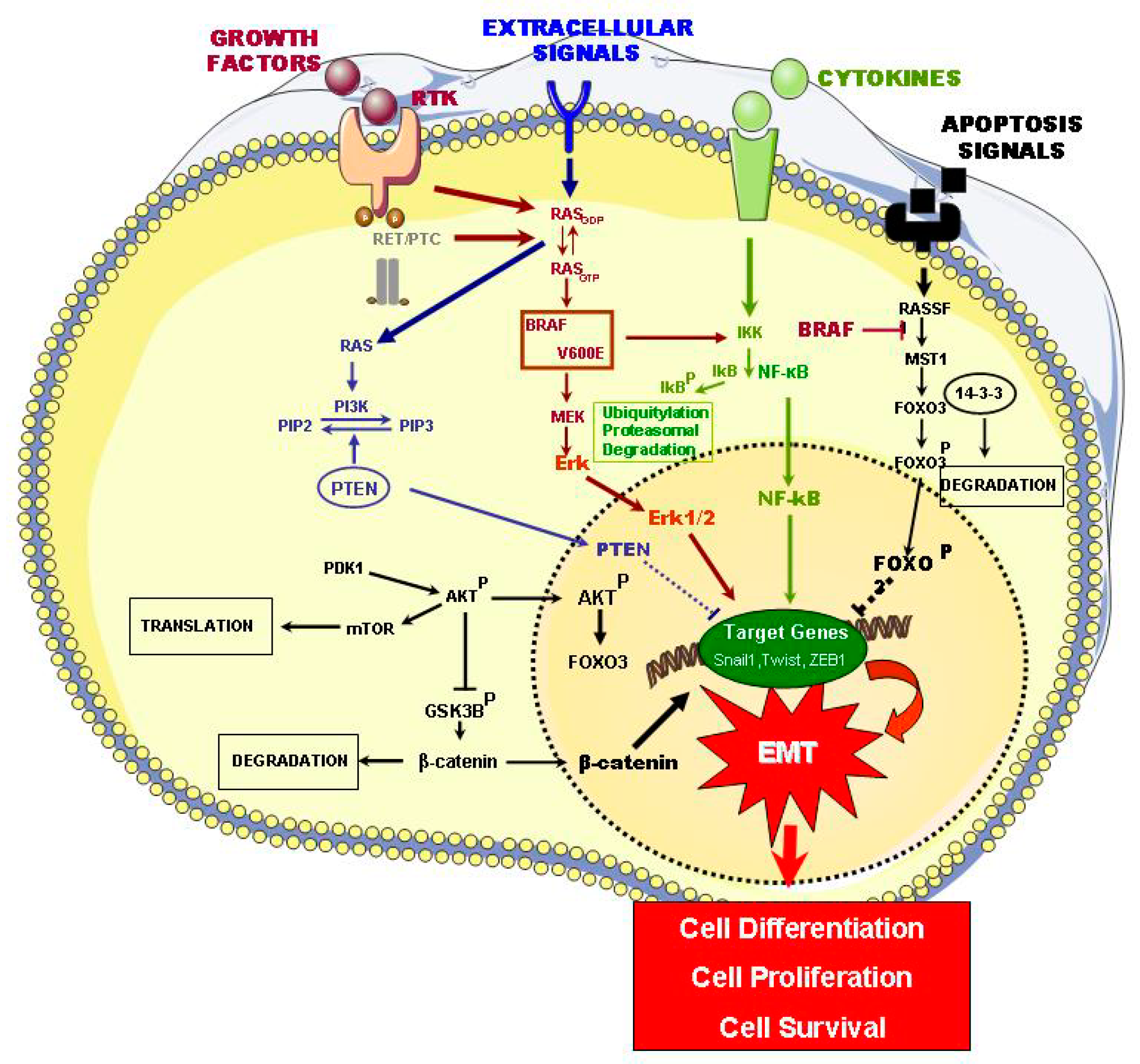
2.1. Signaling Pathways of ETC Oncogenesis

Different common genetic alterations (gene mutation, gene amplifications, copy-number gains, gene translocations and aberrant gene methylation) have been identified in WDTC (PTC and FTC) and, in PDTC, UTC/ATC, being the most frequent: point mutations affecting oncogenes (BRAF, RAS, TP53, and CTNNB1), rearrangements affecting RET-PTC and NTRK genes. Such mutations or genetic disorders directly or indirectly affect different cell signaling pathways, including MAPKinasas or the PI3K-AKT-mTOR, which are essential for many cell biological processes [11,12]. These important findings in the knowledgement of ETC etiopathogenesis will hopefully allow physicians to properly select patients that require aggressive treatment and minimize the risk for those patients with indolent tumors, who may not even require surgery. This would maximize the cure rate and minimize complications [13].
The main signaling pathways that are involved in thyroid carcinogenesis are described below and are summarized in Figure 1.
The MAPK pathways regulate multiple cell progresses, including proliferation, differentiation, and survival. In PTC, the activation of the MAPK pathway by mutated BRAF-V600E, RAS, or RET/PTC and RTK rearrangements has been identified. In contrast, mutations that involve the PI3K/Akt pathway, such as the PI3K, Akt1, and phosphatase and tensin homolog (PTEN), are more frequently found in FTC and in de-differentiated types of thyroid cancer (Figure 1) (Table 1) [11].
The importance of the MAPK pathway in PTC is now well known. The MAPK pathway is affected by activating mutations, such as PRAF and RAS, RET-PTC, and in several cases, by ALK mutation [14]. Tumorigenic activity that is caused by MAPK leads to a wide range of secondary molecular alterations that synergize and amplify oncogenic activity, such as hypermethylation and hypomethylation, as well as the upregulation of different oncogenic proteins, including chemokines, vascular endothelial growth factor A (VEGFA), the mesenchymal–epithelial transition (MET), nuclear factor kB (NF-κB), matrix metalloproteinases (MMPs), prohibitin, vimentin, hypoxia-inducible factor α (HIFIα) prokineticin (also known as EG-VEGF or PROK1), urokinase plasminogen activator (uPA) and its receptor (uPAR), TGF-β1, and thrombospondin (TSP1). All of these proteins play an important role in the human tumorigenesis through different mechanisms that manage cell carcinogenic proliferation, growth, migration and survival, tumor angiogenesis, invasion, and metastases (Figure 1 and Table 1) [15].
Many of these proteins are the critical constituents of the unique microenvironment of the extracellular matrix; this plays an important role in the tumor pathogenesis of the thyroid, which is determined by fundamental oncoproteins, like BRAF-V600E. This microenvironment not only acts as a cell support, but it also has an important influence on the behavior of tumor cells, including their viability, proliferation, adhesion, and mortality [26]. Both tumor and stromal cells (fibroblasts, macrophages) produce proteins that form autocrine and paracrine signaling chains. For example, when activated, the MAPK pathway leads to the liberation of TSP1 in the matrix, where it interacts and modifies proteins, namely, cellular membrane receptors, integrins and non-integrins, matrix proteins, cytokines, VEGFA, and MMPs. These, in turn, activate the signaling system in thyroid cancer cells (CCs) and promote tumor progression and metastasis. Released into the extracellular matrix, the TGF-β1 cytokine is capable of activating the BRAF-V600E/MAPK pathway (Figure 1). The cytokines can create an inflamed microenvironment with the production of reactive oxygen species and oxidative stress, which stimulate the MAPK pathway, thereby elevating tumorigenic activity [27]. It seems that stromal fibroblasts mainly intervene through the expression patterns of two isoforms of fibroblast growth factor receptor type 2 (FGFR2)—FGFR2-IIIb and FGFR2-IIIc—in tumor cells, and fibroblasts express the same type of FGFR2 isoform (Figure 1) [16].
The PI3/Akt signaling pathway plays a significant role in thyroid tumor genesis. It was discovered to be associated with follicular thyroid adenoma (FTA) and FTC, while investigating Cowden syndrome, which is caused by germinal PTEN mutation. The Akt1 and Akt2 isoforms are clearly expressed and activated in thyroid cancer [28]. Studies with human thyroid tumors have suggested that the invasion and metastasis of FTC, as activated by this pathway, mainly affect activation and nuclear localization of Akt1, which coincides with the presence of Akt1 mutations in the metastatic thyroid cancers. This pathway predominates in FCT, while the MAPK pathway predominates in PTC. It is plausible that PI3K/Akt pathway hyperactivation leads to FTA to PTC conversion (Figure 1 and Table 1) [17].
The NF-κB signaling pathway has an important role in the regulation of inflammatory response related to the tumor generation; it also plays a significant part in thyroid cancer. This pathway controls the signaling processes of cellular proliferation and anti-apoptosis in tumoral cells; it also determines the upregulated expression of different oncogenic proteins, which the MAPK pathway also upregulates. In thyroid cancer, with different histological patterns that include PTCs, FTCs, and ATCs, the RET-PTC, RAS, and BRAF-V600E mutations promote the activation of the NF-κB pathway in differentiated tumors, thereby inducing dedifferentiation processes and also increasing the invasiveness (Figure 1 and Table 1) [18,19,20].
The RASSF1/MST1/FOXO3 signaling pathway is extremely important in tumor suppression and apoptosis promotion. BRAF-V600E, but not the native BRAF, directly interacts with the terminal carboxyl group of MST1, which inhibits its protein kinase activation; these results decreased the transactivation of FOXO3, a transcriptional factor that activates pro-apoptotic gene transcription, through its translocation to the nucleus. Hypermethylation of the RASSF1A promoter is associated with its silencing in thyroid cancer (Figure 1) [21]. In summary, BRAF-V600E is independently linked with three principle pathways, namely MEK/MAPK, RASSF1/MST1/FOXO3, and NF-κB, thereby determining a unique and powerful oncogenic mechanism in the process of thyroid tumorigenesis (Table 1).
The WNT/β-catenin signaling pathway plays a well-established role in cell growth and proliferation, as well as in the differentiation of cell progenitors, and its constitutive activation is frequently seen in the oncogenic processes. β-catenin facilitates the transcription of diverse tumor-promoting genes through its upregulation by the WNT signaling pathway [29]. In the case of thyroid cancer, the activation of this pathway is determined by the activating mutation of CTNNB1 (which codifies β-catenin), especially in terms of ATC and PDTC (Figure 1) (Table 1) [22].
In terms of the HIF1α signaling pathway, it is well-known that hypoxia is a strong stimulus for tumor metabolism, growth, and progression. HIF1α is a critical mediator of a response to hypoxia, which binds to HIF1β (also known as ARNT) to form the transcription factor HIF1; this factor induces the expression of diverse genes that are associated with cell metabolism and tumor angiogenesis—a critical process in solid tumor progression and the intratumoral response to hypoxia. VEGFA induces this process, where HIF1 upregulates the expression [23]. HIF1α is not expressed in healthy tissues, but it is expressed in thyroid cancer, especially in more aggressive forms like ATC. Another target of HIF1 is a MET oncogene. It is also important to observe that HIF1 in thyroid cancer is upregulated by the PI3K/Akt and MAPK pathways (Figure 1) (Table 1) [24].
The TSHR signaling pathway plays a fundamental role in the regulation of thyroid cells proliferation, differentiation, and function, as well as the development of the thyroid gland through the TSH receptor (TSHR). TSHR is coupled with the G-protein, and it is linked to a guanine nucleotide that unchains two intracellular signaling pathways, namely, AMP cycloadenililcyclase Gsα-mediated signaling and intracellular Ca2+-inositol 1,4,5-triphosphoolipase Cβ-mediated signaling by Gs or G11 (demonstrated in experimental animals). There is a clinical association between elevated TSH levels and an elevated risk of malignant thyroid nodules in men [30]. However, it is likely that the TSH-TSHR system plays a dual role in thyroid tumorigenesis. It is known that the hyperactivation of TSHR signaling leads to a benign hyperfunction (FTA), which suggests that TSHR signaling (the main mechanism that runs Gsα pathway mutation) can act as a protecting factor against the malignant transformation of thyroid cells. Therefore, the TSH-TSHR system not only suppresses malignant transformation, but it also promotes the growth and progression of thyroid cancer once the oncogenic alteration is started (Figure 1) (Table 1) [25,31]. In contrast, some data show that TSH can induce the phosphorylation of p21-activated kinase 4 (PAK4) via TSHR and promote PTC malignization [32].
Moreover, TSHR can be used as a therapeutic target, since TSH estimulation promotes sodium–iodide symporter (NIS) upregulation and radioiodine uptake of the undifferentiated thyroid cells, being important their TSHR expression in the thyroid metastatic cells [33].

2.2. Thyroid Cancer Cells (CCs) and Cancer Stem Cells (CSCs)

Stemness is a property that some cell populations possess for the maintenance of tissue homeostasis (adult stem cells), but, when these cell populations undergo deregulation, they can also contribute to cancer initiation or progression. This cell population is referred to as cancer stem cells (CSCs), and these cells’ most important characteristic is their high potential plasticity and resistance to stress factors in the tumoral microenvironment. Moreover, these cells share specific markers of human embryonic stem cells (hESCs) and the adult stem cells [34,35,36]. The question will be to clarify whether thyroid tissue can contain the population of the CSCs or other cells with the characteristics of stem cells instead of external/internal signals or whether normal cells can also participate in their formation through the mechanisms that control cell regulation. In this scenario, several studies have confirmed the existence of the CSCs population and different studies have identified specific miRNA expression in this cell population, with a role in signaling pathways that are involved in tumoral progression and metastasis [37,38,39].
Moreover, side population (SP) cells have also been identified in thyroid. This cell population that correspond to one subtype of hematopoietic stem cells (HSC) was described for the first time in 1996 by Goodell et al., and it is involved in the malignancy of tumors, including the resistance to chemotherapy [40,41]. In addition, they are implicated in the EMT with a high capacity for sphere-forming when they are cultured in soft agar assays [42].
It is known that the epithelial cells can acquire characteristics of CSCs through the epithelial mesenchymal transition (EMT). Several inducers that can change the transcriptional pattern and cellular epigenetic reprogramming control this process. These molecular factors are described below.
Moreover, a clear correlation between cell metabolic reprogramming and EMT pathways has been identified, where lipid metabolism has an important role in this process [43,44].

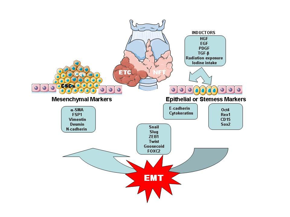
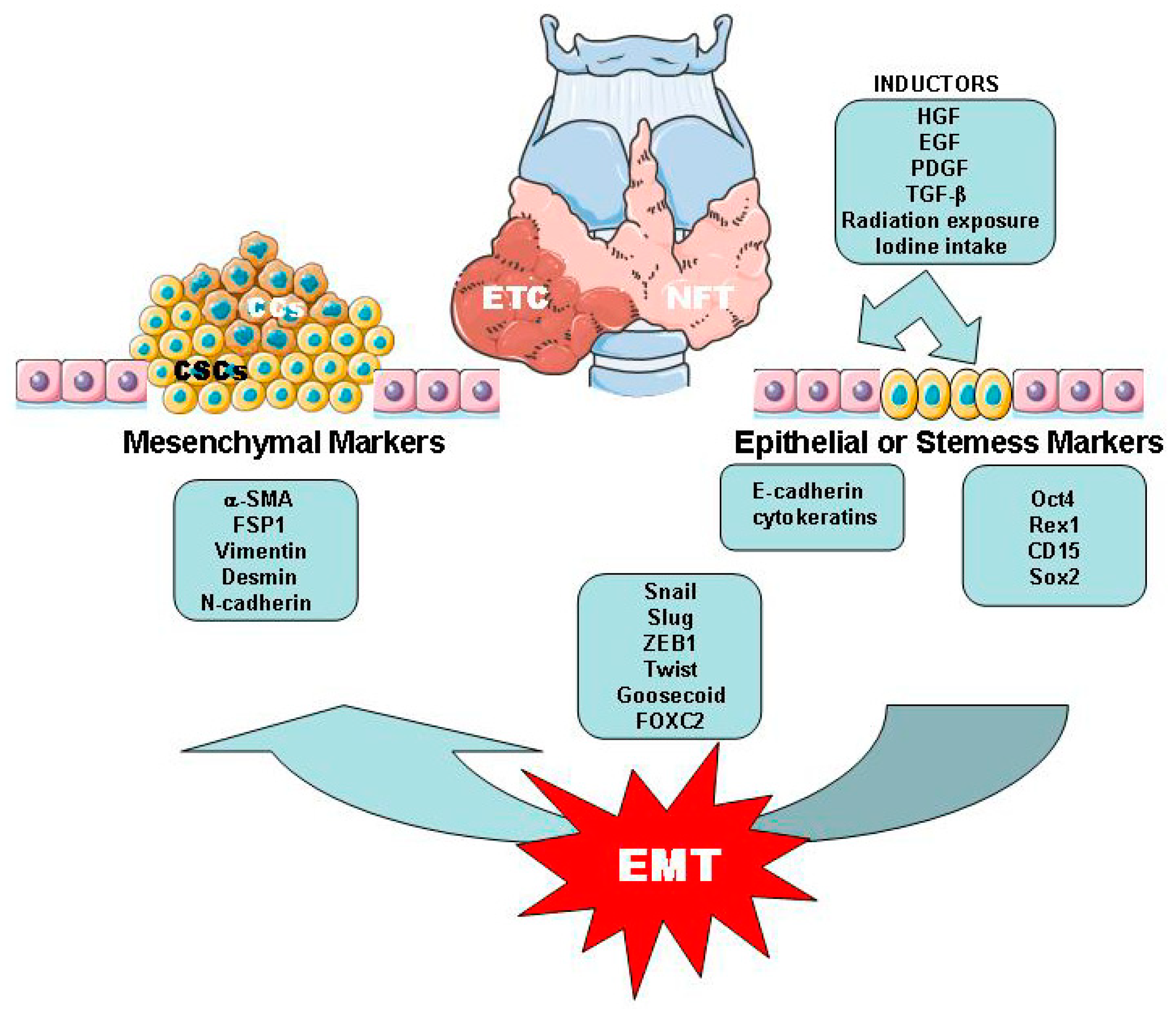
2.3. EMT/MET Processes Related with ETC

EMT is the biological process in which epithelial cells are transformed in cells with a mesenchymal phenotype through multiple biochemical changes [45]. The reverse process is known as the MET. Both are considered to be extremely important in the establishment of the tumors and local or distant metastasis [46,47]. In the EMT/MET, different molecular pathways, different transcription factors, and the expression of specific cell-surface proteins are involved in providing the migratory capacity, invasiveness, and resistance to apoptosis, among other malignancy characteristics, to tumoral cells [45].
The EMT process has been classified, as follows, according to three different contexts: Type 1, which is associated with embryonic development; Type 2, which is essential for tissue regeneration wound healing; and, Type 3, which is linked to cancer progression and metastasis [45,48]. Genetic and epigenetic alterations are potentially responsible for EMT signals originating in the tumor-associated stroma. The loss of epithelial markers and acquisition of mesenchymal markers, such as α-SMA, FSP1, vimentin, and desmin characterizes this process [45,48,49]. In the thyroid, the downregulation of E-cadherin and the upregulation of vimentin expression were found in BRAF-V600E thyroid tumors as compared with normal thyroid tissue (Figure 2) [50].
Several molecules, such as HGF, EGF, PDGF, and TGF-β, have been identified as inducers of the EMT process. One of the most important molecules is TGF-β, and two mechanisms have been proposed for this, namely, Smad-dependent and independent signaling events. In the first case, this pathway can regulate the expression of various transcription factors, which are, as follows: Snail, Slug, zinc finger E-box binding homeobox 1 (ZEB1), Twist, Homeobox protein Goosecoid, and FOXC2. In human and murine thyroid cancer and other carcinomas, the expressions of high levels of Slug, Snail, and Twist and their upregulation are also associated with the transdifferentiation program of thyroid cells into undifferentiated CSC-like cells (Figure 2) [41,47,50]. Moreover, miRNAs play an important role as a regulatory in WDTC and the overexpression of miRNA-146b, miRNA-221, miRNA-222, and miRNA-181b was associated with their malignization and the miRNA-17-92 cluster was identified in ATC [51].
Little is known regarding the MET processes. Some of the authors have suggested that mesenchymal cells and epithelial cells of the tumors engage in cross-talk, facilitating the migration of the latter population (epithelial-like population) and the colonization of other tissues [52,53].

3. Tissue and Inflammatory Mediators in Relation to the EMT Process in ETC

Several data support the idea of the strong correlation between chronic inflammation process and carcinogenesis, with the infiltrated inflammatory cells in the tissue having the capacity to release different cytokines that are involved in the cellular malignization [54].
On the other hand, the incidence of thyroid cancer (especially PTC) has rapidly increased over time, in parallel to the increment of obesity prevalence. A meta-analysis of adiposity measures and thyroid cancer risk using 21 articles yielded data on 12,199 thyroid cancer cases; it found an increased risk (25% excess) for the development of thyroid cancer in overweight individuals and a 55% greater risk in obese individuals [55]. In this scenario, epidemiological studies have established an association between obesity, insulin resistance, type 2 diabetes (DM2), and several cancer types, especially breast and colon cancer [56]. Although the association with ETC is not completely understood, several mechanisms have been proposed for this link, which include elevated levels of TSH, insulin, glucose and triglycerides, insulin resistance, obesity, vitamin D deficiency, and antidiabetic medications [57]. However, several reports, including literature reviews and meta-analyses, have provided conflicting results [58]; nevertheless, according to some authors, obesity may contribute to ETC development [59], whereas it has also been associated with a decreased risk of medullar thyroid cancer [56]. Most of the reports do not offer significant evidence regarding the causal mechanisms of these associations. The link among obesity, DM2, dyslipidemia, and the risk of ETC is not completely understood. It appears reasonable to suggest that insulin resistance and hypothetical factors from adipose tissue (AT) may contribute to a higher risk of follicular thyroid cell proliferation [60].

3.1. Adipose Tissue

AT is considered to be an inflammatory tissue that is able to express both immuno-receptors and immunoregulatory molecules that promote a pro-tumor immune microenvironment. For these reasons, it has been proposed that obesity as an inflammatory process, being the link between obesity and some solid tumors. The identification in the adipose tissue of macrophagic markers that positively correlated with both adipocyte size and body mass supports this hypothesis, which thereby indicates the association between macrophage infiltration in the tumoral tissue and the development of obesity [61].
The macrophage population is divided into two subgroups (M1 and M2) with different cellular actions, thereby the cytokines interferon α (IFNα) or lipopolysaccharide (LPS) can stimulate M1 and produce proinflammatory cytokines; in contrast, cytokines, such as interleukins (ILs) IL-4, IL-10, or IL-13, can stimulate M2, which are related to humoral immune response and they are able to produce anti-inflammatory lymfokines and also participate in tissue repair [62,63]. These two types of macrophages are responsible for almost all adipose tissue TNFα expression and a significant amount of iNOS and IL-6 expression. Moreover, TNFα has an antiproliferative action in a human PTC cell line through a receptor-mediated mechanism [64]. However, the high TNFα exposure that is provided by obesity may induce TNFα resistance that facilitates tumor progression [65].
On the other hand, AT is a critical regulator of the energy balance and substrate metabolism. It plays an important role in energy homeostasis through the production and secretion of several substances with endocrine or paracrine functions. Furthermore, AT is not only considered as a storage organ, but also as an important endocrine organ that releases various proteins that constitute the tumor microenvironment [66]. Many studies have associated an excessive amount of AT with the development of different diseases, such as DM2, premature atherosclerosis, and cardiovascular disease [67]. In ETC, the biological features of AT could play a crucial role in carcinogenic processes. Moreover, it has been demonstrated that many different molecules that are derived from AT, such as leptin, adiponectin (Acrp30), and cholesterol, promote proliferation and invasion processes via the activation of specific transcription factors.

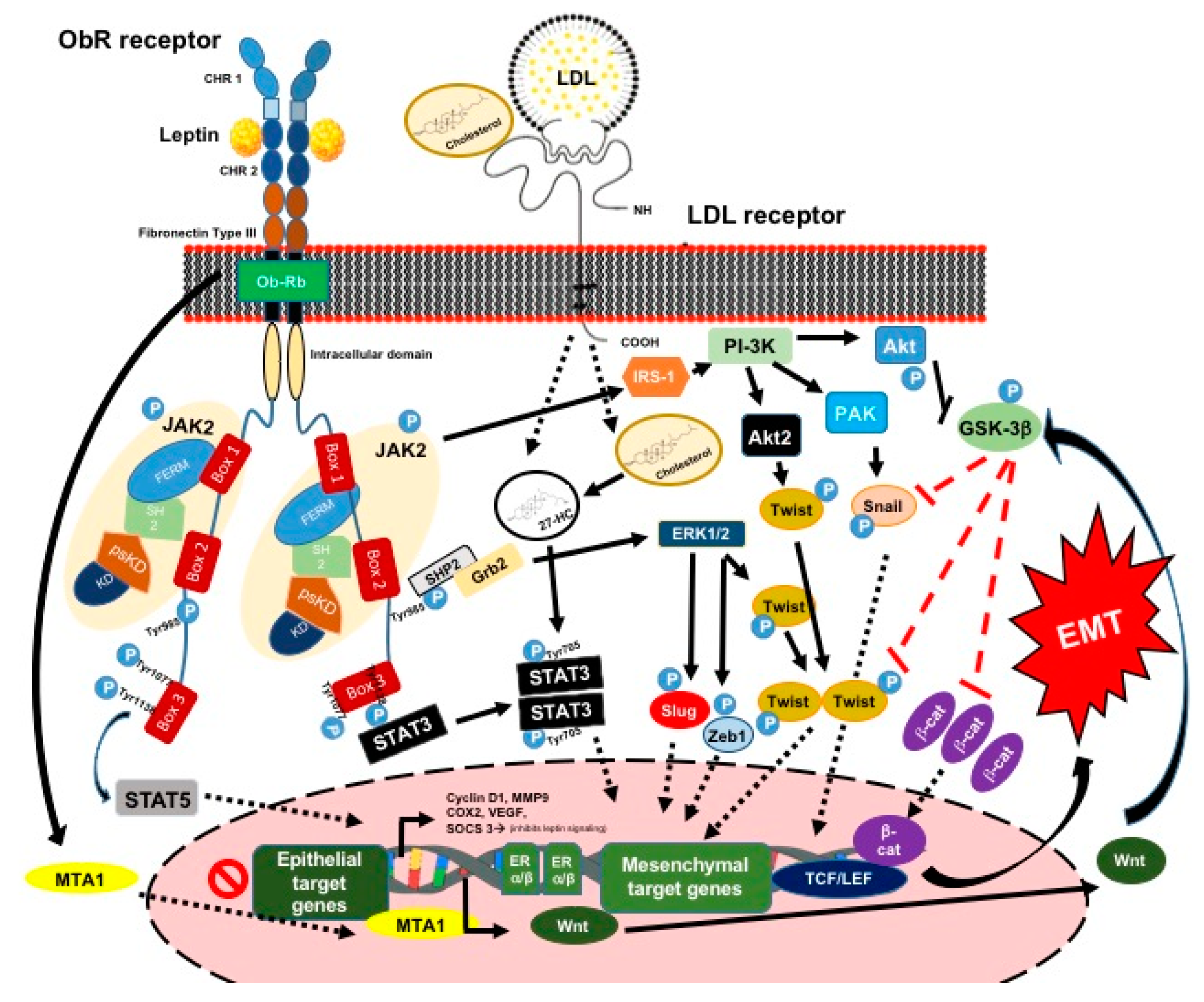
3.1.1. Leptin and Its Signaling Pathway

Leptin is an important adipokine with a molecular weight of 16 kDa. Its major functions are regulating appetite, energy homeostasis, and lipid metabolism, as well as regulating cell differentiation and proliferation of different types of cells. The LEP gene, which is located on chromosome 7q32.1, encodes it, and the AT mainly secretes it [48,68,69].
Leptin is one of the most important mediators between obesity and increased cancer risk. This protein exerts its effects through binding to the OBR receptor, which is widely expressed in a variety of tissues transducing leptin-mediated downstream signaling events [68]. Leptin and OBR receptors have been reported to be overexpressed in numerous types of cancer. What is more, their expression intensity is associated with proliferation, migration, invasion, EMT, and prognosis in several common types of cancer [47,48].
The LEPR gene encodes the OBR receptor, which is located on chromosome 1p31.3. This receptor belongs to the family of class I cytokine receptors and it presents six isoforms that were generated by alternative splicing (OBRa-OBRf). However, only the long form (OBRrb) contains the intracellular motifs that are required for the activation of the signaling [48].
In ETC, the expression of leptin and OBRs is also increased. In a case-control study by Hedayati et al. [70], the leptin levels were higher in ETC patients when compared with healthy subjects. Moreover, in Cheng et al.’s research [71], leptin and its receptor were overexpressed in PTC cells and associated with an aggressive phenotype, larger tumor size, and nodal metastasis. Finally, leptin was also shown to enhance the migration of PTC cells, whereas it inhibited the migration of anaplastic and follicular CCs [72]. In a study on a cohort of Chinese patients with PTC, the leptin and OBRs were expressed in 72.4% and 73.9% of PTC patients [69]. Similarly, in another study of Uddin et al., OBRs and leptin were expressed in 80.1% and 49.1% of PTC cases, respectively [73]. Thus, the previous studies showed that a proportion of PTC tumors express leptin and OBRs, and their expression is associated with aggressive PTC tumor behaviors, poorer disease-free survival, disease persistence, and recurrence [69,70,73].

3.1.2. Leptin and the EMT Process and ETC
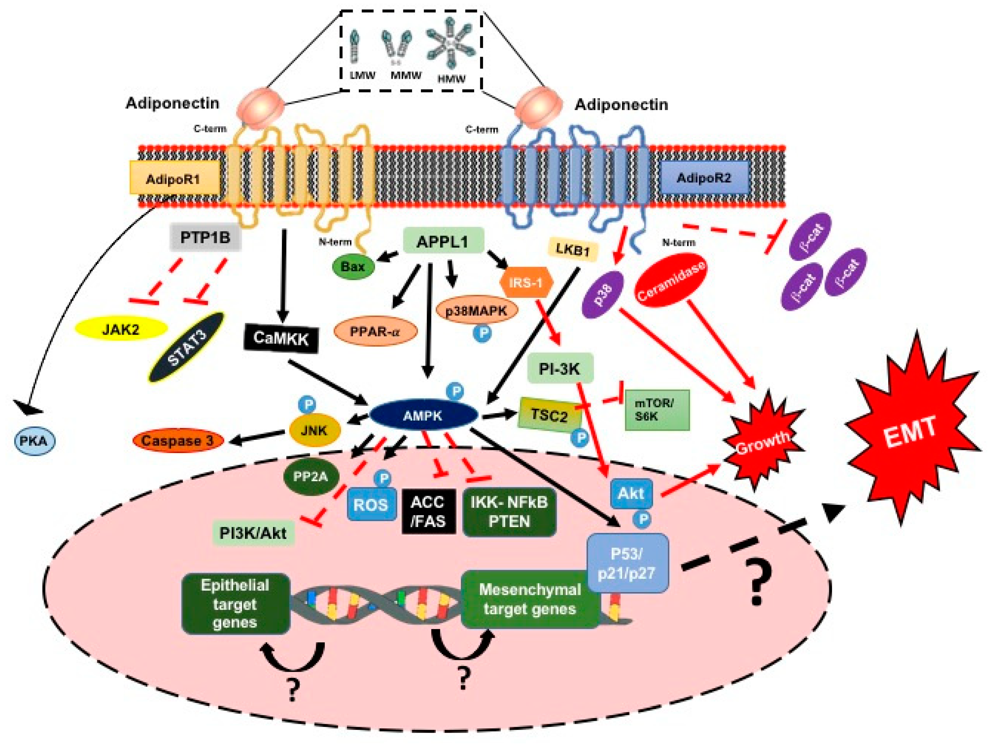
Leptin binds to its receptor through the extracellular region of the OBRrb receptor, which comprises two cytokine homology regions (CHRs). However, only the CHR2 domain is necessary for binding with leptin. Once leptin is bound, the transdimerization of two leptin-OBRrb dimers takes place. This process induces changes in the intracellular region of the OBRrb receptor. Thus, different pathways become activated, which are as follows: the classic cytokine Janus kinase 2/signal transducer and activator of transcription 3 (JAK2/STAT-3) pathway, Ras/extracellular signal-regulated kinases 1/2 (Ras/ERK1/2) signaling cascade, and phosphoinositide 3 kinase/protein kinase B (PI3K/Akt) growth/anti-apoptotic pathway) (Figure 3) [48,68,69].
The intracellular region of the OBRrb contains the three following boxes: a proline-rich region called box 1, which is essential for the binding of the FERM domain of JAK2; a region called box 2 that interacts with the SH2 domain of JAK2; and, box 3, which contains Tyr1077 and Tyr1138, which are necessary residues for the activation of STAT-3 and STAT-5. In addition, JAK2 phosphorylates STAT-3 at Tyr705, inducing its dimerization and translocation to the nucleus, as well as insulin receptor substrate 1 (IRS-1) phosphorylation [74]. Consequently, the PI3K/Akt pathway is activated, which involves the activation of Akt2 and PAK and the phosphorylation of Akt. The latter phosphorylates the GSK-3β, allowing for β-catenin to enter the nucleus and bind to the TCF/LEF complex. Akt2 and PAK phosphorylate Twist and Snail, respectively, promoting their entry into the nucleus. In contrast, JAK2 phosphorylates the Tyr985 of OBRrb, and then the SHP2 protein is anchored and recruits Grb2, which promotes an activation of kinases ERK1/2. Thus, the transcription factors: Slug, Zeb1, and Twist are phosphorylated, entering to the nucleus and promoting the inhibition of the epithelial genes and the overexpression of mesenchymal genes, such as vimentin, cyclin D1, MMP9, VEGF, and COX2, among others (Figure 3) [48,75].
It has been proposed that there are some functional interactions between leptin, metastasis-associated protein 1 (MTA1), and Wnt signaling components [49]. During the EMT process, the expression of Wnt is promoted when the expression of E-cadherin is decreased, and this is associated with high levels of Snail in the nucleus and the nuclear translocation of β-catenin from the cytoplasm, becoming part of the Tcf/LEF complex (Figure 3) [45,68].
In thyroid cancer, there are few studies regarding the relationship between leptin and the EMT process, but some reports have demonstrated that treatment with leptin can promote cell growth and modulated migration of CCs, as well as inhibiting apoptosis through of the upregulation of the XIAP gene [76].

3.1.3. Adiponectin

Acrp30 is the most abundant adipokine that is released by white AT and it circulates oligomers of different molecular weights, which may be of low molecular weight (LMW), medium molecular weight (MMW), or high molecular weight (HMW) [58]. The Acrp30 gene codes for a 244-amino acid polypeptide and it is located on chromosome 3q27 [59]. Its regulation depends on various factors, such as genetic and inflammatory parameters, hormones, or the percentage of body fat and body mass index [47]. Among these, Acrp30 has multiple functions in the regulation of energy homeostasis, insulin sensitivity, and inflammation processes [58].

3.1.4. Adiponectin and the Signaling Pathway

Acrp30 has been considered as a negative regulator of the tumor progression and invasion processes via AdipoR1 and AdipoR2. Furthermore, this molecule plays an important role in increasing apoptosis via the activation of the pAMPK/mTOR pathways (Figure 4) [66,77]. In contrast, other studies have challenged the antitumor role of Acrp30, demonstrating that its presence promotes metastasis and proliferation through the MAPK and NF-κB pathways (Figure 4) [78,79].
The AdipoR1 and AdipoR2 receptors are expressed in macrophages, dendritic cells (DCs), and lymphocytes of various tissues and organs. These two classical Acrp30 receptors share 67% identity of their protein sequence [78]. They belong to the family of seven transmembrane proteins with internal N-terminus and external C-terminus regions, and they may form homo- and hetero-multimers [78,80]. Genes that are situated on the 1p36.13-q41 and 12p13.31 chromosomal regions encode AdipoR1 and AdipoR2, respectively [81].
It seems that Acrp30 exerts antineoplastic effects via two mechanisms: First, it can directly act on CCs by stimulating receptor-mediated signaling pathways. Second, it may act indirectly by modulating insulin sensitivity at the target tissue site, regulating inflammatory responses, and influencing tumor angiogenesis [78]. This review will be focused on the receptor-mediated (AdipoR1 and AdipoR2) signaling pathways.
The evidence suggests that most of the effects of Acrp30 are related to the inhibition of carcinogenesis through AMPK, as well as the inhibition of leptin-induced JAK2 activation and STAT-3 transcriptional activity via increasing the PTP1B protein [78,82]. For example, the activation of cAMP/protein kinase A (PKA), inhibition of β-catenin, and the reduction of reactive oxygen species (ROS) have also been implicated in CCs response to Acrp30 (Figure 4) [78]. Conversely, new molecular data suggest that Acrp30 and its receptor promote proangiogenic effects that could promote tumor growth. This event could be promoted by ceramidase activity from the classical Acrp30 adiponectin receptors [78,83].
Acrp30 binds to AdipoR1 or AdipoR2, initiating a downstream signaling pathway [82]. Bax is induced, thereby promoting apoptosis. Moreover, Ser/Thr kinase LKB1, calcium-dependent kinases (CaMKK), and the adaptor protein APPL-1 are cofactors that activate AMPK, and APPL-1 acts as a signaling pathway mediator in cross-talk with Acrp30 and insulin, which directly interacts with insulin receptor substrates [78,81]. Furthermore, activated AMPK stimulates JNK, PP2A, and p53/p21/p27 (cell cycle regulators) and inhibits fatty acid synthase (FAS)/ACC and the mTOR/S6K axis through TSC2 phosphorylation [81,84]. These factors result in reduced fatty acid synthesis; decreased cellular growth, proliferation, and DNA mutagenesis; and, increased cell cycle arrest and apoptosis, thereby negatively influencing carcinogenesis (Figure 4) [81].
Acrp30 has a negative effect on PI3K/Akt activation. Thus, Akt, which promotes cell proliferation and survival, remains inactivated. PTEN acts as an inhibitor of PI3K signaling, and cell line-specific PTEN deficiency may determine whether AMPK or Akt prevails in regulating mTOR activation. Furthermore, Acrp30 inhibits thioredoxin and the thioredoxin reductase-mediated inhibition of PTEN, thereby influencing PI3K/Akt signaling (Figure 4) [78].
The mediators of the MAPK, cascade, such as JNK, p38, and ERK1/2, can also be related to Acrp30. In different types of cancers, treatment with Acrp30 led to increased JNK activity, resulting in Caspase-3-mediated apoptosis. It seems that AMPK plays a crucial role in JNK phosphorylation. Furthermore, Acrp30 treatment resulted in decreased levels of c-myc, cyclin D, and Bcl-2, while increasing the expression of p53 and Bax, thereby promoting cell cycle arrest and apoptosis (Figure 4) [78]. In contrast, it has been observed that Acrp30 can increase p38 activity, positively influencing the carcinogenesis processes [83].

3.1.5. Adiponectin, the EMT Process and ETC

An EPIC cohort’s study that was published by Dossus et al. [77] compared the levels of various proteins that are associated with inflammation (IL-10) and Acrp30 with ETC risk. The researchers established that women with high Acrp30 levels were less likely to develop ETC, while those with high IL-10 levels had increased TC risk. A few studies suggest that Acrp30 may have a role in EMT, affecting the expression of molecules, like cytokines, vimentin, or E-cadherin. Nigro et al. [80] demonstrated that Acrp30 promotes the apoptosis and oxidative stress in colorectal CCs and increases the pro-inflammatory IL-6 and IL-8 cytokines, which thereby decreases cell survival and migration processes, while reversing the EMT processes [80,85].
In relation to MMPs and tissue inhibitor of MMPs (TIMPs), their role in inducing EMT has been well reported, as well as their part in the migration and invasion processes in ETC [86,87,88]. It has been suggested that ETC may secrete a factor(s) that stimulates MMPs or TIMPs surrounding tissues. Patel et al. demonstrated that the expression of MMPs and TIMPs in recurrent PTC was lower in TIMP when compared with non-recurrent PTC (p = 0.049). Thus, MMPs and TIMPs are all expressed in ETC and could be important in promoting recurrence [86,87,88]. Moreover, it has been reported that Acrp30 can decrease MMP activity via the secretion of human monocyte-derived macrophages via the Syk pathway [85]. Thus, it is well established that tumor cells can induce tumor-associated macrophages (TAMs) that express immunosuppressive cytokine association [66]. Sun et al. [89] revealed that Acrp30 deficiencies promote tumor progression, which reduces the infiltration of macrophages in tumor tissues.
In ETC, Cheng et al. [90] found that the protein levels of the heptahelical transmembrane adiponectin receptors 1 (AdipoR1) and 2 (AdipoR2) modulated by the histone acetylation pattern were increased in some TC patients. This overexpression, which was located in some PTC tissues, was associated with a better prognosis. Moreover, Mitsiadis et al. [91] also reported an inverse relationship of circulating Acrp30 levels with ETC risk.

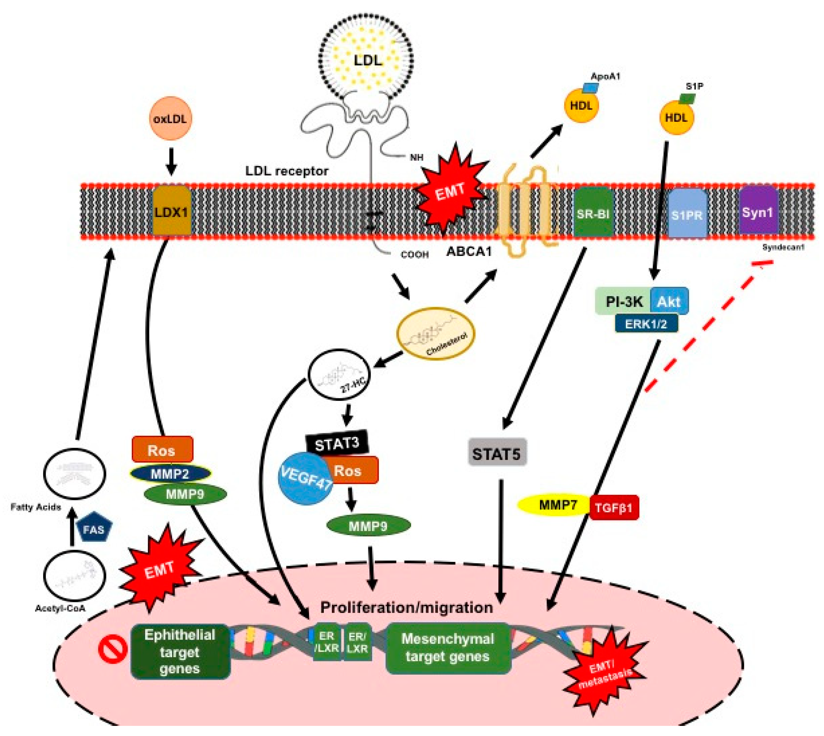
3.2. Plasma Lipoprotein Particles

Significant evidence supports the key role of lipid pathways in cancer development [92,93]. Tumor cells require an increased supply of lipids, which can be obtained by either increasing the exogenous lipids uptake or by upregulating their endogenous synthesis [94,95,96,97]. CCs can take up exogenous cholesterol from low-density lipoproteins (LDL) or be exported into the circulation as high-density lipoproteins (HDLs) to regulate their cholesterol requirement. When cholesterol is in excess, it can be exported into the circulation or locally stored in lipid droplets. Stored cholesterol and one of the main oxidized derivatives of cholesterol, 27-hydroxycholesterol (27-HC), are considered as one of the hallmarks of cancer aggressiveness [98,99]. Alterations in the plasma membrane composition influence the signaling pathways that are associated with EMT processes [44]. Indeed, the lipid composition of epithelial cells in EMT processes are distinct, and they exhibit a significant increase in sphingomyelin and diacylglycerol contents, whereas cholesterol is markedly reduced, which thereby contributes to the enhancement in membrane fluidity and the conversion to the mesenchymal phenotype [100]. However, the statin-induced reduction in intracellular cholesterol levels impaired the cell motility and the metastatic potential in CCs undergoing EMT [101]. These divergent findings suggest a complex relationship between cholesterol, tumor proliferation, and EMT. Cholesterol affects tumor proliferation and metastasis through a range of mechanisms, and it is likely that it plays both promoting and inhibiting roles throughout tumor development. Beyond cholesterol, fatty acid synthesis may also promote the pro-tumorigenic pathways that are involved in EMT-induced cell motility [102]. Indeed, breast CCs undergoing EMT showed an increased expression of FAS and accumulations of saturated fatty acids, thereby regulating lipid rafts’ organization and activating EMT-inducer VEGF/VEGFR2 signaling [102]. FAS inhibition prevented EMT execution, migration, and invasion induced by the hepatocyte growth factor in breast CCs, which is in line with these findings [103]. However, these findings have not been reproduced in other CCs undergoing EMT [104].

3.2.1. Role of Low-Density Lipoproteins

The LDL receptor (LDLR) gene resides on chromosome 19 at the band 19p13.2, which encodes for a cell-surface receptor of 839 amino acids [105]. Increased LDLR expression and, consequently, the uptake of LDL cholesterol has been reported in breast CCs [106,107,108]. In line with these findings, LDL promoted breast CC proliferation, migration, and loss of adhesion, which are hallmarks of the EMT. Furthermore, adhesion molecules, such as cadherin-related family member 3, CD226, Claudin 7, and Ocludin, were downregulated in breast cancer cells exposure to LDL [109]. In this context, the accumulation of 27-HC induced the activation of STAT-3, which promoted the angiogenesis of breast CCs via the ROS/STAT-3/VEGF47 and induced EMT [110]. This mechanism may also contribute to promoting migration and invasion, via STAT-3/MMP-9 and STAT-3/EMT signaling (Figure 5) [111].
Under conditions of oxidative stress, LDL can be oxidatively modified, which results in the formation of lipid peroxidation metabolites, which are closely associated with carcinogenic processes [112]. The levels of serum oxidized LDL (oxLDL) were associated with an increased risk of breast cancer [113], which, in turn, triggers pro-oncogenic signaling in breast CCs [114]. OxLDL lecithin-like receptor 1 (LOX-1) is the main receptor for the internalization of oxLDL. LOX-1 is located in 12p13.2, which encodes a 273-amino acid protein with a short N-terminal intracellular domain, a transmembrane domain, and an extracellular region, followed by a C-type lectin-like domain [115]. Importantly, oxLDL enhanced the LOX-1 expression and lipid accumulation in tubular epithelial cells, which induced the formation of ROS and EMT through ROS [116]. When LOX-1 was overexpressed in prostate CCs and induced by oxLDL, it promoted EMT by downregulating E-cadherin and plakoglobin, whereas it upregulated vimentin, N-cadherin, MMP-2, and MMP-9 (Figure 5) [116].
These studies show the importance of LDL-related pathways in controlling EMT processes and cancer aggressiveness. However, the impact of LDL and oxLDL on EMT processes that are related with thyroid CCs is unknown.

3.2.2. Role of High-Density Lipoproteins

A few studies have addressed the relationship between HDL transporters and EMT in CCs, with divergent results. The gene SCARB1, located in 12q24.31, encodes the scavenger receptor class B, type I (SR-BI), which is the main HDL receptor. This integral membrane protein has 509 amino acids and contains N- and C-terminal cytoplasmic domains, two transmembrane domains, and a large extracellular domain that contains multiple sites for N-linked glycosylation [117]. SR-BI was found to be highly expressed in human metastatic melanoma and regulated EMT processes in melanoma cells [118]. SR-BI downregulation resulted in the loss of the EMT signature in close association with protein glycosylation, including STAT-5, prevented the migration and invasion of melanoma cells, and reduced xenograft tumor growth. However, the SR-BI-mediated effects on EMT were not related to its ability to transport HDL cholesterol [118].
The ABCA1 gene, located in 9q31.1, encodes the ATP binding cassette (ABC) A1 (ABCA1), which is the main cholesterol efflux transporter from cells to HDL. It is ubiquitously expressed as a 220-kDa protein, and it can activate multiple signaling pathways, including the JAK/STAT, PKA, and PKC pathways [119]. Indeed, ABCA1 was significantly upregulated in an EMT of different metastatic CCs [120]. Furthermore, CCs that were induced to undergo an EMT and treated with a range of antimetastatic drugs showed significant ABCA1 downregulation [120]. These results strongly indicate that the ABCA1-mediated effects on plasma membrane cholesterol content and fluidity are critical determinants of their metastatic capacity. In addition, ABCA1 overexpression leads to EMT and the increased invasion of colorectal CCs [121]. However, ABCA1 also inhibits the development of tumors by inhibiting cellular proliferation [122]. ABCA1 reduces mitochondrial cholesterol and ultimately supports CC survival, thereby highlighting the dual role of cholesterol in regulating proliferation and metastasis (Figure 5) [122].
It is noteworthy that apoAI overexpression or exogenous human apoAI, which is the main HDL protein, reduced the malignant properties of colorectal CCs are driven by ABCA1 overexpression [121]. HDL mainly transports sphingosine-1-phosphate (S1P); S1P activates the PI3K/Akt and ERK1/2 signaling pathways, resulting in the MMP-7 upregulation of hepatocellular carcinoma [123]. MMP-7 mediates the shedding of syndecan-1, which, in turn, causes an increase in TGF-β1 production and induces EMT [123]. Based on these results, treatments with some specific HDL components could be considered as potential therapies for treating some cancer invasiveness. However, the effects of lipoproteins and their specific components on EMT processes related to thyroid CCs remain completely unknown.

4. Conclusions

The incidence of ETC, especially PTC, has rapidly increased in recent decades, in parallel with the increase in the prevalence of obesity and other inflammatory diseases. Some reports indicate that inflammatory mediators, such as adipokines, can contribute to cell proliferation and the dedifferentiation of the follicular thyroid epithelium, especially in circumstances of greater genetic susceptibility. These cytokines can induce EMT processes in human PTC cell lines; furthermore, the link between inflammation and tumor metastasis progression in these types of tumors has been demonstrated. However, significant evidence has not been offered regarding the causal mechanisms of these associations and the link between these pathologies. As of yet, neither the risk of ETC nor the molecular mechanism that regulates both EMT and MET are completely understood.
In conclusion, inflammatory mediators, like adipokines and metabolite of cholesterol, can contribute to promoting the aggressive behaviours of thyroid tumors, and the molecular mechanism that controls this process should be investigated.

Author Contributions

G.R., J.C.E.-G., R.C., A.M., and E.M. designed and conducted the literature review and wrote the manuscript. All authors approved the submitted version.

Funding

This work was partly funded by the Ministerio de Sanidad y Consumo, Instituto de Salud Carlos III, and FEDER “Una manera de hacer Europa,” grant FIS 16-00139 (to J.C.E.-G. and E.M.), CIBERDEM and CIBER-BBN (Instituto de Salud Carlos III). We especially want to thank the Societat Catalana d’Endocrinologia i Nutrició (SCEN) (Ajut a la Investigació Bàsica/Clínica 2018) for the financial support for parts in this paper.

Conflicts of Interest

The authors declare no conflict of interest.

Abbreviations

ABCATP binding cassette
AdipoRAdiponectin receptors
ATAdipose tissue
ATCAnaplastic thyroid cancer
CSCCancer stem cells
CCCancer cell
CHRCytokine homology regions
CaMKKCalcium-dependent kinases
DCDendritic cell
DM2Type 2 diabetes
ETCEpithelial thyroid cancer
EMTEpithelial- mesenchymal transition
FTCFollicular thyroid cancer
HMWHigh molecular weight
HDLHigh-density lipoproteins
HSC Hematopoietic stem cells
IFNαinterferon α
ILsinterleukins
LPSlipopolysaccharide
LDLLow-density lipoproteins
LMWLow molecular weight
LOX-1Lecithin-like receptor 1
MTA1Metastasis-associated protein 1
METMesenchymal-epithelial-transition
MMWMedium molecular weight
MMPMatrix metalloproteinase
oxLDLOxidized low-density lipoproteins
PDTCPoorly differentiated thyroid cancer
PTCPapillary thyroid cancer
PTENPhosphatase and tensin homolog
RAIRadioactive iodine
ROSReactive oxygen species
SR-BIReceptor class B type I
TAMsTumor-associated macrophagues
UTCUndifferentiated thyroid carcinoma
VEGFVascular endothelial growth factor
WDTCWell-Differentiated thyroid cancer
ZEB1Zinc finger E-box binding homeobox 1

References

  1. Shah, J.P. Thyroid Carcinoma: Epidemiology, Histology, and Diagnosis. Clin. Adv. Hematol. Oncol. 2015, 13, 3–6. [Google Scholar] [PubMed]
  2. AJCC Cancer Staging Form Supplement (Eight Edition). Available online: https://cancerstaging.org/references-tools/deskreferences/Documents/AJCC%20Cancer%20Staging%20Form%20Supplement.pdf (accessed on 5 May 2019).
  3. Amin, M.B.; Edge, S.; Greene, F.; Byrd, D.R.; Brookland, R.K.; Washington, M.K.; Gershenwald, J.E.; Compton, C.C.; Hess, K.R.; Sullivan, D.C.; et al. AJCC Cancer Staging Manual, 8th ed.; Springer: New York, NY, USA, 2017. [Google Scholar]
  4. Chao, T.C.; Lin, J.D.; Chen, M.F. Insular carcinoma: Infrequent subtype of thyroid cancer with aggressive clinical course. World J. Surg. 2004, 28, 393–396. [Google Scholar] [CrossRef]
  5. Cipriani, N.A.; Nagar, S.; Kaplan, S.P.; White, M.G.; Antic, T.; Sadow, P.M.; Aschebrook-Kilfoy, B.; Angelos, P.; Kaplan, E.L.; Grogan, R.H. Follicular Thyroid Carcinoma: How Have Histologic Diagnoses Changed in the Last Half-Century and What Are the Prognostic Implications? Thyroid 2015, 25, 1209–1216. [Google Scholar] [CrossRef] [PubMed] [Green Version]
  6. Ganly, I.; Nixon, I.J.; Wang, L.Y.; Palmer, F.L.; Migliacci, J.C.; Aniss, A.; Sywak, M.; Eskander, A.E.; Freeman, J.L.; Campbell, M.J.; et al. Survival from Differentiated Thyroid Cancer: What Has Age Got to Do with It? Thyroid 2015, 25, 1106–1114. [Google Scholar] [CrossRef] [Green Version]
  7. Kakudo, K.; Bychkov, A.; Bai, Y.; Li, Y.; Liu, Z.; Jung, C.K. The new 4th edition World Health Organization classification for thyroid tumors, Asian perspectives. Pathol. Int. 2018, 68, 641–664. [Google Scholar] [CrossRef] [PubMed]
  8. Volante, M.; Collini, P.; Nikiforov, Y.E.; Sakamoto, A.; Kakudo, K.; Katoh, R.; Lloyd, R.V.; LiVolsi, V.A.; Papotti, M.; Sobrinho-Simoes, M.; et al. Poorly differentiated thyroid carcinoma: The Turin proposal for the use of uniform diagnostic criteria and algorithmic diagnostic approach. Am. J. Surg. Pathol. 2007, 31, 1256–1264. [Google Scholar] [CrossRef] [PubMed]
  9. Wiseman, S.M.; Loree, T.R.; Rigual, N.R.; Hicks, W.L., Jr.; Douglas, W.G.; Anferson, G.R.; Stoler, D.L. Anaplastic transformation of thyroid cancer. Review of clinical, pathologic, and molecular evidence provides new insights into disease biology and future therapy. Head Neck 2003, 25, 662–670. [Google Scholar] [CrossRef] [PubMed]
  10. Luster, M.; Clarke, S.E.; Dietlein, M.; Lassmann, M.; Lind, P.; Oyen, W.J.G.; Tennvall, J.; Bombardieri, E.; European Association of Nuclear Medicine (EANM). Guidelines for radioiodine therapy of differentiated thyroid cancer. Eur. J. Nucl. Mol. Imaging 2008, 35, 1941–1959. [Google Scholar] [CrossRef] [PubMed]
  11. Nikiforov, Y.E.; Nikiforova, M.N. Molecular genetics and diagnosis of thyroid cancer. Nat. Rev. Endocrinol. 2011, 7, 569–580. [Google Scholar] [CrossRef]
  12. Pak, K.; Suh, S.; Kim, S.J.; Kim, I.J. Prognostic value of genetic mutations in thyroid cancer: A meta-analysis. Thyroid 2015, 25, 63–70. [Google Scholar] [CrossRef]
  13. Patel, K.N. Genetic mutations, molecular markers and future directions in research. Oral Oncol. 2013, 49, 711–721. [Google Scholar] [CrossRef] [PubMed]
  14. Xing, M. Genetic alterations in the phosphatidylinositol-3 kinase/Akt pathway in thyroid cancer. Thyroid 2010, 20, 697–706. [Google Scholar] [CrossRef]
  15. Brzezianska, E.; Pastuszak-Lewandoska, D.A. A minireview: The role of MAPK/ERK and PI3K/Akt pathways in thyroid follicular cell-derived neoplasm. Front Biosci. 2011, 1, 422–439. [Google Scholar] [CrossRef]
  16. Nakao, Y.; Mitsuyasu, T.; Kawano, S.; Nakamura, N.; Kanda, S.; Nakamura, S. Fibroblast growth factors 7 and 10 are involved in ameloblastoma proliferation via the mitogen-activated protein kinase pathway. Int. J. Oncol. 2013, 43, 1377–1384. [Google Scholar] [CrossRef] [PubMed] [Green Version]
  17. O’Malley, Y.; Lal, G.; Howe, J.R.; Weigel, R.J.; Komorowski, R.A.; Shilyansky, J.; Sugg, S.L. Invasion in follicular thyroid cancer cell lines is mediated by EphA2 and pAkt. Surgery 2012, 152, 1218–1224. [Google Scholar] [CrossRef]
  18. Palona, I.; Namba, H.; Mitsutake, N.; Starenki, D.; Podtcheko, A.; Sedliarou, I.; Ohtsuru, A.; Saenko, V.; Nagayama, Y.; Umezawa, K.; et al. BRAFv600E promotes invasiveness of thyroid cancer cells through nuclear factor kB activation. Endocrinology 2006, 147, 5699–5707. [Google Scholar] [CrossRef]
  19. Pyo, J.S.; Kang, G.; Kim, D.H.; Chae, S.W.; Park, C.; Kim, K.; Do, S.I.; Lee, H.J.; Kim, J.H.; Sohn, J.H. Activation of nuclear factor-kappaB contributes to growth and aggressiveness of papillary thyroid carcinoma. Pathol. Res. Pract. 2013, 209, 228–232. [Google Scholar] [CrossRef]
  20. Li, X.; Abdel-Mageed, A.B.; Mondal, D.; Kandil, E. The nuclear factor kappa-B signaling pathway as a therapeutic target against thyroid cancers. Thyroid 2013, 23, 209–218. [Google Scholar] [CrossRef]
  21. Lee, S.J.; Lee, M.H.; Kim, D.W.; Lee, S.; Huang, S.; Ryu, M.J.; Kim, Y.K.; Kim, S.J.; Kim, S.J.; Hwang, J.H.; et al. Cross-regulation between oncogenic BRAF(V600E) kinase and the MST1 pathway in papillary thyroid carcinoma. PLoS ONE 2011, 13, e16180. [Google Scholar] [CrossRef]
  22. Sastre-Perona, A.; Santisteban, P. Role of the wnt pathway in thyroid cancer. Front Endocrinol. 2012, 3, 31. [Google Scholar] [CrossRef]
  23. Enns, L.; Ladiges, W. Mitochondrial redox signaling and cancer invasiveness. J. Bioenerg. Biomembr. 2012, 44, 635–638. [Google Scholar] [CrossRef] [Green Version]
  24. Dubey, R.; Levin, M.D.; Szabo, L.Z.; Laszlo, C.F.; Kushal, S.; Singh, J.B.; Oh, P.; Schnitzer, J.E.; Olenyuk, B.Z. Suppression of tumor growth by designed dimeric epidithiodiketopiperazine targeting hypoxia-inducible transcription factor complex. J. Am. Chem. Soc. 2013, 135, 4537–4549. [Google Scholar] [CrossRef] [PubMed]
  25. García-Jiménez, C.; Santisteban, P. TSH signalling and cancer. Arq. Bras. Endocrinol. Metabol. 2007, 51, 654–671. [Google Scholar] [CrossRef] [PubMed] [Green Version]
  26. Zafon, C.; Obiols, G. The mitogen-activated protein kinase (MAPK) signaling pathway in papillary thyroid cancer. From the molecular bases to clinical practice. Endocrinol. Nutr. 2009, 56, 176–186. [Google Scholar] [CrossRef]
  27. Romitti, M.; Wajner, S.M.; Zennig, N.; Goemann, I.M.; Bueno, A.L.; Meyer, E.L.; Maia, A.L. Increased type 3 deiodinase expression in papillary thyroid carcinoma. Thyroid 2012, 22, 897–904. [Google Scholar] [CrossRef] [PubMed]
  28. Motti, M.L.; Califano, D.; Troncone, G.; De Marco, C.; Migliaccio, I.; Palmieri, E.; Pezzullo, L.; Palombini, L.; Fusco, A.; Viglietto, G. Complex regulation of the cyclin-dependent kinase inhibitor p27kip1 in thyroid cancer cells by the PI3K/Akt pathway: Regulation of p27kip1 expression and localization. Am. J. Pathol. 2005, 166, 737–749. [Google Scholar] [CrossRef]
  29. Serra, R.; Easter, S.L.; Jiang, W.; Baxley, S.E. Wnt5a as an effector of TGFβ in mammary development and cancer. J. Mammary Gland Biol. Neoplasia 2011, 16, 157–167. [Google Scholar] [CrossRef]
  30. Nieto, H.R.; Boelaert, K. Thyroid stimulating hormone in thyroid cancer: Does it matter? Endocr Relat Cancer 2016, 23, 109–121. [Google Scholar] [CrossRef]
  31. Xiaochen, X.; Xiaoguang, S.; Haixia, G.; Qiqiang, G.; Chenling, F.; Wenwu, D. P21-activated kinase 4 involves TSH induced papillary thyroid cancer cell proliferation. Oncotarget 2017, 8, 24882–24891. [Google Scholar] [CrossRef] [Green Version]
  32. Xing, M. Molecular pathogenesis and mechanisms of thyroid cancer. Nat. Rev. Cancer 2013, 13, 184–199. [Google Scholar] [CrossRef] [Green Version]
  33. Rowe, C.W.; Paul, J.W.; Gedye, C.; Tolosa, J.M.; Bendinelli, C.; McGrath, S.; Smith, R. Targeting the TSH receptor in thyroid cancer. Endocr Relat Cancer 2017, 24, 191–202. [Google Scholar] [CrossRef]
  34. Aponte, P.M.; Caicedo, A. Stemness in Cancer: Stem Cells, Cancer Stem Cells, and Their mMicroenvironment. Stem Cells Int. 2017, 2017, 5619472. [Google Scholar] [CrossRef] [PubMed]
  35. Chen, W.; Dong, J.; Haiech, J.; Kilhoffer, M.C.; Zeniou, M. Cancer stem cell quiescence and plasticity as major challenges in cancer therapy. Stem Cells Int. 2016, 2016, 1740936. [Google Scholar] [CrossRef]
  36. Plaks, V.; Kong, N.; Werb, Z. The cancer stem cell niche: How essential is the niche in regulating stemness of tumor cells? Cell Stem Cell 2015, 16, 225–238. [Google Scholar] [CrossRef]
  37. Ma, R.; Minsky, N.; Morshed, S.A.; Davies, T.F. Stemness in human thyroid cancers and derived cell lines: The role of asymmetrically dividing cancer stem cells resistant to chemotherapy. J. Clin. Endocrinol. Metab. 2014, 99, E400–E409. [Google Scholar] [CrossRef] [PubMed]
  38. Garg, M. Emerging role of microRNAs in cancer stem cells: Implications in cancer therapy. World J. Stem Cells 2015, 7, 1078–1089. [Google Scholar] [CrossRef]
  39. Asadzadeh, Z.; Mansoori, B.; Mohammadi, A.; Aghajani, M.; Haji-Asgarzadeh, K.; Safarzadeh, E.; Mokhtarzadeh, A.; Duijf, P.H.G.; Baradaran, B. MicroRNAs in cancer stem cells: Biology, pathways, and therapeutic opportunities. J. Cell Physiol. 2019, 234, 10002–10017. [Google Scholar] [CrossRef]
  40. Goodell, M.A.; Brose, K.; Paradis, G.; Conner, A.S.; Mulligan, R.C. Isolation and functional properties of murine hematopoietic stem cells that are replicating in vivo. J. Exp. Med. 1996, 183, 1797–1806. [Google Scholar] [CrossRef] [Green Version]
  41. Mato, E.; González, C.; Moral, A.; Pérez, J.I.; Bell, O.; Lerma, E.; de Leiva, A. ABCG2/BCRP gene expression is related to epithelial–mesenchymal transition inducer genes in a papillary thyroid carcinoma cell line (TPC-1). J. Mol. Endocrinol. 2014, 52, 289–300. [Google Scholar] [CrossRef] [Green Version]
  42. Lan, L.; Luo, Y.; Cui, D.; Shi, B.Y.; Deng, W.; Huo, L.L.; Chen, H.L.; Zhang, G.Y.; Deng, L.L. Epithelial-mesenchymal transition triggers cancer stem cell generation in human thyroid cancer cells. Int. J. Oncol. 2013, 43, 113–120. [Google Scholar] [CrossRef] [Green Version]
  43. Roche, J. The Epithelial-to-Mesenchymal Transition in Cancer. Cancers 2018, 10, 52. [Google Scholar] [CrossRef]
  44. Morandi, A.; Taddei, M.L.; Chiarugi, P.; Giannoni, E. Targeting the metabolic reprogramming that controls epithelial-to-mesenchymal transition in aggressive tumors. Front. Oncol. 2017, 7, 40. [Google Scholar] [CrossRef] [PubMed]
  45. Kalluri, R.; Weinberg, R.A. The basics of epithelial-mesenchymal transition. J. Clin. Invest. 2009, 119, 1420–1428. [Google Scholar] [CrossRef] [Green Version]
  46. Chaffer, C.L.; Brennan, J.P.; Slavin, J.L.; Blick, T.; Thompson, E.W.; Williams, E.D. Mesenchymal-to-epithelial transition facilitates bladder cancer metastasis: Role of fibroblast growth factor receptor-2. Cancer Res. 2006, 66, 11271–11278. [Google Scholar] [CrossRef] [PubMed]
  47. Montemayor-Garcia, C.; Hardin, H.; Guo, Z.; Larrain, C.; Buehler, D.; Asioli, S.; Chen, H.; Lloyd, R.V. The role of epithelial mesenchymal transition markers in thyroid carcinoma progression. Endocr. Pathol. 2013, 24, 206–212. [Google Scholar] [CrossRef] [PubMed]
  48. Olea-Flores, M.; Juárez-Cruz, J.C.; Mendoza-Catalán, M.A.; Padilla-Benavides, T.; Navarro-Tito, N. Signaling pathways induced by leptin during epithelialmesenchymal transition in breast cancer. International journal of molecular sciences. Int. J. Mol. Sci. 2018, 19, 3493. [Google Scholar] [CrossRef] [PubMed]
  49. Yang, J.; Weinberg, R.A. Epithelial-mesenchymal transition: At the crossroads of development and tumor metastasis. Dev. Cell 2008, 14, 818–829. [Google Scholar] [CrossRef] [PubMed]
  50. Ma, R.; Bonnefond, S.; Morshed, S.A.; Latif, R.; Davies, T.F. Stemness is derived from thyroid cancer cells. Front. Endocrinol. 2014, 5, 114. [Google Scholar] [CrossRef] [PubMed]
  51. Deng, X.; Wu, B.; Xiao, K.; Kang, J.; Xie, J.; Zhang, X.; Fan, Y. MiR-146b-5p promotes metastasis and induces epithelial-mesenchymal transition in thyroid cancer by targeting ZNRF3. Cell Physiol. Biochem. 2015, 35, 71–82. [Google Scholar] [CrossRef] [PubMed]
  52. Scheel, C.; Weinberg, R.A. Cancer stem cells and epithelial-mesenchymal transition: Concepts and molecular links. Semin. Cancer Biol. 2012, 22, 396–403. [Google Scholar] [CrossRef]
  53. Tsuji, T.; Ibaragi, S.; Shima, K.; Hu, M.G.; Katsurano, M.; Sasaki, A.; Hu, G.F. Epithelial-mesenchymal transition induced by growth suppressor p12CDK2-AP1 promotes tumor cell local invasion but suppresses distant colony growth. Cancer Res. 2008, 68, 10377–10386. [Google Scholar] [CrossRef] [PubMed] [Green Version]
  54. Landskron, G.; De la Fuente, M.; Thuwajit, P.; Thuwajit, C.; Hermoso, M.A. Chronic inflammation and cytokines in the tumor microenvironment. J. Immunol. Res. 2014, 2014, 149185. [Google Scholar] [CrossRef] [PubMed]
  55. Schmid, D.; Ricci, C.; Behrens, G.; Leitzmann, M.F. Adiposity and risk of thyroid cancer: A systematic review and meta-analysis. Obes. Rev. 2015, 16, 1042–1054. [Google Scholar] [CrossRef] [PubMed]
  56. Ma, J.; Huang, M.; Wang, L.; Ye, W.; Tong, Y.; Wang, H. Obesity and Risk of Thyroid Cancer: Evidence from a Meta-Analysis of 21 Observational Studies. Med. Sci. Monit. 2015, 21, 283–291. [Google Scholar] [CrossRef] [Green Version]
  57. Shih, S.R.; Chiu, W.Y.; Chang, T.C.; Tseng, C.H. Diabetes and Thyroid Cancer Risk. Literature Review. Exp. Diabetes Res. 2012, 2012, 578285. [Google Scholar] [CrossRef] [PubMed]
  58. Yeo, Y.; Ma, S.H.; Hwang, Y.; Horn-Ross, P.L.; Hsing, A.; Lee, K.E.; Park, Y.J.; Park, D.J.; Yoo, K.Y.; Park, S.K. Diabetes Mellitus and Risk of Thyroid Cancer: A Meta-Analysis. PLoS ONE 2014, 9, e98135. [Google Scholar] [CrossRef]
  59. Zhang, W.; Bai, X.; Ge, H.; Cui, H.; Zhijiang, W.; Han, G. Meta-analysis in the association between obesity and risk of thyroid cancer. Int. J. Clin. Exp. Med. 2014, 7, 5268–5274. [Google Scholar] [PubMed]
  60. Rose, D.P.; Gracheck, P.J.; Vona-Davis, L. The Interactions of Obesity, Inflammation and Insulin Resistance in Breast Cancer. Cancers 2015, 27, 2147–2168. [Google Scholar] [CrossRef] [PubMed]
  61. Zheng, C.; Yang, Q.; Cao, J.; Xie, N.; Liu, K.; Shou, P.; Qian, F.; Wang, Y.; Shi, Y. Local proliferation initiates macrophage accumulation in adipose tissue during obesity. Cell Death Dis. 2016, 7, e2167. [Google Scholar] [CrossRef]
  62. Nielsen, S.R.; Schmid, M.C. Macrophages as Key Drivers of Cancer Progression and Metastasis. Mediators Inflamm. 2017, 2017, 9624760. [Google Scholar] [CrossRef]
  63. Martinez, F.O.; Gordon, S. The M1 and M2 paradigm of macrophage activation: Time for reassessment. F1000Prime Rep. 2014, 6, 13. [Google Scholar] [CrossRef] [PubMed]
  64. Nannan, L.V. Inflammatory mediators, tumor necrosis factor- and interferon-, induce EMT in human PTC cell lines. Oncol Lett. 2015, 10, 2591–2597. [Google Scholar] [CrossRef]
  65. Park, J. Obesity and cancer—Mechanisms underlying tumour progression and recurrence. Nat. Rev. Endocrinol. 2014, 10, 455–465. [Google Scholar] [CrossRef] [PubMed]
  66. Cui, E.; Guo, H.; Shen, M.; Yu, H.; Gu, D.; Mao, W.; Wang, X. Adiponectin inhibits migration and invasion by reversing epithelial-mesenchymal transition in non-small cell lung carcinoma. Oncol. Rep. 2018, 40, 1330–1338. [Google Scholar] [CrossRef] [PubMed]
  67. Cignarelli, A.; Genchi, V.A.; Perrini, S.; Natalicchio, A.; Laviola, L.; Giorgino, F. Insulin and insulin receptors in adipose tissue development. Int. J. Mol. Sci. 2019, 20, 759. [Google Scholar] [CrossRef] [PubMed]
  68. Yan, D.; Avtanski, D.; Saxena, N.K.; Sharma, D. Leptin-induced epithelial-mesenchymal transition in breast cancer cells requires-catenin activation via Akt/GSK3-and MTA1/Wnt1 protein-dependent pathways. J. Biol. Chem. 2012, 287, 8598–8612. [Google Scholar] [CrossRef] [PubMed]
  69. Zhang, G.A.; Hou, S.; Han, S.; Zhou, J.; Wang, X.; Cui, W. Clinicopathological implications of leptin and leptin receptor expression in papillary thyroid cancer. Oncol. Lett. 2013, 5, 797–800. [Google Scholar] [CrossRef] [PubMed] [Green Version]
  70. Hedayati, M.; Yaghmaei, P.; Pooyamanesh, Z.; Yeganeh, M.Z.; Rad, L.H. Leptin: A correlated peptide to papillary thyroid carcinoma? J. Thyroid Res. 2011, 2011, 832163. [Google Scholar] [CrossRef] [PubMed]
  71. Cheng, S.P.; Chi, C.W.; Tzen, C.Y.; Yang, T.L.; Lee, J.J.; Liu, T.P. Clinicopathologic significance of leptin and leptin receptor expressions in papillary thyroid carcinoma. Surgery 2010, 147, 847–853. [Google Scholar] [CrossRef] [PubMed]
  72. Pappa, T.; Alevizaki, M. Obesity and Thyroid Cancer: A Clinical Update. Thyroid 2013, 24, 190–199. [Google Scholar] [CrossRef]
  73. Uddin, S.; Bavi, P.; Siraj, A.K.; Ahmed, M.; Al-Rasheed, M.; Hussain, A.R.; Ahmed, M.; Amin, T.; Alzahrani, A.; Al-Dayel, F.; et al. Leptin-R and its association with PI3K/Akt signaling pathway in papillary thyroid carcinoma. Endocr. Relat. Cancer 2010, 17, 191–202. [Google Scholar] [CrossRef] [PubMed]
  74. Chang, C.C.; Wu, M.J.; Yang, J.Y.; Camarillo, I.G.; Chang, C.J. Leptin-STAT3-G9a signaling promotes obesity-mediated breast cancer progression. Cancer Res. 2015, 75, 2375–2386. [Google Scholar] [CrossRef] [PubMed]
  75. Malaguarnera, R.; Belfiore, A. The emerging role of insulin and insulin-like growth factor signaling in cancer stem cells. Front. Endocrinol. 2014, 5, 10. [Google Scholar] [CrossRef] [PubMed]
  76. Cheng, S.P.; Yin, P.H.; Chang, Y.C.; Lee, C.H.; Huang, S.Y.; Chi, C.W. Differential roles of leptin in regulating cell migration in thyroid cancer cells. Oncol. Rep. 2010, 23, 1721–1727. [Google Scholar] [PubMed]
  77. Dossus, L.; Franceschi, S.; Biessy, C.; Navionis, A.S.; Travis, R.C.; Weiderpass, E.; Scalbert, A.; Romieu, I.; Tjønneland, A.; Olsen, A.; et al. Adipokines and inflammation markers and risk of differentiated thyroid carcinoma: The EPIC study. Int. J. Cancer 2017, 142, 1332–1342. [Google Scholar] [CrossRef] [PubMed] [Green Version]
  78. Dalamaga, M.; Diakopoulos, K.N.; Mantzoros, C.S. The role of adiponectin in cancer: A review of current evidence. Endocr. Rev. 2012, 33, 547–594. [Google Scholar] [CrossRef] [PubMed]
  79. Tang, C.H.; Lu, M.E. Adiponectin increases motility of human prostate cancer cells via AdipoR, p38, AMPK, and NF-κB pathways. Prostate 2009, 69, 1781–1789. [Google Scholar] [CrossRef] [PubMed]
  80. Nigro, E.; Schettino, P.; Polito, R.; Scudiero, O.; Monaco, M.L.; De Palma, G.D. Adiponectin and colon cancer: Evidence for inhibitory effects on viability and migration of human colorectal cell lines. Mol. Cell Biochem. 2018, 448, 125–135. [Google Scholar] [CrossRef] [PubMed]
  81. Achari, A.E.; Jain, S.K. Adiponectin, a therapeutic target for obesity, diabetes, and endothelial dysfunction. Int. J. Mol. Sci. 2017, 18, 1321. [Google Scholar] [CrossRef]
  82. Yuan, J.; Zhang, F.; Niu, R. Multiple regulation pathways and pivotal biological functions of STAT3 in cancer. Sci. Rep. 2015, 5, 17663. [Google Scholar] [CrossRef] [Green Version]
  83. Holland, W.L.; Miller, R.A.; Wang, Z.V.; Sun, K.; Barth, B.M.; Bui, H.H.; Davis, K.E.; Bikman, B.T.; Halberg, N.; Rutkowski, J.M.; et al. Receptor-mediated activation of ceramidase activity initiates the pleiotropic actions of adiponectin. Nat. Med. 2011, 17, 55–63. [Google Scholar] [CrossRef] [PubMed]
  84. Kelesidis, I.; Kelesidis, T.; Mantzoros, C.S. Adiponectin and cancer: A systematic review. Br. J. Cancer 2006, 94, 1221–1225. [Google Scholar] [CrossRef] [PubMed]
  85. Berg, G.; Barchuk, M.; Miksztowicz, V. Behavior of metalloproteinases in adipose tissue, liver and arterial wall: An update of extracellular matrix remodeling. Cells 2019, 8, 158. [Google Scholar] [CrossRef] [PubMed]
  86. Patel, A.; Straight, A.M.; Mann, H.; Duffy, E.; Fenton, C.; Dinauer, C.; Tuttle, R.M.; Francis, G.L. Matrix metalloproteinase (MMP) expression by differentiated thyroid carcinoma of children and adolescents. J. Endocrinol. Invest. 2002, 25, 403–408. [Google Scholar] [CrossRef] [PubMed]
  87. Roth, L.; Kalev-Altman, R.; Monsonego-Ornan, E.; Sela-Donenfeld, D. A new role of the membrane-type matrix metalloproteinase 16 (MMP16/MT3-MMP) in neural crest cell migration. Int. J. Dev. Biol. 2017, 61, 245–256. [Google Scholar] [CrossRef]
  88. Bai, X.; Li, Y.Y.; Zhang, H.Y.; Wang, F.; He, H.L.; Yao, J.C.; Liu, L.; Li, S.S. Role of metalloproteinase-9 in transforming growth factor—β1-induced epithelial-mesenchymal transition in esophageal squamous cell carcinoma. Onco Targets Ther. 2017, 10, 2837–2847. [Google Scholar] [CrossRef] [PubMed]
  89. Sun, Y.; Lodish, H.F. Adiponectin deficiency promotes tumor growth in mice by reducing macrophage infiltration. PLoS ONE 2010, 5, e11987. [Google Scholar] [CrossRef]
  90. Cheng, S.P.; Liu, C.L.; Hsu, Y.C.; Chang, Y.C.; Huang, S.Y.; Lee, J.J. Expression and biologic significance of adiponectin receptors in papillary thyroid carcinoma. Cell Biochem. Biophys. 2013, 65, 203–210. [Google Scholar] [CrossRef] [PubMed]
  91. Mitsiades, N.; Pazaitou-Panayiotou, K.; Aronis, K.N.; Moon, H.S.; Chamberland, J.P.; Liu, X.; Diakopoulos, K.N.; Kyttaris, V.; Panagiotou, V.; Mylvaganam, G.; et al. Circulating adiponectin is inversely associated with risk of thyroid cancer: In vivo and in vitro studies. J. Clin. Endocrinol. Metab. 2011, 96, E2023–E2028. [Google Scholar] [CrossRef] [PubMed]
  92. Cruz, P.M.; Mo, H.; McConathy, W.J.; Sabnis, N.; Lacko, A.G. The role of cholesterol metabolism and cholesterol transport in carcinogenesis: A review of scientific findings, relevant to future cancer therapeutics. Front. Pharmacol. 2013, 4, 119. [Google Scholar] [CrossRef] [PubMed]
  93. Ackerman, D.; Simon, M.C. Hypoxia, lipids, and cancer: Surviving the harsh tumor microenvironment. Trends Cell Biol. 2014, 24, 472–478. [Google Scholar] [CrossRef] [PubMed]
  94. Martínez-Botas, J.; Ferruelo, A.J.; Suárez, Y.; Fernández, C.; Gómez-Coronado, D.; Lasunción, M.A. Dose-dependent effects of lovastatin on cell cycle progression. Distinct requirement of cholesterol and non-sterol mevalonate derivatives. Biochim. Biophys. Acta 2001, 1532, 185–194. [Google Scholar] [CrossRef]
  95. Fernández, C.; Val T. Lobo, M.d.; Gómez-Coronado, D.; Lasunción, M.A. Cholesterol is essential for mitosis progression and its deficiency induces polyploid cell formation. Exp. Cell Res. 2004, 300, 109–120. [Google Scholar] [CrossRef]
  96. Jameel, F.; Phang, M.; Wood, L.G.; Garg, M.L. Acute effects of feeding fructose, glucose and sucrose on blood lipid levels and systemic inflammation. Lipids Health Dis. 2014, 13, 195. [Google Scholar] [CrossRef] [PubMed] [Green Version]
  97. Furuya, Y.; Sekine, Y.; Kato, H.; Miyazawa, Y.; Koike, H.; Suzuki, K. Low-density lipoprotein receptors play an important role in the inhibition of prostate cancer cell proliferation by statins. Prostate Int. 2016, 4, 56–60. [Google Scholar] [CrossRef] [PubMed] [Green Version]
  98. Beloribi-Djefaflia, S.; Vasseur, S.; Guillaumond, F. Lipid metabolic reprogramming in cancer cells. Oncogenesis 2016, 5, e189. [Google Scholar] [CrossRef] [PubMed]
  99. Nelson, E.R. The significance of cholesterol and its metabolite, 27-hydroxycholesterol in breast cancer. Mol. Cell Endocrinol. 2018, 466, 73–80. [Google Scholar] [CrossRef] [PubMed]
  100. Sampaio, J.L.; Gerl, M.J.; Klose, C.; Ejsing, C.S.; Beug, H.; Simons, K.; Shevchenko, A. Membrane lipidome of an epithelial cell line. Proc. Natl. Acad. Sci. USA 2011, 108, 1903–1907. [Google Scholar] [CrossRef] [PubMed] [Green Version]
  101. Warita, K.; Warita, T.; Beckwitt, C.H.; Schurdak, M.E.; Vazquez, A.; Wells, A.; Oltvai, Z.N. Statin-induced mevalonate pathway inhibition attenuates the growth of mesenchymal-like cancer cells that lack functional E-cadherin mediated cell cohesion. Sci. Rep. 2014, 4, 7593. [Google Scholar] [CrossRef] [Green Version]
  102. Li, J.; Dong, L.; Wei, D.; Wang, X.; Zhang, S.; Li, H. Fatty acid synthase mediates the epithelial-mesenchymal transition of breast cancer cells. Int. J. Biol. Sci. 2014, 10, 10171–10180. [Google Scholar] [CrossRef]
  103. Hung, C.M.; Kuo, D.H.; Chou, C.H.; Su, Y.C.; Ho, C.T.; Way, T.D. Osthole suppresses hepatocyte growth factor (HGF)-induced epithelial-mesenchymal transition via repression of the c-Met/Akt/mTOR pathway in human breast cancer cells. J. Agric. Food Chem. 2011, 59, 9683–9690. [Google Scholar] [CrossRef] [PubMed]
  104. Jiang, L.; Xiao, L.; Sugiura, H.; Huang, X.; Ali, A.; Kuro-o, M.; Deberardinis, R.J.; Boothman, D.A. Metabolic reprogramming during TGFβ1-induced epithelial-to-mesenchymal transition. Oncogene 2015, 34, 3908–3916. [Google Scholar] [CrossRef] [PubMed]
  105. Sudhof, T.C.; Goldstein, J.L.; Brown, M.S.; Russell, D.W. The LDL receptor gene: A mosaic of exons shared with different proteins. Science 1985, 228, 815–822. [Google Scholar] [CrossRef]
  106. Rotheneder, M.; Kostner, G.M. Effects of low- and high-density lipoproteins on the proliferation of human breast cancer cells in vitro: Differences between hormone-dependent and hormone-independent cell lines. Int. J. Cancer 1989, 43, 875–879. [Google Scholar] [CrossRef] [PubMed]
  107. De Gonzalo-Calvo, D.; López-Vilaró, L.; Nasarre, L.; Perez-Olabarria, M.; Vázquez, T.; Escuin, D.; Badimon, L.; Barnadas, A.; Lerma, E.; Llorente-Cortés, V. Intratumor cholesteryl ester accumulation is associated with human breast cancer proliferation and aggressive potential: A molecular and clinicopathological study. BMC Cancer 2015, 15, 460. [Google Scholar] [CrossRef] [PubMed]
  108. Gallagher, E.; Zelenko, Z.; Neel, B.A.; Antoniou, I.M.; Rajan, L.; Kase, N.; LeRoith, D. Elevated tumor LDLR expression accelerates LDL cholesterol-mediated breast cancer growth in mouse models of hyperlipidemia. Oncogene 2017, 36, 6462–6471. [Google Scholar] [CrossRef] [PubMed] [Green Version]
  109. Dos Santos, C.R.; Domingues, G.; Matias, I.; Matos, J.; Fonseca, I.; de Almeida, J.M.; Dias, S. LDL-cholesterol signaling induces breast cancer proliferation and invasion. Lipids Health Dis. 2014, 13, 16. [Google Scholar] [CrossRef]
  110. Cruz, P.; Epuñán, M.J.; Ramírez, M.E.; Torres, C.G.; Valladares, L.E.; Sierralta, W.D. 27-hydroxycholesterol and the expression of three estrogen-sensitive proteins in MCF7 cells. Oncol. Rep. 2012, 28, 992–998. [Google Scholar] [CrossRef]
  111. Zhang, Y.; Ma, K.L.; Liu, J.; Wu, Y.; Hu, Z.B.; Liu, L.; Lu, J.; Zhang, X.L.; Liu, B.C. Inflammatory stress exacerbates lipid accumulation and podocyte injuries in diabetic nephropathy. Acta Diabetol. 2015, 52, 1045–1056. [Google Scholar] [CrossRef]
  112. Wiseman, H.; Halliwell, B. Damage to DNA by reactive oxygen and nitrogen species: Role in inflammatory disease and progression to cancer. Biochem. J. 1996, 313, 17–29. [Google Scholar] [CrossRef]
  113. Delimaris, I.; Faviou, E.; Antonakos, G.; Stathopoulou, E.; Zachari, A.; Dionyssiou-Asteriou, A. Oxidized LDL, serum oxidizability and serum lipid levels in patients with breast or ovarian cancer. Clin. Biochem. 2007, 15, 1129–1134. [Google Scholar] [CrossRef] [PubMed]
  114. Khaidakov, M.; Mitra, S.; Kang, B.Y.; Wang, X.; Kadlubar, S.; Novelli, G.; Raj, V.; Winters, M.; Carter, W.C.; Mehta, J.L. Oxidized LDL Receptor 1 (OLR1) as a possible link between obesity, dyslipidemia and cancer. PLoS ONE 2011, 6, e20277. [Google Scholar] [CrossRef] [PubMed]
  115. Kume, N.; Kita, T. Roles of lectin-like oxidized LDL receptor-1 and its soluble forms in atherogenesis. Curr. Opin. Lipidol. 2001, 12, 419–423. [Google Scholar] [CrossRef] [PubMed]
  116. Wang, R.; Ding, G.; Liang, W.; Chen, C.; Yang, H. Role of LOX-1 and ROS in oxidized low-density lipoprotein induced epithelial-mesenchymal transition of NRK52E. Lipids Health Dis. 2010, 9, 120. [Google Scholar] [CrossRef] [PubMed]
  117. Parathath, S.; Darlington, Y.F.; de la Llera Moya, M.; Drazul-Schrader, D.; Williams, D.L.; Phillips, M.C.; Rothblat, G.H.; Connelly, M.A. Effects of amino acid substitutions at glycine 420 on SR-BI cholesterol transport function. J. Lipid Res. 2007, 48, 1386–1395. [Google Scholar] [CrossRef] [PubMed]
  118. Kinslechner, K.; Schörghofer, D.; Schütz, B.; Vallianou, M.; Wingelhofer, B.; Mikulits, W.; Röhrl, C.; Hengstschläger, M.; Moriggl, R.; Stangl, H.; et al. Malignant phenotypes in metastatic melanoma are governed by SR-BI and its association with glycosylation and STAT5 activation. Mol. Cancer Res. 2018, 16, 135–146. [Google Scholar] [CrossRef] [PubMed]
  119. Luu, W.; Sharpe, L.J.; Gelissen, I.C.; Brown, A.J. The role of signalling in cellular cholesterol homeostasis. IUBMB Life 2013, 65, 675–684. [Google Scholar] [CrossRef] [Green Version]
  120. Zhao, W.; Prijic, S.; Urban, B.C.; Tisza, M.J.; Zuo, Y.; Li, L.; Tan, Z.; Chen, X.; Mani, S.A.; Chang, J.T. Candidate antimetastasis drugs suppress the metastatic capacity of breast cancer cells by reducing membrane fluidity. Cancer Res. 2016, 76, 2037–2049. [Google Scholar] [CrossRef] [PubMed]
  121. Aguirre-Portolés, C.; Feliu, J.; Reglero, G.; Ramírez de Molina, A. ABCA1 overexpression worsens colorectal cancer prognosis by facilitating tumour growth and caveolin-1-dependent invasiveness and these effects can be ameliorated using the BET inhibitor apabetalone. Mol. Oncol. 2018, 12, 1735–1752. [Google Scholar] [CrossRef]
  122. Smith, B.; Land, H. Anticancer activity of the cholesterol exporter ABCA1 gene. Cell Rep. 2012, 2, 580–590. [Google Scholar] [CrossRef] [PubMed]
  123. Zeng, Y.; Yao, X.; Chen, L.; Yan, Z.; Liu, J.; Zhang, Y.; Feng, T.; Wu, J.; Liu, X. Sphingosine-1-phosphate induced epithelial-mesenchymal transition of hepatocellular carcinoma via an MMP-7/syndecan-1/TGF-β autocrine loop. Oncotarget 2016, 7, 63324–63337. [Google Scholar] [CrossRef] [PubMed] [Green Version]
Figure 1. Schematic representation of relevant signaling pathways that are involved in epithelial thyroid carcinogenesis (ETC) and their link with epithelial mesenchymal transition (EMT) process. Part of Servier Medical Art by Servier is licensed under a Creative Commons Attribution 3.0 Unported License. (https://smart.servier.com/image-set-download).
Ijms 20 02466 g001
Figure 2. Schematic representation of the most significant signaling inductors involved on the cell dedifferentiation of adult stem cells and normal follicular cells by EMT process. EMT, thyroid cancer cells (CCs) and cancer stem cells (CSCs). Part of Servier Medical Art by Servier is licensed under a Creative Commons Attribution 3.0 Unported License. (https://smart.servier.com/image-set-download).
Ijms 20 02466 g002
Figure 3. Schematic representation of leptin-induced signaling pathway. Leptin, cholesterol and low-density lipoprotein (LDL) particles interact with their respective receptors, which trigger a cascade of signaling events promoting EMT process.
Ijms 20 02466 g003
Figure 4. Schematic representation of adiponectin-induced signaling pathway. Adiponectin interacts with its respective receptors, AdipoR1 and AdipoR2, in order to activate a cascade of signaling events. Further studies are needed in order to clarify the possible activation of EMT processes induced by adiponectin.
Ijms 20 02466 g004
Figure 5. Schematic representation of mechanisms by which LDL, the oxidized form of LDL, high-density lipoprotein (HDL) and their specific receptors induce proliferation, migration, and EMT processes in cancer cells. These lipoprotein particles interact with their respective receptors, which trigger a cascade of signaling events promoting EMT process. The cholesterol that is transported by the lipoproteins and the synthesis of fatty acids play a critical role in these signaling pathways.
Ijms 20 02466 g005
Table 1. Main signaling pathways and gene mutations involved in thyroid carcinogenesis.
Signaling Pathways AffectedMutationsTypes of Thyroid TumorsRefs
MAPKBRAF V600E, RAS, RET/PTC, RTK, ALKPTC[11,14]
PI3K/Akt1, PTENFTC[11]
TGF-β1PTC, FTC, ATC, PDTC[16]
PI3/AKTPTENFTA, FTC[17]
Akt1, Akt2FTC[17]
NF-KBRET-PTC, RAS, BRAF-V600EPTC, FTC, ATC[18,19,20]
RASSF1/MST1/FOXO3MEK/MAPK, RASSF1/MST1/FOXO3, NF-κB, BRAF-V600E, ERK 1/2, AktPTC, FTC, ATC, PDTC[21]
WNT/β-CATENINCTNNB1ATC, PDTC[22]
HIF1AHIF1, VEGFA, METPDTC, FCT, ATC[23,24]
HIF1αATC[19]
TSHRTSH-TSHR, NISPDTC, FCT, ATC, PTC[25]
PAK4PTC[25]

