Article

Multiple Layers of CDK5R1 Regulation in Alzheimer’s Disease Implicate Long Non-Coding RNAs

1
Dipartimento di Biotecnologie Mediche e Medicina Traslazionale, Università degli Studi di Milano, Via Fratelli Cervi 93, 20090 Segrate, Italy
2
Istituto di Neuroscienze, CNR, Via Vanvitelli 32, 20129 Milano, Italy
*
Author to whom correspondence should be addressed.
Int. J. Mol. Sci. 2018, 19(7), 2022; https://doi.org/10.3390/ijms19072022
Submission received: 17 May 2018 / Revised: 28 June 2018 / Accepted: 3 July 2018 / Published: 11 July 2018
(This article belongs to the Special Issue Molecular Mechanism of Alzheimer's Disease)

Abstract

:
Cyclin-dependent kinase 5 regulatory subunit 1 (CDK5R1) gene encodes for p35, the main activator of Cyclin-dependent kinase 5 (CDK5). The active p35/CDK5 complex is involved in numerous aspects of brain development and function, and its deregulation is closely associated to Alzheimer’s disease (AD) onset and progression. We recently showed that miR-15/107 family can negatively regulate CDK5R1 expression modifying mRNA stability. Interestingly, miRNAs belonging to miR-15/107 family are downregulated in AD brain while CDK5R1 is upregulated. Long non-coding RNAs (lncRNAs) are emerging as master regulators of gene expression, including miRNAs, and their dysregulation has been implicated in the pathogenesis of AD. Here, we evaluated the existence of an additional layer of CDK5R1 expression regulation provided by lncRNAs. In particular, we focused on three lncRNAs potentially regulating CDK5R1 expression levels, based on existing data: NEAT1, HOTAIR, and MALAT1. We demonstrated that NEAT1 and HOTAIR negatively regulate CDK5R1 mRNA levels, while MALAT1 has a positive effect. We also showed that all three lncRNAs positively control miR-15/107 family of miRNAs. Moreover, we evaluated the expression of NEAT1, HOTAIR, and MALAT1 in AD and control brain tissues. Interestingly, NEAT1 displayed increased expression levels in temporal cortex and hippocampus of AD patients. Interestingly, we observed a strong positive correlation between CDK5R1 and NEAT1 expression levels in brain tissues, suggesting a possible neuroprotective role of NEAT1 in AD to compensate for increased CDK5R1 levels. Overall, our work provides evidence of another level of CDK5R1 expression regulation mediated by lncRNAs and points to NEAT1 as a biomarker, as well as a potential pharmacological target for AD therapy.

Graphical Abstract

1. Introduction

Alzheimer’s Disease (AD) is the most common neurodegenerative disorder, causing a severe and permanent impairment of both cognitive and behavioral functions. It accounts for about 70% of the 50 million people suffering from dementia worldwide and it is currently estimated that, with global population aging, the prevalence of AD will triple by 2050 [1], with a significant economic and social burden on both patients’ families and society.
AD is characterized by a plethora of pathological features, including neuronal loss, dendritic hypotrophy and synaptic alteration, microglial malfunction, cerebrovascular amyloid angiopathy, inflammation, and mitochondrial dysfunction [2,3]. However, the most distinctive features are the presence of extracellular senile plaques, formed by fibrillary β-amyloid (Aβ) and neurofibrillary tangles (NFTs), composed of hyperphosphorylated Tau [4]. Abnormal kinase activity is believed to play a major role in AD pathogenesis [5]. In particular, deregulation of Cyclin-dependent kinase 5 (CDK5), a proline-directed serine/threonine kinase involved in several developmental and physiological processes in the central nervous system (CNS) [6,7], has been suggested to play a pivotal role in the onset of the two main pathological hallmarks of AD by inducing Aβ peptide production and mediating Tau protein hyperphosphorylation [8].
CDK5 requires the p35 regulatory subunit to become active and its kinase activity is strictly dependent on the amount of its activator. p35 is encoded by the cyclin-dependent kinase 5 regulatory subunit 1 (CDK5R1) gene, which displays a large and highly conserved 3′-UTR, suggestive of an important role of post-transcriptional regulation in the control of its expression. Indeed, we previously demonstrated that CDK5R1 expression is regulated at the post-transcriptional level by neuronal ELAV (nELAV) RNA-binding proteins [9,10] and by heterogeneous nuclear ribonucleoproteins A2/B1 (hnRNP A2/B1) [10]. In addition, we recently found that the miR-15/107 family of microRNAs is also involved in negatively regulating CDK5R1 expression. More interestingly, this group of microRNAs turned out to be downregulated in the hippocampus and cerebral cortex of AD patients while CDK5R1 mRNA levels were upregulated in AD hippocampus [11].
An additional layer of complexity to the regulation of CDK5R1 expression that can be relevant for AD pathogenesis might be provided by long non-coding RNAs (lncRNAs). LncRNAs are a highly heterogeneous class of RNA molecules of more than 200 bases in length with no protein-coding capacity. They are involved in the control of gene expression at multiple levels, from nuclear architecture to transcription regulation, mRNA splicing and maturation to mRNA localization and stability, and protein translation and stability to regulation of miRNA activity [12]. Owing to this versatility, lncRNAs are now considered as master regulators of gene expression [13]. In particular, lncRNAs have been shown to post-transcriptionally regulate the levels of several target genes by the formation of lncRNA/miRNA/target gene axes, and the dysregulation of the crosstalk between the two types of ncRNAs has been found to be a crucial contributor to disease pathogenesis [14].
The role of lncRNAs in malignancies and their significance as both diagnostic and prognostic markers has been extensively studied and is well established [15], but an involvement of lncRNAs in the pathogenesis of neurodegenerative diseases is now clearly emerging. In particular, different lncRNAs have been found dysregulated in Alzheimer’s disease and involved in AD pathogenesis by promoting β-amyloid production, including BACE1-AS, 17A, and NDM29 [16]. For example, the expression of BACE1-AS, the antisense transcript of the β-secretase encoding gene BACE1, is upregulated in AD brains specimens. BACE1-AS was reported to increase the stability of BACE1 mRNA and to prevent the binding of miRNA 485-5p, therefore positively regulating BACE1 protein levels and promoting Aβ42 synthesis [16,17].
In the present work, we focused on three different lncRNAs which had the potential for regulating CDK5R1 expression levels and deserved to be analyzed in AD brain tissues, namely NEAT1, HOTAIR, and MALAT1. NEAT1 (nuclear enriched abundant transcript 1) is a lncRNA that regulates gene expression by binding to the promoter of active chromatin sites [18,19]. Moreover, NEAT1 is known to act as a scaffold for paraspeckles [20], representing specific subnuclear bodies that are involved in gene expression regulation by sequestration and retention of specific RNAs and proteins [21]. Relevantly, NEAT1 levels were found to be deregulated in different neurodegenerative diseases [22]. MALAT1 (metastasis-associated lung adenocarcinoma transcript 1), also known as NEAT2 (nuclear-enriched abundant transcript 2), is predominantly localized to nuclear speckles, where it regulates alternative splicing by modulating the phosphorylation status of SR family of splicing factors [23]. MALAT1 has been linked to several human tumors, in most cases being overexpressed in malignant tissues [24]. Both NEAT1 and MALAT1 have been demonstrated to regulate the expression of members of the miR-15/107 group of miRNAs [25,26], which are known CDK5R1 negative regulators [11]. HOTAIR (HOX antisense intergenic RNA) is transcribed from the antisense strand of the HOXC locus and represses expression of the downstream HOXD locus together with several genes on other chromosomes. HOTAIR is involved in the control of cell apoptosis, growth, metastasis, angiogenesis, DNA repair and, like MALAT1, it has been shown to be upregulated in different types of cancer [27]. Interestingly, HOTAIR can also serve as a scaffold for Lysine-specific histone demethylase 1A (LSD1) complex and polycomb repressive complex 2 (PRC2) [28]. Since the expression of CDK5R1 is repressed by LSD1 [29], HOTAIR can potentially impact CDK5R1 levels.
Here, we demonstrated that NEAT1 and HOTAIR negatively regulate CDK5R1 mRNA levels, while MALAT1 has a positive effect on CDK5R1 expression. We also showed that all three lncRNAs positively control the levels of miR-15/107 family of microRNAs. Moreover, we evaluated the expression of NEAT1, HOTAIR, and MALAT1 in AD and control brain tissues. Interestingly, NEAT1 displayed increased expression levels in temporal cortex and hippocampus of AD patients, compared to controls. In addition, we observed a strong positive correlation between CDK5R1 and NEAT1 expression levels in brain tissues, suggesting a novel molecular marker of AD pathogenesis, warranting further studies. Overall, our work provides evidence of another level of CDK5R1 expression regulation mediated by long non-coding RNAs, which can also impact on Alzheimer’s disease research.

2. Results

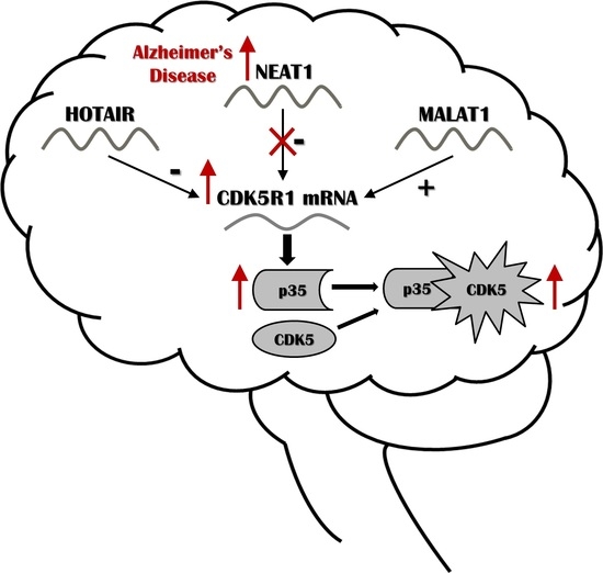
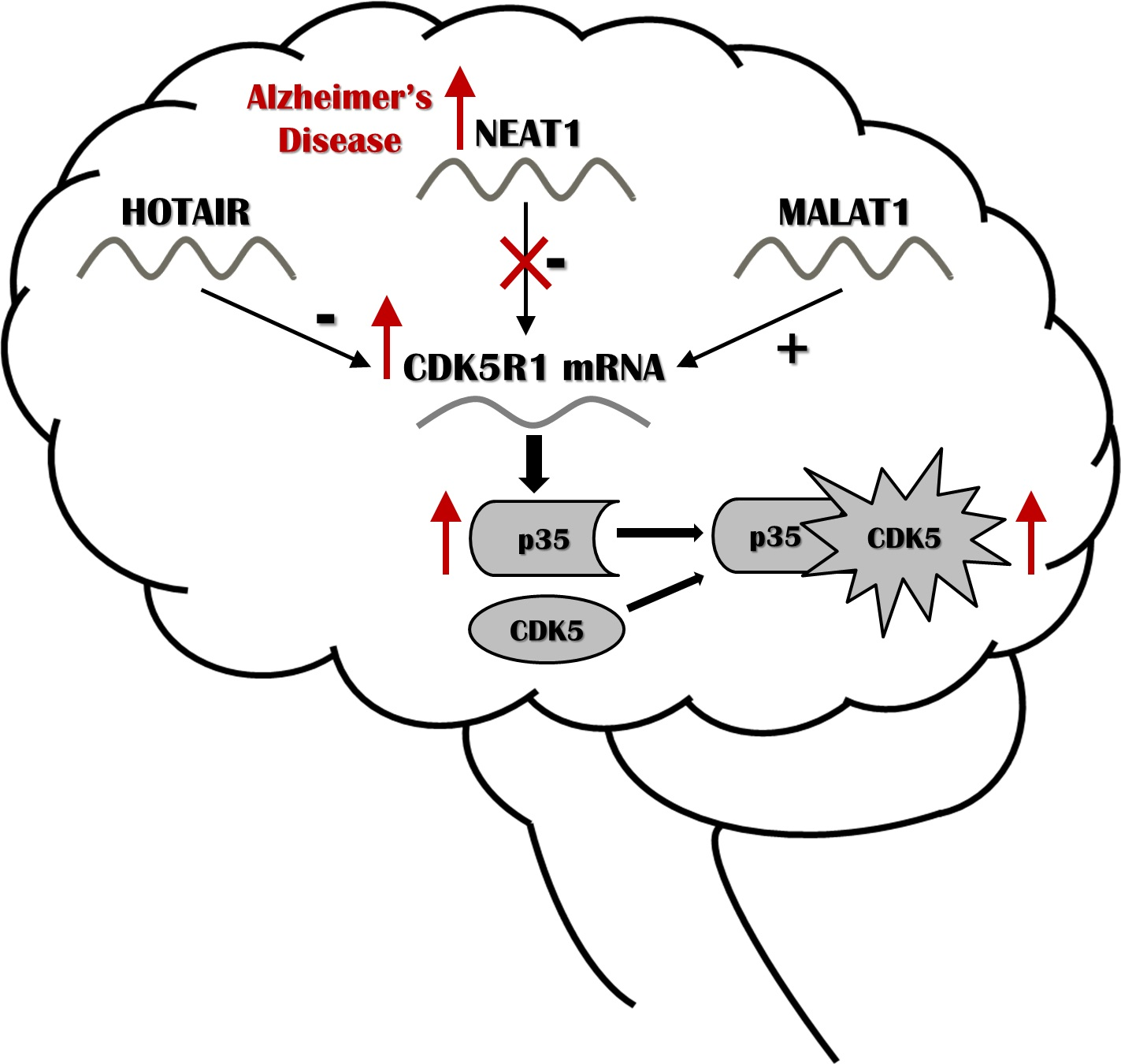
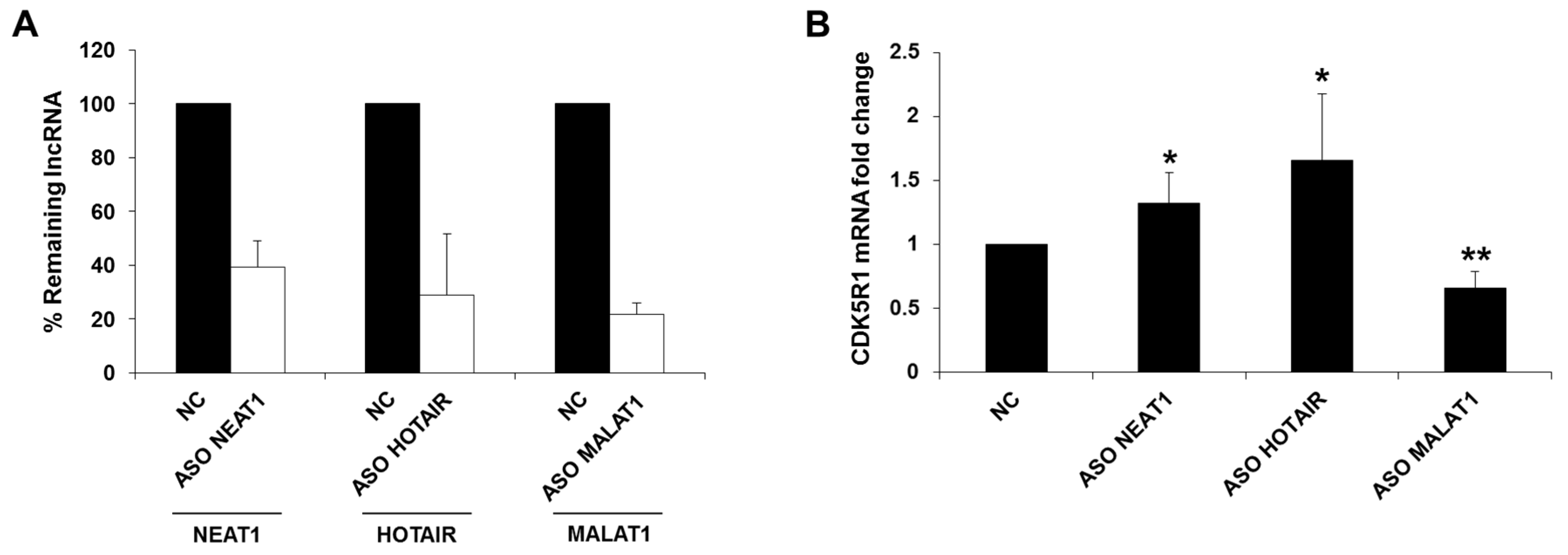
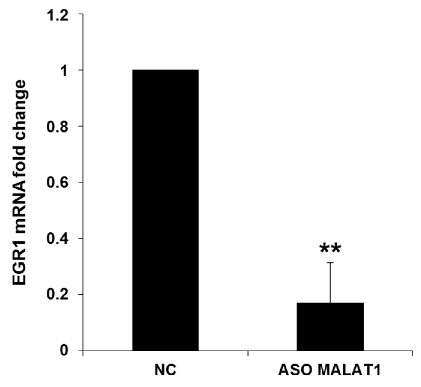
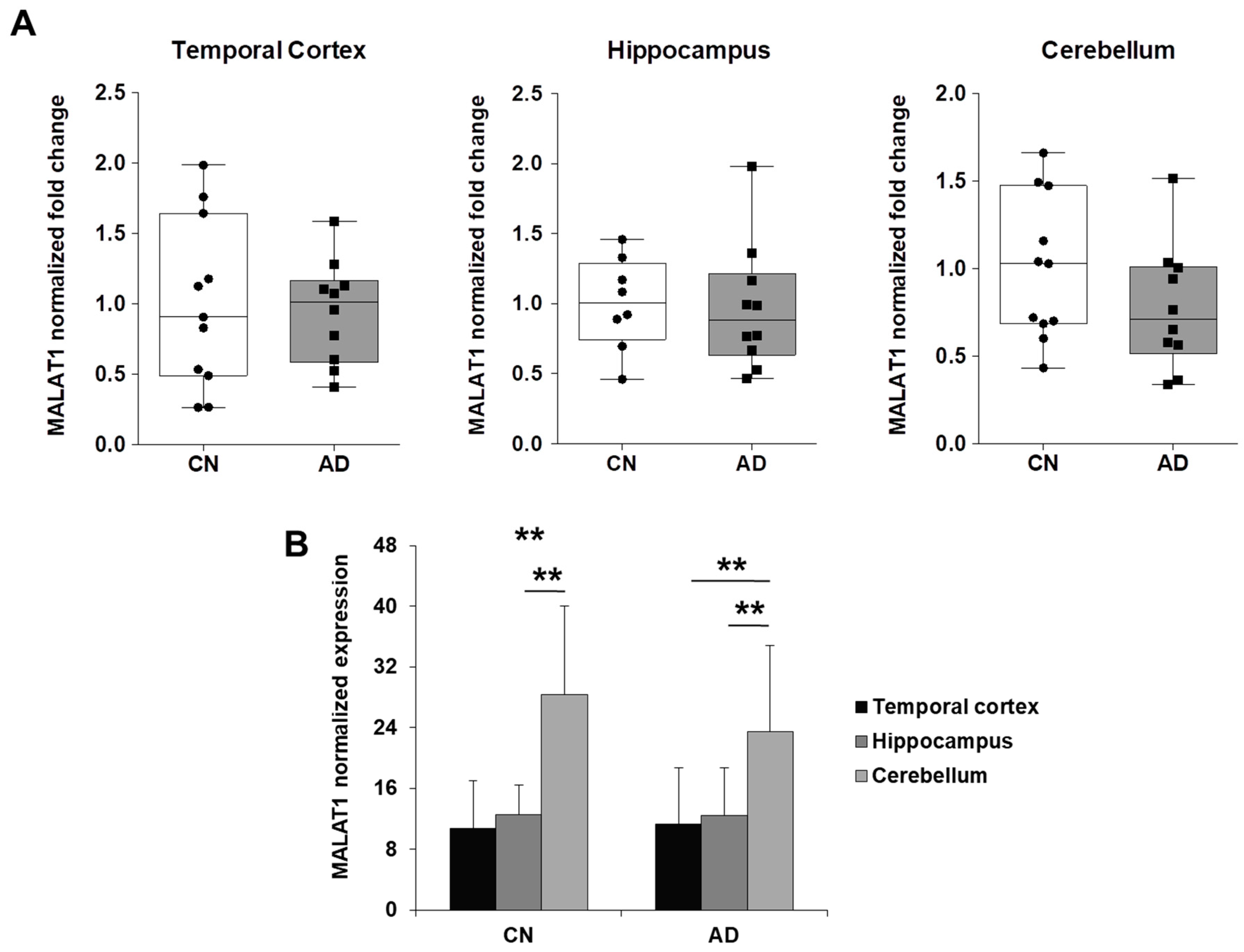
2.1. NEAT1, HOTAIR, and MALAT1 Long Non-Coding RNAs Differently Regulate CDK5R1 Expression

In order to test the hypothesis that lncRNAs might be involved in the regulation of CDK5R1, we analyzed the effect of NEAT1, HOTAIR, and MALAT1 downregulation on CDK5R1 expression. We transfected HeLa cells with 10 nM of specific 2’OMe-PS antisense oligonucleotides (ASO) to specifically knockdown the three lncRNAs. Total RNA was extracted 24 h after transfection and the levels of lncRNAs and CDK5R1 mRNA were assessed by qRT-PCR. The analysis showed that NEAT1, HOTAIR, and MALAT1 levels were reduced by 61%, 71%, and 78% respectively, compared to the control oligonucleotide (Figure 1A). Remarkably, increased CDK5R1 transcript levels were observed after NEAT1 and HOTAIR silencing, meaning that these two lncRNAs negatively regulate CDK5R1 expression (Figure 1B). On the contrary, CDK5R1 mRNA levels were significantly decreased after MALAT1 silencing compared to controls, indicating a positive action of this lncRNA on CDK5R1 expression (Figure 1B).

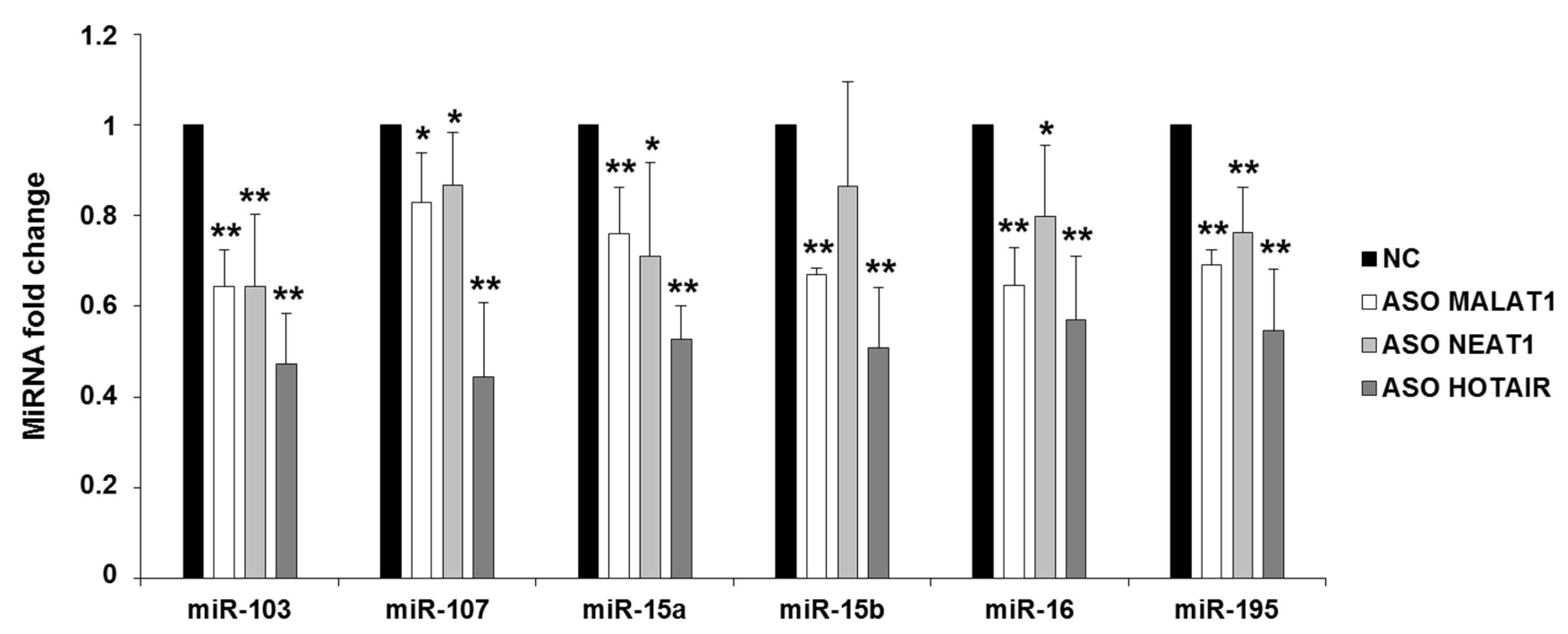
2.2. NEAT1, HOTAIR, and MALAT1 Upregulate miR-15/107 Expression

Since we previously demonstrated that CDK5R1 expression is negatively regulated by the miR-15/107 group of microRNAs [11], we also verified by qRT-PCR on the RNA previously extracted from HeLa cells if NEAT1, HOTAIR, and MALAT1 silencing was able to affect miR-15/107 expression. The levels of all the analyzed miR-15/107 family members were reduced after NEAT1, HOTAIR, and MALAT1 silencing, compared to the control treatment (Figure 2), being HOTAIR the most efficient with a reduction of miRNA targets of about 50%. NEAT1 and MALAT1 led to a less pronounced but significant reduction of all miRNAs, with the exception of miR-15b after NEAT1 knock-down, whose reduction did not reach the statistical significance (Figure 2).
These data suggest that HOTAIR and NEAT1 might negatively regulate CDK5R1 expression through a positive action on miR-15/107 levels. On the contrary, the positive effect of MALAT1 on CDK5R1 mRNA cannot be explained by the action of these miRNAs, and a different mechanism must be involved in MALAT1-mediated positive effect on CDK5R1 expression.
CDK5R1 also represents a target of EGR1 transcription factor whose expression is induced by ERK/MAPK pathway activation [30]. Since MALAT1 has been described to be a positive modulator of ERK/MAPK pathway [31], we evaluated the expression of EGR1 following MALAT1 silencing. Consistently, qRT-PCR analysis showed that the levels of EGR1 are strongly reduced (83%) in cells treated with MALAT1 specific antisense oligonucleotide, compared to normal control (Figure 3). These results suggest that the positive regulation exerted by MALAT1 on CDK5R1 expression can be due to MALAT1-mediated enhancement of EGR1 levels, likely overcoming the concurrent downregulation of miR-15/107 miRNAs.

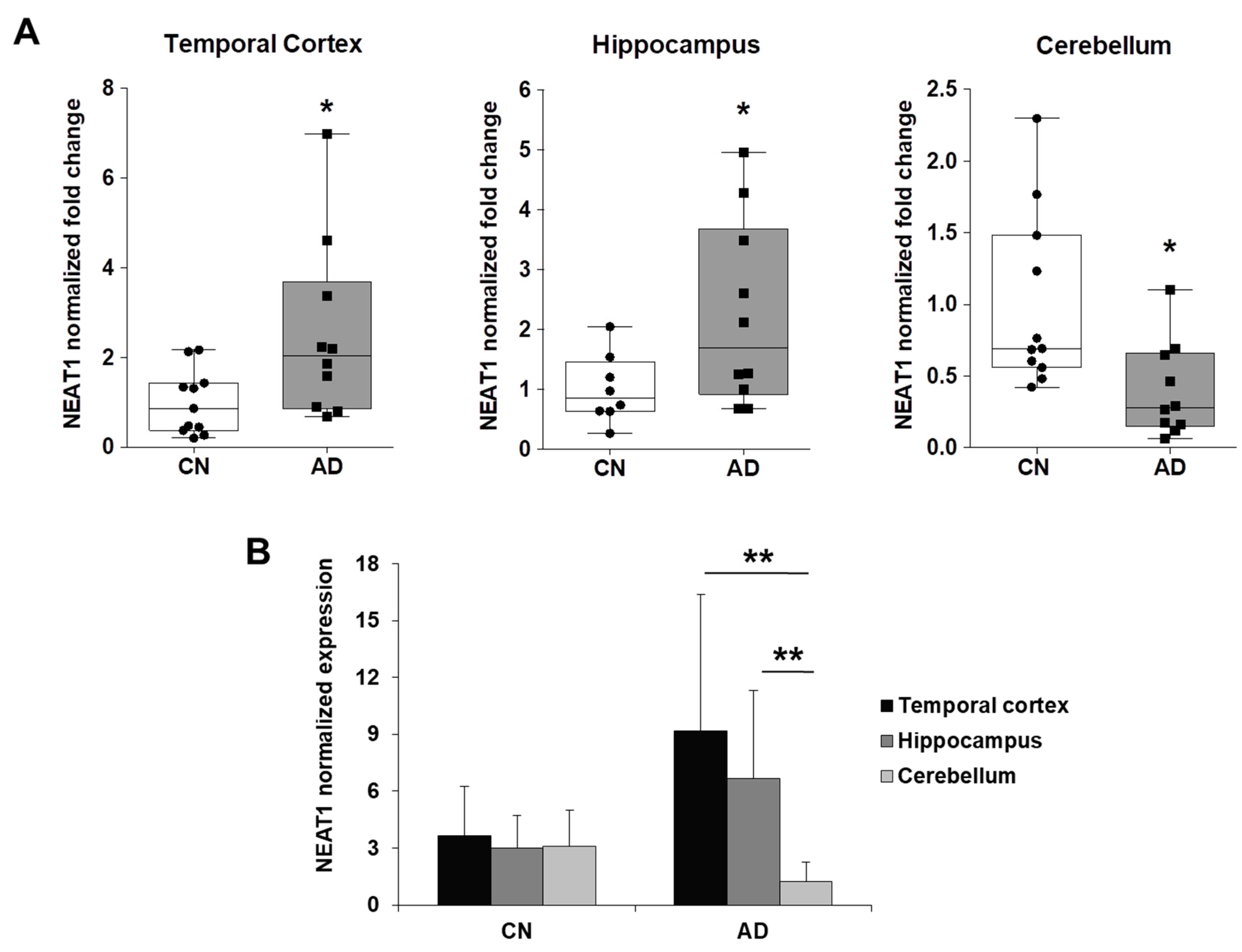
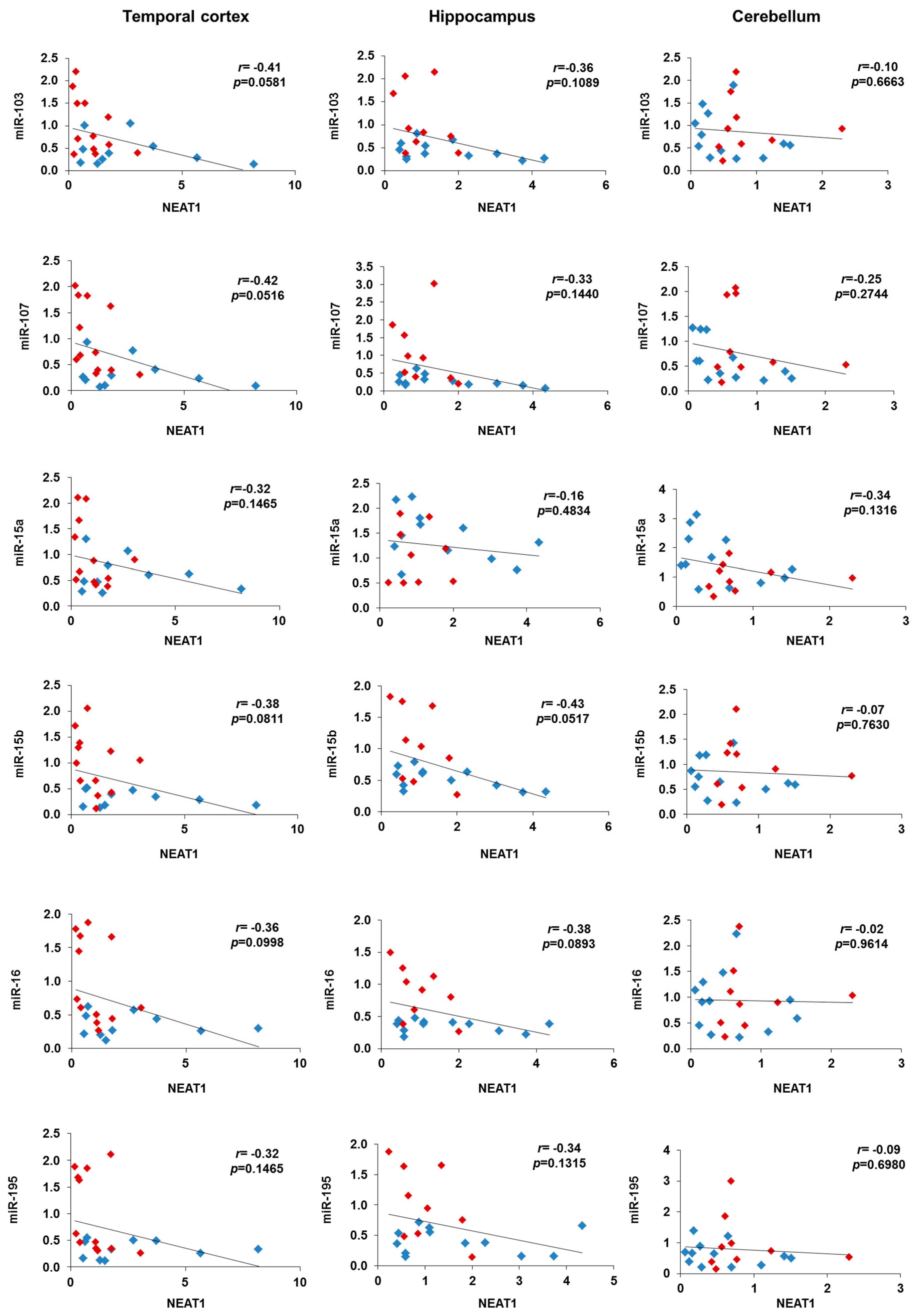
2.3. NEAT1 is Upregulated in AD Temporal Cortex and Hippocampus

We recently showed that miR-15/107 miRNAs level is reduced in the hippocampus and the temporal cortex, but not in the cerebellum, of AD brains. Furthermore, we showed that increased CDK5R1 mRNA levels are displayed by AD hippocampus tissue, compared to controls [11]. These data are consistent with the hypothesis that an increase of CDK5R1 expression, and consequent enhanced CDK5 activity, caused by downregulation of the miR-15/107 family has a role in the pathogenesis of AD.
To verify whether NEAT1, HOTAIR, and MALAT1 expression is also altered in Alzheimer’s disease, we quantified their levels by qRT-PCR in the temporal cortex, hippocampus, and cerebellum of the same AD patients and age-matched healthy controls which were analyzed in our previous work.
Remarkably, we found that NEAT1 was significantly overexpressed in temporal cortex and hippocampus and downregulated in cerebellum of AD patients, compared to control individuals (Figure 4A). Comparing NEAT1 distribution among the different brain areas of control individuals we observed similar expression levels, while NEAT1 was significantly higher in temporal cortex and hippocampus compared to cerebellum in AD patients (Figure 4B). On the contrary, MALAT1 expression showed no difference between AD patients and controls (Figure 5A), even though higher levels were detected in cerebellum, compared to temporal cortex and hippocampus, in both groups (Figure 5B). Finally, HOTAIR was expressed at very low levels in hippocampus and cerebellum and was not detectable in temporal cortex. Particularly, HOTAIR was downregulated in cerebellum in AD patients, compared to controls. No difference in HOTAIR expression between hippocampus and cerebellum was observed in both groups.

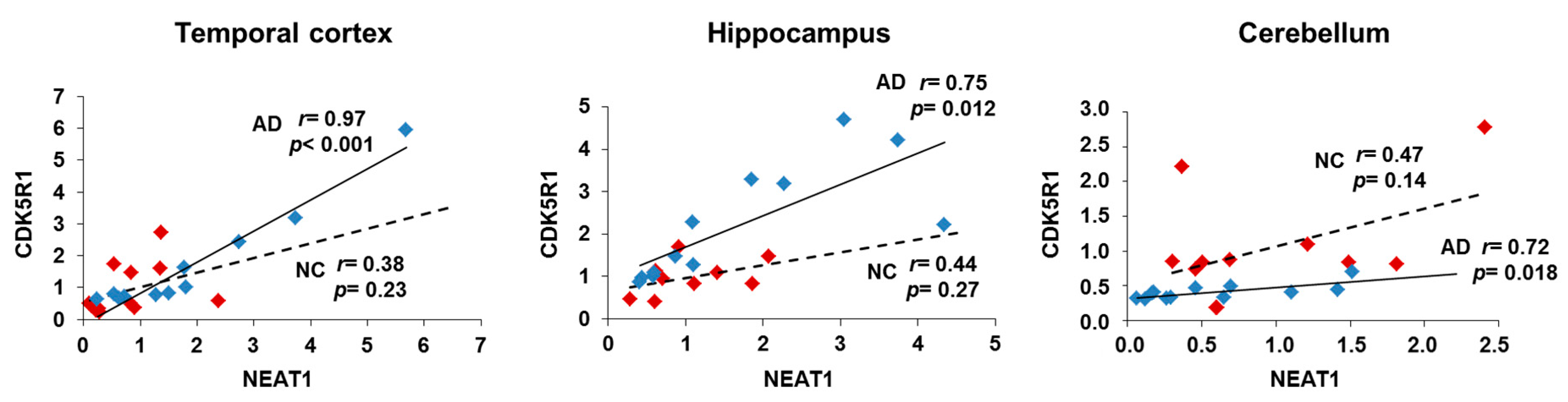
2.4. NEAT1 and CDK5R1 Overexpression as a Biomarker of AD

In order to verify the existence of a correlation between NEAT1 and CDK5R1 expression in AD and control brain tissues, we performed a Pearson’s correlation analysis between the normalized expression levels of NEAT1 and those previously obtained for CDK5R1 [11].
The analysis showed that a significant positive correlation between CDK5R1 and NEAT1 levels was only displayed by AD patients’ postmortem specimens of hippocampi and temporal cortices with very significant values of Pearson’s r (Figure 6). In other words, in AD, NEAT1 increases along with CDK5R1, indicating a peculiar functional relationship (in vitro defined as a negative NEAT1 control over CDK5R1) which is specific for AD and that can be either a protective response-related mechanism aimed at limiting (inefficiently) CDK5R1 upregulation or part of the disease pathogenesis. Notably, in the cerebellum, a brain area that is almost unaffected by the disease and in which CDK5R1 does not appear to be upregulated, the correlation between NEAT1 levels and those of CDK5R1 is still evident only in AD. This observation suggests that in the cerebellum a protective, NEAT1-associated mechanism might efficiently control CDK5R1 levels.
Another interesting observation is the opposite correlation between the expression of NEAT1 and miR-15/107 miRNAs in AD brains and controls. Indeed, while in controls we observed high expression of miR-15/107 and low expression of NEAT1, in AD patients, to higher NEAT1 levels correspond very low miR-15/107 levels, particularly in temporal cortex and hippocampus (Figure 7). In conclusion, a picture emerges in which not only NEAT1 is unable to increase its levels in a sufficient manner to counteract CDK5R1 increase in AD brains, but also, it loses the ability to positively regulate miR-15/107, validated negative regulators of CDK5R1. In this way, converging pathological mechanisms based on a failure of lncRNA NEAT1 and miR-15/107 homeostatic role towards CDK5R1 expression could result in CDK5R1 upregulation.

3. Discussion

Cyclin-dependent kinase 5 (CDK5) has a major role in CNS development and functioning and its deregulation can contribute to different pathological events implicated in the pathogenesis of Alzheimer’s disease [8]. Monomeric CDK5 itself does not display kinase activity and requires, in order to be active, the association with its regulatory subunits, p35 or p39, although p35, encoded by the CDK5R1 gene, is considered the most important CDK5 activator [32]. Multiple layers of regulation govern CDK5R1 expression and ensure p35 levels and CDK5 activity to be tightly controlled. They include transcriptional activation by EGR1 transcription factor and repression by LSD1 demethylase [29,30], as well as well various post-transcriptional mechanisms which involve the binding to the long and evolutionary conserved CDK5R1 3′-UTR of both RNA-binding proteins and microRNAs [9,10,11].
In this work, we took into account another class of non-coding RNAs, long non-coding RNAs (lncRNAs), as potential regulators of CDK5R1 expression. In particular, our attention was focused on three lncRNAs, NEAT1, HOTAIR, and MALAT1. Our results showed that these three lncRNAs are able to influence CDK5R1 expression. In particular, NEAT1 and HOTAIR exert a negative regulatory effect on CDK5R1 levels, while MALAT1 has an opposite, positive action. In addition, all these lncRNAs were proven to positively regulate the miRNAs belonging to the miR-15/107 family.
We hypothesize that the negative regulatory effect of NEAT1 on CDK5R1 expression might depend on its capacity to exert a positive control on miR-15/107 levels. Interestingly, we also found that NEAT1 is significantly overexpressed in temporal cortex and hippocampus of AD patients, compared to control individuals, suggesting that NEAT1 upregulation can be considered a biomarker of the disease. Recent studies have linked altered expression and function of long non-coding RNAs to the pathogenesis of neurodegenerative diseases (reviewed in [22]). In particular, different lncRNAs have been found to be dysregulated in Alzheimer’s disease (e.g., BACE1-AS and NDM29) and to be involved in AD pathogenesis by promoting β-amyloid production. In this work we show that, in vitro, NEAT1 negatively regulates CDK5R1 expression. In line, NEAT1 upregulation in AD patients would predict a corresponding downregulation of CDK5R1. Notably, this was not the case. Indeed, in AD brains the expression of both CDK5R1 and NEAT1 is increased compared to healthy controls. A possibility is that the negative control of NEAT1 over CDK5R1 levels is not efficient either because the ratio between CDK5R1 and NEAT1 typical of controls is increased in AD brains (Figure 6), or because NEAT1 loses its positive control towards miR-15/107 (Figure 7). As a result, we can infer that the critical NEAT1 level that would be necessary to counteract CDK5R1 expression is not reached in AD temporal cortex and hippocampus. For these reasons, NEAT1 overexpression as a pathomechanism in Alzheimer’s disease is unlikely, although our data do not allow to fully reject this hypothesis. Moreover, several lines of evidence suggest that NEAT1 and paraspeckles may have a neuroprotective role in neurodegenerative diseases. An increase in paraspeckles formation and NEAT1 levels has been detected in spinal motor neurons of early phase amyotrophic lateral sclerosis (ALS) patients compared to control individuals [33] and compromised paraspeckles formation has been proposed as a pathogenic factor in FUSopathies [34]. Moreover, NEAT1 levels are also increased in the brains of patients affected by frontotemporal lobar degeneration (FTLD) [35]. Importantly, Sunwoo and colleagues [36] showed that NEAT1 is overexpressed in Huntington’s disease patients and plays a protective role against cell injury. These data suggest that NEAT1 may contribute to neuronal survival in the degenerating brain. Analogously, our work showed that NEAT1 is also overexpressed in AD patients. In this context, putative beneficial effects of NEAT1 are still unknown. However, enhanced amounts of CDK5R1 are predicted to cause CDK5 hyperactivation, which is a typical hallmark of the disease [8]. It is worth noting that CDK5 can phosphorylate p53, which is also known to be upregulated in AD [37], thereby inducing its stabilization and transcriptional activation, contributing to neuronal cell death [38]. Remarkably, p53 was recently demonstrated to activate NEAT1 expression [39]. These findings provide a possible molecular link between CDK5R1 and NEAT1 upregulation in AD brains, albeit they do not indicate the reason why CDK5R1 escapes NEAT1 control in AD condition.
The negative action exerted by HOTAIR on CDK5R1 expression is likely mediated by different converging mechanisms. On the one hand, HOTAIR can negatively regulate CDK5R1 at the post-transcriptional level via the same miR-15/107 miRNA-mediated mechanism as NEAT1, on the other hand it could regulate CDK5R1 also at the transcriptional level participating to recruiting and regulating the LSD1 and PRC2 repressing complexes [28]. Interestingly, HOTAIR also represses the transcription of BDNF [27], which normally induces the ERK-mediated expression of CDK5R1 [29].
On the contrary, our silencing experiments suggest that MALAT1 positively affect CDK5R1 expression. Since reduction of miR-15/107 levels after MALAT1 silencing would predict an increase in the amount of CDK5R1 mRNA, as expected for their inhibitory action, there must be other predominant regulatory mechanisms leading to CDK5R1 upregulation by MALAT1. As we have also shown that MALAT1 silencing causes a strong reduction in the levels of EGR1, which is the main activator of CDK5R1 transcription, we thus speculate that MALAT1 can enhance CDK5R1 expression mainly by upregulating EGR1 transcription factor through activation of ERK/MAPK signaling pathway [31].
Moreover, the activation of this pathway is known to play a critical role in promoting neurite outgrowth [31]. Given that the p35/CDK5 complex is also essential for neurite outgrowth during neuronal differentiation [40], our evidence raises the interesting hypothesis that MALAT1 induces axonal elongation via CDK5R1/p35 upregulation.
Mounting evidence suggests that lncRNAs can function as miRNA sponges, by sequestering the mature miRNA molecules and preventing the binding to their target mRNAs [41]. However, this mechanism is predicted to increase—or leave unchanged—the levels of the sequestered miRNAs when the lncRNA acting as sponge is silenced [14,42]. Since we observed that the silencing of NEAT1, HOTAIR, and MALAT1 lead to a reduction of miR-15/107 miRNAs, we hypothesize that this effect could be mediated by a positive regulatory action of these lncRNAs on transcription factors that promote the expression of this family of miRNAs or, alternatively, by their interaction with the microprocessor to enhance pri-miRNA processing, as already demonstrated for NEAT1 [43].
Overall, our data suggest that lncRNAs can provide a further layer and a higher degree of complexity to the control of CDK5R1 expression. In addition, we show that NEAT1 is upregulated in AD brain, possibly as a part of a protective mechanism against neuronal death, and can be considered a marker of the disease and represents a potential pharmacological target for therapeutic intervention in AD.

4. Materials and Methods

4.1. Cell Cultures

HeLa cells (code CCL-2, ATCC, Manassas, VA, USA) were cultured in DMEM high glucose (Euroclone, Pero, Italy) medium with 10% fetal bovine serum (FBS) (Euroclone), 100 U/mL penicillin-streptomycin (Euroclone) and 0.01 mM l-glutamine (Euroclone). Cultures were maintained at 37 °C in a 5% CO2 incubator.

4.2. Brain Tissues

Post-mortem frozen brain tissue samples of AD patients and age- and sex-matched non-demented individuals were obtained from MRC London Neurodegenerative Diseases Brain Bank (King’s College London), Newcastle Brain Tissue Resource (Newcastle University), and South West Dementia Brain Bank (University of Bristol) and are described in [10]. The approval of the Ethics Committee of the University of Milan was obtained for the use of post-mortem tissues for research purposes (Project identification code: RV_RIC_AT16MVENT_M, 15 June 2016).

4.3. Antisense Oligonucleotides Transfection

2′-O-methyl phosphorothioate antisense oligonucleotides (2′OMe-PS ASO) were designed as described by [44] (NEAT1 1473, HOTAIR 1259, MALAT1 5326, NC1) and purchased from Consorzio Futuro in Ricerca, Università degli Studi di Ferrara (Ferrara, Italy).
ASOs were used at 10 nM concentration. 200 × 103 HeLa cells were seeded in 6-well plates in order to extract total RNA. The cells were transfected 24 h after seeding with 2′OMe-PS ASOs, using Lipofectamine 2000 (Thermo Fisher Scientific, Waltham, MA, USA) transfection reagent according to the manufacturer’s instructions. Cell extracts were prepared for analysis 24 h after the transfection.

4.4. Real-Time PCR

Total RNA from transfected/nontransfected cells and from brain tissues (100 mg of each sample tissue) was isolated using TRIzol reagent (Thermo Fisher Scientific), according to the manufacturer’s instructions. Concentration and purity of RNA were measured using the Nanodrop spectrophotometer (ThermoFisher Scientific). All RNA samples had an A260/280 value of 1.8–2.1.
For the measurement of CDK5R1, EGR1 mRNA, NEAT1, HOTAIR, and MALAT1 RNA, a DNase reaction was performed on 1 μg of total RNA using RQ1 RNase-Free Dnase (Promega, Madison, WI, USA) and then cDNA was synthetized in 20 μL reactions using the High Capacity cDNA Reverse Transcription Kit (Thermo Fisher Scientific), according to the manufacturer’s instructions. SYBR Green Real-Time PCR was performed using the GoTaq qPCR Master Mix (Promega) and the following primers: CDK5R1 fw: TGAGCGGGTCTAGTGGAAAG; CDK5R1 rev: AGCAGCAGACAAGGGGGTAG; EGR1 fw: GAGCACCTGACCGCAGAGTC; EGR1 rev: GTGTTGCCACTGTTGGGTGC; HOTAIR fw: GGCAAGACGGGCACTCACAG; HOTAIR rev: CTGGGCGTTCATGTGGCGAG; MALAT1 fw: AGGGAAAGCGAGTGGTTGGT; MALAT1 rev: GAAATCGGCCTACGTCCCCA; NEAT1 fw: CGGAGGTGAGGGGTGGTCTG; NEAT1 rev: GCAGTCCCCGCCTGTCAAAC; EIF4A2 fw: GGTCAGGGTCAAGTCGTGTT; EIF4A2 rev: CCCCCTCTGCCAATTCTGTG; CYC1 fw: TAGAGTTTGACGATGGCACCC; CYC1 rev: CGTTTTCGATGGTCGTGCTC; SYP fw: CTTCGCCATCTTCGCCTTTG; SYP rev: TACACTTGGTGCAGCCTGAAG; ENO2 fw: CTGAAGCCATCCAAGCGTGC; ENO2 rev: CCCACCACCAGGTCAGCAAT. 20 μL PCR reactions were prepared with 2× SYBR Green mix containing 1.6 μL of reverse transcriptase product and 0.4 μL of each primer (10 µM). The PCR mixtures were incubated at 95 °C for 3 min, followed by 40 cycles of 95 °C for 10 s, 60 °C for 20 s, and 72 °C for 10 s. The calculation of gene expression levels was based on the ΔΔCt method in transfection experiments and on the ΔCt method for gene expression analysis in brain tissues. The geometric mean of the expression values of EIF4A2 and CYC1 housekeeping genes was used as internal control in transfection experiments, while gene expression levels in brain tissues were normalized on the geometric mean of the same housekeeping genes and the neuronal markers SYP and ENO2.
For the measurement of miRNAs, a two-step Taq-Man real-time PCR assay was performed using primers and probes obtained from Thermo Fisher Scientific. The reverse transcriptase reaction was performed using the TaqMan MicroRNA Reverse Transcription kit (Thermo Fisher Scientific), according to the manufacturer’s instructions. cDNA was synthesized from 50 ng of total RNA in 15 μL reactions, using the stem-loop primer for miR-15a (ID000389), miR-15b (ID000390), miR-16 (ID000391), miR-103 (ID000439), miR-107 (ID000443), miR-195 (ID000494), and U6 snRNA (ID001973). The PCR reaction (20 μL) contained 1.3 μL of reverse transcriptase product, 10 μL of Taq-Man Universal PCR Master Mix (Thermo Fisher Scientific) and 1 μL of the appropriate TaqMan MicroRNA Assay (20×) containing primers and probes for the miR of interest. The PCR mixtures were incubated at 95 °C for 10 min, followed by 40 cycles of 95 °C for 15 s and 60 °C for 60 s. The expression of miRs was based on the ΔΔCt methods, using U6 snRNA as an endogenous control. All PCRs were performed in triplicate using an iQ5 Real-Time PCR detection system (Bio-Rad, Hercules, CA, USA).

4.5. Statistical Analysis

Each experiment was carried out at least three times. Histograms represent the mean values and bars indicate the standard deviation of the mean. The box plots show median, 25th and 75th percentile values and whiskers to the minimum and maximum value. The statistical significance of the results was determined using Student’s t-test, with data considered significant when p < 0.05. The degree of linear relationship between CDK5R1 gene and NEAT1 expression levels was calculated using Pearson’s correlation coefficient (r value). The p value was calculated from an extra sum-of-squares F test.

Author Contributions

Conceptualization, M.S., F.R., E.B., and M.V.; Formal analysis, M.S., B.G., F.R., E.B., and M.V.; Funding acquisition, E.B., F.R., and M.V.; Investigation, M.S. and B.G.; Methodology, M.S., B.G., and M.V.; Project administration, M.V.; Resources, E.B. and M.V.; Supervision, M.V.; Validation, M.V.; Visualization, M.S. and M.V.; Writing—original draft, M.S. and M.V.; Writing—review & editing, M.S., B.G., F.R., E.B., and M.V.

Acknowledgments

The authors would like to thank the MRC London Neurodegenerative Diseases Brain Bank, the Newcastle Brain Tissue Resource, and the South West Dementia Brain Bank (SWDBB) for providing brain tissue for this study. The SWDBB is supported by BRACE (Bristol Research into Alzheimer’s and Care of the Elderly), Brains for Dementia Research and the Medical Research Council. This work was supported by the Academic Grant “Bando BIOMETRA—Fondi Incentivo alla Ricerca 2015” (Università degli studi di Milano, project no. RV_RIC_AT16MVENT_M) to M.V., the Ministero dell’Istruzione, dell’Università e della Ricerca (award no. Epigenomics Flagship Project), Telethon Foundation project (award no. GGP14074) and Fondazione Cariplo (award no. 2016-0204) to E.B., and Fondazione Cariplo (award no. 2014-0972) to F.R.

Conflicts of Interest

The authors declare no conflict of interest.

References

  1. Hebert, L.E.; Weuve, J.; Scherr, P.A.; Evans, D.A. Alzheimer disease in the United States (2010–2050) estimated using the 2010 census. Neurology 2013, 80, 1778–1783. [Google Scholar] [CrossRef] [PubMed]
  2. Serrano-Pozo, A.; Frosch, M.P.; Masliah, E.; Hyman, B.T. Neuropathological alterations in Alzheimer disease. Cold Spring Harb. Perspect. Med. 2011, 1, a006189. [Google Scholar] [CrossRef] [PubMed]
  3. Desler, C.; Lillenes, M.S.; Tønjum, T.; Rasmussen, L.J. The role of mitochondrial dysfunction in the progression of Alzheimer’s disease. Curr. Med. Chem. 2017, 24. [Google Scholar] [CrossRef] [PubMed]
  4. Jiang, T.; Chang, R.C.; Rosenmann, H.; Yu, J.T. Advances in Alzheimer’s disease: From bench to bedside. Biomed. Res. Int. 2015, 2015, 202676. [Google Scholar] [CrossRef] [PubMed]
  5. Wang, J.Z.; Grundke-Iqbal, I.; Iqbal, K. Kinases and phosphatases and tau sites involved in Alzheimer neurofibrillary degeneration. Eur. J. Neurosci. 2007, 25, 59–68. [Google Scholar] [CrossRef] [PubMed] [Green Version]
  6. Fischer, A.; Sananbenesi, F.; Schrick, C.; Spiess, J.; Radulovic, J. Cyclin-dependent kinase 5 is required for associative learning. J. Neurosci. 2002, 22, 3700–3707. [Google Scholar] [CrossRef] [PubMed]
  7. McLinden, K.A.; Trunova, S.; Giniger, E. At the fulcrum in health and disease: Cdk5 and the balancing acts of neuronal structure and physiology. Brain Disord. Ther. 2012, 2012 (Suppl. 1), 001. [Google Scholar] [CrossRef] [PubMed]
  8. Liu, S.L.; Wang, C.; Jiang, T.; Tan, L.; Xing, A.; Yu, J.T. The Role of Cdk5 in Alzheimer’s Disease. Mol. Neurobiol. 2016, 53, 4328–4342. [Google Scholar] [CrossRef] [PubMed]
  9. Moncini, S.; Bevilacqua, A.; Venturin, M.; Fallini, C.; Ratti, A.; Nicolin, A.; Riva, P. The 3′ untranslated region of human Cyclin-Dependent Kinase 5 Regulatory subunit 1 contains regulatory elements affecting transcript stability. BMC Mol. Biol. 2007, 8, 111. [Google Scholar] [CrossRef] [PubMed]
  10. Zuccotti, P.; Colombrita, C.; Moncini, S.; Barbieri, A.; Lunghi, M.; Gelfi, C.; de Palma, S.; Nicolin, A.; Ratti, A.; Venturin, M.; et al. hnRNPA2/B1 and nELAV proteins bind to a specific U-rich element in CDK5R1 3′-UTR and oppositely regulate its expression. Biochim. Biophys. Acta 2014, 1839, 506–516. [Google Scholar] [CrossRef] [PubMed]
  11. Moncini, S.; Lunghi, M.; Valmadre, A.; Grasso, M.; del Vescovo, V.; Riva, P.; Denti, M.A.; Venturin, M. The miR-15/107 Family of microRNA Genes Regulates CDK5R1/p35 with Implications for Alzheimer’s Disease Pathogenesis. Mol. Neurobiol. 2017, 54, 4329–4342. [Google Scholar] [CrossRef] [PubMed]
  12. Khorkova, O.; Hsiao, J.; Wahlestedt, C. Basic biology and therapeutic implications of lncRNA. Adv. Drug Deliv. Rev. 2015, 87, 15–24. [Google Scholar] [CrossRef] [PubMed] [Green Version]
  13. Nie, L.; Wu, H.J.; Hsu, J.M.; Chang, S.S.; Labaff, A.M.; Li, C.W.; Wang, Y.; Hsu, J.L.; Hung, M.C. Long non-coding RNAs: Versatile master regulators of gene expression and crucial players in cancer. Am. J. Transl. Res. 2012, 4, 127–150. [Google Scholar] [PubMed]
  14. Bayoumi, A.S.; Sayed, A.; Broskova, Z.; Teoh, J.P.; Wilson, J.; Su, H.; Tang, Y.L.; Kim, I.M. Crosstalk between Long Noncoding RNAs and MicroRNAs in Health and Disease. Int. J. Mol. Sci. 2016, 17, 356. [Google Scholar] [CrossRef] [PubMed]
  15. Li, Y.; Wang, X. Role of long noncoding RNAs in malignant disease (Review). Mol. Med. Rep. 2016, 13, 1463–1469. [Google Scholar] [CrossRef] [PubMed]
  16. Faghihi, M.A.; Modarresi, F.; Khalil, A.M.; Wood, D.E.; Sahagan, B.G.; Morgan, T.E.; Finch, C.E.; St Laurent, G., 3rd; Kenny, P.J.; Wahlestedt, C. Expression of a noncoding RNA is elevated in Alzheimer’s disease and drives rapid feed-forward regulation of beta-secretase. Nat. Med. 2008, 14, 723–730. [Google Scholar] [CrossRef] [PubMed]
  17. Faghihi, M.A.; Zhang, M.; Huang, J.; Modarresi, F.; Van der Brug, M.P.; Nalls, M.A.; Cookson, M.R.; St-Laurent, G., 3rd; Wahlestedt, C. Evidence for natural antisense transcript-mediated inhibition of microRNA function. Genome Biol. 2010, 11, R56. [Google Scholar] [CrossRef] [PubMed]
  18. Chakravarty, D.; Sboner, A.; Nair, S.S.; Giannopoulou, E.; Li, R.; Hennig, S.; Mosquera, J.M.; Pauwels, J.; Park, K.; Kossai, M.; et al. The oestrogen receptor alpha-regulated lncRNA NEAT1 is a critical modulator of prostate cancer. Nat. Commun. 2014, 5, 5383. [Google Scholar] [CrossRef] [PubMed] [Green Version]
  19. West, J.A.; Davis, C.P.; Sunwoo, H.; Simon, M.D.; Sadreyev, R.I.; Wang, P.I.; Tolstorukov, M.Y.; Kingston, R.E. The long noncoding RNAs NEAT1 and MALAT1 bind active chromatin sites. Mol. Cell 2014, 55, 791–802. [Google Scholar] [CrossRef] [PubMed]
  20. Fox, A.H.; Lamond, A.I. Paraspeckles. Cold Spring Harb. Perspect. Biol. 2010, 2, a000687. [Google Scholar] [CrossRef] [PubMed]
  21. Hirose, T.; Virnicchi, G.; Tanigawa, A.; Naganuma, T.; Li, R.; Kimura, H.; Yokoi, T.; Nakagawa, S.; Bénard, M.; Fox, A.H.; et al. NEAT1 long noncoding RNA regulates transcription via protein sequestration within subnuclear bodies. Mol. Biol. Cell 2014, 25, 169–183. [Google Scholar] [CrossRef] [PubMed] [Green Version]
  22. Riva, P.; Ratti, A.; Venturin, M. The Long Non-Coding RNAs in Neurodegenerative Diseases: Novel Mechanisms of Pathogenesis. Curr. Alzheimer Res. 2016, 13, 1219–1231. [Google Scholar] [CrossRef] [PubMed]
  23. Tripathi, V.; Ellis, J.D.; Shen, Z.; Song, D.Y.; Pan, Q.; Watt, A.T.; Freier, S.M.; Bennett, C.F.; Sharma, A.; Bubulya, P.A.; et al. The nuclear-retained noncoding RNA MALAT1 regulates alternative splicing by modulating SR splicing factor phosphorylation. Mol. Cell 2010, 39, 925–938. [Google Scholar] [CrossRef] [PubMed]
  24. Gutschner, T.; Hämmerle, M.; Diederichs, S. MALAT1—A paradigm for long noncoding RNA function in cancer. J. Mol. Med. (Berl.) 2013, 91, 791–801. [Google Scholar] [CrossRef] [PubMed]
  25. Wang, P.; Wu, T.; Zhou, H.; Jin, Q.; He, G.; Yu, H.; Xuan, L.; Wang, X.; Tian, L.; Sun, Y.; et al. Long noncoding RNA NEAT1 promotes laryngeal squamous cell cancer through regulating miR-107/CDK6 pathway. J. Exp. Clin. Cancer Res. 2016, 35, 22. [Google Scholar] [CrossRef] [PubMed] [Green Version]
  26. Zhang, H.; Wang, G.; Yin, R.; Qiu, M.; Xu, L. Comprehensive Identification of MicroRNAs Regulated by Long Non-coding RNA MALAT1. Zhongguo Fei Ai Za Zhi 2016, 19, 247–251. [Google Scholar] [CrossRef] [PubMed]
  27. Yu, X.; Li, Z. Long non-coding RNA HOTAIR: A novel oncogene (Review). Mol. Med. Rep. 2015, 12, 5611–5618. [Google Scholar] [CrossRef] [PubMed]
  28. Tsai, M.C.; Manor, O.; Wan, Y.; Mosammaparast, N.; Wang, J.K.; Lan, F.; Shi, Y.; Segal, E.; Chang, H.Y. Long noncoding RNA as modular scaffold of histone modification complexes. Science 2010, 329, 689–693. [Google Scholar] [CrossRef] [PubMed]
  29. Toffolo, E.; Rusconi, F.; Paganini, L.; Tortorici, M.; Pilotto, S.; Heise, C.; Verpelli, C.; Tedeschi, G.; Maffioli, E.; Sala, C.; et al. Phosphorylation of neuronal Lysine-Specific Demethylase 1 LSD1/KDM1A impairs transcriptional repression by regulating interaction with CoREST and histone deacetylases HDAC1/2. J. Neurochem. 2014, 128, 603–616. [Google Scholar] [CrossRef] [PubMed]
  30. Harada, T.; Morooka, T.; Ogawa, S.; Nishida, E. ERK induces p35, a neuron-specific activator of Cdk5, through induction of Egr1. Nat. Cell Biol. 2001, 3, 453–459. [Google Scholar] [CrossRef] [PubMed]
  31. Chen, L.; Feng, P.; Zhu, X.; He, S.; Duan, J.; Zhou, D. Long non-coding RNA Malat1 promotes neurite outgrowth through activation of ERK/MAPK signalling pathway in N2a cells. J. Cell. Mol. Med. 2016, 20, 2102–2110. [Google Scholar] [CrossRef] [PubMed] [Green Version]
  32. Ko, J.; Humbert, S.; Bronson, R.T.; Takahashi, S.; Kulkarni, A.B.; Li, E.; Tsai, L.H. p35 and p39 are essential for cyclin-dependent kinase 5 function during neurodevelopment. J. Neurosci. 2001, 21, 6758–6771. [Google Scholar] [CrossRef] [PubMed]
  33. Nishimoto, Y.; Nakagawa, S.; Hirose, T.; Okano, H.J.; Takao, M.; Shibata, S.; Suyama, S.; Kuwako, K.; Imai, T.; Murayama, S.; et al. The long non-coding RNA nuclear-enriched abundant transcript 1_2 induces paraspeckle formation in the motor neuron during the early phase of amyotrophic lateral sclerosis. Mol. Brain 2013, 6, 31. [Google Scholar] [CrossRef] [PubMed] [Green Version]
  34. Shelkovnikova, T.A.; Robinson, H.K.; Troakes, C.; Ninkina, N.; Buchman, V.L. Compromised paraspeckle formation as a pathogenic factor in FUSopathies. Hum. Mol. Genet. 2014, 23, 2298–2312. [Google Scholar] [CrossRef] [PubMed]
  35. Tollervey, J.R.; Curk, T.; Rogelj, B.; Briese, M.; Cereda, M.; Kayikci, M.; König, J.; Hortobágyi, T.; Nishimura, A.L.; Zupunski, V.; et al. Characterizing the RNA targets and position-dependent splicing regulation by TDP-43. Nat. Neurosci. 2011, 14, 452–458. [Google Scholar] [CrossRef] [PubMed] [Green Version]
  36. Sunwoo, J.S.; Lee, S.T.; Im, W.; Lee, M.; Byun, J.I.; Jung, K.H.; Park, K.I.; Jung, K.Y.; Lee, S.K.; Chu, K.; et al. Altered Expression of the Long Noncoding RNA NEAT1 in Huntington's Disease. Mol. Neurobiol. 2017, 54, 1577–1586. [Google Scholar] [CrossRef] [PubMed]
  37. Hooper, C.; Meimaridou, E.; Tavassoli, M.; Melino, G.; Lovestone, S.; Killick, R. p53 is upregulated in Alzheimer’s disease and induces tau phosphorylation in HEK293a cells. Neurosci. Lett. 2007, 418, 34–37. [Google Scholar] [CrossRef] [PubMed]
  38. Lee, J.H.; Kim, H.S.; Lee, S.J.; Kim, K.T. Stabilization and activation of p53 induced by Cdk5 contributes to neuronal cell death. J. Cell Sci. 2007, 120, 2259–2271. [Google Scholar] [CrossRef] [PubMed] [Green Version]
  39. Mello, S.S.; Sinow, C.; Raj, N.; Mazur, P.K.; Bieging-Rolett, K.; Broz, D.K.; Imam, J.F.C.; Vogel, H.; Wood, L.D.; Sage, J.; et al. Neat1 is a p53-inducible lincRNA essential for transformation suppression. Genes Dev. 2017, 31, 1095–1108. [Google Scholar] [CrossRef] [PubMed] [Green Version]
  40. Nikolic, M.; Dudek, H.; Kwon, Y.T.; Ramos, Y.F.; Tsai, L.H. The cdk5/p35 kinase is essential for neurite outgrowth during neuronal differentiation. Genes Dev. 1996, 10, 816–825. [Google Scholar] [CrossRef] [PubMed]
  41. Rinn, J.L.; Chang, H.Y. Genome regulation by long noncoding RNAs. Annu. Rev. Biochem. 2012, 81, 145–166. [Google Scholar] [CrossRef] [PubMed]
  42. Yoon, J.H.; Abdelmohsen, K.; Gorospe, M. Functional interactions among microRNAs and long noncoding RNAs. Semin. Cell Dev. Biol. 2014, 34, 9–14. [Google Scholar] [CrossRef] [PubMed] [Green Version]
  43. Jiang, L.; Shao, C.; Wu, Q.J.; Chen, G.; Zhou, J.; Yang, B.; Li, H.; Gou, L.T.; Zhang, Y.; Wang, Y.; et al. NEAT1 scaffolds RNA-binding proteins and the Microprocessor to globally enhance pri-miRNA processing. Nat. Struct. Mol. Biol. 2017, 24, 816–824. [Google Scholar] [CrossRef] [PubMed] [Green Version]
  44. Lennox, K.A.; Behlke, M.A. Cellular localization of long non-coding RNAs affects silencing by RNAi more than by antisense oligonucleotides. Nucleic Acids Res. 2016, 44, 863–877. [Google Scholar] [CrossRef] [PubMed]
Figure 1. Effect of NEAT1, HOTAIR, and MALAT1 silencing on CDK5R1 mRNA levels. (A) NEAT1, HOTAIR, and MALAT1 levels 24 h after transfection with specific ASOs. The levels of each lncRNA were reduced by at least 60%, compared to a control ASO (NC)-transfected cells. (B) Increased CDK5R1 transcript levels were observed after NEAT1 and HOTAIR silencing, compared to the normal control. On the contrary, CDK5R1 mRNA levels were significantly decreased after MALAT1 silencing. n = 5, mean ± s.d., * p < 0.05, ** p < 0.01, Student’s t-test.
Ijms 19 02022 g001
Figure 2. Effect of NEAT1, HOTAIR, and MALAT1 silencing on miR-15/107 levels. Decreased levels of all miR-15/107 miRNAs were detected after the knock-down of the three lncRNAs. n = 5, mean ± s.d., * p < 0.05, ** p < 0.005, Student’s t-test.
Ijms 19 02022 g002
Figure 3. Effect of MALAT1 silencing on EGR1 mRNA levels. EGR1 mRNA levels were significantly decreased after MALAT1 silencing, compared to the normal control (NC). n = 3, mean ± s.d., ** p < 0.005, Student’s t-test.
Ijms 19 02022 g003
Figure 4. Comparison between the levels of NEAT1 expression in AD and control brain tissues. (A) Dot-Box-plots of the levels of NEAT1 expression in three different brain areas (temporal cortex, hippocampus, and cerebellum) of AD patients (n = 10) and controls (n = 8–11). Dark horizontal lines represent the median, with the box representing the 25th and 75th percentiles, the whiskers the 5th and 95th percentiles. The average of control values was set to 1 and all values were calculated relatively. NEAT1 levels are significantly upregulated in temporal cortex and hippocampus and downregulated in cerebellum of AD patients, compared to control individuals. (B) Higher NEAT1 expression levels were observed in temporal cortex and hippocampus, compared to cerebellum, in AD patients, but not in control individuals. * p < 0.05, ** p < 0.01, Student’s t-test.
Ijms 19 02022 g004
Figure 5. Comparison between the levels of MALAT1 expression in AD and control brain tissues. (A) Dot-Box-plots of the levels of MALAT1 expression in three different brain areas (temporal cortex, hippocampus, and cerebellum) of AD patients (n = 10) and controls (n = 8–11). Dark horizontal lines represent the median, with the box representing the 25th and 75th percentiles, the whiskers the 5th and 95th percentiles. The average of control values was set to 1 and all values were calculated relatively. We observed no difference in MALAT1 levels between AD patients and control individuals in any analyzed tissues. (B) Higher MALAT1 expression levels were observed in cerebellum, compared to temporal cortex and hippocampus, in both AD patients and controls individuals. ** p < 0.01, Student’s t-test.
Ijms 19 02022 g005
Figure 6. Correlation analysis between NEAT1 and CDK5R1 expression in temporal cortex, hippocampus and cerebellum samples of AD patients (blue diamonds) and controls (red diamonds). r = Pearson's correlation coefficient, solid line = linear regression line of AD patients, dashed line = linear regression line of normal controls (NC).
Ijms 19 02022 g006
Figure 7. Correlation analysis between miR-103, miR-107, miR-15a, miR-15b, miR-16, and miR-195 and NEAT1 expression in temporal cortex, hippocampus, and cerebellum samples of AD patients (blue diamonds) and controls (red diamonds). r = Pearson’s correlation coefficient.
Ijms 19 02022 g007

