Review

AMP-Activated Protein Kinase as a Key Trigger for the Disuse-Induced Skeletal Muscle Remodeling

by
Natalia A. Vilchinskaya
1,
Igor I. Krivoi
2 and
Boris S. Shenkman
1,*
1
Myology Laboratory, Institute of Biomedical Problems RAS, Moscow 123007, Russia
2
Department of General Physiology, St. Petersburg State University, St. Petersburg 199034, Russia
*
Author to whom correspondence should be addressed.
Int. J. Mol. Sci. 2018, 19(11), 3558; https://doi.org/10.3390/ijms19113558
Submission received: 1 October 2018 / Revised: 2 November 2018 / Accepted: 9 November 2018 / Published: 12 November 2018
(This article belongs to the Special Issue AMP-Activated Protein Kinase Signalling)

Abstract

:
Molecular mechanisms that trigger disuse-induced postural muscle atrophy as well as myosin phenotype transformations are poorly studied. This review will summarize the impact of 5′ adenosine monophosphate -activated protein kinase (AMPK) activity on mammalian target of rapamycin complex 1 (mTORC1)-signaling, nuclear-cytoplasmic traffic of class IIa histone deacetylases (HDAC), and myosin heavy chain gene expression in mammalian postural muscles (mainly, soleus muscle) under disuse conditions, i.e., withdrawal of weight-bearing from ankle extensors. Based on the current literature and the authors’ own experimental data, the present review points out that AMPK plays a key role in the regulation of signaling pathways that determine metabolic, structural, and functional alternations in skeletal muscle fibers under disuse.

1. Introduction

Skeletal muscle is a highly plastic organ, which is able to change its structure and metabolism depending on the mode of contractile activity. Such conditions as hypokinesia, immobilization, paralysis, and weightlessness can lead to a complex of atrophic changes (most pronounced in postural muscles), resulting from a significant reduction in muscle mass and contractile function [1,2]. Skeletal muscle disuse also leads to a reduction in muscle stiffness and slow-to-fast myosin phenotype transformations [2,3,4,5,6,7]. Muscle atrophy observed during muscle inactivation under conditions of real and simulated microgravity, joint immobilization, or spinal isolation is associated with an increase in proteolytic processes and a decrease in protein synthesis [4,8,9,10,11]. Myosin phenotype shift occurs as a result of a decrease in the gene expression of the slow isoform of myosin heavy chain (MyHC) and an increase in the expression of the fast MyHC isoforms [12,13,14].
To study the mechanisms of muscle disuse atrophy, a variety of experimental models with the different rate of reduction in muscle electrical and contractile activity are used. In this sense, one of the most suitable models is a rodent hindlimb suspension (HS) technique, which prevents the hindlimbs from touching any supporting surface, resulting in a cessation of rat soleus neuromuscular activity [15,16,17]. Similar effects are observed during dry immersion in human skeletal muscle [1,18,19]. These models not only provide an almost complete cessation of the soleus muscle contractile activity, simulating the effects of weightlessness, but also allow the experimentalist to avoid invasive procedures (denervation, spinal isolation, administration of toxins, etc.). Hence, this review will mainly summarize the data obtained in HS and dry immersion models.
Despite a large number of studies aimed at the analysis of disuse muscle atrophy, the triggering mechanisms of its development within a few hours/days after withdrawal of weight-bearing from postural muscles are still poorly studied [1,15,16]. The earliest effects of unloading (the first 24 h) on postural muscle include: (1) Depolarization of the sarcolemma due to an inactivation of the α2 subunit of the Na,K-ATPase [20,21], (2) disintegration of cholesterol rafts [22], and (3) translocation of the neuronal NO-synthase from the subsarcolemmal compartment to the cytoplasm [23]. However, the mechanisms of development of these changes and, most importantly, the dependence of these mechanisms on molecular triggers determined by the level of muscle contractile activity/inactivity remain unknown.
It seems natural that the reduction/cessation of electrical and, accordingly, contractile activity of the muscle can lead to changes in the basic physiological mechanisms that directly depend on the activity of muscle fibers.
1. Changes in electrogenic signaling mechanisms due to termination of electrical activity (hypothetical decrease in Na+ concentration inside the muscle fibers, the expected temporary cessation of Ca2+ flow through voltage-dependent L-type calcium channels).
2. Mechanosensory molecular changes due to termination of the mechanical action of extracellular matrix structures on mechanosensory molecules (changes in the state of integrins, etc. [24], termination of the active state of actin stress-fibers, inactivation of mechanosensitive channels, inactivation of mechanosensory myofibrillar proteins).
3. Changes in energy metabolism as a result of termination of ATP expenditure (changes in the ratio of ATP/ADP/AMP and PCr/Cr, accumulation of glycogen and reactive oxygen species).
It is not yet possible to trace the entire complex of processes that link the cessation of electrical activity and the elimination of mechanical loading of muscle fibers with the development of early molecular events during mechanical unloading. As for the consequences of the cessation of metabolic energy expenditure during unloading, the situation is somewhat different. In the 1990s, it was hypothesized that mechanical unloading leads to a change in the balance of high-energy phosphate compounds towards the accumulation of fully phosphorylated compounds [25]. Wakatsuki and co-authors have shown the accumulation of phosphocreatine in the soleus muscle of rats after 10 days of HS [26]. In 1987, Henriksen and Tischler reported a 25% increase in glycogen content in rat soleus during the first three days of HS [27].
If the described changes in energy metabolism are the potential triggers for signaling processes leading to the development of postural muscle atrophy, reduced intrinsic muscle stiffness, and myosin phenotype shift, there should be a specific sensor of the state of energy metabolism. Such a sensor has long been known. It is 5′ adenosine monophosphate -activated protein kinase (AMPK), the cell’s main energy sensor reacting to the changes in the ratio of high-energy phosphates (Figure 1). Therefore, the termination of electrical activity in soleus muscle at the initial stage of gravitational unloading should affect AMPK activity.

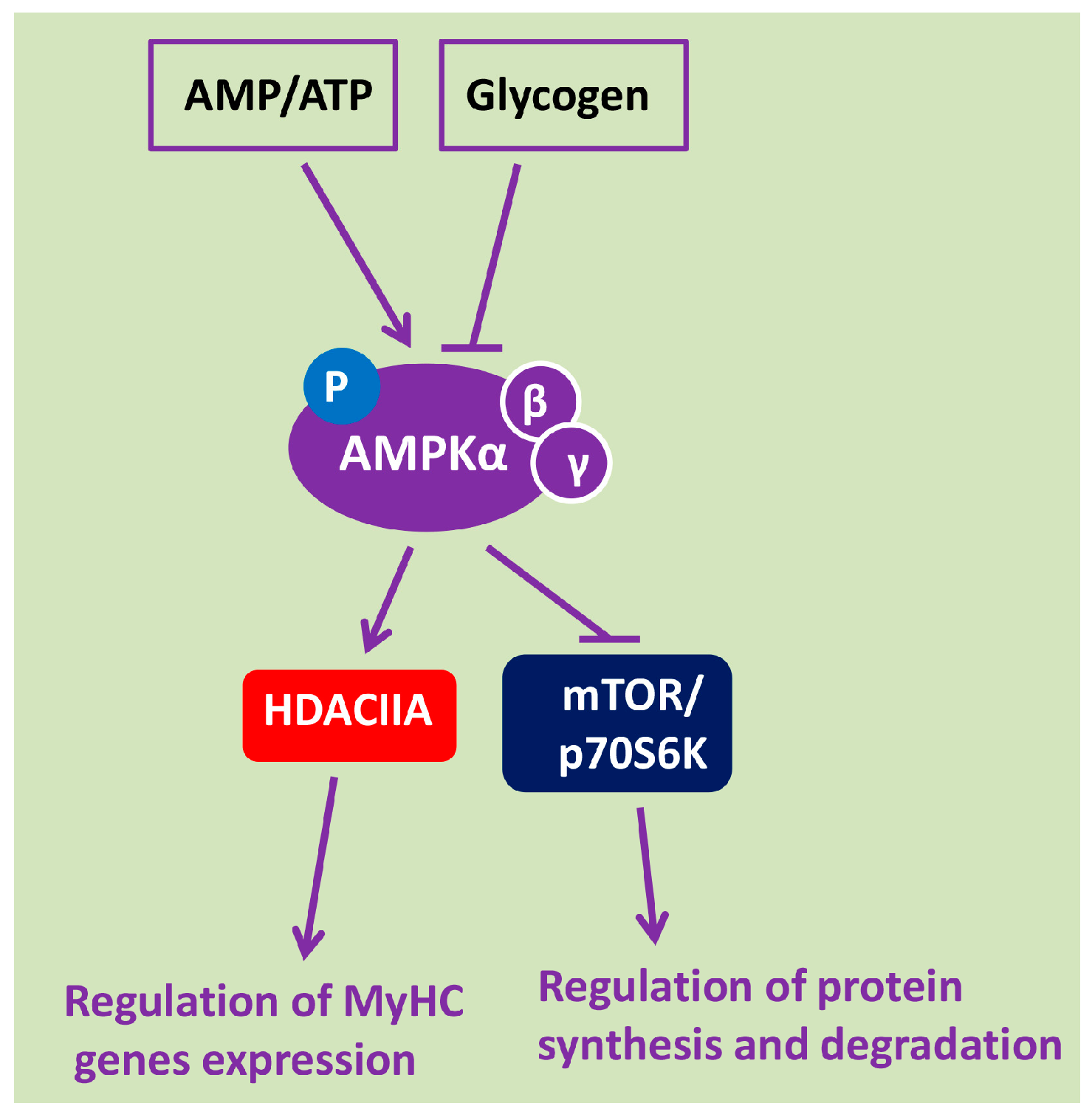
2. AMPK Is a Key Energy Sensor and Metabolic Regulator of Signaling Pathways in Skeletal Muscle Fibers

AMPK is involved in transmission of extracellular and intracellular signals by phosphorylation of various substrates in many metabolic reactions in skeletal muscle. AMRK is a heterotrimeric complex consisting of three proteins: α-subunit, which has its own kinase activity, and two regulatory subunits, β and γ [28].
AMPK activity is regulated both allosterically and by post-translational modifications (phosphorylation). Allosteric activation of AMPK is carried out with the help of AMP and its analogues. Phosphorylation AMPK at Thr172 of the α-subunit leads to its activation. This phosphorylation is regulated by calcium-/calmodulin-dependent kinase kinase 2 (CaMKK2) and liver kinase B1 (LKB1). Further activation of AMPK occurs due to conformational changes occurring upon binding of AMP or ADP to the γ-subunit of AMPK, promoting Thr172 phosphorylation at the α-subunit. The combined effect of Thr172 phosphorylation of the α-subunit of AMPK and allosteric regulation leads to more than a 1000-fold increase in AMPK activity, which makes AMPK highly sensitive to alternations in the energy status of the cell [29].
Activation of AMPK may also occur under the action of extracellular regulatory factors, such as interleukin-6 (IL-6) and brain-derived neurotrophic factor (BDNF) [30]. Interestingly, AMPK is involved in the regulation of IL-6 expression in skeletal muscles [31]. One of the factors of AMPK activation is nitric oxide (NO), the production of which is determined by the activity of neuronal and endothelial NO synthases [32,33]. There is evidence that AMPK activation can result from mechanical stretch via components of the dystrophin-glycoprotein complex (at least in cardiomyocytes) [34].
AMPK can also be phosphorylated on Ser485/491 sites by protein kinase D and some isoforms of protein kinase C [35], which leads to inhibition of AMPK activity. A decrease in AMPK activity is associated with increased glycogen content, as well as accumulation of ATP and creatine phosphate [36,37]. AMPK has a number of molecular targets in skeletal muscle. It is known that AMPK can activate Na,K-ATPase [38,39] and phosphorylate neuronal NO-synthase [40]. AMPK is also involved in the regulation of protein synthesis and degradation [36]. AMPK is a negative regulator of protein synthesis in skeletal muscle. This kinase can inhibit the key regulator of protein synthesis, the mammalian target of rapamycin complex 1 (mTORC1), through phosphorylation of TSC2 [41] and raptor [42]. AMPK can also be involved in the degradation of myofibrillar proteins [43]. Nakashima and co-authors have shown that AMPK participates in the degradation of myofibrillar proteins through the activation of forkhead box proteins (FOXO) transcription factors and subsequent up-regulation of muscle-specific E3 ubiquitin-ligases atrogin-1/MAFbx and MuRF-1 [44].
In addition, AMPK, as a key energy sensor of the cell regulating energy metabolism, participates in the initiation of autophagy [45]. AMPK can directly phosphorylate Unc-51-like kinase (ULK-1) across several sites as well as activate autophagy by inhibiting mTOR activity [46,47].
In recent years, it has been shown that AMPK can influence the expression of a number of genes by phosphorylation of class IIA histone deacetylases (HDAC4, HDAC5, HDAC7), leading to their exclusion from the nucleus and activation of gene expression [48,49,50].
Thus, according to modern concepts, AMPK activity is mainly determined by the state of energy metabolism: AMPK activity increases with increased ATP consumption, AMP accumulation, and glycogen depletion, and decreases with the accumulation of ATP and glycogen in muscle fibers. Activated AMPK phosphorylates and retains class IIA HDACs outside the myonuclei (thereby contributing to the expression of a number of genes) and inhibits the activity of mTORC1 and its primary targets (Figure 1). Dephosphorylated AMPK, on the contrary, promotes HDACs nuclear import and transcriptional suppression of gene expression, while reducing the degree of mTORC1 suppression.

3. AMPK Activity under Conditions of Mechanical Unloading

Until recently, AMPK activity/phosphorylation in skeletal muscle under unloading conditions has been poorly studied. Moreover, the literature reveals contradictory results concerning AMPK Thr172 phosphorylation at various time-points in different models of disuse. It has been shown that four- and seven-day denervation resulted in a significant increase in AMPK phosphorylation in rodent skeletal muscles [51,52], while deletion of AMPKα2 significantly attenuated denervation-induced skeletal muscle wasting and protein degradation [51]. In human skeletal muscle with recent complete cervical spinal cord injury, AMPKα2 protein abundance decreased by 25% during the first year after injury, without significant change in AMPKα1 content. Furthermore, AMPK phosphorylation on Thr172 was significantly decreased during the first year post-spinal cord injury in human vastus lateralis muscle [53]. Thirty-day space flight and subsequent recovery did not affect AMPK Thr172 phosphorylation in murine longissimus dorsi muscle [54]. In terms of fiber-type composition, murine longissimus dorsi is similar to soleus muscle. However, it should be noted that it is not quite correct to compare the data obtained from rat and mouse soleus muscle. It is known that rat soleus muscle, as well as human soleus, comprises about 85% of slow-twitch fibers expressing the slow isoform of MyHC, while mouse soleus consists of approximately 40% slow-twitch fibers [55]. Obviously, this fact determines the essential features of metabolism in the mouse soleus muscle and its response to gravitational unloading. Therefore, unloading-induced changes in mouse soleus can significantly differ from that of rats and humans.
Vilchinskaya et al. (2015) have shown for the first time that short-term (three days) gravitational unloading via dry immersion leads to a significant decrease in the level of AMPK Thr172 phosphorylation in human soleus muscle [56]. The literature data on the AMPK activity in rat soleus following HS are inconsistent. AMPK activity, which is usually assessed by the level of Thr172 phosphorylation [57,58,59], was reported to be reduced in rat soleus after two weeks of HS [60], whereas Hilder and co-authors showed that 14-day HS results in a significant increase in AMPK Thr172 phosphorylation in rat soleus [61]. At the same time, Egawa and others did not find any changes in AMPKα1 и AMPKα2 activity in mouse soleus after 14-day HS, however, the level of ACC phosphorylation was upregulated [62,63].
A significant reduction in AMPK Thr172 phosphorylation was previously observed in rat soleus at the early stage (24 h) of hindlimb unloading [11]. A recent study has also demonstrated a significant decrease in AMPK Thr172 phosphorylation in rat soleus muscle already after 6- and 12-h mechanical unloading [64]. In an inactive skeletal muscle, a rapid accumulation of completely phosphorylated high-energy phosphates can occur, resulting in reduced AMPK activity. Thus, AMPK dephosphorylation at the early stage of gravitational unloading can be caused by a decrease in postural muscle energy consumption and a corresponding change in the ratio of phosphorylated and dephosphorylated adenine nucleotides (ATP, ADP, AMP).
It is known that binding of AMPK to glycogen results in reduced AMPK activity [37]. Therefore, it is possible that a decrease in the activity of AMPK at the initial stages of mechanical unloading may be associated with glycogen accumulation. Indeed, glycogen concentration in rat soleus muscle during the first three days of HS is significantly increased [27].
After seven-day HS, AMPK phosphorylation does not differ from that of control [11], which correlates well with the restoration of electromyographic activity of rat soleus following six to seven days of HS [15]. Moreover, 14-day HS resulted in a significant increase in AMPK Thr172 phosphorylation in rat soleus [61,65]. It is notable that after 14-day HS, the increase of AMPK activity (judging by ACC phosphorylation) [62,63] is less pronounced in murine vs rat soleus.
Now, it is difficult to establish the precise mechanisms that cause an increase in AMPK activity by 14 days of HS. However, some assumptions can be made concerning potential signaling mechanisms leading to this phenomenon. There is evidence that the concentration of interleukin-6 (IL-6) is significantly increased in rodent skeletal muscle after 5- and 14-day HS [66,67] as well as in human skeletal muscle following 60-day bed rest [68]. It is known that IL-6 can increase AMPK activity in rodent skeletal muscle [69], and it is likely that increased concentration of IL-6 during long-term gravitational unloading promotes AMPK hyperphosphorylation. BDNF was also shown to increase AMPK phosphorylation in isolated rat extensor digitorum muscle [30]. Therefore, an increase in BDNF mRNA expression in the spinal cord and soleus muscle of rats after 14 days of HS [70] could be the cause of the increased AMPK Thr172 phosphorylation during this period.
Thus, HS experiments show complex time-course changes in AMPK activity in the rodent soleus muscle under mechanical unloading. It is important to note that the level of AMPK phosphorylation is significantly reduced during the first day of HS, a period that precedes atrophy development. Additionally, the most likely cause of such a decrease is a shift in the ratio of phosphorylated and dephosphorylated adenine nucleotides (AMP/ATP and ADP/ATP ratios).

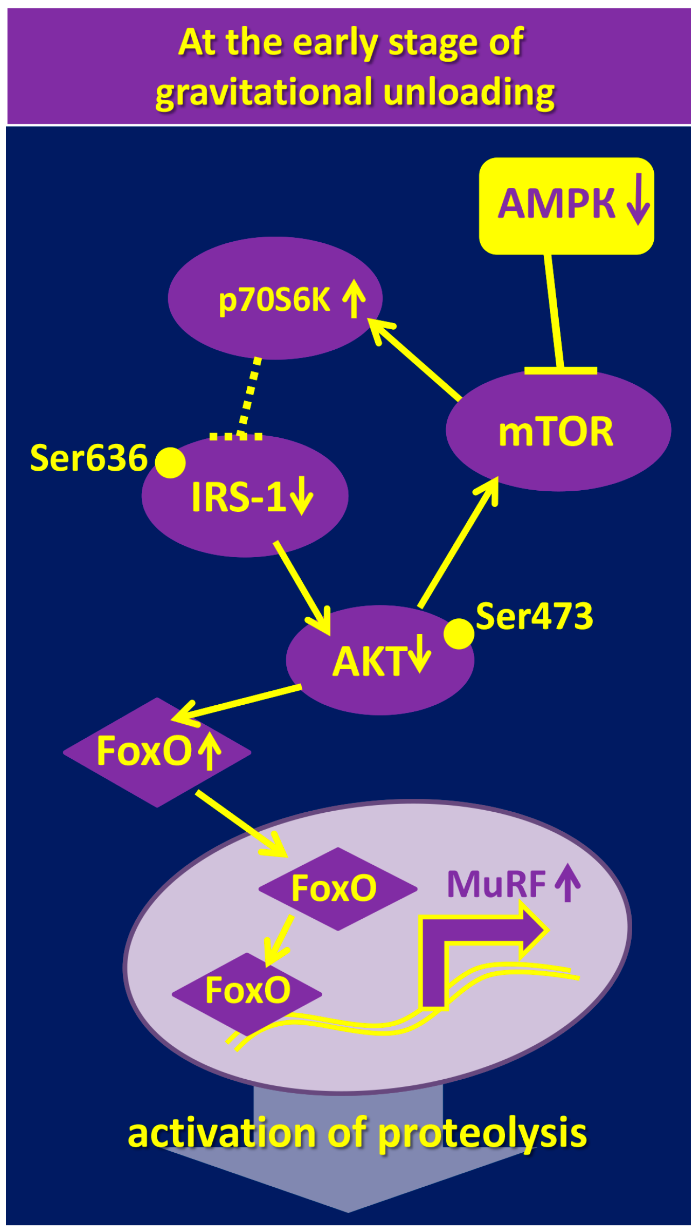
4. The Role of AMPK in the Regulation of mTOR/p70S6K and Akt/FOXO3/MuRF-1/MAFbx/Atrogin-1 Signaling Pathways under Gravitational Unloading

Anabolic processes in skeletal muscle fibers are regulated by a number of signaling pathways, the most important of which is the mammalian target of rapamycin complex 1 (mTORC1) signaling pathway. mTORC1, through its downstream targets (p70S6K, 4E-BP1), stimulates mRNA translation initiation on a ribosome. Activation of protein synthesis following resistance exercise is associated with the increased level of p70S6K (Thr389) phosphorylation, leading to subsequent phosphorylation of ribosomal protein S6 and initiation of protein synthesis. Some authors reported a decrease in p70S6K (Thr389) phosphorylation in rat soleus after four to five days of HS [71,72]. Other studies showed that even 7–10 days of HS did not affect p70S6K phosphorylation, the level of which decreased only following 14 days of unloading [73,74,75,76].
It is well known that activated AMPK has an inhibitory effect on the anabolic processes via suppression of mTORC1 and its key substrate, p70S6K [41,42,43,77,78,79,80]. Summing up the available data, we can assume that changes in p70S6K and AMPK phosphorylation in rat soleus muscle during the first two weeks of HS show reciprocal relations and complex dynamics. It is important to note that during the early stage of HS (one to three days), an increase in p70S6K Thr389 phosphorylation is accompanied by a decrease in the level of AMPK Thr172 phosphorylation. However, after seven days of HS, there is no difference between these parameters and control values [11]. Two-week HS results in an opposite effect: A decrease in p70S6K phosphorylation is accompanied by an increase in AMPK phosphorylation. These data are consistent with the report by Sugiura and co-authors (2005) that showed no changes in p70S6K phosphorylation after 10 days of HS [74], whereas 14-day HS led to a significant decrease in p70S6K phosphorylation as compared to control levels [71,72,75,76,81]. Interestingly, according to Hilder et al. (2005) and Zhang et al. (2018), the level of AMPK phosphorylation following 14-day unloading is significantly increased [61,65]. The high level of AMPK phosphorylation is accompanied by a decrease in phosphorylation of not only p70S6K, but also Akt and FOXO3, which can lead to an upregulation of muscle-specific E3-ubiquitin ligases, MuRF-1 and MAFbx/atrogin-1 [65]. These results are consistent with earlier studies showing the ability of AMPK to stimulate FOXO3 dephosphorylation and the expression of E3-ubiquitin ligases [44,82]. Time-course changes in the level of p70S6K and AMPK phosphorylation during mechanical unloading suggest that an increase in p70S6K phosphorylation at the first day of HS may be due to the low AMPK activity. This hypothesis has been recently tested by Vilchinskaya and co-authors [83]. Pretreatment of rats with 5-aminoimidazole-4-carboxamide ribonucleotide (AICAR) several days before and during 24-h HS prevented both a decrease in AMPK Th172 phosphorylation as well as an increase in p70S6K Thr389 phosphorylation [83]. This result fully confirmed the hypothesis and once again demonstrated the role of AMPK as a negative regulator of mTORC1-signaling [41,42]. The results obtained by Vilchinskaya et al. (2017) suggest that AMPK dephosphorylation in postural soleus muscle at the initial stage of mechanical unloading is one of the reasons for a paradoxical increase in the level of p70S6K phosphorylation. Increased p70S6K phosphorylation is usually considered to be a consequence of (1) inactivation of endogenous mTORC1 inhibitor tuberous sclerosis complex (TSC1/2) due to AMPK dephosphorylation [41] and (2) accumulation of sphingolipid ceramide [84]. Interestingly, it was previously shown that activation of AMPK with AICAR prevents ceramide accumulation in skeletal muscle fibers [85]. Therefore, it can be assumed that AMPK dephosphorylation can contribute to the accumulation of ceramide in rat soleus under gravitational unloading [86,87,88]. Hsieh and co-authors (2014) observed an interesting effect of p70S6K hyperphosphorylation. The authors showed that activated p70S6K promotes phosphorylation of insulin receptor substrate (IRS1) on Ser636-639, which leads to a reduction in IRS-1 activity and, accordingly, dephosphorylation of downstream protein kinase Akt (Figure 2).
Dephosphorylation of Akt on Ser473, as a rule, causes an increased expression of E3-ubiquitin ligases (MuRF-1 and MAFbx/atrogin-1) [88]. It is well known that even short-term (one to three days) gravitational unloading leads to Akt dephosphorylation and upregulation of E3-ubiquitin ligases [10,66]. According to a number of authors, gravitational unloading results in a significant decrease in IRS-1 content [54,89]. Based on these literature data, it can be assumed that AMPK dephosphorylation during the first day of unloading leads to an increase in p70S6K phosphorylation resulting in the increased E3-ubiquitin ligases’ expression and enhanced proteolysis. Recent experiments with inhibition of p70S6K phosphorylation during the first day of gravitational unloading do not contradict this hypothesis: The use of rapamycin (mTORC1 inhibitor) resulted in a significant decrease in MuRF-1 and MAFbx/atrogin-1 expression [88].
Thus, AMPK dephosphorylation at the initial period of HS leads to an increase in the level of p70S6K phosphorylation, which can contribute to the subsequent upregulation of proteolytic enzymes.
AMPK is known to activate autophagy by ULK phosphorylation (see above). Therefore, one would expect to see a reduction in autophagy markers at the initial period of unloading when AMPK activity is downregulated. However, it has been recently shown that most of autophagy markers, except for ULK, were upregulated in rat soleus following 6- and 12-h HS [64]. The authors of the cited report emphasize that this state of autophagy markers is not consistent with a reduced level of AMPK phosphorylation. They explain this phenomenon by the possible unequal phosphorylation of different AMPK isoforms. It is clear that this issue needs further investigation.
At a later stage of unloading, an increase in AMPK phosphorylation is accompanied by a decrease in p70S6K phosphorylation and activation of the proteolytic signaling system [65].
At the same time, no significant changes in phosphorylation of Akt and p70S6K and the rate of protein synthesis were found in the soleus of transgenic mice overexpressing the dominant-negative mutant of AMPK following 14-day HS [62]. Thus, the lack of AMPK activity in the transgenic mice did not affect anabolic signaling following a relatively long period of unloading (14 days). However, 14-day unloading of these transgenic mice induced a significant increase in the expression of both markers of the ubiquitin-proteasome system and ULK, a marker of autophagy. The differences between dominant-negative AMPK mutants and wild-type mice were significant [62]. There were also significant differences in the severity of atrophic changes. The weight of soleus muscle in dominant-negative AMPK mutants after 14-day HS was significantly reduced, but to a lesser extent than that in wild-type mice (difference of 10–15%) [62,63]. Thus, AMPK can contribute to the development of muscle atrophy during unloading. This contribution to muscle atrophy seems to be carried out via the activation of catabolic processes rather than through the suppression of anabolic regulation.
This fact allows us to suggest that, at later stages of gravitational unloading, AMPK activity can act as a key signaling node of protein homeostasis in skeletal muscle.

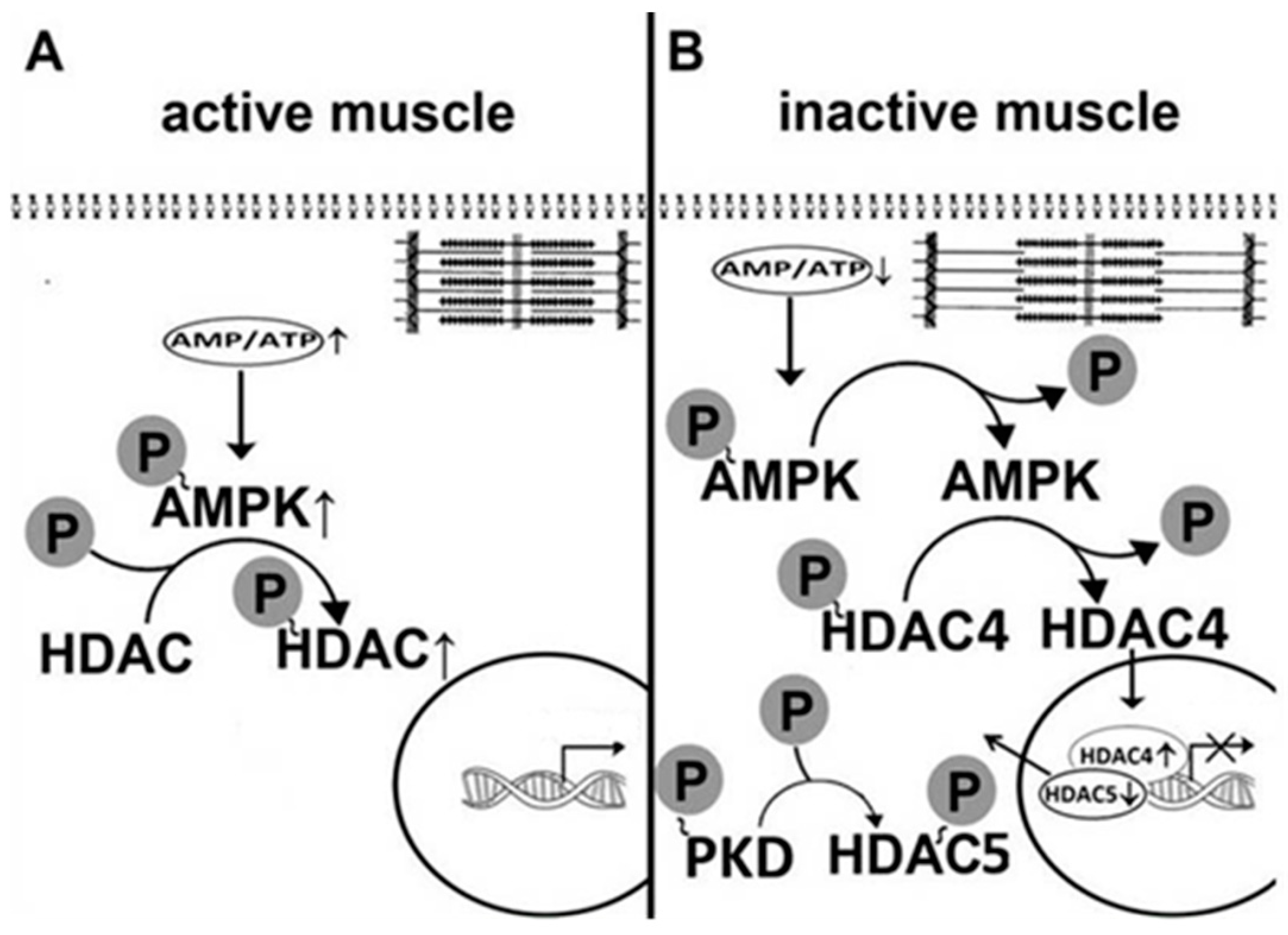
5. AMPK Is Involved in the Regulation of Myosin Phenotype under Mechanical Unloading

Unloading-induced changes in skeletal muscle myosin phenotype have been observed by a number of researchers. It is well established that there is an increase in the expression of fast-type myosin isoforms and a decrease in the expression of slow-type myosin isoform in skeletal muscles of space-flown rodents as well as astronauts [3,4,90,91]. HS results in a significant increase in the content of fast-twitch (type II) fibers and a significant decrease in the proportion of slow-twitch (type I) fibers in rat soleus muscle [7,92,93,94,95]. In samples of soleus muscle, taken from astronauts after a six-month spaceflight, there was a decrease in the proportion of fibers expressing slow MyHC isoform and an increase in the proportion of fibers expressing fast MyHC isoforms [96]. Mechanisms of molecular regulation of myosin phenotype transformation remain largely unexplored. It is known that AMPK can affect the expression of a number of genes by phosphorylation of class IIA histone deacetylases (HDAC4, HDAC5, HDAC7), which leads to HDACs dissociation from gene promoters and removal from the nucleus, thereby allowing for increased gene expression [48,49,50]. It was earlier shown that at the first day of mechanical unloading, mRNA expression of the slow MyHC isoform in rat soleus is reduced [13]. One of the ways of MyHC expression regulation in muscle fibers is associated with phosphorylation of histone deacetylase 4 (HDAC4) [7,97]. However, it remains unclear what mechanisms are implicated in such a rapid decrease in gene expression. Until recently, there have been no studies on the role of AMPK in the regulation of myosin phenotype in muscle fibers under conditions of gravitational unloading. Can AMPK be involved in the regulation of MyHC gene expression in skeletal muscle fibers? To answer this question, Vilchinskaya et al. (2017) carried out an experiment with AICAR pretreatment of Wistar male rats [98]. There was a significant decrease in MyHC I (β) pre-mRNA expression and a pronounced tendency to a decrease in the mature MyHC I(β) mRNA expression in rat soleus muscle after 24 h of HS. These results are in good agreement with the data previously obtained in skeletal muscles of Sprague-Dawley rats under similar conditions [13]. However, when exposed to AICAR, there were not any significant decreases in the MyHC I(β) pre-mRNA and mature mRNA expression. Since one of the possible mechanisms of AMPK gene expression regulation is linked to histone deacetylase 4 and 5 (HDAC4/HDAC5) phosphorylation status [49,50], it was hypothesized that a significant decrease in AMPK Thr172 phosphorylation after one-day HS would result in HDAC4 and HDAC5 nuclear accumulation [83]. This hypothesis is supported by a previous report that showed a significant deacetylation of histone H3 at the MyHC I (β) gene in rat soleus following seven-day HS [99]. In addition, it has been previously shown that HDAC4 is predominantly localized to the nuclei in fast-twitch fibers in contrast to the sarcoplasm in slow-twitch fibers [100]. Indeed, in our experiment with 24-h HS, HDAC4 accumulation in the nuclear fraction was found; however, in the AICAR-pretreated group, the accumulation of HDAC4 in the nuclei did not occur, which correlates well with data on AMPK phosphorylation and confirms the hypothesis of AMPK-dependent control of nucleocytoplasmic trafficking of HDAC4 [98,101]. Recently, Yoshihara and co-authors have found nuclear accumulation of HDAC4 in rat gastrocnemius muscle following 10 days of ankle joint immobilization. This accumulation (as in the experiment of Vilchinskaya and co-authors) was accompanied by a decrease in the level of AMPK phosphorylation [102]. As for HDAC5, even a slight increase in AMPK activity in the control AICAR-pretreated rat was accompanied by a decrease in HDAC5 content in the nuclear fraction. This phenomenon could be associated with HDAC5 phosphorylation via AMPK. However, 24-h HS resulted in a significant decrease in the nuclear HDAC5 content. Such a reduction in HDAC5 content in the nuclear fraction could be associated with HDAC5 degradation or HDAC5 phosphorylation and subsequent nuclear export. HDAC5 can also be a target for protein kinase D (PKD) [103,104]. Since the AMPK activity is significantly reduced within the first day of unloading (see above), it appears that HDAC5 nuclear export during unloading is not directly linked to AMPK. It has been shown that a decrease in AMPK activity can result in an upregulation of protein kinase D (PKD) phosphorylation [103]. Indeed, one-day HS resulted in a significant increase in PKD Ser916 phosphorylation, however, in the HS+AICAR group, PKD phosphorylation did not differ from the control levels. Interestingly, such a decrease in PKD phosphorylation in the HS+AICAR group vs. the HS group can significantly attenuate the loss of nuclear HDAC5. It is noteworthy that an increase in PKD phosphorylation in unloaded animals did not affect the content of nuclear HDAC4. Obviously, under unloading conditions, HDAC4 does not appear to be a target of PKD. This study was the first to observe the reciprocal relationship between AMPK and PKD in an inactivated skeletal muscle. In addition, gravitational unloading led to an increase in the level of negative AMPK phosphorylation on Ser485/491, which is known to be associated with PKD [35]. However, allosteric activation of AMPK by AICAR reduced the intensity of negative phosphorylation, possibly due to a reduction in PKD phosphorylation.
AMPK is known to stimulate the expression of peroxisome proliferator-activated receptor gamma coactivator 1-alpha (PGC1α), the most important regulator of the expression and activity of signaling proteins. In particular, PGC1α is involved in the control of the expression of the slow isoform of MyHC [105]. A significant decrease in PGC1α expression was found in mouse soleus after four days of hindlimb unloading [106]. However, Vilchinskaya et al. (2017) found no changes in PGC1α mRNA expression after 24-h HS [98]. AMPK activation by AICAR pretreatment also did not induce any changes in PGC1α expression. Obviously, the impact of AMPK on the expression of the slow isoform of MyHC during the first day of unloading is carried out primarily through the trafficking of HDAC4, without the involvement of PGC1α.
Thus, the results obtained by Vilchinskaya and the co-authors (2017) clearly show that AMPK Thr172 dephosphorylation within the first day of gravitational unloading has a significant effect on the regulation of myosin phenotype in rat postural muscle. In particular, AMPK Thr172 dephosphorylation led to a decrease in MyHC I(β) pre-mRNA and mRNA expression. The study of Vilchinskaya et al. (2017) indicates that HDAC4 is not a target of PKD at the early stages of unloading, and, probably, HDAC4 nuclear import results from a decrease in the AMPK activity (Figure 3). It is possible that an increase in PKD activity can lead to HDAC5 nuclear export.

6. AMPK and Disuse-Induced Motor Endplate Remodeling

Reliable neuromuscular transmission is essential for normal bodily functions. Motor endplate is a highly specialized sarcolemma region in which the transmission of activity from motor neurons to striated muscle fiber is realized. Endplate structure, including features of nicotinic acetylcholine receptors’ (nAChRs) localization and distribution, are among the factors crucial for maintenance of highly efficient neuromuscular transmission [108,109].
Ultrastructure of the neuromuscular junction strongly depends on the motor activity, and exhibits high morphological and functional plasticity. Different modes of increased activity are manifested in the morphological remodeling, such as the expansion of the neuromuscular junction size. Reduced patterns of neuromuscular activity also trigger endplate remodeling. Alterations in neuromuscular junction stability and integrity progressively increase with age [110,111,112,113,114] and myodystrophy [115,116,117], in animal models of muscle injures [118,119], after denervation [120] and prolonged (four weeks) HS [121]. Although the molecular mechanisms underlying the structural and functional endplate plasticity are intensively studied, they are not completely clear [113,115,116,122,123,124,125].
Currently, many facts point to the involvement of AMPK in neuromuscular junction remodeling. AMPK is linked to a variety of cellular processes and is also considered a crucial activator of autophagy and its downstream target, ULK1 [126]. Autophagy is involved in neuromuscular junction preservation during aging, and impairments in autophagy exacerbate synaptic structure degeneration [111]. AMPK and AMPK-activated autophagy are among the most important factors that maintain neuromuscular junction stability [111,127,128]. Pharmacological activation of AMPK by AICAR administration improves integrity of neuromuscular junctions and prevents skeletal muscle pathology in a mouse model of severe spinal muscular atrophy [128]. Additionally, AICAR treatment has been shown to stimulate autophagy and ameliorate muscular dystrophy in the mdx mice, a model of Duchenne muscular dystrophy, suggesting AMPK as a powerful therapeutic target [129,130]. Also, AMPK may play a role in activating PGC-1α, a key regulatory protein in skeletal muscle adaptation to physical activity. PGC-1α has been reported to play a major role in maintaining neuromuscular junction integrity [122,123].
AMPK also plays a beneficial role in the regulation of skeletal muscle cholesterol synthesis as well as sarcolemma cholesterol levels [131,132]. Direct molecular interaction between membrane cholesterol and the nAChRs has been shown [133]. Moreover, cholesterol and lipid rafts contribute to the orchestration of nAChRs clustering at the endplate region [134,135]. In addition, AMPK can affect the Na,K-ATPase activity [38,39]. The targeting and activity of the Na,K-ATPase are also influenced by the cholesterol environment [136,137,138,139], and reciprocal interactions between cholesterol and the α2 Na,K-ATPase isozyme has been suggested [22,140]. Notably, both the nAChRs and the α2 Na,K-ATPase isozyme are enriched at the endplate region, co-localized, and co-immunoprecipitated, suggesting that these proteins exist as a functional multimolecular complex to regulate electrogenesis and to maintain the effectiveness of neuromuscular transmission [141,142,143].
The loss of the α2 Na,K-ATPase isozyme electrogenic activity accompanied by disturbances in lipid rafts and endplate structure stability were observed in rat soleus muscle even after 6–12 h of HS [21,22,64]. Such acute disuse decreased the endplate area and increased the density of the nAChRs distribution. These changes were accompanied by decreased phosphorylation of AMPK and its substrate, ACC, and increased autophagy [64]. Autophagy is known to be involved in the nAChRs turnover regulation [127] and has a major impact on neuromuscular synaptic function [111]. So, an increase in autophagy after acute HS can reflect an adaptive response to compensate for the endplate area loss through increasing the density of the nAChRs distribution [64].
Notably, pretreatment of the rats with AMPK activator, AICAR, followed by HS, stabilized the resting membrane potential, endplates area, and the nAChRs distribution density, indicating that AMPK activation can prevent disuse-induced endplate structural and functional reorganization (unpublished observation).
In summary, these novel findings indicate that endplate functional and structural characteristics rapidly (within hours) respond to skeletal muscle disuse. Decreased phosphorylation of AMPK accompanied by increased autophagy is the earliest disuse-induced remodeling event preceding the overt skeletal muscle atrophy.

7. Conclusions

AMPK demonstrates multidirectional changes in a mammalian soleus muscle during gravitational unloading: A deep decrease in the level of phosphorylation and kinase activity at the early stages of the process and a significant increase in activity at the later stages. Time-course changes in AMPK activity under unloading are nonlinear and require a more detailed analysis at each stage of the process. The experiments discussed in this review show that changes in AMPK activity under unloading conditions can have a significant impact on the key signaling pathways and molecular structures in skeletal muscle fiber. As a result of reduction in AMPK activity at the initial stage of unloading, paradoxical hyperphosphorylation of p70S6K occurs, which, according to in vitro experiments, can lead to the activation of proteolytic processes in muscle fiber [83,87]. A decrease in the level of phosphorylation and kinase activity of AMPK at the early stages of unloading also affects the change in nucleocytoplasmic traffic of class IIA HDACs, resulting in a decrease in the expression of the myh7 gene (slow isoform of MyHC) and, possibly, a number of other genes controlling energy metabolism. At the later time-points of unloading, an increase in the level of AMPK phosphorylation/activity is accompanied by a decrease in the activity of p70S6K [76], FOXO3 dephosphorylation, and an increase in the expression of the key enzymes of the ubiquitin-proteasome pathway [65], which, obviously, should lead to decreased muscle protein synthesis and enhanced proteolysis. Unfortunately, to date, the precise physiological mechanisms, both intramuscular and systemic, underlying such a complex and nonlinear nature of AMPK activity during gravitational unloading are not known. It is possible that glycogen accumulation [27] or a change in the ratio of dephosphorylated and phosphorylated high-energy phosphates could lead to a deep AMPK dephosphorylation. The cause of a gradual increase in AMPK phosphorylation in rat soleus during the first week of HS remains unclear. It is possible that this increase is due to a gradual increase in the electromyographic activity of the postural muscle [15]. It is difficult to explain a significant increase in AMPK phosphorylation in rat soleus by the end of the second week of HS. Possibly, such systemic factors as BDNF and/or interleukin-6 could play a role in these processes. All these questions have yet to be answered.
Thus, recent studies have revealed the key role of AMPK in the processes of deep remodeling of signaling pathways, leading to changes in metabolism, structure, and function of postural muscle fibers under unloading conditions. Further studies are needed to elucidate new signaling mechanisms that trigger, determine, and limit atrophy development and intrinsic muscle stiffness, as well as myosin phenotype changes, in a mammalian postural muscle under mechanical unloading.

Funding

The work was supported by the Russian Science Foundation (RSF) grant no. 18-15-00107.

Acknowledgments

The authors express their deep gratitude to Timur M. Mirzoev for the participation in the manuscript preparation and creative criticism of the paper.

Conflicts of Interest

The authors declare no conflict of interest.

Abbreviations

AMPK5′ adenosine monophosphate -activated protein kinase
AICAR5-aminoimidazole-4-carboxamide ribonucleotide
BGPAβ-Guanidinopropionic acid
HShindlimb suspension
mTOR mammalian/mechanistic target of rapamycin
p70S6Kribosomal protein S6 kinase beta-1
CaMKKβCa(2+)/calmodulin-dependent protein kinase kinase β
LKB1liver kinase B1
BDNFbrain-derived neurotrophic factor
MuRF-1Muscle RING-finger protein-1
FOXOforkhead box proteins
IRS-1insulin receptor substrate 1
MyHCmyosin heavy chain isoforms
nAChRsnicotinic acetylcholine receptors
ULK-1Unc-51-like kinase
PGC1αperoxisome proliferator-activated receptor gamma coactivator 1-alpha

References

  1. Grigor’ev, A.I.; Kozlovskaia, I.B.; Shenkman, B.S. The role of support afferents in organisation of the tonic muscle system. Rossiiskii fiziologicheskii zhurnal imeni IM Sechenova 2004, 90, 508–521. [Google Scholar]
  2. Kozlovskaia, I.B.; Grigor’eva, L.S.; Gevlich, G.I. Comparative analysis of the effect of weightlessness and its model on the velocity-strength properties and tonus of human skeletal muscles. Kosm. Biol. Aviakosm. Med. 1984, 18, 22–26. [Google Scholar] [PubMed]
  3. Oganov, V.S.; Skuratova, S.A.; Murashko, L.M.; Guba, F.; Takach, O. Effect of short-term space flights on physiological properties and composition of myofibrillar proteins of the skeletal muscles of rats. Kosm. Biol. Aviakosm. Med. 1988, 22, 50–54. [Google Scholar] [PubMed]
  4. Baldwin, K.M.; Haddad, F.; Pandorf, C.E.; Roy, R.R.; Edgerton, V.R. Alterations in muscle mass and contractile phenotype in response to unloading models: Role of transcriptional/pretranslational mechanisms. Front. Physiol. 2013, 4, 284. [Google Scholar] [CrossRef] [PubMed]
  5. Kandarian, S.C.; Stevenson, E.J. Molecular events in skeletal muscle during disuse atrophy. Exerc. Sport Sci. Rev. 2002, 30, 111–116. [Google Scholar] [CrossRef] [PubMed]
  6. Adams, G.R.; Baldwin, K.M. Age dependence of myosin heavy chain transitions induced by creatine depletion in rat skeletal muscle. J. Appl. Physiol. 1995, 78, 368–371. [Google Scholar] [CrossRef] [PubMed] [Green Version]
  7. Shenkman, B.S. From Slow to Fast: Hypogravity-Induced Remodeling of Muscle Fiber Myosin Phenotype. Acta Nat. 2016, 8, 47–59. [Google Scholar]
  8. Chopard, A.; Hillock, S.; Jasmin, B.J. Molecular events and signalling pathways involved in skeletal muscle disuse-induced atrophy and the impact of countermeasures. J. Cell Mol. Med. 2009, 13, 3032–3050. [Google Scholar] [CrossRef] [PubMed] [Green Version]
  9. Bodine, S.C. Disuse-induced muscle wasting. Int. J. Biochem. Cell Biol. 2013, 45, 2200–2208. [Google Scholar] [CrossRef] [PubMed] [Green Version]
  10. Kachaeva, E.V.; Shenkman, B.S. Various jobs of proteolytic enzymes in skeletal muscle during unloading: Facts and speculations. J. Biomed. Biotechnol. 2011, 2012, 493618. [Google Scholar] [CrossRef] [PubMed]
  11. Mirzoev, T.; Tyganov, S.; Vilchinskaya, N.; Lomonosova, Y.; Shenkman, B. Key Markers of mTORC1-Dependent and mTORC1-Independent Signaling Pathways Regulating Protein Synthesis in Rat Soleus Muscle during Early Stages of Hindlimb Unloading. Cell. Physiol. Biochem. 2016, 39, 1011–1020. [Google Scholar] [CrossRef] [PubMed]
  12. Stevens, L.; Sultan, K.R.; Peuker, H.; Gohlsch, B.; Mounier, Y.; Pette, D. Time dependent changes in myosin heavy chain mRNA and protein isoforms 94 in unloaded soleus muscle of rat. Am. J. Physiol. Cell. Physiol. 1999, 277, 1044–1049. [Google Scholar] [CrossRef] [PubMed]
  13. Giger, J.M.; Bodell, P.W.; Zeng, M.; Baldwin, K.M.; Haddad, F. Rapid muscle atrophy response to unloading: Pretranslational processes involving MHC and actin. J. Appl. Physiol. 2009, 107, 1204–1212. [Google Scholar] [CrossRef] [PubMed]
  14. Lomonosova, Y.N.; Turtikova, O.V.; Shenkman, B.S. Reduced expression of MyHC slow isoform in rat soleus during unloading is accompanied by alterations of endogenous inhibitors of calcineurin/NFAT signaling pathway. J. Muscle Res. Cell Motil. 2016, 37, 7–16. [Google Scholar] [CrossRef] [PubMed]
  15. Alford, E.K.; Roy, R.R.; Hodgson, J.A.; Edgerton, V.R. Electromyography of rat soleus, medial gastrocnemius, and tibialis anterior during hind limb suspension. Exp. Neurol. 1987, 96, 635–649. [Google Scholar] [CrossRef]
  16. Kawano, F.; Matsuoka, Y.; Oke, Y.; Higo, Y.; Terada, M.; Wang, X.D.; Nakai, N.; Fukuda, H.; Imajoh-Ohmi, S.; Ohira, Y. Role(s) of nucleoli and phosphorylation of ribosomal protein S6 and/or HSP27 in the regulation of muscle mass. Am. J. Physiol. Cell Physiol. 2007, 293, 35–44. [Google Scholar] [CrossRef] [PubMed]
  17. De-Doncker, L.; Kasri, M.; Picquet, F.; Falempin, M. Physiologically adaptive changes of the L5afferent neurogram and of the rat soleus EMG activity during 14 days of hindlimb unloading and recovery. J. Exp. Biol. 2005, 208, 4585–4592. [Google Scholar] [CrossRef] [PubMed]
  18. Kozlovskaya, I.; Dmitrieva, I.; Grigorieva, L.; Kirenskaya, A.; Kreidich, Y. Gravitational mechanisms in the motor system studies in real and simulated weightlessness. In Stance and Motion Facts and Concepts; Gurfinkel, V.S., Loffe, M.E., Massion, J., Eds.; Springer: New York, NY, USA, 1988; ISBN 978-1-4899-0823-0. [Google Scholar]
  19. Navasiolava, N.M.; Custaud, M.A.; Tomilovskaya, E.S.; Larina, I.M.; Mano, T.; Gauquelin-Koch, G.; Gharib, C.; Kozlovskaya, I.B. Long-term dry immersion: Review and prospects. Eur. J. Appl. Physiol. 2011, 111, 1235–1260. [Google Scholar] [CrossRef] [PubMed] [Green Version]
  20. Kravtsova, V.V.; Matchkov, V.V.; Bouzinova, E.V.; Vasiliev, A.N.; Razgovorova, I.A.; Heiny, J.A.; Krivoi, I.I. Isoform-specific Na,K-ATPase alterations precede disuse-induced atrophy of rat soleus muscle. Biomed. Res. Int. 2015, 720172. [Google Scholar] [CrossRef] [PubMed]
  21. Kravtsova, V.V.; Petrov, A.M.; Matchkov, V.V.; Bouzinova, E.V.; Vasiliev, A.N.; Benziane, B.; Zefirov, A.L.; Chibalin, A.V.; Heiny, J.A.; Krivoi, I.I. Distinct α2 Na,K-ATPase membrane pools are differently involved in early skeletal muscle remodeling during disuse. J. Gen. Physiol. 2016, 147, 175–188. [Google Scholar] [CrossRef] [PubMed]
  22. Petrov, A.M.; Kravtsova, V.V.; Matchkov, V.V.; Vasiliev, A.N.; Zefirov, A.L.; Chibalin, A.V.; Heiny, J.A.; Krivoi, I.I. Membrane lipid rafts are disturbed in the response of rat skeletal muscle to short-term disuse. Am. J. Physiol. Cell Physiol. 2017, 312, 627–C637. [Google Scholar] [CrossRef] [PubMed]
  23. Lechado, I.; Terradas, A.; Vitadello, M.; Traini, L.; Namuduri, A.V.; Gastaldello, S.; Gorza, L. Sarcolemmal loss of active nNOS (Nos1) is an oxidative stress-dependent, early event driving disuse atrophy. J. Pathol. 2018. [Google Scholar] [CrossRef]
  24. Gordon, S.E.; Flück, M.; Booth, F.W. Selected Contribution: Skeletal muscle focal adhesion kinase, paxillin, and serum response factor are loading dependent. J. Appl. Physiol. 2001, 90, 1174–1183. [Google Scholar] [CrossRef] [PubMed]
  25. Ohira, Y.; Yasui, W.; Kariya, F.; Wakatsuki, T.; Nakamura, K.; Asakura, T.; Edgerton, V.R. Metabolic adaptation of skeletal muscles to gravitational unloading. Acta Astronaut. 1994, 33, 113–117. [Google Scholar] [CrossRef]
  26. Wakatsuki, T.; Ohira, Y.; Yasui, W.; Nakamura, K.; Asakura, T.; Ohno, H.; Yamamoto, M. Responses of contractile properties in rat soleus to high-energy phosphates and/or unloading. Jpn. J. Physiol. 1994, 44, 193–204. [Google Scholar] [CrossRef] [PubMed]
  27. Henriksen, E.J.; Tischler, M.E. Time Course of the Response of Carbohydrate Metabolism to Unloading of the Soleus. Metabolism 1988, 37, 201–208. [Google Scholar] [CrossRef]
  28. Witczak, C.A.; Sharoff, C.G.; Goodyear, L.J. AMP-activated protein kinase in skeletal muscle: From structure and localization to its role as a master regulator of cellular metabolism. Cell Mol. Life Sci. 2008, 65, 3737–3755. [Google Scholar] [CrossRef] [PubMed]
  29. Mounier, R.; Théret, M.; Lantier, L.; Foretz, M.; Viollet, B. Expanding roles for AMPK in skeletal muscle plasticity. Trends Endocrinol. Metab. 2015, 26, 275–286. [Google Scholar] [CrossRef] [PubMed] [Green Version]
  30. Matthews, V.B.; Aström, M.B.; Chan, M.H.; Bruce, C.R.; Krabbe, K.S.; Prelovsek, O.; Akerström, T.; Yfanti, C.; Broholm, C.; Mortensen, O.H.; et al. Brain-derived neurotrophic factor is produced by skeletal muscle cells in response to contraction and enhances fat oxidation via activation of AMP-activated protein kinase. Diabetologia 2009, 52, 1409–1418. [Google Scholar] [CrossRef] [PubMed] [Green Version]
  31. Glund, S.; Treebak, J.T.; Long, Y.C.; Barres, R.; Viollet, B.; Wojtaszewski, J.F.; Zierath, J.R. Role of adenosine 5′-monophosphate-activated protein kinase in interleukin-6 release from isolated mouse skeletal muscle. Endocrinology 2009, 150, 600–606. [Google Scholar] [CrossRef] [PubMed]
  32. Lira, V.A.; Soltow, Q.A.; Long, J.H.; Betters, J.L.; Sellman, J.E.; Criswell, D.S. Nitric oxide increases GLUT4 expression and regulates AMPK signaling in skeletal muscle. Am. J. Physiol. Endocrinol. Metab. 2007, 293, 1062–1068. [Google Scholar] [CrossRef] [PubMed]
  33. Lira, V.A.; Brown, D.L.; Lira, A.K.; Kavazis, A.N.; Soltow, Q.A.; Zeanah, E.H.; Criswell, D.S. Nitric oxide and AMPK cooperatively regulate PGC-1α in skeletal muscle. J. Physiol. 2010, 588, 3551–3566. [Google Scholar] [CrossRef] [PubMed]
  34. Garbincius, J.F.; Michele, D.E. Dystrophin-glycoprotein complex regulates muscle nitric oxide production through mechanoregulation of AMPK signaling. Proc. Natl. Acad. Sci. USA 2015, 112, 13663–13668. [Google Scholar] [CrossRef] [PubMed]
  35. Coughlan, K.A.; Valentine, R.J.; Sudit, B.S.; Allen, K.; Dagon, Y.; Kahn, B.B.; Ruderman, N.B.; Saha, A.K. PKD1 Inhibits AMPKα2 through Phosphorylation of Serine 491 and Impairs Insulin Signaling in Skeletal Muscle Cells. Biochem. J. 2016, 473, 4681–4697. [Google Scholar] [CrossRef] [PubMed]
  36. Hardie, D.G.; Ross, F.A.; Hawley, S.A. AMP-activated protein kinase: A target for drugs both ancient and modern. Chem. Biol. 2012, 19, 1222–1236. [Google Scholar] [CrossRef] [PubMed]
  37. Koay, A.; Woodcroft, B.; Petrie, E.J.; Yue, H.; Emanuelle, S.; Bieri, M.; Bailey, M.F.; Hargreaves, M.; Park, J.T.; Park, K.H.; et al. AMPK beta subunits display isoform specific affinities for carbohydrates. FEBS Lett. 2010, 584, 3499–3503. [Google Scholar] [CrossRef] [PubMed]
  38. Ingwersen, M.S.; Kristensen, M.; Pilegaard, H.; Wojtaszewski, J.F.; Richter, E.A.; Juel, C. Na, K-ATPase activity in mouse muscle is regulated by AMPK and PGC-1α. J. Membr. Biol. 2011, 242, 1–10. [Google Scholar] [CrossRef] [PubMed]
  39. Benziane, B.; Bjornholm, M.; Pirkmajer, S.; Austin, R.L.; Kotova, O.; Viollet, B.; Zierath, J.R.; Chibalin, A.V. Activation of AMP-activated protein kinase stimulates Na+,K+-ATPase activity in skeletal muscle cells. J. Biol. Chem. 2012, 287, 23451–23463. [Google Scholar] [CrossRef] [PubMed]
  40. Chen, Z.P.; McConell, G.K.; Belinda, J.; Snow, R.J.; Canny, B.J.; Kemp, B.E. AMPK signaling in contracting human skeletal muscle: Acetyl-CoA carboxylase and NO synthase phosphorylation. Am. J. Physiol. Endocrinol. Metab. 2000, 279, E1202–E1206. [Google Scholar] [CrossRef] [PubMed]
  41. Inoki, K.; Zhu, T.; Guan, K.L. TSC2 mediates cellular energy response to control cell growth and survival. Cell 2003, 115, 577–590. [Google Scholar] [CrossRef]
  42. Gwinn, D.M.; Shackelford, D.B.; Egan, D.F.; Mihaylova, M.M.; Mery, A.; Vasquez, D.S.; Turk, B.E.; Shaw, R.J. AMPK phosphorylation of raptor mediates a metabolic checkpoint. Mol. Cell 2008, 30, 214–226. [Google Scholar] [CrossRef] [PubMed]
  43. Nystrom, G.J.; Lang, C.H. Sepsis and AMPK Activation by AICAR Differentially Regulate FoxO-1, -3 and -4 mRNA in Striated Muscle. Int. J. Clin. Exp. Med. 2008, 1, 50–63. [Google Scholar] [PubMed]
  44. Nakashima, K.; Yakabe, Y.; Ishida, A.; Katsumata, M. Effects of orally administered glycine on myofibrillar proteolysis and expression of proteolytic-related genes of skeletal muscle in chicks. Amino Acids 2008, 35, 451–456. [Google Scholar] [CrossRef] [PubMed]
  45. Kim, J.; Kundu, M.; Viollet, B.; Guan, K. AMPK and mTOR regulate autophagy via direct phosphorylation of ULK1. Nat. Cell Biol. 2011, 13, 132–141. [Google Scholar] [CrossRef] [PubMed]
  46. Egan, D.F.; Shackelford, D.B.; Mihaylova, M.M.; Gelino, S.; Kohnz, R.A.; Mair, W.; Vasquez, D.S.; Joshi, A.; Gwinn, D.M.; Taylor, R.; et al. Phosphorylation of ULK1 (hATG1) by AMP-activated protein kinase connects energy sensing to mitophagy. Science 2011, 331, 456–461. [Google Scholar] [CrossRef] [PubMed]
  47. Dunlop, E.A.; Tee, A.R. The kinase triad, AMPK, mTORC1 and ULK1, maintains energy and nutrient homoeostasis. Biochem. Soc. Trans. 2013, 41, 939–943. [Google Scholar] [CrossRef] [PubMed] [Green Version]
  48. Mihaylova, M.M.; Shaw, R.J. The AMPK signalling pathway coordinates cell growth, autophagy and metabolism. Nat. Cell Biol. 2011, 13, 1016–1023. [Google Scholar] [CrossRef] [PubMed] [Green Version]
  49. Röckl, K.S.; Hirshman, M.F.; Brandauer, J.; Fujii, N.; Witters, L.A.; Goodyear, L.J. Skeletal muscle adaptation to exercise training: AMP-activated protein kinase mediates muscle fiber type shift. Diabetes 2007, 56, 2062–2069. [Google Scholar] [CrossRef] [PubMed]
  50. McGee, S.L.; Hargreaves, M. AMPK-mediated regulation of transcription in skeletal muscle. Clin. Sci. (London) 2010, 118, 507–518. [Google Scholar] [CrossRef] [PubMed] [Green Version]
  51. Guo, Y.; Meng, J.; Tang, Y.; Wang, T.; Wei, B.; Feng, R.; Gong, B.; Wang, H.; Ji, G.; Lu, Z. AMP-activated kinase α2 deficiency protects mice from denervation-induced skeletal muscle atrophy. Arch. Biochem. Biophys. 2016, 600, 56–60. [Google Scholar] [CrossRef] [PubMed]
  52. Gao, H.; Li, Y.F. Distinct signal transductions in fast- and slow- twitch muscles upon denervation. Physiol. Rep. 2018, 6. [Google Scholar] [CrossRef] [PubMed]
  53. Kostovski, E.; Boon, H.; Hjeltnes, N.; Lundell, L.S.; Ahlsén, M.; Chibalin, A.V.; Krook, A.; Iversen, P.O.; Widegren, U. Altered content of AMP-activated protein kinase isoforms in skeletal muscle from spinal cord injured subjects. Am. J. Physiol. Endocrinol. Metab. 2013, 305, E1071–E1080. [Google Scholar] [CrossRef] [PubMed]
  54. Mirzoev, T.M.; Vil’chinskaia, N.A.; Lomonosova, I.N.; Nemirovskaia, T.L.; Shenkman, B.S. Effect of 30-day space flight and subsequent readaptation on the signaling processes in m. longissimus dorsi of mice. Aviakosm. Ekolog. Med. 2014, 48, 12–15. [Google Scholar] [PubMed]
  55. Augusto, V.; Padovani, R.C.; Campos, G.E.R. Skeletal muscle fiber types in C57BL6J mice. Braz. J. Morphol. Sci. 2004, 21, 89–94. [Google Scholar]
  56. Vilchinskaya, N.A.; Mirzoev, T.M.; Lomonosova, Y.N.; Kozlovskaya, I.B.; Shenkman, B.S. Human muscle signaling responses to 3-day head-out dry immersion. J. Musculoskelet. Neuronal Interact. 2015, 15, 286–293. [Google Scholar] [PubMed]
  57. Hawley, S.A.; Boudeau, J.; Reid, J.L.; Mustard, K.J.; Udd, L.; Mäkelä, T.P.; Alessi, D.R.; Hardie, D.G. Complexes between the LKB1 tumor suppressor, STRAD alpha/beta and MO25 alpha/beta are upstream kinases in the AMP-activated protein kinase cascade. J. Biol. 2003, 2, 28. [Google Scholar] [CrossRef] [PubMed] [Green Version]
  58. Hawley, S.A.; Pan, D.A.; Mustard, K.J.; Ross, L.; Bain, J.; Edelman, A.M.; Frenguelli, B.G.; Hardie, D.G. Calmodulin-dependent protein kinase kinase-beta is an alternative upstream kinase for AMP-activated protein kinase. Cell Metab. 2005, 2, 9–19. [Google Scholar] [CrossRef] [PubMed]
  59. Woods, A.; Vertommen, D.; Neumann, D.; Turk, R.; Bayliss, J.; Schlattner, U.; Wallimann, T.; Carling, D.; Rider, M.H. Identification of phosphorylation sites in AMP-activated protein kinase (AMPK) for upstream AMPK kinases and study of their roles by site-directed mutagenesis. J Biol. Chem. 2003, 278, 28434–28442. [Google Scholar] [CrossRef] [PubMed]
  60. Han, B.; Zhu, M.J.; Ma, C.; Du, M. Rat hindlimb unloading down-regulates insulin like growth factor-1 signaling and AMP-activated protein kinase, and leads to severe atrophy of the soleus muscle. Appl. Physiol. Nutr. Metab. 2007, 32, 1115–1123. [Google Scholar] [CrossRef] [PubMed]
  61. Hilder, T.L.; Baer, L.A.; Fuller, P.M.; Fuller, C.A.; Grindeland, R.E.; Wade, C.E.; Graves, L.M. Insulinindependent pathways mediating glucose uptake in hindlimbsuspended skeletal muscle. J. Appl. Physiol. 2005, 99, 2181–2188. [Google Scholar] [CrossRef] [PubMed]
  62. Egawa, T.; Goto, A.; Ohno, Y.; Yokoyama, S.; Ikuta, A.; Suzuki, M.; Sugiura, T.; Ohira, Y.; Yoshioka, T.; Hayashi, T.; et al. Involvement of AMPK in regulating slow-twitch muscle atrophy during hindlimb unloading in mice. Am. J. Physiol. Endocrinol. Metab. 2015, 309, 651–662. [Google Scholar] [CrossRef] [PubMed]
  63. Egawa, T.; Ohno, Y.; Goto, A.; Yokoyama, S.; Hayashi, T.; Goto, K. AMPK Mediates Muscle Mass Change But Not the Transition of Myosin Heavy Chain Isoforms during Unloading and Reloading of Skeletal Muscles in Mice. Int. J. Mol. Sci. 2018, 19, 2954. [Google Scholar] [CrossRef] [PubMed]
  64. Chibalin, A.V.; Benziane, B.; Zakyrjanova, G.F.; Kravtsova, V.V.; Krivoi, I.I. Early endplate remodeling and skeletal muscle signaling events following rat hindlimb suspension. J. Cell. Physiol. 2018, 233, 6329–6336. [Google Scholar] [CrossRef] [PubMed]
  65. Zhang, S.F.; Zhang, Y.; Li, B.; Chen, N. Physical inactivity induces the atrophy of skeletal muscle of rats through activating AMPK/FoxO3 signal pathway. Eur. Rev. Med. Pharmacol. Sci. 2018, 22, 199–209. [Google Scholar] [PubMed]
  66. Grano, M.; Mori, G.; Minielli, V.; Barou, O.; Colucci, S.; Giannelli, G.; Alexandre, C.; Zallone, A.Z.; Vico, L. Rat hindlimb unloading by tail suspension reduces osteoblast differentiation, induces IL-6 secretion, and increases bone resorption in ex vivo cultures. Calcif. Tissue Int. 2002, 70, 176–185. [Google Scholar] [CrossRef] [PubMed]
  67. Yakabe, M.; Ogawa, S.; Ota, H.; Iijima, K.; Eto, M.; Ouchi, Y.; Akishita, M. Inhibition of interleukin-6 decreases atrogene expression and ameliorates tail suspension-induced skeletal muscle atrophy. PLoS ONE 2018, 13, e0191318. [Google Scholar] [CrossRef] [PubMed]
  68. Mutin-Carnino, M.; Carnino, A.; Roffino, S.; Chopard, A. Effect of muscle unloading, reloading and exercise on inflammation during a head-down bed rest. Int. J. Sports Med. 2014, 35, 28–34. [Google Scholar] [CrossRef] [PubMed]
  69. Ruderman, N.B.; Keller, C.; Richard, A.M.; Saha, A.K.; Luo, Z.; Xiang, X.; Giralt, M.; Ritov, V.B.; Menshikova, E.V.; Kelley, D.E.; et al. Interleukin-6 regulation of AMP-activated protein kinase. Potential role in the systemic response to exercise and prevention of the metabolic syndrome. Diabetes 2006, 55, S48–54. [Google Scholar] [CrossRef] [PubMed]
  70. Yang, W.; Zhang, H. Effects of hindlimb unloading on neurotrophins in the rat spinal cord and soleus muscle. Brain Res. 2016, 1630, 1–9. [Google Scholar] [CrossRef] [PubMed]
  71. Dupont, E.; Cieniewski-Bernard, C.; Bastide, B.; Stevens, L. Electrostimulation during hindlimb unloading modulates PI3K-AKT downstream targets without preventing soleus atrophy and restores slow phenotype through ERK. Am. J. Physiol. Regul. Integr. Comp. Physiol. 2011, 300, 408–417. [Google Scholar] [CrossRef] [PubMed]
  72. Bajotto, G.; Sato, Y.; Kitaura, Y.; Shimomura, Y. Effect of branched-chain amino acid supplementation during unloading on regulatory components of protein synthesis in atrophied soleus muscles. Eur. J. Appl. Physiol. 2011, 111, 1815–1828. [Google Scholar] [CrossRef] [PubMed]
  73. Childs, T.E.; Spangenburg, E.E.; Vyas, D.R.; Booth, F.W. Temporal alterations in protein signaling cascades during recovery from muscle atrophy. Am. J. Physiol. Cell. Physiol. 2003, 285, 391–398. [Google Scholar] [CrossRef] [PubMed]
  74. Sugiura, T.; Abe, N.; Nagano, M.; Goto, K.; Sakuma, K.; Naito, H.; Yoshioka, T.; Powers, S.K. Changes in PKB/Akt and calcineurin signaling during recovery in atrophied soleus muscle induced by unloading. Am. J. Physiol. Regul. Integr. Comp. Physiol. 2005, 288, 1273–1278. [Google Scholar] [CrossRef] [PubMed]
  75. Gwag, T.; Lee, K.; Ju, H.; Shin, H.; Lee, J.W.; Choi, I. Stress and signaling responses of rat skeletal muscle to brief endurance exercise during hindlimb unloading: A catch-up process for atrophied muscle. Cell Physiol. Biochem. 2009, 24, 537–546. [Google Scholar] [CrossRef] [PubMed]
  76. Lysenko, E.A.; Turtikova, O.V.; Kachaeva, E.V.; Ushakov, I.B.; Shenkman, B.S. Time course of ribosomal kinase activity during hindlimb unloading. Dokl. Biochem. Biophys. 2010, 434, 223–226. [Google Scholar] [CrossRef] [PubMed]
  77. Hamilton, D.L.; Philp, A.; MacKenzie, M.G.; Patton, A.; Towler, M.C.; Gallagher, I.J.; Bodine, S.C.; Baar, K. Molecular brakes regulating mTORC1 activation in skeletal muscle following synergist ablation. Am. J. Physiol. Endocrinol. Metab. 2014, 307, E365–E373. [Google Scholar] [CrossRef] [PubMed]
  78. Xu, J.; Ji, J.; Yan, X.H. Cross-talk between AMPK and mTOR in regulating energy balance. Crit. Rev. Food. Sci. Nutr. 2012, 52, 373–381. [Google Scholar] [CrossRef] [PubMed]
  79. McGee, S.L.; Mustard, K.J.; Hardie, D.G.; Baar, K. Normal hypertrophy accompanied by phosphoryation and activation of AMP-activated protein kinase alpha1 following overload in LKB1 knockout mice. J. Physiol. 2008, 586, 1731–1741. [Google Scholar] [CrossRef] [PubMed]
  80. Mounier, R.; Lantier, L.; Leclerc, J.; Sotiropoulos, A.; Pende, M.; Daegelen, D.; Sakamoto, K.; Foretz, M.; Viollet, B. Important role for AMPKalpha1 in limiting skeletal muscle cell hypertrophy. FASEB J. 2009, 23, 2264–2273. [Google Scholar] [CrossRef] [PubMed] [Green Version]
  81. Hornberger, T.A.; Hunter, R.B.; Kandarian, S.C.; Esser, K.A. Regulation of translation factors during hindlimb unloading and denervation of skeletal muscle in rats. Am. J. Physiol. Cell Physiol. 2001, 281, 179–187. [Google Scholar] [CrossRef] [PubMed]
  82. Krawiec, B.J.; Nystrom, G.J.; Frost, R.A.; Jefferson, L.S.; Lang, C.H. AMP-activated protein kinase agonists increase mRNA content of the muscle-specific ubiquitin ligases MAFbx and MuRF1 in C2C12 cells. Am. J. Physiol. Endocrinol. Metab. 2007, 292, E1555–E1567. [Google Scholar] [CrossRef] [PubMed]
  83. Vilchinskaya, N.A.; Mochalova, E.P.; Belova, S.P.; Shenkman, B.S. Dephosphorylation of AMP-activated protein kinase in a postural muscle: A key signaling event on the first day of functional unloading. Biophysics 2016, 61, 1019–1025. [Google Scholar] [CrossRef]
  84. Hsieh, C.T.; Chuang, J.H.; Yang, W.C.; Yin, Y.; Lin, Y. Ceramide inhibits insulin-stimulated Akt phosphorylation through activation of Rheb/mTORC1/S6K signaling in skeletal muscle. Cell. Signal. 2014, 26, 1400–1408. [Google Scholar] [CrossRef] [PubMed]
  85. Erickson, K.A.; Smith, M.E.; Anthonymuthu, T.S.; Evanson, M.J.; Brassfield, E.S.; Hodson, A.E.; Bressler, M.A.; Tucker, B.J.; Thatcher, M.O.; Prince, J.T.; et al. AICAR inhibits ceramide biosynthesis in skeletal muscle. Diabetol. Metab. Syndr. 2012, 4, 45. [Google Scholar] [CrossRef] [PubMed] [Green Version]
  86. Bryndina, I.G.; Shalagina, M.N.; Ovechkin, S.V.; Ovchinina, N.G. Sphingolipids in skeletal muscles of C57B1/6 mice after short-term simulated microgravity. Rossiiskii fiziologicheskii zhurnal imeni IM Sechenova 2014, 100, 1280–1286. [Google Scholar]
  87. Salaun, E.; Lefeuvre-Orfila, L.; Cavey, T.; Martin, B.; Turlin, B.; Ropert, M.; Loreal, O.; Derbré, F. Myriocin prevents muscle ceramide accumulation but not muscle fiber atrophy during short-term mechanical unloading. J. Appl. Physiol. 2016, 120, 178–187. [Google Scholar] [CrossRef] [PubMed]
  88. Shenkman, B.; Vilchinskaya, N.; Mochalova, E.; Belova, S.; Nemirovskaya, T. Signaling events at the early stage of muscle disuse. FEBS J. 2017, 284, 367. [Google Scholar] [CrossRef]
  89. Nakao, R.; Hirasaka, K.; Goto, J.; Ishidoh, K.; Yamada, C.; Ohno, A.; Okumura, Y.; Nonaka, I.; Yasutomo, K.; Baldwin, K.M.; et al. Ubiquitin ligase Cbl-b is a negative regulator for insulin-like growth factor 1 signaling during muscle atrophy caused by unloading. Mol. Cell. Biol. 2009, 29, 4798–4811. [Google Scholar] [CrossRef] [PubMed]
  90. Shenkman, B.S.; Nemirovskaya, T.L. Calcium-dependent signaling mechanisms and soleus fiber remodeling under gravitational unloading. J. Muscle Res. Cell Motil. 2008, 29, 221–230. [Google Scholar] [CrossRef] [PubMed]
  91. Fitts, R.H.; Riley, D.R.; Widrick, J.J. Physiology of a microgravity environment invited review: Microgravity and skeletal muscle. J. Appl. Physiol. 2000, 89, 823–839. [Google Scholar] [CrossRef] [PubMed]
  92. Templeton, G.H.; Sweeney, H.L.; Timson, B.F.; Padalino, M.; Dudenhoeffer, G.A. Changes in fiber composition of soleus muscle during rat hindlimb suspension. J. Appl. Physiol. 1988, 65, 1191–1195. [Google Scholar] [CrossRef] [PubMed]
  93. Desplanches, D.; Mayet, M.H.; Sempore, B.; Flandrois, R. Structural and functional responses to prolonged hindlimb suspension in rat muscle. J. Appl. Physiol. 1987, 63, 558–563. [Google Scholar] [CrossRef] [PubMed]
  94. Desplanches, D.; Kayar, S.R.; Sempore, B.; Flandrois, R.; Hoppeler, H. Rat soleus muscle ultrastructure after hindlimb suspension. J. Appl. Physiol. 1990, 69, 504–508. [Google Scholar] [CrossRef] [PubMed]
  95. Riley, D.A.; Slocum, G.R.; Bain, J.L.; Sedlak, F.R.; Sedlak, F.R.; Sowa, T.E.; Mellender, J.W. Rat hindlimb unloading: Soleus histochemistry, ultrastructure, and electromyography. J. Appl. Physiol. 1990, 69, 58–66. [Google Scholar] [CrossRef] [PubMed]
  96. Trappe, S.; Costill, D.; Gallagher, P.; Creer, A.; Peters, J.R.; Evans, H.; Riley, D.A.; Fitts, R.H. Exercise in space: Human skeletal muscle after 6 months aboard the International Space Station. J. Appl. Physiol. 2009, 106, 1159–1168. [Google Scholar] [CrossRef] [PubMed]
  97. Liu, Y.; Randall, W.R.; Martin, F.; Schneider, M.F. Activity-dependent and -independent nuclear fluxes of HDAC4 mediated by different kinases in adult skeletal muscle. J. Cell Biol. 2005, 168, 887–897. [Google Scholar] [CrossRef] [PubMed] [Green Version]
  98. Vilchinskaya, N.A.; Mochalova, E.P.; Nemirovskaya, T.L.; Mirzoev, T.M.; Turtikova, O.V.; Shenkman, B.S. Rapid decline in MyHC I(β) mRNA expression in rat soleus during hindlimb unloading is associated with AMPK dephosphorylation. J. Physiol. 2017, 595, 7123–7134. [Google Scholar] [CrossRef] [PubMed]
  99. Pandorf, C.E.; Haddad, F.; Wright, C.; Bodell, P.W.; Baldwin, K.M. Differential epigenetic modifications of histones at the myosin heavy chain genes in fast and slow skeletal muscle fibers and in response to muscle unloading. Am. J. Physiol. Cell Physiol. 2009, 297, 6–16. [Google Scholar] [CrossRef] [PubMed]
  100. Cohen, T.J.; Choi, M.C.; Kapur, M.; Lira, V.A.; Lira, V.A.; Yan, Z.; Yao, T.P. HDAC4 regulates muscle fiber type-specific gene expression programs. Mol. Cells. 2015, 38, 343–348. [Google Scholar] [CrossRef] [PubMed]
  101. Vilchinskaya, N.A.; Turtikova, O.V.; Shenkman, B.S. Regulation of the Nuclear–Cytoplasmic Traffic of Class IIa Histone Deacethylases in Rat Soleus Muscle at the Early Stage of Gravitational Unloading. Biol. Membr. 2017, 34, 109–115. [Google Scholar] [CrossRef]
  102. Yoshihara, T.; Machida, S.; Kurosaka, Y.; Kakigi, R.; Sugiura, T.; Naito, H. Immobilization induces nuclear accumulation of HDAC4 in rat skeletal muscle. J. Physiol. Sci. 2016, 66, 337–343. [Google Scholar] [CrossRef] [PubMed]
  103. McGee, S.L.; Swinton, C.; Morrison, S.; Gaur, V.; Gaur, V.; Campbell, D.E.; Jorgensen, S.B.; Kemp, B.E.; Baar, K.; Steinberg, G.R.; Hargreaves, M. Compensatory regulation of HDAC5 in muscle maintains metabolic adaptive responses and metabolism in response to energetic stress. Faseb, J. 2014, 28, 3384–3395. [Google Scholar] [CrossRef] [PubMed] [Green Version]
  104. Ya, F.; Rubin, C.S. Protein kinase D: Coupling extracellular stimuli to the regulation of cell physiology. EMBO Rep. 2011, 12, 785–796. [Google Scholar]
  105. Gan, Z.; Rumsey, J.; Hazen, B.C.; Lai, L.; Leone, T.C.; Vega, R.B.; Xie, H.; Conley, KE.; Auwerx, J.; Smith, S.R.; et al. Nuclear receptor/microRNA circuitry links muscle fiber type to energy metabolism. J. Clin. Investig. 2013, 123, 2564–2575. [Google Scholar] [CrossRef] [PubMed]
  106. Cannavino, J.; Brocca, L.; Sandri, M.; Bottinelli, R.; Pellegrino, M.A. PGC1-α over-expression prevents metabolic alterations and soleus muscle atrophy in hindlimb unloaded mice. J. Physiol. 2014, 592, 4575–4589. [Google Scholar] [CrossRef] [PubMed] [Green Version]
  107. Sharlo, C. A.; Lomonosova, Y. N.; Turtikova, O.V.; Mitrofanova, O.V.; Kalamkarov, G.R.; Bugrova, A.E.; Shevchenko, T.F. The Role of GSK-3β Phosphorylation in the Regulation of Slow Myosin Expression in Soleus Muscle during Functional Unloading. Biochem. (Mosc.) Suppl. Ser. A Membr. Cell Biol. 2018, 1, 85–91. [Google Scholar] [CrossRef]
  108. Wood, S.J.; Slater, C.R. Safety factor at the neuromuscular junction. Prog. Neurobiol. 2001, 64, 393–429. [Google Scholar] [CrossRef]
  109. Ruff, R.L. Endplate contributions to the safety factor for neuromuscular transmission. Muscle Nerve 2011, 44, 854–861. [Google Scholar] [CrossRef] [PubMed]
  110. Cheng, A.; Morsch, M.; Murata, Y.; Ghazanfari, N.; Reddel, S.W.; Phillips, W.D. Sequence of age-associated changes to the mouse neuromuscular junction and the protective effects of voluntary exercise. PLoS ONE 2013, 8, e67970. [Google Scholar] [CrossRef] [PubMed]
  111. Carnio, S.; LoVerso, F.; Baraibar, M.A.; Longa, E.; Khan, M.M.; Maffei, M.; Reischl, M.; Canepari, M.; Loefler, S.; Kern, H.; et al. Autophagy impairment in muscle induces neuromuscular junction degeneration and precocious aging. Cell Rep. 2014, 8, 1509–1521. [Google Scholar] [CrossRef] [PubMed]
  112. Willadt, S.; Nash, M.; Slater, C.R. Age-related fragmentation of the motor endplate is not associated with impaired neuromuscular transmission in the mouse diaphragm. Sci. Rep. 2016, 6, 24849. [Google Scholar] [CrossRef] [PubMed] [Green Version]
  113. Lee, K.M.; Chand, K.K.; Hammond, L.A.; Lavidis, N.A.; Noakes, P.G. Functional decline at the aging neuromuscular junction is associated with altered laminin-α4 expression. Aging 2017, 9, 880–899. [Google Scholar] [CrossRef] [PubMed] [Green Version]
  114. Hughes, D.C.; Marcotte, G.R.; Marshall, A.G.; West, D.W.D.; Baehr, L.M.; Wallace, M.A.; Saleh, P.M.; Bodine, S.C.; Baar, K. Age-related differences in dystrophin: Impact on force transfer proteins, membrane integrity, and neuromuscular junction stability. J. Gerontol. A Biol. Sci. Med. Sci. 2017, 72, 640–648. [Google Scholar] [CrossRef] [PubMed]
  115. Rudolf, R.; Khan, M.M.; Labeit, S.; Deschenes, M.R. Degeneration of neuromuscular junction in age and dystrophy. Front. Aging Neurosci. 2014, 6, 99. [Google Scholar] [CrossRef] [PubMed]
  116. Pratt, S.J.; Valencia, A.P.; Le, G.K.; Shah, S.B.; Lovering, R.M. Pre- and postsynaptic changes in the neuromuscular junction in dystrophic mice. Front. Physiol. 2015, 6, 252. [Google Scholar] [CrossRef] [PubMed]
  117. van der Pijl, E.M.; van Putten, M.; Niks, E.H.; Verschuuren, J.J.; Aartsma-Rus, A.; Plomp, J.J. Characterization of neuromuscular synapse function abnormalities in multiple Duchenne muscular dystrophy mouse models. Eur. J. Neurosci. 2016, 43, 1623–1635. [Google Scholar] [CrossRef] [PubMed]
  118. Falk, D.J.; Todd, A.G.; Lee, S.; Soustek, M.S.; ElMallah, M.K.; Fuller, D.D.; Notterpek, L.; Byrne, B.J. Peripheral nerve and neuromuscular junction pathology in Pompe disease. Hum. Mol. Genet. 2015, 24, 625–636. [Google Scholar] [CrossRef] [PubMed]
  119. Tu, H.; Zhang, D.; Corrick, R.M.; Muelleman, R.L.; Wadman, M.C.; Li, Y.L. Morphological regeneration and functional recovery of neuromuscular junctions after tourniquet-induced injuries in mouse hindlimb. Front. Physiol. 2017, 8, 207. [Google Scholar] [CrossRef] [PubMed]
  120. Yampolsky, P.; Pacifici, P.G.; Witzemann, V. Differential muscle-driven synaptic remodeling in the neuromuscular junction after denervation. Eur. J. Neurosci. 2010, 31, 646–658. [Google Scholar] [CrossRef] [PubMed]
  121. Deschenes, M.R.; Wilson, M.H. Age-related differences in synaptic plasticity following muscle unloading. J. Neurobiol. 2003, 57, 246–256. [Google Scholar] [CrossRef] [PubMed] [Green Version]
  122. Nishimune, H.; Stanford, J.A.; Mori, Y. Role of exercise in maintaining the integrity of the neuromuscular junction. Muscle Nerve 2014, 49, 315–324. [Google Scholar] [CrossRef] [PubMed]
  123. Gonzalez-Freire, M.; de Cabo, R.; Studenski, S.A.; Ferrucci, L. The neuromuscular junction: Aging at the crossroad between nerves and muscle. Front. Aging Neurosci. 2014, 6, 208. [Google Scholar] [CrossRef] [PubMed]
  124. Tintignac, L.A.; Brenner, H.R.; Rüegg, M.A. Mechanisms regulating neuromuscular junction development and function and causes of muscle wasting. Physiol. Rev. 2015, 95, 809–852. [Google Scholar] [CrossRef] [PubMed]
  125. Wang, J.; Wang, F.; Zhang, P.; Liu, H.; He, J.; Zhang, C.; Fan, M.; Chen, X. PGC-1α over-expression suppresses the skeletal muscle atrophy and myofiber-type composition during hindlimb unloading. Biosci. Biotechnol. Biochem. 2017, 81, 500–513. [Google Scholar] [CrossRef] [PubMed]
  126. Martin-Rincon, M.; Morales-Alamo, D.; Calbet, J.A.L. Exercise-mediated modulation of autophagy in skeletal muscle. Scand. J. Med. Sci. Sports 2018, 28, 772–781. [Google Scholar] [CrossRef] [PubMed]
  127. Khan, M.M.; Strack, S.; Wild, F.; Hanashima, A.; Gasch, A.; Brohm, K.; Reischl, M.; Carnio, S.; Labeit, D.; Sandri, M.; et al. Role of autophagy, SQSTM1, SH3GLB1, and TRIM63 in the turnover of nicotinic acetylcholine receptors. Autophagy 2014, 10, 123–136. [Google Scholar] [CrossRef] [PubMed]
  128. Cerveró, C.; Montull, N.; Tarabal, O.; Piedrafita, L.; Esquerda, J.E.; Calderó, J. Chronic treatment with the AMPK agonist AICAR prevents skeletal muscle pathology but fails to improve clinical outcome in a mouse model of severe spinal muscular atrophy. Neurotherapeutics 2016, 13, 198–216. [Google Scholar] [CrossRef] [PubMed]
  129. Pauly, M.; Daussin, F.; Burelle, Y.; Li, T.; Godin, R.; Fauconnier, J.; Koechlin-Ramonatxo, C.; Hugon, G.; Lacampagne, A.; Coisy-Quivy, M.; et al. AMPK activation stimulates autophagy and ameliorates muscular dystrophy in the mdx mouse diaphragm. Am. J. Pathol. 2012, 181, 583–592. [Google Scholar] [CrossRef] [PubMed]
  130. Ljubicic, V.; Jasmin, B.J. AMP-activated protein kinase at the nexus of therapeutic skeletal muscle plasticity in Duchenne muscular dystrophy. Trends Mol. Med. 2013, 19, 614–624. [Google Scholar] [CrossRef] [PubMed]
  131. Habegger, K.M.; Hoffman, N.J.; Ridenour, C.M.; Brozinick, J.T.; Elmendorf, J.S. AMPK enhances insulin-stimulated GLUT4 regulation via lowering membrane cholesterol. Endocrinology 2012, 153, 2130–2141. [Google Scholar] [CrossRef] [PubMed]
  132. Ambery, A.G.; Tackett, L.; Penque, B.A.; Brozinick, J.T.; Elmendorf, J.S. Exercise training prevents skeletal muscle plasma membrane cholesterol accumulation, cortical actin filament loss, and insulin resistance in C57BL/6J mice fed a western-style high-fat diet. Physiol. Rep. 2017, 5, e13363. [Google Scholar] [CrossRef] [PubMed]
  133. Brannigan, G.; Hénin, J.; Law, R.; Eckenhoff, R.; Klein, M.L. Embedded cholesterol in the nicotinic acetylcholine receptor. Proc. Natl. Acad. Sci. USA 2008, 105, 14418–14423. [Google Scholar] [CrossRef] [PubMed] [Green Version]
  134. Zhu, D.; Xiong, W.C.; Mei, L. Lipid rafts serve as a signaling platform for nicotinic acetylcholine receptor clustering. J. Neurosci. 2006, 26, 4841–4851. [Google Scholar] [CrossRef] [PubMed]
  135. Willmann, R.; Pun, S.; Stallmach, L.; Sadasivam, G.; Santos, A.F.; Caroni, P.; Fuhrer, C. Cholesterol and lipid microdomains stabilize the postsynapse at the neuromuscular junction. EMBO J. 2006, 25, 4050–4060. [Google Scholar] [CrossRef] [PubMed] [Green Version]
  136. Chen, Y.; Li, X.; Ye, Q.; Tian, J.; Jing, R.; Xie, Z. Regulation of α1 Na/K-ATPase expression by cholesterol. J. Biol. Chem. 2011, 286, 15517–15524. [Google Scholar] [CrossRef] [PubMed]
  137. Kapri-Pardes, E.; Katz, A.; Haviv, H.; Mahmmoud, Y.; Ilan, M.; Khalfin-Penigel, I.; Carmeli, S.; Yarden, O.; Karlish, S.J.D. Stabilization of the α2 isoform of Na,K-ATPase by mutations in a phospholipid binding pocket. J. Biol. Chem. 2011, 286, 42888–42899. [Google Scholar] [CrossRef] [PubMed]
  138. Haviv, H.; Habeck, M.; Kanai, R.; Toyoshima, C.; Karlish, S.J. Neutral phospholipids stimulate Na,K-ATPase activity: A specific lipid-protein interaction. J. Biol. Chem. 2013, 288, 10073–10081. [Google Scholar] [CrossRef] [PubMed]
  139. Cornelius, F.; Habeck, M.; Kanai, R.; Toyoshima, C.; Karlish, S.J. General and specific lipid-protein interactions in Na,K-ATPase. Biochim. Biophys. Acta 2015, 1848, 1729–1743. [Google Scholar] [CrossRef] [PubMed]
  140. Kravtsova, V.V.; Petrov, A.M.; Vasiliev, A.N.; Zefirov, A.L.; Krivoi, I.I. Role of cholesterol in the maintenance of endplate electrogenesis in rat diaphragm. Bull. Exp. Biol. Med. 2015, 158, 298–300. [Google Scholar] [CrossRef] [PubMed]
  141. Heiny, J.A.; Kravtsova, V.V.; Mandel, F.; Radzyukevich, T.L.; Benziane, B.; Prokofiev, A.V.; Pedersen, S.E.; Chibalin, A.V.; Krivoi, I.I. The nicotinic acetylcholine receptor and the Na,K-ATPase α2 isoform interact to regulate membrane electrogenesis in skeletal muscle. J. Biol. Chem. 2010, 285, 28614–28626. [Google Scholar] [CrossRef] [PubMed]
  142. Chibalin, A.V.; Heiny, J.A.; Benziane, B.; Prokofiev, A.V.; Vasiliev, A.N.; Kravtsova, V.V.; Krivoi, I.I. Chronic nicotine exposure modifies skeletal muscle Na,K-ATPase activity through its interaction with the nicotinic acetylcholine receptor and phospholemman. PLoS ONE 2012, 7, e33719. [Google Scholar] [CrossRef] [PubMed]
  143. Matchkov, V.V.; Krivoi, I.I. Specialized functional diversity and interactions of the Na,K-ATPase. Front. Physiol. 2016, 7, 179. [Google Scholar] [CrossRef] [PubMed]
Figure 1. Key physiological regulators of 5′ adenosine monophosphate -activated protein kinase (AMPK) activity in skeletal muscle and physiologically-relevant AMPK targets. (original scheme)
Ijms 19 03558 g001
Figure 2. Hypothetical role of AMPK in the activation of signaling pathways regulating the expression of E3-ubiquitin ligases during gravitational unloading.
Ijms 19 03558 g002
Figure 3. The role of AMPK in class IIa histone deacetylases traffic in rat soleus muscle at the initial stages of gravitational unloading (modified from [98]). (A): active muscle; (B): inactive muscle. Time-course studies on the MyHCI (β) expression demonstrate that a reduction in MyHCI (β) mRNA expression begins on the first day of unloading and then steadily decreases during, at least, two weeks of hindlimb unloading [13,14]. As shown in dominant-negative AMPK mutants, AMPK has no effect on the expression of slow MyHC after 14-day unloading [63]. It is clear that an experiment with transgenic animals does not allow for tracing the effect of AMPK on the expression of MyHC at the different time-points of unloading. However, the results of this experiment indicate that the decrease in the expression of slow isoform of MyHC after the completion of the initial stage of hindlimb unloading is determined not by AMPK, but by some other mechanisms, for example, via inhibition of the calcineurin/ Nuclear factor of activated T-cells (NFAT) signaling pathway [14,107].
Ijms 19 03558 g003

