Article

Physiological and Molecular Analysis of Aluminium-Induced Organic Acid Anion Secretion from Grain Amaranth (Amaranthus hypochondriacus L.) Roots

1
College of Resources and Environment, Yunnan Agricultural University, Kunming 650201, China
2
State Key Laboratory of Plant Physiology and Biochemistry, College of Life Sciences, Zhejiang University, Hangzhou 310058, China
3
Institute of Resource Biology and Biotechnology, Department of Biotechnology, College of Life Science and Technology, Huazhong University of Science and Technology, Wuhan 430074, China
4
College of Life and Environmental Sciences, Hangzhou Normal University, Hangzhou 310036, China
*
Author to whom correspondence should be addressed.
Int. J. Mol. Sci. 2016, 17(5), 608; https://doi.org/10.3390/ijms17050608
Submission received: 23 February 2016 / Revised: 5 April 2016 / Accepted: 8 April 2016 / Published: 30 April 2016
(This article belongs to the Section Molecular Plant Sciences)

Abstract

:
Grain amaranth (Amaranthus hypochondriacus L.) is abundant in oxalate and can secrete oxalate under aluminium (Al) stress. However, the features of Al-induced secretion of organic acid anions (OA) and potential genes responsible for OA secretion are poorly understood. Here, Al-induced OA secretion in grain amaranth roots was characterized by ion charomatography and enzymology methods, and suppression subtractive hybridization (SSH) together with quantitative real-time PCR (qRT-PCR) was used to identify up-regulated genes that are potentially involved in OA secretion. The results showed that grain amaranth roots secrete both oxalate and citrate in response to Al stress. The secretion pattern, however, differs between oxalate and citrate. Neither lanthanum chloride (La) nor cadmium chloride (Cd) induced OA secretion. A total of 84 genes were identified as up-regulated by Al, in which six genes were considered as being potentially involved in OA secretion. The expression pattern of a gene belonging to multidrug and toxic compound extrusion (MATE) family, AhMATE1, was in close agreement with that of citrate secretion. The expression of a gene encoding tonoplast dicarboxylate transporter and four genes encoding ATP-binding cassette transporters was differentially regulated by Al stress, but the expression pattern was not correlated well with that of oxalate secretion. Our results not only reveal the secretion pattern of oxalate and citrate from grain amaranth roots under Al stress, but also provide some genetic information that will be useful for further characterization of genes involved in Al toxicity and tolerance mechanisms.

Graphical Abstract

1. Introduction

Aluminium (Al) toxicity is one of the major constraints for crop production in acid soils, which occupies approximately 50% of potentially arable lands worldwide [1]. The primary visible symptom of Al toxicity is the inhibition of root elongation, which occurs even at micromolar concentrations of Al [2]. A number of possible mechanisms responsible for the Al-induced inhibition of root elongation have been proposed. For example, Al may alter cytoskeletal structure, interfere with DNA replication, disrupt signal transduction pathways, and trigger oxidative stress response [3,4,5]. Transcriptional analysis also revealed that hundreds to thousands of genes were up- and down-regulated within several hours of exposure to Al stress, which are involved in a variety of physiological and molecular processes [6,7,8,9,10]. Thus, the inhibition of root elongation appears to be a toxic syndrome that results from disorder of a series of physiological and biochemical processes.
On the other hand, different plant species or cultivars within the same species have evolved mechanisms to deal with Al toxicity in the acidic soils. Al-induced organic acid anions (OA) secretion has been well established as a very important Al resistance mechanism in a variety of plant species [4]. Among the various secreted OA in response to Al stress, extensively reported are three major species, i.e., malate, citrate, and oxalate. Over the past decade, genes involved in Al-activated malate secretion and Al-activated citrate secretion have been isolated in a number of plant species [11]. However, genes involved in Al-activated oxalate secretion have not been reported in any plant species.
Secretion of oxalate from plant roots in response to Al stress has been reported in several plant species such as taro (Colocasia esculenta) [12], common buckwheat (Fagopyrum esculentum) [13], spinach (Spinacia oleracea) [14], Polygonum species [15], and tea plants (Camellia sinensis) [16]. Being a C4 dicotyledonous plant, Grain amaranth (Amaranthus hypochondriacus L.) belongs to the Amaranhaceae family [17]. Because of the high protein content of the leaves and seeds, and the high levels of the essential amino acid, lysine in the protein, grain amaranth has been becoming one of the world’s most promising foods among health-conscious consumers [17]. In addition, grain amaranths are known to be tolerant to adverse environmental conditions, including drought, acidic, poor and/or saline soils and pests, which can be attributed to their high levels of stored carbon reserves in stem and roots, high water use efficiency, and accumulated compatible solutes and hormones, such as jasmonic acid (JA) [18,19,20,21]. Our previous study showed grain amaranth contains a substantial amount of oxalate and secretes oxalate from roots while suffering from Al stress [22]. Although circumstantial evidence implied that oxalate secretion under Al stress from some oxalate accumulators might be a common feature [22], the physiological and molecular characterization of Al-induced organic acids secretion from grain amaranth roots has not been conducted.
In screening of Al-responsive genes in plants, a number of molecular approaches have been adopted such as differential display reverse transcription-polymerase chain reaction [23], cDNA-amplified fragment length polymorphisms [24], microarray [25], suppression subtractive hybridization (SSH) [26], and mRNA sequencing [9,10,27]. SSH method is a powerful technique to identify multiple differentially expressed sequence tags (EST) in different mRNA populations, and has been used to identify Al-responsive genes in wheat (Triticum aestivum) [28], common bean (Phaseolus vulgaris) [29], alfalfa (Medicago sativa) [30], and rice bean (Vigna umbellata) [31].
In the present study, we analyzed both types and patterns of OA secretion from grain amaranth roots in response to Al stress. The results showed that grain amaranth roots secrete both oxalate and citrate into rhizosphere under Al stress, but secretion pattern differed between oxalate and citrate. Using SSH method, we identified a total of 84 ESTs that were up-regulated by 9 h of Al stress. Finally, 6 genes participating in ion transport process were evaluated with respect to their potential role in Al-induced OA secretion.

2. Results

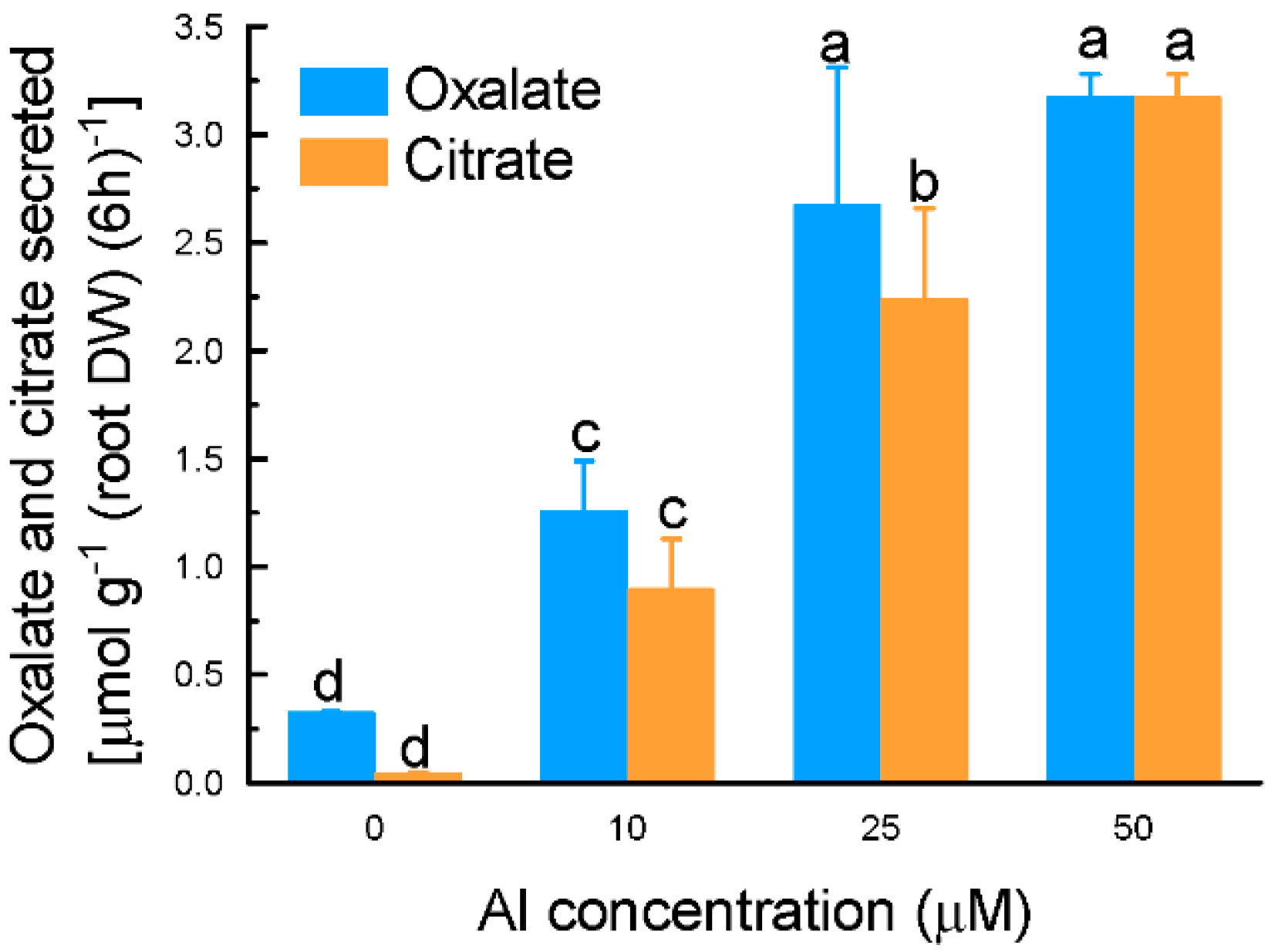
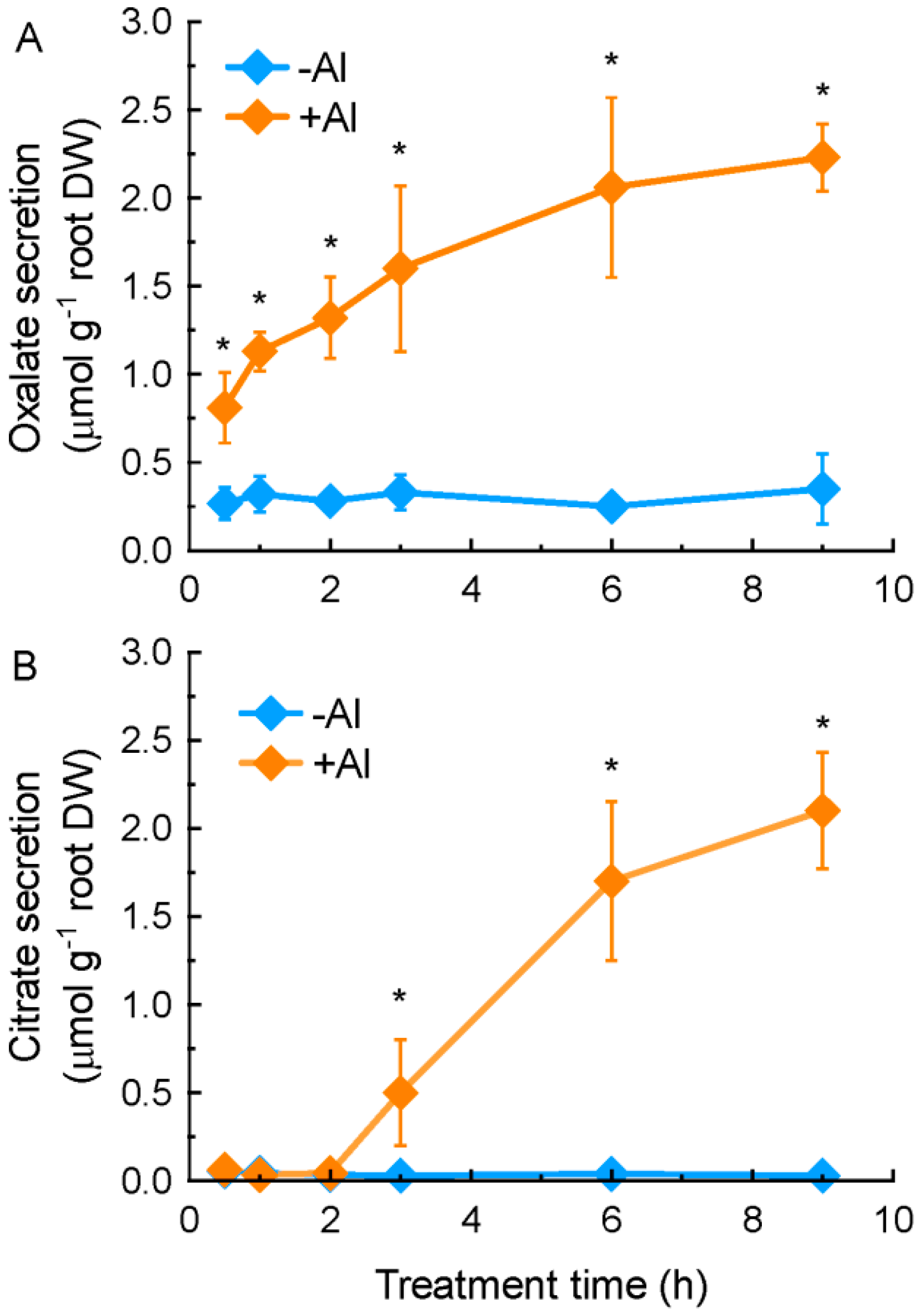
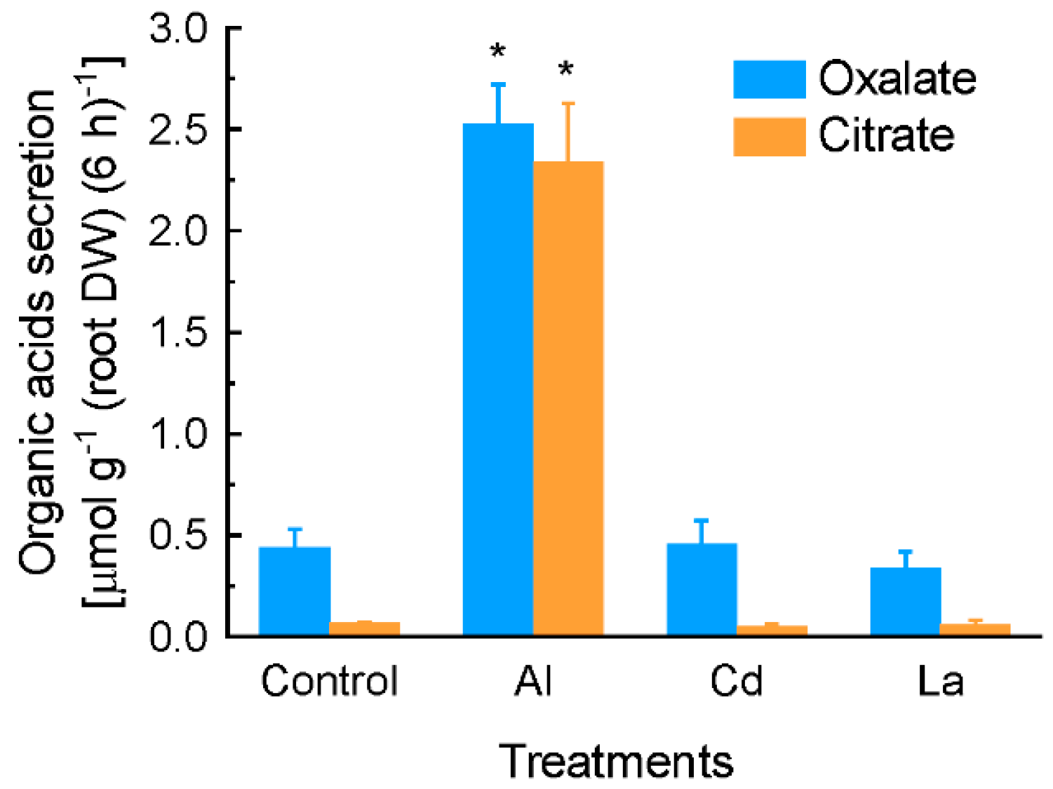
2.1. Characterization of Aluminium (Al)-Induced Organic Acid Anions (OA) Secretion from Grain Amaranth Roots

We have previously demonstrated that some oxalate accumulators can secrete oxalate from their roots in response to Al stress [22]. In this present study, we characterized secreted OA profiles and patterns in an oxalate accumulator, grain amaranth (Amaranthus hypochondriacus L.). Al treatment rapidly (within 30 min of exposure) stimulated the secretion of oxalate from grain amaranth roots (Figure 1A), although oxalate was detected in the root exudates of grain amaranth without Al treatment (Figure S1). During the entire treatment period, the secretion rate of oxalate was at similar levels (Figure 1A). On the other hand, citrate was not detected in the root exudates until 3 h of exposure to 25 µM Al. The amount of secreted citrate was significantly increased after 6 h of exposure. In the absence of Al stress, citrate was almost undetectable (Figure 1B).
The secretion of both oxalate and citrate was in a likely dose-dependent manner. However, there is no significant difference in secreted oxalate between 25 and 50 µM Al treatments (Figure 2).
When grain amaranth roots were exposed to lanthanum chloride (La) or cadmium chloride (Cd) stress for 6 h, both oxalate and citrate secretion were not induced. In contrast, exposure of roots to 10 µM Al induced secretion of both oxalate and citrate significantly (Figure 3).

2.2. Identification of up-Regulated Genes by Al Stress in Amaranth Roots

In order to identify genes potentially associated with Al-induced OA secretion, a forward cDNA library (9 h Al treatment vs. control) was constructed. A total of 84 differentially expressed transcripts were obtained, and were grouped into several functional categories according to the Gene Ontology (GO) biological process defined by the TAIR and UniProt database (Figure S2). Among these functional categories, the one that encompasses genes involved in “metabolism and energy” is most predominant. For example, genes encoding malate dehydrogenase, succinate dehydrogenase, and citrate synthase were observed (Table S1). In addition, up-regulation of eight genes involved in hormone metabolism and signaling transduction pathways confirming the recent observation that hormone may partly share Al signal to regulate Al tolerance mechanism such as OA secretion [32,33]. However, this present study was restricted to the genes which could be potentially involved in the transport processes of OA. Therefore, a total of nine genes involved in ion transport process were selected for further. Among these candidates, genes encoding ADP, ATP carrier protein, plasma membrane intrinsic protein (PIP2;1), and inorganic phosphate transporter were excluded, because they have somewhat defined functions that are not associated with OA secretion (Table S1). Finally, six genes including one gene encoding a multidrug and toxic compound extrusion (AhMATE1) protein, one gene encoding tonoplast dicarboxylate transporter (AhTDT), and four genes encoding ATP-binding cassette (ABC) transporters (AhABCG11, AhABCG21, AhABCA2, and AhABCB21, respectively) were chosen for further expression analysis by qRT-PCR (Table 1).

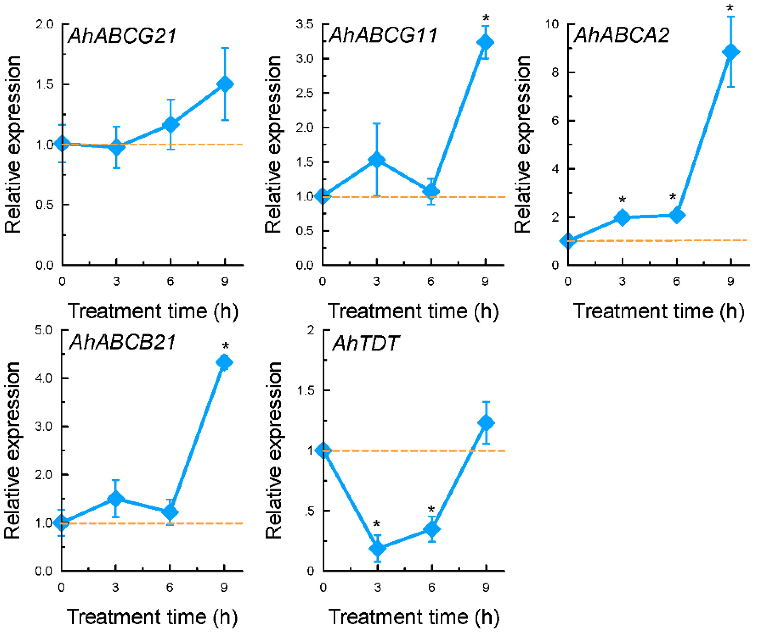
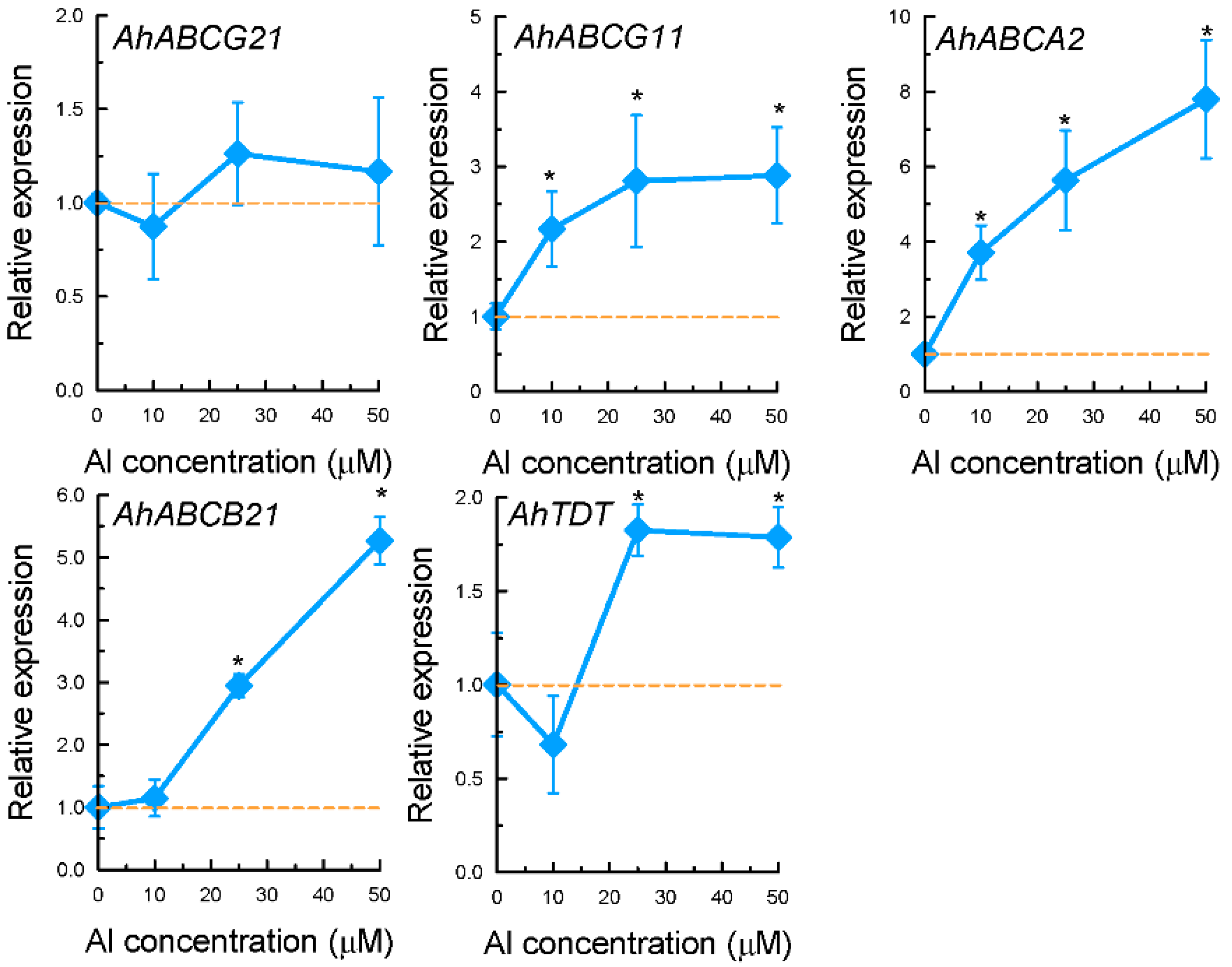
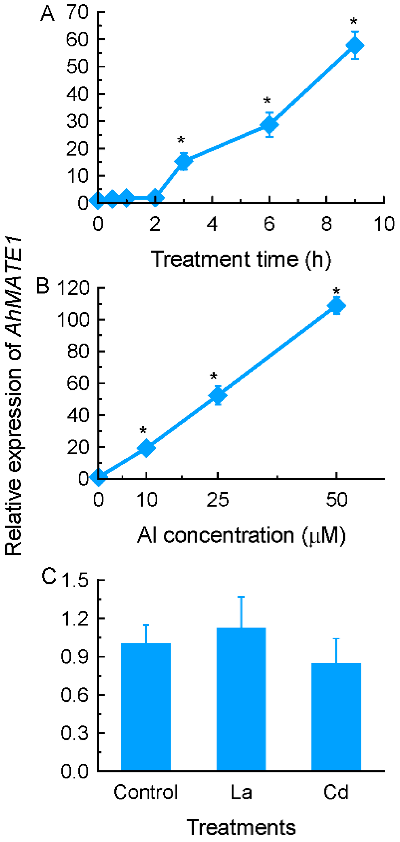
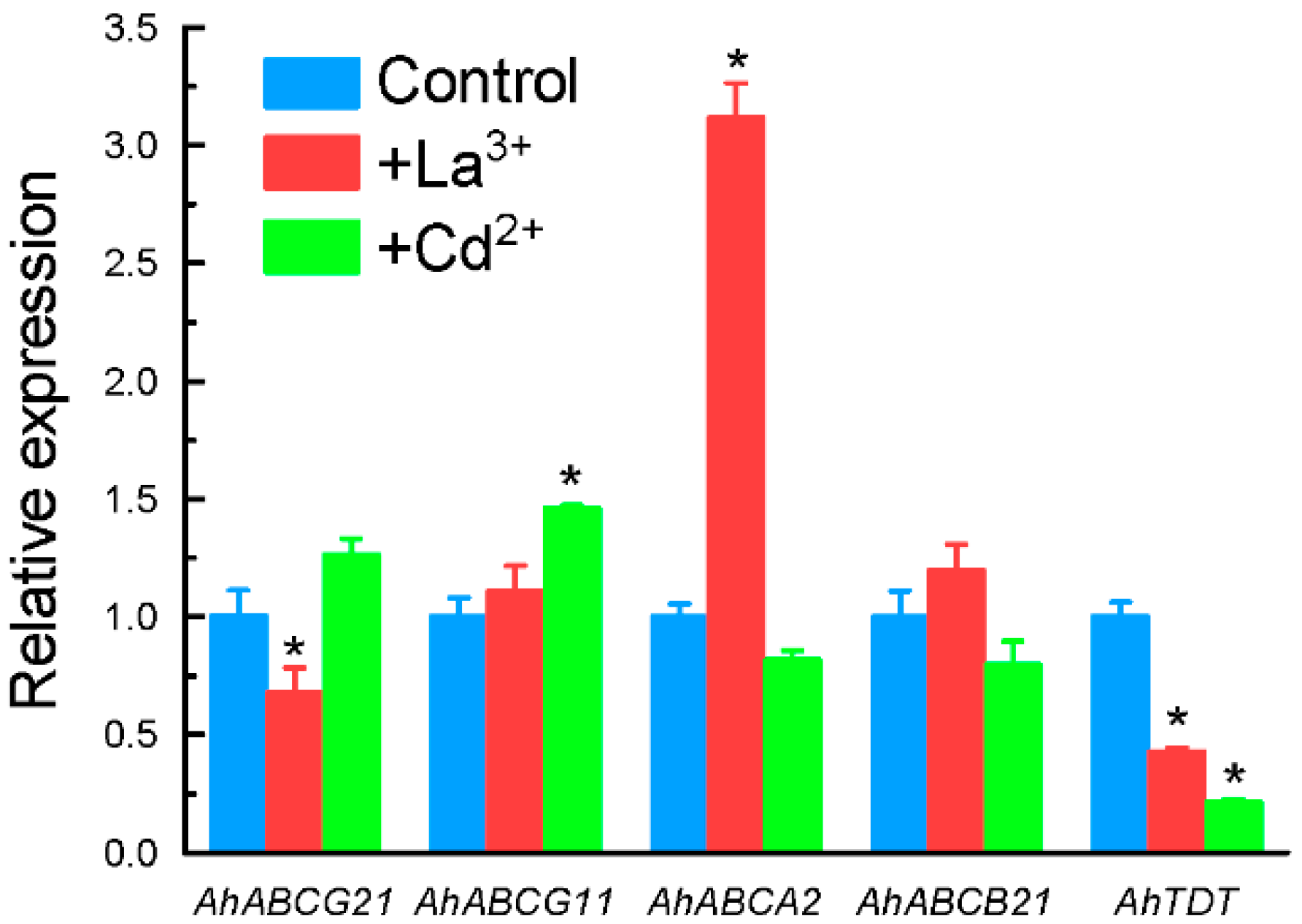
2.3. Expression Patterns of Selected Genes Encoding Transporter Proteins
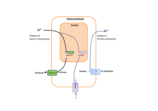
A subgroup of MATE proteins has been reported to be associated with Al-activated citrate transporter in some plant species [11]. Thus, it is likely that the gene encoding MATE protein could be involved in Al-induced citrate secretion from grain amaranth roots. In order to verify whether AhMATE1 is possibly involved in Al-induced citrate secretion, we characterized the expression pattern of AhMATE1. The expression of AhMATE1 is in a time- and dose-dependent manner in response to Al stress (Figure 4A,B), which is in line with citrate secretion pattern (Figure 1 and Figure 2). However, its expression was not affected by La or Cd stress (Figure 4C).
Among the four genes encoding ATP-binding cassette (ABC) transporters, the expression of AhABCG21 was not significantly affected by Al stress in both time-course and dose-response experiments (Figure 5 and Figure 6). The expression of AhABCG11 and AhABCB21 were only significantly increased after 9 h of exposure, whereas that of AhABCA2 was increased within 3 h of exposure (Figure 5). The expression of both AhABCA2 and AhABCB21 displayed dose-dependent increase in response to Al stress (Figure 6). The expression of AhABCG11, however, reached the maximum at 10 µM Al, and cannot be further increased with increased Al concentrations (Figure 6). The expression of these four genes was not affected by either La or Cd stress with the exception of AhABCA2, which was greatly induced by La treatment, and AhABCG21, which was inhibited by La (Figure 7).
The expression of a gene encoding tonoplast dicarboxylate transporter, AhTDT, was observed to be down-regulated within 6 h of exposure to 25 µM Al, and slightly increased after 9 h of exposure (Figure 5). Compared to control conditions, exposure to Al at 10 µM for 9 h slightly inhibited the expression of AhTDT, and higher Al concentrations resulted in a slight increase in expression levels (Figure 6). Both La and Cd significantly inhibited its expression (Figure 7).

3. Discussion

Many plant species secrete OA from roots to neutralize Al toxicity [4,34]. Here we characterized the secretion of OA from grain amaranth roots under Al stress, and some unique features are highlighted. First, roots of grain amaranth secrete both oxalate and citrate in response to Al stress (Figure 1 and Figure 2). While some plant species secrete only a single type of OA into the rhizosphere under Al stress, others secrete two types of OA simultaneously. For example, Al-induced specific secretion of citrate in Cassia tora [35], malate in wheat [36], and oxalate in buckwheat [13] was observed. Both citrate and malate were secreted from rye (Secale cereale) roots when exposed to Al stress [37]. Evidence for a similar situation has been demonstrated in Arabidopsis [38] and some Al-tolerant wheat genotypes [39]. However, this study is the first report on simultaneous secretion of oxalate and citrate from grain amaranth roots. Second, the secretion pattern differed between oxalate and citrate. Based on the timing of secretion, OA secretion can be grouped into two patterns [34,40]. In pattern I, organic acids were immediately secreted after the beginning of Al stress, whereas in the pattern II, an induction period ranging from several hours to several days are required for the initiation of OA secretion. The rapid secretion and similar secretion rate of oxalate during the entire exposure period indicated that oxalate secretion from grain amaranth roots fits pattern I response (Figure 1A), whereas increased secretion rate of citrate over time indicated that it belongs to pattern II response (Figure 1B). Increased evidence showed that patterns I and II operate independently during activated and regulated process [34], suggesting complexity in secretion of OA from grain amaranth in response to Al. Thirdly, the amount of Al-induced oxalate secretion is far more than citrate secretion at 2 h, but both of them were similar almost while treated Al more than 6 h (Figure 1), suggesting that the key role of oxalate in detoxifying Al toxicity at early stage. Finally, the secretion of both oxalate and citrate exhibited specificity to Al stress since other metals failed to stimulate the secretion (Figure 3). In tomato, La stress failed to induce the secretion of oxalate [41]. Cd stress, however, stimulated the secretion of oxalate in a time- and dose-dependent manner [42]. In both Cassia tora and rice bean, Al specifically induced citrate secretion from their roots [35,43]. The specificity of secretion to Al stress indicates that secretion of both OA from grain amaranth roots is regulated by specific transport pathways rather than by a common stress response.
Using the SSH method, we have identified a total of 84 genes that are up-regulated by Al stress in grain amaranth roots (Table S1). These genes were functionally categorized into several physiological and molecular events (Figure S2). For example, Al stress triggered the expression of genes involved in “metabolism and energy” which are most predominant in our library. Similarly, a previous study in rice bean suggested that metabolic changes act as an adaptive mechanism for plants to deal with Al toxicity [31]. The importance of mitochondrial metabolism in Al resistance has recently been summarized [44]. Here, up-regulation of a set of genes involved in tricarboxylic acid cycle points to the possible contribution of citrate metabolism to its secretion (Table S1). However, the key genes encoding enzymes that are involved in the oxalate biosynthesis are still poorly understood. It has been reported that oxalate content is not related to the amount of secretion in amaranth plants, because its content is far more than secreted [22]. Recent genome-wide transcriptome analysis of Al-responsive genes in common buckwheat and tartary buckwheat also found that oxalate metabolism is not a limiting factor of Al-induced oxalate secretion [9,10].
Our data showed that a gene-encoding MATE protein, AhMATE1, is a potential citrate transporter in grain amaranth. MATE proteins represent a large family of transporters in prokaryotes and eukaryotes [45]. For example, there are 58 and at least 40 MATE orthologs in Arabidopsis and rice, respectively. The role of MATE proteins in Al resistance has been reported in a number of plant species such as sorghum (Sorghum bicolor) [46], barley (Hordeum vulgare) [47], wheat [39], rice (Oryza sativa) [48], Arabidopsis [38], rice bean [49], and Eucalyptus camaldulensis [50]. It has been concluded that citrate-transporting MATE proteins are characteristic of having 12 transmembrane domains and one highly conserved amino acid sequence in the cytosolic loop between the second and third transmembrane domains [49]. Amplification of AhMATE1 full length by RACE PCR revealed that it shares all conserved structures with other known citrate transporters (Data not shown). Furthermore, the expression pattern of AhMATE1 fit the secretion pattern of citrate. Therefore, it is very likely that this gene is responsible for Al-induced citrate secretion from grain amaranth roots.
Among 9 cDNA clones identified as ion transporters, nearly half of them belong to the family of ABC transporters (Table S1). ABC transporter represents a very large gene family. Members of this family can be found in all taxa, and shuttle a broad range of substrates such as lipids, phytohormones, carboxylates, heavy metals, chlorophyll catabolites and xenobiotic conjugates [51]. In fact, experimental evidence has pointed to the role of ABC transporters in transporting OA in plants. For example, AtABCB14 has been reported to be implicated in transporting of malate from apoplast into guard cells [52]. Knockout of AtPDR6, a member of the pleiotropic drug resistance protein (PDR) subfamily of ABC transporters, resulted in significant decrease of OA (3-hydroxypropionic acid, succinic acid, fumaric acid, and malic acid) in Arabidopsis root exudates [53]. These results raised the question as to whether ABC transporter is involved in Al-dependent oxalate secretion. In the present study, although both AhABCB21 and AhABCA2 exhibited at least partly time- and dose-dependent expression patterns in response to Al stress (Figure 5 and Figure 6), they did not correlate well with the pattern of Al-dependent oxalate secretion. In addition, the expression of AhABCA2 was also significantly induced by La stress. In wheat, the induction of TaMDR1, a member of ABC transporters, was demonstrated to be associated with Al toxicity, and some calcium channel inhibitors including La could induce the expression of TaMDR1 [54]. It is possible that up-regulation of AhABCA2 might be involved in Al toxicity rather than Al resistance.
While whether ABC transporters are involved in Al-induced OA secretion remains unknown, emerging evidence has suggested the importance of this gene family in other plant Al resistance mechanisms. In a screening to identify genes involved in Arabidopsis Al resistance, both AtALS1 and AtALS3 have been found to contribute to Al detoxification. AtALS1 is a tonoplast-localized half-type ABC transporter, and predicted to function to transport chelated Al into vacuoles [55]. However, AtALS3 encodes a transmembrane domain of a bacterial ABC transporter, and may function to redistribute accumulated Al away from sensitive tissues [56]. Huang et al. (2009) identified two rice genes, OsSTAR1 and OsSTAR2, with the former encoding the nucleotide-binding domain and the later encoding the transmembrane domain of one ABC transporter [57]. Interestingly, OsSTAR1 and OsSTAR2 directly interact with each other to form a functional transporter complex, which possibly transports UDP-glucose to mask cell wall composition to protect the binding of Al [57]. In the present study, expression of some ABC transporter genes in response to Al stress was regulated, suggesting that these genes should have roles in adaptation of grain amaranth to Al toxicity. However, the detailed function of each member of the ABC transporters identified in this study has to be characterized in the future study.
In the present study, the expression of a gene encoding tonoplast dicarboxylate transporter was examined. Oxalate is the simplest dicarboxylate. Thus, transporters involved in oxalate secretion are permeable to dicarboxylates. Ryan and Delhaize (2010) hypothesized that transporters responsible for Al-induced malate or citrate secretion might be evolved from other malate or citrate transporters that originally performed different functions [58]. It is therefore likely that mutations might occur in the coding region of the tonoplast dicarboxylate transporter which alters its subcellular localization and functions. Although this EST was enriched in our forward library, the expression of this gene was only slightly increased at higher Al concentrations with relatively long exposure time (Figure 6). The inconsistency between Al-induced oxalate secretion pattern and the expression pattern of AhTDT precludes it being a potential candidate for oxalate transporter. However, it remains possible that Al mediates oxalate secretion through post-translational regulation of the oxalate transporter gene. For example, in both wheat and barley, TaALMT1 and HvAACT1 are constitutively highly expressed in Al-resistant genotypes, and Al directly or indirectly activates their functions [28,47].

4. Materials and Methods

4.1. Plant Materials and Growth Conditions

Grain amaranth (Amaranthus hypochondriacus L.) seeds were soaked in de-ionized water overnight. Then the seeds were placed on the filter paper moistened with 0.5 mM CaCl2 solution at pH 4.5 and kept in the dark at 26 °C. Germinated seeds were transferred to a net tray, which was floated on a 5 L of 0.5 mM CaCl2 solution at pH 4.5 in a plastic container. The seedlings were kept in the dark at 26 °C for 7 days, and then transferred to a controlled-environment growth room with a 14 h/26 °C day at a light intensity of 300 μmol·photons·m−2·s−1 and 10 h/22 °C night regime. The solution was renewed daily. At day 4, the seedlings were transplanted into 1.2 L plastic pots (four holes per pot, 2 seedlings for each hole) containing aerated nutrient solution. One-fifth-strength Hoagland nutrient solution was used consisting of the following macronutrients in mM: KNO3, 1.0; Ca(NO3)2, 1.0; MgSO4, 0.4; NH4H2PO4, 0.2, and the following micronutrients in μM: NaFeEDTA, 20; H3BO3, 3; MnCl2, 0.5; CuSO4, 0.2; ZnSO4, 0.4; (NH4)6Mo7O24, 1. The solution was adjusted to pH 4.5 with 1 M HCl and renewed every 3 days.

4.2. Treatments

Three-week old seedlings were subjected to the following treatments. Prior to each treatment, the roots were rinsed by soaking in aerated 0.5 mM CaCl2 solution (pH 4.5) overnight. For the time-course experiment, roots of seedling were exposed to 0.5 mM CaCl2 solution (pH 4.5) with or without 25 μM AlCl3 for 0.5, 1, 2, 3, 6, or 9 h. For the dose-response experiment, roots of seedling were exposed to 0.5 mM CaCl2 solution (pH 4.5) containing 0, 10, 25, or 50 μM AlCl3 for 6 or 9 h. For LaCl3 or CdCl2 treatment, seedlings were exposed to 0.5 mM CaCl2 solution (pH 4.5) containing 25 μM LaCl3 or 10 μM CdCl2 for 6 or 9 h. After treatments, root exudates were collected and immediately used to analyze organic acids, and root tissues were frozen immediately in liquid N2 and stored in −80 °C refrigerator until use.

4.3. Collection of Root Exudates

Collected root exudates were passed through a cation-exchange resin column (16 mm × 14 cm) filled with 5 g of Amberlite IR-102 B resin (H+ form), followed by an anion-exchange resin column (16 mm × 14 cm) filled with 2 g of Dowex 1 × 8 resin (100–200 mesh, formate form). Organic acids retained on the anion-exchange resin were eluted with 15 mL of 1 M HCl, and the eluate was concentrated to dryness by a rotary evaporator (40 °C). The residue was dissolved in 2 mL of Milli-Q water and subjected to organic acid determination.

4.4. Measurement of Oxalate and Citrate

Oxalic acid was determined by ion chromatography using an IonPac AS11 anion-exchange analytical column (4 mm × 250 mm) equipped with a guard column (4 mm × 50 mm). The mobile phase was 30 mM NaOH at a flow rate of 0.6 mL·min−1. A standard curve was made by analyzing different concentrations of oxalic acid with the same procedure. Citric acid was determined enzymatically [59]. In briefly, to 1 mL of sample solution, 120 µL 1 M Tris/HCl buffer (pH 7.8) and 15 µL 10 mM NADH were added. After incubation at 25 °C for 40 min, 2 µL enzyme mixture (1.25 U lactate dehydrogenase and 0.5 U malate dehydrogenase) was added and the reaction mixture was incubated for an additional 40 min. the change in A340 was monitored after the reaction was initiated with addition of citrate lyase (0.5 U).

4.5. RNA Isolation and Construction of SSH Library

Total RNA was extracted using the RNAeasy mini kit (Tiangen, Shanghai, China). cDNA was amplified with Super SMART™ PCR cDNA synthesis Kit (Clontech, Palo Alto, CA, USA). For identification of up-regulated genes, forward subtracted cDNA library was constructed. For this, amplified cDNA from 25 μM Al-treated for 9 h roots was used as the “tester”, while amplified cDNA from control as the “driver”. PCR-select cDNA subtraction was performed according to instruction of PCR-Select™ cDNA Subtraction Kit (Clontech, Palo Alto, CA, USA). The PCR products were sub-cloned into the pGEM-Teasy vector (Promega, Madison, WI, USA) and then transformed into chemically competent E. coli (DH5α) cells. Transformed clones were grown on LB plates containing X-gal and ampicillin for blue/white screening. Positive clones were selected and further checked by PCR for the presence of gene inserts after plasmid isolation, and clones with >200 bp single fragment insert were selected for DNA sequencing.

4.6. Sequence Homology and Functional Annotation

The cDNA sequences were compared with the GenBank database using the online Basic Local Alignment Search Tool (BLAST) program [60]. The detailed search procedure was the same as our previous report [31].

4.7. Quantitative Real-Time PCR

Total RNA was isolated from root tissues as described above. First-strand cDNA was synthesized from 1 µg of total RNA using Superscript™ reverse transcriptase (Takara, Dalian, China). 1 μL (100 ng·μL−1) of cDNA in 10 μL volume system were used for quantitative analysis of gene expression performed with SYBR Premix ExTaq (Takara, Dalian, China). The primer pairs for each gene were listed in Table 1, and the PCR amplification conditions were as follows: 94 °C for 5 min; 45 cycles of 94 °C for 10 s, 55 °C for 15 s and 72 °C for 20 s. The endogenous gene glyceraldehyde-3-phosphate dehydrogenase (GAPDH) was used as the internal control with the following primers: 5′-TTCGGTCACAGGAACCCAGA-3′ and 5′-ACCTTCTTGGCACCACCCTT-3′. For each target gene, the PCR reactions were carried out in three biological and technical repeats. The relative expression level was calculated by formula 2–∆∆Cp.

4.8. Statistical Analysis

Each result shown in the figures was the mean of three replicated treatments, and at least two independent experiments were conducted for each result. The significant differences between treatments were statistically evaluated by Students’s t or Tukey test method.

5. Conclusions

We characterized Al-induced OA secretion from grain amaranth roots and observed simultaneous secretion of oxalate and citrate in response to Al stress with the secretion pattern differed. A total of 84 genes up-regulated by Al stress were identified in grain amaranth roots, and several genes encoding transporter proteins were highlighted. Although identification of oxalate transporter remains a great challenge, the present results provide some genetic information that will be useful for further characterization of genes involved in plant Al tolerance and toxicity mechanisms.

Supplementary Materials

Supplementary materials can be found at https://www.mdpi.com/1422-0067/17/4/608/s1.

Acknowledgments

This work was supported by the National Key Basic Research Program of China (2014CB441002), the National Natural Science Foundation of China (31222049, 31501827), and the Fundamental Research Funds for the Central Universities.

Author Contributions

Conceived and designed the experiments: Wei Fan and Jian-Li Yang. Performed the experiments: Wei Fan, Jia-Meng Xu, He-Qiang Lou, Chuan Xiao, Wei-Wei Chen. Analyzed the data: Wei Fan, Jian-Li Yang. Wrote the paper: Wei Fan and Jian-Li Yang.

Conflicts of Interest

The authors declare no conflict of interest.

References

  1. Kochian, L.V. Cellular mechanisms of aluminum toxicity and resistance in plants. Annu. Rev. Plant Physiol. Plant Mol. Biol. 1995, 46, 237–260. [Google Scholar] [CrossRef]
  2. Barcelo, J.; Poschenrieder, C. Fast root growth responses, root exudates, and internal detoxification as clues to the mechanisms of aluminium toxicity and resistance: A review. Environ. Exp. Bot. 2002, 48, 75–92. [Google Scholar] [CrossRef]
  3. Matsumoto, H. Cell biology of aluminum toxicity and tolerance in higher plants. Int. Rev. Cytol. 2000, 200, 1–46. [Google Scholar] [PubMed]
  4. Kochian, L.V.; Hoekenga, O.A.; Piñeros, M.A. How do crop plants tolerate acid soils? Mechanisms of aluminum tolerance and phosphorus efficiency. Annu. Rev. Plant Biol. 2004, 55, 459–493. [Google Scholar] [CrossRef] [PubMed]
  5. Ma, J.F. Syndrome of aluminum toxicity and diversity of aluminum resistance in higher plants. Int. Rev. Cytol. 2007, 264, 225–252. [Google Scholar] [PubMed]
  6. Kumari, M.; Taylor, G.J.; Deyholos, M.K. Transcriptomic responses to aluminum stress in roots of Arabidopsis thaliana. Mol. Genet. Genom. 2008, 279, 339–357. [Google Scholar] [CrossRef] [PubMed]
  7. Goodwin, S.B.; Sutter, T.R. Microarray analysis of Arabidopsis genome response to aluminum stress. Biol. Plant. 2009, 53, 85–99. [Google Scholar] [CrossRef]
  8. Tsutsui, T.; Yamaji, N.; Huang, C.F.; Motoyama, R.; Nagamura, Y.; Ma, J.F. Comparative genome-wide transcriptional analysis of Al-responsive genes reveals novel Al tolerance mechanisms in rice. PLoS ONE 2012, 7, e48197. [Google Scholar] [CrossRef] [PubMed]
  9. Yokosho, K.; Yamaji, N.; Ma, J.F. Global transcriptome analysis of Al-induced genes in an Al-accumulating species, common buckwheat (Fagopyrum esculentum Moench). Plant Cell Physiol. 2014, 55, 2077–2091. [Google Scholar] [CrossRef] [PubMed]
  10. Zhu, H.; Wang, H.; Zhu, Y.; Zou, J.; Zhao, F.J.; Huang, C.F. Genome-wide transcriptomic and phylogenetic analyses reveal distinct aluminum-tolerance mechanisms in the aluminum-accumulating species buckwheat (Fagopyrum tataricum). BMC Plant Biol. 2015, 15, 16. [Google Scholar] [CrossRef] [PubMed]
  11. Delhaize, E.; Ma, J.F.; Ryan, P.R. Transcriptional regulation of aluminium tolerance genes. Trends Plant Sci. 2012, 17, 341–348. [Google Scholar] [CrossRef] [PubMed]
  12. Ma, Z.; Miyasaka, S.C. Oxalate exudation by taro in response to Al. Plant Physiol. 1998, 118, 861–865. [Google Scholar] [CrossRef] [PubMed]
  13. Zheng, S.J.; Ma, J.F.; Matsumoto, H. High aluminum resistance in buckwheat I. Al-induced specific secretion of oxalic acid from root tips. Plant Physiol. 1998, 117, 745–751. [Google Scholar] [CrossRef]
  14. Yang, J.L.; Zheng, S.J.; He, Y.F.; Matsumoto, H. Aluminium resistance requires resistance to acid stress: A case study with spinach that exudes oxalate rapidly when exposed to Al stress. J. Exp. Bot. 2005, 56, 1197–1203. [Google Scholar] [CrossRef] [PubMed]
  15. You, J.F.; He, Y.F.; Yang, J.L.; Zheng, S.J. A comparison of aluminum resistance among Polygonum species originating on strongly acidic and neutral soils. Plant Soil. 2005, 276, 143–151. [Google Scholar] [CrossRef]
  16. Morita, A.; Yanagisawa, O.; Maeda, S.; Takatsu, S.; Ikka, T. Tea plant (Camellia sinensis L.) roots secrete oxalic acid and caffeine into medium containing aluminum. Soil Sci. Plant Nutr. 2011, 57, 796–802. [Google Scholar] [CrossRef]
  17. Brenner, D.M.; Baltensperger, D.D.; Kulakow, P.A.; Lehmann, J.W.; Myers, R.L.; Slabbert, M.M.; Sleugh, B.B. Genetic resources and breeding of Amaranthus. Plant Breed. Rev. 2000, 19, 227–285. [Google Scholar]
  18. Miller, T.E.; Wing, J.S.; Huete, A.R. The agricultural potential of selected C4 plants in arid environments. J. Arid Environ. 1984, 7, 275–286. [Google Scholar]
  19. Johnson, B.L.; Henderson, T.L. Water use patterns of grain amaranth in the northern Great Plains. Agron. J. 2002, 94, 1437–1443. [Google Scholar] [CrossRef]
  20. Huerta-Ocampo, J.A.; LeÓn-Galván, M.F.; Ortega-Cruz, L.B.; Barrera-Pacheco, A.; de León-Rodríguez, A.; Mendoza-Hernández, G.; de la Rosa, A.P. Water stress induces up-regulation of DOF1 and MIF1 transcription factors and down-regulation of proteins involved in secondary metabolism in amaranth roots (Amaranthus hypochondriacus L.). Plant Biol. 2011, 13, 472–482. [Google Scholar] [CrossRef] [PubMed]
  21. Délano-Frier, J.P.; Martínez-Gallardo, N.A.; Martínez-de la Vega, O.; Salas-Araiza, M.D.; Barbosa-Jaramillo, E.R.; Torres, A.; Vargas, P.; Borodanenko, A. The effect of exogenous jasmonic acid on induced resistance and productivity in amaranth (Amaranthus hypochondriacus) is influenced by environmental conditions. J. Chem. Ecol. 2004, 30, 1001–1034. [Google Scholar] [CrossRef] [PubMed]
  22. Yang, J.L.; Zheng, L.; Zheng, S.J. Aluminum-activated oxalate secretion does not associate with internal content among some oxalate accumulators. J. Integr. Plant Biol. 2008, 50, 1103–1107. [Google Scholar] [CrossRef] [PubMed]
  23. Zhang, J.; He, Z.; Tian, H.; Zhu, G.; Peng, X. Identification of aluminium-responsive genes in rice cultivars with different aluminium sensitivities. J. Exp. Bot. 2007, 58, 2269–2278. [Google Scholar] [CrossRef] [PubMed]
  24. Mao, C.; Yi, K.; Yang, L.; Zheng, B.; Wu, Y.; Liu, F.; Wu, P. Identification of aluminium-regulated genes by cDNA-AFLP in rice (Oryza sativa L.): Aluminium-regulated genes for the metabolism of cell wall components. J. Exp. Bot. 2004, 55, 137–143. [Google Scholar] [CrossRef] [PubMed]
  25. Maron, L.G.; Kirst, M.; Mao, C.; Milner, M.J.; Menossi, M.; Kochian, L.V. Transcriptional profiling of aluminum toxicity and tolerance responses in maize roots. New Phytol. 2008, 179, 116–128. [Google Scholar] [CrossRef] [PubMed]
  26. Watt, D.A. Aluminium-responsive genes in sugarcane: Identification and analysis of expression under oxidative stress. J. Exp. Bot. 2003, 54, 1163–1174. [Google Scholar] [CrossRef] [PubMed]
  27. Arenhart, R.A.; Bai, Y.; de Oliveira, L.F.; Neto, L.B.; Schunemann, M.; Maraschin Fdos, S.; Mariath, J.; Silverio, A.; Sachetto-Martins, G.; Margis, R.; et al. New insights into aluminum tolerance in rice: ASR5 protein binds in the STAR1 promoter and other aluminum responsive genes. Mol. Plant 2014, 7, 709–721. [Google Scholar] [CrossRef] [PubMed]
  28. Sasaki, T.; Yamamoto, Y.; Ezaki, B.; Katsuhara, M.; Ahn, S.J.; Ryan, P.R.; Delhaize, E.; Matsumoto, H. A wheat gene encoding an aluminum-activated malate transporter. Plant J. 2004, 37, 645–653. [Google Scholar] [CrossRef] [PubMed]
  29. Eticha, D.; Zahn, M.; Bremer, M.; Yang, Z.; Rangel, A.F.; Rao, I.M.; Horst, W.J. Transcriptomic analysis reveals differnetial gene expression in response to aluminium in common bean (Phaseolus vulgaris) genotypes. Ann. Bot. 2010, 105, 1119–1128. [Google Scholar] [CrossRef] [PubMed]
  30. Chen, Q.; Zhang, X.D.; Wang, S.S.; Wang, Q.F.; Wang, G.Q.; Hian, H.J.; Li, K.Z.; Yu, Y.X.; Chen, L.M. Transcriptional and physiological changes of alfalfa in response to aluminium stress. J. Agric. Sci. 2011, 149, 737–751. [Google Scholar] [CrossRef]
  31. Fan, W.; Lou, H.Q.; Gong, Y.L.; Liu, M.Y.; Wang, Z.Q.; Yang, J.L.; Zheng, S.J. Identification of early Al-responsive genes in rice bean (Vigna umbellata) roots provides new clues to molecular mechanism of Al toxicity and tolerance. Plant Cell Environ. 2014, 37, 1586–1597. [Google Scholar] [CrossRef] [PubMed]
  32. Kobayashi, Y.; Kobayashi, Y.; Sugimoto, M.; Lakshmanan, V.; Luchi, S.; Kobayashi, M.; Bais, H.P.; Koyama, H. Characterization of the complex regulation of AtALMT1 expression in response to phytohormones and other inducers. Plant Physiol. 2013, 162, 732–740. [Google Scholar] [CrossRef] [PubMed]
  33. Tian, Q.; Zhang, X.; Ramesh, S.; Gilliham, M.; Tyerman, S.D.; Zhang, W.H. Ethylene negatively regulates aluminium-induced malate efflux from wheat roots and tobacco cells transformed with TaALMT1. J. Exp. Bot. 2014, 65, 2415–2426. [Google Scholar] [CrossRef] [PubMed]
  34. Ma, J.F.; Ryan, P.R.; Delhaize, E. Aluminium tolerance in plants and the complexing role of organic acids. Trends Plant Sci. 2001, 6, 273–278. [Google Scholar] [CrossRef]
  35. Ma, J.F.; Zheng, S.J.; Matsumoto, H. Specific secretion of citric acid induced by Al stress in Cassia tora L. Plant Cell Physiol. 1997, 38, 1019–1025. [Google Scholar] [CrossRef]
  36. Delhaize, E.; Ryan, P.R.; Randall, P.J. Aluminum tolernace in wheat (Triticum aestivum L.) II. Aluminum-stimulated excretion of malic acid from root apices. Plant Physiol. 1993, 103, 695–702. [Google Scholar] [PubMed]
  37. Li, X.F.; Ma, J.F.; Matsumoto, H. Pattern of aluminum-induced secretion of organic acids differs between rye and wheat. Plant Physiol. 2000, 123, 1537–1544. [Google Scholar] [CrossRef] [PubMed]
  38. Liu, J.P.; Magalhaes, J.V.; Shaff, J.E.; Kochian, L.V. Aluminum-activated citrate and malate transporters from the MATE and ALMT families function independently to confer Arabidopsis aluminum tolerance. Plant J. 2009, 57, 389–399. [Google Scholar] [CrossRef] [PubMed]
  39. Ryan, P.R.; Raman, H.; Gupta, S.; Horst, W.J.; Delhaize, E. A second mechanism for aluminum resistance in wheat relies on the constitutive efflux of citrate from roots. Plant Physiol. 2009, 149, 340–351. [Google Scholar] [CrossRef] [PubMed]
  40. Ma, J.F. Role of organic acids in detoxification of aluminum in higher plants. Plant Cell Physiol. 2000, 41, 383–390. [Google Scholar] [CrossRef] [PubMed]
  41. Yang, J.L.; Zhu, X.F.; Peng, Y.X.; Zheng, C.; Ming, F.; Zheng, S.J. Aluminum regulates oxalate secretion and plasma membrane H+-ATPase activity independently in tomato roots. Planta 2011, 234, 281–291. [Google Scholar] [CrossRef] [PubMed]
  42. Zhu, X.F.; Zheng, C.; Hu, Y.T.; Jiang, T.; Dong, N.Y.; Yang, J.L.; Zheng, S.J. Cadmium-induced oxalate secretion from root apex is associated with cadmium exclusion and resistance in Lycopersicon esculentum. Plant Cell Environ. 2011, 34, 1055–1064. [Google Scholar] [CrossRef] [PubMed]
  43. Yang, J.L.; Zhang, L.; Li, Y.Y.; You, J.F.; Wu, P.; Zheng, S.J. Citrate transporters play a critical role in aluminium-stimulated citrate efflux in rice bean (Vigna umbellata) roots. Ann. Bot. 2006, 97, 579–584. [Google Scholar] [CrossRef] [PubMed]
  44. Nunes-Nesi, A.; Brito, D.S.; Inostroza-Blancheteau, C.; Fernie, A.R.; Araújo, W.L. The complex role of mitochondrial metabolism in plant aluminum resistance. Trends Plant Sci. 2014, 19, 399–407. [Google Scholar] [CrossRef] [PubMed]
  45. Omote, H.; Hiasa, M.; Matsumoto, T.; Otsuka, M.; Moriyama, Y. The MATE proteins as functional transporters of metabolic and xenobiotic organic cations. Trends Pharmacol. Sci. 2007, 11, 587–593. [Google Scholar]
  46. Magalhaes, J.V.; Liu, J.; Guimarães, C.T.; Lana, U.G.; Alves, V.M.; Wang, Y.H.; Schaffert, R.E.; Hoekenga, O.A.; Piñeros, M.A.; Shaff, J.E.; et al. A member of the multidrug and toxic compound extrusion (MATE) family confers aluminum tolerance in sorghum. Nat. Genet. 2007, 39, 1156–1161. [Google Scholar] [CrossRef] [PubMed]
  47. Furukawa, J.; Yamaji, N.; Wang, H.; Mitani, N.; Murata, Y.; Sato, K.; Katsuhara, M.; Takeda, K.; Ma, J.F. An aluminum-activated citrate transporter in barley. Plant Cell Physiol. 2007, 48, 1081–1091. [Google Scholar] [CrossRef] [PubMed]
  48. Yokosho, K.; Yamaji, N.; Ma, J.F. An Al-inducible MATE gene is involved in external detoxification of Al in rice. Plant J. 2011, 68, 1061–1069. [Google Scholar] [CrossRef] [PubMed]
  49. Yang, X.Y.; Yang, J.L.; Zhou, Y.; Piñeros, M.A.; Kochian, L.V.; Li, G.X.; Zheng, S.J. A de novo synthesis citrate transporter, Vigna umbellate multidrug and toxic compound extrusion, implicates in Al-activated citrate efflux in rice bean (Vigna umbellata) root apex. Plant Cell Environ. 2011, 34, 2138–2148. [Google Scholar] [CrossRef] [PubMed]
  50. Sawaki, Y.; Kihara-Doi, T.; Kobayashi, Y.; Nishikubo, N.; Kawazu, T.; Kobayashi, Y.; Koyama, H.; Sato, S. Characterization of Al-responsive citrate excretion and citrate-transporting MATEs in Eucalyptus camaldulensis. Planta 2012, 237, 979–989. [Google Scholar] [CrossRef] [PubMed]
  51. Martinoia, E.; Klein, M.; Geisler, M.; Bovet, L.; Forestier, C.; Kolukisaoglu, U.; Müller-Röber, B.; Schulz, B. Multifunctionality of plant ABC transporters—More than just detoxifiers. Planta 2002, 214, 345–355. [Google Scholar] [CrossRef] [PubMed]
  52. Lee, M.; Choi, Y.; Burla, B.; Kim, Y.Y.; Jeon, B.; Maeshima, M.; Yoo, J.Y.; Martinoia, E.; Lee, Y. The ABC transporter AtABCB14 is a malate importer and modulates stomatal response to CO2. Nat. Cell Biol. 2008, 10, 1227–1223. [Google Scholar] [CrossRef] [PubMed] [Green Version]
  53. Badri, D.V.; Loyola-Vargas, V.M.; Broeckling, C.D.; de-la-Peña, C.; Jasinski, M.; Sanelia, D.; Martinoia, E.; Sumner, L.W.; Banta, L.M.; Stermitz, F.; et al. Altered profile of secondary metabolites in the root exudates of Arabidopsis ATP-binding cassette transporter mutants. Plant Physiol. 2008, 146, 762–771. [Google Scholar] [CrossRef] [PubMed] [Green Version]
  54. Sasaki, T.; Ezaki, B.; Matsumoto, H. A gene encoding multidrug resistance (MDR)-like protein is induced by aluminum and inhibitors of calcium flux in wheat. Plant Cell Physiol. 2002, 43, 177–185. [Google Scholar] [CrossRef] [PubMed]
  55. Larsen, P.B.; Cancel, J.; Rounds, M.; Ochoa, V. Arabidopsis ALS1 encodes a root tip and stele localized half type ABC transporter required for root growth in an aluminum toxic environment. Planta 2007, 225, 1447–1458. [Google Scholar] [CrossRef] [PubMed]
  56. Larsen, P.B.; Geisler, M.J.B.; Jones, C.A.; Williams, K.M.; Cancel, J.D. ALS3 encodes a phloem-localized ABC transporter-like protein that is required for aluminum tolerance in Arabidopsis. Plant J. 2005, 41, 353–363. [Google Scholar] [CrossRef] [PubMed]
  57. Huang, C.F.; Yamaji, N.; Mitani, N.; Yano, M.; Nagamura, Y.; Ma, J.F. A bacterial-type ABC transporter is involved in aluminum tolerance in rice. Plant Cell 2009, 21, 655–667. [Google Scholar] [CrossRef] [PubMed]
  58. Ryan, P.R.; Delhaize, E. The convergent evolution of aluminium resistance in plants exploits a convenient currency. Funct. Plant Biol. 2010, 37, 275–284. [Google Scholar] [CrossRef]
  59. Zhao, Z.; Ma, J.F.; Sato, K.; Takeda, K. Differential Al resistance and citrate secretion in barley (Hordeum vulgare L.). Planta 2003, 217, 794–800. [Google Scholar] [CrossRef] [PubMed]
  60. BLAST®. Available online: http://blast.ncbi.nlm,nig.gov/Blast.cgi (accessed on 23 February 2016).
Figure 1. Time-course of aluminium (Al)-induced oxalic acid (A) and citric acid (B) secretion from grain amaranth roots. Three-week-old seedlings were exposed to 0.5 mM CaCl2 solution (pH 4.5) containing 0 or 25 µM Al. Root exudates were collected at indicated intervals for determination of organic acids. Data are means ± SD (n = 3). Asterisks indicate that mean values are significantly different between Al treatment and control (Student’s t test, p < 0.05).
Ijms 17 00608 g001
Figure 2. Effect of different Al concentrations on oxalic acid and citric acid secretion from grain amaranth roots. Three-week-old seedlings were exposed to 0.5 mM CaCl2 solution containing 0, 10, 25, or 50 µM Al for 6 h. Root exudates were collected for determination of organic acids. Data are means ± SD (n = 3). Different letters indicate statistically significant differences (Tukey test, p < 0.05).
Ijms 17 00608 g002
Figure 3. Effect of lanthanum chloride (La) and cadmium chloride (Cd) on the secretion of organic acids from grain amaranth roots. Three-week-old seedlings were exposed to 0.5 mM CaCl2 solution containing 25 µM Al, 25 µM La, 10 µM Cd, or without metals as control. Root exudates were collected by 6 h for determination of organic acids. Data are means ± SD (n = 3). Asterisks indicate that mean values are significantly different between treatment and control (Student’s t test, p < 0.05).
Ijms 17 00608 g003
Figure 4. Grain amaranth roots AhMATE1 gene expression analysis. (A) Time-course analysis of AhMATE1 expression treated with 25 µM aluminium (Al) after various periods by real-time PCR; (B) dose response analysis of AhMATE1 expression treated with different Al concentrations for 9 h; (C) expression of AhMATE1 in response to 25 μM La or 10 μM Cd for 9 h. Total RNA was extracted from roots and qRT-PCR was performed for each gene with glyceraldehyde-3-phosphate dehydrogenase (GAPDH) as an internal control. Data are means ± SD (n = 3). Asterisks indicate that mean values are significantly different between treatment and control (Student’s t test, p < 0.05).
Ijms 17 00608 g004
Figure 5. Temporal expression pattern of AhABCG21, AhABCG11, AhABCA2, AhABCB21, and AhTDT in response to 25 µM Al. Three-week-old seedlings were exposed to 0.5 mM CaCl2 solution (pH 4.5) containing 0 or 25 µM Al. Total RNA was extracted from roots and qRT-PCR was performed for each gene with GAPDH as an internal control. Data are means ± SD (n = 3). Asterisks indicate that mean values are significantly different between treatment and control (Student’s t test, p < 0.05). Dash line indicates expression level of 1.
Ijms 17 00608 g005
Figure 6. Dosage expression pattern of AhABCG21, AhABCG11, AhABCA2, AhABCB21, and AhTDT in response to 0, 10, 25, or 50 µM Al for 9 h. Three-week-old seedlings were exposed to 0.5 mM CaCl2 solution (pH 4.5) containing different concentrations of Al or not. Total RNA was extracted from roots and qRT-PCR was performed for each gene with GAPDH as an internal control. Data are means ± SD (n = 3). Asterisks indicate that mean values are significantly different between treatment and control (Student’s t test, p < 0.05). Dash line indicates expression level of 1.
Ijms 17 00608 g006
Figure 7. Effect of La and Cd treatment on expression of AhABCG21, AhABCG11, AhABCA2, AhABCB21, and AhTDT in grain amaranth roots. Three-week-old seedlings were exposed to 0.5 mM CaCl2 solution containing 25 µM La, 10 µM Cd for 9 h, or without metals as control. Total RNA was extracted from roots and qRT-PCR was performed for each gene with GAPDH as an internal control. Data are means ± SD (n = 3). Asterisks indicate that mean values are significantly different between treatment and control (Student’s t test, p < 0.05).
Ijms 17 00608 g007
Table 1. List of selected cDNA clones associated with ion transport processes and primer pairs used for qRT-PCR analysis.
Gene Name aAnnotationPrimer PairsAmplicon Size (bp)
AhABCG21ABC transporter G family member 21for: AGGTGACTTGCCTATGGAACT107
rev: TCGTAAGGGTAAGGATAAATG
AhABCG11ABC transporter G family member 11for: AAACACACTTTCTTCAATCCCAT205
rev: ACCCGTTATGATACCCATTAGAA
AhABCA2ABC transporter A family member 2for: ACATCGCAAGACAAGCCG116
rev: CCCCACATACCTGGCTCC
AhABCB21ABC transporter B family member 21for: TGCTATGGGGGAGAAGGT122
rev: AAAGGGGTATGGACGAAA
AhTDTTonoplast dicarboxylate transporterfor: TACAGCGACTTCCGACGACTA267
rev: ACAAGCAACAAAGAACACCCC
AhMATE1MATE proteinfor: GGTCCTTTGGTGCTCCTGC163
rev: CCACTGACACCCAAACGACAT
a Gene name was temporarily assigned to the selected cDNA clones according to their closest homologous genes.

