Review

Cyclic Nucleotide Signalling in Kidney Fibrosis

Pharmacology and Toxicology, University Regensburg, Regensburg 93053, Germany
*
Author to whom correspondence should be addressed.
Int. J. Mol. Sci. 2015, 16(2), 2320-2351; https://doi.org/10.3390/ijms16022320
Submission received: 24 October 2014 / Revised: 14 November 2014 / Accepted: 14 January 2015 / Published: 22 January 2015
(This article belongs to the Special Issue Signalling Molecules and Signal Transduction in Cells 2014)

Abstract

:
Kidney fibrosis is an important factor for the progression of kidney diseases, e.g., diabetes mellitus induced kidney failure, glomerulosclerosis and nephritis resulting in chronic kidney disease or end-stage renal disease. Cyclic adenosine monophosphate (cAMP) and cyclic guanosine monophosphate (cGMP) were implicated to suppress several of the above mentioned renal diseases. In this review article, identified effects and mechanisms of cGMP and cAMP regarding renal fibrosis are summarized. These mechanisms include several signalling pathways of nitric oxide/ANP/guanylyl cyclases/cGMP-dependent protein kinase and cAMP/Epac/adenylyl cyclases/cAMP-dependent protein kinase. Furthermore, diverse possible drugs activating these pathways are discussed. From these diverse mechanisms it is expected that new pharmacological treatments will evolve for the therapy or even prevention of kidney failure.

1. Renal Fibrosis and Involved Signalling Pathways

Cyclic nucleotides cyclic adenosine monophosphate (cAMP) and cyclic guanosine monophosphate (cGMP) are important second messengers regulating chronic kidney disease. These signalling molecules act in a different manner upon various chronic kidney diseases (CKDs) which result from diverse causes. Disorders which often generate CKD comprise diabetic nephropathy, glomerulosclerosis and nephritis.

1.1. Fibrotic Kidney Diseases

Renal fibrosis is commonly found in CKD, e.g., diabectic nephropathy, glomerulosclerosis and lupus nephritis. These CKDs can be caused by oxidative stress, hypoxia, inflammation, autoimmune disease or altered metabolism. Acute insult of the kidney by ischemia or toxins can also finally result in CKD. However, there are common disease patterns in various CKDs, as the formation of myofibroblasts (which secrete in turn extracellular matrix (ECM) proteins) is regularly the first step in the progression of fibrosis. This leads to several renal damages including fibroblast activation, matrix deposition and inflammatory processes like interstitial leukocyte accumulation. The differentiation of progenitor cells, e.g., fibroblasts, pericytes, endothelial cells, epithelial tubular cells or bone-marrow derived fibrocytes into myofibroblasts is very important for the development of fibrosis. It is still not defined and highly discussed which cells are considered to be the primary source here [1,2,3,4]. For a long time, it was suggested that the differentiation of tubular cells into myofibroblast, the epithelial mesenchymal transition (EMT), is the main process for fibrosis induction. Meanwhile, it is thought that pericytes, interstitial fibroblasts and fibrocytes are major sources of this differentiation process [3,5]. In glomerulosclerosis, besides mesangial cells, also podocytes might be a source for pericyte differentiation into myofibroblasts [6]. The differentiation of progenitor cells is enhanced by profibrotic mediators, e.g., transforming growth factor β (TGFβ), connective tissue growth factor (CTGF) or Rho/Rho kinase [7]. The underlying signalling pathways are crucial for fibrotic progression. Therefore, the concentration or activity of these fibrotic markers is often determined to evaluate the development of fibrosis. Following differentiation, myofibroblasts lead to cytokine secretion, e.g., interleukin 1 (IL-1), tumor necrosis factor α (TNFα), platelet derived growth factor (PDGF), and enhanced ECM deposition of collagens or fibronectin. Consequently, the regulation of ECM degrading matrix metalloproteases (MMPs), e.g., MMP2 or MMP9, and their inhibitors TIMPs (tissue inhibitors of MMPs) is very critical for balancing the abundance of extracellular matrix proteins. Though, the MMP/TIMP ratio is often disturbed in fibrotic diseases. Moreover, inflammatory processes, e.g., activation of macrophages or T-lymphocytes, also influence fibrotic disease progression. Profibrotic cytokines, released by leukocytes induce oxidative stress and production of reactive oxygen species (ROS) [8,9]. Besides that, oxidative stress, e.g., caused by aldosterone, is involved in fibrotic disease progression [10]. Moreover, chronic hypoxia or stable expression of the hypoxia inducible factor 1α (HIF-1α) in tubuloepithelial cells promotes renal interstitial fibrosis [11]. A further factor for fibrosis progression might be altered metabolism by mitochondrial dysfunction and thereby generation of mitochondrial ROS [12,13].
Interference of profibrotic signalling pathways is a selective tool for disease suppression [14]. Inhibitors of TGFβ or of its signalling pathways, preventing the myofibroblast differentiation, are valuable as antifibrotic agents. Expression of TGFβ can be reduced e.g., by pirfenidone, which might be effective for treatment of diabetic kidney disease (Table 1). Additionally, it improves oxidative stress induced by mitochondrial dysfunction [15,16]. Signalling of cyclic nucleotides can act on several parts of these fibrotic processes as they suppress, e.g., interstitial fibrosis via reduced TGFβ signalling and myofibroblast formation or reduction of oxidative stress [10,17]. Details of these effects of cyclic nucleotides will be given in Section 2.1. and Section 2.2.
Diabetic nephropathy (DN) is the most important cause for end-stage renal disease and kidney failure. DN is often associated with hypertension and symptoms of DN are albuminuria, glomerulosclerosis and interstitial kidney fibrosis. Development of DN is strongly enhanced in endothelial nitric oxide synthase knockout mice (eNOS−/−), implicating an important function of this signalling defect in DN, most likely through hypertension [18,19]. Increases of the renin angiotensin aldosterone system (RAAS) are associated with diabetic nephropathy and fibrosis. Diverse signalling pathways including JAK/STAT and TGFβ are strongly activated by angiotensin II (ATII) leading to diabetic glomerular fibrosis and sclerosis [20]. Hence, treatments with RAAS inhibitors are a common therapy option of DN (Table 1) [21]. Remarkably, in a diabetic rat model an increase of aldosterone was concomitant with nitric oxide (NO)/cGMP decrease. The reduction of aldosterone by the renin inhibitor aliskiren or the calcium channel blocker amlodipine restored NO/cGMP levels [10]. Further possible treatments are tested regarding Rho kinase inhibition which experimentally improves glomerular haemodynamics in diabetic nephropathy [22]. Diminished blood pressure in hypertension confers protection against fibrosis. However, there are effects which are presumably independent of blood pressure reduction. The combined treatment with the angiotensin receptor blocker telmisartan and the sGC stimulator riociguat improved glomerular and interstitial fibrosis in comparison to telmisartan treatment alone without additional blood pressure reduction [23].
Glomerulosclerosis is frequently found in kidney diseases. A specific form, the idiopathic focal segmental glomeruloclerosis (FSGS), is a major cause of primary kidney disease resulting in nephrotic syndrome and end-stage renal disease [24]. FSGS causes podocyte injury which alters the permeability and selectivity of the glomerular barrier and leads to proteinuria [25,26]. The serum soluble urokinase receptor (suPAR) activating podocyte β3 integrin could be involved in induction of FSGS [27,28]. There are multiple secondary causes for this disease including diverse forms of glomerulonephritis, diabetes mellitus, arterial hypertension or Bartter syndrome. Current therapy options are glucocorticoids, calcineurin inhibitors, cytostatics and/or mycophenolate mofetil (Table 1, summarized in [26]). About half of the patients show total or strong reduction of proteinuria, whereas others are resistant to medication. Recently, rituximab—A monoclonal CD20 antibody which reduces circulation of B cells—Was clinically tested. Steroid-resistant FSGS showed no remission of the disease. However, other studies reported an improvement of FSGS upon rituximab or adrenocorticotropin (ACTH) treatment [25,29,30]. Still, new treatments for resistant FSGS are needed. In this regard, it might be interesting that cyclosporine is protective against FSGS via an increase in intracellular cAMP [31]. Therefore, enhancement of cAMP might be a valuable target for this disease. Furthermore, reduction of glomerulosclerosis by cGMP modulators is also common in renal fibrotic models (see below).
Lupus nephritis is a rare autoimmune disease leading to glomerulosclerosis, tubular atrophy and interstitial fibrosis that all result in renal failure. Enhanced cellular metabolism and hypertrophy was often observed [32]. The TNF-like weak inducer of apoptosis/Fibroblast growth factor-inducible 14 (TWEAK/Fn14) system is an inducing pathway in lupus nephritis and might be a pharmacological target for treatment of this disease [33]. A rodent model of systemic lupus is the MRL/lpr lupus prone mouse in which increased cGMP phosphodiesterase (PDE) and decreased cGMP levels was detected [34]. Actual common treatments of lupus nephritis or systemic lupus erythematodes (SLE) include broad spectrum steroids [35] or belimumab [36]. Besides that, cytostatics azathioprin or cyclophosphamide as well as hydroxychloroquine are used for treatment [37,38].

1.2. Renal Fibrotic Models

Transgenic or knockout animals, particularly mice, in combination with disease models for diverse renal fibrotic diseases are beneficial for understanding the diverse mechanisms in chronic kidney diseases. Knockout (KO) of genes in the AC/cAMP or natriuretic peptide/NO/cGMP signalling pathways (e.g., eNOS−/−, sGC−/−, GCA−/− or PKG1−/− mice) or overexpression of proteins (e.g., PKG transgenic mice) are important tools for studying the various diseases. The use of these mice for the analysis of renal fibrotic diseases will be presented in the diverse parts of this article (see chapter 2). siRNA is also used for examination of cellular signalling pathways, e.g., in Madin-Darby canine kidney epithelial (MDCK) or fibroblast cells.
Several models of diabetic nephropathy exist which are validated for the clinical features of human DN regarding e.g., decrease of kidney function, albuminuria and interstitial fibrosis [18]. However, none of the available models resemble all of these criteria. It is important to note that the induction of DN is dependent on the murine strain used. Furthermore, murine models often reveal only early stages of DN because the induction of interstitial fibrosis is less observed than in human DN. Common models for type 1 diabetes are mice injected with streptozotocin or genetic models e.g., OVE26 mice which carry a transgene overexpressing calmodulin in pancreatic β cells resulting in early onset of type I diabetes, in combination with unilateral nephrectomy [39]. For type 2 diabetes, e.g., ob/ob mice in combination with eNOS−/− mice are used. Furthermore, hypertension e.g., induced with renin, ATII or aldosterone is an important factor enhancing DN and therefore to evaluate its renal effects [10,40]. Deoxycorticosterone acetate (DOCA)-salt hypertensive rats are often analyzed (DOCA and sodium chloride applied to uninephrectomised rats) which develop oxidative stress and inflammation. The effects of cGMP and cAMP modulators on diabetic nephropathy tested with these models will be discussed in the respective parts of Section 2.1.1. and Section 2.2.1.
Unilateral ureteral ligation (UUO) is a common surgery for the analysis of interstitial kidney fibrosis. This model is versatile for the elucidation of fibrotic disease mechanisms, resembles the various factors of interstitial fibrosis, is highly predictable [41] and often applied in the analysis of cyclic nucleotide signalling (see Section 2.2). The outcome is very rapid (between 3 and 14 days) and, therefore, it is discussed whether it features all phases of chronic kidney disease.
Renal ischemia/reperfusion is a preferred model for studying acute kidney injury [42]. It leads to lesions of tubular epithelial cells, inflammation and tubulointerstitial fibrosis. These responses are often not reversible and, therefore, might lead to CKD and kidney dysfunction. This model was used to study effects of tadalafil and CNP in acute kidney injury (see Section PDE Inhibitors and Natriuretic peptides).
Renal injury upon 5/6 nephrectomy is a valuable model for the analysis of mechanisms associated with renal dysfunction of the remnant kidney [41]. Apoptosis, inflammation and fibrosis via tubulointerstitial injury are main factors caused by renal ablation. Notably, there are differences in the responses of diverse murine strains. Furthermore, the analysis of the damaged tissue is limited by the small size of the remnant kidney. Several studies using this model revealed the suppressive effect of the PDE5 inhibitor sildenafil in fibrosis mechanisms (see Section PDE Inhibitors).
cAMP and cGMP are suppressive in several fibrotic diseases which will be explained explicitly in this review. The concentration of these cyclic nucleotides cAMP or cGMP is enhanced by adenylyl cyclases (AC) or guanylyl cyclases (GC), respectively, and modulated by several phosphodiesterases (PDEs). Examples regarding modulators of cyclic nucleotides cAMP or cGMP in renal fibrotic diseases and pharmacological treatments will be given in Section 2.1. and Section 2.2. of this article.
Table 1. Renal fibrotic diseases and its actual or clinically tested treatments.
Renal Fibrotic DiseaseCausesProfibrotic Signalling PathwaysActual or Clinically Tested TreatmentsLiterature
Diabetic nephropathyHyperglykaemia; HypertensionDM I, II; RAAS; JAK/STAT eNOS-dysfunction TGFβRAAS blockade Pirfenidone[16,21]
Glomerulo-sclerosis (e.g., FSGS)DN, Hypertension; Nephrotic syndrome; (FSGS)e.g., DM I, II; RAAS; suPAR (FSGS)RAAS blockade, Pirfenidone, FSGS: Glucocorticoids, Cytostatics, ACTH, Rituximab[25,26,29,30,43]
Lupus nephritisAutoimmune antibodies; Expansion of inflammatory cells [44]TWEAK/Fn14Steroids, Belimumab, Cytostatics: Azathioprin, Cyclophosphamide Hydroxychloroquine[35,36,37,45]
DN: Diabetic nephropathy; DM: Diabetes mellitus; FSGS: Focal segmental glomeruloclerosis; RAAS: Renin angiotensin aldosterone system; suPAR: Serum soluble urokinase receptor; TWEAK/Fn14: TNF-like weak inducer of apoptosis/Fibroblast growth factor-inducible 14 system.

2. Cyclic Nucleotide Signalling Pathways and Their Potential as Therapeutic Options in Renal Fibrosis

Renal failure is a very common consequence of the above mentioned diseases. As the incidence of renal failure is rising worldwide, the prevention or delaying of renal dysfunction that leads to end-stage renal failure is the most important goal for pharmacological treatment of CKD [46]. Cyclic nucleotide modulation could be a therapeutic approach. This review focuses on the most relevant cyclic nucleotide signalling pathways in renal fibrosis as well as diverse drugs involved in cAMP or cGMP pathways which could be useful in the treatment of CKD.

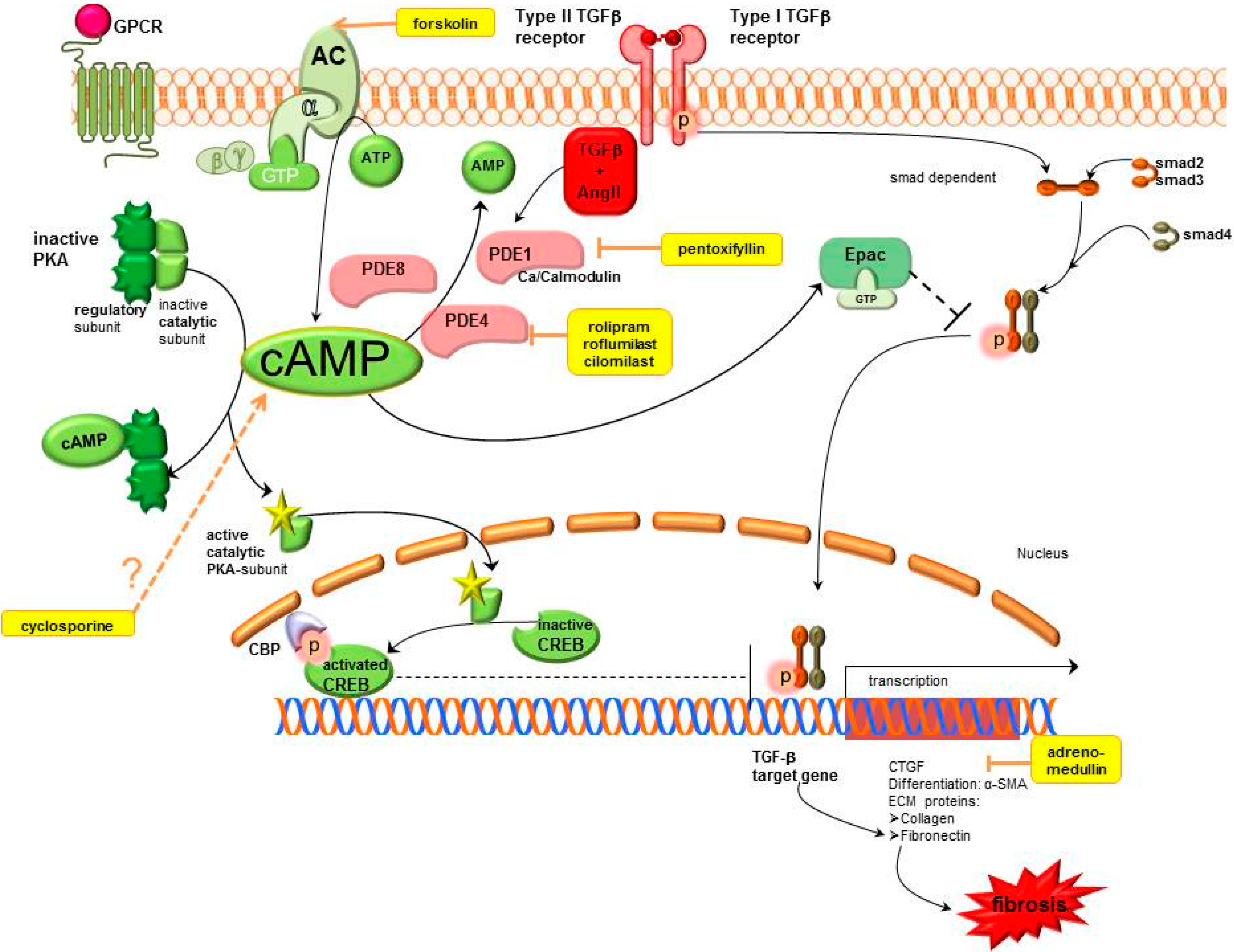
2.1. Cyclic Adenosine Monophosphate (cAMP) Pathway

The cAMP pathway exerts antifbrotic actions which include inhibition of EMT blockade of fibroblast proliferation and activation of the death of fibroblasts. These effects can arise in response to an increase in cAMP by AC activators, PDE inhibitors, cAMP analogues or pharmacological agents like Gs-linked G protein coupled receptors (GPCR) agonists and Gi-linked GPCR antagonists. Increased cAMP levels exert their effects through activation of protein kinase A (PKA) which is the classical signalling pathway. Thereby, cAMP binds to the regulatory subunit of PKA leading to dissociation of the catalytic subunit which subsequently phosphorylates target proteins. Stimulated PKA causes inter alia phosphorylation of cAMP response element binding (CREB) and subsequent CREB-mediated gene transcription. Detailled signalling mechanisms of cAMP are shown in Figure 1.
Figure 1. Cyclic adenosine monophosphate signalling pathways in kidney fibrosis including pharmacological treatment options. AC, adenylyl cyclase; AMP, adenosine monophosphate; ATP, adenosine triphosphate; cAMP, cyclic adenosine monophosphate; CBP, CREB binding protein; CREB, cAMP response element binding protein; CTGF, connective tissue growth factor; ECM, extracellular matrix; Epac, exchange protein directly activated by cAMP; GPCR, G protein coupled receptor; GTP, guanosine triphosphate; PDE, phosphodiesterase; PKA, protein kinase A; SMA, smooth muscle actin; smad, small mothers against decapentaplegic protein, TGFβ, transforming growth factor β.
Ijms 16 02320 g001
cAMP exerts antifibrotic effects in fibrosis, which are mediated by stimulation of PKA and activated CREB that thus blocks TGFβ mediated gene transcription. Furthermore, it activates Epac which blocks TGFβ mediated smad dependent gene transcription. Therapeutic options which enhance cAMP are AC stimulators, PDE inhibitors, cyclosporine and adrenomedullin (shown in yellow). Motifolio PPT Drawing Toolkit was used for designing the figure.

2.1.1. cAMP Modulation

Adenylyl Cyclase (AC)

Adenylyl cyclases are enzymes which convert ATP into cAMP. At least nine isoforms of AC—AC1-9—and two splice variants of AC8 are known [47].
Previous studies have shown antifibrotic effects by an increase of AC5/6 expression in cardiac and pulmonary fibroblasts which prevents the differentiation of cardiac fibroblasts [48] and pulmonary fibroblasts to pathophysiological changed myofibroblasts [49]. This is not yet studied in renal fibroblasts, but AC5 expression was also shown in the kidney [50].
Forskolin is an AC activator which increases intracellular cAMP levels by activation of membrane bound AC. Clinical data about the usage of AC activators are not yet available. As many disorders, e.g., cardiovascular diseases, glaucoma, asthma etc., are discussed for the application of forskolin, the risk-benefit ratio is not yet fully evaluated [51]. Nevertheless, antifibrotic effects of forskolin in renal mesangial cell cultures were already observed by reducing glomerular mesangial cell growth [52] or inhibiting CTGF gene expression by forskolin [53]. CTGF is a growth factor which is not present in healthy kidne but which is induced in fibrotic pathological condition, like glomerulosclerosis or diabetic nephropathy.

Phosphodiesterases (PDEs).

PDEs are enzymes which catalyze the hydrolysis of cAMP and/or cGMP, thereby, regulating the cAMP and cGMP levels [54]. The PDEs can be subdivided into at least 11 structurally related gene families (PDE 1 to 11) based on their ability to hydrolyze either preferentially cAMP, cGMP or both [55]. PDE1, PDE4 and PDE8 are important enzymes for cAMP signalling in the kidney. cAMP might be increased by PDE inhibitors.
PDE1: PDE1 consists of three genes—PDE1A, B and C—And belongs to the Ca2+ calmodulin-activated PDE family. Thereby, the activity of PDE1 family members can be increased up to tenfold in the presence of Ca2+ calmodulin [56,57]. PDE1A represents the predominant isoform but has a higher affinity for cGMP than for cAMP [57]. In cardiac fibroblasts, PDE1A is highly upregulated after stimulation with ATII and TGFβ [58]. Moreover, it is reported that the PDE1 inhibitor IC86340 decreased ATII or TGFβ induced cardiac myofibroblast activation, ECM production, and profibrotic gene expression. Thereby, PDE1 inhibition also mediates the antifbrotic effects via cAMP [58]. The PDE1 isozymes are abundant in the kidney and some isoforms of PDE1C exhibit high affinity for cAMP [59]. Thus, increased cAMP levels induced by specific PDE1 inhibitors could be beneficial in renal disease, but data about PDE1 inhibition in the kidney are lacking.
PDE4: Another PDE isoform, which specifically hydrolyses cAMP, is PDE4. Souness et al., reported that inhibitors of PDE4 exert antiinflammatory effects by suppression of many inflammatory cell responses, TNFα release and ROS generation. Consequently, PDE4 inhibitors can be used in the therapy of different diseases characterized by excessive cytokine production [60]. In renal diseases protective effects—Including antifibrotic effects—Of PDE4 inhibition have already been investigated.
cAMP hydrolysis in the renal mesangial cells is particularly mediated via PDE3 and PDE4 [61]. Inhibition of PDE4 has repeatedly shown suppressive effects on TGFβ signalling which has relevant therapeutic effects on excessive tissue remodeling, e.g., glomerulopathies. PDE4 inhibitors decrease fibroblast activity through elevated cAMP, and thus PKA, by decreasing collagen contraction and fibroblast chemotaxis towards fibronectin, especially in the presence of TGFβ signalling in vitro. Moreover, PDE4 inhibitors normalize the release and activation of tissue-degrading MMPs in TNFα cultured fibroblasts [62,63,64]. In a rat glomerulonephritis model, improvement of renal function and renal structure was observed [65,66].
Roflumilast, a selective PDE4 inhibitor was tested in type1 diabetic nephropathy induced by streptozotocin. Treatment with roflumilast decreased oxidative stress as well as extracellular matrix proteins such as fibronectin, collagen and apoptosis which was demonstrated by the TUNEL assay. The antioxidant enzyme heme oxygenase-1 is elevated in type 1 diabetic kidney whereas FoxO1, a transcription factor involved in oxidative stress is decreased [67]. Roflumilast was able to reverse that pathological condition.
However, the above mentioned PDE4 inhibitors, e.g., roflumilast (see Section 2.1.2) have several side effects, like central nervous or gastrointestinal disturbances [68].
Recently, a selective PDE4 inhibitor TJN-598 was established which showed lower excretion of TGFβ as well as lower mesangial matrix index, but—Inconsistent with the commonly used PDE4 inhibitors—Could not improve protein excretion [69]. (The mesangial matrix index evaluates mesangial expansion by the ratio of mesangial area to total glomerular area, whereas mesangial expansion is a product of glomerular hypercellularity, widening as well as mesangial matrix accumulation.).
NCS613 preferentially inhibits PDE4C which prevents disease progression in a lupus nephritis disease experimental model by ameliorating proteinuria [70].
PDE8: PDE8 is a cAMP specific PDE [60]. Upon TGFβ stimulation cardiac myofibroblasts generate less cAMP compared with fibroblasts because the synthesis of cAMP by AC5/6 expression was down-regulated and the degradation of cAMP by the PDE8 isoform PDE8A was upregulated in myofibroblasts [71]. In the kidney, mRNA expression of AC5/6 [47] and PDE8A [72] was already shown. Therefore, a TGFβ induced downregulation of cAMP in renal myofibroblasts could also be conceivable, but it is not yet examined.
The methylxanthine derivate pentoxifylline is an unspecific PDE inhibitor which has antifibrotic and antiinflammatory properties in several models of disease and is used for peripheral vascular diseases in clinical practice [73].
Renal antifibrotic effects of this unspecific PDE inhibitor were described in several animal models. In an anti-Thy1 nephritic rat model, mRNA levels of type I, type III and type IV collagen as well as fibronectin were decreased in pentoxifylline treated rats compared to controls. Additionally, ICAM-1 and MCP-1 mRNA levels were reduced by pentoxifylline, in which cAMP involvement is supposed but evidence is lacking so far [74]. Similar antifibrotic results were observed in rats with 5/6 subtotal nephrectomy [75].
In tubulointerstitial fibrosis induced by unilateral ureteral obstruction, expression of remodeling biomarkers like Col1A1 and CTGF as well as α-smooth muscle actin (α-SMA), a marker for myofibroblast accumulation, are diminished by pentoxifylline which mediates its action via cAMP and subsequently PKA to abrogate TGFβ signalling [76]. In a similar model, reduction of total volume of interstitial fibrosis was observed as well [77].
Remodeling processes—Particularly renal scarring—Are also a consequence of pyelonephritis. Pentoxifylline prevented renal scar formation after induction of pyelonephritis in an experimental rat model when antibiotic therapy was delayed [78].
The renoprotective effect of pentoxifylline was already shown in the PREDIAN clinical trial, in which patients with type 2 diabetes mellitus and chronic kidney disease had a lower decline in estimated glomerular filtration rate with pentoxifylline treatment in addition to RAAS blockade [79]. These results indicate that multiple inhibitors of PDE isoforms are potential targets in the therapy of renal fibrosis. Inhibitors of PDE4, which specifically prevent degradation of cAMP, are in clinical development.

2.1.2. cAMP Effectors

Protein Kinase A-cAMP Response Element Binding (PKA-CREB)

In human epidermal HaCat keratinocytes, it was shown that cAMP elevating agents such as the AC activator forskolin prevent TGFβ2 signalling via PKA [80]. TGFβ transduces intracellular signals through type1 (TGFβ-R1) and type2 (TGFβ-R2) receptors. Receptor associated smad (R-smads) proteins, such as smad2 and smad3 are phosphorylated and activated by type1 receptors of TGFβ. Upon phosphorylation, the R-smads build a complex with smad4 which is a common mediator for all receptor activated smads. R-smad/smad4 complexes are then translocated into the nucleus where they modulate the transcription of many genes [81,82]. However, the cAMP/PKA/cAMP response element binding (CREB) cascade blocks TGFβ specific smad-dependent transcription. cAMP elevating agents abolished interactions of the TGFβ2 induced smad3/4 complex with the transcription co-activators CREB protein in a PKA dependent manner. Thereby, the smad translocation into the nucleus in response to TGFβ was not affected by cAMP in the examined HaCat keratinocytes [80]. These results are in accordance with Lin et al., that increased cAMP accompanied by PKA induced CREB phosphorylation attenuated renal tubulointerstitial fibrosis. Thereby, increased cAMP levels were achieved by pentoxifylline—A general inhibitor of cAMP dependent PDEs—Which leads to a block of TGFβ induced smad3/4 dependent gene transcription. In this study, the inhibition of the profibrogenic CTGF was shown. However, smad activation and nuclear translocation were also not affected by cAMP/PKA [76].
CREB effects were shown by modulating cAMP via pentoxifylline or forskolin as direct modulators of CREB are lacking.

Exchange Protein Directly Activated by cAMP (Epac)

cAMP is able to stimulate PKA-independent Epac which is a guanine nucleotide exchange protein for the small GTPase Rap1. Activation of Epac by cAMP leads to release of the guanine nucleotide GDP and binding to GTP [83]. Epac regulates different functions like migration, proliferation and apoptosis via Rap1 [84]. It was shown that fibrosis inhibits Epac expression. Therefore, activation of Epac acts antifibrotically by inhibition of collagen type I- and collagen type III expression. Moreover, Epac interacts with TGFβ-R1 resulting in inhibition of phosphorylation of smad2 and transcriptional activation [85]. The importance of the cAMP-Epac-Rap signalling for modulating myofibroblast stimulation and ECM production was also described in the heart by Miller et al. [58].
A model to study EMT, which is a mechanism of tissue fibrosis, is the use of renal epithelial cells such as MDCK cells treated with TGFβ. MDCK cells base on epithelial phenotype with high E-cadherin expression and low α-SMA expression. TGFβ induces EMT by increasing α-SMA (and proteins characteristic of fibroblasts such as collagens) and decreasing E-cadherin expression (and other proteins characteristic of epithelial cells). Treating these MDCK cells with cAMP-derivative inhibits the upregulation of α-SMA via Epac. Therefore, Epac acts antifibrotically via inhibition of profibrotic TGFβ signalling [86].
A recent study by Stolman et al., demonstrated that Epac/Rap stimulation ROS production in the kidney [87]. Uncontrolled production of ROS mediated cellular injury and also occured during renal fibrosis [88].
Thus, activation of Epac/Rap signalling may protect against renal fibrosis, but data about pharmacological modulation of kidney fibrosis are lacking.

2.1.3. Further cAMP Influencing Systems

Adrenomedullin particularly is a vasodilatory agent which was demonstrated to augment NO. Its antifibrotic effects are mediated through cAMP-mediated decrease of CTGF induction and Erk phosphorylation in renal interstitial fibrosis [89]. The renoprotective effects were observed in diverse hypertensive models, e.g., in Dahl salt-sensitive rats or in the DOCA-salt model [90,91].
Cyclosporine is widely used for the treatment of FSGS. Its renoprotective effects are partly mediated through hemodynamic effects by decreasing glomerular perfusion rate and by decreasing intracellular pressure, but also through reduction of T cell mediated cytokines. Presumably, an increase in glomerular cAMP levels following cyclosporine treatment is responsible for the improved permeability characteristics of the glomerular filtration barrier [31].

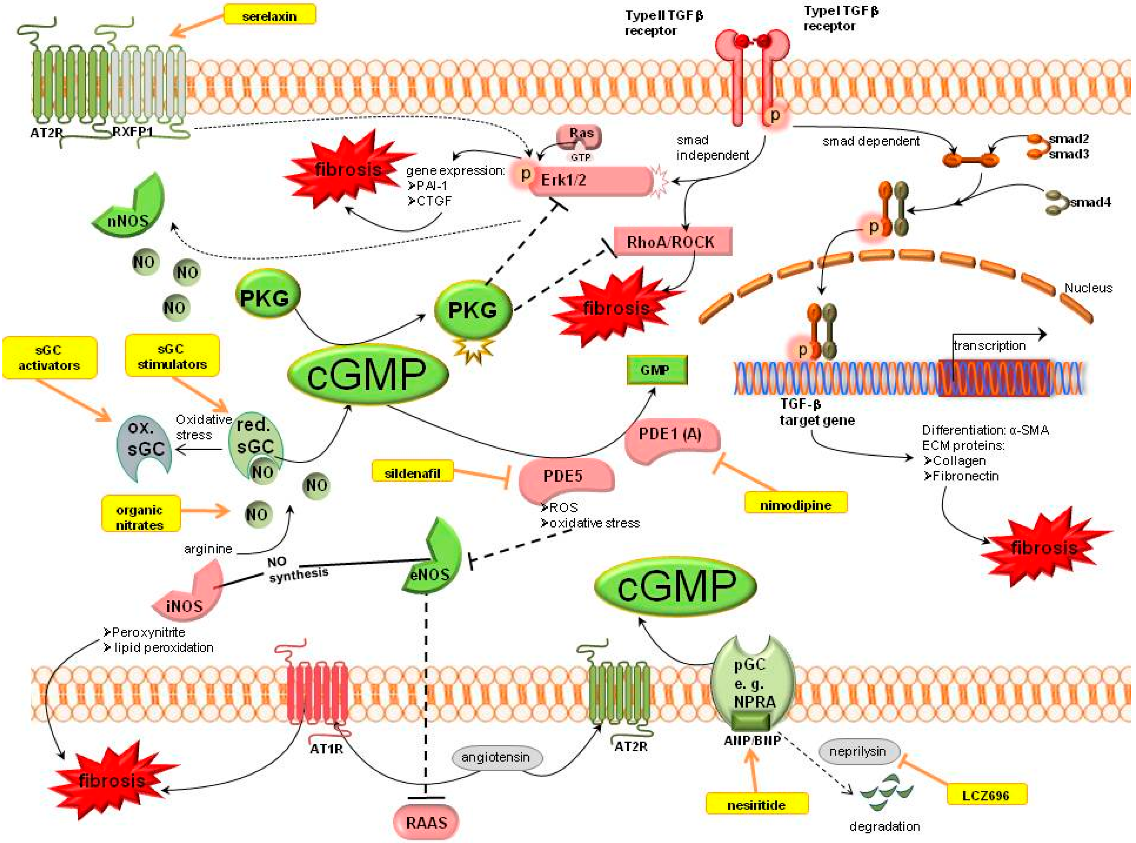
2.2. Cyclic Guanosine Monophosphate (cGMP) Pathway

Chronic organ injury, particularly kidney fibrosis, degrades NO producing cells, such as endothelial cells. The decreased availability of NO leads to reduced cGMP levels. Many studies report that enhanced cGMP levels have an effective antifibrotic benefit in various organs including the kidney [17].
Nitric oxide synthases (NOS) produce NO which activates the soluble guanylyl cyclase (sGC). sGC is the NO receptor mediating the downstream signalling by the generation of cGMP. Increased cGMP levels lead to activation of cGMP dependent protein kinases (PKG). cGMP can also be produced by natriuretic peptides (ANP/BNP) which stimulate the particulate guanylyl cyclase (pGC). The degradation of cGMP is mediated by PDEs. Detailled signalling of cGMP is shown in Figure 2.
cGMP mediates its antifibrotic action (shown in green) via activation of PKG which is able to inhibit the profibrotic TGFβ signalling (shown in red). TGFβ signalling is mediated via a smad dependent pathway to increase target gene transcription, or a smad independent pathway which activates Erk1/2 and RhoA/ROCK signalling. Therapeutic options which enhance cGMP are serelaxin, sGC modulation, organic nitrates, PDE inhibitors or natriuretic peptide analogues or their modulators (shown in yellow). Motifolio PPT Drawing Toolkit was used for designing the figure.
Some treatment options are in development for the augmentation of the cGMP pool [92]. The aim of these agents is to ameliorate or prevent the progression of fibrotic tissue.
Figure 2. Cyclic guanosine monophosphate signalling pathways in kidney fibrosis including pharmacological treatment options. ANP, atrial natriuretic peptide; AT1R, angiotensin II receptor type 1; AT2R, angiotensin II receptor type 2; BNP, brain natriuretic peptide; cGMP, cyclic guanosine monophosphate; CNP-c, type natriuretic peptide; CTGF, connective tissue growth factor; ECM, extracellular matrix; eNOS, endothelial NO synthase; ERK1/2, extracellular-signal regulated kinase; GMP, guanosine monophosphate; GTP, guanosine triphosphate; iNOS, inducible NO synthase; nNOS, neuronal NO synthase; NO, nitric oxide; NPRA, natriuretic peptide receptor A; PAI-1, plasminogen activator inhibitor 1; PDE, phosphodiesterase; pGC, particulate guanylyl cyclase; PKG, cGMP dependent protein kinases; RAAS, renin angiotensin aldosterone system; Ras, Rat sarcoma protein, RhoA, particular Rho protein; ROCK, Rho associated protein kinase; ROS, reactive oxygen species; RXFP1, relaxin family peptide receptor 1; sGC, soluble guanylyl cyclase; SMA, smooth muscle actin; smad, small mothers against decapentaplegic protein; TGFβ, transforming growth factor β.
Ijms 16 02320 g002

2.2.1. cGMP Modulation

Organic Nitrates

Organic nitrates are used for the treatment of cardiovascular disease for the last centuries, as they increase NO availability and thereby support the NO/cGMP signalling pathway. However, this therapy option is limited due to formation of nitrate tolerance as well as accumulation of ROS (e.g., peroxynitrite) under oxidative stress conditions. Beneficial effects in kidney fibrosis were shown in different glomerulonephritis experimental models after administering l-arginine supplements [93,94].

Nitric Oxide Synthase

The generation of NO is catalyzed by three NOS isoforms: type I (neuronal or nNOS), type II (inducible or iNOS) and type III (endothelial or eNOS). All three NOS isoforms are found in the kidney, but the expression of iNOS is more variable [95]. Furthermore, the contribution of eNOS, nNOS and iNOS to renal injury is very different.
eNOS: The eNOS activity is altered in diabetes and associated with the development of nephropathy in type1 and type2 diabetes patients [96]. Diabetic patients often develop nephropathy despite optimal therapy including adjustment of blood glucose. Moreover, superoxide producing enzymes promote an increased formation of ROS and eNOS uncoupling leading to decreased NO bioavailability [97]. Oxidative stress inactivates NO. Thus, reduced NO bioavailability accompanies chronic renal diseases [98]. Comparable results reported Liu et al., that the activity of eNOS was decreased by the fibrotic process in the lung [99]. In the cirrhotic liver, cGMP synthesis was also reduced due to the declined activity of eNOS [100].
Moreover, eNOS counterbalances the activity of the RAAS. Thus, inhibition of NOS increases the activity of the RAAS resulting in augmented expression of fibrosis marker such as fibronectin and α-SMA [101]. Moreover, deletion or reduction of eNOS in animals leads to glomerular abnormalities and tubular cell death. eNOS deletion is realized in eNOS knockout models whereas eNOS reduction is, for example, realized by models of hypertension. Consequently, eNOS protects against renal injury [95,102].
nNOS: In contrast to eNOS, an increase in nNOS facilitates renal injury. It is speculated that increased nNOS expression promotes generation of oxidative stress and formation of ROS within the kidney. Therefore, inhibition of nNOS showed renoprotective effects. Thereby, the inflammatory cells and the number of CD68 positive cells were reduced [95].
iNOS: iNOS produces—In comparison to eNOS and nNOS—large amounts of NO that, under oxidative stress conditions, can form peroxynitrite. Peroxynitrite initiates lipid peroxidation, oxidative protein and DNA modifications [103]. Thus, the downregulation of iNOS expression can reduce toxic peroxynitrite reactions.
Jeong et al., also proposed that decrease of iNOS could induce renoprotective effects in streptozotocin induced diabetic rats [104].

Soluble Guanylyl Cyclase (sGC)

Guanylyl cyclases are enzymes that convert guanosine triphosphate (GTP) to cGMP. Both types of guanylyl cyclases, particulate guanylyl cyclase (pGC) activated by atrial (ANP) and brain natriuretic peptides (BNP), and soluble guanylyl cyclase (sGC) stimulated by nitric oxide, can reduce cardiac fibrosis by increasing intracellular cGMP levels [105]. Furthermore, it is reported that NO via cGMP downregulates CTGF in rat mesangial cells. Thereby, NO activates the soluble form of guanylyl cyclase which leads to increased cGMP levels [106].
sGC modulation could be achieved by either sGC stimulators or sGC activators.
The target of sGC stimulator is the NO sensitive reduced Fe2+ redox state (NO dependent) whereas the target of sGC activator is the NO insensitive oxidized (Fe3+) sGC [107]. sGC stimulators bind NO-independently to a regulatory site of the α-subunit of sGC to stimulate the enzyme. Furthermore, they cause sGC to be more sensitive to endogenous NO.
If NO deficiency is the major issue, the use of sGC stimulators is suitable sensitizing sGC for NO. However, if heme oxidation or heme-free form of sGC is the major cause of disease, sGC activators are an appropriate tool for therapeutic use [97]. sGC activators bind to inactive sGC to additionally increase enzyme activity, when the prosthetic heme-group is oxidized or lacking. Activators might be introduced as therapy option particularly for pathological changes due to oxidative stress [108].
Thereby, it is important to know that oxidative stress, which is an accompanying effect of fibrosis, favours heme oxidation or heme-free sGC. Both sGC stimulators and sGC activators were used in studies for the treatment of kidney diseases. It is suggested that sGC modulators have direct antifibrotic actions that are presumably associated with enhanced NO/sGC/cGMP signalling [108,109,110,111].
sGC Stimulators: It is manyfold reported that the sGC-cGMP axis could be a therapeutic target of fibrosis of various organs. Stimulation of sGC decreased TGFβ induced collagen release by inhibition of Erk1/2 phosphorylation in human fibroblasts. However, nuclear p-smad2 and 3 levels, smad reporter activity and transcription of TGFβ target genes were unaffected by sGC stimulation. In murine sGC KO fibroblasts sGC stimulation showed no effects [112].
YC-1 is a benzylindazole derivative which has multiple actions—cGMP-dependent [113] or -independent [114] effects. cGMP dependent actions are, among others, inhibition of phosphodiesterase activity or sGC modulation [113]. It firstly provided sGC modulation, independent of NO bioavailability [115,116,117].
Antifibrotic effects of YC-1 were observed in diverse tissues, e.g., in a preventive model of hypoxia-induced pulmonary arterial hypertension (PAH), in which YC-1 alleviates right ventricular hypertrophy and pulmonary vascular remodeling [118].
cGMP-dependent antifibrotic effects of YC-1 via PKG1 in the kidney were described by Schinner et al. [119] in an animal model of interstitial kidney fibrosis induced by unilateral ureteral obstruction. YC-1 inhibited the profibrotic RhoA/ROCK pathway by the cGKIα isozyme, which blocked RhoA phosphorylation. As a consequence TGFβ stimulation as well as myofibroblast formation was suppressed. Moreover, the antifibrotic effect of YC-1 was boosted via HIF-1α inhibition in renal kidney fibrosis [11]. However, due to its cGMP-independent effects YC-1 is not applicable for clinical drug testing.
BAY 41-8543 and BAY 41-2272 are pyrazolopyridines which were developed from the structure of YC-1.
BAY 41-2272 attenuated remodeling and limited progression of fibrosis in an anti-Thy1 induced model of progressive kidney disorders, particularly in interstitial fibrosis [17,120]. Additionally, it reduced mesangial proliferation, matrix expansion and proteinuria in a rat model of mesangial proliferative glomerulonephritis compared to placebo [94,121].
BAY 41-8543 showed also a renal protective effect [122]. It restored or preserved renal structure and function in case of obstructive kidney disease by positively influencing α-SMA expression, collagen IV deposition and TGFβ1 mRNA expression [123].
The majority of available research is preclinical. Clinical experiments are limited due to unfavourable pharmacokinetic profile, CYP inhibition or induction and blood pressure decrease in healthy volunteers [124].
Riociguat (Bay 63-2521) is a further sGC stimulator causing antifibrotic effects in the kidney. Riociguat has two benefits—it sensitizes sGC to NO and can also increase sGC activity in the absence of NO. Riociguat (BAY 63-2521) is structurally similar to BAY41-8543 and the single sGC stimulator which has an acceptable oral bioavailability and thus, was successfully completing the clinical development program due to its pharmacokinetic profile. It was approved by health authorities for PAH and chronic thromboembolic pulmonary hypertension (CTEPH) [125]. Its tradename is Adempas®.
Administration of riociguat alone as well as in combination with the AT1R inhibitor telmisartan attenuated progression of renal fibrosis, especially diabetic nephropathy. Thereby the systemic inflammation was decreased which was measured by plasma TNFα levels [23]. In a similar fashion renal fibrotic tissue remodeling was markedly improved in a rodent model of pressure and volume overload by decreasing protein und mRNA expression of profibrotic osteopontin, TIMP1 and PAI-1 (plasminogen activator inhibitor 1) in the renal cortex [126].
Furthermore, in two independent models of hypertension, it was shown a potent protection against renal interstitial fibrosis and partially against glomerulosclerosis [127].
sGC activators: The sGC activators cinaciguat [128] and ataciguat (HMR1766) [129] have antiremodeling effects in the kidney.
Cinaciguat is in the clinical development program for acute heart failure, but in a high dose cinaciguat therapy (>200 µg/h) hypotension occurred as adverse event and subsequently the phase IIb clinical trial was stopped [130].
The NO independent sGC activator cinaciguat (Bay 58-2667) is characterized by activating sGC after oxidation of its hem group. Chronic renal failure is accompanied by impaired NO availability because the generation of NO is decreased, and NO is inactivated by ROS or its physiological action is impeded by dysfunctional sGC. The reduced NO availability promotes renal disease.
Cinaciguat attenuated remodeling and limited progression of fibrosis in models of pulmonary hypertension partially associated with eNOS-dependent generation of nitric oxide [110]. Furthermore, rats with subtotal 5/6 nephrectomy which were treated with cinaciguat had slowing renal disease progression, reduced left ventricular hypertrophy and preserved renal function by targeting oxidized sGC and increasing intracellular cGMP [128].
Beneficial effects of ataciguat (HMR1766) on structural parameters of renal damage and urinary albumin excretion in a remnant kidney model were demonstrated [129]. HMR1766 has blood-pressure-independent and sustainable antifibrotic effects.
sGC activators are promising compounds for the treatment of kidney fibrosis. Data about antifibrotic effects in the kidney are still to be fully elucidated.

PDE Inhibitors

PDE1: As already mentioned, PDE1 consists of three genes—PDE1A, B and C. PDE1A represents the predominant isoform which has a higher affinity for cGMP than for cAMP [57]. Inhibition of PDE1 by IC86340 reduced ATII or TGFβ induced activation of cardiac myofibroblasts, synthesis of ECM and expression of profibrotic genes. Thereby, the antifbrotic effects of this PDE1 inhibitor were also mediated via cGMP/PKG.
The phosphodiesterase 1 inhibitor nimodipine was investigated as new therapeutic strategy for the autoimmune disease systemic lupus. In MRL/lpr lupus-prone mice PDE1 activity was elevated in the kidney, which was accompanied by a decrease in cGMP levels. Treatment with nimodipine resulted in improvement of organ remodeling, especially kidney remodeling, as well as in reduction of hypercellularity [131].
PDE5: The cGMP pool is rapidly degraded by PDEs. PDE5 is the PDE isozyme which specifically hydrolyzes cGMP. At the renal level of rat kidney, PDE5 is expressed in inner medullary collecting duct cells, cortical tubules, mesangial cells, the vasculature and glomeruli. PDE5 inhibitors, including sildenafil and tadalafil, possess antiapoptotic and antioxidant properties. Consequently, they exert nephroprotective effects.
In lung fibrosis, the PDE5 inhibitor (KMUP-1) was able to reduce the fibrotic process. In this study, many possible mechanisms, which mediate the effects of PDE5 inhibition, were shown. Administration of KMUP-1 significantly attenuated the expression of active MMP2 which is upregulated in fibrosis. Moreover, TGFβ and CTGF expression was reduced by KMUP-1. Moreover, the TGFβ induced increase of the p-smad3/smad3 ratio was downregulated by administration of KMUP-1. Furthermore, KMUP-1 inhibited the profibrotic RhoA/ROCK signalling and enhanced eNOS activity which was decreased in fibrosis [99]. These antifibrotic mechanisms of PDE5 inhibition are not yet studied in the kidney although PDE5 inhibitors such as sildenafil and tadalafil are tested in renal diseases. Thus, these mechanisms and signalling pathways in lung fibrosis could also be an approach in renal fibrosis. The PDE5 inhibitor sildenafil was already tested many times regarding kidney function. In a mouse model of interstitial kidney fibrosis induced by UUO renal fibrosis was reduced after administration of sildenafil for 14 days. Amelioration of kidney fibrosis was perceived by increased cGMP levels, mediated partly through regulation of macrophages and tubular cells. This was associated with reduced renal TGFβ/smad signalling and decreased macrophages infiltration. Kidney fibrosis was evaluated via protein and mRNA expression of collagen type I, collagen type III as well as α-SMA [132]. The PDE5 inhibitor sildenafil was administered to rats with 5/6 nephrectomy immediately after renal ablation for 8 weeks. In this setting sildenafil prevented histological damage, inflammation and apoptosis leading to reduced worsening of renal function, ameliorated proteinuria and decreased hypertension. Simultaneously, urinary cGMP excretion was elevated with sildenafil treatment [133]. It additionally suppressed renal arteriolar remodeling as a reason for improved function of the remnant kidney [134]. DOCA-salt hypertensive rats develop renal dysfunction due to fibrotic remodeling processes. In this experiment sildenafil reduced remodeling biomarkers, e.g., α-SMA and fibronectin, and thereby prevented progression of tubulointerstitial fibrosis and glomerulosclerosis [135].
Another study found that sildenafil attenuates infiltration of macrophages (ED1 positive cells) reducing the production of inflammatory stimuli. Moreover, PDE5 inhibition prevented oxidative stress in diabetic nephropathy. Jeong et al., speculated that decrease of iNOS by sildenafil could induce this renoprotective effect [104]. The inhibition of oxidative stress by sildenafil—Determinated by suppression of NADPH oxidase expression and therefore of superoxide formation—Parallels the results of Muzaffar et al. [136].
In the study of Sohotnik et al., the PDE5 inhibitor tadalafil prevented kidney dysfunction and structural renal damage in an experimental model of renal ischemia-reperfusion (I/R) [137,138]. Tadalafil also reduced the urinary excretion of NGAL and KIM-1, which are two biomarkers indicating acute kidney injury.

Relaxin

The pregnancy hormone relaxin—Human relaxin II—Has shown antifibrotic effects in several experiments in vitro and in vivo. Its antifibrotic actions were demonstrated in several cell types, tissues, and organs, including lung, liver, heart, and kidney. The relaxin family peptide receptor 1 (RXFP1)—The most relevant receptor for relaxin’s antifibrotic effects—was already localized in different tissues, including the kidney [139,140].
Relaxin is a hormone that both reduces ECM production and increases its clearance. This was first found in 1929 by Hisaw et al., who showed, that relaxin induces relaxation of the pelvic ligament as the ligament reveales remodeling of the collagen from dense bundles to looser, less structured fibers. In pregnancy, relaxin is responsible for widespread extracellular matrix remodeling in the cervix, vagina and in some species in the pubic symphysis [141].
The antifibrotic signalling mechanism of relaxin is not clearly understood so far and therefore, it has to be fully elucidated in the future. But a lot of research is done to generate a hypothesis for relaxin’s antifibrotic pathway. Relaxin is supposed to mediate its antifibrotic effect in renal myofibroblasts via the RXFP1 and a subsequent Erk1/2 phosphorylation which activates the nNOS/NO/cGMP-dependent pathway. The disruption of TGFβ signalling by relaxin was demonstrated by decreased ECM production and decreased myofibroblast differentiation in humans [142] as well as in rodents [143,144]. Additionally, relaxin leads to upregulation of MMPs—ECM degrading enzymes—And downregulation of TIMPs—Inhibiting enzymes of MMPs—Which is likely mediated through iNOS. This signalling mechanism is increased by blocking the TGFβ1/Smad2 signalling [145]. Current research indicated that relaxin signals through RXFP1-AT2R heterodimer complexes which are formed between RXFP1 and AT2R independent of ligand binding [146], which could explain relaxin’s antifibrotic effects especially in pathological conditions. In the lung, iNOS-NO-cGMP-PKG signalling was demonstrated to inhibit the profibrotic RhoA/ROCK which thus abrogated myofibroblast contractility [147]. Furthermore, eNOS appeared to be more relevant in vasodilating effects of relaxin [148]. This leads to the assumption that relaxin can act on the various NOS isoforms which depends on specific actions and tissues.
Relaxin deficient mice spontaneously develop fibrotic tissue in different organs, including the kidney [149]. They are associated with age-related fibrosis going along with renal hypertrophy, increase in total collagen content, interstitial fibrosis, glomerular sclerosis and a decline in renal function [150,151].
The exogenous infusion of serelaxin led to ameliorated progression of interstitial and glomerular fibrosis in several experimental models of renal diseases.
In aging rats the infusion of relaxin increased glomerular filtration rate as well as renal plasma flow and decreases renal vascular resistance, acutely caused by increased gelatinase activity [152]. Renal fibrosis caused by antiglomerular basement membrane disease decreased glomerulosclerosis and interstitial fibrosis [153]. ATII-induced hypertension preserved glomerular structure as well as reduced oxidative stress after a two week period of relaxin treatment [154].
In another animal model relaxin administration was started one week after induction of renal papillary necrosis by bromoethylamin injection for the duration of 28 days. Initial papillary necrosis results in interstitial fibrosis and renal insufficiency. Relaxin improved renal function by restoration of glomerular filtration rate indicated by reduced TGFβ, macrophage infiltration and reducing fractional area of interstitial collagen staining by 75% [155]. Relaxin was administered for the same duration in models of renal mass reduction [156] and for shorter [157] or longer duration [148] in hypertension which demonstrated decreased interstitial and glomerular fibrosis and normalized collagen accumulation in the kidney. Influence on reducing blood pressure was shown, but matchable results were achieved in blood pressure independent models [155].
In contrast, in ATII-induced organ damage [158] as well as in diabetic renal disease [159] relaxin could not reverse fibrotic processes.
Recently, a novel peptide was identified, CGEN25009, which has relaxin-like activity. Its action is mediated through RXFP1 receptor, involvement of cyclic nucleotides was observed in the antifibrotic mode of action [160]. Data about renal fibrosis are still lacking.

Natriuretic Peptides

Natriuretic peptides are divided into three peptides—Atrial (ANP), brain (BNP) and C-type (CNP) natriuretic peptides [161]. Biological action of ANP and BNP is mediated through binding to NPR-A to induce vasorelaxing, diuretic, natriuretic, antiproliferative, antihypertrophic and antialdosterone effects. NPR-B is the receptor for CNP to cause vasorelaxing and antifibrotic effects as well as playing a role in bone growth regulation and reproduction. Due to its paracrine and autocrine effects CNP as well as its receptor is located in several tissues, including the kidney [162].
It is reported that ANP/cGMP/PKG can abolish TGFβ induced nuclear translocation of p-smad2 and p-smad3 in rat pulmonary arterial smooth muscle cells. However, the phosphorylation of smad2/3 is not influenced by ANP/cGMP. It is discussed that cGMP/PKG phosphorylates smad2 or smad3 on additional serine residues which are different from the TGFβ phosphorylation sites leading to inhibition of nuclear translocation [163]. These results are in agreement with studies in cardiac fibroblasts. Here, the TGFβ induced nuclear translocation of p-smad3 was also abolished by cGMP. As already mentioned, PKG1 phosphorylated smad3, but at sites different from those required for its TGFβ induced nuclear translocation. Furthermore, the stimulation of the cGMP/PKG pathway by ANP reduced TGFβ induced myofibroblast differentiation, proliferation, collagen production and PAI-1 expression [164].
Thus the pharmacological potentiation of endogenous ANP or BNP may be a therapeutic approach for the treatment of renal fibrosis.
Disruption of the gene encoding for GC-A/NPR-A leads to the development of renal fibrosis. Thereby, TNFα and IL-6 are increased [165]. These findings are in accordance with Kumar et al., who showed that increased renal NPR-A/cGMP signalling attenuates renal fibrosis [166]. ANP is a member of a natriuretic peptide family, which counterregulates renal hypoxia and the consequent process of fibrosis, exerting protective effects in response to oxidative stress and fibrosis [167]. An earlier study also demonstrated that activation of the endogenous NPR-A system and administration of ANP increased the cGMP levels leading to reduced renal interstitial fibrosis induced by UUO [168].
Nesiritide is the recombinant form of naturally occurring brain natriuretic peptide (BNP) enhancing cGMP levels via GC-A/NPR-A. This signalling pathway has demonstrated cardiac antihypertrophic and antifibrotic effects in several experiments.
In mutant mice lacking Npr1 gene, which is encoding for GC-A/NPR-A, increased fibrosis, hypertrophic growth and remodeling of the kidney was observed [165].
Chronic excess of BNP has shown to ameliorate glomerular hypertrophy and mesangial expansion after renal ablation, improves immune-mediated renal injury and prevents glomerular injury in progression of diabetic nephropathy [169,170,171].
In the last years new chimeric natriuretic peptides were developed to generate more potent peptides. CBA-NP is a fusion of diverse amino acid sequences of ANP, BNP and CNP. CD-NP is a composition of CNP and DNP, which enables the newly created peptide to act on both natriuretic peptide receptors NPR-A and NPR-B. Both CD-NP and CBA-NP attenuated renal fibrosis resulting in an improved renal function [172,173].
CNP could be a novel approach for the prevention of acute kidney impairment after ischemia-reperfusion injury. Jin et al. demonstrated improved renal vascular function and degraded glomerular and tubular microstructure in an experimental rat model of ischemia reperfusion injury. This effect is supposed to be mediated via cGMP signalling [162].
These observations suggest that natriuretic peptides and their derivatives play a pivotal role in the renal antifibrotic properties. However, nesiritide could not provide improvement of kidney function in the ASCEND-HF clinical trial in acute decompensated heart failure [174].
LCZ696 is the first compound of a new drug class, ARNI (angiotensin receptor neprilysin inhibitor) and is now being investigated in patients with chronic kidney disease [175]. LCZ696 simulaneously blocks AT1R and neprilysin, which is a natriuretic peptide degrading enzyme, resulting in enhanced activity of natriuretic peptides. Recently, clinical data are promising for chronic heart failure patients with reduced ejection fraction as they are superior to angiotensin-converting enzyme (ACE) inhibition by reducing risk of death and hospitalization in heart failure [176]. LCZ696 may provide kidney protection through blocking the profibrotic RAAS and concomitantly stimulating the antifibrotic neprilysin inhibition. Renoprotective evidence is supposed, but evidence is still lacking.

2.2.2. cGMP Effectors

cGMP Dependent Protein Kinases (PKG)

The major downstream effector of cGMP is PKG. Two isoforms of PKG are known—PKG1 and PKG2. However, only PKG1 is studied regarding the involvement in renal fibrosis.
Using PKG1-KO mice, Schinner et al. [92,119] showed that cGMP acts antifibrotically via activation of PKG1 in UUO. Thereby, the antifibrotic effects of cGMP/PKG1 were mediated by inhibition of the profibrotic RhoA/ROCK signalling to inhibit TGFβ signalling and myofibroblast formation (see above).
In human mesangial cells (HMCs), TWEAK, which is an inflammatory cytokine, led to increased TGFβ expression by stimulation of Ras/Erk1/2. Interestingly, PKG1 protein expression and activity was reduced. Thereby, the Ras/Erk1/2 pathway was essential for the downregulation of PKG1 by TWEAK. Stimulation of PKG1 via 8Br-cGMP abolished TWEAK induced upregulation of TGFβ [177]. These results are in accordance with a study of Cui et al., that increasing PKG activity reduces ECM accumulation in renal mesangial cells. PKG can be activated by increased cGMP levels. In addition to pharmacological approach, PKG transgenic mice (Tg mice) were used to demonstrate the antifibrotic effects of PKG. Tg mice, which expressed more PKG1 than WT mice, showed the same results compared to the treatment with PDE5 inhibitor sildenafil [132]. Until today, no direct PKG activators are in clinical trial, only cGMP elevating agents are being tested.

2.2.3. Further cGMP Influencing Systems

Renin Angiotensin Aldosterone System (RAAS)

Angiotensin acts via angiotensin II type 1 receptor (AT1R) and angiotensin II type 2 receptor (AT2R). Interestingly, AT2R has opposite effects of AT1R which induces fibrosis. The expression of AT2R is increased in pathological situations and is able to suppress cardiac fibrosis. Thereby, the AT2R effect is mediated via bradykinin/NO/cGMP pathway AT2R thereby forms a heterodimer with the bradykinin receptor B2R to induce eNOS which subsequently activates NO/cGMP signalling [178]. The effect of AT2R might be promising but is not yet explored in the kidney.
Diabetes mellitus is accompanied by an increase of the RAAS activity, including ATII and aldosterone production. Increased aldosterone was associated with reduced NO/cGMP and increased fibrosis in diabetic kidney [10]. The renin inhibitor aliskiren blocked ATII and aldosterone production [179] and the dihydropyridine-type calcium channel blocker amlodipine reduced aldosterone levels [180]. Decrease of aldosterone and its downstream effectors via aliskiren, amlodipine or the combination of both lead to reduced oxdidative stress and fibrosis via enhanced NO/cGMP availability in the kidney [10].

Kallikrein

Bradykinin mediates vasodilatory and antiinflammatory action through NO signalling. Furthermore, kallikrein positively modulates bradykinin production which subsequently activates eNOS. This mechanism is responsible, among others, for its antifibrotic effects.
Kallikrein reverses salt-induced renal fibrosis and glomerular hypertrophy in the interstitium and vasculature of hypertensive Dahl salt-sensitive rats [181]. The repair of renal tubular damage was also observed in a gentamycin-induced nephrotoxicity in normotensive rats [182]. Similar results were shown in a mineralocorticoid-induced renal fibrosis [183].

All-Trans-Retinoic Acid/Sodium Butyrate

All-trans retinoic acid in combination with sodium butyrate showed synergistical effects in reducing renal fibrotic biomarkers by enhancing Npr1 gene transcription which encodes for the GC-A/NPR-A [184]. Renal fibrosis and immunoexpression of renal α-SMA was reduced by ≥70%, and TNFα as well as IL-6 showed lower plasma and renal levels. This could be an important finding for the prevention of hypertension-related kidney diseases.

3. Conclusions

Cyclic nucleotide signalling plays a prominent role in the development as well as in the prevention or amelioration of progressive renal disease. cGMP concentrations are diminished during kidney fibrosis. Enhancement of cyclic nucleotides improves renal fibrosis at different stages. They reduce TGFβ signalling, transcription of profibrotic cytokines, oxidative stress, myofibroblast formation and subsequently ECM accumulation in different experimental models for studying renal fibrosis in vitro and in vivo. Therapeutic approaches are in development, but treatment options modulating cyclic nucleotides are still lacking in clinical practice. Hence, preclinical experimental research is indispensable for the understanding of cyclic nucleotide dependent antifibrotic signalling and for the generation of drugs that may find their way into clinical practice for the treatment or prevention of kidney failure.

Acknowledgments

The work was supported by the state of Bavaria, the Deutsche Forschungsgemeinschaft DFG and the Sonderforschungsbereich SFB699.

Author Contributions

Elisabeth Schinner, Veronika Wetzl and Jens Schlossmann wrote the manuscript. Jens Schlossmann was primarily involved in the concept. Elisabeth Schinner and Veronika Wetzl designed the figures.

Conflicts of Interest

The authors Elisabeth Schinner and Jens Schlossmann declare no conflict of interest. PhD thesis of Veronika Wetzl is funded by Novartis GmbH. The company had no participation in writing of the review and decision to publish it.

References

  1. Eddy, A.A. The origin of scar-forming kidney myofibroblasts. Nat. Med. 2013, 19, 964–966. [Google Scholar] [CrossRef] [PubMed]
  2. Honda, E.; Park, A.-M.; Yoshida, K.; Tabuchi, M.; Munakata, H. Myofibroblasts: Biochemical and proteomic approaches to fibrosis. Tohoku J. Exp. Med. 2013, 230, 67–73. [Google Scholar] [CrossRef] [PubMed]
  3. LeBleu, V.S.; Taduri, G.; O’Connell, J.; Teng, Y.; Cooke, V.G.; Woda, C.; Sugimoto, H.; Kalluri, R. Origin and function of myofibroblasts in kidney fibrosis. Nat. Med. 2013, 19, 1047–1053. [Google Scholar] [CrossRef] [PubMed]
  4. Mack, M.; Yanagita, M. Origin of myofibroblasts and cellular events triggering fibrosis. Kidney Int. 2014. [CrossRef]
  5. Lin, S.L.; Kisseleva, T.; Brenner, D.A.; Duffield, J.S. Pericytes and perivascular fibroblasts are the primary source of collagen-producing cells in obstructive fibrosis of the kidney. Am. J. Pathol. 2008, 173, 1617–1627. [Google Scholar] [CrossRef] [PubMed]
  6. Campanholle, G.; Ligresti, G.; Gharib, S.A.; Duffield, J.S. Cellular mechanisms of tissue fibrosis. 3. Novel mechanisms of kidney fibrosis. Am. J. Physiol. Cell Physiol. 2013, 304, C591–C603. [Google Scholar] [CrossRef] [PubMed]
  7. Wakino, S.; Kanda, T.; Hayashi, K. Rho/Rho kinase as a potential target for the treatment of renal disease. Drug News Perspect. 2005, 18, 639–643. [Google Scholar] [CrossRef] [PubMed]
  8. Liu, Y. Cellular and molecular mechanisms of renal fibrosis. Nat. Rev. Nephrol. 2011, 7, 684–696. [Google Scholar] [CrossRef] [PubMed]
  9. Vernon, M.A.; Mylonas, K.J.; Hughes, J. Macrophages and renal fibrosis. Semin. Nephrol. 2010, 30, 302–317. [Google Scholar] [CrossRef] [PubMed]
  10. Matavelli, L.C.; Siragy, H.M. Reduction of aldosterone production improves renal oxidative stress and fibrosis in diabetic rats. J. Cardiovasc. Pharmacol. 2013, 61, 17–22. [Google Scholar] [CrossRef] [PubMed]
  11. Kimura, K.; Iwano, M.; Higgins, D.F.; Yamaguchi, Y.; Nakatani, K.; Harada, K.; Kubo, A.; Akai, Y.; Rankin, E.B.; Neilson, E.G.; et al. Stable expression of HIF-1α in tubular epithelial cells promotes interstitial fibrosis. Am. J. Physiol. Renal Physiol. 2008, 295, F1023–F1029. [Google Scholar] [CrossRef] [PubMed]
  12. Higgins, G.C.; Coughlan, M.T. Mitochondrial dysfunction and mitophagy: The beginning and end to diabetic nephropathy? Br. J. Pharmacol. 2014, 171, 1917–1942. [Google Scholar] [CrossRef] [PubMed]
  13. Lee, D.Y.; Wauquier, F.; Eid, A.A.; Roman, L.J.; Ghosh-Choudhury, G.; Khazim, K.; Block, K.; Gorin, Y. Nox4 NADPH oxidase mediates peroxynitrite-dependent uncoupling of endothelial nitric-oxide synthase and fibronectin expression in response to angiotensin II: Role of mitochondrial reactive oxygen species. J. Biol. Chem. 2013, 288, 28668–28686. [Google Scholar] [CrossRef] [PubMed]
  14. Rosenbloom, J.; Mendoza, F.A.; Jimenez, S.A. Strategies for anti-fibrotic therapies. Biochim. Biophys. Acta 2013, 1832, 1088–1103. [Google Scholar] [CrossRef] [PubMed]
  15. Chen, J.F.; Liu, H.; Ni, H.F.; Lv, L.L.; Zhang, M.H.; Zhang, A.H.; Tang, R.N.; Chen, P.S.; Liu, B.C. Improved mitochondrial function underlies the protective effect of pirfenidone against tubulointerstitial fibrosis in 5/6 nephrectomized rats. PLoS One 2013, 8, e83593. [Google Scholar] [CrossRef] [PubMed]
  16. RamachandraRao, S.P.; Zhu, Y.; Ravasi, T.; McGowan, T.A.; Toh, I.; Dunn, S.R.; Okada, S.; Shaw, M.A.; Sharma, K. Pirfenidone is renoprotective in diabetic kidney disease. J. Am. Soc. Nephrol. 2009, 20, 1765–1775. [Google Scholar] [CrossRef] [PubMed]
  17. Wang, Y.; Krämer, S.; Loof, T.; Martini, S.; Kron, S.; Kawachi, H.; Shimizu, F.; Neumayer, H.; Peters, H. Enhancing cGMP in experimental progressive renal fibrosis: Soluble guanylate cyclase stimulation vs. phosphodiesterase inhibition. Am. J. Physiol. Renal Physiol. 2006, 290, F167–F176. [Google Scholar] [CrossRef] [PubMed]
  18. Betz, B.; Conway, B.R. Recent advances in animal models of diabetic nephropathy. Nephron. Exp Nephrol. 2014, 126, 191–195. [Google Scholar] [CrossRef] [PubMed]
  19. Hodgin, J.B.; Nair, V.; Zhang, H.; Randolph, A.; Harris, R.C.; Nelson, R.G.; Weil, E.J.; Cavalcoli, J.D.; Patel, J.M.; Brosius, F.C., III; et al. Identification of cross-species shared transcriptional networks of diabetic nephropathy in human and mouse glomeruli. Diabetes 2013, 62, 299–308. [Google Scholar] [CrossRef] [PubMed]
  20. Brosius, F.C., III. New insights into the mechanisms of fibrosis and sclerosis in diabetic nephropathy. Rev. Endocr. Metab. Disord. 2008, 9, 245–254. [Google Scholar] [CrossRef] [PubMed]
  21. Roscioni, S.S.; Heerspink, H.J.; de Zeeuw, D. The effect of RAAS blockade on the progression of diabetic nephropathy. Nat. Rev. Nephrol. 2014, 10, 77–87. [Google Scholar] [CrossRef] [PubMed]
  22. Komers, R.; Oyama, T.T.; Beard, D.R.; Tikellis, C.; Xu, B.; Lotspeich, D.F.; Anderson, S. Rho kinase inhibition protects kidneys from diabetic nephropathy without reducing blood pressure. Kidney Int. 2011, 79, 432–442. [Google Scholar] [CrossRef] [PubMed]
  23. Ott, I.M.; Alter, M.L.; von Websky, K.; Kretschmer, A.; Tsuprykov, O.; Sharkovska, Y.; Krause-Relle, K.; Raila, J.; Henze, A.; Stasch, J.P.; et al. Effects of stimulation of soluble guanylate cyclase on diabetic nephropathy in diabetic eNOS knockout mice on top of angiotensin II receptor blockade. PLoS One 2012, 7, e42623. [Google Scholar] [CrossRef] [PubMed]
  24. Braun, N.; Schweisfurth, A.; Lohofener, C.; Lange, C.; Grundemann, C.; Kundt, G.; Grone, H.J. Epidemiology of glomerulonephritis in Northern Germany. Int. Urol. Nephrol. 2011, 43, 1117–1126. [Google Scholar] [CrossRef] [PubMed]
  25. Appel, G.B.; Appel, A.S. New diagnostic tests and new therapies for glomerular diseases. Blood Purif. 2013, 35, 81–85. [Google Scholar] [CrossRef] [PubMed]
  26. Ponticelli, C.; Graziani, G. Current and emerging treatments for idiopathic focal and segmental glomerulosclerosis in adults. Expert. Rev. Clin. Immunol. 2013, 9, 251–261. [Google Scholar] [CrossRef] [PubMed]
  27. Shankland, S.J.; Pollak, M.R. A suPAR circulating factor causes kidney disease. Nat. Med. 2011, 17, 926–927. [Google Scholar] [CrossRef] [PubMed]
  28. Wei, C.; El Hindi, S.; Li, J.; Fornoni, A.; Goes, N.; Sageshima, J.; Maiguel, D.; Karumanchi, S.A.; Yap, H.K.; Saleem, M.; et al. Circulating urokinase receptor as a cause of focal segmental glomerulosclerosis. Nat. Med. 2011, 17, 952–960. [Google Scholar] [CrossRef] [PubMed]
  29. Hogan, J.; Bomback, A.S.; Mehta, K.; Canetta, P.A.; Rao, M.K.; Appel, G.B.; Radhakrishnan, J.; Lafayette, R.A. Treatment of idiopathic FSGS with adrenocorticotropic hormone gel. Clin. J. Am. Soc. Nephrol. 2013, 8, 2072–2081. [Google Scholar] [CrossRef] [PubMed]
  30. Kronbichler, A.; Kerschbaum, J.; Fernandez-Fresnedo, G.; Hoxha, E.; Kurschat, C.E.; Busch, M.; Bruchfeld, A.; Mayer, G.; Rudnicki, M. Rituximab treatment for relapsing minimal change disease and focal segmental glomerulosclerosis: A systematic review. Am. J. Nephrol. 2014, 39, 322–330. [Google Scholar] [CrossRef] [PubMed]
  31. Sharma, R.; Ix, J.H.; Mathew, A.V.; Cho, M.; Pflueger, A.; Dunn, S.R.; Francos, B.; Sharma, S.; Falkner, B.; McGowan, T.A.; et al. Cyclosporine protects glomeruli from FSGS factor via an increase in glomerular cAMP. Transplantation 1996, 62, 1916–1920. [Google Scholar] [CrossRef] [PubMed]
  32. Balow, J.E.; Austin, H.A., III. Renal disease in systemic lupus erythematosus. Rheum. Dis. Clin. N. Am. 1988, 14, 117–133. [Google Scholar]
  33. Michaelson, J.S.; Wisniacki, N.; Burkly, L.C.; Putterman, C. Role of TWEAK in lupus nephritis: A bench-to-bedside review. J. Autoimmun. 2012, 39, 130–142. [Google Scholar] [CrossRef] [PubMed]
  34. Crampton, S.P.; Morawski, P.A.; Bolland, S. Linking susceptibility genes and pathogenesis mechanisms using mouse models of systemic lupus erythematosus. Dis. Model. Mech. 2014, 7, 1033–1046. [Google Scholar] [CrossRef] [PubMed]
  35. Liu, D.; Ahmet, A.; Ward, L.; Krishnamoorthy, P.; Mandelcorn, E.D.; Leigh, R.; Brown, J.P.; Cohen, A.; Kim, H. A practical guide to the monitoring and management of the complications of systemic corticosteroid therapy. Allergy Asthma. Clin. Immunol. 2013, 9, 30. [Google Scholar] [CrossRef] [PubMed]
  36. Runkel, L.; Stacey, J. Lupus clinical development: Will belimumab’s approval catalyse a new paradigm for SLE drug development? Expert Opin. Biol. Ther. 2014, 14, 491–501. [Google Scholar] [CrossRef] [PubMed]
  37. Donadio, J.V., Jr.; Glassock, R.J. Immunosuppressive drug therapy in lupus nephritis. Am. J. Kidney Dis. 1993, 21, 239–250. [Google Scholar] [CrossRef] [PubMed]
  38. Fox, R.I. Mechanism of action of hydroxychloroquine as an antirheumatic drug. Semin. Arthritis. Rheum. 1993, 23, 82–91. [Google Scholar] [CrossRef] [PubMed]
  39. Zheng, S.; Huang, Y.; Yang, L.; Chen, T.; Xu, J.; Epstein, P.N. Uninephrectomy of diabetic OVE26 mice greatly accelerates albuminuria, fibrosis, inflammatory cell infiltration and changes in gene expression. Nephron. Exp. Nephrol. 2011, 119, e21–e32. [Google Scholar] [CrossRef] [PubMed]
  40. Conway, B.R.; Rennie, J.; Bailey, M.A.; Dunbar, D.R.; Manning, J.R.; Bellamy, C.O.; Hughes, J.; Mullins, J.J. Hyperglycemia and renin-dependent hypertension synergize to model diabetic nephropathy. J. Am. Soc. Nephrol. 2012, 23, 405–411. [Google Scholar] [CrossRef] [PubMed]
  41. Eddy, A.A.; Lopez-Guisa, J.M.; Okamura, D.M.; Yamaguchi, I. Investigating mechanisms of chronic kidney disease in mouse models. Pediatr. Nephrol. 2012, 27, 1233–1247. [Google Scholar] [CrossRef] [PubMed]
  42. Bonventre, J.V.; Yang, L. Cellular pathophysiology of ischemic acute kidney injury. J. Clin. Investig. 2011, 121, 4210–4221. [Google Scholar] [CrossRef] [PubMed]
  43. Komers, R. Rho kinase inhibition in diabetic kidney disease. Br. J. Clin. Pharmacol. 2013, 76, 551–559. [Google Scholar] [PubMed]
  44. Wakeland, E.K.; Liu, K.; Graham, R.R.; Behrens, T.W. Delineating the genetic basis of systemic lupus erythematosus. Immunity 2001, 15, 397–408. [Google Scholar] [CrossRef] [PubMed]
  45. Fox, R.I.; Kang, H.I. Mechanism of action of antimalarial drugs: Inhibition of antigen processing and presentation. Lupus 1993, 2, S9–S12. [Google Scholar] [CrossRef] [PubMed]
  46. Schmidt, H.H.; Schmidt, P.M.; Stasch, J.P. NO- and haem-independent soluble guanylate cyclase activators. Handb. Exp. Pharmacol. 2009, 191, 309–339. [Google Scholar] [PubMed]
  47. Defer, N.; Best-Belpomme, M.; Hanoune, J. Tissue specificity and physiological relevance of various isoforms of adenylyl cyclase. Am. J. Physiol. Renal Physiol. 2000, 279, F400–F416. [Google Scholar] [PubMed]
  48. Swaney, J.S.; Roth, D.M.; Olson, E.R.; Naugle, J.E.; Meszaros, J.G.; Insel, P.A. Inhibition of cardiac myofibroblast formation and collagen synthesis by activation and overexpression of adenylyl cyclase. Proc. Natl. Acad. Sci. USA 2005, 102, 437–442. [Google Scholar] [CrossRef] [PubMed]
  49. Dunkern, T.R.; Torsten, R.; Feurstein, D.; Rossi, G.A.; Sabatini, F.; Hatzelmann, A. Inhibition of TGFβ induced lung fibroblast to myofibroblast conversion by phosphodiesterase inhibiting drugs and activators of soluble guanylyl cyclase. Eur. J. Pharmacol. 2007, 572, 12–22. [Google Scholar] [CrossRef] [PubMed]
  50. Vatner, S.F.; Yan, L.; Ishikawa, Y.; Vatner, D.E.; Sadoshima, J. Adenylyl cyclase type 5 disruption prolongs longevity and protects the heart against stress. Circ. J. 2009, 73, 195–200. [Google Scholar] [CrossRef] [PubMed]
  51. Kavitha, C.; Rajamani, K.; Vadivel, E. Coleus forskohlii—A comprehensive review on morphology, phytochemistry and pharmacological aspects. J. Med. Plants Res. 2010, 4, 278–285. [Google Scholar]
  52. Dubey, R.K.; Rosselli, M.; Gillespie, D.G.; Mi, Z.; Jackson, E.K. Extracellular 3',5'-cAMP-adenosine pathway inhibits glomerular mesangial cell growth. J. Pharmacol. Exp. Ther. 2010, 333, 808–815. [Google Scholar] [CrossRef] [PubMed]
  53. Yu, Z.; Kong, Q.; Kone, B.C. CREB trans-activation of disruptor of telomeric silencing-1 mediates forskolin inhibition of CTGF transcription in mesangial cells. Am. J. Physiol. Renal Physiol. 2010, 298, F617–F624. [Google Scholar] [CrossRef] [PubMed]
  54. Kwak, H.J.; Park, K.M.; Choi, H.E.; Chung, K.S.; Lim, H.J.; Park, H.Y. PDE4 inhibitor, roflumilast protects cardiomyocytes against NO-induced apoptosis via activation of PKA and Epac dual pathways. Cell Signal. 2008, 20, 803–814. [Google Scholar] [CrossRef] [PubMed]
  55. Francis, S.H.; Turko, I.V.; Corbin, J.D. Cyclic nucleotide phosphodiesterases: Relating structure and function. Prog. Nucleic Acid Res. Mol. Biol. 2001, 65, 1–52. [Google Scholar] [PubMed]
  56. Sonnenburg, W.K.; Seger, D.; Beavo, J.A. Molecular cloning of a cDNA encoding the “61 kDa” calmodulin-stimulated cyclic nucleotide phosphodiesterase. Tissue-specific expression of structurally related isoforms. J. Biol. Chem. 1993, 268, 645–652. [Google Scholar] [PubMed]
  57. Snyder, P.B.; Florio, V.A.; Ferguson, K.; Loughney, K. Isolation, expression and analysis of splice variants of a human Ca2+/calmodulin-stimulated phosphodiesterase (PDE1A). Cell Signal. 1999, 11, 535–544. [Google Scholar] [CrossRef] [PubMed]
  58. Miller, C.L.; Cai, Y.; Oikawa, M.; Thomas, T.; Dostmann, W.R.; Zaccolo, M.; Fujiwara, K.; Yan, C. Cyclic nucleotide phosphodiesterase 1A: A key regulator of cardiac fibroblast activation and extracellular matrix remodeling in the heart. Basic Res. Cardiol. 2011, 106, 1023–1039. [Google Scholar] [CrossRef] [PubMed]
  59. Dousa, T.P. Cyclic-3',5'-nucleotide phosphodiesterase isozymes in cell biology and pathophysiology of the kidney. Kidney Int. 1999, 55, 29–62. [Google Scholar] [CrossRef] [PubMed]
  60. Souness, J.E.; Aldous, D.; Sargent, C. Immunosuppressive and anti-inflammatory effects of cyclic AMP phosphodiesterase (PDE) type 4 inhibitors. Immunopharmacology 2000, 47, 127–162. [Google Scholar] [CrossRef] [PubMed]
  61. Chini, C.C.; Grande, J.P.; Chini, E.N.; Dousa, T.P. Compartmentalization of cAMP signaling in mesangial cells by phosphodiesterase isozymes PDE3 and PDE4. Regulation of superoxidation and mitogenesis. J. Biol. Chem. 1997, 272, 9854–9859. [Google Scholar] [CrossRef] [PubMed]
  62. Kohyama, T.; Liu, X.; Wen, F.Q.; Zhu, Y.K.; Wang, H.; Kim, H.J.; Takizawa, H.; Cieslinski, L.B.; Barnette, M.S.; Rennard, S.I. PDE4 inhibitors attenuate fibroblast chemotaxis and contraction of native collagen gels. Am. J. Respir. Cell Mol. Biol. 2002, 26, 694–701. [Google Scholar] [CrossRef] [PubMed]
  63. Kohyama, T.; Liu, X.; Zhu, Y.K.; Wen, F.Q.; Wang, H.J.; Fang, Q.; Kobayashi, T.; Rennard, S.I. Phosphodiesterase 4 inhibitor cilomilast inhibits fibroblast-mediated collagen gel degradation induced by tumor necrosis factor-α and neutrophil elastase. Am. J. Respir. Cell Mol. Biol. 2002, 27, 487–494. [Google Scholar] [CrossRef] [PubMed]
  64. Togo, S.; Liu, X.; Wang, X.; Sugiura, H.; Kamio, K.; Kawasaki, S.; Kobayashi, T.; Ertl, R.F.; Ahn, Y.; Holz, O.; et al. PDE4 inhibitors roflumilast and rolipram augment PGE2 inhibition of TGFβ1-stimulated fibroblasts. Am. J. Physiol. Lung Cell Mol. Physiol. 2009, 296, L959–L969. [Google Scholar] [CrossRef] [PubMed]
  65. Tsuboi, Y.; Shankland, S.J.; Grande, J.P.; Walker, H.J.; Johnson, R.J.; Dousa, T.P. Suppression of mesangial proliferative glomerulonephritis development in rats by inhibitors of cAMP phosphodiesterase isozymes types III and IV. J. Clin. Investig. 1996, 98, 262–270. [Google Scholar] [CrossRef] [PubMed]
  66. Tam, F.W.; Smith, J.; Agarwal, S.; Karkar, A.M.; Morel, D.; Thompson, E.M.; Pusey, C.D. Type IV phosphodiesterase inhibitor is effective in prevention and treatment of experimental crescentic glomerulonephritis. Nephron 2000, 84, 58–66. [Google Scholar] [CrossRef] [PubMed]
  67. Tikoo, K.; Lodea, S.; Karpe, P.A.; Kumar, S. Calorie restriction mimicking effects of roflumilast prevents diabetic nephropathy. Biochem. Biophys. Res. Commun. 2014, 450, 1581–1586. [Google Scholar] [CrossRef] [PubMed]
  68. Calverley, P.M.; Rabe, K.F.; Goehring, U.M.; Kristiansen, S.; Fabbri, L.M.; Martinez, F.J. Roflumilast in symptomatic chronic obstructive pulmonary disease: Two randomised clinical trials. Lancet 2009, 374, 685–694. [Google Scholar] [CrossRef] [PubMed]
  69. Sadakane, C.; Kase, Y.; Koseki, J.; Hasegawa, Y.; Shindo, S.; Maruyama, H.; Takeda, S.; Takeda, H.; Hattori, T. Effects of TJN-598, a new selective phosphodiesterase type IV inhibitor on anti-Thy1 nephritis in rats. Clin. Exp. Nephrol. 2011, 15, 14–24. [Google Scholar] [CrossRef] [PubMed]
  70. Keravis, T.; Monneaux, F.; Yougbare, I.; Gazi, L.; Bourguignon, J.J.; Muller, S.; Lugnier, C. Disease progression in MRL/lpr lupus-prone mice is reduced by NCS 613, a specific cyclic nucleotide phosphodiesterase type 4 (PDE4) inhibitor. PLoS One 2012, 7, e28899. [Google Scholar] [CrossRef] [PubMed]
  71. Lu, D.; Aroonsakool, N.; Yokoyama, U.; Patel, H.H.; Insel, P.A. Increase in cellular cyclic AMP concentrations reverses the profibrogenic phenotype of cardiac myofibroblasts: A novel therapeutic approach for cardiac fibrosis. Mol. Pharmacol. 2013, 84, 787–793. [Google Scholar] [CrossRef] [PubMed]
  72. Lakics, V.; Karran, E.H.; Boess, F.G. Quantitative comparison of phosphodiesterase mRNA distribution in human brain and peripheral tissues. Neuropharmacology 2010, 59, 367–374. [Google Scholar] [CrossRef] [PubMed]
  73. Ward, A.; Clissold, S.P. Pentoxifylline—A review of its pharmacodynamic and pharmacokinetic properties, and its therapeutic efficacy. Drugs 1987, 34, 50–97. [Google Scholar] [CrossRef] [PubMed]
  74. Chen, Y.M.; Chien, C.T.; Hu-Tsai, M.I.; Wu, K.D.; Tsai, C.C.; Wu, M.S.; Tsai, T.J. Pentoxifylline attenuates experimental mesangial proliferative glomerulonephritis. Kidney Int. 1999, 56, 932–943. [Google Scholar] [CrossRef] [PubMed]
  75. Lin, S.L.; Chen, Y.M.; Chien, C.T.; Chiang, W.C.; Tsai, C.C.; Tsai, T.J. Pentoxifylline attenuated the renal disease progression in rats with remnant kidney. J. Am. Soc. Nephrol. 2002, 13, 2916–2929. [Google Scholar] [CrossRef] [PubMed]
  76. Lin, S.L.; Chen, R.H.; Chen, Y.M.; Chiang, W.C.; Lai, C.F.; Wu, K.D.; Tsai, T.J. Pentoxifylline attenuates tubulointerstitial fibrosis by blocking Smad3/4-activated transcription and profibrogenic effects of connective tissue growth factor. J. Am. Soc. Nephrol. 2005, 16, 2702–2713. [Google Scholar] [CrossRef] [PubMed]
  77. Shirazi, M.; Noorafshan, A.; Farrokhi, A. Effects of pentoxifylline on renal structure after urethral obstruction in rat: A stereological study. Cent. Eur. J. Urol. 2011, 64, 30–33. [Google Scholar] [CrossRef]
  78. Yagmurlu, A.; Boleken, M.E.; Ertoy, D.; Ozsan, M.; Gokcora, I.H.; Dindar, H. Preventive effect of pentoxifylline on renal scarring in rat model of pyelonephritis. Urology 2003, 61, 1037–1041. [Google Scholar] [CrossRef] [PubMed]
  79. He, T.; Cooper, M.E. Diabetic nephropathy: Renoprotective effects of pentoxifylline in the PREDIAN trial. Nat. Rev. Nephrol. 2014, 10, 547–548. [Google Scholar] [CrossRef] [PubMed]
  80. Schiller, M.; Verrecchia, F.; Mauviel, A. Cyclic adenosine 3',5'-monophosphate-elevating agents inhibit transforming growth factor-β-induced SMAD3/4-dependent transcription via a protein kinase A-dependent mechanism. Oncogene 2003, 22, 8881–8890. [Google Scholar] [CrossRef] [PubMed]
  81. Mehra, A.; Wrana, J.L. TGFβ and the Smad signal transduction pathway. Biochem. Cell Biol. 2002, 80, 605–622. [Google Scholar] [CrossRef] [PubMed]
  82. Shi, Y.; Massague, J. Mechanisms of TGFβ signaling from cell membrane to the nucleus. Cell 2003, 113, 685–700. [Google Scholar] [CrossRef]
  83. De Rooij, J.; Zwartkruis, F.J.T.; Verheijen, M.H.G.; Cool, R.H.; Nijman, S.M.B.; Wittinghofer, A.; Bos, J.L. Epac is a Rap1 guanine-nucleotide-exchange factor directly activated by cyclic AMP. Nature 1998, 396, 474–477. [Google Scholar] [CrossRef] [PubMed]
  84. Okumura, S.; Fujita, T.; Cai, W.; Jin, M.; Namekata, I.; Mototani, Y.; Jin, H.; Ohnuki, Y.; Tsuneoka, Y.; Kurotani, R.; et al. Epac1-dependent phospholamban phosphorylation mediates the cardiac response to stresses. J. Clin. Investig. 2014, 124, 2785–2801. [Google Scholar] [CrossRef] [PubMed]
  85. Yokoyama, U.; Minamisawa, S.; Quan, H.; Akaike, T.; Suzuki, S.; Jin, M.; Jiao, Q.; Watanabe, M.; Otsu, K.; Iwasaki, S.; et al. Prostaglandin E2-activated Epac promotes neointimal formation of the rat ductus arteriosus by a process distinct from that of cAMP-dependent protein kinase A. J. Biol. Chem. 2008, 283, 28702–28709. [Google Scholar] [CrossRef] [PubMed]
  86. Insel, P.A.; Murray, F.; Yokoyama, U.; Romano, S.; Yun, H.; Brown, L.; Snead, A.; Lu, D.; Aroonsakool, N. cAMP and Epac in the regulation of tissue fibrosis. Br. J. Pharmacol. 2012, 166, 447–456. [Google Scholar] [CrossRef] [PubMed]
  87. Stokman, G.; Qin, Y.; Booij, T.H.; Ramaiahgari, S.; Lacombe, M.; Dolman, M.E.; van Dorenmalen, K.M.; Teske, G.J.; Florquin, S.; Schwede, F.; et al. Epac-Rap signaling reduces oxidative stress in the tubular epithelium. J. Am. Soc. Nephrol. 2014, 25, 1474–1485. [Google Scholar] [CrossRef] [PubMed]
  88. Dendooven, A.; Ishola, D.A., Jr.; Nguyen, T.Q.; van der Giezen, D.M.; Kok, R.J.; Goldschmeding, R.; Joles, J.A. Oxidative stress in obstructive nephropathy. Int. J. Exp. Pathol. 2011, 92, 202–210. [Google Scholar] [CrossRef] [PubMed]
  89. Nagae, T.; Mori, K.; Mukoyama, M.; Kasahara, M.; Yokoi, H.; Suganami, T.; Sawai, K.; Yoshioka, T.; Koshikawa, M.; Saito, Y.; et al. Adrenomedullin inhibits connective tissue growth factor expression, extracellular signal-regulated kinase activation and renal fibrosis. Kidney Int. 2008, 74, 70–80. [Google Scholar] [CrossRef] [PubMed]
  90. Chao, J.; Kato, K.; Zhang, J.J.; Dobrzynski, E.; Wang, C.; Agata, J.; Chao, L. Human adrenomedullin gene delivery protects against cardiovascular remodeling and renal injury. Peptides 2001, 22, 1731–1737. [Google Scholar] [CrossRef] [PubMed]
  91. Zhang, J.J.; Yoshida, H.; Chao, L.; Chao, J. Human adrenomedullin gene delivery protects against cardiac hypertrophy, fibrosis, and renal damage in hypertensive dahl salt-sensitive rats. Hum. Gene Ther. 2000, 11, 1817–1827. [Google Scholar] [CrossRef] [PubMed]
  92. Schlossmann, J.; Schinner, E. cGMP becomes a drug target. Naunyn Schmiedebergs Arch. Pharmacol. 2012, 385, 243–252. [Google Scholar] [CrossRef] [Green Version]
  93. Peters, H.; Border, W.A.; Noble, N.A. Tandem antifibrotic actions of l-arginine supplementation and low protein diet during the repair phase of experimental glomerulonephritis. Kidney Int. 2000, 57, 992–1001. [Google Scholar] [CrossRef] [PubMed]
  94. Peters, H.; Daig, U.; Martini, S.; Ruckert, M.; Schaper, F.; Liefeldt, L.; Kramer, S.; Neumayer, H.H. NO mediates antifibrotic actions of l-arginine supplementation following induction of anti-thy1 glomerulonephritis. Kidney Int. 2003, 64, 509–518. [Google Scholar] [CrossRef] [PubMed]
  95. Yamaleyeva, L.M.; Lindsey, S.H.; Varagic, J.; Zhang, L.L.; Gallagher, P.E.; Chen, A.F.; Chappell, M.C. Amelioration of renal injury and oxidative stress by the nNOS inhibitor L-VNIO in the salt-sensitive mRen2.Lewis congenic rat. J. Cardiovasc. Pharmacol. 2012, 59, 529–538. [Google Scholar] [CrossRef] [PubMed]
  96. Ezzidi, I.; Mtiraoui, N.; Mohamed, M.B.; Mahjoub, T.; Kacem, M.; Almawi, W.Y. Association of endothelial nitric oxide synthase Glu298Asp, 4b/a, and −786T>C gene variants with diabetic nephropathy. J. Diabetes Complicat. 2008, 22, 331–338. [Google Scholar] [CrossRef] [PubMed]
  97. Gheorghiade, M.; Marti, C.N.; Sabbah, H.N.; Roessig, L.; Greene, S.J.; Bohm, M.; Burnett, J.C.; Campia, U.; Cleland, J.G.; Collins, S.P.; et al. Soluble guanylate cyclase: A potential therapeutic target for heart failure. Heart Fail. Rev. 2013, 18, 123–134. [Google Scholar] [CrossRef] [PubMed]
  98. Vaziri, N.D.; Ni, Z.; Oveisi, F.; Liang, K.; Pandian, R. Enhanced nitric oxide inactivation and protein nitration by reactive oxygen species in renal insufficiency. Hypertension 2002, 39, 135–141. [Google Scholar] [CrossRef] [PubMed]
  99. Liu, C.P.; Kuo, M.S.; Wu, B.N.; Chai, C.Y.; Huang, H.T.; Chung, P.W.; Chen, I.J. NO-releasing xanthine KMUP-1 bonded by simvastatin attenuates bleomycin-induced lung inflammation and delayed fibrosis. Pulm Pharmacol. Ther. 2014, 27, 17–28. [Google Scholar] [CrossRef] [PubMed]
  100. Knorr, A.; Hirth-Dietrich, C.; Alonso-Alija, C.; Harter, M.; Hahn, M.; Keim, Y.; Wunder, F.; Stasch, J.P. Nitric oxide-independent activation of soluble guanylate cyclase by BAY 60–2770 in experimental liver fibrosis. Arzneimittelforschung 2008, 58, 71–80. [Google Scholar] [PubMed]
  101. Kashiwagi, M.; Shinozaki, M.; Hirakata, H.; Tamaki, K.; Hirano, T.; Tokumoto, M.; Goto, H.; Okuda, S.; Fujishima, M. Locally activated renin-angiotensin system associated with TGFβ1 as a major factor for renal injury induced by chronic inhibition of nitric oxide synthase in rats. J. Am. Soc. Nephrol. 2000, 11, 616–624. [Google Scholar] [PubMed]
  102. Forbes, M.S.; Thornhill, B.A.; Park, M.H.; Chevalier, R.L. Lack of endothelial nitric-oxide synthase leads to progressive focal renal injury. Am. J. Pathol. 2007, 170, 87–99. [Google Scholar] [CrossRef] [PubMed]
  103. Szabo, C. Multiple pathways of peroxynitrite cytotoxicity. Toxicol. Lett. 2003, 140, 105–112. [Google Scholar] [CrossRef] [PubMed]
  104. Jeong, K.H.; Lee, T.W.; Ihm, C.G.; Lee, S.H.; Moon, J.Y.; Lim, S.J. Effects of sildenafil on oxidative and inflammatory injuries of the kidney in streptozotocin-induced diabetic rats. Am. J. Nephrol. 2009, 29, 274–282. [Google Scholar] [CrossRef] [PubMed]
  105. Masuyama, H.; Tsuruda, T.; Kato, J.; Imamura, T.; Asada, Y.; Stasch, J.P.; Kitamura, K.; Eto, T. Pressure-independent effects of pharmacological stimulation of soluble guanylate cyclase on fibrosis in pressure-overloaded rat heart. Hypertens. Res. 2009, 32, 597–603. [Google Scholar] [CrossRef] [PubMed]
  106. Keil, A.; Blom, I.E.; Goldschmeding, R.; Rupprecht, H.D. Nitric oxide down-regulates connective tissue growth factor in rat mesangial cells. Kidney Int. 2002, 62, 401–411. [Google Scholar] [CrossRef] [PubMed]
  107. Schmidt, H.H.; Hofmann, F.; Stasch, J.P. Handbook of Experimental Pharmacology 191. cGMP: Generators, effectors and therapeutic implications. Pref. Handb. Exp. Pharmacol. 2009, 191, v–vi. [Google Scholar]
  108. Nossaman, B.; Pankey, E.; Kadowitz, P. Stimulators and activators of soluble guanylate cyclase: Review and potential therapeutic indications. Crit. Care Res. Pract. 2012, 2012, 290805. [Google Scholar] [PubMed]
  109. Deruelle, P.; Balasubramaniam, V.; Kunig, A.M.; Seedorf, G.J.; Markham, N.E.; Abman, S.H. BAY 41–2272, a direct activator of soluble guanylate cyclase, reduces right ventricular hypertrophy and prevents pulmonary vascular remodeling during chronic hypoxia in neonatal rats. Neonatology 2006, 90, 135–144. [Google Scholar]
  110. Dumitrascu, R.; Weissmann, N.; Ghofrani, H.A.; Dony, E.; Beuerlein, K.; Schmidt, H.; Stasch, J.P.; Gnoth, M.J.; Seeger, W.; Grimminger, F.; et al. Activation of soluble guanylate cyclase reverses experimental pulmonary hypertension and vascular remodeling. Circulation 2006, 113, 286–295. [Google Scholar] [CrossRef]
  111. Masuyama, H.; Tsuruda, T.; Kato, J.; Imamura, T.; Asada, Y.; Stasch, J.P.; Kitamura, K.; Eto, T. Soluble guanylate cyclase stimulation on cardiovascular remodeling in angiotensin II-induced hypertensive rats. Hypertension 2006, 48, 972–978. [Google Scholar] [CrossRef] [PubMed]
  112. Beyer, C.; Zenzmaier, C.; Palumbo-Zerr, K.; Mancuso, R.; Distler, A.; Dees, C.; Zerr, P.; Huang, J.; Maier, C.; Pachowsky, M.L.; et al. Stimulation of the soluble guanylate cyclase (sGC) inhibits fibrosis by blocking non-canonical TGFβ signalling. Ann. Rheum. Dis. 2014. [Google Scholar] [CrossRef]
  113. Friebe, A.; Mullershausen, F.; Smolenski, A.; Walter, U.; Schultz, G.; Koesling, D. YC-1 potentiates nitric oxide- and carbon monoxide-induced cyclic GMP effects in human platelets. Mol. Pharmacol. 1998, 54, 962–967. [Google Scholar]
  114. Hwang, T.L.; Wu, C.C.; Guh, J.H.; Teng, C.M. Potentiation of tumor necrosis factor-α expression by YC-1 in alveolar macrophages through a cyclic GMP-independent pathway. Biochem. Pharmacol. 2003, 66, 149–156. [Google Scholar] [CrossRef] [PubMed]
  115. Feelisch, M.; Kotsonis, P.; Siebe, J.; Clement, B.; Schmidt, H.H. The soluble guanylyl cyclase inhibitor 1H-[1,2,4]oxadiazolo[4,3,-a] quinoxalin-1-one is a nonselective heme protein inhibitor of nitric oxide synthase and other cytochrome P-450 enzymes involved in nitric oxide donor bioactivation. Mol. Pharmacol. 1999, 56, 243–253. [Google Scholar] [PubMed]
  116. Ko, F.N.; Wu, C.C.; Kuo, S.C.; Lee, F.Y.; Teng, C.M. YC-1, a novel activator of platelet guanylate cyclase. Blood 1994, 84, 4226–4233. [Google Scholar] [PubMed]
  117. Wu, C.C.; Ko, F.N.; Kuo, S.C.; Lee, F.Y.; Teng, C.M. YC-1 inhibited human platelet aggregation through NO-independent activation of soluble guanylate cyclase. Br. J. Pharmacol. 1995, 116, 1973–1978. [Google Scholar] [CrossRef] [PubMed]
  118. Huh, J.W.; Kim, S.Y.; Lee, J.H.; Lee, Y.S. YC-1 attenuates hypoxia-induced pulmonary arterial hypertension in mice. Pulm Pharmacol. Ther. 2011, 24, 638–646. [Google Scholar] [CrossRef] [PubMed]
  119. Schinner, E.; Schramm, A.; Kees, F.; Hofmann, F.; Schlossmann, J. The cyclic GMP-dependent protein kinase Iα suppresses kidney fibrosis. Kidney Int. 2013, 84, 1198–1206. [Google Scholar] [CrossRef] [PubMed]
  120. Wang, Y.; Kramer, S.; Loof, T.; Martini, S.; Kron, S.; Kawachi, H.; Shimizu, F.; Neumayer, H.H.; Peters, H. Stimulation of soluble guanylate cyclase slows progression in anti-thy1-induced chronic glomerulosclerosis. Kidney Int. 2005, 68, 47–61. [Google Scholar] [CrossRef] [PubMed]
  121. Hohenstein, B.; Daniel, C.; Wagner, A.; Stasch, J.P.; Hugo, C. Stimulation of soluble guanylyl cyclase inhibits mesangial cell proliferation and matrix accumulation in experimental glomerulonephritis. Am. J. Physiol Renal Physiol. 2005, 288, F685–F693. [Google Scholar] [CrossRef] [PubMed]
  122. Stasch, J.P.; Dembowsky, K.; Perzborn, E.; Stahl, E.; Schramm, M. Cardiovascular actions of a novel NO-independent guanylyl cyclase stimulator, BAY 41-8543: In vivo studies. Br. J. Pharmacol. 2002, 135, 344–355. [Google Scholar] [CrossRef] [PubMed]
  123. Wang-Rosenke, Y.; Mika, A.; Khadzhynov, D.; Loof, T.; Neumayer, H.H.; Peters, H. Stimulation of soluble guanylate cyclase improves renal recovery after relief of unilateral ureteral obstruction. J. Urol. 2011, 186, 1142–1149. [Google Scholar] [CrossRef] [PubMed]
  124. Stasch, J.P.; Hobbs, A.J. NO-independent, haem-dependent soluble guanylate cyclase stimulators. Handb. Exp. Pharmacol. 2009, 191, 277–308. [Google Scholar] [PubMed]
  125. Conole, D.; Scott, L.J. Riociguat: First global approval. Drugs 2013, 73, 1967–1975. [Google Scholar] [CrossRef] [PubMed]
  126. Geschka, S.; Kretschmer, A.; Sharkovska, Y.; Evgenov, O.V.; Lawrenz, B.; Hucke, A.; Hocher, B.; Stasch, J.P. Soluble guanylate cyclase stimulation prevents fibrotic tissue remodeling and improves survival in salt-sensitive Dahl rats. PLoS One 2011, 6, e21853. [Google Scholar] [CrossRef] [PubMed]
  127. Sharkovska, Y.; Kalk, P.; Lawrenz, B.; Godes, M.; Hoffmann, L.S.; Wellkisch, K.; Geschka, S.; Relle, K.; Hocher, B.; Stasch, J.P. Nitric oxide-independent stimulation of soluble guanylate cyclase reduces organ damage in experimental low-renin and high-renin models. J. Hypertens. 2010, 28, 1666–1675. [Google Scholar] [CrossRef]
  128. Kalk, P.; Godes, M.; Relle, K.; Rothkegel, C.; Hucke, A.; Stasch, J.P.; Hocher, B. NO-independent activation of soluble guanylate cyclase prevents disease progression in rats with 5/6 nephrectomy. Br. J. Pharmacol. 2006, 148, 853–859. [Google Scholar] [CrossRef] [PubMed]
  129. Benz, K.; Orth, S.R.; Simonaviciene, A.; Linz, W.; Schindler, U.; Rutten, H.; Amann, K. Blood pressure-independent effect of long-term treatment with the soluble heme-independent guanylyl cyclase activator HMR1766 on progression in a model of noninflammatory chronic renal damage. Kidney Blood Press Res. 2007, 30, 224–233. [Google Scholar] [CrossRef] [PubMed]
  130. Erdmann, E.; Semigran, M.J.; Nieminen, M.S.; Gheorghiade, M.; Agrawal, R.; Mitrovic, V.; Mebazaa, A. Cinaciguat, a soluble guanylate cyclase activator, unloads the heart but also causes hypotension in acute decompensated heart failure. Eur. Heart J. 2013, 34, 57–67. [Google Scholar] [CrossRef] [PubMed]
  131. Yougbare, I.; Keravis, T.; Abusnina, A.; Decossas, M.; Schall, N.; Muller, S.; Lugnier, C. Cyclic GMP catabolism up-regulation in MRL/lpr lupus-prone mice is associated with organ remodeling. Biochim. Biophys. Acta 2014, 1842, 916–926. [Google Scholar] [CrossRef] [PubMed]
  132. Cui, W.; Maimaitiyiming, H.; Qi, X.; Norman, H.; Zhou, Q.; Wang, X.; Fu, J.; Wang, S. Increasing cGMP-dependent protein kinase activity attenuates unilateral ureteral obstruction-induced renal fibrosis. Am. J. Physiol. Renal Physiol. 2014, 306, F996–F1007. [Google Scholar] [CrossRef] [PubMed]
  133. Rodriguez-Iturbe, B.; Ferrebuz, A.; Vanegas, V.; Quiroz, Y.; Espinoza, F.; Pons, H.; Vaziri, N.D. Early treatment with cGMP phosphodiesterase inhibitor ameliorates progression of renal damage. Kidney Int. 2005, 68, 2131–2142. [Google Scholar] [CrossRef] [PubMed]
  134. Tapia, E.; Sanchez-Lozada, L.G.; Soto, V.; Manrique, A.M.; Ortiz-Vega, K.M.; Santamaria, J.; Medina-Campos, O.N.; Cristobal, M.; Avila-Casado, C.; Pedraza-Chaverri, J.; et al. Sildenafil treatment prevents glomerular hypertension and hyperfiltration in rats with renal ablation. Kidney Blood Press Res. 2012, 35, 273–280. [Google Scholar] [CrossRef] [PubMed]
  135. Bae, E.H.; Kim, I.J.; Joo, S.Y.; Kim, E.Y.; Kim, C.S.; Choi, J.S.; Ma, S.K.; Kim, S.H.; Lee, J.U.; Kim, S.W. Renoprotective effects of sildenafil in DOCA-salt hypertensive rats. Kidney Blood Press Res. 2012, 36, 248–257. [Google Scholar] [CrossRef] [PubMed]
  136. Muzaffar, S.; Shukla, N.; Srivastava, A.; Angelini, G.D.; Jeremy, J.Y. Sildenafil citrate and sildenafil nitrate (NCX 911) are potent inhibitors of superoxide formation and gp91phox expression in porcine pulmonary artery endothelial cells. Br. J. Pharmacol. 2005, 146, 109–117. [Google Scholar] [CrossRef] [PubMed]
  137. Guzeloglu, M.; Yalcinkaya, F.; Atmaca, S.; Bagriyanik, A.; Oktar, S.; Yuksel, O.; Fansa, I.; Hazan, E. The beneficial effects of tadalafil on renal ischemia-reperfusion injury in rats. Urol. Int. 2011, 86, 197–203. [Google Scholar] [CrossRef] [PubMed]
  138. Sohotnik, R.; Nativ, O.; Abbasi, A.; Awad, H.; Frajewicki, V.; Bishara, B.; Sukhotnik, I.; Armaly, Z.; Aronson, D.; Heyman, S.N.; et al. Phosphodiesterase-5 inhibition attenuates early renal ischemia-reperfusion-induced acute kidney injury: Assessment by quantitative measurement of urinary NGAL and KIM-1. Am. J. Physiol. Renal Physiol. 2013, 304, F1099–F1104. [Google Scholar] [CrossRef] [PubMed]
  139. Hsu, S.Y.; Nakabayashi, K.; Nishi, S.; Kumagai, J.; Kudo, M.; Sherwood, O.D.; Hsueh, A.J. Activation of orphan receptors by the hormone relaxin. Science 2002, 295, 671–674. [Google Scholar] [CrossRef] [PubMed]
  140. Bathgate, R.A.; Halls, M.L.; van der Westhuizen, E.T.; Callander, G.E.; Kocan, M.; Summers, R.J. International Union of Pharmacology LVII: Recommendations for the nomenclature of receptors for relaxin family peptides. Pharmacol. Rev. 2006, 58, 7–31. [Google Scholar] [CrossRef] [PubMed]
  141. Bennett, R.G. Relaxin and its role in the development and treatment of fibrosis. Transl. Res. 2009, 154, 1–6. [Google Scholar] [CrossRef] [PubMed]
  142. Heeg, M.H.; Koziolek, M.J.; Vasko, R.; Schaefer, L.; Sharma, K.; Muller, G.A.; Strutz, F. The antifibrotic effects of relaxin in human renal fibroblasts are mediated in part by inhibition of the Smad2 pathway. Kidney Int. 2005, 68, 96–109. [Google Scholar] [CrossRef] [PubMed]
  143. Mookerjee, I.; Hewitson, T.D.; Halls, M.L.; Summers, R.J.; Mathai, M.L.; Bathgate, R.A.; Tregear, G.W.; Samuel, C.S. Relaxin inhibits renal myofibroblast differentiation via RXFP1, the nitric oxide pathway, and Smad2. FASEB J. 2009, 23, 1219–1229. [Google Scholar] [CrossRef] [PubMed]
  144. Masterson, R.; Hewitson, T.D.; Kelynack, K.; Martic, M.; Parry, L.; Bathgate, R.; Darby, I.; Becker, G. Relaxin down-regulates renal fibroblast function and promotes matrix remodelling in vitro. Nephrol. Dial. Transplant. 2004, 19, 544–552. [Google Scholar] [CrossRef] [PubMed]
  145. Chow, B.S.; Chew, E.G.; Zhao, C.; Bathgate, R.A.; Hewitson, T.D.; Samuel, C.S. Relaxin signals through a RXFP1-pERK-nNOS-NO-cGMP-dependent pathway to up-regulate matrix metalloproteinases: The additional involvement of iNOS. PLoS One 2012, 7, e42714. [Google Scholar] [CrossRef] [PubMed]
  146. Chow, B.S.; Kocan, M.; Bosnyak, S.; Sarwar, M.; Wigg, B.; Jones, E.S.; Widdop, R.E.; Summers, R.J.; Bathgate, R.A.; Hewitson, T.D.; et al. Relaxin requires the angiotensin II type 2 receptor to abrogate renal interstitial fibrosis. Kidney Int. 2014, 86, 75–85. [Google Scholar] [CrossRef] [PubMed]
  147. Huang, X.; Gai, Y.; Yang, N.; Lu, B.; Samuel, C.S.; Thannickal, V.J.; Zhou, Y. Relaxin regulates myofibroblast contractility and protects against lung fibrosis. Am. J. Pathol. 2011, 179, 2751–2765. [Google Scholar] [CrossRef] [PubMed]
  148. Yoshida, T.; Kumagai, H.; Suzuki, A.; Kobayashi, N.; Ohkawa, S.; Odamaki, M.; Kohsaka, T.; Yamamoto, T.; Ikegaya, N. Relaxin ameliorates salt-sensitive hypertension and renal fibrosis. Nephrol. Dial. Transplant. 2012, 27, 2190–2197. [Google Scholar] [CrossRef] [PubMed]
  149. Hewitson, T.D.; Mookerjee, I.; Masterson, R.; Zhao, C.; Tregear, G.W.; Becker, G.J.; Samuel, C.S. Endogenous relaxin is a naturally occurring modulator of experimental renal tubulointerstitial fibrosis. Endocrinology 2007, 148, 660–669. [Google Scholar] [CrossRef] [PubMed]
  150. Hewitson, T.D.; Zhao, C.; Wigg, B.; Lee, S.W.; Simpson, E.R.; Boon, W.C.; Samuel, C.S. Relaxin and castration in male mice protect from, but testosterone exacerbates, age-related cardiac and renal fibrosis, whereas estrogens are an independent determinant of organ size. Endocrinology 2012, 153, 188–199. [Google Scholar] [CrossRef] [PubMed]
  151. Samuel, C.S.; Zhao, C.; Bond, C.P.; Hewitson, T.D.; Amento, E.P.; Summers, R.J. Relaxin-1-deficient mice develop an age-related progression of renal fibrosis. Kidney Int. 2004, 65, 2054–2064. [Google Scholar] [CrossRef] [PubMed]
  152. Danielson, L.A.; Welford, A.; Harris, A. Relaxin improves renal function and histology in aging Munich Wistar rats. J. Am. Soc. Nephrol. 2006, 17, 1325–1333. [Google Scholar] [CrossRef] [PubMed]
  153. McDonald, G.A.; Sarkar, P.; Rennke, H.; Unemori, E.; Kalluri, R.; Sukhatme, V.P. Relaxin increases ubiquitin-dependent degradation of fibronectin in vitro and ameliorates renal fibrosis in vivo. Am. J. Physiol. Renal Physiol. 2003, 285, F59–F67. [Google Scholar] [PubMed]
  154. Sasser, J.M.; Molnar, M.; Baylis, C. Relaxin ameliorates hypertension and increases nitric oxide metabolite excretion in angiotensin II but not Nω-nitro-l-arginine methyl ester hypertensive rats. Hypertension 2011, 58, 197–204. [Google Scholar] [CrossRef] [PubMed]
  155. Garber, S.L.; Mirochnik, Y.; Brecklin, C.S.; Unemori, E.N.; Singh, A.K.; Slobodskoy, L.; Grove, B.H.; Arruda, J.A.; Dunea, G. Relaxin decreases renal interstitial fibrosis and slows progression of renal disease. Kidney Int. 2001, 59, 876–882. [Google Scholar] [CrossRef] [PubMed]
  156. Garber, S.L.; Mirochnik, Y.; Brecklin, C.; Slobodskoy, L.; Arruda, J.A.; Dunea, G. Effect of relaxin in two models of renal mass reduction. Am. J. Nephrol. 2003, 23, 8–12. [Google Scholar] [CrossRef] [PubMed]
  157. Lekgabe, E.D.; Kiriazis, H.; Zhao, C.; Xu, Q.; Moore, X.L.; Su, Y.; Bathgate, R.A.; Du, X.J.; Samuel, C.S. Relaxin reverses cardiac and renal fibrosis in spontaneously hypertensive rats. Hypertension 2005, 46, 412–418. [Google Scholar] [CrossRef] [PubMed]
  158. Haase, N.; Rugor, J.; Przybyl, L.; Qadri, F.; Muller, D.N.; Dechend, R. Relaxin does not improve angiotensin II-induced target-organ damage. PLoS One 2014, 9, e93743. [Google Scholar] [CrossRef] [PubMed]
  159. Wong, S.E.; Samuel, C.S.; Kelly, D.J.; Zhang, Y.; Becker, G.J.; Hewitson, T.D. The anti-fibrotic hormone relaxin is not reno-protective, despite being active, in an experimental model of type 1 diabetes. Protein Pept. Lett. 2013, 20, 1029–1038. [Google Scholar] [CrossRef] [PubMed]
  160. Pini, A.; Shemesh, R.; Samuel, C.S.; Bathgate, R.A.; Zauberman, A.; Hermesh, C.; Wool, A.; Bani, D.; Rotman, G. Prevention of bleomycin-induced pulmonary fibrosis by a novel antifibrotic peptide with relaxin-like activity. J. Pharmacol. Exp. Ther. 2010, 335, 589–599. [Google Scholar] [CrossRef] [PubMed]
  161. Sager, G. Cyclic GMP transporters. Neurochem. Int. 2004, 45, 865–873. [Google Scholar] [CrossRef] [PubMed]
  162. Jin, X.; Zhang, Y.; Li, X.; Zhang, J.; Xu, D. C-type natriuretic peptide ameliorates ischemia/reperfusion-induced acute kidney injury by inhibiting apoptosis and oxidative stress in rats. Life Sci. 2014, 117, 40–45. [Google Scholar] [CrossRef] [PubMed]
  163. Li, P.; Oparil, S.; Novak, L.; Cao, X.; Shi, W.; Lucas, J.; Chen, Y.F. ANP signaling inhibits TGFβ-induced Smad2 and Smad3 nuclear translocation and extracellular matrix expression in rat pulmonary arterial smooth muscle cells. J. Appl. Physiol. 2007, 102, 390–398. [Google Scholar] [CrossRef] [PubMed]
  164. Li, P.; Wang, D.; Lucas, J.; Oparil, S.; Xing, D.; Cao, X.; Novak, L.; Renfrow, M.B.; Chen, Y.F. Atrial natriuretic peptide inhibits transforming growth factor β-induced Smad signaling and myofibroblast transformation in mouse cardiac fibroblasts. Circ. Res. 2008, 102, 185–192. [Google Scholar] [CrossRef] [PubMed]
  165. Das, S.; Au, E.; Krazit, S.T.; Pandey, K.N. Targeted disruption of guanylyl cyclase-A/natriuretic peptide receptor-A gene provokes renal fibrosis and remodeling in null mutant mice: Role of proinflammatory cytokines. Endocrinology 2010, 151, 5841–5850. [Google Scholar] [CrossRef] [PubMed]
  166. Kumar, P.; Periyasamy, R.; Das, S.; Neerukonda, S.; Mani, I.; Pandey, K.N. All-trans retinoic acid and sodium butyrate enhance natriuretic peptide receptor a gene transcription: Role of histone modification. Mol. Pharmacol. 2014, 85, 946–957. [Google Scholar] [CrossRef] [PubMed]
  167. Ogawa, Y.; Mukoyama, M.; Yokoi, H.; Kasahara, M.; Mori, K.; Kato, Y.; Kuwabara, T.; Imamaki, H.; Kawanishi, T.; Koga, K.; et al. Natriuretic peptide receptor guanylyl cyclase-A protects podocytes from aldosterone-induced glomerular injury. J. Am. Soc. Nephrol. 2012, 23, 1198–1209. [Google Scholar] [CrossRef] [PubMed]
  168. Nishikimi, T.; Inaba-Iemura, C.; Ishimura, K.; Tadokoro, K.; Koshikawa, S.; Ishikawa, K.; Akimoto, K.; Hattori, Y.; Kasai, K.; Minamino, N.; et al. Natriuretic peptide/natriuretic peptide receptor-A (NPR-A) system has inhibitory effects in renal fibrosis in mice. Regul. Pept. 2009, 154, 44–53. [Google Scholar] [CrossRef] [PubMed]
  169. Makino, H.; Mukoyama, M.; Mori, K.; Suganami, T.; Kasahara, M.; Yahata, K.; Nagae, T.; Yokoi, H.; Sawai, K.; Ogawa, Y.; et al. Transgenic overexpression of brain natriuretic peptide prevents the progression of diabetic nephropathy in mice. Diabetologia 2006, 49, 2514–2524. [Google Scholar] [CrossRef] [PubMed]
  170. Suganami, T.; Mukoyama, M.; Sugawara, A.; Mori, K.; Nagae, T.; Kasahara, M.; Yahata, K.; Makino, H.; Fujinaga, Y.; Ogawa, Y.; et al. Overexpression of brain natriuretic peptide in mice ameliorates immune-mediated renal injury. J. Am. Soc. Nephrol. 2001, 12, 2652–2663. [Google Scholar] [PubMed]
  171. Kasahara, M.; Mukoyama, M.; Sugawara, A.; Makino, H.; Suganami, T.; Ogawa, Y.; Nakagawa, M.; Yahata, K.; Goto, M.; Ishibashi, R.; et al. Ameliorated glomerular injury in mice overexpressing brain natriuretic peptide with renal ablation. J. Am. Soc. Nephrol. 2000, 11, 1691–1701. [Google Scholar] [PubMed]
  172. Ameenuddin, S.; Oehler, E.A.; Burnett, J.C., Jr.; Chen, H.H. Treatment with CBA-NP a novel chimeric natriuretic peptide attenuates cardiorenal fibrosis and improves diastolic dysfunction in diabetic rat model. J. Card. Fail. 2011, 17, S22. [Google Scholar] [CrossRef]
  173. Martin, F.L.; Sangaralingham, S.J.; McKie, P.M.; Huntley, B.K.; Harders, G.E.; Chen, H.H.; Burnett, J.C. Prevention of cardiorenal fibrosis and suppression of proteinuria and aldosterone activation following experimental myocardial infarction with the novel natriuretic peptide CD-NP. J. Card. Fail. 2009, 15, S3. [Google Scholar] [CrossRef]
  174. Testani, J.M. Renal subanalysis of the acute study of clinical effectiveness of nesiritide in decompensated heart failure (ASCEND-HF): The end of nesiritide as a cardiorenal therapeutic? Circulation 2014, 130, 936–938. [Google Scholar] [CrossRef] [PubMed]
  175. Judge, P.; Haynes, R.; Landray, M.J.; Baigent, C. Neprilysin inhibition in chronic kidney disease. Nephrol. Dial. Transplant. 2014. [CrossRef]
  176. McMurray, J.J.; Packer, M.; Desai, A.S.; Gong, J.; Lefkowitz, M.P.; Rizkala, A.R.; Rouleau, J.L.; Shi, V.C.; Solomon, S.D.; Swedberg, K.; et al. Angiotensin-neprilysin inhibition versus enalapril in heart failure. N. Engl. J. Med. 2014, 371, 993–1004. [Google Scholar] [CrossRef] [PubMed]
  177. Martin, P.; Mora, I.; Cortes, M.A.; Calleros, L.; Garcia-Jerez, A.; Ortiz, A.; Rodriguez-Puyol, M.; Rodriguez-Puyol, D.; Olmos, G. Relevant role of PKG in the progression of fibrosis induced by TNF-like weak inducer of apoptosis. Am. J. Physiol. Renal Physiol. 2014, 307, F75–F85. [Google Scholar] [CrossRef] [PubMed]
  178. Faria-Costa, G.; Leite-Moreira, A.; Henriques-Coelho, T. Cardiovascular effects of the angiotensin type 2 receptor. Rev. Port. Cardiol. 2014, 33, 439–449. [Google Scholar] [PubMed]
  179. Persson, F.; Lewis, J.B.; Lewis, E.J.; Rossing, P.; Hollenberg, N.K.; Hans-Henrik, P. Impact of aliskiren treatment on urinary aldosterone levels in patients with type 2 diabetes and nephropathy: An AVOID substudy. J. Renin Angiotensin Aldosterone Syst. 2012, 13, 118–121. [Google Scholar] [CrossRef] [PubMed]
  180. Nadler, J.L.; Hsueh, W.; Horton, R. Therapeutic effect of calcium channel blockade in primary aldosteronism. J. Clin. Endocrinol. Metab. 1985, 60, 896–899. [Google Scholar] [CrossRef] [PubMed]
  181. Zhang, J.J.; Bledsoe, G.; Kato, K.; Chao, L.; Chao, J. Tissue kallikrein attenuates salt-induced renal fibrosis by inhibition of oxidative stress. Kidney Int. 2004, 66, 722–732. [Google Scholar] [CrossRef] [PubMed]
  182. Murakami, H.; Yayama, K.; Chao, L.; Chao, J. Human kallikrein gene delivery protects against gentamycin-induced nephrotoxicity in rats. Kidney Int. 1998, 53, 1305–1313. [Google Scholar] [CrossRef] [PubMed]
  183. Xia, C.-F.; Bledsoe, G.; Chao, L.; Chao, J. Kallikrein gene transfer reduces renal fibrosis, hypertrophy, and proliferation in DOCA-salt hypertensive rats. Am. J. Physiol. Renal Physiol. 2005, 289, F622–F631. [Google Scholar] [CrossRef] [PubMed]
  184. Kumar, P.; Periyasamy, R.; Das, S.; Pandey, K. Retinoic acid and sodium butyrate attenuate renal fibrosis and inflammation in guanylyl cyclase-A/natriuretic peptide receptor-A gene-targeted mice (796.13). FASEB J. 2014, 28 Supplement 796-13. [Google Scholar] [PubMed]

