Article

Multilineage Potential Research of Bovine Amniotic Fluid Mesenchymal Stem Cells

1
Institute of Animal Sciences, Chinese Academy of Agricultural Sciences, Beijing 100193, China
2
College of Wildlife Resources, Northeast Forestry University, Harbin 150040, China
3
Harbin Institute of Physical Education, Harbin 150008, Heilongjiang, China
4
College of Veterinary Medicine, Shaanxi Centre of Stem Cells Engineering and Technology, Key Lab for Animal Biotechnology of Agriculture Ministry of China, Northwest A&F University, Yangling 712100, Shaanxi, China
*
Author to whom correspondence should be addressed.
Int. J. Mol. Sci. 2014, 15(3), 3698-3710; https://doi.org/10.3390/ijms15033698
Submission received: 17 November 2013 / Revised: 27 November 2013 / Accepted: 18 December 2013 / Published: 28 February 2014
(This article belongs to the Section Biochemistry)

Abstract

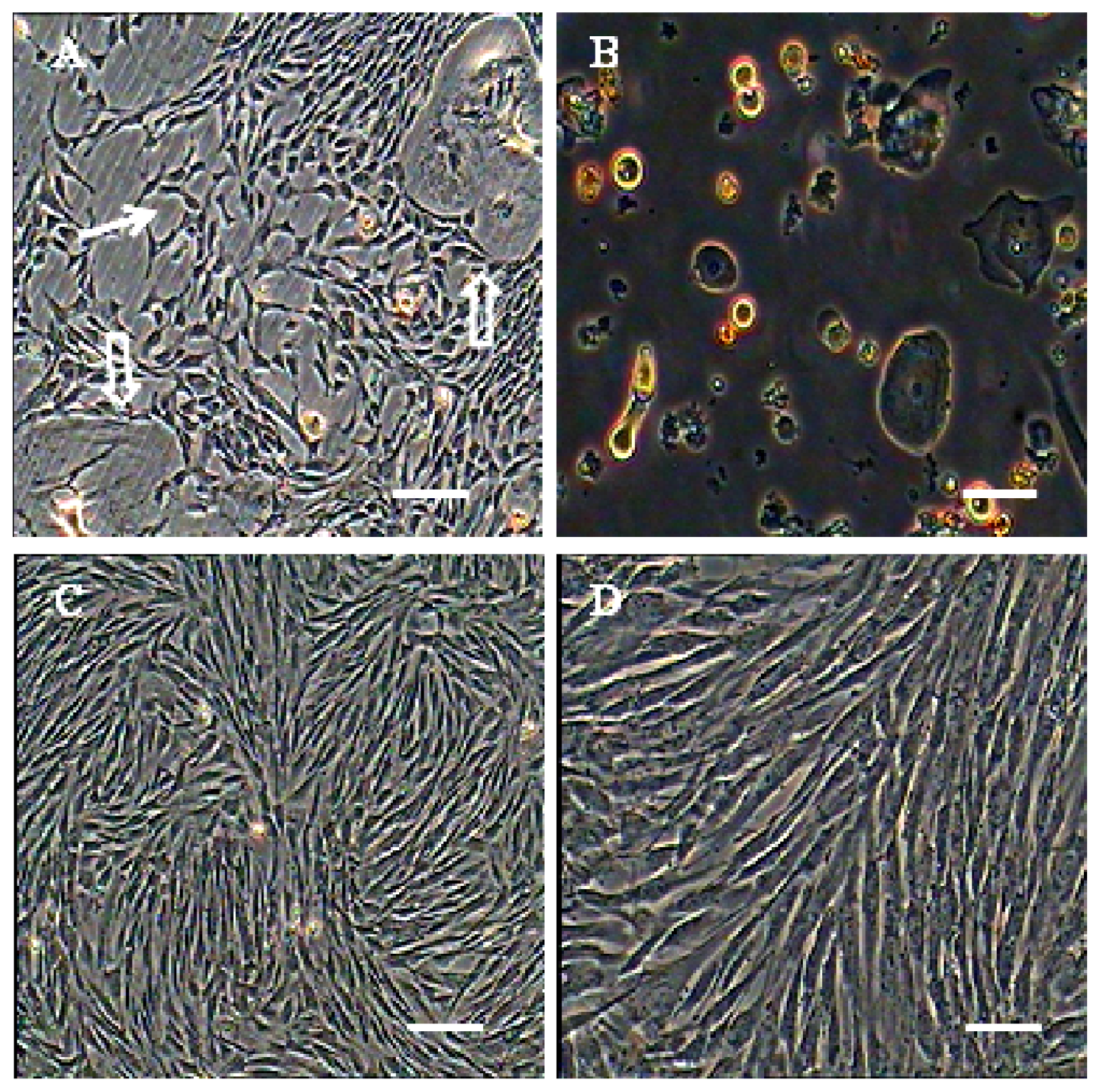
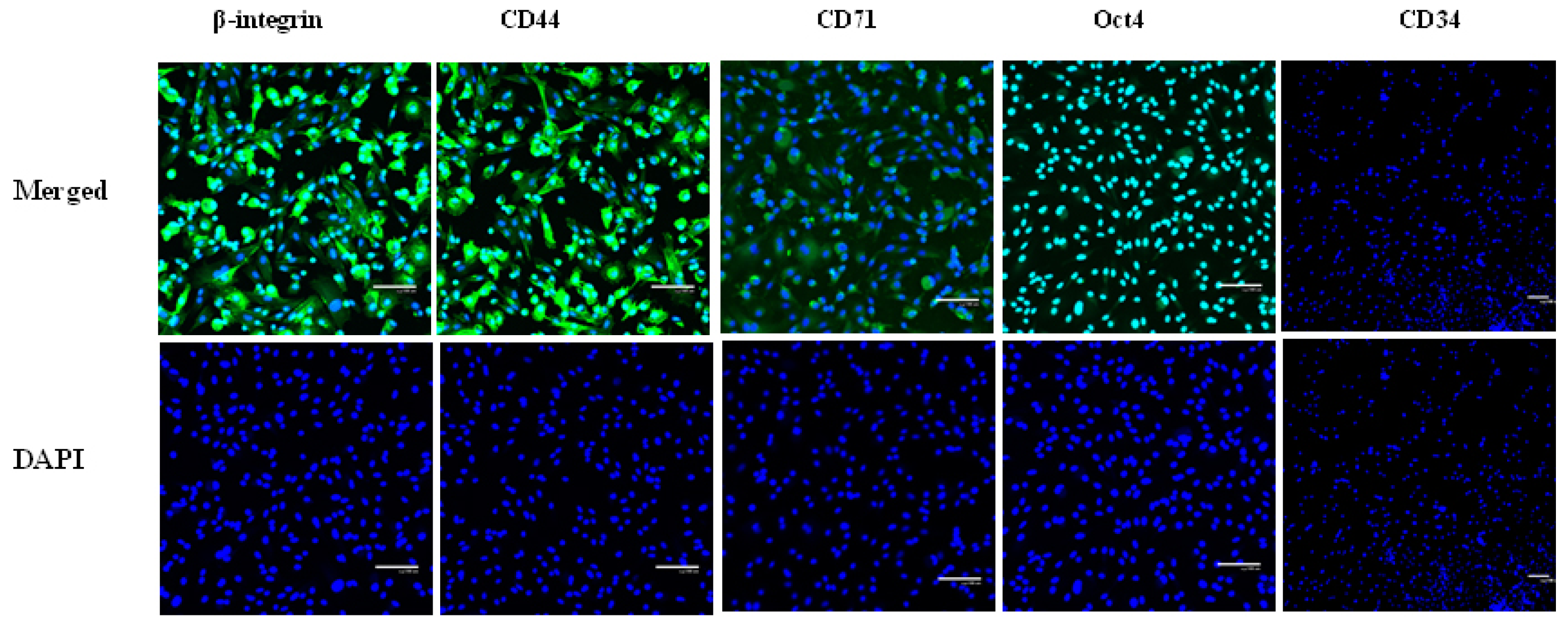
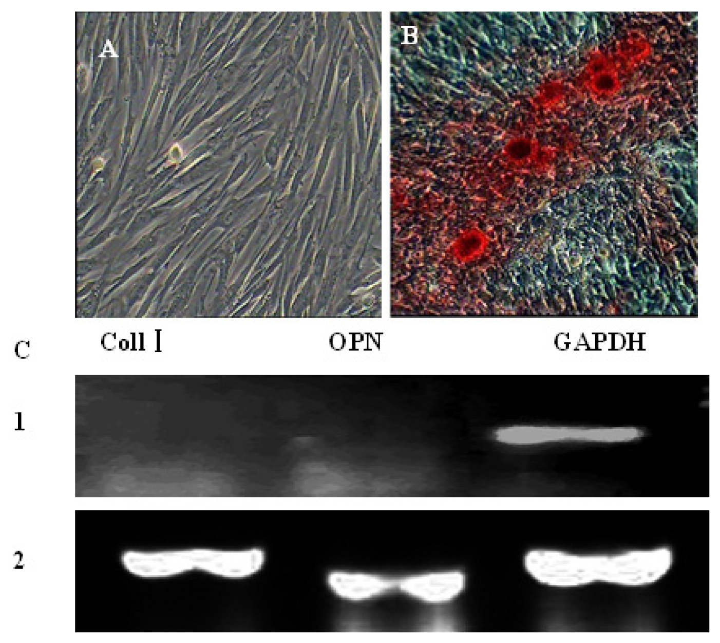
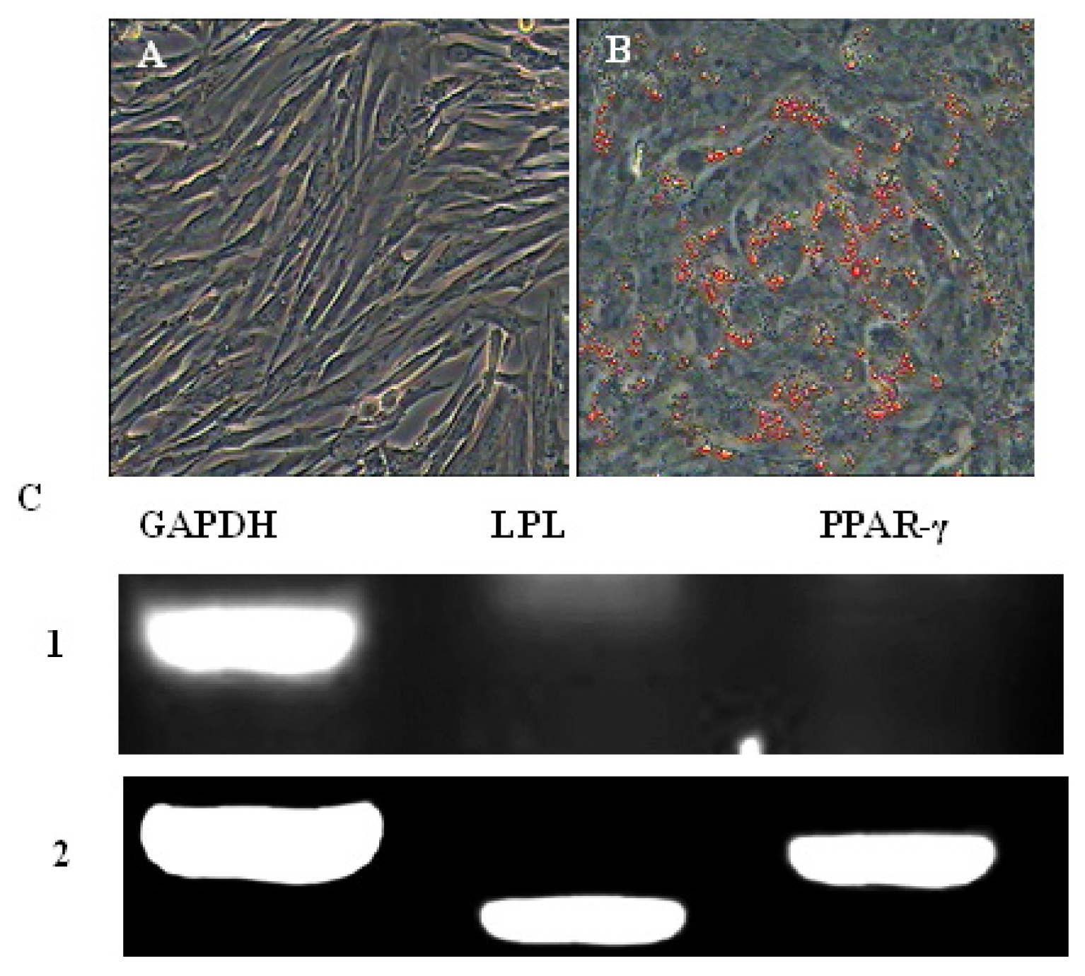
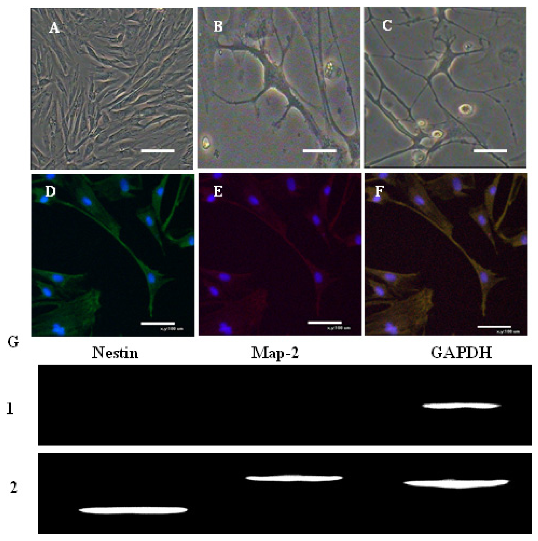
The use of amnion and amniotic fluid (AF) are abundant sources of mesenchymal stem cells (MSCs) that can be harvested at low cost and do not pose ethical conflicts. In human and veterinary research, stem cells derived from these tissues are promising candidates for disease treatment, specifically for their plasticity, their reduced immunogenicity, and high anti-inflammatory potential. This work aimed to obtain and characterize bovine amniotic fluid mesenchymal stem cells (AFMSC). The bovine AF from the amniotic cavity of pregnant gilts in the early stages of gestation (3- and 4-m-old bovine embryos) was collected. AFMSCs exhibit a fibroblastic-like morphology only starting from the fourth passage, being heterogeneous during the primary culture. Immunofluorescence results showed that AFMSCs were positive for β-integrin, CD44, CD73 and CD166, but negative for CD34, CD45. Meanwhile, AFMSCs expressed ES cell markers, such as Oct4, and when appropriately induced, are capable of differentiating into ectodermal and mesodermal lineages. This study reinforces the emerging importance of these cells as ideal tools in veterinary medicine; future studies aimed at a deeper evaluation of their immunological properties will allow a better understanding of their role in cellular therapy.
Keywords: bovine; amniotic fluid mesenchymal stem cell; multiply differentiation bovine; amniotic fluid mesenchymal stem cell; multiply differentiation