Article

Identification of a Plastid-Localized Bifunctional Nerolidol/Linalool Synthase in Relation to Linalool Biosynthesis in Young Grape Berries

1
Centre for Viticulture & Enology, College of Food Science and Nutritional Engineering, China Agricultural University, Beijing 100083, China
2
Department of Food Science and Engineering, College of Biological Sciences and Technology, Beijing Forestry University, Beijing 100083, China
3
Qingdao Huadong Winery Co., Ltd., Northside of Lisha Road, Laoshan District, Qingdao 266000, China
*
Author to whom correspondence should be addressed.
These authors contributed equally to this work.
Int. J. Mol. Sci. 2014, 15(12), 21992-22010; https://doi.org/10.3390/ijms151221992
Submission received: 2 September 2014 / Revised: 13 October 2014 / Accepted: 29 October 2014 / Published: 1 December 2014
(This article belongs to the Section Biochemistry)

Abstract

:
Monoterpenoids are a diverse class of natural products and contribute to the important varietal aroma of certain Vitis vinifera grape cultivars. Among the typical monoterpenoids, linalool exists in almost all grape varieties. A gene coding for a nerolidol/linalool (NES/LINS) synthase was evaluated in the role of linalool biosynthesis in grape berries. Enzyme activity assay of this recombinant protein revealed that it could convert geranyl diphosphate and farnesyl diphosphate into linalool and nerolidol in vitro, respectively, and thus it was named VvRILinNer. However, localization experiment showed that this enzyme was only localized to chloroplasts, which indicates that VvRILinNer functions in the linalool production in vivo. The patterns of gene expression and linalool accumulation were analyzed in the berries of three grape cultivars (“Riesling”, “Cabernet Sauvignon”, “Gewurztraminer”) with significantly different levels of monoterpenoids. The VvRILinNer was considered to be mainly responsible for the synthesis of linalool at the early developmental stage. This finding has provided us with new knowledge to uncover the complex monoterpene biosynthesis in grapes.

1. Introduction

Vitis vinifera is an economical and useful grape vine, and its numerous varieties facilitate the making of high-quality wines. Terpenenoids, especially monoterpenoids and sesquiterpenoids, greatly contribute to characteristic varietal aroma of the grapes and wines. Based on the level of free monoterpenoids produced in the berries, Mateo and Jiménez proposed that grape varieties could be generally classified into muscat varieties (up to 6 mg/L of free monoterpenoids), non-muscat aromatic varieties (between 1–4 mg/L) and neutral varieties (less than 1 mg/L) [1]. Typical monoterpenol components of aroma-rich grape varieties are linalool, geraniol, nerol, citronellol and α-terpineol. In particular, linalool is one of the most odour-active odorants [2,3] and has been shown as a crucial constituent of grape berry aroma. For example, in Burgundy region, “Chardonnay 809” is featured “muscat” aroma as a result of its very high concentration of linalool, which is distinguished from “Chardonnay 76” specified as being neutral variety [4]. Linalool also represents the characteristic varietal aroma of non-muscat “Riesling” berries [5,6]. So it is necessary to explore the mechanism of linalool accumulation during the development of grape berries.
Much evidence has demonstrated that higher plants like grape produce terpenenoid precursor, isopentenyl diphosphate (IPP), through two distinct routes: cytosolic mevalonic acid (MVA) pathway and plastidial 2C-methyl-d-erythritol 4-phosphate (MEP) pathway [7,8]. The condensation of IPP and its allylic isomer dimethylallyl diphosphate (DMAPP) yields geranyl diphosphate (GPP), farnesyl diphosphate (FPP) and geranylgeranyl diphosphate (GGPP) under the action of GPP synthase, FPP synthase and GGPP synthase, respectively. Afterwards, these products successively convert into different kinds of terpenoids, such as monoterpenes (C10), sesquiterpenes (C15), diterpenes (C20), triterpenes (C30), carotenoids (C40), and so on [8]. In this process, terpene synthase (TPS) plays a critical role in the formation of monoterpenes and sesquiterpenes [9,10]. According to previous reports on phylogenetic analysis of TPS amino acid sequences [11,12,13], TPS family is classified into seven subfamilies designated as TPS-a, TPS-b, TPS-c, TPS-d, TPS-e/f, TPS-g and TPS-h [10]. Among these, the members of TPS-g and TPS-b have been in vitro demonstrated to convert GPP, FPP and GGPP into monoterpenes, sesquiterpene and diterpene, respectively [10,13,14,15].
Two natural types of linalool isomers (3S and 3R) exist in higher plants, while the 3S-type are the predominant in grape berries [16]. The genes encoding linalool synthase, have been isolated and characterized from many angiosperms, such as Clarkia breweri [17], Arabidopsis thaliana [18], rice [19], snapdragon [15], strawberry, poplar and others [20]. According to the stereochemistry of their catalytic products, these known linalool synthases in plants are classified into two groups: 3S-linalool synthase (EC 4.2.3.25) and 3R-linalool synthase (EC 4.2.3.26). The former one belongs to the TPS-g subfamily, which includes nearly all the group I LIS responsible for the synthesizing 3S isomer of linalool. And the latter one belongs to the TPS-b subfamily responsible for the synthesis of 3R-linalool.
In Vitis vinifera groups, multiple terpene synthases (VvTPS) have been functionally identified [14]. The researchers have provided comprehensive annotation of the large VvTPS gene family, including chromosomal localization, enzyme functions and phylogeny relevant to the overall plant TPS gene family. Several genes of the TPS-g sub family from Pinot Noir and Cabernet Sauvignon have been considered to be potential 3S-linalool synthases. In vitro experiments showed that VvPNLinNer1, VvPNLinNer2 and VvCSLinNer were bi-functional enzymes and they could convert not only GPP to 3S-linalool but also FPP to (E)-nerolidol. Similarly, VvPNLNG1-4 also generates 3S-linalool, (E)-nerolidoland (E,E)-geranyl linalool when used GPP, FPP and GGPP, respectively, as substrates. However, the in vivo role of these enzymes remains unclear. It has generally been accepted that GPP and monoterpenes and triterpene such as 3S-linalool and (E,E)-geranyl linalool are synthesized in plastids, whereas FPP and sesquiterpenes like (E)-nerolidol are produced in the cytosol [7,9,21]. In grape berries, stable isotope feeding and tracing experiment also has supported that monoterpenes are synthesized through MEP pathway in plastids [16,22]. Since biosynthetic pathways of monoterpenes and sesquiterpenes are strictly compartmentalized in plant cells [15,21], subcellular localization analysis should be a powerful approach to clearly illustrate real roles of these bi-functional enzymes in vivo. However so far there is no report involving subcellular localization of grape bi-functional enzymes related to monoterpene synthesis.
In the present study, a gene encoding a bifunctional linalool/nerolidol synthase was isolated from “Riesling” berries. The biochemical properties of the corresponding enzyme were assessed in vitro and its plastic localization was clearly determined to be relevant to linalool synthesis. Additionally, the correspondence analysis was conducted between this gene expression and linalool accumulation in the berries of three cultivars with significantly different monoterpene levels. The result revealed that this enzyme greatly contributed to linalool production at the early stage of berry development. This finding would undoubtedly promote further understanding of the complex monoterpene biosynthesis in grape berry.

2. Results and Discussion

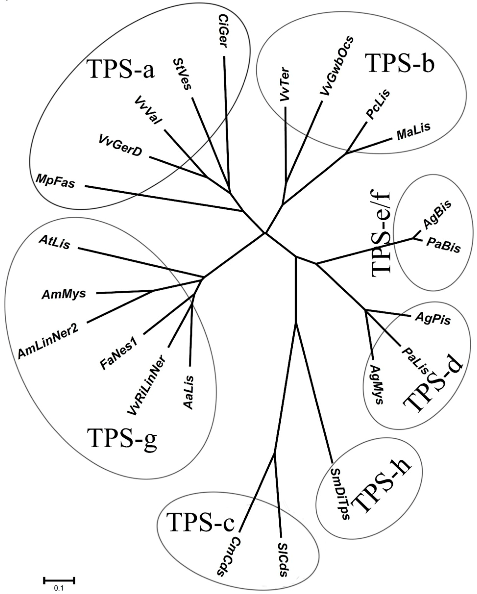
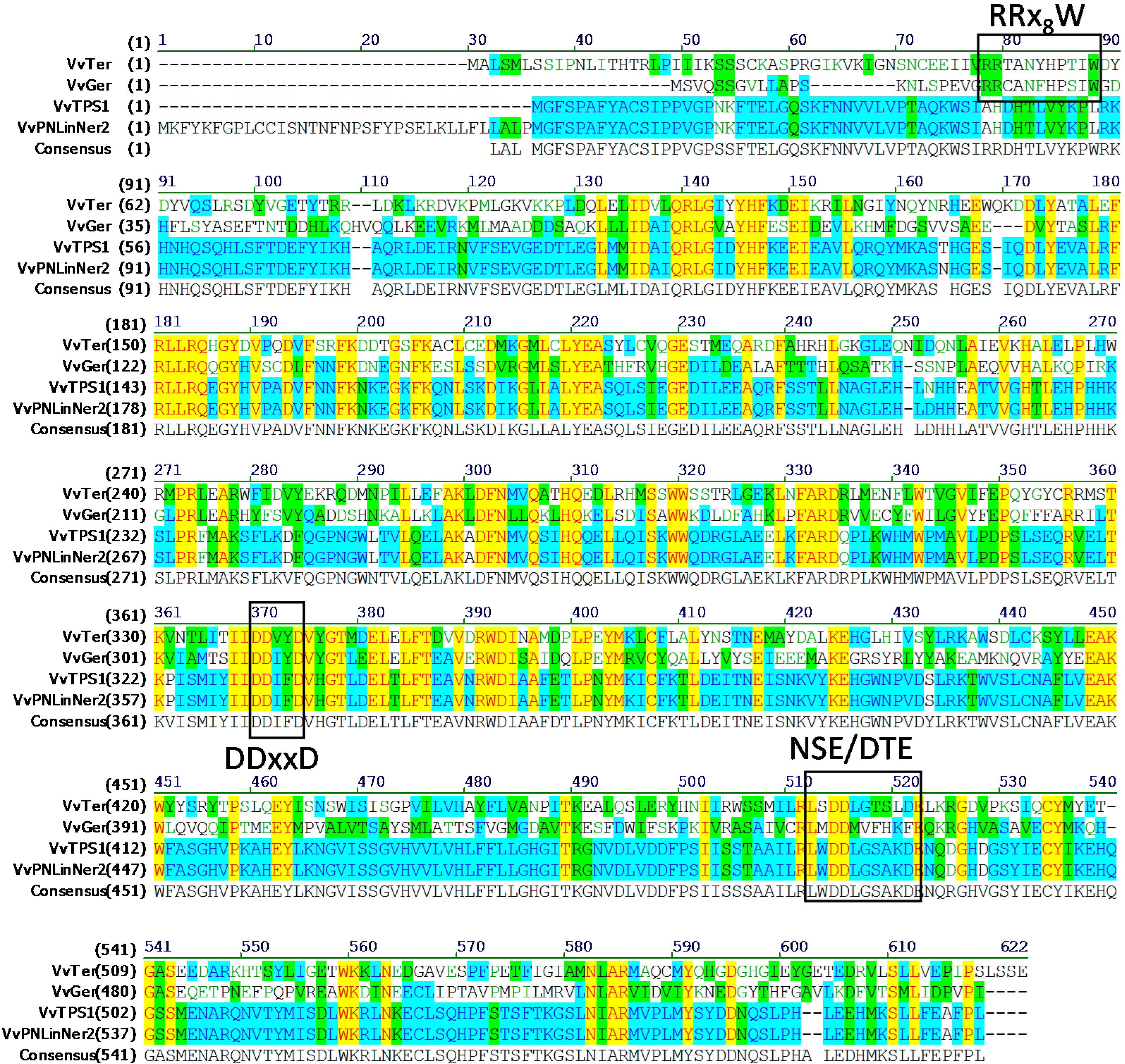
2.1. Identification of Gene Encoding 3S-Linalool Synthase from Grape Berries

According to the report of Martin et al. [14], a total of 32 gene models were annotated as linalool synthase genes or bifunctional linalool/nerolidol synthase in 12× grape genome based on nucleotide sequence similarity. Eighteen gene models out of the above 32 genes were considered to be pseudo genes that might be generated by mutation like nucleotide deletion in the progress of evolution. Only seven full-length cDNA fragments corresponding to six putative gene models (TPS54, 56–58, 61, 63) were screened out from grape cultivars Pinot Noir and Cabernet Sauvignon (PN and CS) cDNA libraries. These fragments coded for VvLinNer, and most of them were from the mixed PN template extracted from stems, berries, flowers and leaves. Here it should be noticed that VvPNLNGl1-4 (corresponding to TPS 57, TPS 63, TPS 58 and TPS 61) all lacked a transit signal peptide at their N-terminal. It is suggested that they might only catalyze the generation of nerolidol rather than linalool in vivo since sesquiterpenes like nerolidol are synthesized in cytoplasm and monoterpenes like linalool are synthesized in plastid. In the present study, we had been trying to get full-length cDNAs of the TPS54 and TPS56 gene models from “Riesling” berries, but only VvRiLinNer, corresponding to TPS56 was successfully obtained from young “Riesling” berries (Table 1). This VvRiLinNer sequence (accession numbers: JQ062931) contained an open reading frame (ORF) of 1734 base pairs for a predicted protein of 577 amino acids, a molecular weight of 66.1 kDa and a calculated isoelectric point of 6.22 (conducted by NAMAN6.0). Martin et al. [14] previously cloned VvPNLinNer2 (HM807392 in Genbank) from Vitis vinifera Pinot Noir and VvCSLinNer (HM807393 in Genbank) from Vitis vinifera Carbernet Sauvignon based on the designated gene VvTPS56. Here, the VvRiLinNer fragment obtained shared very high similarity in ORF with VvPNLinNer2 (99.9%) and VvCSLinNer (99.1%). Accordingly, it is suggested that VvRiLinNer, VvCSLinNerand VvPNLinNer2 were considered as allele genes of VvTPS56 (GSVIVT01005272001) distributing in different grape cultivars. However, the length of VvPNLinNer2 from “Pinot Noir” was 1839 bp, which had an additional 105 nucleotides in its 5'-terminus compared with the sequences of VvRiLinNer and VvCSLinNer. Several primers were designed on the basis of GSVIVT01005272001 nucleotide sequence to clone this putative 105-nucleotide sequence from Vitis vinifera Riesling berries, but no PCR product was attained. Interestingly, the localization of the VvRiLinNer was in silico predicted using three different databases (TargetP, PSORT and ChloroP). The results all showed that this enzyme was present in chloroplast, which is totally the same as VvPNLinNer2 with an additional 105 bp sequence. This phenomenon is difficult to explain at present.
Table 1. Annotation of the three terpenoids synthase (VvTPS) genes.
GenBank NumberGene ID (12×)VvTPS Gene Model Cloned GenesNote
XM002270140.1GSVIVT01005272001VvTPS56VvRiLinNer (JQ062931, this study) VvPNLinNer2 (HM807392) VvCSLinNer (HM807393)Full
XM002270071.1GSVIVT01005271001VvTPS65No gene has been clonedStop in first exon
XM002266813.1GSVIVT01005221001VvTPS54VvPNLinNer1(HM807391)Full
and , The “VvTPS Gene Model” and “Note” are annotated by Marin et al. [14].
Here it was worthwhile to note that the VvRiLinNer exhibited only approximately 55% and 54% amino acid homology with Antirrhinum majus nerolidol/linalool synthases [15], approximately 48% with Arabidopsis thaliana linalool synthase [18], as well as about 55% with Actinidia arguta linalool synthase [23]. Based on putative amino acid sequences, the VvRiLinNer was grouped into the TPS-g subfamily (Figure 1), which lacks the RRx8W motif in the N-terminal domain, a key feature of all monoterpene synthases of the TPS-b and TPS-d groups [11,24]. In the C-terminal domain, the VvRiLinNer contained two highly-conserved aspartate-rich motifs, DDxxD and NSE/DTE that are functional in the metal-dependent ionization of prenyl diphosphate substrates (Figure 2). The VvRiLinNer also showed the same feature sequences as VvPNLinNer2 and VvCSLinNer (Figure 2). This homology analysis strongly supports this view that the VvRiLinNer performs almost the same biological function as VvPNLinNer2 and VvCSLinNer in grape berry.
Figure 1. Phylogenetic tree illustrating the VvRiLinNer as a member of terpene synthase (TPS)-g subfamily. The unrooted neighbor-joining tree was created using ClustalX and visualised in TreeView. Division into subfamilies is based on cluster analysis (Bohlmann et al. 1998 [11]; Dudareva 2003 [12]; Chen et al., 2011 [10]). Only a subset of the known plant TPSs is shown. Sequence abbreviations: AaLis, Actinidia argute linalool synthase (ADD81294); AgBis, Abies grandis (E)-α-bisabolene synthase; AgMys, Abies grandis, myrcene synthase (AAB71084); AgPis, Abies grandis, pinene synthase (AAB71085); AmLinNer2, Antirrhinum majus linalool/nerolidol synthase (ABR24418); AmMys, Antirrhinum majusmyrcene synthase (AAO41726); AtLis, Arabidopsis thaliana linalool synthase (AAO85533); CiGer, Cichorium intybus germacrene A synthase (AAM21658); CmCds, Cucurbita maxima copalyl diphosphate synthase (AAD04292); FaNes1, Fragaria × ananassa nerolidol synthase1 (CAD57083); MaLis, Mentha aquatic linalool synthase (AAL99381); MpFas, Mentha × piperita (E)-β-farnesene synthase (AAB95209); PaBis, Picea abies (E)-α-bisabolene synthase (AAS47689); PaLis, Picea abies (−)-linalool synthase (AAS47693); PcLis, Perilla citriodora linalool synthase (AAX16075); SlCds, Solanum lycopersicum copalyl diphosphate synthase (BAA84918); SmDiTPS, Selaginella moellendorffii a bifunctionalditerpene synthase (AEK75338); StVes, Solanum tuberosum vetispiradiene synthase (BAA82092); VvGerD, Vitis vinifera (−)-germacrene D synthase (AAS66357); VvGwbOcs, Vitis vinifera L. Gewürztraminer (E)-beta-ocimene synthase (ADR74204); VvRiLinNer, Vitis vinifera L. Riesling linalool/nerolidol synthase (JQ062931); VvTer, Vitis vinifera (−)-α-terpineol synthase (AAS79352); VvVal, Vitis vinifera (+)-valencene synthase (AAS66358).
Ijms 15 21992 g001
Figure 2. Alignment of the amino acid sequence of VvRiLinNer with other VvTPS. The alignment of VvRiLinNer predicted amino acid sequence with other VvTPS was performed using Align X (Vector NTI Advance 11, Invitrogen). VvTer (GenBank No. AAS79351): (−)-α-terpineol synthase (Martin and Bohlmann 2004 [25]); VvGerD (GenBank No.Q6Q3H3): (−)-germacrene D synthase (Lücker et al. 2004 [26]); VvPNLinNer2 (GenBank No. HM807392): linalool/nerlidol synthase, VvCSLinNer (GenBank No. HM807393): linalool/nerlidol synthase (Martin et al. 2010 [14]). Letters in different colors or background colors indicate different degrees of sequence similarity between. Following are the explanations in detail. Letters in black font without the background: nonsimilar; green font without the background: weakly similar; green background: block of similar; blue background: conservative; yellow background: identical.
Ijms 15 21992 g002
To investigate the catalytic properties of the VvRiLinNer protein, we expressed VvRiLinNer in E. coil cells and purified the recombinant protein (Figure 3A). In vitro reaction analysis showed that VvRiLinNer could convert GPP into (3S)-linalool and convert FPP into (E)-nerolidol (Figure 3B,C). This means that VvRiLinNer is a bi-functional enzyme in vitro, which is similar to VvPNLinNer2 and VvCSLinNer [14]. In addition, we also detected trace levels of β-myrcene, (E)-β-ocimene, (Z)-β-ocimene and alloocimene in the GPP-containing reaction system, as well as trace levels of α-bisabolol, α-farnesene and β-farnesene in the FPP-containing reaction system (Figure 3B,C). No monoterpene or sesquiterpene was detected in two controls.
Figure 3. Analysis of SDS polyacrylamide gels of the purified recombinant VvRiLinNer proteins (A) and GC-MS analysis of the products of recombinant VvRiLinNer reaction system (B,C). In (A), M: Protein Molecular Weight Marker; 1: the purified protein VvRiLinNer. The apparent molecular weight of expressed recombinant proteins VvRiLinNer was estimated approximately 82 kDa, which is bigger than the predicted proteins from cDNA sequences because of the presence of the His-Tag (approximately 16 kDa); In (B) and (C), the reaction system contained the prokaryotically-expressed VvRiLinNer and geranyl diphosphate (B) or farnesyl diphosphate (C). The reaction product (3S)-linalool or (E)-neroidol was authentically identified. Compounds marked in numbers of (B) and (C) are listed as follows: 1 β-myrcene, 2 (Z)-β-ocimene, 3 (E)-β-ocimene, 4 alloocimene, 5 β-farnesene, 6 α-bisabolol, 7 α-farnesene. Among them, the compounds one & five were authentically identified, the others were tentatively identified according to MS spectra comparison with NIST 05 library.
Ijms 15 21992 g003

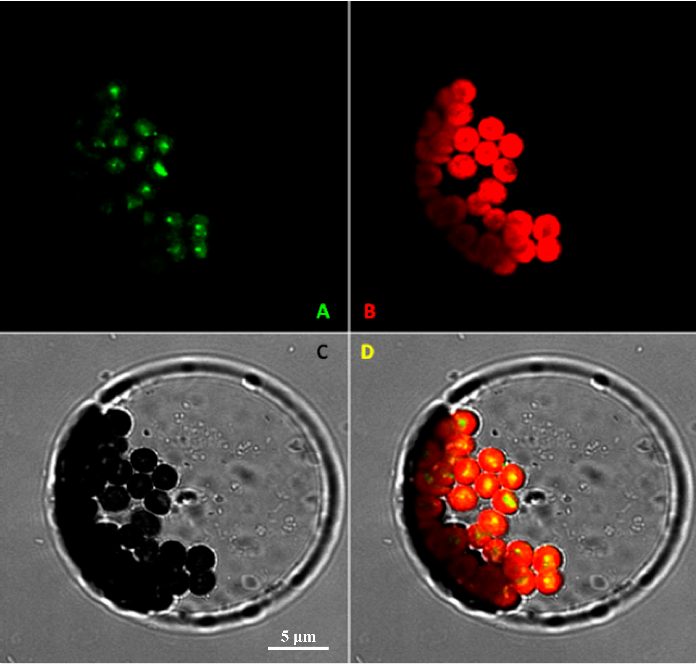
2.2. Subcellular Localization of VvRiLinNer Protein

Differential subcellular compartmentalization for the biosynthesis of FPP and GPP (the in vitro substrates of VvRiLinNerin plastids and the cytosol, respectively) was found in most plants including grape [8,9,21,27]. To determine in vivo localization of this enzyme, we examined the subcellular localization of the VvRiLinNer through transient expression of VvRiLinNer fused with a GFP reporter gene in Arabidopsis protoplasts. GFP fluorescence was clearly restricted in the chloroplasts, and no fluorescence signal was observed in the cytoplasm (Figure 4), which means that the present VvRiLinNer is mainly responsible for the synthesis of 3S-linalool in vivo. This inference was made mainly from two aspects of evidence. Firstly, using intact plastids separated from cell suspensions of Vitis vinifera Muscat de Frontignan, the authors have found that this organelle exhibits the activity of VvGPPS responsible for the synthesis of monoterpene precursor GPP [27]. Secondly, although FPPS and trace FPP pool in wild tomato also have been found to be chloroplast-localized [21,28], the feeding experiment in grape berries with the deuterium labeled precursors [5,5-H-2(2)]-1-deoxy-d-xylulose and [5,5-H-2(2)]-mevalonic acid lactone, a cytosolic MVA pathway-specific precursor, has recently demonstrated that adding IPP concentration would promote more production of sesquiterpenes, but hardly alter level of monoterpenes [22]. IPP is produced from the cytosolic MVA pathway. Up to date, there is still no evidence on presence of FPPS or FPP pool in the plastid of grape berry. Moreover, VvFPPS lacks a plastid signal peptide at N-terminal [29]. Combined the present subcellular localization with previous reports, we inferred that the VvRiLinNer acquired in this study should not participate in the synthesis of sesquiterpenes in grape berry.
Figure 4. Confocal laser scanning microscopy of transiently expressed VvRiLinNer-GFP fusions in Arabidopsis protoplasts (A) Green fluorescent protein fluorescence of VvRiLinNer-GFP; (B) Chlorophyll autofluorescence; (C) Light-microscopy image of intact protoplast. Bar: 5 μm; (D) Merged images. All images were taken with the 40× water-immersion objectives on the Zeiss LSM 510 (Zeiss, Germany).
Ijms 15 21992 g004

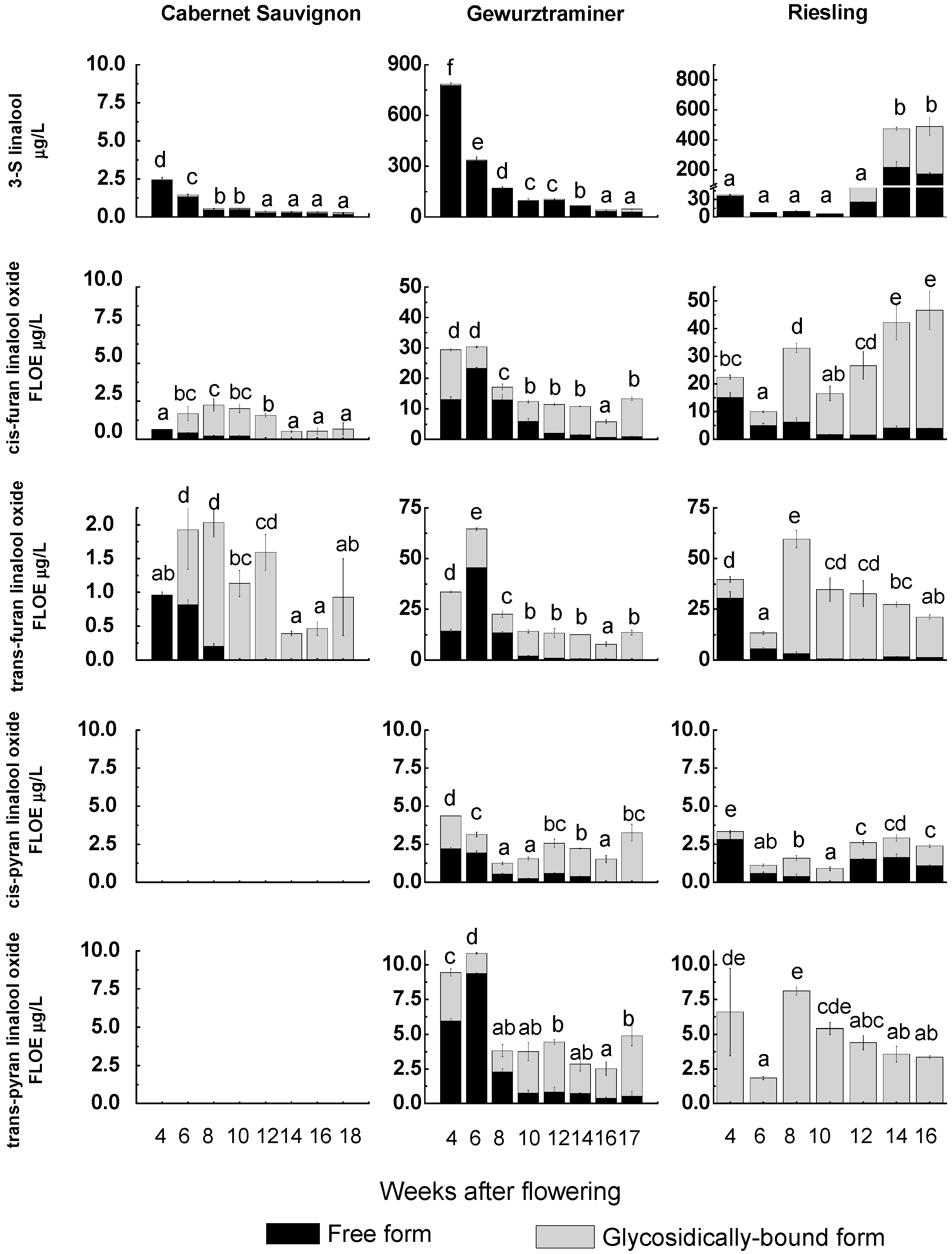
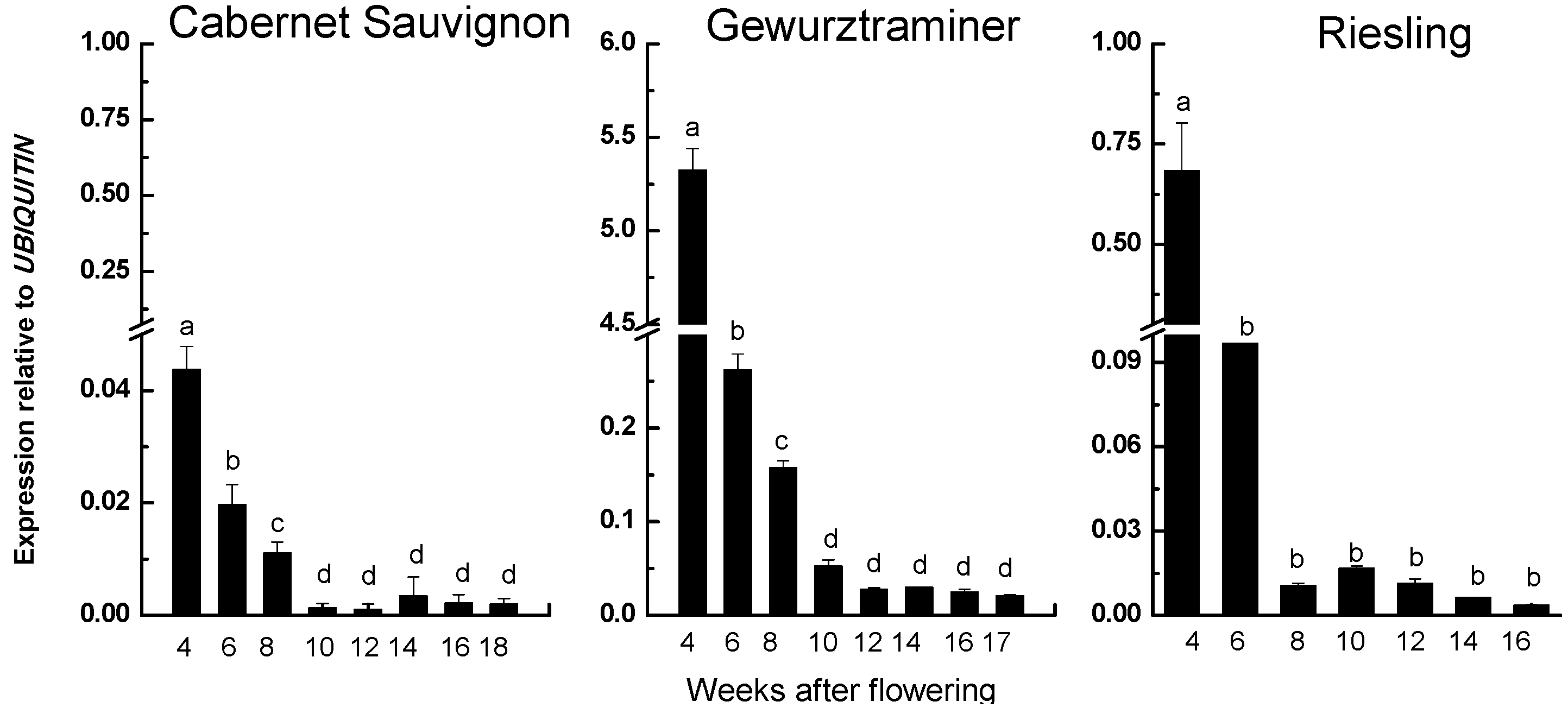
2.3. Correspondence Relationship between VvRiLinNer Expression and Linalool Accumulation in Three Grape Cultivars

To further determine the role of the VvRiLinNer in linalool accumulation during grape berry development, we examined free and glycosidically-bound forms of linalool and its structure-linked oxides in three grape cultivars containing Cabernet Sauvignon (CS, a neutral cultivar), Gewurztraminer (Gw, a muscat cultivar) and Riesling (a non-muscat aromatic cultivar) (Figure 5).
Figure 5. Profiling of linalool and its related oxide in developing grape berries. Each data point is the mean of three values and their standard deviation. Different letters (a–f) indicate that the concentrations of the compound had significantly difference at p < 0.05 according to Duncan’s multiple range test in SPSS. Furan linalool oxide equivalents (FLOE).
Ijms 15 21992 g005
Linalool showed an obvious decreasing trend during the development of both CS and Gw berries and existed predominantly in free form. At each development stage, the total concentration of free and glycosidically-bound 3S-linalool in Gw berries was always much higher than that in CS berries, a neutral grape cultivar, which was in good agreement with previous observations [2,30,31]. 3S-Linalool in young Riesling berries (4 Weeks after flowering “WAF” to 10 “WAF”) was mostly present in free form and declined in the concentration, which was similar to that in Gw and CS varieties. However, a sharp increase in the level of 3S-linalool appeared from 12 WAF to 14 WAF. This accumulation pattern was also observed in several muscat vaireties, such as Muscat Hamburg, Moscato Bianco and Muscat de Frontignan [2,31,32,33,34]. This high level was kept till technological harvest (16 WAF). Furthermore, the mature Riesling berries had almost equal contents of free and glycosidically-bound 3S-linalool, which was significantly different from Gw and CS berries.
Four linalool oxides were identified from Gw and Riesling berries, including trans (cis)-furan linalool oxides and trans (cis)-pyran linalool oxides, whereas only two furan forms were found in CS berries. These linalool oxides existed as both free form and glycosidically-bound form in the grape cultivars studied, apart from only bound-form trans-pyran linalool oxide detected in the Reisling berries. For most of the individual linalool oxide, free form generally accounted for higher proportion in young berries and glycosidically-bound form took up higher proportion in mature berries. The concentration of trans- and cis-furan linalool oxides in CS berries was much lower than those in other two cultivars. In CS and Gw cultivars, total concentration of individual linalool oxide (free form plus glycosidically-bound form, “furan linalool oxide equivalents”) in young berries were generally higher than those in mature berries. The concentration of the four glycosidically-bound linalool oxides (FLOE) all showed a significant increase from eight WAF to 10 WAF and then slightly fluctuated till mature in Gw berries, which is similar to Martin et al.’s previous study [29]. Meanwhile the free-type linalool oxides and 3-S-linalool kept decreasing. Regarding to “Riesling” cultivar, the concentrations of various linalool oxides in the four WAF berries were all higher than those in the six WAF berries, and at eight WAF these compounds increased with different magnitudes and total concentration of these linalool oxides (FLOE) showed a high level in mature berries. Generally, the overall levels of monoterpenes increase during grape maturation in muscat grape varieties [31,32]. In Muscat Hamburg berries, the main free monoterpenes including linalool increased from veraison [2], while in Muscat de Frontignan free linalool accumulated two weeks after veraison, reached the peak level two weeks later and then decreased until harvest [33]. However the present study showed a different result, that is, total concentration of linalool and its derivatives (FLOE) presented an overall decline trend along the mature of Gw berries.
The transcript abundance of the VvRiLinNer was assessed throughout the whole berry development stages. The expression trends of this gene were quite similar in the three grape cultivars (Figure 6). A sharp decrease in expression level was observed from four WAF to eight WAF, which highly positively correlated to the change of total concentration of linalool and its structure-linked compounds. After that, the transcripts of VvRiLinNer maintained at low level till berry harvest, which corresponded to low accumulation of linalool in both CS and Gw berries, but was inconsistent with the increase of linalool concentration after veraison in Riesling berries. Based on the above results, we considered that VvRiLinNer plays an important role in the synthesis of linalool at early stage of berry development.
The high level of linalool at late developmental stage of Riesling berries could be explained by the existence of other 3S-linalool synthases responsible for the production of linalool and its oxides at middle and late developmental stages of “Riesling” berries, as suggested by other researchers. Ebang-Oke et al. (2003) observed that a good correlation was observed between the expression of linalool synthase gene (Lis) and the accumulation of 3S-linalool along with ripening of Muscat de Frontignan grape berries [33]. Matarese et al. (2013) recently mentioned that the gene encoding for a bifunctional enzyme VvPNLinNer1 from Vitis vinifera Pinot Noir was greatly expressed in ripening Moscato Bianco and Aleatico berries [34].
Figure 6. VvLinNer relative expression in developing grape berries. Each data point is the mean of six values (multiplying three biological replicates by two technical replicates) and their standard deviation. Different letters (a–d) indicate that the transcript abundance of the gene had significantly difference at p < 0.05 according to Duncan’s multiple range test in SPSS.
Ijms 15 21992 g006

3. Experimental Section

3.1. Plant Materials

Grape berries (Vitis vinifera L. “Riesling”, “Cabernet Sauvignon”, “Gewurztraminer”) were sampled randomly every two weeks from four weeks after full bloom (WAF) to commercial harvest from 10-year-old grapevines grown in a vineyard located in the suburbs of Beijing, China. Grape berries earlier than four-week were not considered in this study because it was difficult to fully remove the seeds from these small berries. At this location, the commercial harvest date for these three varieties was different: 16 WAF for “Riesling”, 18 WAF for “Cabernet Sauvignon” and 17 WAF for “Gewurztraminer”. At each sampling, about 1000 berries were selected from at least 100-cluster selections at similar position of 50 whole vine selections. Any physically injured, abnormal and infected berries were not selected. The sampling time was fixed at around 10:00 A.M. in the morning. Samples were placed in foam boxes, and transported to the laboratory within two hours. The selected berries were washed with distilled water and dried briefly at room temperature. The seeds of these berries were removed by hand and the remainders were then immediately frozen in liquid nitrogen and stored at −80 °C until use.

3.2. Total RNA Extraction, Purification and cDNA Synthesis

The total RNA were extracted from grape berries without seeds by using an improved CTAB (cetyl triethyl ammonium bromide) method [35]. The total RNA were treated with RNase-free DNase to remove grape genome DNA and then purified with the RNA Purification Kit (Bio Basic Inc., Markham, ON, Canada). The quality of the total RNA was verified by demonstration of intact ribosomal bands following agarose gel electrophoresis and the absorbance ratio (A260/A280). For cDNA synthesis, the purified RNA was treated by using Reverse Transcription System Kit (Promega, Madison, WI, USA) and oligo d(T)18 Primer (Takara, Shiga, Japan) according to the supplier’s instruction. The synthesized cDNA was used as template in subsequent study of gene cloning and gene expression. The cDNA from the five-week “Riesling” berries was used to clone the target gene.

3.3. Isolation of Linalool Synthase (VvRiLin) Gene from Vitis vinifera Riesling Berries

The gene models which were annotated as putative linalool synthase genes (LIN) were obtained from database of VitisNet [36] and FLAGdb++ [37] both based on grape genome [38]. The redundant sequences, together with imperfect models caused by insert/deletion mutation and frame shift, were dropped. The amino acid sequences encoded by the rest of putative full length LIN genes were compared with these known plant LINs. Their subcellular localization was predicted through three web tools: ChloroP [39], TargetP [40] and SignalP [41]. The gene models that were predicted to contain a chloroplast-located signal peptide were selected as putative grape linalool synthase genes (in short, VvLins). A blast program was performed to screen DFCI Grape Gene Index database [42] to find EST fragments corresponding to putative VvLins. The in silico expression profile of VvLins was analyzed.
Specific primers covering the open reading frame (ORF) of VvLin genes were designed with the help of Prime 5.0 software package (Premier Biosoft International, Palo Alto, CA, USA) and were then used to amplify their nucleic acid fragments from grape berry cDNA through polymerase chain reaction (PCR).
The PCR system of a total volume of 20 μL was composed of 1 μL cDNA template, 1 μL of each primer (20 μM), 10 μL 2× Taq PCR plus master mix and 7 μL ddH2O (Sinopharm Chemical Reagent Beijing Co., Ltd., Beijing, China) according to the protocol of the supplier with slight modification. The temperature gradient PCR was conducted under various conditions: pre-denaturation at 95 °C for 5 min, denaturation at 94 °C for 30 s, primer annealing at 55 ± 10 °C for 1 min and extension at 72 °C for 1 min 30 s at 35 cycles, a final extension at 72 °C for 10 min, and then cooled to 10 °C in a thermal cycler (Techne TC-512 PCR System, Fisher Scientific UK Ltd., Leicestershire, UK). A band of about 1800 bp was gel-purified, subcloned into a pMD19-T Easy vector (Takara, Shiga, Japan) and sequenced using ABI3700 DNA sequencer (Applied Biosystems, Foster City, CA, USA).

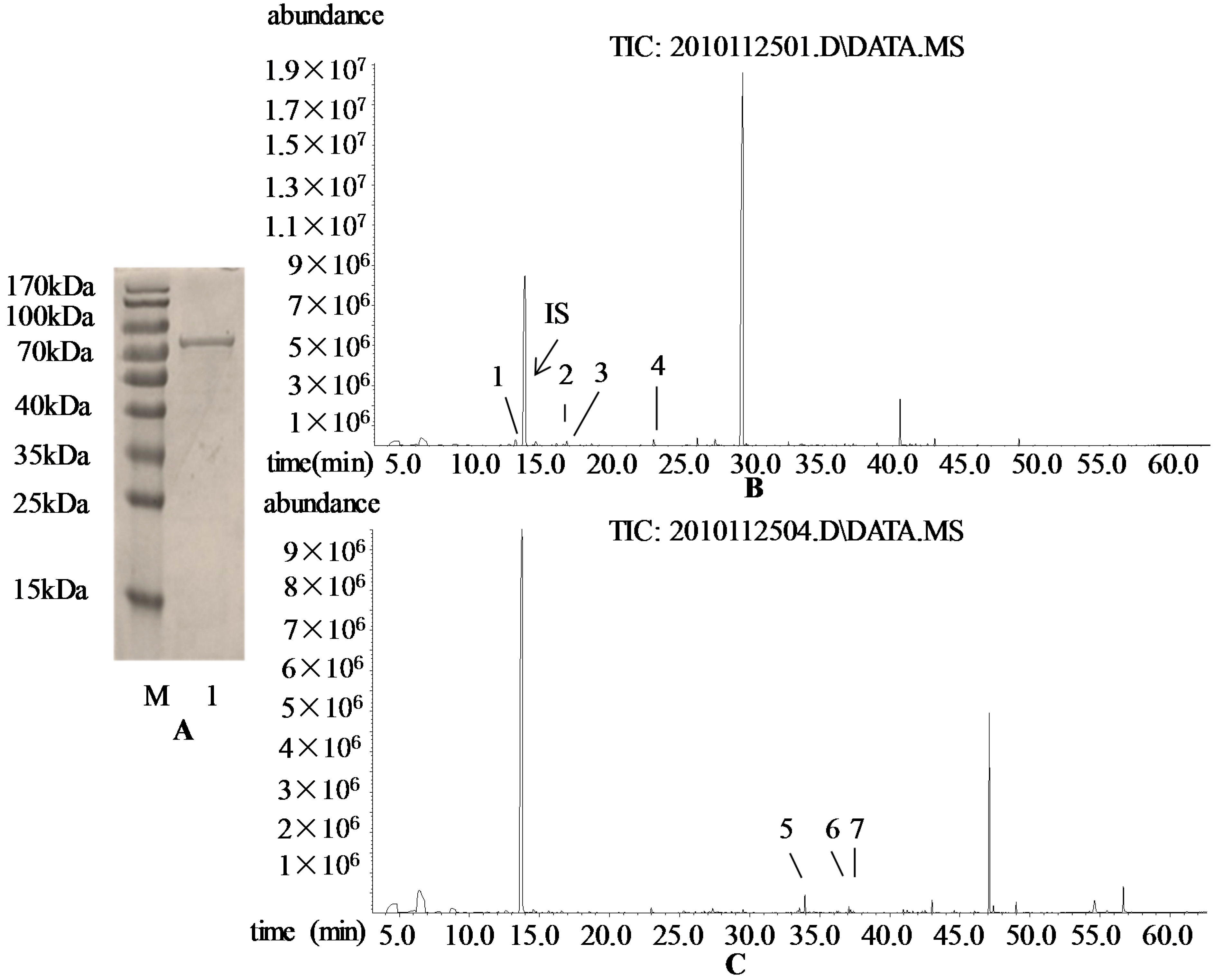
3.4. Prokaryotic Expression of VvRiLin and Purification of Recombinant Protein

To obtain prokaryotically-expressed protein, we subcloned the fragment containing VvRiLin into a pET32a expression vector (Novagen, Madison, WI, USA). Besides, the corresponding ORF was amplified from the reconstructive plasmid pMD19T-VvRiLin by PCR using Pfu DNA polymerase. Forward primer used was 5'-CGGAATTCATGGGATTCTCTCTTGCCTTTTACGC-3' containing an EcoR I site (underlined) at the initiating ATG codon. Reverse primer was 5'-CTTGACGCGTCGACCTACAGGGGAAATGCTTCAAAG-3' including a Sal I site (underlined). The encoding region of VvRiLin amplified by PCR was subcloned into the pET32a expression vector downstream of the (His)6 tag. The reconstructive plasmid pET32a-VvRiLin was transformed into E. coli Rosetta (DE3). A plasmid DNA was purified and verified by sequencing. Single colonies of E. coli Rosetta (DE3) with pET32a-VvRiLin were incubated overnight at 37 °C and 180 rpm in a 5-mL Luria-Bertani medium supplemented with ampicillin (100 μg/mL). After that, the medium of 500 μL was transferred into a 500-mL volume of fresh Lysogeny broth (LB) medium containing ampicillin (100 μg/mL). The bacterial culture was kept shaking in a water bath at 37 °C until reaching an optical density of 0.8 at a wave length of 600 nm. Isopropyl-β-d-thiogalactoside (IPTG) was then added into this medium to a final concentration of 1 mM. The medium was incubated at 16 °C and 150 rpm for another 16 h to reach an optimal protein concentration. The E. coli cells containing target protein were collected by centrifugation and further subjected to a purification procedure using Ni-NTA His Band Resin (Novagen, Madison, WI, USA). The purified protein was dialyzed against a buffer (30 mM HEPES, pH 7.2, 7.5 mM MgCl2, 20 μM MnCl2, 5% glycerol, 5 mM DTT) [25] as described by Fäldt et al. [43]. The analysis of SDS-Polyacrylamide gel electrophoresis showed that there existed a single band corresponding to the target protein on a 10% polyacrylamide gel (Biodee Biotechnology Co., Ltd., Beijing, China), which indicated that the prokaryotically-expressed protein obtained could meet the requirement of identification of biochemical function.

3.5. Catalytic Activity of the Purified VvRiLinNer Protein

Catalytic activity of the prokaryotically-expressed protein was performed in a 500-μL reaction buffer (pH 7.2) containing 30 mM HEPES (Biodee Biotechnology Co., Ltd., Beijing, China), 7.5 mM MgCl2, 20 μM MnCl2, 5% glycerol and 5 mM DTT (Sinopharm Chemical Reagent Beijing Co., Ltd., Beijing, China) and recombinant protein (0.5–0.8 μg) and 55/46 μM GPP/FPP (Echelon Bioscience Inc., Salt Lake City, UT, USA). Two types of the control were taken: one was to use the extracts from E. coli cells containing pET32a without the target gene instead of the recombinant VvRiLinNer protein and the other to use the heat-denatured recombinant VvRiLinNer protein. After incubation for 1 h at 30 °C, the reaction products were extracted with headspace-solid-phase micro-extraction (HS-SPME), and determined through GC–MS. The HS-SPME procedures were shown as follows: 500 μL reaction mixture, 1 μL 4-methyl-2-pentanol (1.039 mg/L water, internal standard) and 0.20 g NaCl were blended in a 15-mL sample vial tightly capped with a PTFE silicon septum and containing a magnetic stirrer. The vial was equilibrated at 40 °C for 30 min on a heating platform agitation. Then activated SPME fiber (50/30 μm DVB/Carboxen/PDMS, Supelco, Bellefonte, PA, USA; pretreated at 270 °C for 1 h) was inserted into the headspace for 30 min with continued heatingand agitation. Finally, the fiber was instantly desorbed in the GC injector for 8 min. Three replicates were conducted as to each reaction.
Quantitative analysis was performed on an Agilent 6890 Series GC System equipped with an Agilent 5975 Network Mass Selective Detector. The injector port was maintained at 250 °C. Helium (Millennium City Gas Co., Ltd., Beijing, China) was used as the carrier gas at 1 mL/min, and GC inlet was set in the splitless mode. The HP-INNOWAX capillary column (60 m, 0.25 mm ID, 0.25 μm film thickness) was used for peak identification. The procedure was as follows: an initial temperature of 50 °C (1 min hold) was increased to 220 °C at 3 °C/min and held at 220 °C for 5 min. Mass spectra in the electron impact mode at a voltage of 70 eV ionization energy was set to scan from m/z 20 to 450 and operated in the selective ion mode under autotune conditions. The products were identified through authentic standards or mass spectra matching in the standard NIST05 library.

3.6. Subcellular Localization of VvRiLinNer

For the observation of subcellular localization of VvRiLinNer, the open reading frame of VvRiLinNer was PCR-amplified with reference to the method described in “Prokaryotic expression of VvRiLinNer” section above. Forward primer (5'-CGGAATTCATGGGATTCTCTCCTGCCTT-3') and reverse primer (5'-CGGGATCCAGGGGATATGCTTCAAAGAG-3') were adopted. The amplicon was digested with EcoRI/Hind III (underlined) restriction enzymes. The PCR product was fused with the upstream of the enhanced GFP in the cauliflower mosaic virus 35S-EGFP-Ocs 3' vector (p-EZS-NL vector; [44]). This p-EZS-NL vector does not express GFP well without adding a coding sequence to the 5' end of the ORF of GFP; thus, the control cells do not show fluorescence of GFP [45]. The pEZS-NL vector and the Arabidopsis (ecotype Columbia) seeds were kindly provided by Professor Zhang Dapeng (School of Life Sciences, Tsinghua University, Beijing, China). Sequencing analysis confirmed the accuracy of fusion. Arabidopsis protoplasts were isolated from the leaves of three- to four-week old plants of Arabidopsis and transiently transformed through polyethylene glycol as described by Sheen (2002) [46]. Transient expression of GFP fusion protein was observed with a confocal laser-scanning microscope (LSM 510 META, Zeiss, Germany) after incubation at 23 °C for 16 h.

3.7. Analysis of VvRiLinNer Expression by Real Time PCR

Analysis of gene expression was performed by Real time PCR using the SYBR green method on an ABI 7300 Real-Time System (Applied Biosystems, Foster City, CA, USA). RNA isolation and cDNA preparation were conducted as described above. The synthesized cDNA was quantified and all the tested samples were adjusted to the same concentration. Each Real time PCR reaction (30 μL) contained 15 μL SYBR® Premix Ex Taq™, 0.6 μLRox Reference Dye (50×) (TaKaRa , Shiga, Japan), 13.5 μL ddH2O, 0.5 μL cDNA and 1 μL primer mixture (forward primers and reverse primers, 10 μM). A 162-bp fragment in the 3' region of the coding sequence was amplified by using specific primers designed by PerlPrimer (http://perlprimer.sourceforge.net/) [47]. The primers used were as follows: forward primer (5'-ATGACAATCAGAGTCTTCCACAC-3') and reverse primer (5'-AAGAAACGAAGAGACAATCCTG-3'). VvUbiquitin (GenBank No. AY684131) that is stably expressed in grape berry was selected as the internal control, and primers for it were Ubiquitin F (5'-GTGGTATTATTGAGCCATCCTT-3') and Ubiquitin R (5'-AACCTCCAATCCAGTCATCTAC-3'). The specification of the primers was verified on the basis of the following two facts: firstly, the dissociation curve for a pair of primers presented a single peak, and secondly, the fragment size of PCR product was consistent with the theoretical prediction, and the fragment sequence was shown to be correct through nucleotide sequencing.
For real time PCR, the template cDNA was denatured at 95 °C for 30 s followed by 40 cycles of amplification with 94 °C for 10 s, 60 °C for 31 s [48]. As to each grape sample, three independent RNA extraction replicates were done and each replicate was performed with two amplification reactions. The average cycle threshold (Ct) of VvUbiquitin at each sampling point was determined and transcript abundance of the target gene, normalized to VvUbiquitin, was calculated using the formula 2−∆Ct, where ∆Ct = Ct,targetCt,VvUbquitin. The mean and the standard derivation (SD) were estimated after 2−∆Ct calculation.

3.8. Quantification of Linalool and Its Derivatives by GC–MS

The berry samples without seeds, stored at −80 °C, were crushed and steeped for 140 min at 4 °C, and then were immediately centrifuged at 4 °C and 6000× g for 10 min. The supernatant was collected. The glycosidically-bound linalool and its related compounds were isolated by the Solid-Phase Extraction (in sort, SPE) method. In short, 5 mL of the clear juice was passed through the Cleanert PEP–SPE column (Bonna-agela Technologies ,Tianjin, China, 200 mg/6 mL), which was activated in advance with 10 mL methanol (Sinopharm Chemical Reagent Beijing Co., Ltd., Beijing, China) and 10 mL water. And then the water-soluble compounds were eluted with 5 mL of water, the free volatiles with 10 mL of dichloromethane (Sinopharm Chemical Reagent Beijing Co., Ltd., Beijing, China) and the target bound volatiles with 20 mL of methanol, sequentially. The flow rate was about 2 mL/min. The methanol elute was evaporated to dryness by reduced pressure distillation at 30 °C and then redissolved in 5 mL citrate-phosphate buffer solution (2 M, pH 5.0). Subsequently, 100 μL of the AR 2000 solution (DSM Food Specialties, Séclin, France, 100 mg/mL) was added to the glycoside extract and the mixture was vortexed and enzymatically hydrolyzed in an incubatorat 40 °C for 16 h to liberate the free aroma according to optimum conditions described previously [49]. The tube containing the mixture was stored at 4 °C and the liberated volatiles were extracted through SPME fiber within two days. Free types of linalool and its related compounds were extracted using HS-SPME according to the method of Wu et al. (2009) with minor modification [49]. That is, the grape juice of 5 mL, 10 μL 4-methyl-2-pentanol (1.039 mg/mL water, internal standard, Sigma-Aldrich) and 1.00 g NaCl (Sinopharm Chemical Reagent Beijing Co., Ltd., Beijing, China) were blended in a 15-mL sample vial tightly capped with a PTFE–silicon septum and containing a magnetic stirrer. The procedures of HS-SPME and GC–MS analysis were performed as described in the above section “assay of terpene synthase activity”. As to each sampling point, three independent extraction replicates were conducted.
The quantification procedure used was based on our prior research with some modifications [50]. According to the average concentration of sugar and acids in grape juice, the model solution consisted of 180 g/L glucose and 7 g/L tartaric acid and pH value was adjusted to 3.3 with NaOH (Sinopharm Chemical Reagent Beijing Co., Ltd., Beijing, China). The chemical standards used for quantification were linalool (96.5%, Sigma-Aldrich, Switzerland) and furan linalool oxides (97% purity, mixture of cis- and trans-furan linalool oxide, Fluka, Japan). Each standard was dissolved with ethanol (HPLC quanlity, Anpel Co., Ltd., Shanghai, China). All the standard stock solutions were then combined together, and eight different and successive concentrations of mixed standard solutions were prepared in juice model solution. Standard solutions of each level was extracted by HS-SPME and analyzed by GC–MS under the same condition as that of grape samples. The calibration curve of linalool was obtained and directly used for linalool quantification. As for furan linalool oxide, the calibration curve was acquired using the sum of two peak areas corresponding to cis- and trans-isomers, respectively. To quantify either of furan isomers in grape berry, we used this calibration curve to estimate the content of trans- or cis-furan linalool oxide on the basis of the peak area of single isomer. Also two pyran linalool oxide isomers were quantified using this furan calibration curve because there were no pyran linalool oxide standards bought. Similarly, only the peak area of the corresponding compound was considered when the content of trans- or cis-pyran linalool oxide was estimated. In this study, both linalool and furan linalool oxide calibration curves had above 99% of regression coefficient.

3.9. Statistics Analysis

A one-way analysis of variance (ANOVA) was carried out using the IBM SPPS Statistics 20 software (IBM Corp, Armonk, NY, USA). The statistical significance of the differences in the levels of linalool related compounds or gene expression among the berries of different developmental stages was examined employing Duncan’s multiple range test at p < 0.05.

4. Conclusions

The present study highlights subcellular localization of the VvRiLinNer from Vitisvinifera “Riesling” berry and the role of this enzyme in the synthesis of linalool and its derivatives in three grape cultivars with significantly different monoterpene levels. Combined strict chloroplast localization and previous reports, the VvRiLinNer obtained in this study is determined to be only responsible for linalool synthesis in grape berry although in vitro experiment shows that it is a bi-functional enzyme, catalyzing the conversion of GPP into linalool and FPP into nerolidol. Considering all linalool-related components including free and glucosidically-bound linalool as well as their derivatives, a close correlation exists between linalool accumulation and VvRiLinNer expression in young grape berries. These observations have added some new knowledgeof the accumulation of a wide verity of terpenes in grape berries. Meanwhile, some new questions arise, for example, what factors regulate the gene expression of VvRiLinNer in developing grape berries; what other genes are responsible for the synthesis of linalool and its oxides in middle and late developmental stages of Riesling berries; what differences exist between nucleotide sequences of these unknown genes and VvRiLinNer, and so on. Future studies are to be made to solve these problems.

Acknowledgments

This work was financially supported by the National Natural Science Foundation of China (Grant No. 31272118 to Qiu-Hong Pan and No. 31301732 to Bao-Qing Zhu). We owe thanks to Professor Zhang Dapeng and his team (School of Life Sciences, Tsinghua University, Beijing, China) for their kindly help in subcellular localization experiment.

Author Contributions

Qiu-Hong Pan and Chang-Qing Duan conceived and designed the experiments; Bao-Qing Zhu and Zhi-Qun Wang performed the experiments; Bao-Qing Zhu and Jian Cai analyzed the data; Xiao-Qing Xu contributed materials and analysis tools; Bao-Qing Zhu wrote the paper. Authorship must be limited to those who have contributed substantially to the work reported.

Conflicts of Interest

The authors declare no conflict of interest.

References

  1. Mateo, J.; Jiménez, M. Monoterpenes in grape juice and wines. J. Chromatogr. 2000, 881, 557–567. [Google Scholar] [CrossRef]
  2. Fenoll, J.; Manso, A.; Hellin, P.; Ruiz, L.; Flores, P. Changes in the aromatic composition of the Vitis vinifera grape muscat hamburg during ripening. Food Chem. 2009, 114, 420–428. [Google Scholar] [CrossRef]
  3. Caven-Quantrill, D.J.; Buglass, A.J. Seasonal variation of flavour content of English vineyard grapes, determined by stir-bar sorptive extraction-gas chromatography-mass spectrometry. Flavour Fragr. J. 2008, 23, 239–248. [Google Scholar] [CrossRef]
  4. Duchêne, E.; Legras, J.L.; Karst, F.; Merdinoglu, D.; Claudel, P.; Jaegli, N.; Pelsy, F. Variation of linalool and geraniol content within two pairs of aromatic and non-aromatic grapevine clones. Aust. J. Grape Wine Res. 2009, 15, 120–130. [Google Scholar] [CrossRef]
  5. Ebeler, S.E. Analytical chemistry: Unlocking the secrets of wine flavor. Food Rev. Int. 2001, 17, 45–64. [Google Scholar] [CrossRef]
  6. Skinkis, P.A.; Bordelon, B.P.; Wood, K.V. Comparison of monoterpene constituents in Traminette, Gewürztraminer, and Riesling winegrapes. Am. J. Enol. Vitic. 2008, 59, 440–445. [Google Scholar]
  7. Pichersky, E.; Noel, J.P.; Dudareva, N. Biosynthesis of plant volatiles: Nature’s diversity and ingenuity. Science 2006, 311, 808–811. [Google Scholar] [CrossRef] [PubMed]
  8. Hemmerlin, A.; Harwood, J.L.; Bach, T.J. A raison d’êtrefor two distinct pathways in the early steps of plant isoprenoid biosynthesis? Prog. Lipid Res. 2012, 51, 95–148. [Google Scholar] [CrossRef] [PubMed]
  9. Dudareva, N.; Negre, F.; Nagegowda, D.A.; Orlova, I. Plant volatiles: Recent advances and future perspectives. Crit. Rev. Plant Sci. 2006, 25, 417–440. [Google Scholar] [CrossRef]
  10. Chen, F.; Tholl, D.; Bohlmann, J.; Pichersky, E. The family of terpene synthases in plants: A mid-size family of genes for specialized metabolism that is highly diversified throughout the kingdom. Plant J. 2011, 66, 212–229. [Google Scholar] [CrossRef] [PubMed]
  11. Bohlmann, J.; Meyer-Gauen, G.; Croteau, R. Plant terpenoid synthases: Molecular biology and phylogenetic analysis. Proc. Natl. Acad. Sci. USA 1998, 95, 4126–4133. [Google Scholar] [CrossRef] [PubMed]
  12. Dudareva, N.; Martin, D.; Kish, C.M.; Kolosova, N.; Gorenstein, N.; Fäldt, J.; Miller, B.; Bohlmann, J. (E)-β-Ocimene and myrcene synthase genes of floral scent biosynthesis in snapdragon: Function and expression of three terpene synthase genes of a new terpene synthase subfamily. Plant Cell 2003, 15, 1227–1241. [Google Scholar] [CrossRef] [PubMed]
  13. Aharoni, A.; Giri, A.P.; Verstappen, F.W.; Bertea, C.M.; Sevenier, R.; Sun, Z.; Jongsma, M.A.; Schwab, W.; Bouwmeester, H.J. Gain and loss of fruit flavor compounds produced by wild and cultivated strawberry species. Plant Cell 2004, 16, 3110–3131. [Google Scholar] [CrossRef] [PubMed]
  14. Martin, D.; Aubourg, S.; Schouwey, M.; Daviet, L.; Schalk, M.; Toub, O.; Lund, S.; Bohlmann, J. Functional annotation, genome organization and phylogeny of the grapevine (Vitis vinifera) terpene synthase gene family based on genome assembly, flcDNA cloning, and enzyme assays. BMC Plant Biol. 2010, 10, 226. [Google Scholar] [CrossRef] [PubMed]
  15. Nagegowda, D.A.; Gutensohn, M.; Wilkerson, C.G.; Dudareva, N. Two nearly identical terpene synthases catalyze the formation of nerolidol and linalool in snapdragon flowers. Plant J. 2008, 55, 224–239. [Google Scholar] [CrossRef]
  16. Luan, F.; Wüst, M. Differential incorporation of 1-deoxy-d-xylulose into (3S)-linalool and geraniol in grape berry exocarp and mesocarp. Phytochemistry 2002, 60, 451–459. [Google Scholar] [CrossRef] [PubMed]
  17. Dudareva, N.; Cseke, L.; Blanc, V.M.; Pichersky, E. Evolution of floral scent in clarkia: Novel patterns of S-linalool synthase gene expression in the c. Breweri flower. Plant Cell 1996, 8, 1137–1148. [Google Scholar] [CrossRef] [PubMed]
  18. Chen, F.; Tholl, D.; D’Auria, J.C.; Farooq, A.; Pichersky, E.; Gershenzon, J. Biosynthesis and emission of terpenoid volatiles from arabidopsis flowers. Plant Cell 2003, 15, 481–494. [Google Scholar] [CrossRef] [PubMed]
  19. Yuan, J.S.; Köllner, T.G.; Wiggins, G.; Grant, J.; Degenhardt, J.; Chen, F. Molecular and genomic basis of volatile-mediated indirect defense against insects in rice. Plant J. 2008, 55, 491–503. [Google Scholar] [CrossRef] [PubMed]
  20. Lin, Y.L.; Lee, Y.R.; Huang, W.K.; Chang, S.T.; Chu, F.H. Characterization of S-(+)-linalool synthase from several provenances of Cinnamomum osmophloeum. Tree Genet. Genomes 2014, 10, 75–86. [Google Scholar] [CrossRef]
  21. Nagegowda, D.A. Plant volatile terpenoid metabolism: Biosynthetic genes, transcriptional regulation and subcellular compartmentation. FEBS Lett. 2010, 584, 2965–2973. [Google Scholar] [CrossRef] [PubMed]
  22. May, B.; Lange, B.M.; Wüst, M. Biosynthesis of sesquiterpenes in grape berry exocarp of Vitis vinifera L.: Evidence for a transport of farnesyl diphosphate precursors from plastids to the cytosol. Phytochemistry 2013, 95, 135–144. [Google Scholar] [CrossRef] [PubMed]
  23. Chen, X.; Yauk, Y.K.; Nieuwenhuizen, N.J.; Matich, A.J.; Wang, M.Y.; Perez, R.L.; Atkinson, R.G.; Beuning, L.L. Characterisation of an (S)-linalool synthase from kiwifruit (Actinidia arguta) that catalyses the first committed step in the production of floral lilac compounds. Funct. Plant Biol. 2010, 37, 232–243. [Google Scholar] [CrossRef]
  24. Aubourg, S.; Lecharny, A.; Bohlmann, J. Genomic analysis of the terpenoid synthase (AtTPS) gene family of Arabidopsis thaliana. Mol. Genet. Genomics 2002, 267, 730–745. [Google Scholar] [CrossRef] [PubMed]
  25. Martin, D.M.; Bohlmann, J. Identification of vitis vinifera (−)-α-terpineol synthase by in silico screening of full-length cdna ests and functional characterization of recombinant terpene synthase. Phytochemistry 2004, 65, 1223–1229. [Google Scholar] [CrossRef] [PubMed]
  26. Lücker, J.; Bowen, P.; Bohlmann, J. Vitis vinifera terpenoid cyclases: Functional identification of two sesquiterpene synthase cDNAs encoding (+)-valencene synthase and (−)-germacrene D synthase and expression of mono- and sesquiterpene synthases in grapevine flowers and berries. Phytochemistry 2004, 65, 2649–2659. [Google Scholar] [CrossRef] [PubMed]
  27. Soler, E.; Feron, G.; Clastre, M.; Dargent, R.; Gleizes, M.; Ambid, C. Evidence for a geranyl-diphosphate synthase located within the plastids of Vitis vinifera L. Cultivated in vitro. Planta 1992, 187, 171–175. [Google Scholar] [CrossRef] [PubMed]
  28. Sallaud, C.; Rontein, D.; Onillon, S.; Jabès, F.; Duffé, P.; Giacalone, C.; Thoraval, S.; Escoffier, C.; Herbette, G.; Leonhardt, N. A novel pathway for sesquiterpene biosynthesis from Z,Z-farnesyl pyrophosphate in the wild tomato solanum habrochaites. Plant Cell 2009, 21, 301–317. [Google Scholar] [CrossRef] [PubMed]
  29. Martin, D.M.; Chiang, A.; Lund, S.T.; Bohlmann, J. Biosynthesis of wine aroma: Transcript profiles of hydroxymethylbutenyl diphosphate reductase, geranyl diphosphate synthase, and linalool/nerolidol synthase parallel monoterpenol glycoside accumulation in Gewürztraminer grapes. Planta 2012, 236, 919–929. [Google Scholar] [CrossRef] [PubMed]
  30. Kalua, C.M.; Boss, P.K. Evolution of volatile compounds during the development of cabernet sauvignon grapes (Vitis viniferaL.). J. Agric. Food Chem. 2009, 57, 3818–3830. [Google Scholar] [CrossRef] [PubMed]
  31. Wilson, B.; Strauss, C.R.; Williams, P.J. Changes in free and glycosidically bound monoterpenes in developing muscat grapes. J. Agric. Food Chem. 1984, 32, 919–924. [Google Scholar] [CrossRef]
  32. Ebeler, S.E.; Thorngate, J.H. Wine chemistry and flavor: Looking into the crystal glass. J. Agric. Food Chem. 2009, 57, 8098–8108. [Google Scholar] [CrossRef] [PubMed]
  33. Ebang-Oke, J.; Billerbeck, G.D.; Ambid, C.; le Quéré, J.; Étiévant, P. Temporal expression of the lis gene from vitis vinifera L., cv. Muscat de frontignan. In Flavour Research at the Dawn of the Twenty-first Century. Proceedings of the 10th Weurman Flavour Research Symposium, Beaune, France, 25–28 June 2002; Editions Tec & Doc, Lavoisier Intercept: London, UK, 2003; pp. 321–325. [Google Scholar]
  34. Matarese, F.; Scalabrelli, G.; D’Onofrio, C. Analysis of the expression of terpene synthase genes in relation to aroma content in two aromatic Vitis vinifera varieties. Funct. Plant Biol. 2013, 40, 552–565. [Google Scholar] [CrossRef]
  35. He, F.; Fang, X.X.; Hu, M.; Pan, Q.H.; Shi, Y.; Duan, C.Q. Preparation and biological application of antibodies against leucoanthocyanidin reductase and anthocyanidin reductase from grape berry. Vitis J. Grapevine Res. 2009, 48, 69–75. [Google Scholar]
  36. Grimplet, J.; Cramer, G.R.; Dickerson, J.A.; Mathiason, K.; van Hemert, J.; Fennell, A.Y. Vitisnet: “Omics” integration through grapevine molecular networks. PLoS One 2009, 4, e8365. [Google Scholar] [CrossRef] [PubMed] [Green Version]
  37. Sandra, D.; Franck, S.; Jean-Philippe, T.; Cécile, G.; Véronique, B.; Philippe, G.; Séverine, G.; Philippe, L.; Jean-Charles, L.; Alain, L. Exploration of plant genomes in the FLAGdb++ environment. Plant Methods 2011, 7, 8. [Google Scholar] [CrossRef] [PubMed]
  38. Jaillon, O.; Aury, J.-M.; Noel, B.; Policriti, A.; Clepet, C.; Casagrande, A.; Choisne, N.; Aubourg, S.; Vitulo, N.; Jubin, C. The grapevine genome sequence suggests ancestral hexaploidization in major angiosperm phyla. Nature 2007, 449, 463–467. [Google Scholar] [CrossRef] [PubMed]
  39. ChloroP. Available online: http://www.cbs.dtu.dk/services/ChloroP (accessed on 10 July 2010).
  40. TargetP. Available online: http://www.cbs.dtu.dk/services/TargetP (accessed on 10 July 2010).
  41. SignalP. Available online: http://www.cbs.dtu.dk/services/SignalP (accessed on 10 July 2010).
  42. DFCI Grape gene index. Available online: http://compbio.dfci.harvard.edu/tgi/cgi-bin/tgi/gimain.pl?gudb=grape (accessed on 20 June 2010).
  43. Fäldt, J.; Martin, D.; Miller, B.; Rawat, S.; Bohlmann, J. Traumatic resin defense in norway spruce (Picea abies): Methyl jasmonate-induced terpene synthase gene expression, and cDNA cloning and functional characterization of (+)-3-carene synthase. Plant Mol. Biol. 2003, 51, 119–133. [Google Scholar] [CrossRef] [PubMed]
  44. The Map of p-EZS-NL Vector. Available online: https://deepgreen.dpb.carnegiescience.edu (accessed on 1 August 2010).
  45. Fan, R.C.; Peng, C.C.; Xu, Y.H.; Wang, X.F.; Li, Y.; Shang, Y.; Du, S.Y.; Zhao, R.; Zhang, X.Y.; Zhang, L.Y.; et al. Apple sucrose transporter SUT1 and sorbitol transporter SOT6 interact with cytochrome b5 to regulate their affinity for substrate sugars. Plant Physiol. 2009, 150, 1880–1901. [Google Scholar] [CrossRef] [PubMed]
  46. Sheen, J. A Transient Expression Assay Using Arabidopsis Mesophyll Protoplasts. 2002. Protocols of Jen Sheen’s Lab. Available online: http://molbio.mgh.harvard.edu/sheenweb/protocols_reg.html (accessed on 29 October 2009).
  47. Marshall, O.J. Perlprimer: Cross-platform, graphical primer design for standard, bisulphite and real-time PCR. Bioinformatics 2004, 20, 2471–2472. [Google Scholar] [CrossRef] [PubMed]
  48. Zhang, Z.Z.; Li, X.X.; Zhu, B.Q.; Wen, Y.Q.; Duan, C.Q.; Pan, Q.H. Molecular characterization and expression analysis on two isogenes encoding 3-deoxy-d-arabino-heptulosonate 7-phosphate synthase in grapes. Mol. Biol. Rep. 2011, 38, 4739–4747. [Google Scholar] [CrossRef] [PubMed]
  49. Loscos, N.; Hernandez-Orte, P.; Cacho, J.; Ferreira, V. Evolution of the aroma composition of wines supplemented with grape flavour precursors from different varietals during accelerated wine ageing. Food Chem. 2010, 120, 205–216. [Google Scholar] [CrossRef]
  50. Wu, Y.; Pan, Q.; Qu, W.; Duan, C. Comparison of volatile profiles of nine litchi (litchi chinensis sonn.) cultivars from southern china. J. Agric. Food Chem. 2009, 57, 9676–9681. [Google Scholar] [CrossRef] [PubMed]

