Review

Activation of Defense Mechanisms against Pathogens in Mosses and Flowering Plants

1
Departamento de Biología Molecular, Instituto de Investigaciones Biológicas Clemente Estable, Avenida Italia 3318, CP 11600, Montevideo, Uruguay
2
Laboratorio de Fisiología Vegetal, Centro de Investigaciones Nucleares, Facultad de Ciencias, Mataojo 2055, CP 11400, Montevideo, Uruguay
*
Author to whom correspondence should be addressed.
Int. J. Mol. Sci. 2013, 14(2), 3178-3200; https://doi.org/10.3390/ijms14023178
Submission received: 4 January 2013 / Revised: 23 January 2013 / Accepted: 23 January 2013 / Published: 4 February 2013
(This article belongs to the Special Issue Abiotic and Biotic Stress Tolerance Mechanisms in Plants)

Abstract

:
During evolution, plants have developed mechanisms to cope with and adapt to different types of stress, including microbial infection. Once the stress is sensed, signaling pathways are activated, leading to the induced expression of genes with different roles in defense. Mosses (Bryophytes) are non-vascular plants that diverged from flowering plants more than 450 million years ago, allowing comparative studies of the evolution of defense-related genes and defensive metabolites produced after microbial infection. The ancestral position among land plants, the sequenced genome and the feasibility of generating targeted knock-out mutants by homologous recombination has made the moss Physcomitrella patens an attractive model to perform functional studies of plant genes involved in stress responses. This paper reviews the current knowledge of inducible defense mechanisms in P. patens and compares them to those activated in flowering plants after pathogen assault, including the reinforcement of the cell wall, ROS production, programmed cell death, activation of defense genes and synthesis of secondary metabolites and defense hormones. The knowledge generated in P. patens together with comparative studies in flowering plants will help to identify key components in plant defense responses and to design novel strategies to enhance resistance to biotic stress.

1. Introduction

Plants are in permanent contact with a variety of microbial pathogens, such as fungi, oomycetes, bacteria and viruses. To ward off these pathogens, plants must recognize the invaders and activate fast and effective defense mechanisms that arrest the pathogen. Perception of the pathogens is central to the activation of a successful plant defense response. Plant cells are capable of sensing evolutionarily conserved microbial molecular signatures, collectively named pathogen-associated molecular patterns (PAMPs) or microbe-associated molecular patterns (MAMPs), by plant pattern recognition receptors (PRRs) [13]. MAMPs are molecules that are essential for microbe fitness and survival and are conserved between different species, resulting in an efficient form to sense the presence of pathogens by the plant. Perception of PAMPs by PRRs activates an immune response, referred to as PAMP-triggered immunity (PTI), which provides protection against non-host pathogens and limits disease caused by virulent pathogens [4]. Pathogens adapted to their host plants can deliver virulence effector proteins into plant cells, which target key PTI components and inhibit plant defense [59]. In turn, plants have evolved resistance (R) proteins to detect directly or indirectly the effector proteins and trigger disease resistance effector-triggered immunity (ETI), which is highly specific and often accompanied by the hypersensitive response (HR) and systemic acquired resistance (SAR). An additional surveillance system for the presence of pathogens is the release or production of endogenous damage associated molecular patterns (DAMPs), including plant cell wall and cutin fragments that are released by the enzymatic action of pathogens and also trigger immune responses [3,10,11]. Thus, plant immunity can be divided in two phases: PTI triggered by PAMPs and ETI triggered by effectors, with the difference being that activated immune responses in ETI are faster and amplified compared to those in PTI [4,12]. ETI and PTI pathways result in activation of an overlapping set of downstream immune responses, suggesting that there is a continuum between PTI and ETI [13]. These downstream defense responses include the activation of multiple signaling pathways and transcription of specific genes that limit pathogen proliferation and/or disease symptom expression. In addition, antimicrobial compounds are produced, reactive oxygen species (ROS) accumulate, cell wall defense mechanisms are activated and defense hormones, such as salicylic acid (SA), ethylene and jasmonic acid (JA) accumulate [4,1417].
During the last few years, some progress has been made on the defense mechanisms activated in mosses (Bryophytes) during pathogen assault. The moss Physcomitrella patens (P. patens) is an interesting model plant to perform functional studies of genes involved in stress responses, because its genome has been sequenced, targeted knock-out mutants can be generated by homologous recombination and it has a dominant haploid phase during its life cycle [1820]. Mosses are non-vascular plants that diverged from flowering plants more than 450 million years ago [21]. P. patens, together with the sequenced vascular spikemoss Selaginella moellendorffii [22], provide an evolutionary link between green algae and angiosperms, allowing comparative studies of the evolution of plant defense mechanisms and gene function. In nature, mosses are infected with microbial pathogens, resulting in chlorosis and necrosis of plant tissues [2325]. Necrotrophic pathogens are capable of infecting and colonizing P. patens tissues, leading to the activation of defense responses [2632]. Most likely, P. patens utilizes similar mechanisms for pathogen recognition as flowering plants, since chitin (PAMP) [31] and probably cell wall fragments generated by the action of cell wall degrading enzymes from bacterial pathogens (DAMPs) [26] are sensed by P. patens cells and typical PRRs and R genes homologues are present in its genome [3335]. In addition, many of the cellular and molecular defense reactions activated in P. patens are similar to those reported in flowering plants. The present paper reviews the current knowledge of defense responses activated in P. patens and compares them to those activated in flowering plants after pathogen assault.

2. Broad Host Range Pathogens Infect both Mosses and Flowering Plants

Broad host range pathogens are capable of infecting a variety of plant species, including flowering plants and mosses. These are successful pathogens, which have adapted and developed effective invasion strategies causing disease by producing different compounds, including enzymes and toxins that interfere with metabolic targets common to many plant species. In this review, we focus on the broad host range fungus Botrytis cinerea, the bacterium Pectobacterium carotovorum subsp. carotovorum and the oomycetes Pythium irregulare and Pythium debaryanum. These are necrotrophic pathogens that actively kill host tissue prior to or during colonization and thrive on the contents of dead or dying cells [36].
B. cinerea is a necrotrophic fungal pathogen that attacks over 200 different plant species [37] and penetrates plant tissues by producing toxins and multiple cell wall degrading enzymes (CWDEs), including pectinolytic enzymes and cutinases that kill the host cells causing grey mould disease in many crop plants [38]. B. cinerea is primarily a pathogen of dicotyledonous plants, but some monocot species, including onions and lilies, are also infected [39,40]. B. cinerea also infect P. patens plants, producing maceration of the tissues and browning of stems and juvenile protonemal filaments [26,28].
P.c. carotovorum (ex Erwinia carotovora subsp. carotovora) cause soft rot in a wide range of plant species, including vegetables, potato and Arabidopsis [41]. P.c. carotovorum is often described as a brute-force pathogen, because its virulence strategy relies on plant CWDEs, including cellulases, proteases and pectinases, which disrupt host cell integrity and promote tissue maceration [42,43]. Cell-free culture filtrate (CF) containing CWDEs from P.c. carotovorum produces similar symptoms (Figure 1) and defense gene expression as those caused by P.c. carotovorum infection, demonstrating that CWDEs are the main virulence factors [4348]. In addition, these CWDEs release cell wall fragments, including oligogalacturonides that act as DAMPS activating an immune response in plant cells evidenced by the activation of defense related genes and phytoalexin accumulation [44,4951]. Recently, it was shown that two strains of P.c. carotovorum, SCC1, harboring the harpin-encoding hrpN gene, which is an elicitor of the hypersensitive response (HR) [52], and the HrpN-negative P.c. carotovorum strain (SCC3193) [53] infect and cause maceration in leaves of P. patens [26]. Green fluorescent protein (GFP) labeled- P.c. carotovorum, was detected in the apoplast, as well as the space of P. patens invaded leaf cells (Figure 2). Treatments with CFs of these strains also caused symptom development in moss tissues, evidenced by tissue maceration and browning, which was more severe with the HrpN-positive strain, suggesting that harpin may contribute to P.c. carotovorum virulence [26].
Pythium species are soil-borne vascular pathogens, which infect the plants through the root tissues and under humid conditions cause pre-/post-emergence damping-off and root and stem rots in important crop species. Pythium infect host young tissues, and maceration is caused by both toxins and cell wall degrading enzymes, such as pectinases, hemicellulases, cellulases and proteinases [54,55]. P. irregulare and P. debaryanum infect P. patens, producing tissue maceration and browning of young protonemal tissues, stems and leaves [29]. In nature, Pythium ultimum infect mosses, causing the formation of areas of dead moss tissues [24]. In all these moss-pathogen interactions, multiple defense reactions are activated in plant cells, although they are not sufficient to stop infection, and after a few days, moss tissues are degraded, leading to plant decay.

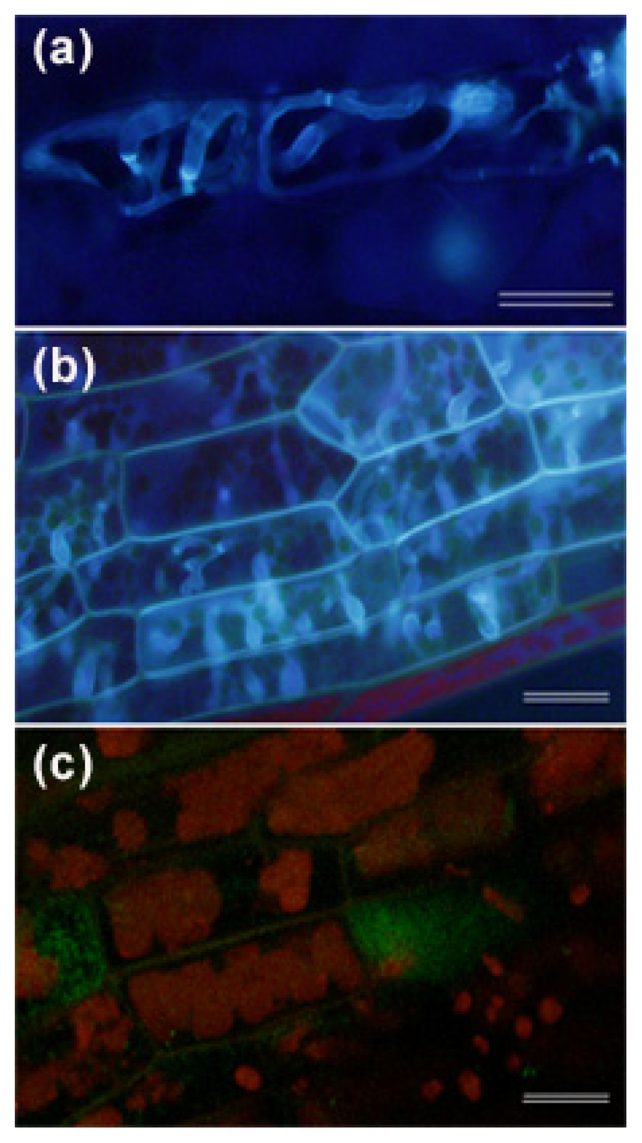
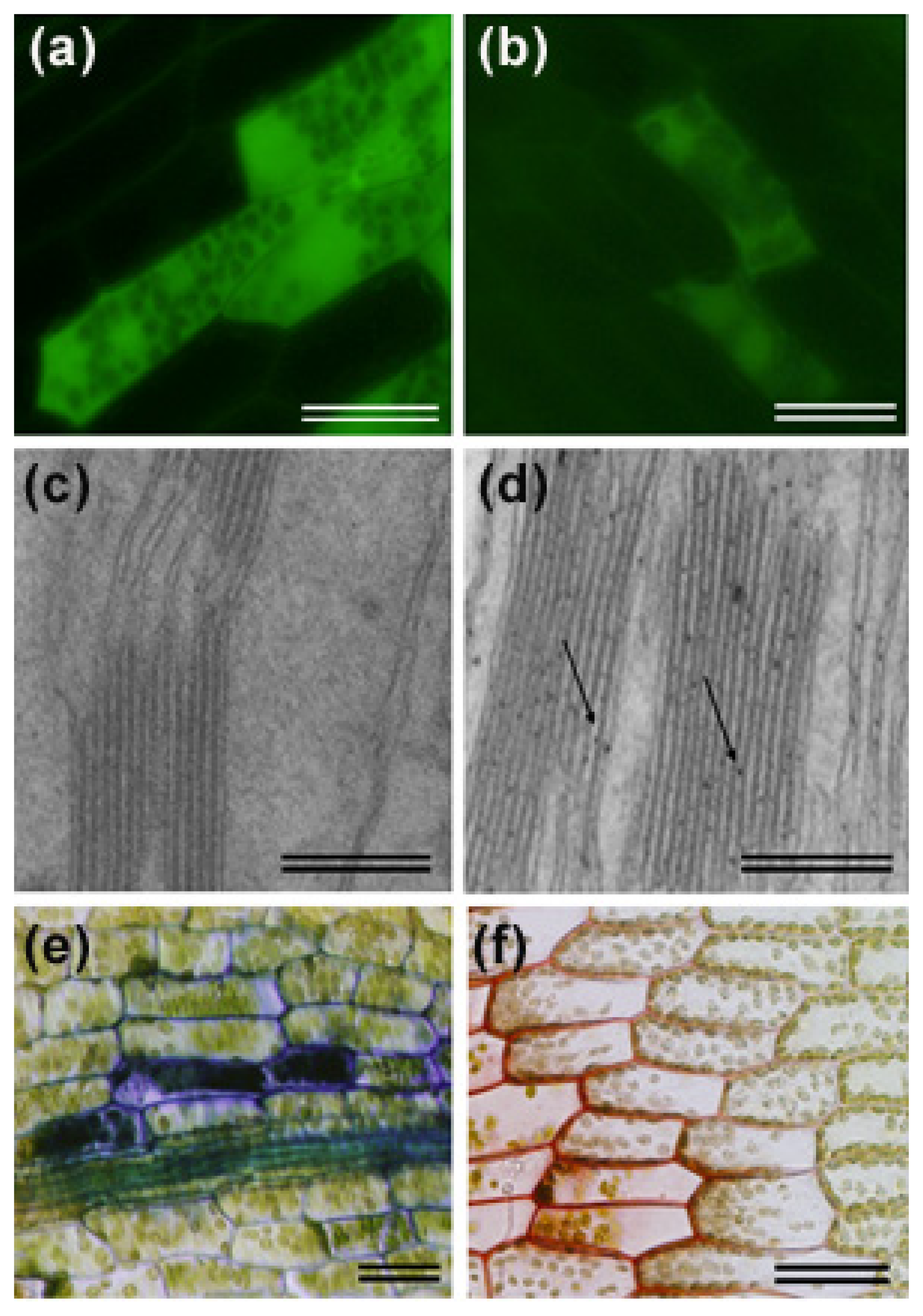
3. Activation of Cell Wall Associated Defense Responses

Pathogens are capable of penetrating the plant cell wall and gain access to cellular nutrients. Plant cells have developed pre-invasive structural defenses, including the cuticle and modifications of the cell wall that serve as barriers for the advance of potential pathogens [38,56]. Modification of the plant cell wall is an important defense mechanism operating in the defense response of flowering plants against necrotrophs [57,58]. Reinforcement of the cell wall involves accumulation of phenolic compounds, ROS and callose deposition at attempted penetration sites, making the cell wall less vulnerable to degradation by CWDEs. Callose is a high–molecular weight β-(1,3)-glucan polymer that is usually associated, together with phenolic compounds, polysaccharides and antimicrobial proteins, with cell wall appositions, called papillae, which are proposed to be effective barriers that are induced at the sites of pathogen attack [59,60]. Callose depositions are formed during early stages of pathogen invasion to inhibit pathogen penetration and are sites of accumulation of antimicrobial secondary metabolites [61]. Callose deposition plays a role in the defense response of Arabidopsis thaliana against P. irregulare, since the callose synthase mutant pmr4 is more susceptible to this oomycete compared with wild-type plants [62]. Phenolic compounds are also incorporated in cell walls of Pythium-infected tissues of flowering plants [63]. Similarly, the P. patens defense response against P. irregulare and P. debaryanum involves the accumulation of phenolic compounds, which were observed in the entire cell wall of infected cells (Figure 3) [29]. In contrast to P. irregulare-infected Arabidopsis plants [62], callose-containing wall appositions were usually not detected in Pythium-infected moss tissues [29]. However, callose depositions were observed when an old Pythium inoculum was used and colonization was not extensive, showing that these cell wall appositions can be formed at attempted infection sites, halting the progress of the invading pathogen [29].
Modification of the plant cell wall by the incorporation of phenolic compounds is also an important defense mechanism in the response of flowering plants against B. cinerea [57,58]. Increased activity of type III cell wall peroxidases, which probably influence the degree of crosslinking, resulted in enhanced resistance to B. cinerea [64]. Upon B. cinerea infection, P. patens incorporates phenolic compounds in the cell wall and increases expression of dirigent (DIR) encoding gene(s) [28]. DIR proteins are thought to mediate the coupling of monolignol plant phenols to yield lignans and lignins [65], and it is suggested that they participate in the defense response against pathogens [66,67]. Consistently, enzymes involved in monolignol biosynthesis, including putative cinnamoyl-CoA reductases, increase in Arabidopsis plants inoculated with B. cinerea [68].
The genome of P. patens contains orthologs of all the core lignin biosynthetic enzymes with the exception of ferulate 5-hydroxylase (F5H), which converts G (guaiacyl) monolignol to S (syringyl) monolignol [69]. The occurrence of lignins in bryophytes is still controversial, and instead, mosses may have wall-bound phenolics that resemble lignin [70,71]. The lack of genuine lignin together with the absence of S monolignols in P. patens could contribute to the high susceptibility observed in Pythium and B. cinerea infected moss tissues [28,29]. Recently, Lloyd and coworkers suggested that syringyl-type lignols in particular are important for successful defense of flowering plants against B. cinerea [72].

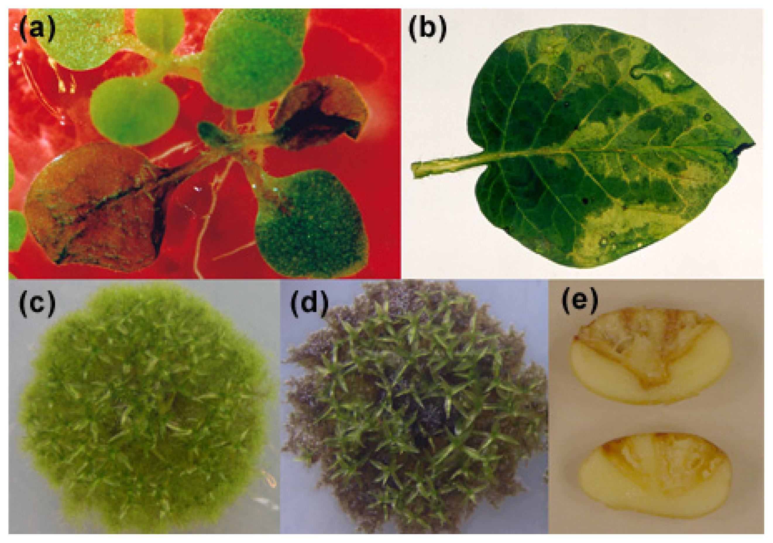
4. ROS Accumulation and Programmed Cell Death in Pathogen-Infected and Elicitor-Treated Plant Tissues

The production of ROS is one of the earliest plant cell responses following pathogen recognition and is involved in cell wall strengthening via cross-linking of glycoproteins, defense signaling and induction of the hypersensitive response [73]. Plant cells produce ROS after B. cinerea attack, which assist fungal colonization, since treatments with antioxidants suppress fungal infection [57]. Aggressiveness of different B. cinerea isolates correlates with the amount of H2O2 and hydroxyl radicals present in leaf tissues during infection [74]. In addition to increased ROS production generated by the host plant as part of a defense mechanism, B. cinerea itself produces ROS, including hydrogen peroxide, which accumulates in germinating conidia during the early steps of tissue infection [75,76]. Inactivation of the major B. cinerea H2O2-generating superoxide dismutase (SOD) retarded development of disease lesions, indicating that this enzyme is a virulence factor leading to the accumulation of phytotoxic levels of hydrogen peroxide in plant tissues [77]. Thus, ROS production is an important component of B. cinerea virulence, and increased levels of ROS in plant cells contributes to host cell death and favors fungal infection [78]. ROS production also increased in moss tissues after B. cinerea, P. irregulare and P. debaryanum infection (Figure 3) [28,29]. Single cells respond rapidly to B. cinerea hyphae contact by generating ROS, suggesting that, like vascular plants [78,79], the oxidative burst is probably induced before and during B. cinerea invasion.
P.c. carotovorum elicitor treatment also increases ROS production in P. patens tissues (Ponce de León et al., unpublished results), similarly to flowering plants [80]. In addition, the fungal elicitor chitin and chitosan caused an oxidative burst in P. patens cells [30,32]. The importance of ROS production as a defense mechanism against microbial pathogen in mosses was demonstrated in the P. patens class III peroxidase knock-out mutant Prx34, which showed enhanced susceptibility to fungal pathogens compared to wild-type P. patens plants [30]. This mutant is unable to generate an oxidative burst after elicitor treatment. While a saprophytic fungal isolate of genus Irpex and a pathogenic isolate of Fusarium sp. caused only mild symptom development in wild-type plants, hyphal growth was abundant and symptoms were severe in Prx34 knock-out plants, leading to moss decay [30]. Class III peroxidases from flowering plants are known to have antifungal activity [81], and recently, it was shown that the secreted effector Pep1 from the fungus Ustilago maydis directly interacts with a class III peroxidase from maize, suppressing the plant defense response by interfering with ROS production [82]. The functional relevance of the Pep1-peroxidase (POX12) interaction was demonstrated with POX12 silenced plants, which were infected by the pep1 deletion mutant, indicating that inhibition of this peroxidase by Pep1 is crucial for U. maydis infection [82]. In addition, PpTSPO1 moss knock-out mutants, which are impaired in mitochondrial protoporphyrin IX uptake and produce elevated levels of intracellular ROS [83], exhibited increased susceptibility to a fungal necrotrophic pathogen, including Irpex sp. and Fusarium avenaceum, suggesting that PpTSPO1 controls redox homeostasis, which is necessary for efficient resistance against pathogens [32].
Cell death plays a different role in plant response to biotrophs and necrotrophs. The hypersensitive response (HR) is a type of programmed cell death (PCD) with features of two types of cell death recently described, vacuolar cell death and necrotic cell death [84]. HR cell death contributes to resistance to biotrophic pathogens by confining the pathogen and limiting its growth [4]. Biotrophic pathogens actively suppress the HR by using effectors. Pseudomonas syringae and Xanthomonas campestris deliver 15 to 30 effectors into host cells using type III secretion systems to suppress PTI and ETI, including the HR [85]. In contrast, necrotrophic pathogens actively stimulate the HR, which enhances tissues colonization and host susceptibility. Plant mutants with enhanced cell death have increased resistance to biotrophic pathogens, but higher susceptibility to necrotrophic fungi [86,87]. B. cinerea produces nonspecific phytotoxic metabolites, which contribute to cell death on different plant hosts [76]. As part of its invasion strategy, B. cinerea promotes PCD in plant cells [78], and studies in flowering plants suggest that B. cinerea needs HR to achieve full pathogenicity [78,88]. Arabidopsis mutants with an accelerated cell death response are more susceptible to B. cinerea, while mutants with reduced or delayed cell death are generally more resistant [89]. P. patens also activate an HR-like response after B. cinerea colonization, evidenced by protoplast shrinkage, accumulation of ROS and autofluorescent compounds, chloroplasts breakdown and TUNEL (terminal deoxynucleotidyl transferase-mediated dUTP nick end labeling) positive nuclei of infected cells [26,28]. Pathogen-infected P. patens tissues also showed other characteristics of PCD, including nucleus condensation and DNA fragmentation, presence of nuclease activities and formation of cytoplasmic vacuoles [31]. Treatments with elicitors, such as CFs of P.c. carotovorum and chitosan, also provoked cell death in P. patens tissues [26,31]. Harpin proteins from Pectobacterium sp. [90,91], Xanthomonas axonopodis [92] or Pseudomonas syringae [93] elicit HR in flowering plants. Consistently, moss cells treated with the CF of the P.c. carotovorum harpin-positive strain SCC1 showed hallmarks of PCD, including protoplast shrinkage, accumulation of autofluorescent compounds and chloroplasts breakdown, while none of these features were detectable in CF treatments with the P.c. carotovorum harpin-negative strain SCC3193 [26]. Chitosan induces ROS production and cell death with hallmarks of PCD in young protonemal tissues and gametophores [31]. Interestingly, genes involved in plant PCD, such as those encoding proteases, deoxiribonucleases and ribonucleases and the antiapoptotic Bax Inhibitor-1 (BI-1) are induced after pathogen or elicitor treatment of P. patens [31]. The most convincing evidence indicating that genetically programmed cell death occurs in moss cells in response to some pathogens, comes from studies showing that transgenic P. patens plants overexpressing BI-1 are more resistance to necrotrophic fungal pathogens [31].

5. Induced Expression of Defense-Related Genes and Synthesis of Metabolites

Perception of a pathogen by a plant triggers rapid defense responses via multiple signaling pathways that lead to the induced expression of genes with different roles in defense. These include genes encoding functionally diverse pathogenesis-related (PR) proteins, transcription factors and enzymes involved in the production of metabolites (e.g., phenylpropanoids) and hormones [15,94,95]. Transcriptional reprogramming occurs rapidly after pathogen infection, and in the case of Arabidopsis-B cinerea interaction, a high-resolution temporal analysis demonstrated that approximately one-third of the Arabidopsis genome is differentially expressed during the initial stages of infection [96]. As expected, P. patens also sense the presence of pathogens and elicitors and respond rapidly by activating defense gene expression. B. cinerea, P. irregulare and P. debaryanum induce the expression of PAL (phenylalanine ammonia-lyase), CHS (chalcone synthase) and LOX1 (lipoxygenase) in P. patens tissues [26,28,29]. PAL is a key enzyme in the synthesis of phenylpropanoids, including lignin monomers, phytoalexin antibiotics and the production of SA and CHS is the first enzyme in the synthesis of flavonoids [95]. LOXs are enzymes involved in the synthesis of oxygenated fatty acids (oxylipins), including JA and aldehydes, which play important functions in plant defense against microbial infection and insects [97]. Elicitors of P.c. carotovorum also induce PpPAL, PpCHS, PpLOX1 and the pathogenesis-related gene PpPR-1 [26]. ROS-responsive genes encoding alternative oxidase (PpAOX), NADPH-oxidase (PpNOX) and LOX (PpLOX7) are induced by chitosan [32], while B. cinerea and P.c. carotovorum elicitors induce the expression of P. patens genes encoding glutathione S-transferases and ascorbate peroxidases (Ponce de León et al., unpublished data).
Mosses are known to contain a whole range of secondary metabolites, which are not present in flowering plants. The P. patens genome has been duplicated 30 and 60 million years ago, and metabolic genes seem to have been retained in excess following duplication, leading probably, in part, to the high versatility of moss metabolism [98]. Some of these metabolites, such as flavonoids, have played important roles in the adaptation of plants to land, to cope with a variety of stresses, including ultraviolet-B (UV-B) radiation, desiccation stress and co-evolving herbivores and pathogens. For example, P. patens has a higher number of members composing PAL and CHS multigene families as compared to flowering plants [99,100], and some specific genes could contribute to host defense. Consistently, several genes of the phenylpropanoid pathway leading to flavonoids synthesis, including 4-coumarate:coenzyme A ligase, several CHS and chalcone isomerase are induced in P. patens tissues after P.c. carotovorum elicitor treatments (Navarrete and Ponce de León et al., unpublished results). Moreover, recent studies showed that P. patens accumulated quercetin derivatives in response to UV-B radiation [99]. These flavonoids could also be involved in moss defense responses, since quercetin induces a resistance mechanism in Arabidopsis tissues in response to Pseudomonas syringae pv. tomato DC3000 infection, evidenced by an oxidative burst, callose deposition, and induced expression of PR-1 and PAL [101]. In addition, the Pythium and B. cinerea inducible PpLOX1 [26,28] can use arachidonic acid as a substrate leading to the production of oxylipins, which are not present in flowering plants [102104] and could contribute to the P. patens defense response. PpLOX1 and PpLOX2 can produce 12-hydroperoxy eicosatetraenoic acid (12-HPETE) from arachidonate, which in turn serves as substrate for a hydroperoxide lyase (HPL) [102,105] or PpLOX1 and PpLOX2, which posses hydroperoxide cleaving activity [102,103], leading to the production of different C8- and C9-oxylipins. P. patens HPL can also use 9-hydroperoxides of C18-fatty acids as substrate, producing (2E)-nonenal and C8-volatiles [105]. The aldehyde (2E)-nonenal could contribute to the defense of P. patens, since it has antimicrobial activity against certain pathogens, including Pseudomonas syringae pv. tomato and Phytophthora infestans [106].
Chitosan induces the production of secondary metabolites in P. patens, such as cyclic diterpenes, and increases transcript levels of genes encoding key biosynthetic enzymes of this metabolic pathway [31,107]. Inducible ent-kaurane–related diterpenoids play important roles in protecting vascular plants against microbial pathogens, as is the case for the causal agent of rice blast disease, Magnaporthe grisea [108], and Rhizopus microsporus and Colletotrichum graminicola, which cause stalk rot in maize [109].

6. Defense Hormones

Plant hormones, including SA, JA, ethylene, abscisic acid (ABA) and auxins, are involved in the defense response of flowering plants against pathogens, and the role played by these hormones is related to the particular host-pathogen interaction [110]. In general, SA is effective in mediating plant resistance against biotrophs, whereas JA and ethylene are effective in mediating resistance against necrotrophs [111114]. The interplay between these defense hormones, both agonistic and antagonistic, will determine the outcome of the interaction and minimizes fitness costs, generating a flexible signaling network that allows fine tuning of the inducible defense mechanisms [110,115,116].
P. patens is capable of producing ABA, auxin and cytokinin [117119], and during the last few years, most studies on moss hormones have been focused on ABA-dependent abiotic stress responses and the regulation of development processes by auxin and cytokinin [120124]. Until present, only a few studies have been focused on moss hormones in plant-pathogen interactions. The role of ABA in defense responses depends on the infection stage, the type of tissue infected and the specific host pathogen interaction [125]. Evidence indicates that ABA plays a role in the resistance of flowering plants, including stomatal closure, defense gene expression and ROS production/scavenging [57,125128]. In flowering plants, ABA antagonizes resistance to B. cinerea, since ABA-deficient mutants are more resistant to infection [58,62,129]. Consistently, increased ABA levels contribute to the development of grey mould in tomato [57,125]. B. cinerea-infected P. patens plants showed a small increase in ABA content when mycelium growth was extensive, suggesting that ABA could be produced by B. cinerea itself [130] to promote susceptibility by interfering with defense signaling, like the SA pathway, as has been reported previously for flowering plants [131,132].
Bryophytes produce ethylene [133,134] and the P. patens genome encodes proteins homologous to ethylene signaling components [18,135]. There are seven putative ethylene receptor proteins in P. patens [135] and genes encoding EIN3, EIL and ERF-type components, although the existence of a CTR1 component of ethylene signaling is less clear [136]. A mutation of the presumed ethylene binding site of PpETR7 inhibits the P. patens ethylene response, indicating that P. patens perceives ethylene using PpETR7 [136]. Ethylene induces defense mechanisms in flowering plants, including the production of phytoalexins, PR proteins, the induction of the phenylpropanoid pathway and cell wall modifications [137]. Resistance against B. cinerea is thought to be influenced by ethylene [138140]. B. cinerea produces ethylene itself and can interfere in this way with plant defense signaling [141]. Ethylene production increases in Arabidopsis after B. cinerea infection [142], and pretreatment of tomato plants with ethylene results in increased resistance against B. cinerea, evidenced by decreased disease symptoms and fungal biomass [137]. In addition, ethylene influenced phenylpropanoid metabolism, leading to accumulation of hydroxycinnamates and monolignols at the plant cell wall, is linked to ethylene-mediated resistance against B. cinerea [72]. Although studies on the effect of ethylene on the P. patens defense system has not been addressed, the ethylene precursor, 1-aminocyclopropane-1-carboxylic acid (ACC), induces the expression of some defense genes in P. patens (Ponce de León et al., unpublished results), suggesting that, like flowering plants, ethylene participates in the moss defense response. The use of the candidate ethylene receptors mutant Ppetr7-1 will contribute to understanding the role played by ethylene in the defense of P. patens against pathogen infection.
Until very recently, it was unknown if bryophytes produce SA and JA. The P. patens genome has 14 putative genes encoding PALs [99] and several putative homologues of isochorismate synthases, supporting the synthesis of SA in this moss. In addition, P. patens synthesizes at least seven LOXs [104], two allene oxide synthase (AOS) [143,144], three allene oxide cyclase (AOC) [145,146] and several putative 12-oxo-phytodienoic acid (OPDA) reductases genes [147,148], which encodes enzymes leading to the production of JA. Until present, enzymatic activity has been confirmed for LOXs, AOSs and AOC [104,143146], although OPR3 activity, which is the only enzyme capable of converting cis-(+)-OPDA to JA, is still missing [147]. Like flowering plants, P. patens responds to B. cinerea and P. irregulare infection by increasing endogenous levels of the precursor of JA, OPDA [28,29,62,149]. Transcript levels of genes encoding enzymes involved in OPDA biosynthesis, including LOX and AOS, are induced in B. cinerea infected tissues [28]. OPDA reductase transcript levels also increase in P. patens tissues in response to B. cinerea inoculation [28]. However, JA could not be detected in healthy, pathogen-infected, elicitor-treated or wounded P. patens tissues, suggesting that oxylipins are not further metabolized to JA [28,145,150]. Thus, cis-(+)-OPDA might function as a signaling molecule in P. patens instead of JA. Studies with the Arabidopsis opr3 mutant have shown that OPDA is active as a defense signal against pathogens and regulates defense gene expression [150152]. Interestingly, moss tissues respond to the presence of OPDA and JA by decreasing rhizoid length and moss colony size [28], similarly to the reduced growth of seedlings and roots observed in OPDA and Methyl Jasmonate (MeJA) treated Arabidopsis [153156]. Moreover, JA, MeJA and OPDA induced the expression of PAL in P. patens, showing that the presence of these oxylipins is sensed by this moss and signal transduction events are activated, leading to increased levels of defense-related transcripts [29]. The P. patens genome has six putative genes encoding the JA-isoleucine receptor COI (coronatine insensitive) and six encoding the repressor JAZ (jasmonate ZIM-domain) [157]. P. patens COI-like receptors could bind other oxylipins instead of JA-isoleucine, including cis-(+)-OPDA and/or cis-(+)-OPDA-isoleucine. Thus, the JA signaling pathway could have evolved after divergence of bryophytes and vascular plants. In addition, the similarities between the auxin receptor (TIR1) and COI1 suggest that COI-1 could have evolved from a TIR1 ancestor by gene duplication, leading to perception of JA-isoleucine by successive mutations [157].
Salicylic acid levels increase in response to B. cinerea infection in flowering plants [158,159] and in P. patens [28]. Like flowering plants, SA seems to play an important role in the defense of P. patens against microbial pathogens. SA treatment of moss tissues induces the expression of the defense gene PAL [28], and SA application induced defense mechanisms and increased resistance to P.c. carotovorum in P. patens colonies [160]. SA-mediated resistance could be due to activation of similar defense mechanisms in mosses and flowering plants, since exogenous SA application to tobacco plants also increase resistance against P.c. carotovorum [161]. In flowering plants, SA plays a key role in the activation of defense mechanisms associated with the HR and participates in a feedback amplification loop, both upstream and downstream of cell death [162,163]. The generation of SA-deficient NahG transgenic moss plants will help to elucidate SA involvement in moss defense, including the HR-like response.

7. Conclusions

During land colonization, plants gradually evolved defense strategies to cope with radiation, desiccation stress and airborne pathogens by newly acquired specialized metabolic pathways, such as the phenylpropanoid metabolism. Recently, significant progress has been made on sequencing genomes of plants that occupy interesting positions within the evolutionary history of plants, including the non-vascular moss P. patens and the vascular spikemoss S. moellendorffii [18,22]. P. patens occupies a key position halfway between green algae and flowering plants, allowing evolutionary and comparative studies of defense mechanisms across the green plant lineage. Interestingly, it was recently shown that P. patens has acquired genes related directly or indirectly with defense mechanisms by means of horizontal gene transfer from fungi and viruses [164]. The possible uptake of foreign DNA from fungi associated with early land plants could have facilitated the transition to a hostile land environment [164,165]. P. patens respond to pathogen infection or elicitor treatment by inducing defense-related gene expression and producing metabolites and hormones that could play different roles in defense. Several defense mechanisms are shared between P. patens and flowering plants, and functional conservation of some signaling pathways probably indicate common ancestral defense strategies [2830,32,136]. While the JA signaling pathway may have evolved after the divergence of bryophytes and vascular plants, ethylene, ABA and SA likely have their origins in the early stages of land colonization. The use of P. patens mutants in key components of these signaling pathways will help to determine the role played by these hormones in moss defense. P. patens also offers the possibility to identify novel metabolites, some of which are not present in flowering plants, including arachidonic acid-derived oxylipins that could play a role in defense responses. In addition, experimentation with P. patens could help to unravel defense pathways and gene functions in plants through the generation of knock-out mutants and single point mutations of genes involved in disease resistance and to identify clear mutant phenotypes due to the presence of a dominant gametophytic haploid phase [19]. Large-scale analyses of transcripts from pathogen-infected or elicitor-treated moss plants together with functional genomic and comparative studies with flowering plants will help to identify key components in the plant defense response and to design strategies to enhance plant resistance to biotic stress.

Acknowledgments

The authors thank ICGEB (International Centre for Genetic Engineering and Biotechnology, CRP/URU07-03), ANII (Fondo Clemente Estable, FCE2007-376) and Pedeciba, Uruguay, for financial support.

Conflict of Interest

The authors declare no conflict of interest.

References

  1. Ausubel, F.M. Are innate immune signaling pathways in plants and animals conserved? Nat. Immunol 2005, 6, 973–979. [Google Scholar]
  2. Bittel, P.; Robatzek, S. Microbe-associated molecular patterns (MAMPs) probe plant immunity. Curr. Opin. Plant Biol 2007, 10, 335–341. [Google Scholar]
  3. Boller, T.; Felix, G. A renaissance of elicitors: perception of microbe-associated molecular patterns and danger signals by pattern-recognition receptors. Annu. Rev. Plant Biol 2009, 60, 379–406. [Google Scholar]
  4. Jones, J.D.; Dangl, J.L. The plant immune system. Nature 2006, 444, 323–329. [Google Scholar]
  5. Abramovitch, R.B.; Janjusevic, R.; Stebbins, C.E.; Martin, G.B. Type III effector AvrPtoB requires intrinsic E3 ubiquitin ligase activity to suppress plant cell death and immunity. Proc. Natl. Acad. Sci. USA 2006, 103, 2851–2856. [Google Scholar]
  6. Boller, T.; He, S.Y. Innate immunity in plants: An arms race between pattern recognition receptors in plants and effectors in microbial pathogens. Science 2009, 324, 742–744. [Google Scholar]
  7. Cui, H.; Wang, Y.; Xue, L.; Chu, J.; Yan, C.; Fu, J.; Chen, M.; Innes, R.W.; Zhou, J.M. Pseudomonas syringae effector protein AvrB perturbs Arabidopsis hormone signaling by activating MAP kinase 4. Cell Host Microbe 2010, 7, 164–175. [Google Scholar]
  8. Grant, S.R.; Fisher, E.J.; Chang, J.H.; Mole, B.M.; Dangl, J.L. Subterfuge and manipulation: Type III effector proteins of phytopathogenic bacteria. Annu. Rev. Microbiol 2006, 60, 425–449. [Google Scholar]
  9. Zhou, J.M.; Chai, J.J. Plant pathogenic bacterial type III effectors subdue host responses. Curr. Opin. Microbiol 2008, 11, 179–185. [Google Scholar]
  10. Denoux, C.; Galletti, R.; Mammarella, N.; Gopalan, S.; Werck, D.; De Lorenzo, G.; Ferrari, S.; Ausubel, F.M.; Dewdney, J. Activation of defense response pathways by OGs and flg22 elicitors in Arabidopsis seedlings. Mol. Plant 2008, 1, 423–445. [Google Scholar]
  11. Lotze, M.T.; Zeh, H.J.; Rubartelli, A.; Sparvero, L.J.; Amoscato, A.A.; Washburn, N.R.; Devera, M.E.; Liang, X.; Tör, M.; Billiar, T. The grateful dead: damage associated molecular pattern molecules and reduction/oxidation regulate immunity. Immunol. Rev 2007, 220, 60–81. [Google Scholar]
  12. Tsuda, K.; Katagiri, F. Comparing signaling mechanisms engaged in pattern-triggered and effector-triggered immunity. Curr. Opin. Plant Biol 2010, 13, 459–465. [Google Scholar]
  13. Thomma, BP.; Nurnberger, T.; Joosten, MH. Of PAMPs and effectors: the blurred PTI-ETI dichotomy. Plant Cell 2011, 23, 4–15. [Google Scholar]
  14. Nicaise, V.; Roux, M.; Zipfel, C. Recent advances in PAMP-triggered immunity against bacteria: Pattern recognition receptors watch over and raise the alarm. Plant Physiol 2009, 150, 1638–1647. [Google Scholar]
  15. van Loon, L.C.; Rep, M.; Pieterse, C.M.J. Significance of inducible defense-related proteins in infected plants. Annu. Rev. Phytopathol 2006, 44, 135–162. [Google Scholar]
  16. Bent, A.F.; Mackey, D. Elicitors, effectors, and R genes: The new paradigm and a lifetime supply of questions. Annu. Rev. Phytopathol 2007, 45, 399–436. [Google Scholar]
  17. Zipfel, C. Early molecular events in PAMP-triggered immunity. Curr. Opin. Plant Biol 2009, 12, 414–420. [Google Scholar]
  18. Rensing, S.A.; Lang, D.; Zimmer, A.D.; Terry, A.; Salamov, A.; Shapiro, H.; Nishiyama, T.; Perroud, P.F.; Lindquist, E.A.; Kamisugi, Y.; et al. The Physcomitrella genome reveals evolutionary insights into the conquest of land by plants. Science 2008, 319, 64–69. [Google Scholar]
  19. Schaefer, D.G. A new moss genetics: targeted mutagenesis in Physcomitrella patens. Annu. Rev. Plant Biol 2002, 53, 477–501. [Google Scholar]
  20. Cove, D. The moss Physcomitrella patens. Annu. Rev. Genet 2005, 39, 339–358. [Google Scholar]
  21. Lewis, L.A.; McCourt, R.M. Green algae and the origin of land plants. Am. J. Bot 2004, 91, 1535–1556. [Google Scholar]
  22. Banks, J.A.; Nishiyama, T.; Hasebe, M.; Bowman, J.L.; Gribskov, M.; dePamphilis, C.; Albert, V.A.; Aono, N.; Aoyama, T.; Ambrose, B.A.; et al. The Selaginella genome identifies genetic changes associated with the evolution of vascular plants. Science 2011, 332, 960–963. [Google Scholar]
  23. Döbbeler, P. Biodiversity of bryophilous ascomycetes. Biodivers. Conserv 1997, 6, 721–738. [Google Scholar]
  24. Davey, M.L.; Currah, R.S. Interactions between mosses (Bryophyta) and fungi. Can. J. Bot 2006, 84, 1509–1519. [Google Scholar]
  25. Davey, M.L.; Tsuneda, A.; Currah, R.S. Pathogenesis of bryophyte hosts by the ascomycete Atradidymella muscivora. Am. J. Bot 2009, 96, 1274–1280. [Google Scholar]
  26. Ponce de León, I.; Oliver, J.P.; Castro, A.; Gaggero, C.; Bentancor, M.; Vidal, S. Erwinia carotovora elicitors and Botrytis cinerea activate defense responses in Physcomitrella patens. BMC Plant Biol 2007, 7, 52. [Google Scholar]
  27. Ponce de León, I. The moss Physcomitrella patens as a model system to study interactions between plants and phytopathogenic fungi and oomycetes. J. Pathog. 2011. [Google Scholar] [CrossRef]
  28. Ponce De León, I.; Schmelz, E.A.; Gaggero, C.; Castro, A.; Álvarez, A.; Montesano, M. Physcomitrella patens activates reinforcement of the cell wall, programmed cell death and accumulation of evolutionary conserved defence signals, such as salicylic acid and 12-oxo-phytodienoic acid, but not jasmonic acid, upon Botrytis cinerea infection. Mol. Plant Pathol 2012, 13, 960–974. [Google Scholar]
  29. Oliver, J.P.; Castro, A.; Gaggero, C.; Cascón, T.; Schmelz, E.A.; Castresana, C.; Ponce de León, I. Pythium infection activates conserved plant defense responses in mosses. Planta 2009, 230, 569–579. [Google Scholar]
  30. Lehtonen, M.T.; Akita, M.; Kalkkinen, N.; Ahola-Iivarinen, E.; Rönnholm, G.; Somervuo, P.; Thelander, M.; Valkonen, J.P. Quickly-released peroxidase of moss in defense against fungal invaders. New Phytol 2009, 183, 432–443. [Google Scholar]
  31. Lawton, M.; Saidasan, H. Pathogenesis in mosses. Annu. Plant Rev 2009, 36, 298–339. [Google Scholar]
  32. Lehtonen, M.T.; Akita, M.; Frank, W.; Reski, R.; Valkonen, J.P. Involvement of a class III peroxidase and the mitochondrial protein TSPO in oxidative burst upon treatment of moss plants with a fungal elicitor. Mol. Plant-Microbe Interact 2012, 25, 363–371. [Google Scholar]
  33. Schwessinger, B.; Ronald, P.C. Plant innate immunity: Perception of conserved microbial signatures. Annu. Rev. Plant Biol 2012, 63, 451–482. [Google Scholar]
  34. Akita, M.; Valkonen, J.P.T. A novel gene family in moss (Physcomitrella patens) shows sequence homology and a phylogenetic relationship with the TIR-NBS class of plant disease resistance genes. J. Mol. Evol 2002, 55, 595–605. [Google Scholar]
  35. Xue, J.Y.; Wang, Y.; Wu, P.; Wang, Q.; Yang, L.T.; Pan, X.H.; Wang, B.; Chen, J.Q. A primary survey on bryophyte species reveals two novel classes of nucleotide-binding site (NBS) genes. PLoS One 2012, 7, e36700. [Google Scholar]
  36. Stone, J.K. Necrotroph. In Encyclopedia of Plant Pathology; Maloy, O.C., Murray, T.D., Eds.; Wiley: New York, NY, USA, 2001; Volume 2, pp. 676–677. [Google Scholar]
  37. Elad, Y.; Williamson, B.; Tudzynski, P.; Delen, N. Botrytis: Biology, Pathology and Control; Kluwer Academic Publishers: Dordrecht, The Netherlands, 2004. [Google Scholar]
  38. van Kan, J.A. Licensed to kill: The lifestyle of a necrotrophic plant pathogen. Trends Plant Sci 2006, 11, 247–253. [Google Scholar]
  39. Prins, T.W.; Tudzynski, P.; Tiedemann, A.V.; Tudzynski, B.; ten Have, A.; Hansen, M.E.; Tenberge, K.; van Kan, J.A.L. Infection strategies of Botrytis cinerea and related necrotrophic pathogens. In Fungal Pathology; Kronstad, J.W., Ed.; Kluwer Academic Publishers: Dordrecht, The Netherlands, 2000; pp. 33–64. [Google Scholar]
  40. Staats, M.; van Baarlen, P.; van Kan, J.A. Molecular phylogeny of the plant pathogenic genus Botrytis and the evolution of host specificity. Mol. Biol. Evol 2005, 22, 333–346. [Google Scholar]
  41. Perombelon, M.C.M.; Kelman, A. Ecology of the Soft Rot Erwinias. Annu Rev. Phytopathol 1980, 18, 361–387. [Google Scholar]
  42. Toth, I.K.; Birch, P.R. Rotting softly and stealthily. Curr. Opin. Plant Biol 2005, 8, 424–429. [Google Scholar]
  43. Palva, T.K.; Holmström, K.O.; Heino, P.; Palva, E.T. Induction of plant defense response by exoenzymes of Erwinia carotovora ssp. carotovora. Mol. Plant-Microbe Interact 1993, 6, 190–196. [Google Scholar]
  44. Norman, C.; Vidal, S.; Palva, E.T. Oligogalacturonide-mediated induction of a gene involved in jasmonic acid synthesis in response to the cell-wall-degrading enzymes of the plant pathogen Erwinia carotovora. Mol. Plant-Microbe Interact 1999, 12, 640–644. [Google Scholar]
  45. Norman-Setterblad, C.; Vidal, S.; Palva, E.T. Interacting signal pathways control defense gene expression in Arabidopsis in response to cell wall-degrading enzymes from Erwinia carotovora. Mol. Plant-Microbe Interact 2000, 13, 430–438. [Google Scholar]
  46. Vidal, S.; Ponce de León, I.; Denecke, J.; Palva, E.T. Salicylic acid and the plant pathogen Erwinia carotovora induce defense genes via antagonistic pathways. Plant J 1997, 11, 115–123. [Google Scholar]
  47. Vidal, S.; Eriksson, A.R.B.; Montesano, M.; Denecke, J.; Palva, E.T. Cell wall degrading enzymes from Erwinia carotovora cooperate in the salicylic acid-independent induction of a plant defense response. Mol. Plant-Microbe Interact. 1998, 11, 23–32. [Google Scholar]
  48. Montesano, M.; Brader, G.; Ponce de Leon, I.; Palva, ET. Multiple defense signals induced by Erwinia carotovora ssp. carotovora in potato. Mol. Plant Pathol. 2005, 6, 541–549. [Google Scholar]
  49. Montesano, M.; Kõiv, V.; Mãe, A.; Palva, E.T. Novel receptor-like protein kinases induced by Erwinia carotovora and short oligogalacturonides in potato. Mol. Plant Pathol 2001, 2, 339–346. [Google Scholar]
  50. Davis, K.R.; Lyon, G.D.; Darvill, A.G.; Albersheim, P. Host-pathogen interactions: XXV. Endopolygalacturonic acid lyase from Erwinia carotovora elicits phytoalexin accumulation by releasing plant cell wall fragments. Plant Physiol 1984, 74, 52–60. [Google Scholar]
  51. Nothnagel, E.A.; McNeil, M.; Albersheim, P.; Dell, A. Host-pathogen interactions: XXII. A galacturonic acid oligosaccharide from plant cell walls elicits phytoalexins. Plant Physiol 1983, 71, 916–926. [Google Scholar]
  52. Rantakari, A.; Virtaharju, O.; Vähämiko, S.; Taira, S.; Palva, E.T.; Saarilahti, H.T.; Romantschuk, M. Type III secretion contributes to the pathogenesis of the soft-rot pathogen Erwinia carotovora partial characterization of the hrp gene cluster. Mol. Plant-Microbe Interact 2001, 14, 962–968. [Google Scholar]
  53. Mattinen, L.; Tshuikina, M.; Mäe, A.; Pirhonen, M. Identification and characterization of Nip, necrosis-inducing virulence protein of Erwinia carotovora subsp. carotovora. Mol. Plant-Microbe Interact 2004, 17, 1366–1375. [Google Scholar]
  54. Martin, F. Pythium. In Pathogenesis and Host Specificity in Plant Diseases: Histopathological, Biochemical, Genetic and Molecular Bases; Komoto, K., Singh, U.S., Singh, R.P., Eds.; Pergamon Press: Oxford, UK, 1994; pp. 17–36. [Google Scholar]
  55. Campion, C.; Massiot, P.; Rouxel, F. Aggressiveness and production of cell-wall degrading enzymes by Pythium violae, Pythium sulcatum and Pythium ultimum, responsible for cavity spot on carrots. Eur. J. Plant Pathol 1997, 103, 725–735. [Google Scholar]
  56. Mendgen, K.; Hahn, M.; Deising, H. Morphogenesis and mechanisms of penetration by plant pathogenic fungi. Annu. Rev. Phytopathol 1996, 34, 367–386. [Google Scholar]
  57. Asselbergh, B.; Curvers, K.; Franca, S.C.; Audenaert, K.; Vuylsteke, M.; Van Breusegem, F.; Höfte, M. Resistance to Botrytis cinerea in sitiens, an abscisic acid-deficient tomato mutant, involves timely production of hydrogen peroxide and cell wall modifications in the epidermis. Plant Physiol 2007, 144, 1863–1877. [Google Scholar]
  58. Curvers, K.; Seifi, H.; Mouille, G.; de Rycke, R.; Asselbergh, B.; Van Hecke, A.; Vanderschaeghe, D.; Höfte, H.; Callewaert, N.; van Breusegem, F.; Höfte, M. Abscisic acid deficiency causes changes in cuticle permeability and pectin composition that influence tomato resistance to Botrytis cinerea. Plant Physiol 2010, 154, 847–860. [Google Scholar]
  59. Jacobs, A.K.; Lipka, V.; Burton, R.A.; Panstruga, R.; Strizhov, N.; Schulze-Lefert, P.; Fincher, G.B. An Arabidopsis callose synthase, GSL5, is required for wound and papillary callose formation. Plant Cell 2003, 15, 2503–2513. [Google Scholar]
  60. Ton, J.; Mauch-Mani, B. Beta-amino-butyric acid-induced resistance against necrotrophic pathogens is based on ABA-dependent priming for callose. Plant J 2004, 38, 119–130. [Google Scholar]
  61. Luna, E.; Pastor, V.; Robert, J.; Flors, V.; Mauch-Mani, B.; Ton, J. Callose deposition: A multifaceted plant defense response. Mol. Plant-Microbe Interact 2011, 24, 183–193. [Google Scholar]
  62. Adie, B.A.; Pérez-Pérez, J.; Pérez-Pérez, M.M.; Godoy, M.; Sánchez-Serrano, J.J.; Schmelz, E.A.; Solano, R. ABA is an essential signal for plant resistance to pathogens affecting JA biosynthesis and the activation of defenses in Arabidopsis. Plant Cell 2007, 19, 1665–1681. [Google Scholar]
  63. Benhamou, N.; Bélanger, R. Induction of systemic resistance to Pythium damping-off in cucumber plants by benzothiadiazole: ultrastructure and cytochemistry of the host response. Plant J 1998, 14, 13–21. [Google Scholar]
  64. Ramírez, V.; Agorio, A.; Coego, A.; García-Andrade, J.; Hernández, M.J.; Balaguer, B.; Ouwerkerk, P.B.; Zarra, I.; Vera, P. MYB46 modulates disease susceptibility to Botrytis cinerea in Arabidopsis. Plant Physiol 2011, 155, 1920–1935. [Google Scholar]
  65. Davin, L.B.; Lewis, N.G. Dirigent proteins and dirigent sites explain the mystery of specificity of radical precursor coupling in lignan and lignin biosynthesis. Plant Physiol 2000, 123, 453–462. [Google Scholar]
  66. Coram, T.E.; Wang, M.; Chen, X. Transcriptome analysis of the wheat–Puccinia striiformis f. sp. tritici interaction. Mol. Plant Pathol 2008, 9, 157–169. [Google Scholar]
  67. Chakravarthy, S.; Velásquez, A.C.; Ekengren, S.K.; Collmer, A.; Martin, G.B. Identification of Nicotiana benthamiana genes involved in pathogen associated molecular pattern-triggered immunity. Mol. Plant-Microbe Interact 2010, 23, 715–726. [Google Scholar]
  68. Ferrari, S.; Gallettim, R.; Denoux, C.; De Lorenzo, G.; Ausubel, F.M.; Dewdney, J. Resistance to Botrytis cinerea induced in Arabidopsis by elicitors is independent of salicylic acid, ethylene or jasmonate signaling but requires PAD3. Plant Physiol 2007, 144, 367–379. [Google Scholar]
  69. Xu, Z.; Zhang, D.; Hu, J.; Zhou, X.; Ye, X.; Reichel, K.L.; Stewart, N.R.; Syrenne, R.D.; Yang, X.; et al. Comparative genome analysis of lignin biosynthesis gene families across the plant kingdom. BMC Bioinformatics 2009, 10, S3. [Google Scholar]
  70. Popper, Z.A. Evolution and diversity of green plant cell walls. Curr. Opin. Plant Biol 2008, 11, 286–292. [Google Scholar]
  71. Weng, J.K.; Chapple, C. The origin and evolution of lignin biosynthesis. New Phytol 2010, 187, 273–285. [Google Scholar]
  72. Lloyd, A.J.; William Allwood, J.; Winder, C.L.; Dunn, W.B.; Heald, J.K.; Cristescu, S.M.; Sivakumaran, A.; Harren, F.J.; Mulema, J.; Denby, K.; et al. Metabolomic approaches reveal that cell wall modifications play a major role in ethylene-mediated resistance against. Botrytis cinerea. Plant J. 2011, 67, 852–868. [Google Scholar]
  73. Torres, M.A.; Jones, J.D.; Dangl, J.L. Reactive oxygen species signaling in response to pathogens. Plant Physiol 2006, 141, 373–378. [Google Scholar]
  74. Tiedemann, A.V. Evidence for a primary role of active oxygen species in induction of host cell death during infection of bean leaves with Botrytis cinerea. Physiol. Mol. Plant Pathol 1997, 50, 151–166. [Google Scholar]
  75. Schouten, A.; Tenberge, K.B.; Vermeer, J.; Stewart, J.; Wagemakers, L.; Williamson, B.; van Kan, J.A. Functional analysis of an extracellular catalase of Botrytis cinerea. Mol. Plant Pathol 2002, 3, 227–238. [Google Scholar]
  76. Choquer, M.; Fournier, E.; Kunz, C.; Levis, C.; Pradier, J.M.; Simon, A.; Viaud, M. Botrytis cinerea virulence factors: New insights into a necrotrophic and polyphageous pathogen. FEMS Microbiol. Lett 2007, 277, 1–10. [Google Scholar]
  77. Rolke, Y.; Liu, S.; Quidde, T.; Williamson, B.; Schouten, A.; Weltring, K.M.; Siewers, V.; Tenberge, K.B.; Tudzynski, B.; Tudzynski, P. Functional analysis of H2O2-generating systems in Botrytis cinerea: The major Cu-Zn-superoxide dismutase (BCSOD1) contributes to virulence on French bean, whereas a glucose oxidase (BCGOD1) is dispensable. Mol. Plant Pathol. 2004, 5, 17–27. [Google Scholar]
  78. Govrin, E.M.; Levine, A. The hypersensitive response facilitates plant infection by the necrotrophic pathogen Botrytis cinerea. Curr. Biol 2000, 10, 751–757. [Google Scholar]
  79. Asai, S.; Yoshioka, H. Nitric oxide as a partner of reactive oxygen species participates in disease resistance to necrotrophic pathogen Botrytis cinerea in Nicotiana benthamiana. Mol. Plant-Microbe Interact 2009, 22, 619–629. [Google Scholar]
  80. Montesano, M.; Scheller, H.V.; Wettstein, R.; Palva, E.T. Down-regulation of photosystem I by Erwinia carotovora-derived elicitors correlates with H2O2 accumulation in chloroplasts of potato. Mol. Plant Pathol 2004, 5, 115–123. [Google Scholar]
  81. Ghosh, M. Antifungal properties of haem peroxidase from Acorus calamus. Ann. Bot 2006, 98, 1145–1153. [Google Scholar]
  82. Hemetsberger, C.; Herrberger, C.; Zechmann, B.; Hillmer, M.; Doehlemann, G. The Ustilago maydis effector Pep1 suppresses plant immunity by inhibition of host peroxidase activity. PLoS Pathog 2012, 8, e1002684. [Google Scholar]
  83. Frank, W.; Baar, K.; Qudeimat, E.; Woriedh, M.; Alawady, A.; Ratnadewi, D.; Gremillon, L.; Grimm, B.; Reski, R. A mitochondrial protein homologous to the mammalian peripheral-type benzodiazepine receptor is essential for stress adaptation in plants. Plant J 2007, 51, 1004–1018. [Google Scholar]
  84. van Doorn, W.G.; Beers, E.P.; Dangl, J.L.; Franklin-Tong, V.E.; Gallois, P.; Hara-Nishimura, I.; Jones, A.M.; Kawai-Yamada, M.; Lam, E.; Mundy, J.; et al. Morphological classification of plant cell deaths. Cell Death Differ. 2011, 18, 1241–1246. [Google Scholar]
  85. Kjemtrup, S.; Nimchuk, Z.; Dangl, J.L. Effector proteins of phytopathogenic bacteria: Bifunctional signals in virulence and host recognition. Curr. Opin. Microbiol 2000, 3, 73–78. [Google Scholar]
  86. Kachroo, P.; Shanklin, J.; Shah, J.; Whittle, E.J.; Klessig, D.F. A fatty acid desaturase modulates the activation of defense signaling pathways in plants. Proc. Natl. Acad. Sci. USA 2001, 98, 9448–9453. [Google Scholar]
  87. Veronese, P.; Chen, X.; Bluhm, B.; Salmeron, J.; Dietrich, R.; Mengiste, T. The BOS loci of Arabidopsis are required for resistance to Botrytis cinerea infection. Plant J 2004, 40, 558–574. [Google Scholar]
  88. Dickman, M.B.; Park, Y.K.; Oltersdorf, T.; Li, W.; Clemente, T.; French, R. Abrogation of disease development in plants expressing animal antiapoptotic genes. Proc. Natl. Acad. Sci. USA 2001, 98, 6957–6962. [Google Scholar]
  89. van Baarlen, P.; Woltering, E.J.; Staats, M.; van Kan, J.A.L. Histochemical and genetic analysis of host and non-host interactions of Arabidopsis with three Botrytis species: an important role for cell death control. Mol. Plant Pathol 2007, 8, 41–54. [Google Scholar]
  90. Wei, Z.M.; Laby, R.J.; Zumoff, C.H.; Bauer, D.W.; He, S.Y.; Collmer, A.; Beer, S.V. Harpin, elicitor of the hypersensitive response produced by the plant pathogen Erwinia amylovora. Science 1992, 257, 85–88. [Google Scholar]
  91. Kariola, T.; Palomäki, T.A.; Brader, G.; Palva, E.T. Erwinia carotovora subsp. carotovora and Erwinia -derived elicitors HrpN and PehA trigger distinct but interacting defense responses and cell death in Arabidopsis. Mol. Plant-Microbe Interact 2003, 16, 179–187. [Google Scholar]
  92. Kim, J.G.; Jeon, E.; Oh, J.; Moon, J.S.; Hwang, I. Mutational analysis of Xanthomonas harpin HpaG identifies a key functional region that elicits the hypersensitive response in nonhost plants. J. Bacteriol 2004, 186, 6239–6247. [Google Scholar]
  93. Alfano, J.R.; Bauer, D.W.; Milos, T.M.; Collmer, A. Analysis of the role of the Pseudomonas syringae pv. syringae HrpZ harpin in elicitation of the hypersensitive response in tobacco using functionally non-polar hrpZ deletion mutations, truncated HrpZ fragments, and hrmA mutations. Mol. Microbiol 1996, 19, 715–728. [Google Scholar]
  94. Zhu, Q.; Dröge-Laser, W.; Dixon, R.A.; Lamb, C. Transcriptional activation of plant defense genes. Curr. Opin. Genet. Dev 1996, 6, 624–630. [Google Scholar]
  95. Dixon, R.A.; Paiva, N.L. Stress-induced phenylpropanoid metabolism. Plant Cell 1995, 7, 1085–1097. [Google Scholar]
  96. Windram, O.; Madhou, P.; McHattie, S.; Hill, C.; Hickman, R.; Cooke, E.; Jenkins, D.J.; Penfold, C.A.; Baxter, L.; Breeze, E.; et al. Arabidopsis Defense against Botrytis cinerea: Chronology and Regulation Deciphered by High-Resolution Temporal Transcriptomic Analysis. Plant Cell 2012, 24, 3530–3557. [Google Scholar]
  97. Feussner, I.; Wasternack, C. The lipoxygenase pathway. Annu. Rev. Plant Biol 2002, 53, 275–297. [Google Scholar]
  98. Rensing, S.A.; Ick, J.; Fawcett, J.A.; Lang, D.; Zimmer, A.; Van de Peer, Y.; Reski, R. An ancient genome duplication contributed to the abundance of metabolic genes in the moss Physcomitrella patens. BMC Evol. Biol 2007, 7, 130. [Google Scholar]
  99. Wolf, L.; Rizzini, L.; Stracke, R.; Ulm, R.; Rensing, S.A. The molecular and physiological responses of Physcomitrella patens to ultraviolet-B radiation. Plant Physiol 2010, 153, 1123–1134. [Google Scholar]
  100. Koduri, P.K.; Gordon, G.S.; Barker, E.I.; Colpitts, C.C.; Ashton, N.W.; Suh, D.Y. Genome-wide analysis of the chalcone synthase superfamily genes of Physcomitrella patens. Plant Mol. Biol 2010, 72, 247–263. [Google Scholar]
  101. Jia, Z.; Zou, B.; Wang, X.; Qiu, J.; Ma, H.; Gou, Z.; Song, S.; Dong, H. Quercetin-induced H(2)O(2) mediates the pathogen resistance against Pseudomonas syringae pv. Tomato DC3000 in Arabidopsis thaliana. Biochem. Biophys. Res. Commun 2010, 28, 522–527. [Google Scholar]
  102. Wichard, T.; Göbel, C.; Feussner, I.; Pohnert, V. Unprecedented lipoxygenase/hydroperoxide lyase pathways in the moss Physcomitrella patens. Angew. Chem. Int. Edit 2004, 44, 158–161. [Google Scholar]
  103. Senger, T.; Wichard, T.; Kunze, S.; Gobel, C.; Lerchl, J.; Pohnert, G.; Feussner, I. A multifunctional lipoxygenase with fatty acid hydroperoxide cleaving activity from the moss Physcomitrella patens. J. Biol. Chem 2005, 280, 7588–7596. [Google Scholar]
  104. Anterola, A.; Göbel, C.; Hornung, E.; Sellhorn, G.; Feussner, I.; Grimes, H. Physcomitrella patens has lipoxygenases for both eicosanoid and octadecanoid pathways. Phytochemistry 2009, 70, 40–52. [Google Scholar]
  105. Stumpe, M.; Bode, J.; Gobel, C.; Wichard, T.; Schaaf, A.; Frank, W.; Frank, M.; Reski, R.; Pohnert, G.; Feussner, I. Biosynthesis of C9-aldehydes in the moss Physcomitrella patens. Biochim. Biophys. Acta 2006, 1761, 301–312. [Google Scholar]
  106. Prost, I.; Dhondt, S.; Rothe, G.; Vicente, J.; Rodriguez, M.J.; Kift, N.; Carbonne, F.; Griffiths, G.; Esquerré-Tugayé, M.T.; Rosahl, S.; et al. Evaluation of the antimicrobial activities of plant oxylipins supports their involvement in defense against pathogens. Plant Physiol. 2005, 139, 1902–1913. [Google Scholar]
  107. Von Schwartzenberg, K.; Schultze, W.; Kassner, H. The moss Physcomitrella patens releases a tetracyclic diterpene. Plant Cell Rep 2004, 22, 780–786. [Google Scholar]
  108. Peters, R.J. Uncovering the complex metabolic network underlying diterpenoid phytoalexin biosynthesis in rice and other cereal crop plants. Phytochemistry 2006, 67, 2307–2317. [Google Scholar]
  109. Schmelz, E.A.; Kaplan, F.; Huffaker, A.; Dafoe, N.J.; Vaughan, M.M.; Ni, X.; Rocca, J.R.; Alborn, H.T.; Teal, P.E. Identity, regulation, and activity of inducible diterpenoid phytoalexins in maize. Proc. Natl. Acad. Sci. USA 2011, 108, 5455–5460. [Google Scholar]
  110. López, M.A.; Bannenberg, G.; Castresana, C. Controlling hormone signaling is a plant and pathogen challenge for growth and survival. Curr. Opin. Plant Biol 2008, 11, 420–427. [Google Scholar]
  111. Feys, B.J.; Parker, J.E. Interplay of signaling pathways in plant disease resistance. Trends Genet 2000, 16, 449–455. [Google Scholar]
  112. Lund, S.T.; Stall, R.E.; Klee, H.J. Ethylene regulates the susceptible response to pathogen infection in tomato. Plant Cell 1998, 10, 371–382. [Google Scholar]
  113. Greenberg, J.T.; Silverman, F.P.; Liang, H. Uncoupling salicylic acid-dependent cell death and defense-related responses from disease resistance in the Arabidopsis mutant acd5. Genetics 2000, 156, 341–350. [Google Scholar]
  114. Pilloff, R.K.; Devadas, S.K.; Enyedi, A.; Raina, R. The Arabidopsis gain-of-function mutant Dll1 spontaneously develops lesions mimicking cell death associated with disease. Plant J 2002, 30, 61–70. [Google Scholar]
  115. Mur, L.A.; Kenton, P.; Atzorn, R.; Miersch, O.; Wasternack, C. The outcomes of concentration-specific interactions between salicylate and jasmonate signaling include synergy, antagonism, and oxidative stress leading to cell death. Plant Physiol 2006, 140, 249–262. [Google Scholar]
  116. Koornneef, A.; Pieterse, C.M. Cross talk in defense signaling. Plant Physiol 2008, 146, 839–844. [Google Scholar]
  117. Komatsu, K.; Nishikawa, Y.; Ohtsuka, T.; Taji, T.; Quatrano, R.S.; Tanaka, S.; Sakata, Y. Functional analyses of the ABI1-related protein phosphatase type 2C reveal evolutionarily conserved regulation of abscisic acid signaling between Arabidopsis and the moss Physcomitrella patens. Plant Mol. Biol 2009, 70, 327–340. [Google Scholar]
  118. Bierfreund, N.M.; Reski, R.; Decker, E.L. Use of an inducible reporter gene system for the analysis of auxin distribution in the moss Physcomitrella patens. Plant Cell Rep 2003, 21, 1143–1152. [Google Scholar]
  119. Schwartzenberg, K.V.; Nunez, M.F.; Blaschke, H.; Dobrev, P.I.; Novak, D.O.; Motyka, V.; Strnad, M. Cytokinins in the bryophyte Physcomitrella patens: analyses of activity, distribution, and cytokinin oxidase/dehydrogenase overexpression reveal the role of extracellular cytokinins. Plant Physiol 2007, 145, 786–800. [Google Scholar]
  120. Khandelwal, A.; Cho, S.H.; Marella, H.; Sakata, Y.; Perroud, P.F.; Pan, A.; Quatrano, R.S. Role of ABA and ABI3 in desiccation tolerance. Science 2010, 327, 546. [Google Scholar]
  121. Bhyan, S.B.; Minami, A.; Kaneko, Y.; Suzuki, S.; Arakawa, K.; Sakata, Y.; Takezawa, D. Cold acclimation in the moss Physcomitrella patens involves abscisic acid-dependent signaling. J. Plant Physiol 2012, 169, 137–145. [Google Scholar]
  122. Prigge, M.J.; Bezanilla, M. Evolutionary crossroads in developmental biology: Physcomitrella patens. Development 2010, 137, 3535–3543. [Google Scholar]
  123. Jang, G.; Dolan, L. Auxin promotes the transition from chloronema to caulonema in moss protonema by positively regulating PpRSL1and PpRSL2 in Physcomitrella patens. New Phytol 2011, 192, 319–327. [Google Scholar]
  124. Saleh, O.; Issman, N.; Seumel, G.I.; Stav, R.; Samach, A.; Reski, R.; Frank, W.; Arazi, T. MicroRNA534a control of BLADE-ON-PETIOLE 1 and 2 mediates juvenile-to-adult gametophyte transition in Physcomitrella patens. Plant J 2011, 65, 661–674. [Google Scholar]
  125. Ton, J.; Flors, V.; Mauch-Mani, B. The multifaceted role of ABA in disease resistance. Trends Plant Sci 2009, 14, 310–317. [Google Scholar]
  126. Mauch-Mani, B.; Mauch, F. The role of abscisic acid in plant-pathogen interactions. Curr. Opin. Plant Biol 2005, 8, 409–414. [Google Scholar]
  127. Bari, R.; Jones, J.D. Role of plant hormones in plant defence responses. Plant Mol. Biol 2009, 69, 473–488. [Google Scholar]
  128. Grant, M.R.; Jones, J.D. Hormone (dis)harmony moulds plant health and disease. Science 2009, 324, 750–752. [Google Scholar]
  129. AbuQamar, S.; Chen, X.; Dhawan, R.; Bluhm, B.; Salmeron, J.; Lam, S.; Dietrich, R.A.; Mengiste, T. Expression profiling and mutant analysis reveals complex regulatory networks involved in Arabidopsis response to Botrytis infection. Plant J 2006, 48, 28–44. [Google Scholar]
  130. Siewers, V.; Smedsgaard, J.; Tudzynski, P. The P450 monooxygenase BcABA1 is essential for abscisic acid biosynthesis in Botrytis cinerea. Appl. Environ. Microbiol 2004, 70, 3868–3876. [Google Scholar]
  131. Audenaert, K.; De Meyer, G.B.; Höfte, M.M. Abscisic acid determines basal susceptibility of tomato to Botrytis cinerea and suppresses salicylic acid dependent signaling mechanisms. Plant Physiol 2002, 128, 491–501. [Google Scholar]
  132. Yasuda, M.; Ishikawa, A.; Jikumaru, Y.; Seki, M.; Umezawa, T.; Asami, T.; Maruyama-Nakashita, A.; Kudo, T.; Shinozaki, K.; Yoshida, S.; et al. Antagonistic interaction between systemic acquired resistance and the abscisic acid-mediated abiotic stress response in. Arabidopsis. Plant Cell 2008, 20, 1678–1692. [Google Scholar]
  133. Rohwer, F.; Bopp, M. Ethylene Synthesis in Moss Protonema. J. Plant Physiol 1985, 117, 331–338. [Google Scholar]
  134. Osborne, D.J.; Walters, J.; Milborrow, B.V.; Norville, A.; Stange, L.M.C. Evidence for a non-ACC ethylene biosynthesis pathway in lower plants. Phytochemistry 1995, 42, 51–60. [Google Scholar]
  135. Ishida, K.; Yamashino, T.; Nakanishi, H.; Mizuno, T. Classification of the genes involved in the two-component system of the moss Physcomitrella patens. Biosci. Biotechnol. Biochem 2010, 74, 2542–2545. [Google Scholar]
  136. Yasumura, Y.; Pierik, R.; Fricker, M.D.; Voesenek, L.A.; Harberd, N.P. Studies of Physcomitrella patens reveal that ethylene-mediated submergence responses arose relatively early in land-plant evolution. Plant J 2012, 72, 947–959. [Google Scholar]
  137. Díaz, J.; ten Have, A.; van Kan, J.A. The role of ethylene and wound signaling in resistance of tomato to Botrytis cinerea. Plant Physiol 2002, 129, 1341–1351. [Google Scholar]
  138. Penninckx, I.A.M.A.; Eggermont, K.; Terras, F.R.G.; Thomma, B.P.H.J.; De Samblanx, G.W.; Buchala, A.; Métraux, J.P.; Manners, J.M.; Broekaert, W.F. Pathogen-induced systemic activation of a plant defensin gene in Arabidopsis follows a salicylic acid-independent pathway. Plant Cell 1996, 8, 2309–2323. [Google Scholar]
  139. Thomma, B.P.H.J.; Eggermont, K.; Tierens, K.F.M.J.; Broekaert, W.F. Requirement of functional ethylene-insensitive 2 gene for efficient resistance of Arabidopsis to infection by Botrytis cinerea. Plant Physiol 1999, 121, 1093–1101. [Google Scholar]
  140. Berrocal-Lobo, M.; Molina, A.; Solano, R. Constitutive expression of ETHYLENE-RESPONSEFACTOR1 in Arabidopsis confers resistance to several necrotrophic fungi. Plant J 2002, 29, 23–32. [Google Scholar]
  141. Chague, V.; Elad, Y.; Barakat, R.; Tudzynski, P.; Sharon, A. Ethylene biosynthesis in Botrytis cinerea. FEMS Microbiol. Ecol 2002, 40, 143–149. [Google Scholar]
  142. Han, L.; Li, G.J.; Yang, K.Y.; Mao, G.; Wang, R.; Liu, Y.; Zhang, S. Mitogen-activated protein kinase 3 and 6 regulate Botrytis cinerea-induced ethylene production in Arabidopsis. Plant J 2010, 64, 114–127. [Google Scholar]
  143. Bandara, P.K.; Takahashi, K.; Sato, M.; Matsuura, H.; Nabeta, K. Cloning and functional analysis of an allene oxide synthase in Physcomitrella patens. Biosci. Biotechnol. Biochem 2009, 73, 2356–2359. [Google Scholar]
  144. Scholz, J.; Dickmanns, A.; Feussner, I.; Ficner, R. Crystal Structures of Physcomitrella patens AOC1 and AOC2: Insights into the Enzyme Mechanism and Differences in Substrate Specificity. Plant Physiol 2012, 160, 1251–1266. [Google Scholar]
  145. Stumpe, M.; Göbel, C.; Faltin, B.; Beike, A.K.; Hause, B.; Himmelsbach, K.; Bode, J.; Kramell, R.; Wasternack, C.; Frank, W.; et al. The moss Physcomitrella patens contains cyclopentenones but no jasmonates: mutations in allene oxide cyclase lead to reduced fertility and altered sporophyte morphology. New Phytol. 2010, 188, 740–749. [Google Scholar]
  146. Hashimoto, T.; Takahashi, K.; Sato, M.; Bandara, P.K.G.S.S.; Nabeta, K. Cloning and characterization of an allene oxide cyclase, PpAOC3, in Physcomitrella patens. Plant Growth Regul. 2011, 65, 239–245. [Google Scholar]
  147. Breithaupt, C.; Kurzbauer, R.; Schaller, F.; Stintzi, A.; Schaller, A.; Huber, R.; Macheroux, P.; Clausen, T. Structural basis of substrate specificity of plant 12-oxophytodienoate reductases. J. Mol. Biol 2009, 392, 1266–1277. [Google Scholar]
  148. Li, W.; Liu, B.; Yu, L.; Feng, D.; Wang, H.; Wang, J. Phylogenetic analysis, structural evolution and functional divergence of the 12-oxophytodienoate acid reductase gene family in plants. BMC Evol. Biol 2009, 9, 90. [Google Scholar]
  149. Vicedo, B.; Flors, V.; de la O Leyva, M.; Finiti, I.; Kravchuk, Z.; Real, M.D.; García-Agustín, P.; González-Bosch, C. Hexanoic acid-induced resistance against Botrytis cinerea in tomato plants. Mol. Plant-Microbe Interact 2009, 22, 1455–1465. [Google Scholar]
  150. Browse, J. Jasmonate passes muster: A receptor and targets for the defense hormone. Annu. Rev. Plant Biol 2009, 60, 183–205. [Google Scholar]
  151. Stintzi, A.; Weber, H.; Reymond, P.; Browse, J.; Farmer, E.E. Plant defense in the absence of jasmonic acid: The role of cyclopentenones. Proc. Natl. Acad. Sci. USA 2001, 98, 12837–12842. [Google Scholar]
  152. Taki, N.; Sasaki-Sekimoto, Y.; Obayashi, T.; Kikuta, A.; Kobayashi, K.; Ainai, T.; Yagi, K.; Sakurai, N.; Suzuki, H.; Masuda, T.; et al. 12-Oxo-phytodienoic acid triggers expression of a distinct set of genes and plays a role in wound-induced gene expression in Arabidopsis. Plant Physiol. 2005, 139, 1268–1283. [Google Scholar]
  153. Mueller, S.; Hilbert, B.; Dueckershoff, K.; Roitsch, T.; Krischke, M.; Mueller, M.J.; Berger, S. General detoxification and stress responses are mediated by oxidized lipids through TGA transcription factors in Arabidopsis. Plant Cell 2008, 20, 768–785. [Google Scholar]
  154. Staswick, P.E.; Su, W.P.; Howell, S.H. Methyl jasmonate inhibition of root-growth and induction of a leaf protein are decreased in an Arabidopsis thaliana mutant. Proc. Natl. Acad. Sci. USA 1992, 89, 6837–6840. [Google Scholar]
  155. Vellosillo, T.; Martinez, M.; Lopez, M.A.; Vicente, J.; Cascon, T.; Dolan, L.; Hamberg, M.; Castresana, C. Oxylipins produced by the 9-lipoxygenase pathway in Arabidopsis regulate lateral root development and defense responses through a specific signaling cascade. Plant Cell 2007, 19, 831–846. [Google Scholar]
  156. Yan, Y.; Stolz, S.; Chételat, A.; Reymond, P.; Pagni, M.; Dubugnon, L.; Farmer, E.E. A downstream mediator in the growth repression limb of the jasmonate pathway. Plant Cell 2007, 19, 2470–2483. [Google Scholar]
  157. Chico, J.M.; Chini, A.; Fonseca, S.; Solano, R. JAZ repressors set the rhythm in jasmonate signaling. Curr. Opin. Plant Biol 2008, 11, 486–494. [Google Scholar]
  158. El Oirdi, M.; El Rahman, T.A.; Rigano, L.; El Hadrami, A.; Rodriguez, M.C.; Daayf, F.; Vojnov, A.; Bouarab, K. Botrytis cinerea manipulates the antagonistic effects between immune pathways to promote disease development in tomato. Plant Cell 2011, 23, 2405–2421. [Google Scholar]
  159. Veronese, P.; Nakagami, H.; Bluhm, B.; Abuqamar, S.; Chen, X.; Salmeron, J.; Dietrich, R.A.; Hirt, H.; Mengiste, T. The membrane-anchored Botrytis induced kinase1 plays distinct roles in Arabidopsis resistance to necrotrophic and biotrophic pathogens. Plant Cell 2006, 18, 257–273. [Google Scholar]
  160. Andersson, R.A.; Akita, M.; Pirhonen, M.; Gammelgård, E.; Valkonen, J.P.T. Moss-Erwinia pathosystem reveals possible similarities in pathogenesis and pathogen defense in vascular and nonvascular plants. J. Gen. Plant Pathol 2005, 71, 23–28. [Google Scholar]
  161. Palva, TK.; Hurtig, M.; Saindrenan, P.; Palva, ET. Salicylic Acid Induced Resistance to Erwinia carotovora subsp, carotovora in tobacco. Mol. Plant-Microbe Interact. 1994, 7, 356–363. [Google Scholar]
  162. Dangl, J.L.; Dietrich, R.A.; Richberg, M.H. Death Don’t Have No Mercy: Cell Death Programs in Plant-Microbe Interactions. Plant Cell 1996, 8, 1793–1807. [Google Scholar]
  163. Alvarez, M.E. Salicylic acid in the machinery of hypersensitive cell death and disease resistance. Plant Mol. Biol 2000, 44, 429–442. [Google Scholar]
  164. Yue, J.; Hu, X.; Sun, H.; Yang, Y.; Huang, J. Widespread impact of horizontal gene transfer on plant colonization of land. Nat. Commun. 2012, 3. [Google Scholar] [CrossRef]
  165. Wang, B.; Yeun, L.H.; Xue, J.Y.; Liu, Y.; Ané, J.M.; Qiu, Y.L. Presence of three mycorrhizal genes in the common ancestor of land plants suggests a key role of mycorrhizas in the colonization of land by plants. New Phytol 2010, 186, 514–525. [Google Scholar]
Figure 1. Disease symptoms evidenced by tissue maceration of plants inoculated with Pectobacterium carotovorum subsp. carotovorum (P.c. carotovorum) or treated with elicitors of this pathogen. (a) Nicotiana tabacum leaves inoculated with P.c. carotovorumSCC3193 at 48 h post-inoculation; (b) Solanum tuberosum leaf treated during 72 h with elicitors of P.c. carotovorumSCC3193; (c) water-treated P. patens colony; (d) P. patens colony treated during 48 h with elicitors of P.c. carotovorumSCC1; (e) Solanum tuberosum tubers inoculated with P.c. carotovorumSCC3193 (upper tuber) or treated with elicitors of this strain (lower tuber) during 24 h.
Ijms 14 03178f1
Figure 2. Colonization of P. patens leaves by B. cinerea, P. debaryanum and P.c. carotovorum. Stained hyphae are visualized with the fluorescent dye solophenyl flavine 7GFE 500 after 24 h of B. cinerea inoculation (a) and (b) 48 h of P. debaryanum inoculation. (c) Leaves of P. patens inoculated with P.c. carotovorumSCC3193 carrying a GFP-expressing plasmid at 48 h post-inoculation. The scale bar represents 20 μm.
Ijms 14 03178f2
Figure 3. Reactive oxygen species (ROS) production and cell wall reinforcement in pathogen-infected plant tissues. Generation of intracellular ROS was observed using 2′,7′-dichlorodihydrofluorescein diacetate in P. patens leaves inoculated with P. irregulare (a) and B. cinerea (b) at 24 hpi. Hydrogen peroxide accumulation was detected by cerium chloride staining and transmission electron microscopy in Solanum tuberosum leaves treated with water (c) and treated with elicitors of P.c. carotovorum (d). Arrows indicate examples of electron-dense deposits of cerium perhydroxides in chloroplasts. Cell wall associated defenses were detected with toluidine blue staining of a B. cinerea-infected leaf (e) and safranin-O staining of a P. irregulare infected leaf (f) showing incorporation of phenolic compounds into the cell walls. The scale bar in a, b, e and f represents 20 μm, while in c and d, the scale bar represents 200 nm.
Ijms 14 03178f3

