Review

Unlocking the Potential of Gallic Acid-Based Metal Phenolic Networks for Innovative Adsorbent Design

by
Shella Permatasari Santoso
1,2,3,†,
Artik Elisa Angkawijaya
4,†,
Kuan-Chen Cheng
5,6,7,8,†,
Shin-Ping Lin
9,10,11,
Hsien-Yi Hsu
12,13,
Chang-Wei Hsieh
14,
Astrid Rahmawati
15,
Osamu Shimomura
15 and
Suryadi Ismadji
1,2,3,*
1
Chemical Engineering Department, Faculty of Engineering, Universitas Katolik Widya Mandala Surabaya, Jl. Kalijudan 37, Surabaya 60114, East Java, Indonesia
2
Chemical Engineering Master Program, Widya Mandala Surabaya Catholic University, Kalijudan 37, Surabaya 60114, East Java, Indonesia
3
Collaborative Research Center for Zero Waste and Sustainability, Jl. Kalijudan 37, Surabaya 60114, East Java, Indonesia
4
RIKEN Center for Sustainable Resource Science, Yokohama 230-0045, Japan
5
Institute of Biotechnology, National Taiwan University, #1 Roosevelt Rd., Sec. 4, Taipei 10617, Taiwan
6
Department of Optometry, Asia University, 500, Lioufeng Rd., Wufeng, Taichung 41354, Taiwan
7
Graduate Institute of Food Science and Technology, National Taiwan University, 1 Roosevelt Rd., Sec. 4, Taipei 10617, Taiwan
8
Department of Medical Research, China Medical University Hospital, China Medical University, 91 Hsueh-Shih Rd., Taichung 40402, Taiwan
9
School of Food Safety, Taipei Medical University, 250 Wu-Hsing Street, Taipei 11031, Taiwan
10
TMU Research Center for Digestive Medicine, Taipei Medical University, 250 Wu-Hsing Street, Taipei 11031, Taiwan
11
Research Center of Biomedical Device, Taipei Medical University, 250 Wu-Hsing Street, Taipei 11031, Taiwan
12
School of Energy and Environment, Department of Materials Science and Engineering, Centre for Functional Photonics (CFP), City University of Hong Kong, Kowloon Tong, Hong Kong, China
13
Shenzhen Research Institute of City University of Hong Kong, Shenzhen 518057, China
14
Department of Food Science and Biotechnology, National Chung Hsing University, 145 Xingda Rd., South Dist., Taichung 40227, Taiwan
15
Department of Applied Chemistry, Osaka Institute of Technology, 5-16-1 Omiya, Ashahi-ku, Osaka 535-8585, Japan
*
Author to whom correspondence should be addressed.
These authors contributed equally to this work.
Molecules 2025, 30(6), 1218; https://doi.org/10.3390/molecules30061218
Submission received: 15 January 2025 / Revised: 28 February 2025 / Accepted: 3 March 2025 / Published: 8 March 2025

Abstract

Metal phenolic networks (MPNs) have attracted significant attention due to their environmentally benign nature, broad compatibility, and universal adhesive properties, making them highly effective for modifying adsorbent surfaces. These supramolecular complexes are formed through the coordination of metal ions with natural phenolic ligands, resulting in stable structures while retaining the active adsorption sites of the ligands, thereby enhancing the adsorption performance of unmodified substrates. Among various MPNs, metal ion gallic acid (GA) networks are particularly well-known for their exceptional stability, biological activity, and superior adsorption ability. This review offers a comprehensive examination of GA-based MPN adsorbents, focusing on their formation chemistry, characterization techniques, and applications. The coordination chemistry underlying the stability of GA–metal complexes is analyzed through equilibrium studies, which are critical for understanding the robustness of MPNs. The main analytical methods for assessing metal ligand interactions are discussed, along with additional characterization techniques for evaluating adsorbent properties. This review also explores various synthesis and performance enhancement strategies for GA-based MPN adsorbents, including stand-alone MPNs, MPN-mediated mesoporous materials, MPN-MOF composites, and MPN-coated substrates. By consolidating current advancements in MPN-based adsorbents and offering fundamental insights into their chemistry and characterization, this review serves as a valuable resource for researchers seeking to develop stable, functional metal-organic materials. It aims to drive innovation in sustainable and efficient adsorbent technologies for diverse environmental and industrial applications.

1. Introduction

Surface modification is a widely adopted strategy to introduce specific functional groups into adsorbents, enhancing their adsorption capacity [1,2], selectivity [3], reactivity [1,4], and stability [5]. This approach offers a practical and economical alternative to commercial activated carbon, which is often expensive. By modifying low-cost and locally available materials such as clays, zeolites, or cellulosic biomasses, adsorbents with increased active adsorption sites and improved capacity can be developed. Among the various modification techniques, surface coating stands out as a simple yet effective method. In particular, metal-phenolic network (MPN) coatings have emerged as a promising and sustainable approach [6,7,8,9,10]. In adsorption-based applications, MPN coatings introduce additional active sites through their metal and organic components [6,11], while their supramolecular structure ensures robust interconnections between the adsorbent, metal, and organic modifier. This not only enhances adsorption performance but also prevents leaching and secondary contamination, making MPN coatings a viable solution for sustainable adsorbent development.
MPNs are formed through the coordination of phenolic compounds with metal ions, creating stable metal-organic complexes. Due to their universal adherence properties, MPNs offer remarkable versatility in surface modification across various materials, including adsorbents [12,13]. Tannic acid has become the most extensively studied phenolic compound for MPN preparation, particularly following the pioneering work of Ejima et al. (2013) [14]. However, the predominant focus on tannic acid-based MPNs has overshadowed the potential of other phenolic compounds, such as gallic acid (GA), which remains underexplored despite its promising structural and functional attributes.
Gallic acid (GA), chemically known as 3,4,5-trihydroxybenzoic acid, is a naturally occurring polyphenol with a planar molecular structure. It exhibits an aromatic ring and galloyl group, which consists of one carboxylic group and three hydroxyl phenol groups. This galloyl group enables GA to function as a strong organic linker, chelating metal ions through a coordination self-assembly process [15,16,17]. This interaction leads to the formation of metal complexes with adhesive properties, capable of forming thin films on various substrate surfaces upon coordination with metal ions [18].
While previous reviews have extensively covered MPNs in general [13,19], studies specifically dedicated to GA-based MPNs remain scarce. This review provides the first comprehensive evaluation of GA-based MPNs for adsorbent preparation. The review begins by elucidating the coordination chemistry of GA-based metal complexes, offering critical insights into metal ligand interactions and the selection criteria for organic linkers. Next, the review examines the stability of GA-based metal ligand complexes, a fundamental yet often overlooked aspect of MPN chemistry that directly influences adsorbent performance. Additionally, key characterization techniques used to confirm metal-complex formation are discussed.
Furthermore, this review explores the potential of GA-based MPNs in adsorbent preparation, summarizing various MPN-based adsorbents derived from GA, including standalone MPN adsorbents, MPN-mediated mesoporous materials, MPN-MOF composites, and MPN-coated substrates. By integrating fundamental principles with practical insights, this review addresses a critical knowledge gap and offers a structured framework for researchers developing sustainable and high-performance MPN-based adsorbents. Through an in-depth discussion of the coordination chemistry and stability of GA-based MPNs, it establishes a foundation for future advancements in MPN-modified adsorption materials.

2. Evolution of Gallic Acid-Based Metal Complexes

The study of gallic acid-based metal complexes has evolved from fundamental coordination chemistry to advanced applications in material science, therapeutics, and environmental technology. Figure 1 illustrates a timeline highlighting the evolution of gallic acid-based metal complexes and their applications in multifunctional fields.
Early research (1954–1985) focused on elucidating the coordination chemistry of gallic acid-metal interactions, particularly with Ti4+ [20,21], Mo2+ [22], Fe2+, Fe3+ [23], Ca2+, Mg2+, and Zn2+ [24,25]. With advancements in computational modeling and spectroscopy between 2008 and 2016, researchers refined their understanding of transition metal coordination [17], protonation equilibria [26], and electron distribution in GA-based metal complexes [27]. These studies extended beyond analytical chemistry to applications in cultural heritage preservation [28] and antioxidant research [29]. Due to its high metal-binding affinity, GA has also been explored for metal detection in solutions and soil, playing a crucial role in phytoremediation, where GA-rich plants enhance metal sequestration from contaminated environments [30].
Between 2015 and 2020, research expanded into biomedical and material science applications. GA-metal complexes were employed in electrochemical sensors [29], cancer therapy [31], and neurodegenerative treatments [32]. The emergence of metal-organic frameworks (MOFs) and metal phenolic networks (MPNs) revolutionized drug delivery and photodynamic therapy [33]. MPNs, in particular, gained attention for their biocompatibility, self-assembly properties, and redox-responsive behavior, making them highly effective in biomedical coatings and controlled drug release [34]. During this period, adsorption applications also gained prominence, particularly in environmental remediation. GA-based MOFs, such as Zr- and Cu-based frameworks [35,36,37], demonstrated excellent performance in enhanced mass spectrometry and dye removal from wastewater, leveraging GA’s strong affinity for metal ions and organic pollutants.
Recent studies have focused on MOFs for wastewater treatment, targeted drug delivery, and sustainable materials. The development of gallic acid-calcium grafts [38] and bismuth-gallic acid MOFs [39] has enhanced tumor therapy and tissue regeneration. Meanwhile, MPNs have evolved into tunable coatings, mimicking natural protective barriers and expanding their applications in biomedicine [40] and advanced materials. Adsorption technologies have continued to advance, with AI-optimized green synthesis of GA-based MOFs enhancing efficiency in heavy metal and dye removal [41]. Additionally, GA’s role in metal sequestration and phytoremediation continues to grow, reinforcing its importance in environmental sustainability.
The evolution of GA-based metal complexes reflects their versatility, from historical ink formulations and metal detection to cutting-edge biomedical, adsorption, and environmental applications. Among these developments, MPNs and MOFs stand out as transformative innovations, unlocking new possibilities in biomedicine, coatings, adsorption technologies, and sustainable materials.

3. Gallic Acid-Based Metal Complexes, an MPN or MOF?

MOFs and MPNs are supramolecular materials composed of metal ions or clusters coordinated with multidirectional organic ligands. Their shared precursors often lead to the misconception that they are interchangeable, yet their structural and synthetic distinctions are significant. MOFs are highly crystalline, exhibiting well-organized frameworks that provide exceptional porosity and internal surface area [42]. Their formation typically requires heat and pressure [43,44,45], though some MOFs can be synthesized under milder conditions, often resulting in varied particle morphologies [46,47]. In contrast, MPNs are amorphous coordination compounds that form spontaneously under ambient conditions [19]. They exist as molecular species in solution but can aggregate into solid structures at high precursor concentrations or prolonged reaction times. Unlike MOFs, which often adopt well-defined geometric shapes (e.g., cubic, octahedral, or tetrahedral), MPNs generally lack distinct morphological features due to their cluster-based assembly.
For GA-based metal complexes, their behavior aligns more closely with MPNs. While GA-metal systems can remain in solution, they can also form solid frameworks resembling MOFs. For instance, Yilmaz (2024) reported the formation of two metal complexes with nanoflower-like structures, synthesized by reacting GA and copper at 4 °C for 3 days, and the other by reacting GA and zinc at 25 °C for 24 h [48]. These materials exhibited broad-spectrum antimicrobial activity and anticancer properties. The solid formation of these materials likely resulted from the uninterrupted clusterization of metal-GA networks, which aggregated into supramolecular structures (Figure 2a,b). Similarly, Santoso et al. (2021) synthesized spherical MPN particles from Cu2+ and GA, with N-functionalization achieved through glycine addition (Figure 2c) [37]. Meanwhile, Sharma et al. (2019) produced Cu-gallate solids with nanorod morphology by reacting copper acetate and GA in DMF with a micellar surfactant at 70–80 °C for 12 h [33]. The resulting material exhibited polycrystalline XRD patterns akin to MIL-53 and aggregated rod-like morphology (Figure 2d,e).
MOFs are renowned for their exceptionally high internal surface areas, which contribute to their superior porosity and gas adsorption properties. Notable examples include Zr-MOF NU-1103 (6550 m2/g) [50], Cu-MOF HKUST-1 (1850 m2/g) [51], and Cu-tbo-MOF-5 (3971 m2/g) [52], all of which demonstrate excellent gas storage potential. In contrast, MOFs with lower surface areas are often employed for aqueous-phase adsorption. For instance, MIL-101(Fe)/WO3 (20.74 m2/g) effectively adsorbs tetracycline-HCl from solution [53], MIL-101(Fe)/Bi2MoO6 (39.72 m2/g) exhibits both photodegradation and photofixation capabilities toward tetracycline-HCl [54]. For GA-based metal complexes, surface area variations significantly influence adsorption performance. A nitrogen-grafted CuGA complex, despite its low surface area (2.00 m2/g) [37]. In contrast, a non-grafted CuGA complex, with a higher surface area (198.22 m2/g), shows slightly lower adsorption efficiency for the same dye [36]. NiGA exhibits a surface area of 196 m2/g, showing antimicrobial and anticancer activity [55].
These studies highlight the nuanced classification of GA-based metal complexes. While they often align with MPNs due to their amorphous nature, mild synthesis conditions, and spontaneous self-assembly, some may resemble MOFs. Additionally, GA’s susceptibility to oxidation under high-temperature conditions limits its suitability as a ligand for MOF synthesis. Collectively, these factors underscore the advantages of MPNs, particularly for applications that demand flexible and low-energy synthesis.

4. Coordination Chemistry of GA-Based Metal Complexes

4.1. Protonation and Deprotonation of Ligands

Understanding the ionization behavior of ligands is critical for elucidating the formation of chelate complexes [56]. Phenolic compounds, as ligands, possess ionizable functional groups that can undergo protonation or deprotonation, processes that are strongly modulated by pH. In the case of gallic acid (GA), it contains four ionizable groups, that is, one carboxylic moiety and three hydroxy phenolic moieties [57,58]. The carboxylic moiety, being the most acidic group, deprotonates first as the pH increases, followed by the sequential deprotonation of the hydrogen ions (H+) from the three hydroxy phenolic moieties. This stepwise deprotonation of GA can be expressed by the following equilibrium Equations (1)–(4) and is illustrated in Figure 3:
G A H 4 H + + G A H 3 [ H + ] G A H 3 = K a 1 [ G A H 4 ]
G A H 3 H + + G A H 2 2 [ H + ] G A H 2 2 = K a 2 [ G A H 3 ]
G A H 2 2 H + + G A H 3 [ H + ] G A H 3 = K a 3 [ G A H 2 2 ]
G A H 3 H + + G A 4 [ H + ] G A 4 = K a 4 [ G A H 3 ]
The protonated ligand is designated by the general formula AHxz, where A denotes the ligand abbreviation (e.g., GA for gallic acid), H represents the proton, x is the number of protons associated with all ionizable groups, and z is the net charge of the species at its current state. The protonation constant (Ka, also referred to as acidity constant, ionization constant, or acid dissociation constant) reflects the relative capacity of a compound to donate a proton [59]. Its logarithmic value, pKa, indicates the pH at which protonation or deprotonation of the ionizable group occurs.
In its fully protonated state, GA possesses four protons, one at each ionizable group, and is denoted as GAH4, indicating four protons and a neutral charge. The first deprotonation occurs at the carboxylic group, with a pKa1 value approximately 4, forming the negatively charged species of GAH3. The second deprotonation occurs at one of the hydroxy groups, with a pKa2 value of approximately 8, forming GAH22−. The third and fourth deprotonations, with pKa3 and pKa4 values of approximately 11 (GAH3−) and 12 (GA4−), respectively, require highly alkaline conditions.
Table 1 summarizes the reported pKa values of GA from various publications, ranging from older to more recent studies. The reported values are generally consistent for pKa1 and pKa2. However, significant variations in the reported pKa3 and pKa4 values is likely due to the extreme alkaline conditions required for accurate determination. Additionally, discrepancies may arise from differences in experimental conditions, such as the use of different salts and ionic strengths. For instance, salts such as KCl are highly reactive and can significantly influence ionization behavior.

4.2. Metal Complex Formation and Stability Constant

The interaction between ligands and metal ions is governed by the specific functional groups of the ligand, as well as the charge density and polarizability of the metal ion. According to Pearson’s Hard and Soft Acids and Bases (HSAB) theory (see Supplementary Information Table S2), metal ions act as Lewis acids (electron acceptors), while ligands function as Lewis bases (electron donors) [63]. Hard acids, characterized by high charge density and low polarizability, preferentially bind to hard bases through ionic interactions. In contrast, soft acids, which have low charge density and high polarizability, form covalent bonds with soft bases. Gallic acid (GA), with its hydroxyl (–OH) functional groups, behaves as a hard base, forming stable coordination bonds with hard acids such as Fe3+ [17,27], a metal ion commonly used in metal-phenolic network (MPN) synthesis. While GA can also coordinate with borderline and soft acids, these complexes exhibit lower stability.
Crystal field theory (CFT) further explains metal ligand stability through ionic potential (charge-to-radius ratio), underpinning the Irving–Williams Series (IWS). IWS predicts the stability trend of first-row transition metal complexes (i.e., Mn2+ to Zn2+) [64]. According to IWS, decreasing ionic radius from Mn2+ to Zn2+ leads to increased stabilization energy, enhancing complex stability. However, Cu2+ deviates from this trend due to the Jahn-Teller effect [65], which provides additional stabilization, making Cu2+ complexes more stable than those of Ni2+. The general stability trend follows: Mn2+ < Fe2+ < Co2+ < Ni2+ < Cu2+ > Zn2+.
To quantitatively assess metal ligand interaction strength, the stability constant (logK) is a key parameter for predicting complex formation and stability [66,67], serving as a fundamental metric for evaluating MPN stability. The stepwise stability constant (Kn) for each stage of metal ligand complexation is expressed as Equation (5).
M L n 1 + L M L n K n = M L n M L n 1 L
where M is the metal ion, L is the ligand, and n represents the number of ligands coordinated to the metal. The overall stability of the metal complex is represented by the cumulative stability constant (logβ), given by Equation (6).
M + n L M L n β n = M L n M L n
β n = K 1 × K 2 × K 3 × × K n = i = 1 n K i
where βn denotes the overall formation constant for a complex with n ligands. The higher the βn value, the greater the stability of the resulting metal ligand complex, making it a crucial factor in determining the feasibility of MPN formation and adsorption efficiency.
Before complex formation, ligand deprotonation occurs, enhancing its electronegativity and facilitating metal ion attraction [68]. The metal-to-ligand ratio and pH are key factors affecting complex type and stability. Table 2 presents the logK values for complex formation between GA and various transition metal ions, such as Cu2+, Zn2+, Ni2+, Fe3+, and Co2+. Among these ions, Fe3+, with its higher charge density and smaller ionic radius, facilitates strong binding with GA, leading to the formation of the most stable complex, aligning with HSAB theory. The observed GA-metal stability trend (Co2+ < Ni2+ < Cu2+ > Zn2+) also aligns with the IWS prediction.
Phenolic compounds, including GA, can form complexes with metal ions, but the stability of these complexes varies significantly and must be carefully evaluated. Not all phenolic compounds form highly stable complexes. For instance, Radalla (2015) reported logK values for several phenolic acids complexed with transition metal ions, demonstrating that GA complexes exhibit the highest stability among phenolic acid ligands [61]. Similarly, Sursyakova et al. (2017) demonstrated that GA forms more stable complexes than succinic acid, with logK values of 2.89 for the CuGA complex and 3.88 for Cu(GA)2, with CuGA being the predominant species in the pH range of 3.5 to 8 [69]. The stability of metal ligand complexes is heavily influenced by the nature of the ligand. Some ligands form only sigma (σ) bonds with metal ions, whereas others, such as GA, can form both σ and pi (π) bonds. Ligands containing a benzene ring are particularly effective due to their ability to donate electrons through π-symmetric interactions, which not only influence the geometrical configuration but also significantly enhance the stability of the resulting complexes [70,71].

5. Analysis Techniques for Predicting the Metal Complex Formation

MPNs are (supra)molecular structures that form in aqueous solutions through the coordination of metal ions with organic ligands. The process begins with the deprotonation of the ligand, generating a negatively charged ligand field that strongly attracts positively charged metal ions. This deprotonation, involving the release of protons from the ligand, can be quantitatively analyzed using potentiometric titration, where each ionizable group’s deprotonation appears as an inflection point in the titration curve. The presence of metal ions often facilitates proton release, shifting the titration curve toward lower pH values and providing valuable insights into the complexation process.
To characterize metal ligand complex formation at the molecular level, advanced analytical techniques are crucial. Spectroscopic methods such as Raman, electron paramagnetic resonance (EPR), UV-Vis, Fourier-transform infrared (FTIR), and nuclear magnetic resonance (NMR) spectroscopy are particularly effective in evaluating complex formation. These techniques offer detailed structural and electronic information, enabling a comprehensive understanding of the coordination chemistry governing MPN assembly.

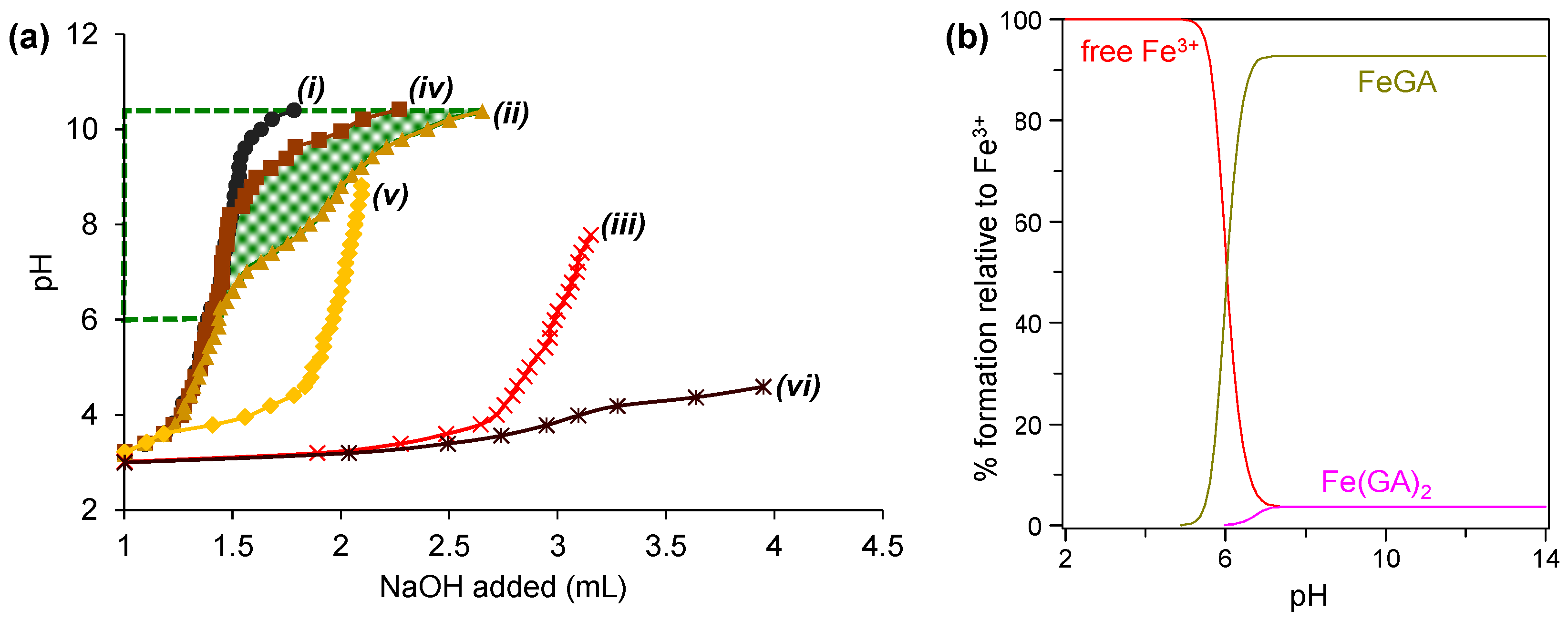
5.1. Potentiometric Titration

Potentiometric titration is a robust and widely used analytical technique for predicting metal ligand complex formation across a broad pH range. This method provides critical insights into the likelihood of complexation by analyzing changes in protonation behavior and stability constants. Figure 4a illustrates the potentiometric titration curves for the titration of Fe3+ with GA (and/or glycine ligand). The protonation constant (pKa) of GA and the stability constant (logK) of its metal complex can be determined by analyzing the difference in the volume of NaOH required to achieve the same pH value in titration curves (i) and (ii) for pKa, and (i) and (iv) for logK. Similarly, for glycine, pKa and logK are derived from the differences in NaOH consumption between curves (i) and (iii) and curves (i) and (v), respectively, following the Irving and Rossotti methodology [17]. The formation of metal complexes is evidenced by the shift in the titration curve for the system containing Fe3+ and GA (curve iv) to lower pH values compared to the curve for GA only (curve i), as highlighted by the green shaded area in Figure 4a. This shift indicates the binding of Fe3+ to GA, which alters the protonation behavior of the ligand.
Using the open-access Hyperquad Simulation and Speciation 2009 (HySS2009) software [72], the logK values corresponding to the stability constants of the metal complexes can be stimulated, allowing for visualization of the distribution of metal complex species across various pH ranges [73]. Illustrated in Figure 4b is the species distribution of Fe3+ and GA complexes. FeGA begins forming at pH 5.0 and is predominantly abundant between pH 7.0 and highly alkaline pH. This observation aligns well with the shift observed in the titration curve in Figure 4a. In contrast, Fe(GA)2 forms at lower abundance in the same pH range, due to its reduced stability (lower logK), which is attributed to weaker electrostatic interactions between Fe3+ and the second ligand molecules [74]. The structural representation of the typical Fe3+-GA complexes at various metal-to-ligand ratios is presented in Figure 5.
The formation of the complexes is influenced by two primary factors: (i) pH, which dictates ligand deprotonation and the availability of deprotonated groups for coordination, and (ii) metal-to-ligand ratios, where higher metal or ligand concentrations can favor the formation of complexes with higher stoichiometry. At acidic pH (2.0–6.0), uncoordinated Fe3+ and GA, dominate because the ligand remains mostly protonated, limiting strong electrostatic interactions. At pH > 6.0, abundant deprotonated ligand species readily coordinate with Fe3+, explaining why MPN synthesis typically occurs under alkaline conditions. However, while FeGA persists under extreme alkaline pH, synthesis in such conditions is impractical due to the need for excessive alkaline compounds. High OH concentrations can lead to the formation or precipitation of metal hydroxide, especially in concentrated metal ligand solutions often used in MPN synthesis. This highlights the importance of optimizing synthesis conditions to promote effective complex formation.

5.2. Spectrophotometric-Based Analyses

Spectrophotometric techniques are well suited to analyze metal complexes because of their unique electronic structures and optical properties, which arise from the interactions between metal ions and ligands. Crystal field theory (CFT) and ligand field theory (LFT) provide the basic framework for understanding these properties, explaining how ligands create electric fields that partition d orbitals into different energy levels and how molecular orbital interactions influence electronic transitions [76,77]. Transition metal complexes, especially those involving d- and f-block elements, exhibit diverse oxidation states and electronic configurations, leading to characteristic transitions such as d-d, charge transfer, and ligand-centered transitions, all of which can be examined using spectrophotometry. Advanced computational methods, including molecular orbital (MO) and density functional theory (DFT), further enhance the ability to predict and interpret absorption and emission spectra, offering insights into stability, reactivity, and bonding interactions [76,78]. These capabilities make spectrophotometry a versatile and indispensable tool, playing a crucial role in advancing research in areas such as analytical chemistry, supramolecular chemistry, and the development of functional materials such as metal-organic frameworks (MOFs), molecular machines, including MPNs [76,77,79,80].

5.2.1. Raman Spectroscopy

Raman spectroscopy, particularly when coupled with Surface-Enhanced Raman Spectroscopy (SERS), serves as a highly sensitive and powerful tool for probing the functional groups of ligands involved in interactions with metal ions during the formation of metal complexes. These vibrational spectroscopy techniques enable the detection and identification of target analytes at the single-molecule level, offering unparalleled insights into molecular interactions [81]. For instance, Sánchez-Cortés and García-Ramos (2000) demonstrated the effectiveness of these techniques by examining the spectral changes in GA upon its interaction with Ag colloid [82]. In their study, the band corresponding to the carboxylic (–COOH) group, observed at 1690 cm−1 in the solid-state spectrum of GA (Figure 6(ai)), disappeared in the SERS spectrum of the GA dissolved in ethanol (Figure 6(aii), red-highlighted spectrum). This disappearance indicates the ionization of the –COOH group at neutral pH. Additionally, the SERS spectrum of GA on Ag colloid (Figure 6(aiii), green-highlighted spectrum) revealed the emergence of new spectral features, reflecting chemical changes in GA induced by the presence of the Ag colloid. These spectral alterations are consistent with the formation of metal ligand coordination complexes, highlighting the utility of SERS in elucidating such interactions.
Similarly, Espina et al. (2022) investigated the Raman spectra of iron gall ink species, specifically the Fe-GA complex, and observed significant restructuring of the vibrational spectra between free GA and metal-complexed GA (Figure 6(bi,bii) compared to Figure 6(biii,biv)) [83]. Three prominent bands at 1470 cm−1, 1322 cm−1, and 576 cm−1 were identified as characteristic of iron gall ink species, with these bands being widely documented in prior studies [84,85,86]. Density functional theory (DFT) calculations on the Fe-GA complex revealed that the band at 1470 cm−1 arises from benzene ring vibrations coupled to C–O stretching and C–H bending. This band intensifies in the spectrum of metal-complexed GA due to changes in polarizability induced by metal coordination. The band at 1322 cm−1 corresponds to ring stretching coupled to C–O stretching and C–H bending, while the band at 576 cm−1, exclusive to the metal-complexed GA, is attributed to Fe–O stretching, confirming metal coordination.
These findings underscore the exceptional capability of SERS to provide detailed insights into molecular interactions and structural transformations at the nanoscale. By capturing subtle spectral changes and correlating them with specific molecular vibrations, SERS enables a deeper understanding of the mechanisms underlying metal ligand complexation and related chemical processes.

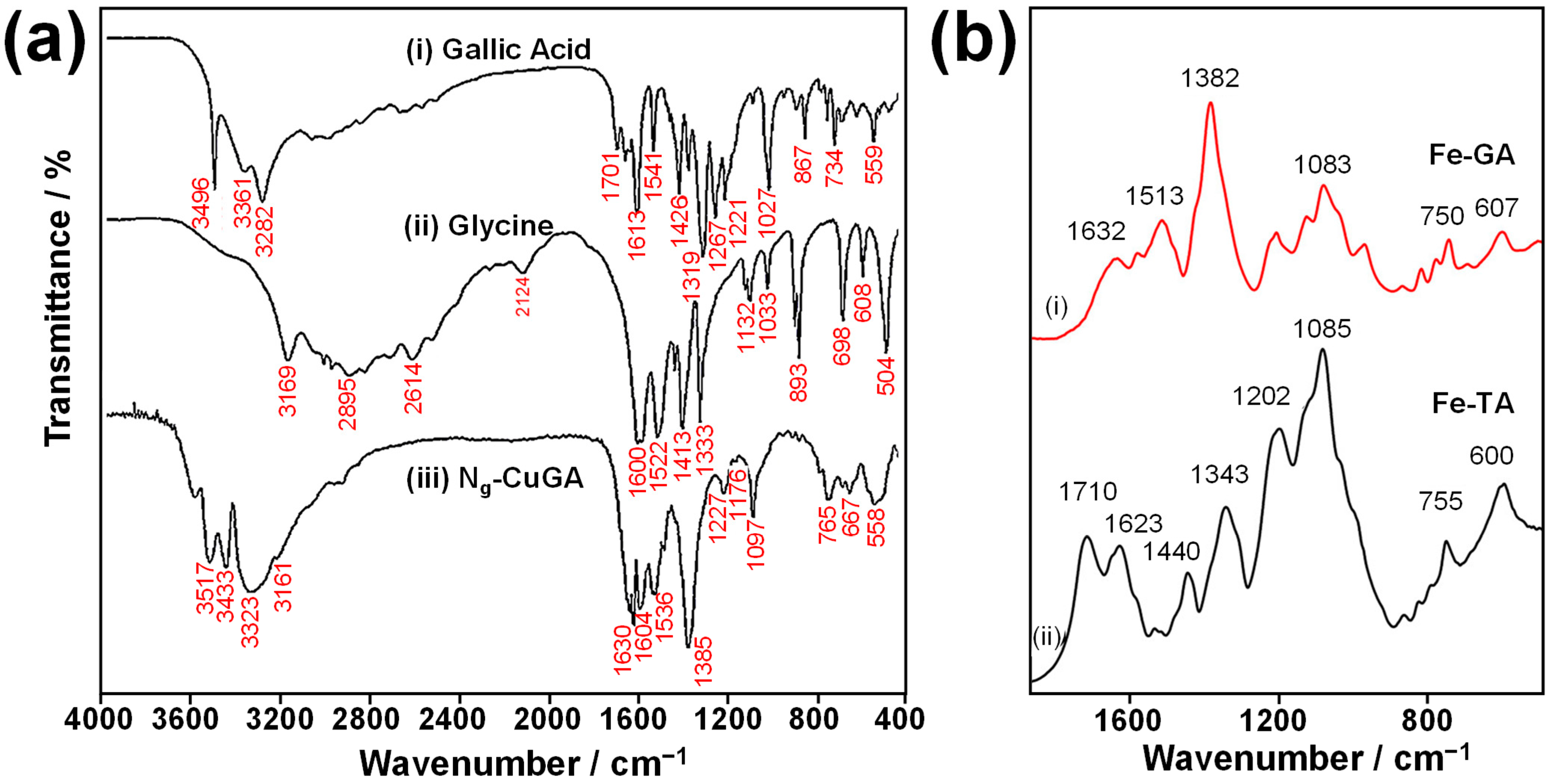
5.2.2. Fourier Transform Infrared (FTIR) Spectroscopy

FTIR spectroscopy is a valuable tool for confirming the successful formation of metal ligand complexes by comparing the spectra of the complex with those of the original ligand. It is particularly effective in detecting prominent vibrations of oxygen-containing functional groups. For example, Santoso et al. (2021) demonstrated the use of FTIR to confirm the formation of the metal complex Ng-CuGA from GA and Cu2+, with additional nitrogen group functionalization via ternary complexation with glycine [37]. The complexation of GA with Cu2+ induces vibrational band shifts in the galloyl group, specifically in the –OH groups, as indicated by the shifting of the bands between the wavenumbers 3282 and 3496 cm−1 (Figure 7(aiii)). Furthermore, nitrogen functionalization is confirmed by the presence of a band at 3161 cm−1 in the Ng-CuGA spectra, indicating the –NH group. The observed shift in this amine group’s spectral position, compared to glycine alone, suggests its involvement in metal ion coordination during complex formation.
Similarly, Espina et al. (2022) highlighted the robustness of FTIR in identifying functional groups involved in metal chelation [78]. Upon complexation with Fe3+, the band corresponding to C–O stretching (1309 cm−1) and C–OH bending (1014 cm−1) decreases in intensity compared to GA alone, while a strong band at 1083 cm−1 appears instead (Figure 7(bi)), indicating Fe3+ coordination with the –OH of the phenolic group. Additionally, the appearance of a strong band at 1382 cm−1 has been attributed to the interaction between Fe3+ and the –COOH group. The Fe–O coordination is observed at 607 cm−1 for the FeGA complex and at 600 cm−1 for the FeTA complex (Figure 7(bii)).
Several other works also reported changes in the spectra as induced by the complexation. Lunardi et al. (2023) reported a reduction from a doublet to a single peak near ~2900 cm−1 [6], while Liu et al. (2021) observed the disappearance of the O–H vibration peak at ~3300 cm−1 [87]. Espina et al. (2022) documented alterations in peaks within the 1300–1000 cm−1 range [83]. These spectral changes provide critical insights into the coordination environment and structural transformations of the ligand upon complexation.

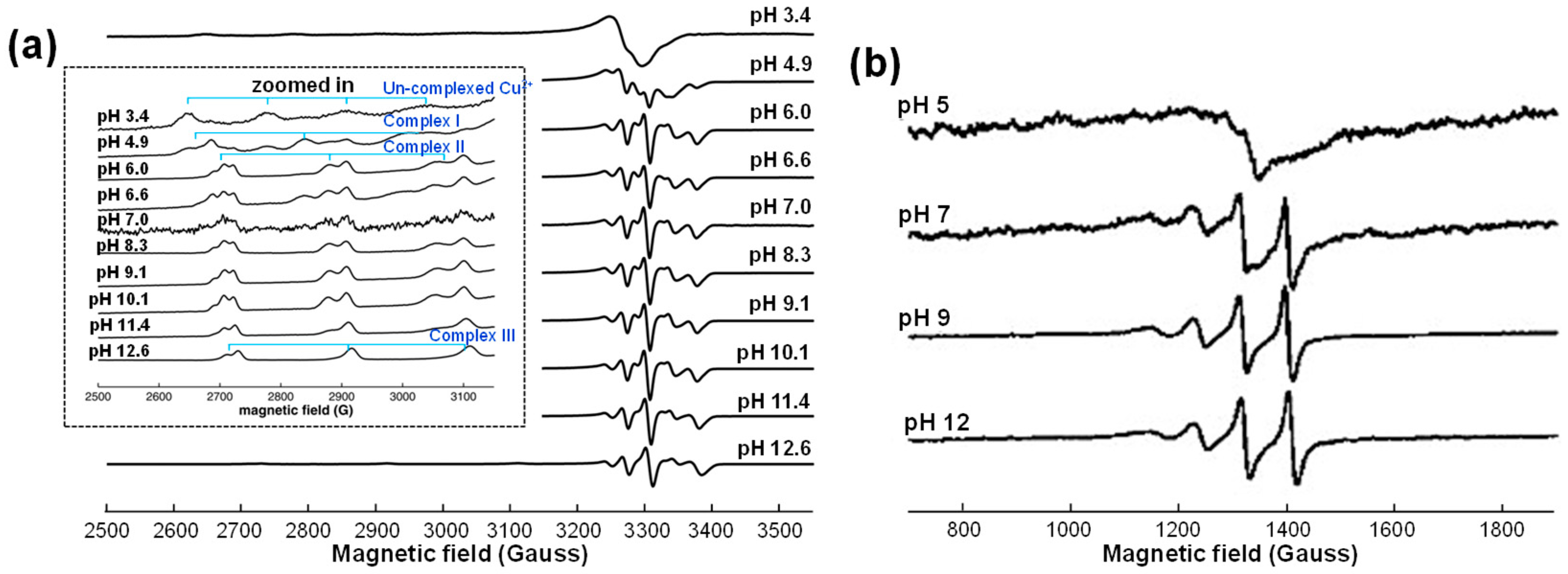
5.2.3. Electron Paramagnetic Resonance (EPR) Spectroscopy

EPR spectroscopy is a powerful technique for characterizing ligand fields (the negative point charges of ligands) and determining the oxidation states of metal complexes. It provides valuable insights into the reaction chemistry between metals and ligands across a wide range of experimental conditions [88]. For instance, Pirker et al. (2012) successfully employed EPR spectroscopy to investigate the formation of mononuclear Cu2+ complexes with GA [89]. Their study identified three distinct Cu-GA complex species through S-band and X-band EPR spectra, denoted as Complexes I, II, and III.
Complexes I and II were assigned as mono- and bis-Cu2+ GA complexes, respectively, with Cu2+ coordinating to two hydroxy-phenolic groups of GA, as illustrated in Figure 5 for ML and ML2 complexes. Complex III, on the other hand, was hypothesized to form through additional coordination of glycerol. The S-band EPR spectra at an acidic pH of 5 revealed the presence of hydrated, uncomplexed Cu2+, [Cu(H2O)6]2+. While Complex I was hardly detectable in the S-band spectra (Figure 8a), it was clearly observed in the X-band spectra (Figure 8b). At pH 7, the S-band spectra exhibited overlapping signals from Complexes II and III, whereas only Complex III was detected at higher pH values.

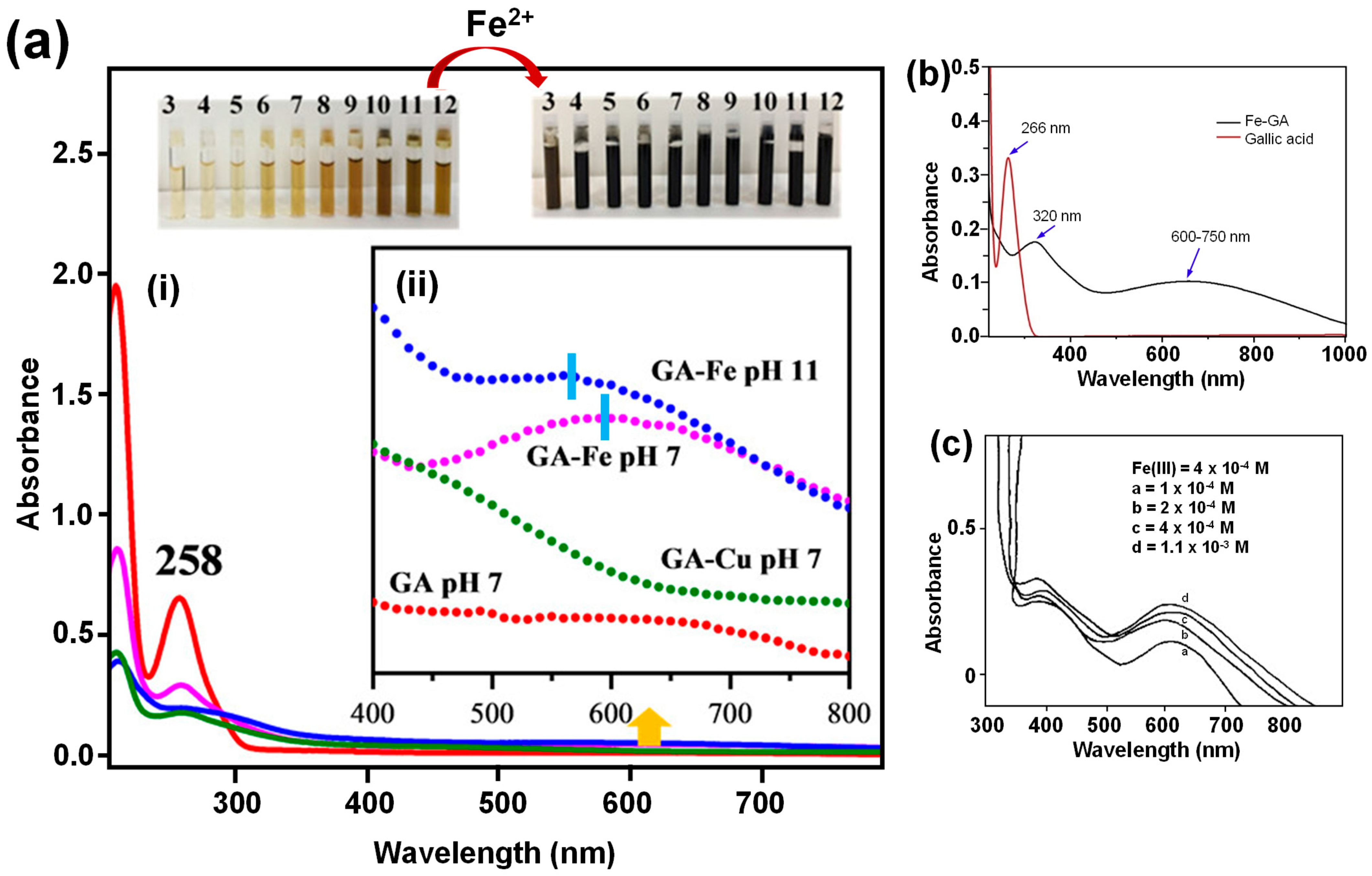
5.2.4. UV-Vis Spectroscopy

The stability of metal ligand complexes in aqueous solutions is commonly quantified using the stability constant (logK), while their structural characteristics are elucidated through spectroscopic techniques. These methods are crucial for confirming the successful formation of metal phenolic networks (MPNs) or modifications induced by MPNs on substrates. The complexation of metal ions with polyphenols under varying pH conditions often results in distinct visible color changes, which arise from different modes of metal ligand coordination and the formation of diverse complex species. UV-visible absorption spectroscopy provides a straightforward means to analyze ligand-to-metal charge transfer (LMCT), which is responsible for these color changes [27,83,90,91,92].
Free GA molecules exhibit two characteristic absorption bands at ~210 nm and ~260 nm, corresponding to the 1La and 1Lb singlet excited states, respectively, associated with π π * transitions. Upon complexation with metal ions at pH 7, these bands significantly decrease in intensity and may undergo a redshift. Additionally, a new, broad absorption band emerges in the visible region at ~600 nm (Figure 9(aii), indicated by the sky-blue colored bar), attributed to LMCT between the metal and the ligand [83]. This band is primarily responsible for the pronounced color changes observed during complexation. For metal complexes formed at pH 11, the band at ~600 nm undergoes a blueshift, which is attributed to the auto-oxidation of GA to quinone structures. Similar observations have been reported by Yadav et al. [49] and Masoud et al. (2014) [27], Figure 9b,c, where complexation with Fe3+ results in the appearance of a band in the visible range with λmax~600 nm.
UV-Vis spectrophotometry is a simple yet effective technique for confirming the successful coordination of metals and ligands in the formation of MPNs. This technique is applicable to various types of ligands. For instance, Mazaheri et al. (2022) [93] demonstrated the formation of Fe3+-tannic acid (TA) MPN through the absorption band within the range 450–650 nm with λmax at 475 nm, corresponding to the formation of the mono Fe3+-TA complexes. This study also highlighted the stability of MPNs over prolonged aging times, noting a decrease in absorption intensity with increasing aging time due to complexation with acetonitrile, which was used as the solvent in their work.

5.2.5. Nuclear Magnetic Resonance (NMR) Spectroscopy

Nuclear magnetic resonance (NMR) spectroscopy is a powerful, non-destructive analytical technique widely employed for molecular structure identification. It operates by exploiting the magnetic properties of atomic nuclei, providing detailed insights into molecular environments and interactions. In the context of metal complex characterization, NMR is particularly valuable for discerning oxidation states, coordination environments, and ligand-metal interactions. To elucidate comprehensive structural information, NMR is often combined with complementary techniques such as polarization transfer, relaxometry, and multidimensional NMR spectroscopy [80,94].
Fazary et al. (2009) have demonstrated the use of 1H and 13C NMR spectroscopy to characterize the isolated solid FeGA complex. Their analysis revealed the presence of functional ionizable groups in the ligand; however, the data did not provide conclusive evidence for the complexation between the ligand and the metal ion. In a more recent study, Etou et al. (2024) demonstrated the efficacy of liquid-state NMR spectroscopy, coupled with density functional theory (DFT) calculations, for elucidating the structure of metal complexes involving gallic acid (GA) and Al3+ [95]. By employing 27Al NMR, they compared the spectra of a solution containing only Al-salt with that of an Al3+-GA mixed solution. While a single peak at 0 ppm was observed for the Al-salt alone, two distinct peaks at 1.17 ppm and 15.7 ppm were detected in the Al3+-GA system. These peaks were attributed to the formation of monodentate and bidentate AlGA complexes, respectively, with the former arising from coordination between Al3+ and the –COOH group of GA, and the latter resulting from coordination with two –OH groups of GA. Further confirmation was provided by 1H NMR analysis, which compared the spectra of GA alone with the Al3+-GA mixed solution. A peak at 7.14 ppm, corresponding to GA alone, was accompanied by two additional peaks at 6.95 ppm and 7.04 ppm in the Al3+-GA system, consistent with the formation of the bidentate complex. This study highlights the utility of NMR spectroscopy, particularly when integrated with computational methods, for unraveling the structural intricacies of metal ligand interactions.

6. Versatility of GA-Based MPNs as Adsorbents

MPNs stand out as unique materials characterized by their amorphous structure, achieved through low-energy synthesis. Unlike other frameworks, MPNs are synthesized at room temperature, within short durations, and under moderate concentrations of metals and ligands. This mild synthesis process preserves the intrinsic properties of (poly)phenols, enabling strong interactions with various macromolecular substrates and metal ions. These interactions grant MPNs exceptional adhesive properties, making them highly versatile for surface engineering and adsorbent applications [96].

6.1. MPN as Standalone Adsorbent

MPNs have been extensively utilized as surface modifiers due to their strong adhesive properties. However, this focus has often overshadowed the exploration of MPNs as standalone materials. MPNs can be isolated as solid particles by carefully controlling parameters in the reaction, including the use of minimal amounts of solvent based on the solubility of the ligand, controlling the pH, and regulating the concentrations of metal ions and ligands during synthesis. This well-established method has been employed for decades. For instance, Patel et al. (1971) successfully isolated Ni2+ complexes with ethylenediamine (en) ligands, such as catechol and pyrogallol, in 1:2 and 1:3 ratios, forming Ni(en)2 and Ni(en)3. These complexes precipitated as the solution pH reached 7 upon ligand addition [60]. Ni(en)2 required less water and ligand, while Ni(en)3 necessitated larger quantities of both. Heating is sometimes applied to accelerate the formation of solid precipitates, though the temperatures required are significantly lower than those used in the synthesis of MOFs. Figure 10 illustrates the assembly of the metal complexes into MPN supramolecule. The formation of solid metal complexes resembles the agglomeration of coordinated metal ligand molecular compounds, which combine to form large clusters of supramolecular complexes. This clustering is driven by charge neutralization, which occurs as metal ligand interactions are prolonged or heating is applied. The neutralization process may also be influenced by attached water molecules to the metal ions.
A major challenge in this process is the potential decomposition of the ligand during cluster formation, particularly due to oxygen exposure. Ligand decomposition can lead to the formation of metal oxides or hydroxides instead of the desired metal ligand complex. Morales et al. (2023) reported such phenomena, noting that ligand decomposition is more likely to occur when metal acetate salts are used as the metal source [98]. The acetate ions can induce the oxidation of gallic acid (GA), leading to its decomposition into phenols, quinones, and hydroxyquinones. To prevent ligand decomposition, metal chloride salts can be used instead of metal acetates, as chloride ions do not promote oxidation.
Santoso et al. (2021) demonstrated a technique for isolating MPNs as solid particles to prepare an adsorbent [37]. The MPN adsorbent was synthesized by reacting an equimolar concentration of GA with CuCl2·5H2O in a minimal amount of water. The solid MPN precipitate was isolated by adjusting the reaction pH to 8 with NaOH. To enhance the adsorption performance, glycine was introduced, providing nitrogen (N)-containing adsorption sites. The resulting solid MPN can be employed in the adsorption system at various pH levels, starting from a pH of 2 to 10, indicating the material’s capability to withstand various environmental pH levels. The MPN has excellent adsorption efficiency for methylene blue, with an adsorption capacity of 190.81 mg/g. This efficiency was attributed to interactions such as electrostatic attraction, hydrogen bonding, dipole-dipole interactions, and n–π stacking.
Solution pH plays a critical role in the formation of MPN complexes, which directly affects their adsorption performance. Azhar et al. [36] demonstrated the impact of pH on the physicochemical properties of complexes formed between Cu2+ and GA, as well as their adsorption efficiency toward methylene blue and Congo red. At low pH (limited NaOH addition), insufficient hydroxide ions hinder the complete deprotonation of GA, limiting its coordination with Cu2+. Conversely, excessive NaOH addition can result in the oxidation of Cu2+, forming Cu2O instead of the desired complexes. Moreover, the synthesis temperature was also shown to influence the formation of the complexes. The CuGA complexes with minimal Cu2O impurities were achieved at a Cu:GA:NaOH molar ratio of 1:1:2.2, resulting in adsorption capacities of 124.64 mg/g for methylene blue and 344.54 mg/g for Congo red, where the molecular interaction between the adsorbent and adsorbate is shown in Figure 10c,d. In another study, Azhar et al. also demonstrated a high adsorption capacity for basic red 9 (115.08 mg/g) [41].
Isolated MPN complexes typically do not exhibit well-defined porosity. To overcome this, Lin et al. (2019) [97] developed a method to create highly ordered mesoporous MPN particles using a sacrificial templating technique. In this approach, cubosome templates were created from PS217-b-PEO45 block copolymers (PC) via a cosolvent method. An MPN precursor solution containing GA or epigallocatechin (EGCG) and FeCl3.6H2O was added to the cubosome-containing solution, and the pH was adjusted to 6.5 to drive coordination-driven assembly. The stability of Fe-ligand bonds was demonstrated under physiological conditions (pH 7.4), with Fe3+ ions detaching at acidic pH levels. This pH-dependent disassembly behavior is also confirmed in other works [99,100,101]. These mesoporous MPN materials exhibited exceptional protein-loading capacities (362 mg/g for glucose oxidase and 486 mg/g for horseradish peroxidase), outperforming commercial mesoporous SiO2 particles.
Despite the promising capabilities of GA-derived metal phenolic networks (MPNs), their adsorption potential remains underexplored, presenting an exciting avenue for future research. For instance, FeGA MPNs have demonstrated electrochemical and redox properties [34], which could be harnessed not only for metal adsorption but also for the removal of hazardous compounds through oxidation pathways. Fe-containing adsorbents have shown the ability to facilitate Fenton-like oxidation, further enhancing the degradation of organic contaminants in water. Additionally, the presence of GA in the Fenton process can enhance the efficiency of the process [102].

6.2. MPN for Surface Modification of Adsorbent

The surface modification of adsorbents with MPNs significantly enhances their adsorption performance by improving binding affinity and capacity. Additionally, MPN moieties impart pH-responsive behavior to the modified adsorbents. For instance, Zeng et al. [103] developed the development of Fe3+-GA (FeGA) MPNs that are stable at pH 5 and non-cytotoxic to mouse cells. These FeGA MPNs also exhibit photon absorption properties, making them potential photothermal guidance agents. At pH 7, FeGA undergoes decomposition due to the metabolic activity of the tested mouse cells with decomposition products efficiently excreted without causing cytotoxicity. The non-cytotoxic nature of FeGA is advantageous in applications targeting aquatic ecosystems, as it minimizes adverse effects on surrounding biota.
Figure 11a illustrates the self-assembly process of MPN formation as a film coating on a substrate. The process begins with the deposition of metal ions onto the substrate surface. Using a substrate that contains functional groups capable of binding metal ions, such as carboxylic or amino groups, can enhance the stability of the resulting MPN coating [104]. Upon the addition of phenolic acids (e.g., GA) under suitable pH conditions, iron complexes form spontaneously, leading to the development of a supramolecular MPN coating on the substrate surface. For example, the modification of cellulose using MPN via the coordination of GA and Fe3+ has been successfully demonstrated by Lunardi et al. (2024) [6]. This process involved immersing the cellulose substrate in a Fe3+-containing solution, followed by the addition of a GA solution. MPN self-assembly was achieved by adjusting the solution pH to 8 by adding NaOH. Although the microscale size of the MPN particles hindered direct morphological observation, EDX analysis confirmed the presence of Fe, indicating successful MPN coating (Figure 11b). The modification was further evidenced by a color change on the cellulose surface, with distinct colors observed for different metal-to-ligand ratios (Figure 11c). This characteristic coloration of Fe-GA MPN has also been utilized in the preparation of dyes and pigments, such as iron gall inks [75,104]. Cellulose modified with MPN at different metal-to-ligand ratios exhibited varying removal efficiencies. The highest removal efficiency (~90%) was achieved at a 1:3 ratio, while lower efficiencies were observed at ratios of 1:1 and 3:1. The maximum adsorption capacity of MPN-modified cellulose for Cr6+ was 73.63 mg/g, approximately 1.6 times higher than that of unmodified cellulose. Increasing the temperature further enhanced the adsorption capacity to 116.8 mg/g.
The combination of MPNs and MOFs has become a widely adopted strategy to enhance the functionality of MOFs. FeGA MPNs, for example, offer photodynamic activity for theragnostic applications, antibacterial and antifungal properties ideal for surface coatings, and pH-responsive behavior suitable for adsorption and controlled drug release. The integration of MPNs with MOFs often involves bridging bonds, such as the Fe–O–Cu bond observed in the combination of Cu2+-GA (CuGA) MPN with NH2-MIL-88B MOF, as reported by Li et al. [105]. In this system, GA served as a chelating agent, binding Fe-metal clusters in the MOF while simultaneously coordinating with free Cu2+ ions. This adhesion, resulting in the formation of CuGA@NH2-MIL-88B, enhances the photodynamic behavior of the MOF by facilitating electron transfer from photons to the MOF through the bridging bond. Additionally, this composite demonstrated significant adsorption capability as demonstrated by a significant reduction in glutathione absorbance in the solution within 30 min of contact.
The weak adhesive nature of MOFs poses a challenge for their integration with various substrates, as direct growth of MOFs on substrate surfaces often results in particle separation due to insufficient bonding. In this context, MPNs can serve as effective bridging molecules, facilitating MOF adhesion to substrates. This process involves coating the substrate with MPNs under mild conditions, such as room temperature in aqueous solvents. The adhered MPN molecules provide active coordination sites, facilitating interactions with MOF precursors and promoting MOF growth on the substrate surface (Figure 11d). This approach has been successfully used to prepare self-standing hollow MPN@ZIF-8 structures, which function as effective H2/CH4 gas adsorbents [96]. Additionally, Luo et al. (2019) demonstrated the ease of MPN coating by applying it to a biomass-derived microporous membrane, enhancing its ability to effectively capture uranium from seawater [106].
Rahim et al. [107] demonstrated the grafting of GA onto Fe3O4 magnetic nanoparticles (FMNPs) using alkoxysilanes and the amine-containing compound 1,2-ethylenediamine (1,2-en) as mediators. In this process, the oxygen-containing group of the silane compound forms a chelation-like coordination bond with FMNPs, while the silicon moiety reacts with the ethyl group of 1,2-en via a nucleophilic-driven reaction. The amine group of 1,2-en interacts with GA through hydrogen bonding and electrostatic interactions. This composite exhibited a high adsorption capacity, binding 4.23–4.98 mmol of Fe3+ ions per gram of adsorbent.
Pirozzi et al. [108] explored a reverse adsorption strategy leveraging coordination chemistry between GA and metal ions. In their study, GA molecules were adsorbed onto a magnetic metal-ceramic nanocomposite by coordinating with active Lewis acid sites on the nanocomposite surface. The highest GA uptake was observed at pH 5, where the surface charge of the nanocomposite is nearly neutral, minimizing electrostatic repulsion with the moderately protonated GA. This optimal pH balanced the nanocomposite’s surface charge, GA protonation state, and Lewis acidic sites availability of for interaction.
In a recent study by Nilnit et al. [109], an in-situ strategy was developed for synthesizing a magnetic adsorbent coated with a phenolic compound. In this method, phenolics extracted from the Hevea brasiliensis Muell. Arg. bark was directly reacted with FeSO4.7H2O at 65 °C under sonication. The phenolic acted as ligand, chelating Fe2+ ions to form Fe-phenolic complexes. Sodium hydroxide was then added to the reaction mixture to precipitate Fe(OH)2-phenolic, which subsequently oxidized in water to produce magnetite Fe3O4-phenolic particles. This strategy resembles conventional MPN synthesis but differs in the absence of an exterior substrate, with Fe3O4 serving as both a substrate and a metal cluster. The phenolic-coated Fe3O4 selectively adsorbed tetracycline residue in honey via a solid-phase extraction process, facilitating the preconcentration of the residues before HPLC-UV analysis. The adsorbed tetracycline residues ranged from 12 to 127.2 mg/g. However, the phenolic-coated Fe3O4 particles were prone to thermal and chemical degradation, rendering them unsuitable for reuse.
While GA-MPN adsorbents offer versatile functionalities, their stability under acidic conditions remains a challenge due to protonation and the subsequent weakening of metal ligand coordination bonds, requiring further research to address this issue. One promising approach is hybridization with inorganic materials, such as metal oxides and MOFs, which can enhance structural rigidity and protect coordination sites from protonation. Additionally, complexation with metal ions that results in a higher stability constant can provide greater resistance to acidic environments. By optimizing metal ligand interactions, GA-MPNs can potentially be tailored for improved durability in low-pH applications.

6.3. Other MPNs-Based Adsorbent

Tannic acid (TA)—a natural biopolymer composed of eight or more gallic acid (GA) molecules—was the first phenolic compound introduced for metal-phenolic network (MPN) synthesis, as reported in the pioneering work of Ejima et al. (2013) [14]. As a structurally undefined dendritic polyphenol, it is widely used as a ligand in MPN preparation due to its abundant carboxylic and phenolic hydroxyl groups. These functional groups contribute to the complexity of determining its pKa value [110]. As pH increases, TA undergoes deprotonation, generating negatively charged ligand species that readily donate electron pairs to metal ions, forming stable complexes [83].
FeTA MPNs are particularly notable for their antifungal activity. Hou et al. demonstrated that FeTA coatings on Ag-based MOFs exhibit a synergistic antifungal effect, effectively inhibiting the growth of Fusarium oxysporum [111]. The FeTA coating not only provides active sites for myclobutanil loading but also enables controlled, pH-responsive release, enhancing the efficiency and precision of antifungal delivery. Furthermore, FeTA MPNs are non-cytotoxic and ecologically compatible, as evidenced by their lack of inhibitory effects on Pakchoi seed germination.
Rahim et al. (2019) explored the chelating ability of phenolic ligands for metal ion sequestration, developing a metal-phenolic sorbent (MPS) via a straightforward sol-gel process by mixing TA and Zr4+ in a 1:1.2 molar ratio at 85 °C for 3 min, followed by a 10 min gelation period at room temperature [112]. The resulting material exhibited nearly 100% sequestration efficiency across a broad range of metals, attributed to the active catechol and gallol groups in TA that remained unbound to Zr4+. Investigating GA as a ligand for MPS synthesis could provide further insights into optimizing metal sequestration performance.
MPNs also show promise as controlled-release agents for nutrients, offering a practical alternative to post-application urea removal from soil or water. Mazaheri et al. (2022) demonstrated FeTA MPN self-assembly in acetonitrile for urea coating, where complexation occurred immediately upon urea addition, leading to uniform deposition [93]. A solvent-free approach was later developed via mechanochemical grinding of MPN and urea granules, followed by aging, which enhanced complexation and coating stability [113]. The most recent work by Mazaheri et al. (2024) further improved nutrient release control by incorporating silicate into MPN-encapsulated urea [114]. Applying similar strategies to GA-based MPNs could unlock new opportunities for sustainable nutrient delivery, leveraging GA’s unique binding properties for enhanced stability and controlled release.

7. Conclusions and Future Perspective

7.1. Concluding Remark

Gallic acid (GA)-based phenolic-metal networks (GA-MPNs) have emerged as a versatile class of functional materials with broad applications in adsorption and surface modification. Their environmentally benign nature, universal adhesion properties, and tunable coordination chemistry make them highly effective for enhancing adsorbent performance. This review has provided a comprehensive overview of the fundamental chemistry governing the formation of GA-MPNs, their structural and chemical characterization, and their roles in various adsorbent architectures, including stand-alone MPNs, MPN-mediated mesoporous materials, MPN-MOF composites, and MPN-coated substrates. By consolidating recent advances in GA-based MPNs, this work contributes to the fundamental understanding required for the rational design of next-generation MPN-based adsorbents.

7.2. Future Research Directions

To unlock the full potential of GA-based MPNs, future research should focus on three important aspects: (1) advancing their synthesis, (2) expanding their applications in emerging fields, and (3) improving their sustainability through biomass-derived ligands.
  • Advancing MPN Synthesis and Structural Control: While GA-based MPNs have demonstrated promising adsorption properties, their amorphous nature presents challenges in precisely tuning pore size, morphology, and surface area. Future efforts should focus on:
    • Template-Assisted Synthesis: Using sacrificial templates or structure-directing agents to achieve better control over pore architecture and surface area.
    • Ligand-to-Metal Ratio Optimization: Fine-tuning coordination chemistry to enhance stability, redox properties, and adsorption performance.
    • Post-Synthesis Modifications: Functionalizing MPNs with catalytic sites, redox-active moieties, or hybrid nanomaterials to broaden their utility.
    Additionally, exploring a wider range of metal centers with tailored functionalities—such as enhanced redox activity, photodegradability, or photocatalytic properties—could expand MPN applications in energy and environmental fields.
2.
Expanding Applications in Emerging Fields: Beyond adsorption and surface modification, GA-MPNs hold potential for various high-impact applications:
  • Biomedical Applications: GA’s intrinsic bioactivity, combined with metal coordination, enables potential use in antimicrobial coatings, drug delivery systems, and biosensors. Investigating MPNs as biodegradable, metal-coordinated drug carriers or bioadhesives could open new biomedical frontiers.
  • Environmental Remediation: Functionalized GA-MPNs could be engineered for targeted pollutant removal, photocatalytic degradation of contaminants, and recovery of critical metals from wastewater. Additionally, integrating MPNs with membranes or composite materials could improve their practicality in filtration technologies.
  • Energy Storage and Catalysis: MPNs with redox-active metals may serve as electrode materials in supercapacitors or electrocatalysts for water splitting and CO₂ reduction.
3.
Sustainable Development Using Biomass-Derived Ligands: A promising avenue for cost-effective and eco-friendly MPN development is the use of crude biomass extracts as sources of phenolic ligand instead of purified GA. However, key challenges remain:
  • Extraction Optimization: Developing efficient, scalable methods to obtain high-phenolic-content extracts with minimal impurities.
  • Ligand Purity Control: Understanding the impact of natural extract variability on MPN formation and performance.
  • Complexation Efficiency: Investigating how mixed phenolic compounds in crude extracts influence coordination chemistry and material stability.
By addressing these challenges, GA-based MPNs could become a sustainable and economically viable alternative for adsorption, catalysis, and biomedicine. Continued interdisciplinary research integrating chemistry, materials science, and engineering will be essential to fully realize their potential in both scientific and industrial domains.

Supplementary Materials

The following supporting information can be downloaded at: https://www.mdpi.com/article/10.3390/molecules30061218/s1, Table S1: Gallic acid-based metal complexes; Table S2: Hard-Soft Acid-Base List; Table S3: Data point of titration curves of solution containing gallic acid, Fe3+, and glycine at 0.1 M ionic strength; Table S4: HySS2009-Concentration table. References [115,116,117,118,119] are cited in the supplementary materials.

Author Contributions

Writing—original draft preparation, S.P.S., A.E.A. and K.-C.C.; writing—review and editing, S.-P.L., C.-W.H. and O.S.; visualization, S.P.S., H.-Y.H. and A.R.; supervision, S.I. All authors have read and agreed to the published version of the manuscript.

Funding

This research received no external funding.

Institutional Review Board Statement

Not applicable.

Informed Consent Statement

Not applicable.

Data Availability Statement

No primary research results have been included and no new data were generated or analyzed as part of this review.

Conflicts of Interest

The authors declare no conflicts of interest.

Abbreviations

The following abbreviations are used in this manuscript:
MPNsMetal-phenolic networks
MOFsMetal-organic frameworks
GAGallic acid
TATannic acid
HSABHard-Soft Acid-Base
IWSIrving Williams Series
SEMScanning electron microscopy
EDXEnergy dispersive X-ray
SERSSurface-Enhanced Raman Spectroscopy
FTIRFourier transform infrared spectroscopy
EPRElectron Paramagnetic Resonance
NMRNuclear Magnetic Resonance
TEMTransmission electron microscopy
HPLCHigh performance liquid chromatography
DFTDensity functional theory
SSASurface specific area
PCPS217-b-PEO45 block copolymers
EGCGEpigallocatechin
enEthylenediamine
FMNPsFe3O4 magnetic nanoparticles
LMCTLigand-to-metal charge transfer

References

  1. Manyangadze, M.; Chikuruwo, N.H.M.; Narsaiah, T.B.; Chakra, C.S.; Radhakumari, M.; Danha, G. Enhancing adsorption capacity of nano-adsorbents via surface modification: A review. S. Afr. J. Chem. Eng. 2020, 31, 25–32. [Google Scholar] [CrossRef]
  2. Jha, M.K.; Joshi, S.; Sharma, R.K.; Kim, A.A.; Pant, B.; Park, M.; Pant, H.R. Surface Modified Activated Carbons: Sustainable Bio-Based Materials for Environmental Remediation. Nanomaterials 2021, 11, 3140. [Google Scholar] [CrossRef]
  3. Petrovic, B.; Gorbounov, M.; Soltani, S.M. Influence of surface modification on selective CO2 adsorption: A technical review on mechanisms and methods. Microporous Mesoporous Mater. 2021, 312, 110751. [Google Scholar] [CrossRef]
  4. Abegunde, S.M.; Idowu, K.S.; Adejuwon, O.M.; Adeyemi-Adejolu, T. A review on the influence of chemical modification on the performance of adsorbents. Resour. Environ. Sustain. 2020, 1, 100001. [Google Scholar] [CrossRef]
  5. Liang, B.; Zhu, P.; Gu, J.; Yuan, W.; Xiao, B.; Hu, H.; Rao, M. Advancing Adsorption and Separation with Modified SBA-15: A Comprehensive Review and Future Perspectives. Molecules 2024, 29, 3543. [Google Scholar] [CrossRef]
  6. Lunardi, V.B.; Cheng, K.-C.; Lin, S.-P.; Angkawijaya, A.E.; Go, A.W.; Soetaredjo, F.E.; Ismadji, S.; Hsu, H.-Y.; Hsieh, C.-W.; Santoso, S.P. Modification of cellulosic adsorbent via iron-based metal phenolic networks coating for efficient removal of chromium ion. J. Hazard. Mater. 2024, 464, 132973. [Google Scholar] [CrossRef]
  7. Zhang, Y.; Shen, L.; Zhong, Q.-Z.; Li, J. Metal-phenolic network coatings for engineering bioactive Interfaces. Colloids Surf. B Biointerfaces 2021, 205, 111851. [Google Scholar] [CrossRef]
  8. Cheng, X.; Zhu, Y.; Tang, S.; Lu, R.; Zhang, X.; Li, N.; Zan, X. Material priority engineered metal-polyphenol networks: Mechanism and platform for multifunctionalities. J. Nanobiotechnol. 2022, 20, 255. [Google Scholar] [CrossRef]
  9. Wen, Y.; Yang, X.; Li, Y.; Yan, L.; Zhao, Y.; Shao, L. Progress reports of metal-phenolic network engineered membranes for water treatment. Sep. Purif. Technol. 2023, 320, 124225. [Google Scholar] [CrossRef]
  10. Zhong, Q.-Z.; Pan, S.; Rahim, M.A.; Yun, G.; Li, J.; Ju, Y.; Lin, Z.; Han, Y.; Ma, Y.; Richardson, J.J.; et al. Spray Assembly of Metal–Phenolic Networks: Formation, Growth, and Applications. ACS Appl. Mater. Interfaces 2018, 10, 33721–33729. [Google Scholar] [CrossRef]
  11. Välimets, A.; Koort, K.; Mortimer, M. Biocompatibility of Metal–Phenolic Network-Coated Nanoparticles. Proceedings 2023, 92, 33. [Google Scholar] [CrossRef]
  12. Fan, G.; Cottet, J.; Rodriguez-Otero, M.R.; Wasuwanich, P.; Furst, A.L. Metal–Phenolic Networks as Versatile Coating Materials for Biomedical Applications. ACS Appl. Bio Mater. 2022, 5, 4687–4695. [Google Scholar] [CrossRef]
  13. Geng, H.; Zhong, Q.-Z.; Li, J.; Lin, Z.; Cui, J.; Caruso, F.; Hao, J. Metal Ion-Directed Functional Metal–Phenolic Materials. Chem. Rev. 2022, 122, 11432–11473. [Google Scholar] [CrossRef]
  14. Ejima, H.; Richardson, J.J.; Liang, K.; Best, J.P.; Koeverden, M.P.V.; Such, G.K.; Cui, J.; Caruso, F. One-Step Assembly of Coordination Complexes for Versatile Film and Particle Engineering. Science 2013, 341, 154–157. [Google Scholar] [CrossRef] [PubMed]
  15. Wianowska, D.; Olszowy-Tomczyk, M. A Concise Profile of Gallic Acid—From Its Natural Sources through Biological Properties and Chemical Methods of Determination. Molecules 2023, 28, 1186. [Google Scholar] [CrossRef]
  16. Andjelković, M.; Camp, J.V.; Meulenaer, B.D.; Depaemelaere, G.; Socaciu, C.; Verloo, M.; Verhe, R. Iron-chelation properties of phenolic acids bearing catechol and galloyl groups. Food Chem. 2006, 98, 23–31. [Google Scholar] [CrossRef]
  17. Fazary, A.E.; Taha, M.; Ju, Y.-H. Iron Complexation Studies of Gallic Acid. J. Chem. Eng. Data 2008, 54, 35–42. [Google Scholar] [CrossRef]
  18. Rahim, M.A.; Kempe, K.; Müllner, M.; Ejima, H.; Ju, Y.; Koeverden, M.P.v.; Suma, T.; Braunger, J.A.; Leeming, M.G.; Abrahams, B.F.; et al. Surface-Confined Amorphous Films from Metal-Coordinated Simple Phenolic Ligands. Chem. Mater. 2015, 27, 5825–5832. [Google Scholar] [CrossRef]
  19. Lin, Z.; Liu, H.; Richardson, J.J.; Xu, W.; Chen, J.; Zhou, J.; Caruso, F. Metal–phenolic network composites: From fundamentals to applications. Chem. Soc. Rev. 2024, 53, 10800–10826. [Google Scholar] [CrossRef]
  20. Venkateswarlu, C.; Das, M.S.; Athavale, V.T. Studies of gallic acid complexes with metals and their analytical applications. Part I. Spectrophotometric Investigation. Proc. Indian Acad. Sci. Sect. A 1954, 40, 260–269. [Google Scholar] [CrossRef]
  21. Venkateswarlu, C.; Das, M.S.; Athavale, V.T. Studies of gallic acid complexes with metals and their analytical applications—Part III. Spectrophotometric estimation of gallic acid. Proc. Indian Acad. Sci. Sect. A 1956, 44, 241–246. [Google Scholar] [CrossRef]
  22. Varde, M.S.; Athavale, V.T. Studies of gallic acid complexes with metals and their analytical applications—Part II. A Spectrophotometric study of molybdenum complexes. Proc. Indian Acad. Sci. Sect. A 1956, 44, 228–240. [Google Scholar] [CrossRef]
  23. Powell, H.K.J.; Taylor, M.C. Interactions of Iron(II) and Iron(III) with Gallic Acid and its Homologues: A Potentiometric and Spectrophotometric Study. Aust. J. Chem. 1982, 35, 739–756. [Google Scholar] [CrossRef]
  24. Sandmann, B.J.; Chien, M.H.; Sandmann, R.A. Stability Constants of Calcium, Magnesium and Zinc Gallate Using a Divalent Ion-Selective Electrode. Anal. Lett. 1985, 18, 149–159. [Google Scholar] [CrossRef]
  25. Elinany, G.A.; Ebeid, F.M.; Zahra, A.M.; Ziedan, F.I. Polarography of Metal-Gallic Complexes. J. Electroanal. Chem. 1976, 72, 363–369. [Google Scholar] [CrossRef]
  26. Rahim, S.A.; Hussain, S.; Farooqui, M. Protonation Equilibria of Gallic Acid (GA) and Stability Constants of Its Complexes with Transition Metal Ions in Aqueous Media. J. Chem. Biol. Phys. Sci. 2017, 7, 267–273. [Google Scholar]
  27. Masoud, M.S.; Ali, A.E.; Haggag, S.S.; Nasr, N.M. Spectroscopic studies on gallic acid and its azo derivatives and their iron(III) complexes. Spectrochim. Acta Part A Mol. Biomol. Spectrosc. 2014, 120, 505–511. [Google Scholar] [CrossRef]
  28. Zaccaron, S.; Ganzerla, R.; Bortoluzzi, M. Iron complexes with gallic acid: A computational study on coordination compounds of interest for the preservation of cultural heritage. J. Coord. Chem. 2012, 66, 1709–1719. [Google Scholar] [CrossRef]
  29. Gao, F.; Zheng, D.; Tanaka, H.; Zhan, F.; Yuan, X.; Gao, F.; Wang, Q. An electrochemical sensor for gallic acid based on Fe2O3/electro-reduced graphene oxide composite: Estimation for the antioxidant capacity index of wines. Mater. Sci. Eng. C-Mater. Biol. Appl. 2015, 57, 279–287. [Google Scholar] [CrossRef]
  30. Mishra, B.; Chandra, M. Evaluation of phytoremediation potential of aromatic plants: A systematic review. J. Appl. Res. Med. Aromat. Plants 2022, 31, 100405. [Google Scholar] [CrossRef]
  31. Daduang, J.; Palasap, A.; Daduang, S.; Boonsiri, P.; Suwannalert, P.; Limpaiboon, T. Gallic acid enhancement of gold nanoparticle anticancer activity in cervical cancer cells. Asian Pac. J. Cancer Prev. 2015, 16, 169–174. [Google Scholar] [CrossRef] [PubMed]
  32. Khan, A.N.; Hassan, M.N.; Khan, R.H. Gallic acid: A naturally occurring bifunctional inhibitor of amyloid and metal induced aggregation with possible implication in metal-based therapy. J. Mol. Liq. 2019, 285, 27–37. [Google Scholar] [CrossRef]
  33. Sharma, S.; Mittal, D.; Verma, A.K.; Roy, I. Copper-Gallic Acid Nanoscale Metal–Organic Framework for Combined Drug Delivery and Photodynamic Therapy. ACS Appl. Bio Mater. 2019, 2, 2092–2101. [Google Scholar] [CrossRef]
  34. Cherepanov, P.V.; Rahim, M.A.; Bertleff-Zieschang, N.; Sayeed, M.A.; O’Mullane, A.P.; Moulton, S.E.; Caruso, F. Electrochemical Behavior and Redox-Dependent Disassembly of Gallic Acid/Fe III Metal–Phenolic Networks. ACS Appl. Mater. Interfaces 2018, 10, 5828–5834. [Google Scholar] [CrossRef] [PubMed]
  35. Wu, J.; Ouyang, D.; He, Y.; Su, H.; Yang, B.; Li, J.; Sun, Q.; Lin, Z.; Cai, Z. Synergistic Effect of Metal–Organic Framework/Gallic Acid in Enhanced Laser Desorption/Ionization Mass Spectrometry. ACS Appl. Mater. Interfaces 2019, 11, 38255–38264. [Google Scholar] [CrossRef]
  36. Azhar, B.; Angkawijaya, A.E.; Santoso, S.P.; Gunarto, C.; Ayucitra, A.; Go, A.W.; Tran-Nguyen, P.L.; Ismadji, S.; Ju, Y.-H. Aqueous synthesis of highly adsorptive copper–gallic acid metal–organic framework. Sci. Rep. 2020, 10, 19212. [Google Scholar] [CrossRef]
  37. Santoso, S.P.; Bundjaja, V.; Angkawijaya, A.E.; Gunarto, C.; Go, A.W.; Yuliana, M.; Tran-Nguyen, P.L.; Hsieh, C.-W.; Ju, Y.-H. One-step synthesis of nitrogen-grafted copper-gallic acid for enhanced methylene blue removal. Sci. Rep. 2021, 11, 12021. [Google Scholar] [CrossRef]
  38. Hou, X.; Zhang, L.; Chen, Y.; Liu, Z.; Zhao, X.; Lu, B.; Luo, Y.; Qu, X.; Musskaya, O.; Glazov, I.; et al. Photothermal switch by gallic acid-calcium grafts synthesized by coordination chemistry for sequential treatment of bone tumor and regeneration. Biomaterials 2025, 312, 122724. [Google Scholar] [CrossRef]
  39. Gu, L.; Li, X.; Chen, G.; Yang, H.; Qian, H.; Pan, J.; Miao, Y.; Li, Y. A glutathione-activated bismuth-gallic acid metal-organic framework nano-prodrug for enhanced sonodynamic therapy of breast tumor. J. Colloid Interface Sci. 2025, 679, 214–223. [Google Scholar] [CrossRef]
  40. Wasuwanich, P.; Fan, G.; Burke, B.; Furst, A.L. Metal-phenolic networks as tuneable spore coat mimetics. J. Mater. Chem. B 2022, 10, 7600–7606. [Google Scholar] [CrossRef]
  41. Azhar, B.; Avian, C.; Tiwikrama, A.H. Green synthesis optimization with artificial intelligence studies of copper–gallic acid metal–organic framework and its application in dye removal from wastewater. J. Mol. Liq. 2023, 389, 122844. [Google Scholar] [CrossRef]
  42. Zhou, H.-C.; Long, J.R.; Yaghi, O.M. Introduction to Metal–Organic Frameworks. Chem. Rev. 2012, 112, 673–674. [Google Scholar] [CrossRef] [PubMed]
  43. Lin, K.-S.; Adhikari, A.K.; Ku, C.-N.; Chiang, C.-L.; Kuo, H. Synthesis and characterization of porous HKUST-1 metal organic frameworks for hydrogen storage. Int. J. Hydrogen Energy 2012, 37, 13865–13871. [Google Scholar] [CrossRef]
  44. Mahmoodi, N.M.; Abdi, J.; Oveisi, M.; Asli, M.A.; Vossoughi, M. Metal-organic framework (MIL-100 (Fe)): Synthesis, detailed photocatalytic dye degradation ability in colored textile wastewater and recycling. Mater. Res. Bull. 2018, 100, 357–366. [Google Scholar] [CrossRef]
  45. Zhao, H.; Li, Q.; Wang, Z.; Wu, T.; Zhang, M. Synthesis of MIL-101(Cr) and its water adsorption performance. Microporous Mesoporous Mater. 2020, 2020, 110044. [Google Scholar] [CrossRef]
  46. Mu, X.; Chen, Y.; Lester, E.; Wu, T. Optimized synthesis of nano-scale high quality HKUST-1 under mild conditions and its application in CO2 capture. Microporous Mesoporous Mater. 2018, 270, 249–257. [Google Scholar] [CrossRef]
  47. Zou, M.; Dong, M.; Zhao, T. Advances in Metal-Organic Frameworks MIL-101(Cr). Int. J. Mol. Sci. 2022, 23, 9396. [Google Scholar] [CrossRef]
  48. Yilmaz, B.S. Antimicrobial and Anticancer Activity of Gallic Acid–Cu(II) Hybrid Nanoflowers and Gallic Acid–Zn(II) Hybrid Nanoflowers. J. Inorg. Organomet. Polym. Mater. 2024, 34, 5329–5341. [Google Scholar] [CrossRef]
  49. Yadav, A.; Sharma, A.; Sharma, R.K. Mesoporous iron gallate nanocomplex for adsorption and degradation of organic dyes. Colloids Surf. A Physicochem. Eng. Asp. 2019, 579, 123694. [Google Scholar] [CrossRef]
  50. Wang, T.C.; Bury, W.; Gómez-Gualdrón, D.A.; Vermeulen, N.A.; Mondloch, J.E.; Deria, P.; Zhang, K.; Moghadam, P.Z.; Sarjeant, A.A.; Snurr, R.Q.; et al. Ultrahigh Surface Area Zirconium MOFs and Insights into the Applicability of the BET Theory. J. Am. Chem. Soc. 2015, 137, 3585–3591. [Google Scholar] [CrossRef]
  51. Peng, Y.; Krungleviciute, V.; Eryazici, I.; Hupp, J.T.; Farha, O.K.; Yildirim, T. Methane storage in metal-organic frameworks: Current records, surprise findings, and challenges. J. Am. Chem. Soc. 2013, 135, 118877–118894. [Google Scholar] [CrossRef] [PubMed]
  52. Spanopoulos, I.; Tsangarakis, C.; Klontzas, E.; Tylianakis, E.; Froudakis, G.; Adil, K.; Belmabkhout, Y.; Eddaoudi, M.; Trikalitis, P.N. Reticular Synthesis of HKUST-like tbo-MOFs with Enhanced CH4 Storage. J. Am. Chem. Soc. 2015, 138, 1568–1574. [Google Scholar] [CrossRef]
  53. Yang, H.; Zhao, Z.-C.; Yang, Y.-P.; Zhang, Z.; Chen, W.; Yan, R.-Q.; Jin, Y.; Zhang, J. Defective WO3 nanoplates controllably decorated with MIL-101(Fe) nanoparticles to efficiently remove tetracycline hydrochloride by S-scheme mechanism. Sep. Purif. Technol. 2022, 300, 121846. [Google Scholar] [CrossRef]
  54. Zhao, Z.-C.; Wang, K.; Chang, L.; Yan, R.-Q.; Zhang, J.; Zhang, M.; Wang, L.; Chen, W.; Huang, G.-B. Construction of S-scheme MIL-101(Fe)/Bi2MoO6 heterostructures for enhanced catalytic activities towards tetracycline hydrochloride photodegradation and nitrogen photofixation. Sol. Energy 2023, 264, 112042. [Google Scholar] [CrossRef]
  55. El-Shahawy, A.A.G.; Dief, E.M.; El-Dek, S.I.; Farghali, A.A.; El-Ela, F.I.A. Nickel-gallate metal–organic framework as an efficient antimicrobial and anticancer agent: In vitro study. Cancer Nanotechnol. 2023, 14, 60. [Google Scholar] [CrossRef]
  56. Chen, Z.; Świsłocka, R.; Choińska, R.; Marszałek, K.; Dąbrowska, A.; Lewandowski, W.; Lewandowska, H. Exploring the Correlation Between the Molecular Structure and Biological Activities of Metal–Phenolic Compound Complexes: Research and Description of the Role of Metal Ions in Improving the Antioxidant Activities of Phenolic Compounds. Int. J. Mol. Sci. 2024, 25, 11775. [Google Scholar] [CrossRef]
  57. Molski, M. Computation of the pKa Values of Gallic Acid and Its Anionic Forms in Aqueous Solution: A Self-Similar Transformation Approach for Accurate Proton Hydration Free Energy Estimation. Molecules 2025, 30, 742. [Google Scholar] [CrossRef]
  58. Jabbari, M. Solvent dependence of protonation equilibria for gallic acid in water and different acetonitrile–water cosolvent systems. J. Mol. Liq. 2015, 208, 5–10. [Google Scholar] [CrossRef]
  59. Roy, K.; Kar, S.; Das, R.N. Chemical Information and Descriptors. In Understanding the Basics of QSAR for Applications in Pharmaceutical Sciences and Risk Assessment, 1st ed.; Academic Press: Cambridge, MA, USA; Elsevier Inc.: Amsterdam, The Netherlands, 2015; pp. 47–80. [Google Scholar] [CrossRef]
  60. Patel, D.C.; Bhattacharya, P.K. A study of ligand exchange in some nickel complexes. J. Inorg. Nucl. Chem. 1971, 33, 529–533. [Google Scholar] [CrossRef]
  61. Radalla, A.M. Potentiometric studies on ternary complexes involving some divalent transition metal ions, gallic acid and biologically abundant aliphatic dicarboxylic acids in aqueous solutions. Beni-Suef Univ. J. Basic Appl. Sci. 2015, 4, 174–182. [Google Scholar] [CrossRef]
  62. Şişmanoğlu, T.; İçhedef, Ç.; Akdut, G.; Soylu, G.S.P.; Medine, E.İ.; Teksöz, S. Complexation of gallic acid involving La3+, Sm3+, Th4+ and UO22+ ions in aqueous solution by potentiometry at various temperatures. J. Radioanal. Nucl. Chem. 2024, 334, 623–636. [Google Scholar] [CrossRef]
  63. Soldatović, T. Correlation between HSAB Principle and Substitution Reactions in Bioinorganic Reactions. In Photophysics, Photochemical and Substitution Reactions—Recent Advances; Saha, S., Kanaparthi, R.K., Soldatovic, T., Eds.; IntechOpen: London, UK, 2020. [Google Scholar]
  64. Miličević, A.; Branica, G.; Raos, N. Irving-Williams Order in the Framework of Connectivity Index 3χv Enables Simultaneous Prediction of Stability Constants of Bivalent Transition Metal Complexes. Molecules 2011, 16, 1103–1112. [Google Scholar] [CrossRef] [PubMed]
  65. Moreno, O.P.; Araiza, O.R.P.; Portillo, M.C.; Téllez, V.C.; Garrido, M.A.V. Jahn-Teller effect analysis at coordination complex [Cu(NH3)4]2+ ion, growth by green synthesis in CuS nanocrystals. Optik 2022, 251, 168470. [Google Scholar] [CrossRef]
  66. Singh, J.; Srivastav, A.N.; Singh, N.; Singh, A. Stability Constants of Metal Complexes in Solution. In Stability and Applications of Coordination Compounds; Srivastva, A.N., Ed.; IntechOpen: London, UK, 2020. [Google Scholar] [CrossRef]
  67. Sóvágó, I.; Kállay, C.; Várnagy, K. Peptides as complexing agents: Factors influencing the structure and thermodynamic stability of peptide complexes. Coord. Chem. Rev. 2012, 256, 2225–2233. [Google Scholar] [CrossRef]
  68. Lihi, N.; Lukács, M.; Raics, M.; Szunyog, G.; Várnagy, K.; Kállay, C. The effect of carboxylate groups on the complexation of metal ion with oligopeptides—Potentiometric investigation. Inorganica Chim. Acta 2018, 472, 165–173. [Google Scholar] [CrossRef]
  69. Sursyakova, V.V.; Burmakina, G.V.; Rubaylo, A.I. Composition and stability constants of copper(II) complexes with succinic acid determined by capillary electrophoresis. J. Coord. Chem. 2017, 70, 431–440. [Google Scholar] [CrossRef]
  70. El-Megharbel, S.M.; Hamza, R.Z. Synthesis, spectroscopic characterizations, conductometric titration and investigation of potent antioxidant activities of gallic acid complexes with Ca (II), Cu (II), Zn(III), Cr(III) and Se (IV) metal ions. J. Mol. Liq. 2022, 358, 119196. [Google Scholar] [CrossRef]
  71. Santoso, S.P.; Angkawijaya, A.E.; Ju, Y.-H.; Soetaredjo, F.E.; Ismadji, S.; Ayucitra, A. Synthesis, characterization, thermodynamics and biological studies of binary and ternary complexes including some divalent metal ions, 2, 3-dihydroxybenzoic acid and N -acetylcysteine. J. Taiwan Inst. Chem. Eng. 2016, 68, 23–30. [Google Scholar] [CrossRef]
  72. HySS2009, Hyperquad Simulation and Speciation. Available online: http://www.hyperquad.co.uk/ (accessed on 3 January 2025).
  73. Alderighi, L.; Gans, P.; Ienco, A.; Peters, D.; Sabatini, A.; Vacca, A. Hyperquad simulation and speciation (HySS): A utility program for the investigation of equilibria involving soluble and partially soluble species. Coord. Chem. Rev. 1999, 184, 311–318. [Google Scholar] [CrossRef]
  74. Santoso, S.P.; Chandra, I.K.; Soetaredjo, F.E.; Angkawijaya, A.E.; Ju, Y.-H. Equilibrium Studies of Complexes between N-Acetylcysteine and Divalent Metal Ions in Aqueous Solutions. J. Chem. Eng. Data 2014, 59, 1661–1666. [Google Scholar] [CrossRef]
  75. Boyatzis, S.C.; Velivasaki, G.; Malea, E. A study of the deterioration of aged parchment marked with laboratory iron gall inks using FTIR-ATR spectroscopy and micro hot table. Herit. Sci. 2016, 4, 13. [Google Scholar] [CrossRef]
  76. Tanase, T. Introduction: What Is a Metal Complex? Tanase, T., Ishii, Y., Eds.; Royal Society of Chemistry: London, UK, 2024; pp. 1–12. [Google Scholar]
  77. Elattar, R.H.; El-Malla, S.F.; Kamal, A.H.; Mansour, F.R. Applications of metal complexes in analytical chemistry: A review article. Coord. Chem. Rev. 2024, 501, 215568. [Google Scholar] [CrossRef]
  78. Frešer, F.; Hostnik, G.; Tošović, J.; Bren, U. Dependence of the Fe(II)-Gallic Acid Coordination Compound Formation Constant on the pH. Foods 2021, 10, 2689. [Google Scholar] [CrossRef]
  79. Ye, H.; Jiang, S.; Yan, Y.; Zhao, B.; Grant, E.R.; Kitts, D.D.; Yada, R.Y.; Pratap-Singh, A.; Baldelli, A.; Yang, T. Integrating Metal–Phenolic Networks-Mediated Separation and Machine Learning-Aided Surface-Enhanced Raman Spectroscopy for Accurate Nanoplastics Quantification and Classification. ACS Nano 2024, 18, 26281–26296. [Google Scholar] [CrossRef]
  80. Shin, J.; Lim, M.H.; Han, J. NMR spectroscopic investigations of transition metal complexes in organometallic and bioinorganic chemistry. Bull. Korean Chem. Soc. 2024, 45, 593–613. [Google Scholar] [CrossRef]
  81. Pérez-Jiménez, A.I.; Lyu, D.; Lu, Z.; Liu, G.; Ren, B. Surface-enhanced Raman spectroscopy: Benefits, trade-offs and future developments. Chem. Sci. 2020, 11, 4563–4577. [Google Scholar] [CrossRef]
  82. Sánchez-Cortés, S.; García-Ramos, J.V. Adsorption and Chemical Modification of Phenols on a Silver Surface. J. Colloid Interface Sci. 2000, 231, 98–106. [Google Scholar] [CrossRef]
  83. Espina, A.; Cañamares, M.V.; Jurašeková, Z.; Sanchez-Cortes, S. Analysis of Iron Complexes of Tannic Acid and Other Related Polyphenols as Revealed by Spectroscopic Techniques: Implications in the Identification and Characterization of Iron Gall Inks in Historical Manuscripts. ACS Omega 2022, 7, 27937–27949. [Google Scholar] [CrossRef]
  84. Carter, E.A.; Perez, F.R.; Garcia, J.M.; Edwards, H.G.M. Raman Spectroscopic Analysis of an Important Visigothic Historiated Manuscript. Philos. Transcations R. Soc. A Math. Phys. Eng. Sci. 2016, 2082, 20160041. [Google Scholar] [CrossRef]
  85. Nastova, I.; Grupče, O.; Minčeva-Šukarova, B.; Turan, S.; Yaygingol, M.; Ozcatal, M.; Martinovska, V.; Jakovlevska-Spirovska, Z. Micro-Raman spectroscopic analysis of inks and pigments in illuminated medieval old-Slavonic manuscripts. J. Raman Spectrosc. 2012, 43, 1729–1736. [Google Scholar] [CrossRef]
  86. Burgio, L.; Clark, R.J.H.; Hark, R.R. Raman microscopy and x-ray fluorescence analysis of pigments on medieval and Renaissance Italian manuscript cuttings. Proc. Natl. Acad. Sci. USA 2010, 107, 5726–5731. [Google Scholar] [CrossRef] [PubMed]
  87. Liu, C.; Li, C.; Jiang, S.; Zhang, C.; Tian, Y. pH-responsive hollow Fe–gallic acid coordination polymer for multimodal synergistic-therapy and MRI of cancer. Nanoscale Adv. 2021, 4, 173–181. [Google Scholar] [CrossRef] [PubMed]
  88. Severino, J.F.; Goodman, B.A.; Reichenauer, T.G.; Pirker, K.F. Is there a redox reaction between Cu(II) and gallic acid? Free Radic. Res. 2011, 45, 123–132. [Google Scholar] [CrossRef] [PubMed]
  89. Pirker, K.F.; Baratto, M.C.; Basosi, R.; Goodman, B.A. Influence of pH on the speciation of copper(II) in reactions with the green tea polyphenols, epigallocatechin gallate and gallic acid. J. Inorg. Biochem. 2012, 112, 10–16. [Google Scholar] [CrossRef]
  90. Santoso, S.P.; Angkawijaya, A.E.; Soetaredjo, F.E.; Ismadji, S.; Ju, Y.-H. Complex equilibrium study of some hydroxy aromatic ligands with beryllium ion. J. Mol. Liq. 2015, 212, 524–531. [Google Scholar] [CrossRef]
  91. Sherwani, I.A.H.A.; Köse, A.; Güngör, Ö.; Kırpık, H.; Güngör, S.A.; Köse, M. Synthesis, characterization and investigation of photophysical and biological properties of Cu(II) and Zn(II) complexes of benzimidazole ligands. Appl. Organomet. Chem. 2022, 36, e6585. [Google Scholar] [CrossRef]
  92. Berto, S.; Alladio, E. Application of Chemometrics Tools to the Study of the Fe(III)–Tannic Acid Interaction. Front. Chem. 2020, 8, 614171. [Google Scholar] [CrossRef]
  93. Mazaheri, O.; Alivand, M.S.; Zavabeti, A.; Spoljaric, S.; Pan, S.; Chen, D.; Caruso, F.; Suter, H.C.; Mumford, K.A. Assembly of Metal–Phenolic Networks on Water-Soluble Substrates in Nonaqueous Media. Adv. Funct. Mater. 2022, 32, 2111942. [Google Scholar] [CrossRef]
  94. Novotny, J.; Komorovsky, S.; Marek, R. Paramagnetic Effects in NMR Spectroscopy of Transition-Metal Complexes: Principles and Chemical Concepts. Acc. Chem. Res. 2024, 57, 1467–1477. [Google Scholar] [CrossRef]
  95. Etou, M.; Yoshida, M.; Tsuji, Y.; Murakami, M.; Inoue, T. Gallic acid complexation with Al3+ under acidic condition—27Al NMR and DFT study. Inorganica Chim. Acta 2024, 571, 122229. [Google Scholar] [CrossRef]
  96. Wang, T.; Lin, Z.; Mazaheri, O.; Chen, J.; Xu, W.; Pan, S.; Kim, C.-J.; Zhou, J.; Richardson, J.J.; Caruso, F. Crystalline Metal–Organic Framework Coatings Engineered via Metal–Phenolic Network Interfaces. Angew. Chem. Int. Ed. 2024, 63, e202410043. [Google Scholar] [CrossRef] [PubMed]
  97. Lin, Z.; Zhou, J.; Cortez-Jugo, C.; Han, Y.; Ma, Y.; Pan, S.; Hanssen, E.; Richardson, J.J.; Caruso, F. Ordered Mesoporous Metal–Phenolic Network Particles. J. Am. Chem. Soc. 2019, 142, 335–341. [Google Scholar] [CrossRef] [PubMed]
  98. Moralles, V.A.; Davolos, M.R.; Cebim, M.A. Europium(III) and gallic acid ligand coordination compounds: Synthesis, characterization, photophysical processes, and optimization of luminescent properties. Inorganica Chim. Acta 2023, 558, 121748. [Google Scholar] [CrossRef]
  99. Kim, N.; Lee, I.-S.; Choi, Y.; Ryu, J. Molecular design of heterogeneous electrocatalysts using tannic acid-derived metal-phenolic networks. Nanoscale 2021, 13, 20374–20386. [Google Scholar] [CrossRef]
  100. Wei, Y.; Wei, Z.; Luo, P.; Wei, W.; Liu, S. pH-sensitive metal-phenolic network capsules for targeted photodynamic therapy against cancer cells. Artif. Cells Nanomed. Biotechnol. 2018, 46, 1552–1561. [Google Scholar] [CrossRef]
  101. Tomasetig, D.; Wang, C.; Hondl, N.; Friedl, A.; Ejima, H. Exploring Caffeic Acid and Lignosulfonate as Key Phenolic Ligands for Metal-Phenolic Network Assembly. ACS Omega 2024, 9, 20444–20453. [Google Scholar] [CrossRef]
  102. Pereira, J.P.; Borges, C.H.; Tabelini; Aguiar, A. A Review of Gallic Acid-Mediated Fenton Processes for Degrading Emerging Pollutants and Dyes. Molecules 2023, 28, 1166. [Google Scholar] [CrossRef]
  103. Zeng, J.; Cheng, M.; Wang, Y.; Wen, L.; Chen, L.; Li, Z.; Wu, Y.; Gao, M.; Chai, Z. pH-Responsive Fe(III)–Gallic Acid Nanoparticles for In Vivo Photoacoustic-Imaging-Guided Photothermal Therapy. Adv. Healthc. Mater. 2016, 5, 772–780. [Google Scholar] [CrossRef]
  104. Fitz-Binder, C.; Manian, A.P.; Lenninger, M.; Ortlieb, S.; Bechtold, T.; Pham, T. Dyeing behaviour of iron(III)-gallic acid complexes on wool as function of pH-dependent iron(III)-complex stoichiometry. Dye. Pigment. 2025, 233, 112502. [Google Scholar] [CrossRef]
  105. Li, Y.; Li, C.; Liu, S.; Wang, Q.; Tang, Z.; Qu, J.; Ye, J.; Lu, Y.; Wang, J.; Zhang, K.; et al. Nano-photosensitizers with gallic acid-involved Fe–O–Cu “electronic storage station” bridging ligand-to-metal charge transfer for efficient catalytic theranostics. J. Colloid Interface Sci. 2024, 676, 974–988. [Google Scholar] [CrossRef]
  106. Luo, W.; Xiao, G.; Tian, F.; Richardson, J.J.; Wang, Y.; Zhou, J.; Guo, J.; Liao, X.; Shi, B. Engineering robust metal–phenolic network membranes for uranium extraction from seawater. Energy Environ. Sci. 2019, 12, 607–614. [Google Scholar] [CrossRef]
  107. Rahim, A.M.A.; Ahmed, S.A.; Soliman, E.M. Adsorptive removal of Fe(III) using gallic acid anchored iron magnetic nano-adsorbents synthesized via two different routes under microwave irradiation. Indian J. Chem. 2020, 59A, 9–20. [Google Scholar]
  108. Pirozzi, D.; Pansini, M.; Marocco, A.; Esposito, S.; Barrera, G.; Tiberto, P.; Allia, P.; Sannino, F. Adsorption of gallic acid by tailor-made magnetic metal-ceramic nanocomposites. J. Mol. Liq. 2023, 371, 121083. [Google Scholar] [CrossRef]
  109. Nilnit, T.; Supharoek, S.-A.; Siriangkhawut, W.; Vichapong, J.; Ponhong, K. Ultrasound-assisted continuous flow synthesis of natural phenolic-coated Fe3O4 for magnetic solid phase extraction of tetracycline residues in honey. Food Chem. 2024, 464, 141642. [Google Scholar] [CrossRef]
  110. Yan, W.; Shi, M.; Dong, C.; Liu, L.; Gao, C. Applications of tannic acid in membrane technologies: A review. Adv. Colloid Interface Sci. 2020, 284, 102267. [Google Scholar] [CrossRef]
  111. Hou, Y.; Zhang, Y.; Huang, Y.; Zhou, A.; Han, J.; Yang, K.; Zhao, Y.; Zhou, J.; Wang, J.; Chen, G.; et al. A pH-responsive MOFs@MPN nanocarrier with enhancing antifungal activity for sustainable controlling myclobutanil release. Chem. Eng. J. 2024, 497, 155713. [Google Scholar] [CrossRef]
  112. Rahim, M.A.; Lin, G.; Tomanin, P.P.; Ju, Y.; Barlow, A.; Bjonmalm, M.; Caruso, F. Self-Assembly of a Metal−Phenolic Sorbent for Broad-Spectrum Metal Sequestration. ACS Appl. Mater. Interfaces 2020, 12, 3746–3754. [Google Scholar] [CrossRef]
  113. Mazaheri, O.; Zavabeti, A.; McQuillan, R.V.; Lin, Z.; Alivand, M.S.; Gaspera, E.D.; Chen, D.; Caruso, F.; Suter, H.; Mumford, K.A. Solid-State Encapsulation of Urea via Mechanochemistry-Driven Engineering of Metal–Phenolic Networks. Chem. Mater. 2023, 35, 7800–7813. [Google Scholar] [CrossRef]
  114. Mazaheri, O.; Lin, Z.; Xu, W.; Mohankumar, M.; Wang, T.; Zavabeti, A.; McQuillan, R.V.; Chen, J.; Richardson, J.J.; Mumford, K.A.; et al. Assembly of Silicate–Phenolic Network Coatings with Tunable Properties for Controlled Release of Small Molecules. Adv. Mater. 2024, 36, 2413349. [Google Scholar] [CrossRef]
  115. Wang, Y.; Zhang, J.; Zhang, C.; Li, B.; Wang, J.; Zhang, X.; Li, D.; Sun, S.-K. Functional-Protein-Assisted Fabrication of Fe–Gallic Acid Coordination Polymer Nanonetworks for Localized Photothermal Therapy. ACS Sustain. Chem. Eng. 2018, 7, 994–1005. [Google Scholar] [CrossRef]
  116. Jing, Z.; Li, M.; Wang, H.; Yang, Z.; Zhou, S.; Ma, J.; Meng, E.; Zhang, H.; Liang, W.; Hu, W.; et al. Gallic acid-gold nanoparticles enhance radiation-induced cell death of human glioma U251 cells. IUBMB Life 2021, 73, 398–407. [Google Scholar] [CrossRef] [PubMed]
  117. Novaković, T.B.; Pavlović, S.M.; Pagnacco, M.C.; Banković, P.T.; Mojović, Z.D. The Application of Alumina for Electroanalytical Determination of Gallic Acid. Electrocatalysis 2023, 14, 18–28. [Google Scholar] [CrossRef]
  118. Goman, D.; Stanković, A.; Galović, O.; Džakula, B.N.; Kontrec, J.; Medvidović-Kosanović, M. Complexation of gallic acid with calcium: Electrochemical, potentiometric, and UV-VIS studies. Anal. Methods 2024, 16, 391–395. [Google Scholar] [CrossRef] [PubMed]
  119. Hamedi, H.; Javanbakht, S.; Mohammadi, R. In-situ synthesis of copper-gallic acid metal–organic framework into the gentamicin-loaded chitosan hydrogel bead: A synergistic enhancement of antibacterial properties. J. Ind. Eng. Chem. 2024, 133, 454–463. [Google Scholar] [CrossRef]
Figure 1. Timeline illustrating the development of gallic acid-based metal complexes, compiled from the publication list in Supplementary Information Table S1.
Molecules 30 01218 g001
Figure 2. (a,b) Morphological characterization of gallic acid (GA)-based metal complexes. SEM images of supramolecular materials formed through coordination between GA and copper (a) and GA and zinc (b). Images reproduced from Yilmaz (2024) [48], under a Creative Commons 4.0 license. (c) SEM image of N-grafted GA-copper MPN. Image adapted from Santoso et al. (2021) [37], licensed under CC-BY. (d,e) TEM and SEM images of copper-gallic acid metal-organic framework. Image reprinted from Sharma et al. (2019) [33], with permission from the American Chemical Society. (f) TEM image of iron gallate particles. Image reproduced from Yadav et al. (2019) [49], with permission from Elsevier B.V. All rights reserved.
Molecules 30 01218 g002
Figure 3. Stepwise ionization constants of gallic acid (GA). GAH4 represents the fully protonated molecule. Each deprotonation step leads to the sequential formation of negatively charged species: GAH3, GAH22−, GAH3−, and GA4−. In the molecular structure, oxygen atoms are represented by red spheres, hydrogen atoms by white spheres (with larger spheres indicating non-ionizable hydrogen atoms and smaller spheres indicating ionizable hydrogen atoms), and carbon atoms by grey spheres. The red dashed circles highlight the sites of subsequent deprotonation.
Molecules 30 01218 g003
Figure 4. (a) Titration curves of a solution containing gallic acid, Fe3+, and glycine at 0.1 M ionic strength: (i) 3 mM HNO3, (ii) solution i + 1 mM gallic acid (ligand only), (iii) solution ii + 0.4 mM Fe3+, (iv) solution i + 1 mM glycine, (v) solution iv + 0.4 mM Fe3+, (vi) solution i + 1 mM gallic acid + 1 mM glycine + 0.4 mM Fe3+. The green shaded area highlights the shift in the gallic acid curve resulting from the addition of Fe3+. The figure was redrawn from Ref. [17] by extracting data points using Origin 2024(10.1) software. The data points for drawing these curves are provided in Supplementary Table S3. (b) Species distribution curve as a function of pH for GA-Fe3+ system, with percentages of formation relative to Fe3+. The figure was created using HySS2009 software to model metal ligand complex distribution, with input pKa and logK values obtained from Ref. [17]. The data points for drawing the curve are provided in Supplementary Table S4.
Molecules 30 01218 g004
Figure 5. Structural representation of typical binary Fe3+-GA complexes. Typical binary complexes are formed with metal-to-ligand ratios of 1:1 (FeGA), 1:2 (Fe(GA)2), and ratios such as Fe2(GA)2 and Fe4(GA)n. Structural illustrations adapted and redrawn from Refs. [17,75]. In the molecular structure, oxygen atoms are represented by red spheres, hydrogen atoms by white spheres (with larger spheres indicating non-ionizable hydrogen atoms and smaller spheres indicating ionizable hydrogen atoms), carbon atoms by grey spheres, and iron atoms by the brown spheres.
Molecules 30 01218 g005
Figure 6. (a) Raman spectra illustrating the interaction of gallic acid (GA) with silver (Ag) in a colloidal system: (i) solid GA, (ii) 60 mM GA in ethanol, and (iii) 5 mM GA on Ag colloid at pH 3.9. The red-highlighted region corresponds to the vibrational modes of the carboxylic group in GA, which disappear due to deprotonation upon dissolution in ethanol. The green-highlighted region represents new spectral bands in GA induced by its interaction with the Ag colloid. Image adapted from Sánchez-Cortés and García-Ramos (2000) [82], with permission from Academic Press. All rights reserved. (b) Raman spectra of the metal complex between gallic acid (GA) and Fe3+: (i) solid GA, (ii) 20 mM GA in aqueous solution, (iii) Fe-GA complex on paper, and (iv) Fe-GA complex in solution at a ligand-to-metal molar ratio of 1:3 and pH of 7. Image adapted from Espina et al. (2022) [83], licensed under CC-BY-NC-ND 4.0.
Molecules 30 01218 g006
Figure 7. (a) FTIR spectra of the solid-state metal complexes formed by the coordination of Cu2+ with gallic acid (GA): (i) solid GA, (ii) solid glycine, and (iii) solid ternary metal complex. Image adapted from Santoso et al. (2021) [37], licensed under CC-BY. (b) FTIR spectra of solid-phase metal complexes of Fe3+ with gallic acid (GA) or tannic acid (TA): (i) Fe-GA complex and (ii) Fe-TA complex. Image adapted from Espina et al. (2022) [83], licensed under CC-BY-NC-ND 4.0.
Molecules 30 01218 g007
Figure 8. EPR spectra of gallic acid (GA) complexed with Cu2+ metal ions at metal-to-ligand molar ratio of 1:5 at different pH values: (a) S-band spectra and (b) X-band spectra. Images were reproduced from Pirker et al. (2021) [89], with permission from Elsevier Inc.
Molecules 30 01218 g008
Figure 9. (a) UV-Vis absorption spectra of the complex between Fe3+ and gallic acid (GA): (i) 0.066 mM GA solution and (ii) metal complex at a metal-to-ligand molar ratio of 3:1. The red line represents the GA solution at pH 7, the green line represents the GA-Cu2+ complex at pH 7, the pink line represents the GA-Fe3+ complex at pH 7, and the blue line represents the GA-Fe3+ complex at pH 11. Image reproduced from Espina et al. (2022) [83], licensed under CC-BY-NC-ND 4.0. (b) Electronic absorption spectra of GA and its corresponding iron gallate nanocomplex. Image reproduced from Yadav et al. (2019) [49], with permission from Elsevier B.V. All rights reserved. (c) UV-Vis spectra of GA complexes with Fe3+ at various molar concentrations. The image was reproduced from Masoud et al. (2014) [27], with permission from Elsevier B.V. All rights reserved.
Molecules 30 01218 g009
Figure 10. (a) Schematic representation of the molecular assembly process of metal phenolic networks as a standalone material. (b) Schematic representation of the preparation process of ordered mesoporous MPN. Adapted with permission from Lin et al. (2019) [97], Copyright © 2019, American Chemical Society. (c,d) Schematic illustration of the molecular structure of Cu2+-gallic acid (CuGA) complexes and their subsequent interaction mode toward Congo red and methylene blue adsorbates. Image reproduced from Azhar et al. (2020) [36], licensed under CC-BY.
Molecules 30 01218 g010
Figure 11. (a) Schematic illustration of MPN assembly on cellulose substrates. (b,c) SEM-EDX image and visual appearance of adsorbent composite prepared from coating of cellulose (DCell) with Fe-gallic acid MPN. Adapted with permission from Lunardi et al. (2024) [6], Copyright © 2023. Elsevier B.V. All rights reserved. (d) MPN-mediated MOF coatings: schematic representation of tunable MOF coating thickness on substrates. Adapted with permission from Wang et al. (2024) [96], Copyright © 2024 John Wiley and Sons, Inc.
Molecules 30 01218 g011
Table 1. Ionization or dissociation constant of the gallic acid.
ConditionpKa1pKa2pKa3pKa4Ref.
I = 0.2 M 1 and T = 25 °C4.228.6911.19 [60]
I = 0.1 M 1 and T = 25 °C4.48.611.212[24]
I = 0.1 M NaNO3 and T = 25 °C4.108.38 [17]
I = 0.1 M NaNO3 and T = 25 °C4.128.32 [61]
I = 0.1 M KCl and T = 25 °C3.757.509.5010.50[62]
1 Salt source undefined.
Table 2. Stability constant (logK) of complexes between GA and transition metal ions.
MetalComplex Species MpLqConditionlogKRef.
pq
Cu2+11I = 0.1 M NaNO3, T = 25 °C9.75[61]
I = 1 N NaNO3, T = 27 °C9.80[26]
12I = 0.1 M NaNO3, T = 25 °C6.75[61]
Zn2+11I = 0.1 M NaNO3, T = 25 °C8.56[61]
I = 1 N NaNO3, T = 27 °C7.98[26]
12I = 0.1 M NaNO3, T = 25 °C5.83[61]
21I = 0.1 M CaCl2, T = 25 °C, pH = 811.38[24]
Ni2+11I = 0.1 M NaNO3, T = 25 °C8.00[61]
I = 1 N NaNO3, T = 27 °C6.74[26]
12I = 0.1 M NaNO3, T = 25 °C5.50[61]
Fe3+11I = 0.1 M NaNO3, T = 25 °C14.73[17]
I = 1 N NaNO3, T = 27 °C10.98[26]
12I = 0.1 M NaNO3, T = 25 °C11.93[61]
Co2+11I = 0.1 M NaNO3, T = 25 °C7.25[61]
I = 1 N NaNO3, T = 27 °C7.13[26]
12I = 0.1 M NaNO3, T = 25 °C4.75[61]
Disclaimer/Publisher’s Note: The statements, opinions and data contained in all publications are solely those of the individual author(s) and contributor(s) and not of MDPI and/or the editor(s). MDPI and/or the editor(s) disclaim responsibility for any injury to people or property resulting from any ideas, methods, instructions or products referred to in the content.

