Article

Synthesis, Antimicrobial Activities, and Model of Action of Indolyl Derivatives Containing Amino-Guanidinium Moieties

1
Key Lab of New Animal Drug of Gansu Province, Key Lab of Veterinary Pharmaceutical Development of Ministry of Agriculture and Rural Affairs, Lanzhou Institute of Husbandry and Pharmaceutical Sciences of Chinese Academy of Agricultural Sciences, Lanzhou 730050, China
2
School of Health Nursing, Fuyang Vocational Technical College, Fuyang 236000, China
*
Authors to whom correspondence should be addressed.
The authors contributed equally to this work.
Molecules 2025, 30(4), 887; https://doi.org/10.3390/molecules30040887
Submission received: 14 December 2024 / Revised: 11 February 2025 / Accepted: 13 February 2025 / Published: 14 February 2025

Abstract

:
The objectives of the study were to design, synthesize, and evaluate the antibacterial activity of a series of novel aminoguanidine-indole derivatives. Thirty-seven new compounds were effectively synthesized through nucleophilic substitution reaction and guanidinylation reaction. Chemical structures of all the desired compounds were identified by NMR and HR-MS spectroscopy. Most of the synthesized compounds showed significant antibacterial activity against ESKAPE pathogens and clinical resistant Klebsiella pneumoniae (K. pneumoniae) isolates. K. pneumoniae is an important opportunistic pathogen that often threatens the health of immunocompromised people such as the elderly, children, and ICU patients. The most active compound 4P showed rapid bactericidal activity against resistant K. pneumoniae 2108 with MIC and MBC values that were 4 and 8 µg/mL, respectively. The hemolytic activity of 4P was low, with an HC50 value of 123.6 µg/mL. Compound 4P induced the depolarization of the bacterial membrane and disrupted bacterial membrane integrity and was not prone to antibiotic resistance. The dihydrofolate reductase (DHFR) activity was also notably inhibited by 4P in vitro. Molecular docking revealed that the aminoguanidine moiety and indole structure of 4P played an important role in binding to the target site of the K. pneumoniae dihydrofolate reductase (DHFR) receptor. In the mouse pneumonia model caused by K. pneumoniae, 4P improved the survival rate of mice, reduced bacterial loads, and alleviated tissues’ pathological injuries at a dosage of 4 mg/kg. Therefore, compound 4P may be a promising lead compound or drug candidate for antibacterial purposes against K. pneumoniae.

1. Introduction

The emergence of drug-resistant pathogenic bacteria has become an increasing threat to global health in recent years [1,2]. In particular, the ESKAPE pathogens (Enterococcus faecalis, Staphylococcus aureus, Klebsiella pneumoniae, Acinetobacter baumannii, Pseudomonas aeruginosa, and Escherichia coli), released by the WHO in 2017, draw much attention due to their rapid development of resistance to conventional antibiotics [3]. ESKAPE pathogens have been associated with a variety of infections, including urinary tract infections, pneumonia, and bloodstream infections, which can be fatal in severe cases [4]. As an important opportunistic pathogen, K. pneumoniae can colonize on the surface of the respiratory tract, the digestive tract, and medical devices [5,6], threatening the health of immunocompromised people such as the elderly [7], children [8], and ICU patients [9]. Healthy adults can also be infected by hypervirulent K. pneumoniae [10]. The WHO has recently emphasized the importance of K. pneumoniae in clinical practice [11]. The inappropriate use of antibiotics has led to the development of a frightening level of resistance, in particular the emergence of multidrug-resistant (MDR) bacteria and even ‘superbugs’, contributing to a significant economic burden [12,13]. The escalating threat posed by these pathogens requires not only the rational use of available antibiotics, but also the development of new classes of drugs to reduce deaths from infectious diseases.
Indole and its derivatives, as important biological signaling molecules, are widely distributed in nature and affect various aspects of bacterial physiology, including spore formation, cell division, plasmid stability, drug resistance, biofilm formation and virulence [14,15]. Recently, indole derivatives have received particular attention in the pharmaceutical field as molecules with antimicrobial activity [16,17]. Indole derivatives have great potential for treating methicillin-resistant Staphylococcus aureus (MRSA) [18]. A series of indole derivatives have been synthesized and some compounds have shown promising antimicrobial activity against Gram-positive and Gram-negative bacteria, and fungi [19,20]. This makes indole an excellent backbone for the synthesis of new antimicrobial compounds.
Antimicrobial activity depends on the penetration of the antimicrobial agent through the multilayered bacterial envelope and its interaction with the target pathogen. [21]. For Gram-negative bacteria, this process is more complicated due to the presence of an outer membrane, which requires additional attention in the design of novel antibacterial agents [22,23]. The accumulation abilities of 180 compounds were investigated in E. coli, with results showing that amphiphilic and rigid small molecules containing amines with low sphericity were most likely to accumulate [24]. The importance of ionizable nitrogen, low three-dimensionality, and rigidity for small molecules entering Gram-negative bacteria was also reviewed [25]. Guanidine was useful for enhancing the activity of compounds and was widely used in many drug molecules. N-Alkyl guanidiniums promoted the uptake of compounds in bacteria, and aminoguanidines were discovered with better activity [19,26,27].
Colistin is regarded as the final line of defense in the treatment of Gram-negative bacterial infections [28]. Consequently, the development of a novel antibiotic with comparable bactericidal efficacy to colistin and low resistance is therefore of considerable practical importance. In this study, a series of aminoguanidyl indole derivatives (3A-3V, 4L-4P, 5L-5P and 6L-6P) were first designed and synthesized. These included indoles and 7-azaindoles with various substituted benzyl bromides and aminoguanidine to produce the target compound, then their structures were characterized. Antibacterial activity against both Gram-positive and Gram-negative bacteria was assessed using colistin as a positive control. The role of the active molecule 4P in disrupting cell membrane integrity was investigated. A molecular docking study was performed between 4P and K. pneumoniae dihydrofolate reductase (DHFR) to confirm the target specificity and elucidate the binding mechanism. Furthermore, the hemolytic activity of 4P was also examined. This study provides a class of compounds for the development of novel antibacterial agents.

2. Result and Discussion
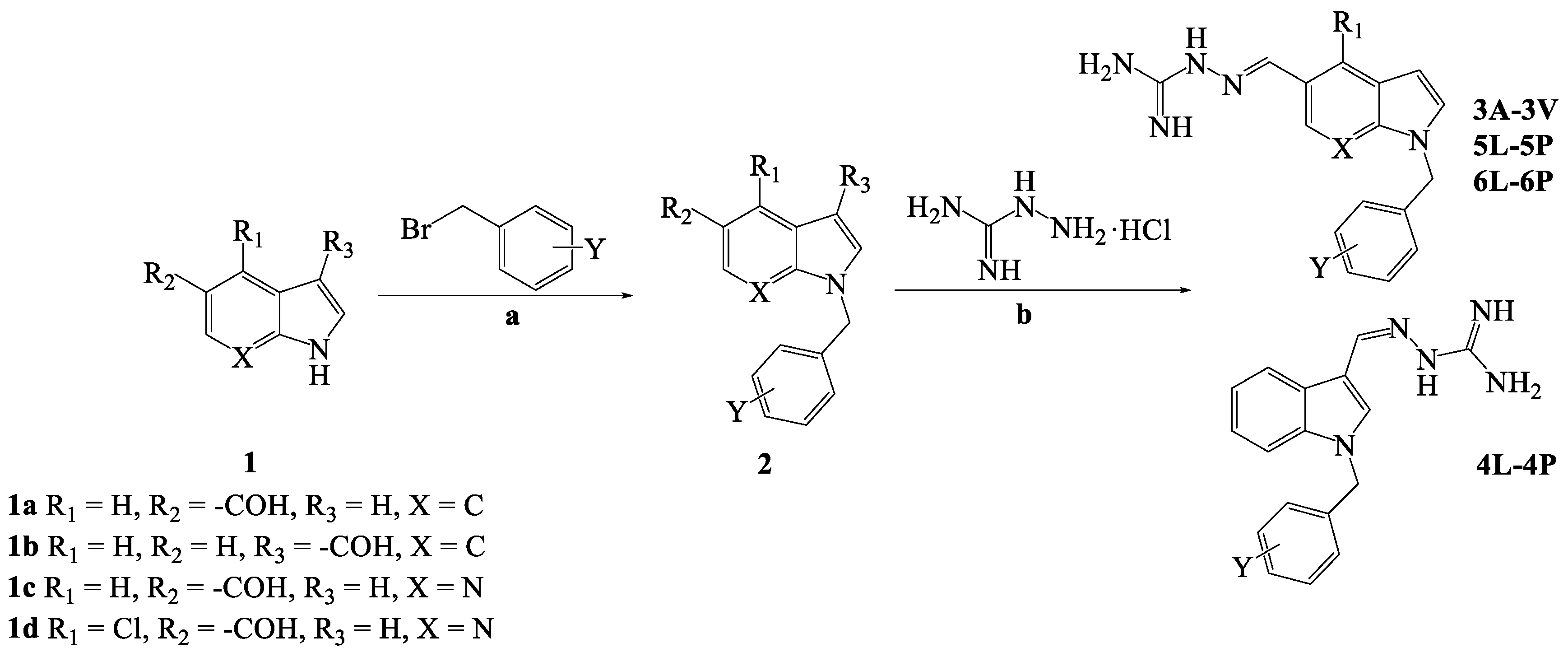
2.1. Chemistry

The synthetic route for the preparation of the guanidyl indole derivatives (3A-3V, 4L-4P, 5L-5P, and 6L-6P) and intermediates is shown in Scheme 1. Compounds (2) were prepared from starting material 1 and substituted benzyl bromides under alkaline conditions. With the key intermediate in hand, all the target derivatives (Table 1) were prepared directly in the presence of aminoguanidine hydrochloride, and concentrated hydrochloric acid by stirring in an oil-bath for 2–6 h at 50 °C [29]. Finally, the structures of the desired compounds were characterized by 1H NMR, 13C NMR, 19F NMR and high-resolution mass spectrometry (HR-MS) (Supplementary Material S1). The purity of all the compounds was 100% using the area normalization method by HPLC (Supplementary Material S2).

2.2. Evaluation of In Vitro Antimicrobial Activity

The in vitro antimicrobial activity of the synthesized compounds was evaluated to obtain the minimum inhibitory concentrations (MICs) and minimum bactericidal concentrations (MBCs) using a serial two-fold dilution method against ESKAPE strains, MRSA and clinical K. pneumoniae isolates. Colistin was selected as the positive control and DMSO (0.1%) was selected as the negative control.
Initial screening results are presented in Table 2. In general, ESKAPE strains and MRSA strains were susceptible to most of the compounds with MICs in the range of 2–64 µg/mL. Compounds 3I-3U and 4L-4P based on indole and compounds 5N-5P based on 7-azaindole showed potent antibacterial activities with MICs in the range of 2–16 µg/mL (except for Pseudomonas aeruginosa ATCC27853). It was also found that both Gram-positive and Gram-negative bacteria had approximately the same susceptibility to the test compounds. N-benzyl indole derivatives containing trifluoromethyl (CF3) groups, Cl atoms, Br atoms, or both Cl and F atoms showed higher antibacterial activity. The Cl atom on the 7-azaindole ring significantly reduced the activity of the compounds 6L-6P. This suggests that the presence and the position of halogen atoms are critical for the efficacy of these aminoguanidyl indole derivatives. Comparing 3L-3P and 4L-4P, the activity was slightly influenced by the position of the aminoguanidine. Comparing 3L-3P and 5L-5P, the activity was declined by the 7-azaindole ring. Among these compounds, the bacteriostatic activities of 16 selected compounds (3I-3U, 4O, 4P and 5P) were tested against clinical K. pneumoniae isolates (Table 3). All 16 compounds showed antibacterial activity (4–16 µg/mL) against clinical K. pneumoniae isolates (including susceptible and drug-resistant bacteria). 3O, 3P, 4O and 4P showed the best antibacterial effect with MICs ranging from 4 to 8 µg/mL. The antibacterial activity of 3O, 3P, 4O and 4P was generally comparable to that of colistin. Moreover, the MBCs for compounds with MIC values below 64 µg/mL were evaluated. Most compounds displayed MBCs that were 1–4 times higher than their MICs against the tested strains.
As the most effective compound, the bacteriostatic and time-kill assay of 4P was investigated in vitro. 4P was able to suppress the growth of MDR strain K.P. 2108 at concentrations of 1MIC, 2MIC and 4MIC. Colistin at 32 µg/mL did not completely inhibit the growth of K.P. 2108 (Figure 1A). The time-kill assay showed that different concentrations of 4P exhibited a concentration-dependent and time-dependent bactericidal response (Figure 1B).

2.3. 4P Disrupted Bacteria Viability and Morphology

Flow cytometry (FC) analysis showed that the fluorescence intensity of the 4P (32 µg/mL) group was approximately twice that of the colistin (32 µg/mL) group, although the fluorescence intensity of two groups increased to some extent after drug treatment (Figure 2A). Moreover, the effect of 4P was concentration dependent. The effect of 4P and colistin on the bacterial structure was further visualized by scanning electron microscopy (SEM) analysis (Figure 2B). Bacteria in the control group and colistin (32 µg/mL) were normal, rod-shaped, and full with no breaks in morphology. However, most of the bacteria treated with 4P (32 µg/mL) were severely damaged and lost the normal bacterial morphology, were atrophic, and completely collapsed.

2.4. 4P Exerts Its Antibacterial Action on the Bacterial Membrane

The effect of 4P on bacterial membrane perturbation was evaluated using the fluorescent dye DiSC3(5). DiSC3(5) localizes in the bacterial membrane depending on an intact the membrane potential gradient, where self-quenching occurs [30]. 4P was able to depolarize the membrane potential, releasing membrane-bound DiSC3(5) into the detection medium, where the fluorescence intensity was measured. The fluorescence intensity of DiSC3(5) was detected 30 min after treatment with 4P. As shown in Figure 3A, a dose-dependent increase in the level of membrane depolarization was observed after treatment. This result indicated that the bacterial membrane was disrupted after treatment with 4P. The membrane integrity of the bacteria was examined by measuring the fluorescence values of propidium iodide (PI). The fluorescence intensity increased when treated with the concentrations above 1MIC, and fluorescence intensities increased with increasing 4P concentrations (Figure 3B). These findings demonstrated that 4P caused damage to the plasma membrane.

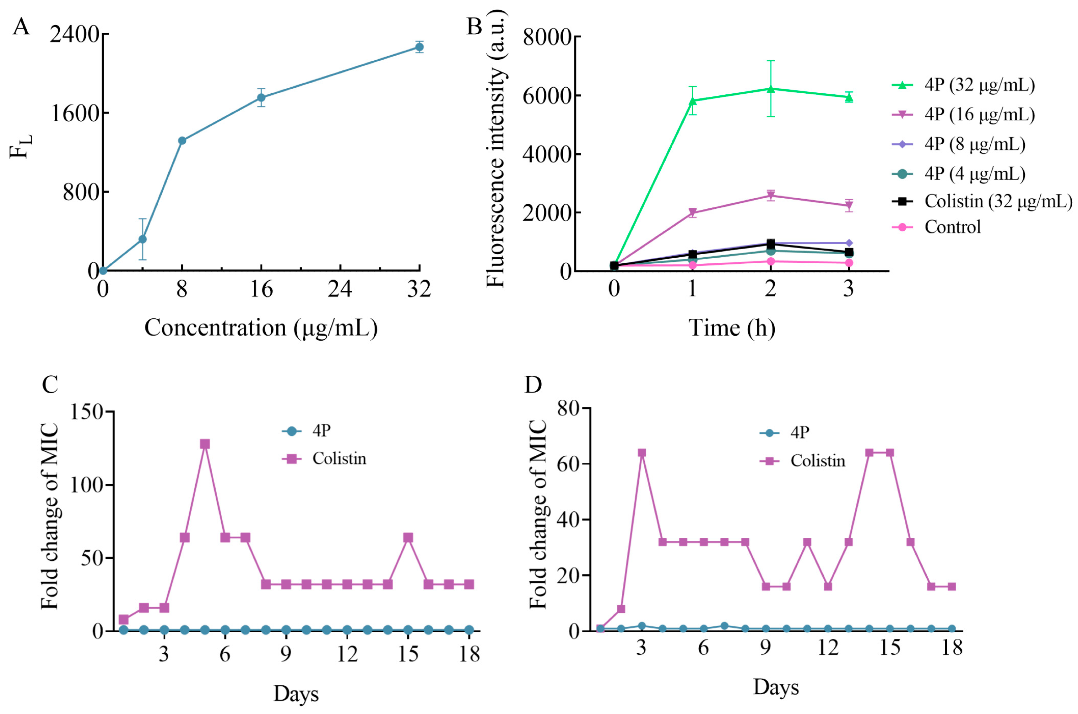
2.5. 4P Was Not Prone to Antibiotic Resistance

The development of bacterial resistance to conventional antibiotics has become a major public health concern [31]. Resistance is commonly characterized as a greater than 4-fold increase from the initial MIC measurement [32]. Therefore, the tendency to inhibit drug resistance is one of the most important properties of an antibacterial agent [33]. The ability of 4P to inhibit the development of antibacterial resistance was examined in K. pneumoniae ATCC700603 and K.P. 2112. As shown in Figure 3C,D, after 18 days of serial challenge, two strains exhibited an increase in colistin resistance, with an 8- to 128-fold increase in colistin MICs, and the MIC value of 4P showed minimal change. Therefore, 4P was less susceptible, promoting antibacterial resistance. The results for colistin resistance induced to K. pneumoniae under laboratory conditions were consistent with related studies [34,35].

2.6. Docking Analysis

It was previously shown that carbazole derivatives with an aminoguanidine group have strong binding capabilities with E. coli dihydrofolate reductase (DHFR) [36]. Therefore, the binding capacity of the interaction between 4P and the K. pneumoniae DHFR protein was investigated. DHFR has been identified as one of the important antimicrobial targets because it plays a crucial role in the regulation of bacterial metabolism [37,38]. As shown in the molecular docking results (Figure 4A), the docked pose of 4P with DHFR had a CDOCKER ENERGY of 30.6912, suggesting that 4P can bind to potential binding sites in the DHFR. Eight active site residues (ALA6, ASP27, LEU28, PHE31, LYS32, LEU54, ILE94 and THR113) were involved in recognition by 4P (Figure 4B). In DHFR, the ALA6 and THR113 side chains participated in conventional hydrogen bond interaction with the amino group of guanidium of 4P, the PHE31 side chain participated in Pi-Pi stacked with 4P, the ASP27 side chain participated in an attractive charge with 4P, and the LEU54, LYS32, LEU28, and ILE94 side chains were involved in alkyl stacking with 4P. In addition to these, 4P can interact with acid residues of DHFR through a carbon-hydrogen bond. Indole and aryl groups enhanced the hydrophobic interactions between 4P and the residues in the DHFR active pocket. These docking results indicated that guanidium and indole groups played an important role in binding with the K. pneumoniae DHFR protein.

2.7. 4P Inhibited DHFR Activities In Vitro

An in vitro enzyme activity assay was performed to investigate whether 4P can bind and inhibit the DHFR protein. Different concentrations of 4P (1/2MIC, 1MIC, 2MIC, 4MIC, and 8MIC) were used to test their inhibitory effects on the DHFR protein. The results showed that the enzyme activity was significantly inhibited at 1MIC when compared to the untreated group. Furthermore, when bacteria were treated with 4P at different concentrations, the enzyme activity of DHFR activity was significantly inhibited in a concentration-dependent manner (Figure 4C). These results suggested that 4P exerted its antibacterial activity by interfering with DHFR. Certainly, this was probably not the only antibacterial mechanism for 4P.

2.8. Hemolytic Activity

The antibacterial mechanism of guanidine compounds is considered to interfere with the bacterial cell membrane [39], which is consistent with our experimental results. To investigate the potential clinical applicability of compounds 3P, 4P and 5P, hemolysis assays were performed (Table 4). The results showed that 4P had a lower hemolysis against sheep blood red cells, with an HC50 value of 123.6 µg/mL (Figure 4D). 4P exhibited excellent selectivity for bacterial and mammalian red blood cells (selectivity index = 30.90).
Considering the MIC and hemolysis of the compounds, 4P was selected for subsequent experiments.

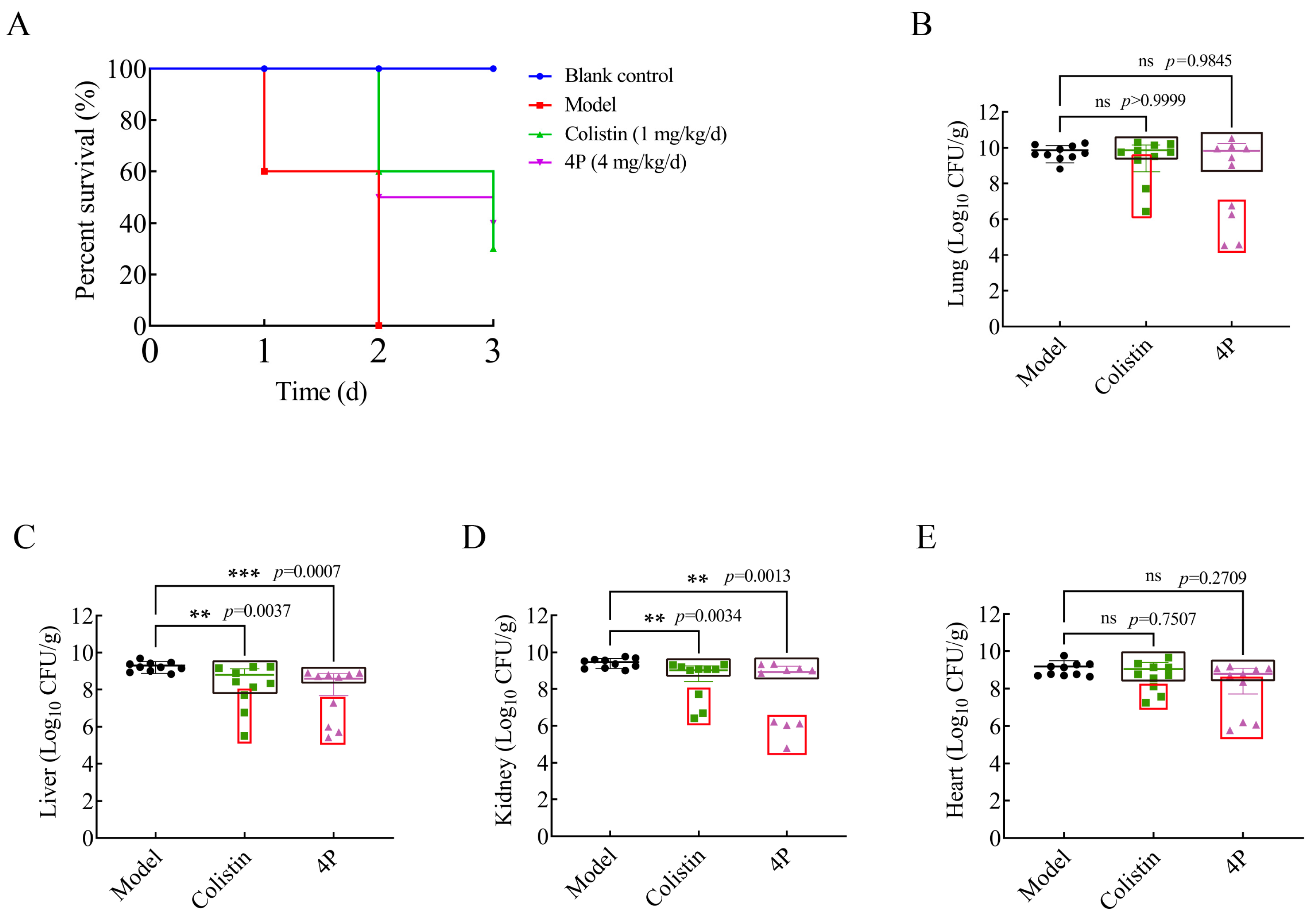
2.9. In Vivo Efficacy

As 4P showed good bacteriostatic and bactericidal capabilities in vitro, its in vivo efficacy was further evaluated by measuring the survival rate of mice after a single lethal challenge of K. pneumoniae. The lethality rate of mice in the untreated group was 100% by day 2 post-infection. There was a significant improvement in survival under 4P, with slightly higher survival rates than colistin treatment during this observation period (Figure 5A). Four tissues (heart, liver, lung, and kidney) were harvested at the time of death for bacterial load determination and histopathological observation. The results of the bacterial load analysis showed that there were reductions in bacterial load after 4P treatment compared to the untreated group, although no significant differences were detected in the lung and heart tissues. In contrast, the tissues from surviving mice exhibited a lower bacterial load (black box). Moreover, the p-values in the 4P treatment group were lower than those in the colistin group, indicating that 4P displayed a lower bacterial load compared with the colistin group (Figure 5B–E). The histopathological changes were examined by light microscopy to investigate whether 4P reduced the tissue injury caused by K. pneumoniae. Compared with the blank group, there were significant pathological changes in different tissues of the model group. The heart tissue showed the necrosis of cardiac myofibers, along with the congestion and dilatation of the blood vessels. Hepatocellular degeneration and hepatic sinusoids were observed in the liver, and the kidney showed extensive tubular necrosis. The most severe lesions were observed in the lung with the necrosis and edema of alveolar epithelial cells, alveolar edema, and inflammatory cell infiltration. After treatment with 4P, all the different tissues were normal, with no significant lesions (Figure 6).

3. Materials and Methods

3.1. Chemistry

3.1.1. General

All chemicals used in this study were of analytical reagents and solvents were not specially treated before use. The reaction process was monitored by Thin Layer Chromatography (TLC), and the products were purified by silica gel column chromatography. The 1H, 13C and 19F NMR spectra were recorded on an Agilent DD2-600MHz NMR spectrometer at room temperature, operating at 600 MHz for 1H NMR, 151 MHz for 13C NMR and 376 MHz for 19F NMR by using DMSO-d6 as solvent. The mass spectra were obtained at Agilent 6530 QTOF. The purity of all the title compounds was obtained with the area normalization method by HPLC (Agilent Technologies 1290 Infinity).

3.1.2. Synthesis of Intermediates (2)

A 50 mL round-bottom flask equipped with a magnetic stirring bar was filled with 5 mL of DMF and 224 mg (4 mmol) of potassium hydroxide. The mixture was stirred at room temperature for 5 min before the addition of 1 mmol of compound 1. We continued stirring for 45 min, then 1 mmol of benzyl bromide with a substituent on the benzene ring was added. It was stirred again for 0.75–4 h. When the reaction was complete, 40 mL of water was added. The mixture was filtered or extracted with ethyl acetate (the ethyl acetate layer was washed twice with saturated salt water, anhydrous sodium sulfate was used to remove water, and ethyl acetate was removed by vacuum distillation). The obtained crude products were recrystallized in ethanol or purified by silica gel column chromatography to yield the desired compounds.

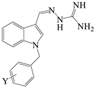
3.1.3. General Procedure for the Synthesis of Compounds 3A-3V, 4L-4P, 5L-5P and 6L-6P

One mmol of compound 2 and 110 mg (1 mmol) of aminoguanidine hydrochloride were stirred in 10 mL of ethanol. Then, 10 drops of concentrated hydrochloric acid and 2 mL of water were added. The mixture was stirred in an oil bath at 50 °C for 2–6 h. Subsequently, the solution was evaporated to dryness under reduced pressure, and the residue was purified by silica gel column chromatography (CH2Cl2:CH3OH = 15:1~10:1) to yield the desired compounds.
The spectral data of the novel compounds are listed below.
(E)-2-((1-(2-fluorobenzyl)-1H-indol-5-yl) methylene) hydrazine-1-carboximidamide (3A). Yield: 65.5%; 1H NMR (600 MHz, DMSO-d6) δ 11.89 (br s, 1H), 8.21 (s, 1H), 7.95 (d, J = 1.1 Hz, 1H), 7.84 (br s, 3H), 7.73 (dd, J = 8.7, 1.5 Hz, 1H), 7.53 (d, J = 8.7 Hz, 1H), 7.49 (d, J = 3.1 Hz, 1H), 7.30 (ddd, J = 15.4, 5.5, 1.9 Hz, 1H), 7.22−7.17 (m, 1H), 7.11–7.02 (m, 2H), 6.54 (dd, J = 3.2, 0.5 Hz, 1H), 5.49 (s, 2H); 13C NMR (151 MHz, DMSO-d6) δ160.36 (d, JCF = 245.2 Hz), 155.74, 148.74, 137.40, 130.72, 130.27 (d, JCF = 8.2 Hz), 129.96 (d, JCF = 4.0 Hz), 128.49, 125.47, 125.13 (d, JCF = 12.0 Hz), 125.07, 122.16, 120.83, 115.90 (d, JCF = 20.9 Hz), 110.86, 102.69, 43.74 (d, JCF = 4.0 Hz); 19F NMR (376 MHz, DMSO-d6) δ −117.72–117.85 (m, 1F); HRMS (ESI+): m/z calcd for C17H16FN5 [M + H]+ 310.1463, found 310.1477.
(E)-2-((1-(3-fluorobenzyl)-1H-indol-5-yl) methylene) hydrazine-1-carboximidamide (3B). Yield: 91.8%; 1H NMR (600 MHz, DMSO-d6) 11.90 (br s, 1H), 8.20 (s, 1H), 7.96 (d, J = 1.0 Hz, 1H), 7.80 (br s, 3H), 7.71 (dd, J = 8.7, 1.4 Hz, 1H), 7.57 (d, J = 3.2 Hz, 1H), 7.52 (d, J = 8.7 Hz, 1H), 7.32 (td, J = 8.1, 6.3 Hz, 1H), 7.07–6.98 (m, 3H), 6.55 (d, J = 3.2 Hz, 1H), 5.46 (s, 2H); 13C NMR (151 MHz, DMSO-d6) δ162.62 (d, JCF = 244.0 Hz), 155.75, 148.75, 141.37 (d, JCF = 7.1 Hz), 137.37, 131.04 (d, JCF = 8.4 Hz), 130.82, 128.60, 125.48, 123.52 (d, JCF = 2.7 Hz), 122.16, 120.84, 114.67 (d, JCF = 20.9 Hz), 114.28 (d, JCF = 21.8 Hz), 111.03, 102.67, 49.03; 19F NMR (376 MHz, DMSO-d6) δ −113.06 (td, J = 9.3, 6.2 Hz, 1F); HRMS (ESI+): m/z calcd for C17H16FN5 [M + H]+ 310.1463, found 310.1460.
(E)-2-((1-(4-fluorobenzyl)-1H-indol-5-yl) methylene) hydrazine-1-carboximidamide (3C). Yield: 98.0%; 1H NMR (600 MHz, DMSO-d6) δ 11.92 (br s, 1H), 8.20 (s, 1H), 7.94 (d, J = 1.0 Hz, 1H), 7.78 (br s, 3H), 7.71 (dd, J = 8.7, 1.4 Hz, 1H), 7.55 (d, J = 3.2 Hz, 1H), 7.52 (d, J = 8.7 Hz, 1H), 7.28–7.22 (m, 2H), 7.14–7.08 (m, 2H), 6.53 (d, J = 3.1 Hz, 1H), 5.42 (s, 2H); 13C NMR (151 MHz, DMSO-d6) δ161.88 (d, JCF = 243.2 Hz), 155.76, 148.76, 137.30, 134.65 (d, JCF = 3.0 Hz), 130.68, 129.63 (d, JCF = 8.3 Hz, 2C), 128.61, 125.40, 122.14, 120.75, 115.78 (d, JCF = 21.4 Hz, 2C), 111.05, 102.56, 48.86; 19F NMR (376 MHz, DMSO-d6) δ −115.11–115.21 (m, 1F); HRMS (ESI+): m/z calcd for C17H16FN5 [M + H]+ 310.1463, found 310.1460.
(E)-2-((1-(2,4-difluorobenzyl)-1H-indol-5-yl) methylene) hydrazine-1-carboximidamide (3D): Yield: 25.4%; 1H NMR (600 MHz, DMSO-d6) δ 11.91 (br s, 1H), 8.20 (s, 1H), 7.95 (d, J = 1.2 Hz, 1H), 7.83 (br s, 3H), 7.73 (dd, J = 8.7, 1.4 Hz, 1H), 7.54 (d, J = 8.7 Hz, 1H), 7.48 (d, J = 3.2 Hz, 1H), 7.28–7.21 (m, 1H), 7.16 (td, J = 8.6, 6.8 Hz, 1H), 7.00 (td, J = 8.6, 2.6 Hz, 1H), 6.54 (d, J = 3.2 Hz, 1H), 5.46 (s, 2H); 13C NMR (151 MHz, DMSO-d6) δ162.27 (dd, JCF = 246.6, 12.0 Hz), 160.46 (dd, JCF = 248.2, 12.5 Hz), 155.76, 148.69, 137.32, 131.29 (dd, JCF = 9.9, 5.6 Hz), 130.60, 128.50, 125.53, 122.17, 121.58 (dd, JCF = 15.2, 3.7 Hz), 120.87, 112.12 (dd, JCF = 21.3, 3.6 Hz), 110.83, 104.56 (t, JCF = 25.9 Hz), 102.77, 43.30 (d, JCF = 3.2 Hz); 19F NMR (376 MHz, DMSO-d6) δ −110.60–110.79 (m, 1F), −113.14 (dd, J = 18.0, 8.7 Hz, 1F); HRMS (ESI+): m/z calcd for C17H15F2N5 [M + H]+ 328.1368, found 328.1352.
(E)-2-((1-(2,5-difluorobenzyl)-1H-indol-5-yl) methylene) hydrazine-1-carboximidamide (3E): Yield: 91.4%; 1H NMR (600 MHz, DMSO-d6) δ 11.88 (br s, 1H), 8.20 (s, 1H), 7.95 (d, J = 1.2 Hz, 1H), 7.74 (dd, J = 8.7, 1.5 Hz, 1H), 7.55 (d, J = 8.7 Hz, 1H), 7.64 (br s, 3H), 7.51 (d, J = 3.2 Hz, 1H), 7.26 (td, J = 9.3, 4.5 Hz, 1H), 7.15 (ddd, J = 12.2, 8.3, 3.6 Hz, 1H), 6.88 (ddd, J = 8.9, 5.7, 3.2 Hz, 1H), 6.55 (d, J = 3.2 Hz, 1H), 5.49 (s, 2H); 13C NMR (151 MHz, DMSO-d6) δ158.50 (dd, JCF = 240.6, 2.0 Hz), 156.51 (dd, JCF = 241.6, 2.2 Hz), 155.88, 148.58, 137.30, 130.66, 128.50, 127.19 (dd, JCF = 17.9, 7.5 Hz), 125.71, 122.12, 120.95, 117.59 (dd, JCF = 24.3, 8.8 Hz), 116.60 (d, JCF = 24.1 Hz), 116.27 (dd, JCF = 24.9, 4.4 Hz), 110.82, 102.89, 43.61; 19F NMR (376 MHz, DMSO-d6) δ −118.35 (dtd, J = 13.0, 8.4, 4.5 Hz, 1F), −122.96 (ddt, J = 18.5, 9.3, 4.7 Hz, 1F); HRMS (ESI+): m/z calcd for C17H15F2N5 [M + H]+ 328.1368, found 328.1360.
(E)-2-((1-(2,6-difluorobenzyl)-1H-indol-5-yl) methylene) hydrazine-1-carboximidamide (3F): Yield: 98.7%; 1H NMR (600 MHz, DMSO-d6) δ 11.93 (br s, 1H), 8.19 (s, 1H), 7.93 (d, J = 1.1 Hz, 1H), 7.81 (br s, 3H), 7.77 (dd, J = 8.7, 1.4 Hz, 1H), 7.54 (d, J = 8.7 Hz, 1H), 7.42 (td, J = 8.4, 4.2 Hz, 1H), 7.38 (d, J = 3.1 Hz, 1H), 7.15–7.09 (m, 2H), 6.50 (dd, J = 3.2, 0.4 Hz, 1H), 5.46 (s, 2H); 13C NMR (151 MHz, DMSO-d6) δ161.23 (dd, JCF = 248.1, 7.9 Hz, 2C), 155.80, 148.56, 137.12, 131.49 (t, JCF = 10.6 Hz), 130.41, 128.37, 125.55, 122.11, 120.93, 113.27 (t, JCF = 19.5 Hz), 112.40 (dd, JCF = 20.8, 4.6 Hz, 2C), 110.34, 102.81, 37.71; 19F NMR (376 MHz, DMSO-d6) δ −114.07 (t, J = 7.0 Hz, 2F); HRMS (ESI+): m/z calcd for C17H15F2N5 [M + H]+ 328.1368, found 328.1361.
(E)-2-((1-(3,4-difluorobenzyl)-1H-indol-5-yl) methylene) hydrazine-1-carboximidamide (3G): Yield: 98.0%; 1H NMR (600 MHz, DMSO-d6) δ 11.93 (br s, 1H), 8.20 (s, 1H), 7.95 (d, J = 1.0 Hz, 1H), 7.78 (br s, 3H), 7.72 (dd, J = 8.7, 1.4 Hz, 1H), 7.58 (d, J = 3.2 Hz, 1H), 7.55 (d, J = 8.7 Hz, 1H), 7.33 (ddd, J = 11.6, 9.3, 5.2 Hz, 2H), 7.05–7.00 (m, 1H), 6.54 (d, J = 3.1 Hz, 1H), 5.43 (s, 2H); 13C NMR (151 MHz, DMSO-d6) δ 155.81, 149.71 (dd, JCF = 246.3, 12.9 Hz), 149.15 (dd, JCF = 245.4, 12.4 Hz), 148.67, 137.25, 136.20 (dd, JCF = 5.2, 3.8 Hz), 130.68, 128.63, 125.55, 124.44 (dd, JCF = 6.6, 3.3 Hz), 122.14, 120.87, 118.10 (d, JCF = 17.2 Hz), 116.78 (d, JCF = 17.4 Hz), 111.02, 102.75, 48.52; 19F NMR (376 MHz, DMSO-d6) δ −138.31–138.48 (m, 1F), −140.33–140.51 (m, 1F); HRMS (ESI+): m/z calcd for C17H15F2N5 [M + H]+ 328.1368, found 328.1359.
(E)-2-((1-(3,5-difluorobenzyl)-1H-indol-5-yl) methylene) hydrazine-1-carboximidamide (3H): Yield: 97.0%; 1H NMR (600 MHz, DMSO-d6) δ 11.89 (br s, 1H), 8.20 (s, 1H), 7.96 (s, 1H), 7.77 (br s, 3H), 7.72 (dd, J = 8.7, 1.2 Hz, 1H), 7.59 (d, J = 3.2 Hz, 1H), 7.54 (d, J = 8.7 Hz, 1H), 7.10 (tt, J = 9.3, 2.2 Hz, 1H), 6.92–6.86 (m, 2H), 6.56 (d, J = 3.1 Hz, 1H), 5.48 (s, 2H); 13C NMR (151 MHz, DMSO-d6) δ162.85 (dd, JCF = 246.8, 13.3 Hz, 2C), 155.92, 148.55, 143.11 (t, JCF = 8.7 Hz), 137.29, 130.79, 128.62, 125.73, 122.11, 120.96, 110.98, 110.68 (dd, JCF = 20.2, 5.1 Hz, 2C), 103.37 (t, JCF = 25.8 Hz), 102.86, 48.73; 19F NMR (376 MHz, DMSO-d6) δ −109.43–109.52 (m, 2F); HRMS (ESI+): m/z calcd for C17H15F2N5 [M + H]+ 328.1368, found 328.1355.
(E)-2-((1-(3-(trifluoromethyl) benzyl)-1H-indol-5-yl) methylene) hydrazine-1-carboximidamide (3I): Yield: 92.2%; 1H NMR (600 MHz, DMSO-d6) δ 11.94 (br s, 1H), 8.19 (s, 1H), 7.95 (s, 1H), 7.72 (d, J = 8.7 Hz, 1H), 7.64 (br s, 3H), 7.60 (t, J = 5.9 Hz, 3H), 7.56–7.50 (m, 2H), 7.44 (d, J = 7.7 Hz, 1H), 6.56 (d, J = 3.0 Hz, 1H), 5.56 (s, 2H); 13C NMR (151 MHz, DMSO-d6) δ 155.98, 148.52, 140.00, 137.29, 131.58, 130.72, 130.16, 129.67 (q, JCF = 31.4 Hz), 128.61, 125.72, 124.64 (d, JCF = 3.6 Hz), 124.51 (d, JCF = 272.4 Hz), 124.06 (q, JCF = 3.6 Hz), 122.14, 120.83, 110.96, 102.80, 48.98; 19F NMR (376 MHz, DMSO-d6) δ −61.15 (s, 3F); HRMS (ESI+): m/z calcd for C18H16F3N5 [M + H]+360.1431, found 360.1415.
(E)-2-((1-(4-(trifluoromethyl) benzyl)-1H-indol-5-yl) methylene) hydrazine-1-carboximidamide (3J): Yield: 57.9%; 1H NMR (600 MHz, DMSO-d6) δ 11.91 (br s, 1H), 8.20 (s, 1H), 7.97 (d, J = 1.0 Hz, 1H), 7.71 (dd, J = 8.7, 1.4 Hz, 1H), 7.68 (br s, 3H), 7.65 (d, J = 8.2 Hz, 2H), 7.58 (d, J = 3.1 Hz, 1H), 7.49 (d, J = 8.7 Hz, 1H), 7.34 (d, J = 8.1 Hz, 2H), 6.57 (d, J = 3.0 Hz, 1H), 5.56 (s, 2H); 13C NMR (151 MHz, DMSO-d6) δ 155.80, 148.68, 143.30, 137.39, 130.86, 128.63, 128.46 (d, JCF = 31.7 Hz), 128.06 (2C), 125.93 (q, JCF = 3.7 Hz,2C), 125.58, 124.60 (d, JCF = 272.1 Hz), 122.17, 120.89, 110.99, 102.80, 49.08; 19F NMR (376 MHz, DMSO-d6) δ −60.97 (s, 3F); HRMS (ESI+): m/z calcd for C18H16F3N5 [M + H]+ 360.1431, found 360.1443.
(E)-2-((1-(3,5-bis(trifluoromethyl) benzyl)-1H-indol-5-yl) methylene) hydrazine-1-carboximidamide (3K): Yield: 93.0%; 1H NMR (600 MHz, DMSO-d6) δ 11.88 (br s, 1H), 8.19 (s, 1H), 7.98 (s, 1H), 7.95 (d, J = 0.9 Hz, 1H), 7.91 (s, 2H), 7.75 (dd, J = 8.7, 1.4 Hz, 1H), 7.70 (br s, 3H), 7.68 (d, J = 3.2 Hz, 1H), 7.61 (d, J = 8.7 Hz, 1H), 6.58 (d, J = 3.1 Hz, 1H), 5.67 (s, 2H); 13C NMR (151 MHz, DMSO-d6) δ 155.93, 148.49, 142.02, 137.22, 130.86 (q, JCF = 32.9 Hz,2C), 130.63, 128.64, 128.42 (d, JCF = 3.7 Hz,2C), 125.88, 123.60 (d, JCF = 272.9 Hz,2C), 122.28, 121.88–121.68 (m), 120.96, 110.89, 103.17, 48.48; 19F NMR (376 MHz, DMSO-d6) δ −61.44 (s, 6F); HRMS (ESI+): m/z calcd for C19H15F6N5 [M + H]+ 428.1304, found 428.1286.
(E)-2-((1-(2-chlorobenzyl)-1H-indol-5-yl) methylene) hydrazine-1-carboximidamide (3L): Yield: 84.0%; 1H NMR (600 MHz, DMSO-d6) δ 8.19 (s, 1H), 7.96 (d, J = 0.9 Hz, 1H), 7.70 (dd, J = 8.7, 1.4 Hz, 1H), 7.59 (br s, 4H), 7.50–7.46 (m, 2H), 7.44 (d, J = 8.7 Hz, 1H), 7.28 (td, J = 7.8, 1.5 Hz, 1H), 7.20 (td, J = 7.6, 1.0 Hz, 1H), 6.70–6.67 (m, 1H), 6.57 (d, J = 3.2 Hz, 1H), 5.52 (s, 2H); 13C NMR (151 MHz, DMSO-d6) δ 156.26, 148.26, 137.45, 135.63, 132.25, 130.82, 129.94, 129.74, 128.88, 128.53, 127.95, 125.97, 121.97, 120.85, 110.89, 102.76, 47.48; HRMS (ESI+): m/z calcd for C17H16ClN5 [M + H]+ 326.1167, found 326.1181.
(E)-2-((1-(3-chlorobenzyl)-1H-indol-5-yl) methylene) hydrazine-1-carboximidamide (3M): Yield: 34.7%; 1H NMR (600 MHz, DMSO-d6) δ 11.92 (br s, 1H), 8.20 (s, 1H), 7.96 (d, J = 1.0 Hz, 1H), 7.77 (br s, 3H), 7.72 (dd, J = 8.7, 1.4 Hz, 1H), 7.58 (d, J = 3.2 Hz, 1H), 7.53 (d, J= 8.7 Hz, 1H), 7.33–7.24 (m, 3H), 7.14 (d, J = 7.3 Hz, 1H), 6.55 (d, J = 3.1 Hz, 1H), 5.46 (s, 2H); 13C NMR (151 MHz, DMSO-d6) δ 155.77, 148.71, 141.01, 137.34, 133.61, 130.93, 130.80, 128.60, 127.84, 127.32, 126.19, 125.51, 122.18, 120.86, 111.03, 102.73, 48.93; HRMS (ESI+): m/z calcd for C17H16ClN5 [M + H]+ 326.1167, found 326.1162.
(E)-2-((1-(4-chlorobenzyl)-1H-indol-5-yl) methylene) hydrazine-1-carboximidamide (3N): Yield: 70%; 1H NMR (600 MHz, DMSO-d6) δ 11.92 (br s, 1H), 8.20 (s, 1H), 7.95 (d, J = 1.0 Hz, 1H), 7.78 (br s, 3H), 7.70 (dd, J = 8.7, 1.4 Hz, 1H), 7.55 (d, J = 3.2 Hz, 1H), 7.50 (d, J = 8.7 Hz, 1H), 7.36–7.32 (m, 2H), 7.20 (d, J = 8.5 Hz, 2H), 6.54 (d, J = 3.1 Hz, 1H), 5.44 (s, 2H); 13C NMR (151 MHz, DMSO-d6) δ 155.78, 148.73, 137.48, 137.33, 132.47,130.75,129.36(2C), 128.97(2C),128.62,125.45,122.15, 120.80, 111.03, 102.64, 48.88; HRMS (ESI+): m/z calcd for C17H16ClN5 [M + H]+ 326.1167, found 326.1168.
(E)-2-((1-(2,4-dichlorobenzyl)-1H-indol-5-yl) methylene) hydrazine-1-carboximidamide (3O): Yield: 67.2%; 1H NMR (600 MHz, DMSO-d6) δ 11.92 (br s, 1H), 8.21 (s, 1H), 7.98 (d, J = 1.0 Hz, 1H), 7.72 (dd, J = 8.7, 1.4 Hz, 1H), 7.69 (br s, 3H), 7.65 (d, J = 2.1 Hz, 1H), 7.47 (d, J = 3.2 Hz, 1H), 7.45 (d, J = 8.7 Hz, 1H), 7.30 (dd, J = 8.4, 2.1 Hz, 1H), 6.66 (d, J = 8.4 Hz, 1H), 6.58 (d, J = 3.1 Hz, 1H), 5.51 (s, 2H); 13C NMR (151 MHz, DMSO-d6) δ 155.77, 148.67, 137.51, 134.89, 133.36, 133.20, 130.84, 130.11, 129.42, 128.53, 128.14, 125.68, 122.25, 120.99, 110.92, 102.98, 47.06; HRMS (ESI+): m/z calcd for C17H15Cl2N5 [M + H]+360.0777, found 360.0763.
(E)-2-((1-(3,4-dichlorobenzyl)-1H-indol-5-yl) methylene) hydrazine-1-carboximidamide (3P): Yield: 31.5%; 1H NMR (600 MHz, DMSO-d6) δ 8.19 (s, 1H), 7.95 (d, J = 1.0 Hz, 1H), 7.72 (dd, J = 8.7, 1.4 Hz, 1H), 7.64 (br s, 4H), 7.58 (d, J = 3.2 Hz, 1H), 7.54 (d, J = 8.3 Hz, 2H), 7.49 (d, J = 1.9 Hz, 1H), 7.13 (dd, J = 8.3, 2.0 Hz, 1H), 6.55 (d, J = 3.2 Hz, 1H), 5.45 (s, 2H); 13C NMR (151 MHz, DMSO-d6) δ 155.91, 148.56, 139.64, 137.24, 131.57, 131.24, 130.71, 130.50, 129.56, 128.63, 127.85, 125.69, 122.13, 120.91, 110.99, 102.84, 48.36; HRMS (ESI+): m/z calcd for C17H15Cl2N5 [M + H]+ 360.0777, found 360.0775.
(E)-2-((1-(2-bromobenzyl)-1H-indol-5-yl) methylene) hydrazine-1-carboximidamide (3Q): Yield: 95.0%; 1H NMR (600 MHz, DMSO-d6) δ 11.93 (br s, 1H), 8.21 (s, 1H), 7.99 (d, J = 1.1 Hz, 1H), 7.72 (dd, J = 8.7, 1.4 Hz, 1H), 7.69 (br s, 3H), 7.65 (dd, J = 7.7, 1.4 Hz, 1H), 7.47 (d, J = 3.2 Hz, 1H), 7.43 (d, J = 8.7 Hz, 1H), 7.25–7.18 (m, 2H), 6.58 (dd, J = 6.5, 2.5 Hz, 2H), 5.49 (s, 2H); 13C NMR (151 MHz, DMSO-d6) δ 155.78, 148.70, 137.57, 137.15, 133.20, 130.88, 129.98, 128.77, 128.53, 128.51, 125.62, 122.40, 122.25, 120.94, 110.95, 102.86, 49.90; HRMS (ESI+): m/z calcd for C17H16BrN5 [M + H]+370.0662, found 370.0645.
(E)-2-((1-(3-bromobenzyl)-1H-indol-5-yl) methylene) hydrazine-1-carboximidamide (3R): Yield: 17.8%; 1H NMR (600 MHz, DMSO-d6) δ 11.87 (br s, 1H), 8.20 (s, 1H), 7.95 (d, J = 1.1 Hz, 1H), 7.72 (dd, J = 8.7, 1.4 Hz, 1H), 7.64 (br s, 3H), 7.58 (d, J = 3.2 Hz, 1H), 7.53 (d, J = 8.7 Hz, 1H), 7.44–7.39 (m, 2H), 7.25 (t, J = 7.8 Hz, 1H), 7.17 (d, J = 7.8 Hz, 1H), 6.55 (d, J = 3.1 Hz, 1H), 5.45 (s, 2H); 13C NMR (151 MHz, DMSO-d6) δ 155.76, 148.72, 141.26, 137.33, 131.22, 130.79, 130.74, 130.21, 128.59, 126.58, 125.51, 122.23, 122.19, 120.85, 111.03, 102.73, 48.87; HRMS (ESI+): m/z calcd for C17H16BrN5 [M + H]+ 370.0662, found 370.0645.
(E)-2-((1-(4-bromobenzyl)-1H-indol-5-yl) methylene) hydrazine-1-carboximidamide (3S): Yield: 37.35%; 1H NMR (600 MHz, DMSO-d6) δ 11.91 (br s, 1H), 8.20 (s, 1H), 7.95 (d, J = 1.0 Hz, 1H), 7.70 (dd, J = 8.7, 1.4 Hz, 1H), 7.66 (br s, 3H), 7.54 (d, J = 3.2 Hz, 1H), 7.51–7.46 (m, 3H), 7.13 (d, J = 8.4 Hz, 2H), 6.54 (d, J = 3.1 Hz, 1H), 5.42 (s, 2H); 13C NMR (151 MHz, DMSO-d6) δ 155.76, 148.74, 137.91, 137.33, 131.90(2C), 130.77, 129.70(2C), 128.61, 125.45, 122.15, 120.98, 120.81, 111.03, 102.64, 48.94; HRMS (ESI+): m/z calcd for C17H16BrN5 [M + H]+ 370.0662, found 370.0644.
(E)-2-((1-(2-chloro-4-fluorobenzyl)-1H-indol-5-yl) methylene) hydrazine-1-carboximidamide (3T): Yield: 69.8%; 1H NMR (600 MHz, DMSO-d6) δ 11.92 (br s, 1H), 8.21 (s, 1H), 7.97 (d, J = 1.1 Hz, 1H), 7.72 (dd, J = 8.7, 1.4 Hz, 1H), 7.65 (br s, 3H), 7.50–7.45 (m, 3H), 7.11 (td, J = 8.5, 2.6 Hz, 1H), 6.78 (dd, J = 8.7, 6.1 Hz, 1H), 6.57 (d, J = 3.1 Hz, 1H), 5.50 (s, 2H); 13C NMR (151 MHz, DMSO-d6) δ161.69 (d, JCF = 247.4 Hz), 155.90, 148.57, 137.46, 133.12 (d, JCF = 10.8 Hz), 132.04 (d, JCF = 3.4 Hz), 130.75, 130.52 (d, JCF = 9.1 Hz), 128.53, 125.73, 122.17, 120.93, 117.32 (d, JCF = 25.2 Hz), 115.12 (d, JCF = 21.2 Hz), 110.92, 102.88, 46.96; 19F NMR (376 MHz, DMSO-d6) δ −112.63 (dd, J = 14.8, 8.4 Hz); HRMS (ESI+): m/z calcd for C17H15ClFN5 [M + H]+344.1073, found 344.1061.
(E)-2-((1-(3-chloro-4-fluorobenzyl)-1H-indol-5-yl) methylene) hydrazine-1-carboximidamide (3U): Yield: 47.4%; 1H NMR (600 MHz, DMSO-d6) δ 11.98 (br s, 1H), 8.19 (s, 1H), 7.94 (d, J = 0.8 Hz, 1H), 7.72 (dd, J = 8.7, 1.4 Hz, 1H), 7.66 (br s, 3H), 7.58 (d, J = 3.1 Hz, 1H), 7.56 (d, J = 8.7 Hz, 1H), 7.47 (dd, J = 7.1, 2.1 Hz, 1H), 7.33 (t, J = 9.0 Hz, 1H), 7.20 (ddd, J = 8.4, 4.6, 2.2 Hz, 1H), 6.54 (d, J = 3.2 Hz, 1H), 5.43 (s, 2H); 13C NMR (151 MHz, DMSO-d6) δ156.93 (d, JCF = 246.4 Hz), 155.86, 148.62, 137.21, 136.38 (d, JCF = 3.6 Hz), 130.65, 129.72, 128.63, 128.34 (d, JCF = 7.5 Hz), 125.63, 122.13, 120.87, 119.89 (d, JCF = 17.8 Hz), 117.50 (d, JCF = 21.1 Hz), 111.01, 102.78, 48.34; 19F NMR (376 MHz, DMSO-d6) δ −118.17 (ddd, J = 9.3, 7.3, 4.9 Hz); HRMS (ESI+): m/z calcd for C17H15ClFN5 [M + H]+ 344.1073, found 344.1059.
(E)-2-((1-(4-cyanobenzyl)-1H-indol-5-yl) methylene) hydrazine-1-carboximidamide (3V): Yield: 34.6%; 1H NMR (600 MHz, DMSO-d6) δ 8.06 (s, 1H), 7.77–7.73 (m, 3H), 7.56 (dd, J = 8.6, 1.4 Hz, 1H), 7.47 (d, J = 3.1 Hz, 1H), 7.34 (d, J = 8.6 Hz, 1H), 7.28 (d, J = 8.3 Hz, 2H), 6.50–6.48 (m, 1H), 5.51 (s, 2H); 13C NMR (151 MHz, DMSO-d6) δ 160.10, 145.58, 144.46, 136.26, 132.96 (2C), 130.09, 129.19, 128.77, 128.13 (2C), 120.48, 119.97, 119.12, 110.55, 110.54, 102.38, 49.15; HRMS (ESI+): m/z calcd for C18H16N6 [M + H]+ 317.1509, found 317.1506.
(Z)-2-((1-(2-chlorobenzyl)-1H-indol-3-yl) methylene) hydrazine-1-carboximidamide (4L). Yield: 73.6%; 1H NMR (600 MHz, DMSO-d6) δ 11.74 (br s, 1H), 8.34 (d, J = 5.9 Hz, 2H), 7.96 (s, 1H), 7.50 (dd, J = 8.0, 0.9 Hz, 1H), 7.44 (d, J = 8.2 Hz, 1H),7.42 (br s, 3H), 7.31 (td, J = 7.8, 1.5 Hz, 1H), 7.22 (ddd, J = 13.1, 7.2, 0.9 Hz, 2H), 7.19–7.14 (m, 1H), 6.85–6.80 (m, 1H), 5.54 (s, 2H); 13C NMR (151 MHz, DMSO-d6) δ 155.29, 144.62, 137.49, 135.26, 134.93, 132.46, 130.05, 129.98, 129.21, 128.05, 124.86, 123.66, 123.24, 121.71, 111.02, 110.89, 47.71; HRMS(ESI+): m/z calcd for C17H16ClN5[M+H]+ 326.1167, found 326.1183.
(Z)-2-((1-(3-chlorobenzyl)-1H-indol-3-yl) methylene) hydrazine-1-carboximidamide (4M). Yield: 97.1%; 1H NMR (600 MHz, DMSO-d6) δ 11.83 (br s, 1H), 8.35 (s, 1H), 8.32 (d, J = 7.9 Hz, 1H), 8.07 (s, 1H), 8.00–7.10 (br s, 3H), 7.51 (d, J = 8.2 Hz, 1H), 7.34–7.29 (m, 3H), 7.24–7.20 (m, 1H), 7.19–7.14 (m, 2H), 5.47 (s, 2H); 13C NMR (151 MHz, DMSO-d6) δ 155.31, 144.57, 140.38, 137.27, 135.14, 133.69, 131.02, 128.03, 127.42, 126.28, 124.94, 123.59, 123.23, 121.67, 111.01, 110.95, 49.12; HRMS(ESI+): m/z calcd for C17H16ClN5[M+H]+ 326.1167, found 326.1170.
(Z)-2-((1-(4-chlorobenzyl)-1H-indol-3-yl) methylene) hydrazine-1-carboximidamide (4N). Yield: 76.9%; 1H NMR (600 MHz, DMSO-d6) δ 11.75 (br s, 1H), 8.33 (d, J = 1.7 Hz, 1H), 8.30 (d, J = 8.9 Hz, 1H), 8.05 (d, J = 1.7 Hz, 1H), 7.40 (br s, 3H), 7.49 (d, J = 8.2 Hz, 1H), 7.38–7.34 (m, 2H), 7.26–7.23 (m, 2H), 7.23–7.18 (m, 1H), 7.17–7.13 (m, 1H), 5.45 (s, 2H); 13C NMR (151 MHz, DMSO-d6) δ 155.26, 144.61, 137.27, 136.85, 135.11, 132.66, 129.49 (2C), 129.07 (2C), 124.97, 123.53, 123.18, 121.62, 111.04, 110.88, 49.08; HRMS(ESI+): m/z calcd for C17H16ClN5[M+H]+ 326.1167, found 326.1168.
(Z)-2-((1-(2,4-dichlorobenzyl)-1H-indol-3-yl) methylene) hydrazine-1-carboximidamide (4O). Yield: 82.0%; 1H NMR (600 MHz, DMSO-d6) δ 11.74 (s, 1H), 8.33 (d, J = 9.1 Hz, 2H), 7.95 (s, 1H), 7.68 (d, J = 2.1 Hz, 1H), 7.47 (br s, 3H), 7.44 (d, J = 8.2 Hz, 1H), 7.34 (dd, J = 8.4, 2.1 Hz, 1H), 7.23 (t, J = 8.0 Hz, 1H), 7.17 (t, J = 7.4 Hz, 1H), 6.82 (d, J = 8.4 Hz, 1H), 5.52 (s, 2H); 13C NMR (151 MHz, DMSO-d6) δ 155.27, 144.59, 137.42, 135.17, 134.17, 133.61, 133.46, 130.53, 129.54, 128.24, 124.87, 123.73, 123.28, 121.79, 111.14, 110.86, 47.27; HRMS(ESI+): m/z calcd for C17H15Cl2N5[M+H]+ 360.0777, found 360.0781.
(Z)-2-((1-(3,4-dichlorobenzyl)-1H-indol-3-yl) methylene) hydrazine-1-carboximidamide (4P). Yield: 78.0%; 1H NMR (600 MHz, DMSO-d6) δ 11.75 (br s, 1H), 8.33 (s, 1H), 8.31 (d, J = 7.9 Hz, 1H), 8.07 (s, 1H), 7.57–7.50 (m, 3H), 7.41 (br s, 3H), 7.25–7.20 (m, 1H), 7.19–7.13 (m, 2H), 5.47 (s, 2H); 13C NMR (151 MHz, DMSO-d6) δ 155.27, 144.57, 139.00, 137.21, 135.06, 131.65, 131.33, 130.71, 129.71, 127.97, 124.95, 123.65, 123.25, 121.72, 111.07, 110.98, 48.55; HRMS(ESI+): m/z calcd for C17H15Cl2N5[M+H]+ 360.0777, found 360.0774.
(Z)-2-((1-(2-chlorobenzyl)-1H-pyrrolo[2,3-b]pyridin-5-yl) methylene) hydrazine-1-carboximidamide (5L). Yield: 94.9%; 1H NMR (600 MHz, DMSO-d6) δ 11.98 (br s, 1H), 8.69 (d, J = 1.8 Hz, 1H), 8.52 (d, J = 1.8 Hz, 1H), 8.28 (s, 1H), 7.82 (br s, 1H), 7.64 (d, J = 3.5 Hz, 1H), 7.48 (d, J = 7.9 Hz, 1H), 7.29 (td, J = 7.9, 1.3 Hz, 1H), 7.21 (t, J = 7.6 Hz, 1H), 6.76 (d, J = 7.7 Hz, 1H), 6.61 (d, J = 3.5 Hz, 1H), 5.57 (s, 2H); 13C NMR (151 MHz, DMSO-d6) δ 155.88, 148.40, 146.21, 143.69, 135.67, 132.21, 131.27, 129.87, 129.72, 129.10, 127.97, 127.91, 122.78, 120.26, 101.13, 45.73; HRMS(ESI+): m/z calcd for C16H15ClN6[M+H]+ 327.1119, found 326.1133.
(Z)-2-((1-(3-chlorobenzyl)-1H-pyrrolo[2,3-b]pyridin-5-yl) methylene) hydrazine-1-carboximidamide (5M). Yield: 91.7%; 1H NMR (600 MHz, DMSO-d6) δ 12.20 (s, 1H), 8.74 (s, 1H), 8.52 (s, 1H), 8.29 (s, 1H), 8.3–7.4 (br s, 3H), 7.74 (s, 1H), 7.30 (d, J = 11.8 Hz, 3H), 7.19 (d, J = 6.3 Hz, 1H), 6.58 (s, 1H), 5.50 (s, 2H); 13C NMR (151 MHz, DMSO-d6) δ 155.96, 147.82, 145.99, 143.25, 140.94, 133.54, 131.27, 130.95, 128.34, 127.89, 127.67, 126.53, 122.71, 120.54, 101.22, 47.35; HRMS(ESI+): m/z calcd for C16H15ClN6[M+H]+ 327.1119, found 326.1131.
(Z)-2-((1-(4-chlorobenzyl)-1H-pyrrolo[2,3-b] pyridin-5-yl) methylene) hydrazine-1-carboximidamide (5N). Yield: 92.5%; 1H NMR (600 MHz, DMSO-d6) δ 12.02 (s, 1H), 8.72 (d, J = 1.7 Hz, 1H), 8.49 (d, J = 1.7 Hz, 1H), 8.28 (s, 1H), 7.89 (br s, 3H), 7.70 (d, J = 3.5 Hz, 1H), 7.34 (d, J = 8.4 Hz, 2H), 7.25 (d, J = 8.4 Hz, 2H), 6.57 (d, J = 3.5 Hz, 1H), 5.47 (s, 2H); 13C NMR (151 MHz, DMSO-d6) δ 155.84, 148.20, 146.25, 143.60, 137.54, 132.51, 131.10, 129.69 (2C), 128.96 (2C), 127.97, 122.60, 120.33, 101.01, 47.17; HRMS(ESI+): m/z calcd for C16H15ClN6[M+H]+ 327.1119, found 326.1129.
(Z)-2-((1-(2,4-dichlorobenzyl)-1H-pyrrolo[2,3-b] pyridin-5-yl) methylene) hydrazine-1-carboximidamide (5O). Yield: 93.6%; 1H NMR (600 MHz, DMSO-d6) δ 12.08 (s, 1H), 8.69 (d, J = 1.8 Hz, 1H), 8.53 (d, J = 1.9 Hz, 1H), 8.28 (s, 1H), 7.77 (br s, 3H), 7.65 (t, J = 2.6 Hz, 2H), 7.31 (dd, J = 8.4, 2.1 Hz, 1H), 6.79 (d, J = 8.4 Hz, 1H), 6.62 (d, J = 3.5 Hz, 1H), 5.55 (s, 2H); 13C NMR (151 MHz, DMSO-d6) δ 155.87, 148.25, 146.12, 143.63, 134.85, 133.36, 133.20, 131.27, 130.51, 129.35, 128.12, 128.09, 122.83, 120.35, 101.29, 45.38; HRMS(ESI+): m/z calcd for C16H14Cl2N6[M+H]+ 361.0730, found 361.0737.
(Z)-2-((1-(3,4-dichlorobenzyl)-1H-pyrrolo[2,3-b] pyridin-5-yl) methylene) hydrazine-1-carboximidamide (5P). Yield: 94.6%; 1H NMR (600 MHz, DMSO-d6) δ 12.14 (s, 1H), 8.74 (d, J = 1.7 Hz, 1H), 8.51 (d, J = 1.8 Hz, 1H), 8.28 (s, 1H), 7.92 (br s, 3H), 7.75 (d, J = 3.5 Hz, 1H), 7.54 (dd, J = 5.1, 3.2 Hz, 2H), 7.20 (dd, J = 8.4, 1.8 Hz, 1H), 6.59 (d, J = 3.5 Hz, 1H), 5.49 (s, 2H); 13C NMR (151 MHz, DMSO-d6) δ 155.92, 147.90, 146.03, 143.39, 139.59, 131.49, 131.25, 131.17, 130.56, 129.92, 128.28, 128.22, 122.77, 120.51, 101.26, 46.80; HRMS(ESI+): m/z calcd for C16H14Cl2N6[M+H]+ 361.0730, found 361.0737.
(Z)-2-((4-chloro-1-(2-chlorobenzyl)-1H-pyrrolo[2,3-b] pyridin-5-yl) methylene) hydrazine-1-carboximidamide (6L). Yield: 57.9%; 1H NMR (600 MHz, DMSO-d6) δ 12.26 (br s, 1H), 9.09 (s, 1H), 8.59 (s, 1H), 7.80 (br s,3H), 7.72 (d, J = 3.5 Hz, 1H), 7.48 (dd, J = 8.0, 1.0 Hz, 1H), 7.30 (td, J = 7.8, 1.6 Hz, 1H), 7.22 (td, J = 7.6, 1.1 Hz, 1H), 6.84 (dd, J = 7.7, 1.2 Hz, 1H), 6.67 (d, J = 3.5 Hz, 1H), 5.59 (s, 2H); 13C NMR (151 MHz, DMSO-d6) δ 155.82, 148.04, 143.00, 142.30, 135.25, 134.97, 132.30, 131.67, 129.91, 129.88, 129.38, 127.95, 119.77, 118.95, 99.46, 46.16; HRMS(ESI+): m/z calcd for C16H14Cl2N6[M+H]+ 361.0730, found 361.0745.
(Z)-2-((4-chloro-1-(3-chlorobenzyl)-1H-pyrrolo[2,3-b] pyridin-5-yl) methylene) hydrazine-1-carboximidamide (6M). Yield: 65.6%; 1H NMR (600 MHz, DMSO-d6) δ 12.22 (br s, 1H), 9.12 (s, 1H), 8.58 (s, 1H), 7.83 (d, J = 3.5 Hz, 1H), 7.76 (br s, 3H), 7.36–7.28 (m, 3H), 7.19 (d, J = 6.3 Hz, 1H), 6.63 (d, J = 3.5 Hz, 1H), 5.50 (s, 2H); 13C NMR (151 MHz, DMSO-d6) δ 155.98, 147.82, 142.99, 142.18, 140.61, 134.89, 133.56, 131.47, 130.97, 128.00, 127.78, 126.60, 119.79, 119.02, 99.40, 47.69; HRMS(ESI+): m/z calcd for C16H14Cl2N6[M+H]+361.0730, found 361.0739.
(Z)-2-((4-chloro-1-(4-chlorobenzyl)-1H-pyrrolo[2,3-b] pyridin-5-yl) methylene) hydrazine-1-carboximidamide (6N). Yield: 78.1%; 1H NMR (600 MHz, DMSO-d6) δ 12.25 (br s, 1H), 9.11 (s, 1H), 8.58 (s, 1H), 7.79 (d, J = 7.0 Hz, 1H), 7.75 (br s, 3H), 7.35 (d, J = 8.4 Hz, 2H), 7.26 (d, J = 8.4 Hz, 2H), 6.62 (d, J = 7.1 Hz, 1H), 5.49 (s, 2H); 13C NMR (151 MHz, DMSO-d6) δ 155.94, 147.84, 142.94, 142.22, 137.16, 134.86, 132.64, 131.44, 129.75 (2C), 128.99 (2C), 119.71, 119.00, 99.35, 47.59; HRMS(ESI+): m/z calcd for C16H14Cl2N6[M+H]+ 361.0730, found 361.0740.
(Z)-2-((4-chloro-1-(2,4-dichlorobenzyl)-1H-pyrrolo[2,3-b] pyridin-5-yl) methylene) hydrazine-1-carboximidamide (6O). Yield: 78.0%; 1H NMR (600 MHz, DMSO-d6) δ 12.23 (br s, 1H), 9.08 (s, 1H), 8.57 (s, 1H), 7.72 (d, J = 3.5 Hz, 1H), 7.68 (br s, 3H), 7.65 (d, J = 2.1 Hz, 1H), 7.32 (dd, J = 8.4, 2.1 Hz, 1H), 6.86 (d, J = 8.4 Hz, 1H), 6.66 (d, J = 3.5 Hz, 1H), 5.57 (s, 2H); 13C NMR (151 MHz, DMSO-d6) δ 156.16, 147.91, 143.02, 141.98, 134.85, 134.47, 133.51, 133.29, 131.60, 130.77, 129.38, 128.11, 120.05, 119.00, 99.53, 45.77; HRMS(ESI+): m/z calcd for C16H13Cl3N6[M+H]+ 395.0340, found 395.0348.
(Z)-2-((4-chloro-1-(3,4-dichlorobenzyl)-1H-pyrrolo[2,3-b] pyridin-5-yl) methylene) hydrazine-1-carboximidamide (6P). Yield: 61.3%; 1H NMR (600 MHz, DMSO-d6) δ 12.23 (br s, 1H), 9.12 (s, 1H), 8.57 (s, 1H), 7.83 (d, J = 3.5 Hz, 1H), 7.70 (br s, 3H), 7.57 (d, J = 2.0 Hz, 1H), 7.55 (d, J = 8.3 Hz, 1H), 7.20 (dd, J = 8.3, 2.0 Hz, 1H), 6.63 (d, J = 3.5 Hz, 1H), 5.50 (s, 2H); 13C NMR (151 MHz, DMSO-d6) δ 156.10, 147.76, 143.01, 142.05, 139.22, 134.87, 131.52, 131.40, 131.27, 130.69, 130.04, 128.28, 119.93, 119.07, 99.48, 47.17; HRMS(ESI+): m/z calcd for C16H13Cl3N6[M+H]+ 395.0340, found 395.0347.

3.2. Antibacterial Evaluation

3.2.1. MIC and MBC Testing

The standard microdilution test was used for determining the MICs of novel compounds [40]. Briefly, all compounds were dissolved in DMSO and mixed with Mueller–Hinton (MH) broth. Subsequently, the compounds were two-fold serially diluted in 100 µL in 96-well plates. The ESKAPE pathogens (E. coli ATCC25922, S. aureus ATCC29223, K. pneumoniae ATCC700603, A. baumannii ATCC19606, P. aeruginosa ATCC27853, and E. faecium ATCC35667), methicillin-resistant Staphylococcus aureus (MRSA) and clinical K. pneumoniae isolates (K.P. 2102, K.P. 2105, K.P. 2107, K.P. 2108, K.P. 2109, K.P. 2112, K.P. 2118, K.P. 2125, K.P. 2134, K.P. 2135, and K.P. 2138) were grown in MH broth overnight at 37 °C with shaking until the mid-logarithmic growth phase. The bacteria were diluted to 106 CFU/mL in MH broth media. A volume of 100 µL of the bacterial solution was added in triplicate to the wells of a 96-well plate containing different concentrations of the test compound. The MIC values were recorded after 18 h of incubation at 37 °C. The MIC of the compounds was recorded as the lowest concentration at which no visible growth was observed by optical density (OD) measurement at 600 nm. Subsequently, the MBC values were also determined (only for the compounds with MIC values of <64 µg/mL). Colistin was used as a positive control and DMSO (0.1%) was used as a negative control.

3.2.2. Growth Curve and Bactericidal Time-Kill Kinetics Assay

The growth and time-dependent killing abilities of 4P were evaluated for the MDR clinical K. pneumoniae isolate K.P. 2108. K.P. 2108 was grown in MH broth overnight until the mid-logarithmic growth phase and diluted to 106 CFU/mL in MH broth media. Different concentrations of 4P were added to the diluted bacterial solution in assay plates (final concentration of 2, 4, 8, and 16 µg/mL). Colistin (32 µg/mL) was used as a drug control. Blank medium and untreated bacterial solutions were used as negative and positive controls, respectively. The OD600 values of the solutions at different incubation times (0, 1, 2, 3, 4, 6, 8, 10, 12 and 24 h) were measured.
K.P. 2108 was grown in MH broth until the mid-logarithmic growth phase and diluted to 107 CFU/mL in MH broth media. Different concentrations of 4P (4, 8, 16, and 32 µg/mL) and colistin (32 µg/mL) were inoculated with the aliquoted bacteria resuspended in fresh medium. After specified time intervals (0, 1, 2, 3, 4, 6, 8, 10, 12, and 24 h), 100 µL aliquots were subjected to serial dilution to concentrations ranging from 10−1 to 10−9 in 0.9% saline solution. The resultant dilutions were plated on sterile MH agar plates and incubated at 37 °C for 24 h. The viable colonies were counted and represented as log10 (CFU/mL).

3.3. Mode of Action Study

3.3.1. FC Analysis

The LIVE/DEAD Baclight Bacterial Viability kit (Invitrogen, Carlsbad, CA, USA) was used to evaluate the bacterial viability after treatment with 4P. K.P. 2108 was incubated until the mid-logarithmic phase, after which it was washed thrice with PBS and resuspended to an OD600 of 0.5. The bacteria were treated with 4P (4, 8, 16, 32 µg/mL) and colistin (32 µg/mL), and were harvested for staining after the completion of the treatment. After an additional incubation for 10 min at 37 °C, protected from light, the stained bacteria were harvested and immediately analyzed using a flow cytometer (BD), with the acquisition of 300,000 events. The data were processed with FlowJo software v10.8.1(FlowJo, LLC, Ashland, OR, USA).

3.3.2. Scanning Electron Microscopy (SEM) Analysis

The bacteria were treated in the same manner as the FC samples. After incubation with 4P (32 µg/mL) and colistin (32 µg/mL), the bacteria were washed thrice with PBS and fixed with 2.5% glutaraldehyde fixation buffer. SEM observation was performed after dehydration with gradient alcohol and gold spraying.

3.3.3. Cytoplasmic Membrane Depolarization Assay

The ability of 4P to depolarize cytoplasmic membranes was evaluated by using the membrane potential-sensitive fluorescent dye 3,3′-dipropylthiadicarbocyanine iodide (DiSC3(5)), following the previous method [41]. In brief, K.P. 2108 was cultured overnight at 37 °C until the mid-logarithmic phase, after which it was washed thrice with PBS and resuspended to an OD600 of 0.5. Then, DiSC3(5) (1 µM) was added to the suspension. After incubation for 20 min at 37 °C, an aliquot (190 µL) of dilution was transferred to a 96-well microtiter plate, and different concentrations of 4P (10 µL) were added to each well to achieve final concentrations ranging from 4 to 32 µg/mL. Wells containing only the suspension served as a blank control. The fluorescence leakage (FL) was defined by the following equation:
FL = (FF − FB) − (FI − FB)
FF: the final fluorescence intensity in the assay medium after 30 minutes of treatment with 4P;
FI: the initial fluorescence intensity of the bacterial suspension;
FB: the fluorescence intensity of the blank.

3.3.4. Membrane Integrity Assay

An overnight culture of K.P. 2108 was washed and resuspended to obtain an OD600 of 0.5 with PBS, and different concentrations of 4P (4, 8, 16, and 32 µg/mL) were added for treatment. Samples were collected at 1, 2 and 3 h. After washing with PBS, 5 µM pf PI was added and incubated at 37 °C while protected from light. After 20 min, the unbound PI was removed with PBS. The excitation wavelength was 535 nm, and fluorescence detection was at 615 nm.

3.3.5. Propensity of Bacterial Resistance Development

According to the methods used in previous studies [42], the propensity for the development of bacterial resistance in K. pneumoniae ATCC700603 and K.P. 2112 towards 4P and colistin was investigated. Firstly, the initial MIC values of 4P and colistin were determined, and the compounds were challenged repeatedly at the 0.5MIC level. Then, serial passage was initiated by transferring a microbial suspension that had been grown at a sub-MIC of 4P or colistin (at 0.5MIC) for another MIC assay. After a 20 h incubation period, the bacteria were transferred and assayed for MIC once more. The process was repeated for 18 passages, and the MICs for test compounds were assayed at every passage as described above.

3.3.6. Predicted Binding Mode of 4P in DHFR

The possible mechanism underlying the bactericidal effect of 4P was investigated through a preliminary docking study. Molecular docking of 4P into the K. pneumoniae dihydrofolate reductase (DHFR, PDB: 4OR7) was carried out using the Discovery Studio (version 4.0) via the graphical user interface DS-CDOCKER protocol. The 3D structure of 4OR7 used in docking study was downloaded from the Protein Data Bank (https://www.rcsb.org/structure/4OR7, accessed on 17 February 2024.). The 3D structure of 4P was constructed using Chem3D 20.0 software (Chemical Structure Drawing Standard; Cambridge Soft corporation, Cambridge, MA, USA (2019)). Hydrogen atoms were added to the DHFR structure, and water molecules and bound ligands were manually removed. During the docking simulation, the 3D conformer of 4P was placed within the binding pocket of DHFR. Types of interactions between the docked protein and 4P were analyzed after the end of molecular docking and were sorted by—CDOCKER ENERGY.

3.3.7. Inhibition of DHFR Activities In Vitro

The inhibition of DHFR activity was measured using enzyme-linked immunosorbent assay (ELISA) kits (mlbio, Shanghai, China) under different concentrations (2, 4, 8, 16 and 32 μg/mL) of 4P, according to the manufacturer′s instructions. The procedure is as follows: The diluted standard solution and prepared samples should be added to the ELISA plate in accordance with the protocol set out in the instruction manual. This should be followed for the incubation, washing, coloration and measurement of the absorbance (OD value) at 450nm within the specified time frame. Calculate the DHFR activity based on the OD value readings.

3.4. Hemolysis Assay

The hemolysis assay was carried out according to the procedure of the reported method with some modifications [43]. Sterile sheep red blood cells were centrifuged and resuspended in a saline solution. The test compounds were added in a range from 0.25 to 64 µg/mL and allowed to incubate at 37 °C for 1 h. The mixture was centrifuged, and the supernatant was collected. The optical density of the supernatant was detected at 570 nm. Triton-X100 (0.1%) was used as a positive control, and untreated cells were used as a negative control. The hemolysis rate was calculated according to the following formula:
Hemolysis rate (%) = (ODTreatment − ODNegative)/(ODPositive − ODNegative) × 100%

3.5. In Vivo Infection Model

The mouse model of K. pneumoniae-induced pneumonia was used to assess the in vivo antibacterial effects of 4P. Eight-week-old female BALB/c mice weighing 20 ± 2 g were purchased from Beijing SiPeiFu Biotechnology Co., Ltd., Beijing, China. The animal experiments were approved by the Ethics Committee of Research Involving Animals of the Lanzhou Institute of Husbandry and Pharmaceutical Science of CAAS and performed in accordance with the guidelines of the Ethics Committee for Animal Experiments. Prior to the formal experiment, all mice were acclimated for 5 days. The mice were kept in a pathogen-free environment with a 12 h light/12 h dark cycle, 50% ± 10% humidity, and a temperature of 24 °C ± 2 °C. Following growth in MH broth to an OD600 nm of 0.4, the mice were semi-anesthetized by intraperitoneal injection with sodium Ulatan (TCI, 750 mg/kg). Then, 50 µL of K. pneumoniae suspension (approximately 7 × 10⁷ CFU) was delivered by nasal instillation. The mice in the model group exhibited mortality rates of approximately 50% on the first day following the bacterial attack and 100% mortality within two days, indicating that the model was successful. The mice were divided into four groups (10 mice per group), three of which received the control solvent, 4P, and colistin which were delivered via intraperitoneal injection at 1 h after infection, respectively. An additional group was injected with saline as a blank control group. Drug and solvent injections were performed once every day for 3 days. The dosages of 4P and colistin were 4 and 1 mg/kg/d, respectively. After the death or sacrifice of the mice, various tissues were immediately harvested, and subjected to the detection of the bacterial load or a histopathology assay, respectively.

3.6. Statistical Analysis

The results of all experiments were presented as the mean ± SD. The statistical significance of the data was analyzed by GraphPad Prism 8.0 software (GraphPad Software, San Diego, CA, USA) with an unpaired Student’s t-test or a non-parametric one-way ANOVA. Significant values are represented by an asterisk: * p < 0.05, ** p < 0.01, *** p < 0.001, **** p < 0.0001.

4. Conclusions

A total of 37 novel aminoguanidine indole derivatives were designed, synthesized and identified. The synthesized compounds showed similar activity against both Gram-positive and Gram-negative bacteria. Compounds 3I-3U, 4O, 4P, and 5P demonstrated strong antibacterial activity against the tested strains, including multidrug-resistant ones, with MIC values ranging from 2 to 16 μg/mL. Compound 4P, with the most antibacterial activity, had a lower risk of inducing resistance in K. pneumoniae and a higher in vivo protection rate than colistin. The main mechanisms of 4P might be the destruction of the inner membrane integrity and the inhibition of DHFR. Furthermore, the hemolytic effect of 4P was insignificant on sheep red blood cells. Therefore, compound 4P, as an aminoguanidine derivative with an indole ring, will be a promising candidate for a novel broad-spectrum antibacterial agent.

Supplementary Materials

The following supporting information can be downloaded at: https://www.mdpi.com/article/10.3390/molecules30040887/s1, Supplementary Materials S1 and S2: HRMS, 1H NMR, 13C NMR and 19F NMR spectrum of the compounds; HPLC profiles of all compounds.

Author Contributions

X.-W.L. and J.-Y.L. conceived of and proposed the idea, and designed the study. Y.-X.L. synthesized and identified the compounds. X.G., Y.-X.L., Q.T. and R.-C.H. evaluated the biological activities. X.G. and Y.-J.Y. performed the acquisition and analysis of data. Y.-X.L., X.G., X.-W.L. and J.-Y.L. wrote the manuscript. All authors have read and agreed to the published version of the manuscript.

Funding

This study was supported by grants from the National Key R&D Program of China (2021YFD1800900) and the Science and Technology Innovation Engineering of CAAS (25-LZIHPS-02).

Institutional Review Board Statement

Animal experiments were approved by the guidelines of the Animal Experimental Ethical Committee of Lanzhou Institute of Husbandry and Pharmaceutical Science of CAAS (2023–017).

Informed Consent Statement

Not applicable.

Data Availability Statement

The raw data supporting the conclusion of this study will be made available by the authors.

Acknowledgments

We are thankful to Xu Chunyan (Henan Agriculture University) for gifting us the clinical K. pneumoniae strains.

Conflicts of Interest

The authors declare no conflicts of interest.

Abbreviations

K. pneumoniaeKlebsiella pneumoniae
DMFN,N-Dimethylformamide
MDRMultidrug-resistant
MCR-1Mobile colistin resistance gene
MICMinimum inhibitory concentrations
CLSIClinical and Laboratory Standards Institute
PBSPhosphate-buffered saline
CLSMConfocal laser scanning microscope
SEMScanning electron microscopy
PIPropidium iodide
DiOC3(5)3,3′-dipropylthiadicarbocyanine iodides

References

  1. Ruzante, J.M.; Harris, B.; Plummer, P.; Raineri, R.R.; Loy, J.D.; Jacob, M.; Sahin, O.; Kreuder, A.J. Surveillance of Antimicrobial Resistance in Veterinary Medicine in the United States: Current Efforts, Challenges, and Opportunities. Front. Vet. Sci. 2022, 9, 1068406. [Google Scholar] [CrossRef] [PubMed]
  2. Larsson, D.G.J.; Flach, C.-F. Antibiotic Resistance in the Environment. Nat. Rev. Microbiol. 2022, 20, 257–269. [Google Scholar] [CrossRef] [PubMed]
  3. World Health Organization. Prioritization of Pathogens to Guide Discovery, Research and Development of New Antibiotics for Drug-Resistant Bacterial Infections, Including Tuberculosis. Available online: https://www.who.int/publications/i/item/WHO-EMP-IAU-2017.12 (accessed on 24 October 2024).
  4. Denissen, J.; Reyneke, B.; Waso-Reyneke, M.; Havenga, B.; Barnard, T.; Khan, S.; Khan, W. Prevalence of ESKAPE Pathogens in the Environment: Antibiotic Resistance Status, Community-Acquired Infection and Risk to Human Health. Int. J. Hyg. Environ. Health 2022, 244, 114006. [Google Scholar] [CrossRef] [PubMed]
  5. Martin, R.M.; Bachman, M.A. Colonization, Infection, and the Accessory Genome of Klebsiella Pneumoniae. Front. Cell. Infect. Microbiol. 2018, 8, 4. [Google Scholar] [CrossRef]
  6. Chen, Q.; Wang, M.; Han, M.; Xu, L.; Zhang, H. Molecular Basis of Klebsiella Pneumoniae Colonization in Host. Microb. Pathog. 2023, 177, 106026. [Google Scholar] [CrossRef] [PubMed]
  7. Liu, Y.-N.; Zhang, Y.-F.; Xu, Q.; Qiu, Y.; Lu, Q.-B.; Wang, T.; Zhang, X.-A.; Lin, S.-H.; Lv, C.-L.; Jiang, B.-G.; et al. Infection and Co-Infection Patterns of Community-Acquired Pneumonia in Patients of Different Ages in China from 2009 to 2020: A National Surveillance Study. Lancet Microbe 2023, 4, e330–e339. [Google Scholar] [CrossRef]
  8. Verani, J.R.; Blau, D.M.; Gurley, E.S.; Akelo, V.; Assefa, N.; Baillie, V.; Bassat, Q.; Berhane, M.; Bunn, J.; Cossa, A.C.A.; et al. Child Deaths Caused by Klebsiella Pneumoniae in Sub-Saharan Africa and South Asia: A Secondary Analysis of Child Health and Mortality Prevention Surveillance (CHAMPS) Data. Lancet Microbe 2024, 5, e131–e141. [Google Scholar] [CrossRef] [PubMed]
  9. Abban, M.K.; Ayerakwa, E.A.; Mosi, L.; Isawumi, A. The Burden of Hospital Acquired Infections and Antimicrobial Resistance. Heliyon 2023, 9, e20561. [Google Scholar] [CrossRef] [PubMed]
  10. Choby, J.E.; Howard-Anderson, J.; Weiss, D.S. Hypervirulent Klebsiella Pneumoniae—Clinical and Molecular Perspectives. J. Intern. Med. 2020, 287, 283–300. [Google Scholar] [CrossRef]
  11. World Health Organization. WHO Bacterial Priority Pathogens List, 2024: Bacterial Pathogens of Public Health Importance to Guide Research, Development and Strategies to Prevent and Control Antimicrobial Resistance. Available online: https://www.who.int/publications/i/item/9789240093461 (accessed on 1 November 2024).
  12. Gray, D.A.; Wenzel, M. Multitarget Approaches against Multiresistant Superbugs. ACS Infect. Dis. 2020, 6, 1346–1365. [Google Scholar] [CrossRef] [PubMed]
  13. De Oliveira, D.M.P.; Forde, B.M.; Kidd, T.J.; Harris, P.N.A.; Schembri, M.A.; Beatson, S.A.; Paterson, D.L.; Walker, M.J. Antimicrobial Resistance in ESKAPE Pathogens. Clin. Microbiol. Rev. 2020, 33, e00181-19. [Google Scholar] [CrossRef] [PubMed]
  14. Lee, J.-H.; Lee, J. Indole as an Intercellular Signal in Microbial Communities. FEMS Microbiol. Rev. 2010, 34, 426–444. [Google Scholar] [CrossRef] [PubMed]
  15. Lee, J.-H.; Wood, T.K.; Lee, J. Roles of Indole as an Interspecies and Interkingdom Signaling Molecule. Trends Microbiol. 2015, 23, 707–718. [Google Scholar] [CrossRef] [PubMed]
  16. Kumar, S.; Ritika, A. Brief Review of the Biological Potential of Indole Derivatives. Future J. Pharm. Sci. 2020, 6, 121. [Google Scholar] [CrossRef]
  17. Hu, Y.; Chen, S.; Yang, F.; Dong, S. Marine Indole Alkaloids—Isolation, Structure and Bioactivities. Mar. Drugs 2021, 19, 658. [Google Scholar] [CrossRef] [PubMed]
  18. Qin, H.-L.; Liu, J.; Fang, W.-Y.; Ravindar, L.; Rakesh, K.P. Indole-Based Derivatives as Potential Antibacterial Activity against Methicillin-Resistance Staphylococcus Aureus (MRSA). Eur. J. Med. Chem. 2020, 194, 112245. [Google Scholar] [CrossRef] [PubMed]
  19. Song, M.; Wang, S.; Wang, Z.; Fu, Z.; Zhou, S.; Cheng, H.; Liang, Z.; Deng, X. Synthesis, Antimicrobial and Cytotoxic Activities, and Molecular Docking Studies of N-Arylsulfonylindoles Containing an Aminoguanidine, a Semicarbazide, and a Thiosemicarbazide Moiety. Eur. J. Med. Chem. 2019, 166, 108–118. [Google Scholar] [CrossRef] [PubMed]
  20. Konus, M.; Çetin, D.; Kızılkan, N.D.; Yılmaz, C.; Fidan, C.; Algso, M.; Kavak, E.; Kivrak, A.; Kurt-Kızıldoğan, A.; Otur, Ç.; et al. Synthesis and Biological Activity of New Indole Based Derivatives as Potent Anticancer, Antioxidant and Antimicrobial Agents. J. Mol. Struct. 2022, 1263, 133168. [Google Scholar] [CrossRef]
  21. Castro, R.A.D.; Borrell, S.; Gagneux, S. The Within-Host Evolution of Antimicrobial Resistance in Mycobacterium Tuberculosis. FEMS Microbiol. Rev. 2021, 45, fuaa071. [Google Scholar] [CrossRef] [PubMed]
  22. Saxena, D.; Maitra, R.; Bormon, R.; Czekanska, M.; Meiers, J.; Titz, A.; Verma, S.; Chopra, S. Tackling the Outer Membrane: Facilitating Compound Entry into Gram-Negative Bacterial Pathogens. npj Antimicrob. Resist. 2023, 1, 17. [Google Scholar] [CrossRef]
  23. Prajapati, J.D.; Kleinekathöfer, U.; Winterhalter, M. How to Enter a Bacterium: Bacterial Porins and the Permeation of Antibiotics. Chem. Rev. 2021, 121, 5158–5192. [Google Scholar] [CrossRef] [PubMed]
  24. Richter, M.F.; Drown, B.S.; Riley, A.P.; Garcia, A.; Shirai, T.; Svec, R.L.; Hergenrother, P.J. Predictive Compound Accumulation Rules Yield a Broad-Spectrum Antibiotic. Nature 2017, 545, 299–304. [Google Scholar] [CrossRef] [PubMed]
  25. Muñoz, K.A.; Hergenrother, P.J. Facilitating Compound Entry as a Means to Discover Antibiotics for Gram-Negative Bacteria. Acc. Chem. Res. 2021, 54, 1322–1333. [Google Scholar] [CrossRef]
  26. Perlmutter, S.J.; Geddes, E.J.; Drown, B.S.; Motika, S.E.; Lee, M.R.; Hergenrother, P.J. Compound Uptake into E. Coli Can Be Facilitated by N-Alkyl Guanidiniums and Pyridiniums. ACS Infect. Dis. 2021, 7, 162–173. [Google Scholar] [CrossRef]
  27. Kim, S.-H.; Semenya, D.; Castagnolo, D. Antimicrobial Drugs Bearing Guanidine Moieties: A Review. Eur. J. Med. Chem. 2021, 216, 113293. [Google Scholar] [CrossRef] [PubMed]
  28. El-Sayed Ahmed, M.A.E.-G.; Zhong, L.-L.; Shen, C.; Yang, Y.; Doi, Y.; Tian, G.-B. Colistin and Its Role in the Era of Antibiotic Resistance: An Extended Review (2000–2019). Emerg. Microbes Infect. 2020, 9, 868–885. [Google Scholar] [CrossRef] [PubMed]
  29. Liu, X.; Li, Y.; Geng, X.; Ge, W.; Bai, L.; Li, Z.; Yang, Y.; Li, J. Amino-Guanidine Indole Derivative, and Preparation Method Therefor and Use Thereof. CN 119080667 A, 6 December 2024. [Google Scholar]
  30. Weiss, A.; Delavenne, E.; Matias, C.; Lagler, H.; Simon, D.; Li, P.; Hansen, J.U.; dos Santos, T.P.; Jana, B.; Priemel, P.; et al. Topical Niclosamide (ATx201) Reduces Staphylococcus Aureus Colonization and Increases Shannon Diversity of the Skin Microbiome in Atopic Dermatitis Patients in a Randomized, Double-blind, Placebo-controlled Phase 2 Trial. Clin. Transl. Med. 2022, 12, e790. [Google Scholar] [CrossRef] [PubMed]
  31. Zhang, Y.; Walsh, T.R.; Wang, Y.; Shen, J.; Yang, M. Minimizing Risks of Antimicrobial Resistance Development in the Environment from a Public One Health Perspective. China CDC Wkly. 2022, 4, 1105. [Google Scholar] [CrossRef] [PubMed]
  32. Kosowska-Shick, K.; Clark, C.; Pankuch, G.A.; McGhee, P.; Dewasse, B.; Beachel, L.; Appelbaum, P.C. Activity of Telavancin against Staphylococci and Enterococci Determined by MIC and Resistance Selection Studies. Antimicrob. Agents Chemother. 2009, 53, 4217–4224. [Google Scholar] [CrossRef] [PubMed]
  33. Darby, E.M.; Trampari, E.; Siasat, P.; Gaya, M.S.; Alav, I.; Webber, M.A.; Blair, J.M.A. Molecular Mechanisms of Antibiotic Resistance Revisited. Nat. Rev. Microbiol. 2023, 21, 280–295. [Google Scholar] [CrossRef]
  34. Braspenning, A.J.M.M.; Rajakani, S.G.; Sey, A.; Bounja, M.E.; Lammens, C.; Glupczynski, Y.; Malhotra-Kumar, S. Assessment of Colistin Heteroresistance among Multidrug-Resistant Klebsiella Pneumoniae Isolated from Intensive Care Patients in Europe. Antibiotics 2024, 13, 281. [Google Scholar] [CrossRef] [PubMed]
  35. Sánchez-León, I.; Pérez-Nadales, E.; Marín-Sanz, J.A.; García-Martínez, T.; Martínez-Martínez, L. Heteroresistance to Colistin in Wild-Type Klebsiella Pneumoniae Isolates from Clinical Origin. Microbiol. Spectr. 2023, 11, e02238-23. [Google Scholar] [CrossRef] [PubMed]
  36. Xue, Y.-J.; Li, M.-Y.; Jin, X.-J.; Zheng, C.-J.; Piao, H.-R. Design, Synthesis and Evaluation of Carbazole Derivatives as Potential Antimicrobial Agents. J. Enzym. Inhib. Med. Chem. 2021, 36, 296. [Google Scholar] [CrossRef] [PubMed]
  37. Askari, B.S.; Krajinovic, M. Dihydrofolate Reductase Gene Variations in Susceptibility to Disease and Treatment Outcomes. Curr. Genom. 2010, 11, 578. [Google Scholar] [CrossRef] [PubMed]
  38. Palmer, A.C.; Kishony, R. Understanding, Predicting and Manipulating the Genotypic Evolution of Antibiotic Resistance. Nat. Rev. Genet. 2013, 14, 243–248. [Google Scholar] [CrossRef] [PubMed]
  39. Gomes, A.R.; Varela, C.L.; Pires, A.S.; Tavares-da-Silva, E.J.; Roleira, F.M.F. Synthetic and Natural Guanidine Derivatives as Antitumor and Antimicrobial Agents: A Review. Bioorganic Chem. 2023, 138, 106600. [Google Scholar] [CrossRef] [PubMed]
  40. Wiegand, I.; Hilpert, K.; Hancock, R.E.W. Agar and Broth Dilution Methods to Determine the Minimal Inhibitory Concentration (MIC) of Antimicrobial Substances. Nat. Protoc. 2008, 3, 163–175. [Google Scholar] [CrossRef] [PubMed]
  41. Cheng, M.; Huang, J.X.; Ramu, S.; Butler, M.S.; Cooper, M.A. Ramoplanin at Bactericidal Concentrations Induces Bacterial Membrane Depolarization in Staphylococcus Aureus. Antimicrob. Agents Chemother. 2014, 58, 6819–6827. [Google Scholar] [CrossRef] [PubMed]
  42. Guo, Y.; Xu, T.; Bao, C.; Liu, Z.; Fan, J.; Yang, R.; Qin, S. Design and Synthesis of New Norfloxacin-1,3,4-Oxadiazole Hybrids as Antibacterial Agents against Methicillin-Resistant Staphylococcus Aureus (MRSA). Eur. J. Pharm. Sci. 2019, 136, 104966. [Google Scholar] [CrossRef]
  43. Ridder, M.J.; Daly, S.M.; Hall, P.R.; Bose, J.L. Quantitative Hemolysis Assays. In Staphylococcus aureus: Methods and Protocols; Rice, K.C., Ed.; Springer: New York, NY, USA, 2021; pp. 25–30. ISBN 978-1-07-161550-8. [Google Scholar]
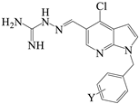
Scheme 1. The synthesis route of compounds 3A-3V, 4L-4P, 5L-5P and 6L-6P. Reagents and Conditions: (a) dry DMF, KOH, RT, 0.75–4 h; (b) ethanol absolute, 10 drops of concentrated HCl, 50 °C, 2–6 h.
Molecules 30 00887 sch001
Figure 1. The result of the growth curve and bactericidal time-kill kinetics assay. (A) Growth curve of K.P. 2108 in the presence of 4P (2, 4, 8, and 16 µg/mL) and colistin (32 µg/mL). (B) Time-kill curve of K.P. 2108 in the presence of 4P (4, 8, 16, and 32 µg/mL) and colistin (32 µg/mL).
Molecules 30 00887 g001
Figure 2. The result of FC and SEM. (A) Quantified results of the flow cytometry analysis after the treatment of K.P. 2108 with colistin (32 µg/mL) and 4P (4, 8, 16, 32 µg/mL). (B) Scanning electron microscope images of K.P. 2108 under the treatment with colistin (32 µg/mL) and 4P (32 µg/mL). The asterisk denotes statistically significant differences: **** p < 0.0001.
Molecules 30 00887 g002
Figure 3. The effect of 4P on bacterial membrane and bacterial resistance development. (A) Increased membrane permeability after treatment with different concentrations of 4P. (B) The dynamic curves of the integrity of the inner membrane probed with PI for K.P. 2108, under the treatment of 4P (4, 8, 16, and 32 µg/mL) and colistin (32 µg/mL). (C,D) Propensity of the development of K. pneumoniae resistance to 4P and colistin after repetitive treatments for 18 days (C for ATCC 700603, D for K.P. 2112).
Molecules 30 00887 g003
Figure 4. The docking result and enzymatic activity of 4P and DHFR, and hemolysis. (A,B) Interaction of 4P with the DHFR protein ((A) for two-dimensional, (B) for three-dimensional). (C) The inhibition rate of 4P on the in vitro activity of the DHFR protein. (D) Hemolysis rate of 4P. The value with “ns” is not significant; the asterisk denotes statistically significant differences: * p < 0.05; **** p < 0.0001.
Molecules 30 00887 g004
Figure 5. 4P can treat K. pneumoniae infection in mice. (A) Survival rates of female BALB/c mice (n = 10) infected with K. pneumoniae. (BE) Bacterial loads in infected lungs, livers, kidneys, and hearts of mice (n = 10). The black box represents the bacterial load of mice that died due to infection; the red box represents the bacterial load of the surviving mice. Each symbol represents a mouse. The value with “ns” is not significant; the asterisk denotes statistically significant differences: ** p < 0.01; *** p < 0.001.
Molecules 30 00887 g005
Figure 6. Histopathological analysis of different tissues using hematoxylin-eosin (HE) staining. The heart, liver, lung, and kidney were histologically analyzed in the mouse pneumoniae model. The green arrow indicates the necrosis of cardiac myofibers, the congestion and dilatation of the blood vessels in heart tissue. The red arrow indicates hepatocellular degeneration and hepatic sinusoids in liver tissue. The blue arrow indicates the necrosis and edema of alveolar epithelial cells, alveolar edema, and inflammatory cell infiltration in lung tissue. The yellow arrow indicates extensive tubular necrosis in the kidney. Scale bar, 20 µm.
Molecules 30 00887 g006
Table 1. Chemical structures of target aminoguanidine indole derivatives.
StructureCompoundR1XYMW
Molecules 30 00887 i0013AHC2-F309.35
3BHC3-F309.35
3CHC4-F309.35
3DHC2-F, 4-F327.34
3EHC2-F,5-F327.34
3FHC2-F, 6-F327.34
3GHC3-F, 4-F327.34
3HHC3-F, 5-F327.34
3IHC3-CF3359.36
3JHC4-CF3359.36
3KHC3-CF3, 5-CF3427.35
3LHC2-Cl325.80
3MHC3-Cl325.80
3NHC4-Cl325.80
3OHC2-Cl, 4-Cl360.24
3PHC3-Cl, 4-Cl360.24
3QHC2-Br370.25
3RHC3-Br370.25
3SHC4-Br370.25
3THC2-Cl, 4-F343.79
3UHC3-Cl, 4-F343.79
3VHC4-CN316.37
Molecules 30 00887 i0024LHC2-Cl325.80
4MHC3-Cl325.80
4NHC4-Cl325.80
4OHC2-Cl, 4-Cl360.24
4PHC3-Cl, 4-Cl360.24
Molecules 30 00887 i0035LHN2-Cl326.79
5MHN3-Cl326.79
5NHN4-Cl326.79
5OHN2-Cl, 4-Cl361.23
5PHN3-Cl, 4-Cl361.23
Molecules 30 00887 i0046LClN2-Cl361.23
6MClN3-Cl361.23
6NClN4-Cl361.23
6OClN2-Cl, 4-Cl395.67
6PClN3-Cl, 4-Cl395.67
Table 2. The MICs and MBCs of target compounds against ESKAPE and MRSA strains.
CompoundsStrains (MIC/MBC, µg/mL)
E. faecium
ATCC35667
S. aureus
ATCC29223
MRSAK. pneumoniae
ATCC700603
A. baumannii
ATCC19606
P. aeruginosa
ATCC27853
E. coli
ATCC25922
3A8/168/3216/1632/648/6416/1632/64
3B8/168/3216/-32/328/6416/3232/32
3C8/164/1616/3232/6416/1616/3216/32
3D8/168/3216/1632/328/1616/6432/-
3E8/168/1616/1632/648/6432/6432/32
3F8/164/64>64/->64/-8/32>64/-64/-
3G8/168/3216/6432/328/1616/3232/32
3H8/164/1616/1632/328/1616/1616/32
3I4/84/88/816/644/1616/648/16
3J4/162/328/88/84/3216/168/16
3K4/42/88/88/164/432/648/32
3L4/82/88/816/168/816/3216/64
3M4/84/816/3216/644/816/1616/16
3N4/84/88/88/644/816/3216/16
3O4/84/84/88/324/816/644/32
3P4/42/84/48/322/416/324/16
3Q4/162/88/648/324/1632/648/16
3R4/82/328/816/164/416/328/16
3S4/328/3216/3216/644/1632/-16/32
3T8/164/88/328/644/816/3216/16
3U4/84/88/6416/324/416/168/8
3V32/328/3264/64>64/-32/3264/6464/64
4L8/162/82/816/328/816/168/16
4M8/162/82/816/328/1616/168/16
4N4/162/42/816/168/168/168/8
4O2/162/82/44/164/88/164/8
4P2/82/42/44/164/328/162/4
5L32/328/328/3232/3232/3216/1616/16
5M>64/-16/6416/32>64/->64/->64/->64/-
5N16/324/328/1616/3216/3216/3216/16
5O16/162/84/3216/6416/1616/328/16
5P16/164/84/328/3216/1616/328/8
6L>64/-16/6416/64>64/->64/->64/->64/-
6M8/648/164/32>64/-8/8>64/-16/16
6N32/-8/328/16>64/-16/32>64/-16/64
6O>64/->64/->64/->64/->64/->64/->64/-
6P16/328/648/64>64/-32/32>64/->64/-
Colistin>64/->64/-64/-2/80.5/0.51/21/2
-: not detected.
Table 3. The MICs and MBCs of active compounds against clinical K. pneumoniae isolates.
CompoundsStrains (MIC/MBC, µg/mL)
K.P.
2102
K.P.
2105
K.P.
2107
K.P.
2108
K.P.
2109
K.P.
2112
K.P.
2118
K.P.
2125
K.P.
2134
K.P.
2135
K.P.
2138
3I8/88/328/168/168/88/648/88/168/328/328/8
3J8/328/168/88/88/168/88/88/88/88/88/8
3K8/6416/648/168/3216/328/328/168/1616/328/168/32
3L8/1616/168/88/88/168/88/88/168/88/88/8
3M8/1616/168/168/88/88/88/6416/168/168/328/8
3N8/816/328/168/88/168/328/6416/168/168/88/8
3O8/88/324/168/168/644/644/48/328/644/84/16
3P4/168/164/84/164/644/84/48/168/84/84/8
3Q8/88/-8/88/88/88/88/328/88/88/168/16
3R8/816/328/88/88/88/88/6416/168/88/328/8
3S8/1616/328/168/648/168/168/88/168/88/88/16
3T8/816/168/88/88/88/88/88/88/88/88/16
3U8/88/88//328/88168/88/88/88/88/88/8
4O4/84/84/84/88/84/44/84/48/84/164/16
4P4/84/84/44/84/164/44/84/84/84/44/4
5P8/168/816/168/328/328/88/88/88/88/188/16
Colistin2/4>64/-8/8>64/->64/-4/48/88/168/320.5/44/8
-: not detected.
Table 4. The hemolysis values of 3P, 4P and 5P.
Compounds (µg/mL)
3P4P5P
HC50245.0123.6243.7
SI *30.6330.9030.46
* Selectivity index (SI), SI = HC50/MICATCC 700603.
Disclaimer/Publisher’s Note: The statements, opinions and data contained in all publications are solely those of the individual author(s) and contributor(s) and not of MDPI and/or the editor(s). MDPI and/or the editor(s) disclaim responsibility for any injury to people or property resulting from any ideas, methods, instructions or products referred to in the content.

