Article

Inhibition Profiling of Rhodiola crenulata on Lactate Dehydrogenase Isoenzymes

The Research Center of Chiral Drugs, Shanghai Frontiers Science Center for TCM Chemical Biology, Innovation Research Institute of Traditional Chinese Medicine, Shanghai University of Traditional Chinese Medicine, 1200 Cailun Road, Shanghai 201203, China
*
Authors to whom correspondence should be addressed.
These authors contributed equally to this work.
Molecules 2025, 30(21), 4199; https://doi.org/10.3390/molecules30214199
Submission received: 18 September 2025 / Revised: 18 October 2025 / Accepted: 22 October 2025 / Published: 27 October 2025

Abstract

Although Rhodiola crenulata (R. crenulata) exhibits anti-tumor effects, its mechanism of action has yet to be elucidated. Lactate dehydrogenase (LDH), a key glycolytic enzyme in tumor metabolism, has emerged as a promising target for anticancer drug development. To elucidate the anticancer mechanism of R. crenulata, this study systematically screened its bioactive components for inhibitory activity against LDHA and LDHB subunits. First, the main components of R. crenulata were identified using HPLC-QTOF MS. Then, the inhibitory potency of these identified components was assessed against recombinant LDHA and LDHB. Finally, isoenzyme inhibition of the bioactive components was elucidated through structure-based molecular docking and cell viability assays. The results showed that five R. crenulata compounds—quercetin, luteolin, kaempferol, epicatechin gallate, and ellagic acid—showed significant LDH inhibition, with stronger effects on LDHA than on LDHB. Against LDHA, the IC50 values for quercetin, luteolin, kaempferol, and epicatechin gallate were 0.27 ± 0.02 µM, 1.19 ± 0.09 µM, 0.70 ± 0.13 µM, and 2.27 ± 0.23 µM, respectively. Against LDHB, the values for quercetin, luteolin, and kaempferol were 0.87 ± 0.07 µM, 2.71 ± 0.39 µM, and 8.69 ± 0.85 µM, respectively. Molecular docking simulations and cell viability assays of the five bioactive compounds revealed their interactions with LDH subunits and supported their inhibitory effects. This study provides the first comprehensive inhibition profile of R. crenulata targeting LDH isoenzymes. It underscores the potential of R. crenulata in LDH-targeted therapeutics and supports its further development for cancer treatment.

1. Introduction

Cancer has garnered significant and growing attention as a high-priority research area within global scientific and medical communities. A defining hallmark of cancer is metabolic reprogramming, wherein cells rewire nutrient utilization to sustain rapid proliferation and survival [1]. This reprogramming encompasses heightened glutaminolysis, aberrant lipid synthesis, and, most prominently, a shift toward aerobic glycolysis—the Warburg effect [1,2]. By accelerating glycolytic flux, tumors generate intermediates for macromolecular synthesis and organelle biogenesis, thereby sustaining tumor expansion [3]. A key player in cancer metabolism is lactate dehydrogenase (LDH), the enzyme responsible for the final step of aerobic glycolysis, catalyzing the reversible interconversion of pyruvate and lactate while simultaneously mediating the redox cycling of NADH and NAD+ [4].
Structurally, LDH functions as a tetramer primarily composed of the LDHA (M subunit) and LDHB (H subunit) subunits, which assemble into five distinct isoenzymes: LDH-1 (H4), LDH-2 (M1H3), LDH-3 (M2H2), LDH-4 (M3H1), and LDH-5 (M4) [5]. While LDHA and LDHB share similar tertiary structures, variations in charged residues near the active sites confer them distinct kinetic properties [6]. LDHA exhibits higher catalytic efficiency for pyruvate reduction to lactate, while LDHB preferentially drives lactate oxidation back to pyruvate [5]. During catalysis, each subunit functions independently, simultaneously binding one substrate molecule and one coenzyme molecule to complete the redox reaction. Elevated LDHA expression has been widely detected in diverse malignancies, where it drives tumorigenic processes including uncontrolled proliferation, malignant maintenance, and invasion [7,8,9,10]. Unlike the consistently tumorigenic role of LDHA, LDHB displays significant functional plasticity across cancers [7,11]. For example, LDHB exhibits tumor-promoting effects in pancreatic cancer [12,13], breast cancer [14,15], lung cancer [16], and colorectal cancer [17,18], while demonstrating tumor-suppressive activity in prostate cancer [19,20] and liver cancer [21,22].
Given the crucial role of LDH in cancer metabolism, there has been increasing interest in developing LDH-targeted antitumor drugs. However, no clinically effective inhibitors have been developed to date, and most reported compounds exhibit poor subunit selectivity [23]. Despite the high structural conservation between LDHA and LDHB, their distinct biological functions and expression across tumor types suggest that designing subunit-selective inhibitors offers a promising strategy for developing clinically viable LDH inhibitors.
Herbal medicines are generally valued for their gentle bioactivity, low toxicity, and good clinical tolerability [24]. These distinctive properties have driven growing interest in target-based screening of bioactive constituents from herbal medicines. Nevertheless, the conventional approach of screening inhibitors extensively against the drug database fails to capture the holistic therapeutic principles and synergistic effects that characterize herbal medicine therapy. Currently, multiple technologies have been developed to address this challenge, including radioligand binding assays [25], affinity mass spectrometry [26], and computer-aided virtual screening [27]. A critical limitation persists, as these technologies primarily assess binding thermodynamics rather than functional bioactivity, resulting in substantial false-positive identification rates [28,29].
R. crenulata is a perennial herbaceous plant native to high-altitude regions. Several contemporary studies have validated its therapeutic applications in cardiovascular dysfunction [30,31], renal impairment [32], neurodegenerative disorders [33,34], hepatic fibrogenesis [35], immune dysregulation [36], and malignant tumors [37,38,39]. However, these studies have predominantly attributed the antitumor effects of R. crenulata to isolated constituents (e.g., salidroside), rather than systematically elucidating its complex polypharmacological mechanisms of action [40].
To systematically elucidate the anticancer mechanisms and bioactive constituents of R. crenulata, we selected LDH isoenzymes as targets for profiling its inhibitory activity. After characterizing the isoenzyme inhibition of the R. crenulata extract, we employed HPLC-QTOF MS to identify its main constituents and evaluated their individual enzymatic inhibitory effects using the enzyme activity assay. The inhibition of the screened bioactive components was further elucidated through structure-based molecular docking and cell viability assays.

2. Results and Discussion

2.1. Substrate-Specific Kinetic Parameters of LDHA and LDHB

LDH catalyzes the reversible interconversion of pyruvate and lactate while mediating the redox cycling of NADH and NAD+. To characterize the enzymatic properties of recombinant LDHA and LDHB, we monitored their activities toward pyruvate and NADH by measuring absorbance changes at 340 nm. Initial reaction velocities were determined from progress curves generated across a range of substrate concentrations. Subsequent Michaelis-Menten analysis, performed by fitting the velocity versus substrate concentration plots (Figure 1), yielded the kinetic parameters (Km and Vmax). Notably, the Km value serves as an inverse indicator of substrate binding affinity, with lower values reflecting stronger enzyme-substrate interactions.
LDHA displayed Km values of 0.160 ± 0.027 mM for pyruvate and 0.039 ± 0.006 mM for NADH, with corresponding Vmax values of 15.11 ± 0.69 μM/min and 15.37 ± 0.57 μM/min. For LDHB, the Km values were 0.074 ± 0.019 mM for pyruvate and 0.040 ± 0.005 mM for NADH, with Vmax values of 11.36 ± 0.76 μM/min and 10.05 ± 0.33 µM/min, respectively. Although the Km values for NADH were nearly equal between LDHA and LDHB, LDHA showed a significantly higher Km for pyruvate compared to LDHB, indicating stronger binding affinity of LDHB for pyruvate. These results are consistent with LDHB’s physiological role in oxidizing lactate back to pyruvate [41,42]. The higher Vmax values for LDHA indicate its specialization in rapid pyruvate reduction during glycolysis. Collectively, these findings demonstrate that while LDHA is optimized for glycolytic pyruvate reduction, LDHB is specialized for efficient pyruvate binding under aerobic conditions. Furthermore, these results suggest that inhibitors targeting the pyruvate-binding site are more likely to achieve isoenzyme-selective inhibition.

2.2. Validation of the LDHA and LDHB Inhibitor Screening Platforms

To ensure the reliable identification of inhibitors and minimize false positives, we rigorously validated the screening platform using established quality metrics. The Z′ factor, a critical parameter for assessing assay robustness, was used to evaluate our screening platform. The obtained values of 0.94 for LDHA and 0.82 for LDHB demonstrate excellent system performance, as they significantly exceed the accepted threshold of 0.5 for robust assays [43] and satisfy the more stringent criterion (Z′ > 0.7) for high-throughput screening. These high Z′ values indicated the high signal-to-background ratio and minimal well-to-well variability of the screening platform.
For pharmacological validation, gossypol (a benchmark LDH inhibitor) was employed as a reference inhibitor. Dose-response curves were generated by plotting percentage inhibition against the logarithm of inhibitor concentrations (log10[gossypol]), with IC50 values determined by nonlinear regression analysis. Quantitative analysis yielded IC50 values of 0.19 ± 0.05 µM for LDHA and 0.27 ± 0.04 µM for LDHB (Figure 2). The submicromolar inhibitory potency observed for both isoforms demonstrates the platform’s sensitivity in detecting biologically relevant inhibition and confirms its utility for high-throughput screening applications.

2.3. Differential Inhibitory Effects of R. crenulata Extract on LDHA and LDHB

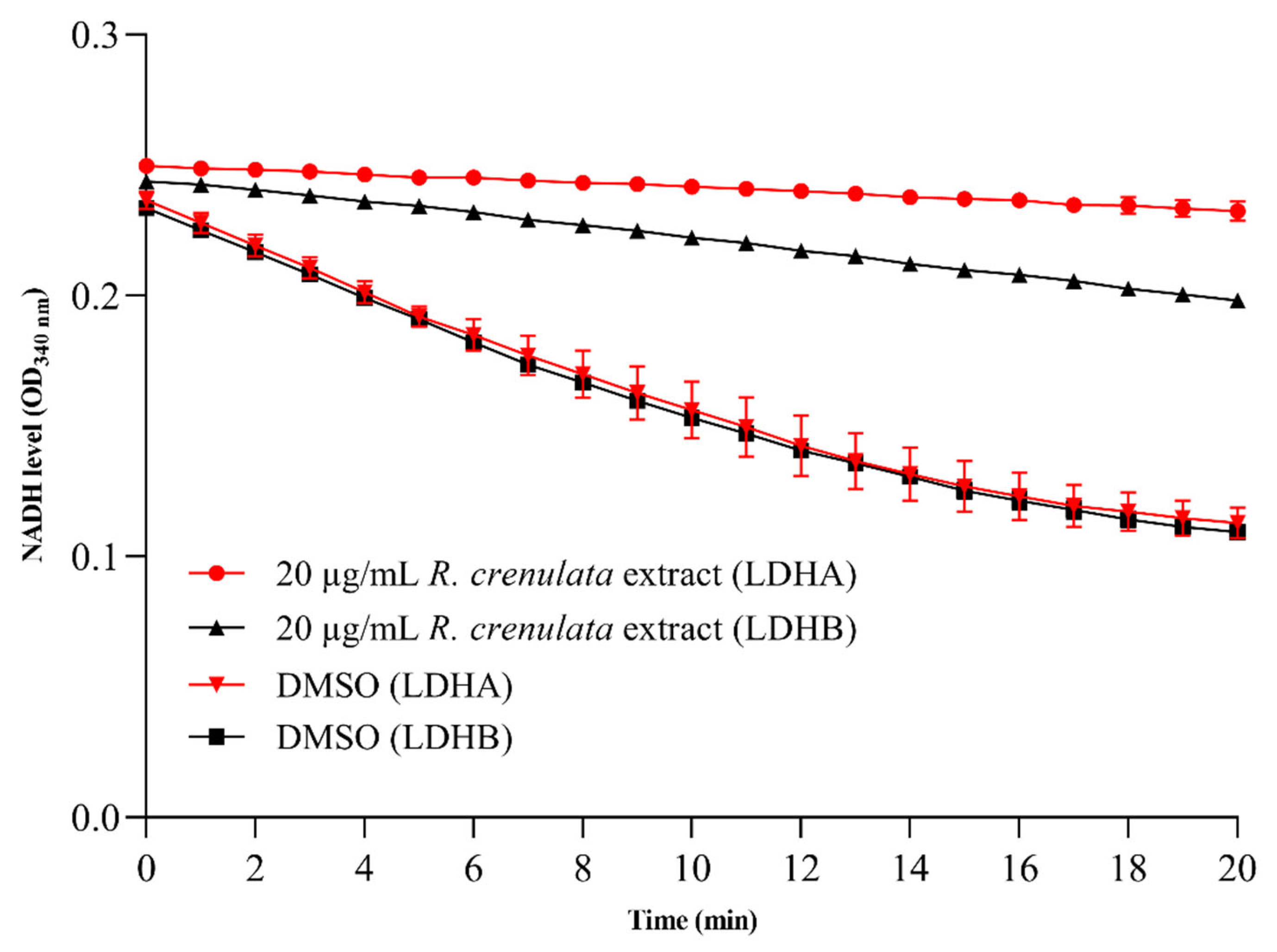
While some isolated components (e.g., salidroside) have been extensively studied for antitumor effects, the antitumor potential of R. crenulata extract still remains insufficiently investigated. Given the crucial role of LDH in cancer metabolism, we evaluated the inhibitory effects of R. crenulata extract on both LDHA and LDHB. As shown in Figure 3, enzyme activity was monitored in real time by measuring the absorbance decrease at 340 nm, which reflects NADH consumption. Comparative analysis of enzymatic progress curves revealed that dimethyl sulfoxide (DMSO)-treated controls maintained high, stable activity throughout the entire recording window, whereas the extract induced a time-dependent decline in both isoforms. In addition, R. crenulata extract showed greater inhibition of LDHA compared to LDHB. Based on the inhibition rate formula, the extract exhibited differential inhibitory effects of 90.9 ± 2.6% on LDHA and 73.1 ± 0.8% on LDHB. These results indicate that some bioactive components in R. crenulata differentially target LDH subunits, with significantly greater efficacy against LDHA. Therefore, it is valuable to identify the specific inhibitory constituents in R. crenulata and evaluate their therapeutic potential.

2.4. Constituent Profiling of R. crenulata

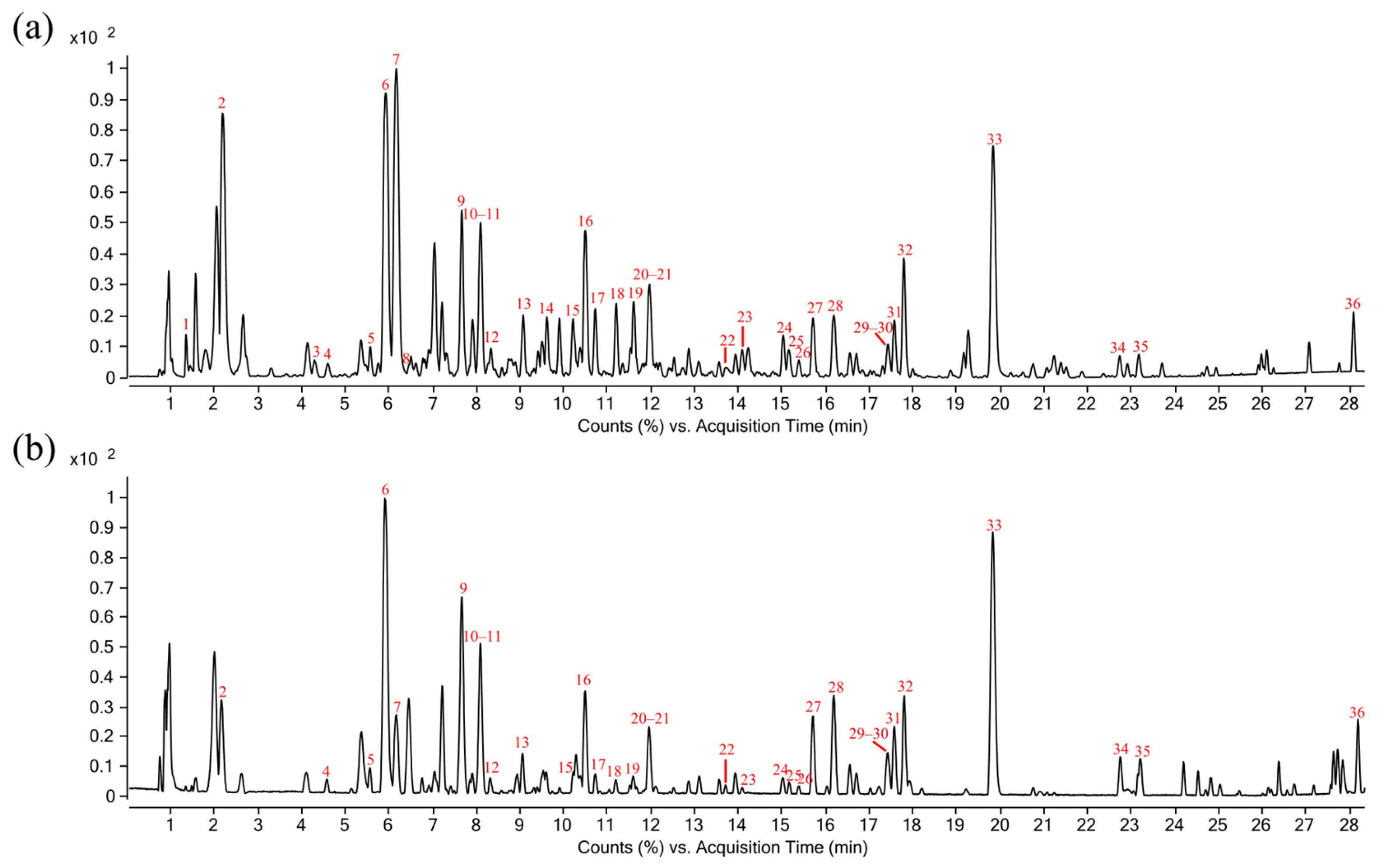
R. crenulata is known to biosynthesize a diverse array of bioactive metabolites, including phenylethanoid glycosides, flavonoids, organic acids, phenylpropanoids, and polyphenols [44,45]. To systematically characterize its chemical constituents, we performed an untargeted metabolomic analysis using HPLC-QTOF MS with data-dependent acquisition. The Base Peak Chromatograms (BPCs) acquired in both positive and negative ionization modes showed a complex metabolite profile in Figure 4. By comparing acquired mass spectra against chemical standards, mass spectral databases (e.g., GNPS), and published literature, we successfully identified and annotated 36 major bioactive compounds. The complete chemical profile of these compounds—including retention times (RT), precursor ions and their mass error, characteristic fragment ions (MS/MS), and molecular formulas—is comprehensively detailed in Table 1.

2.5. Identification of LDHA and LDHB Inhibitors from R. crenulata Extract

Given the differential expression patterns of LDHA and LDHB observed across different cancer types, these subunits emerge as promising candidates for tissue-specific targeted therapies. The structural complexity of LDH isoenzymes in vivo, which form five distinct tetrameric combinations, presents significant challenges for the development of selective inhibitors [53]. In this study, recombinant LDHA and LDHB were employed to effectively circumvent the heterogeneity of mixed tetramers found in physiological contexts. Based on these recombinant proteins, we established a screening platform and evaluated its robustness, sensitivity, and applicability. As individual subunits within the LDH tetramer retain independent catalytic activity, inhibitors targeting specific subunits identified by this platform represent a promising therapeutic strategy.
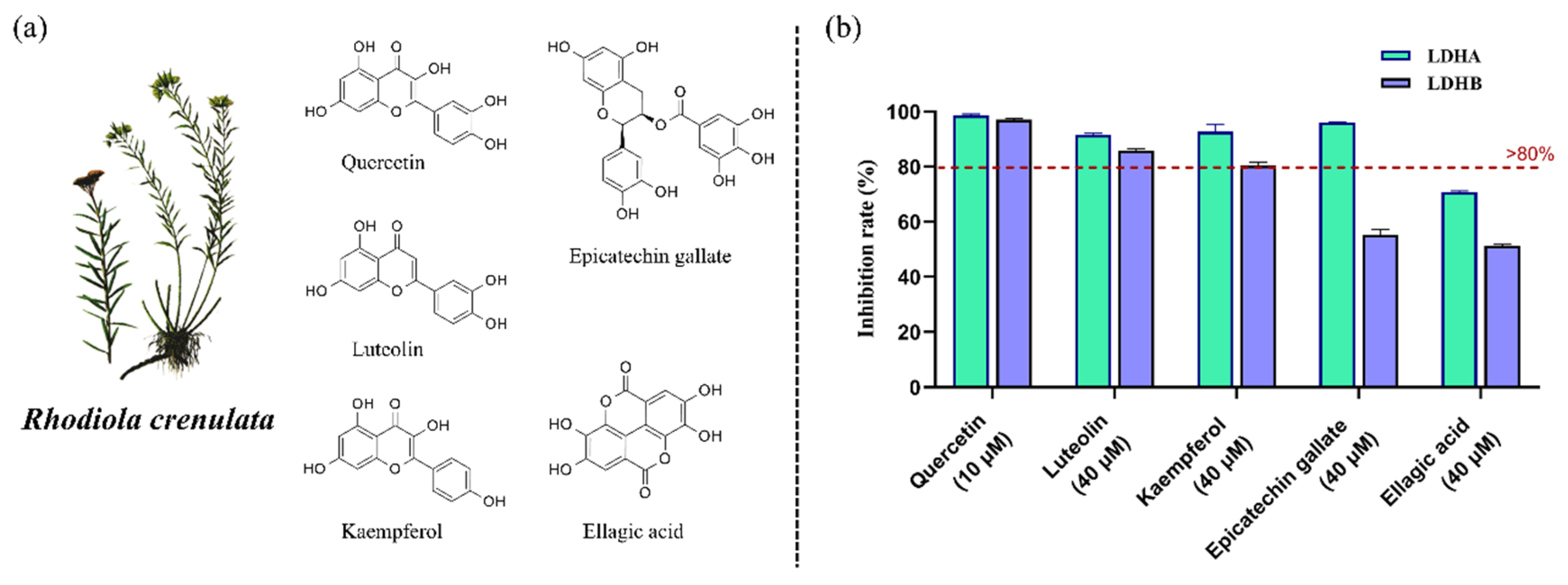
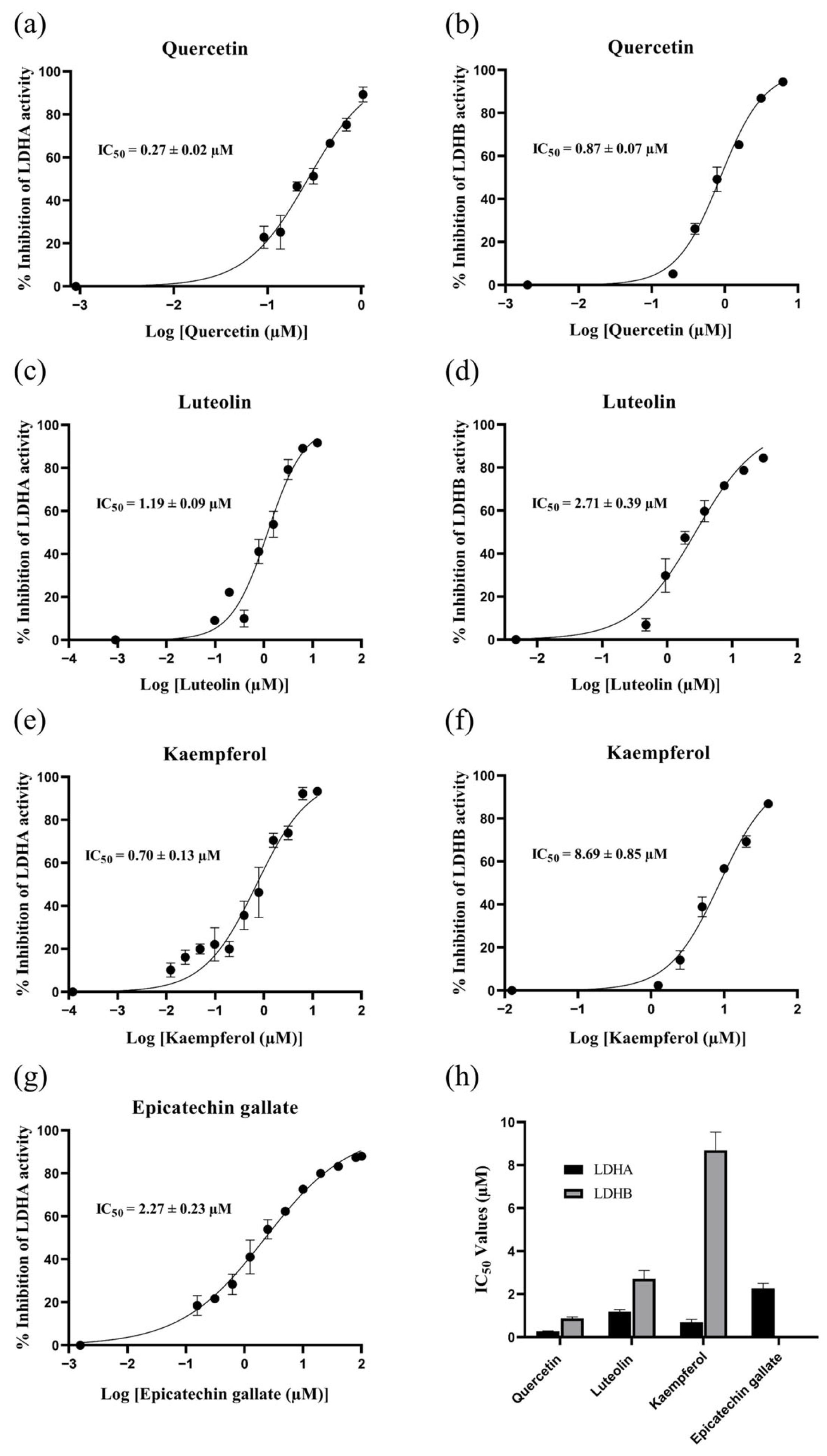
As preliminary screening revealed that the R. crenulata crude extract differentially inhibited LDHA and LDHB, we employed this validated platform to screen the extract for specific LDH inhibitors. Using this platform, we assessed 19 commercially available standards corresponding to identified metabolites in Table 1. Upon comparing the inhibition rates, five compounds—quercetin, luteolin, kaempferol, epicatechin gallate, and ellagic acid—exhibited significant LDH inhibitory activity, with stronger inhibition against LDHA than LDHB (Figure 5). To further characterize the inhibitory properties, components exhibiting inhibition rates exceeding 80% were subjected to dose-response evaluation. Quercetin, luteolin, and kaempferol—each active against both isoforms—along with epicatechin gallate, which was active against LDHA, were selected for IC50 determination. In Figure 6, the comparison of IC50 values revealed that kaempferol was more than 10-fold more potent in inhibiting LDHA (0.70 ± 0.13 µM) than LDHB (8.69 ± 0.85 µM). Similarly, both quercetin and luteolin exhibited a marked preference for inhibiting LDHA over LDHB, with IC50 values of 0.27 ± 0.02 µM and 1.19 ± 0.09 µM for LDHA, compared to 0.87 ± 0.07 µM and 2.71 ± 0.39 µM for LDHB, respectively. Although robust activity against LDHA and LDHB was confirmed in this study, the potential for off-target effects in subsequent experiments should be considered due to the known nonspecificity of some polyphenols. Collectively, these findings underscore the potential of R. crenulata as a valuable source of LDH inhibitors. Furthermore, these natural scaffolds offer promising starting points for further optimization to enhance potency or isoform specificity.

2.6. Molecular Docking of Inhibitory Components to LDHA and LDHB

Molecular docking is a computational approach that predicts the optimal binding mode between a receptor and a ligand by evaluating their geometric, energetic, and chemical complementarity. This technique plays a pivotal role in drug discovery and the investigation of potential therapeutics. Binding energy and key intermolecular interactions serve as the primary indicators for evaluating binding affinity. Lower binding energy values correspond to greater binding stability and stronger molecular interactions. Typically, docking energies ≤ −5.0 kcal/mol suggest substantial binding activity, while values ≤ −7.0 kcal/mol indicate highly strong binding affinity [54]. For intermolecular interactions, given the critical role of hydrogen bonds and hydrophobic interactions in complex stability, their occupancy and surface area were analyzed as key descriptors [55].
To elucidate the interactions of five R. crenulata components with LDH isoenzymes, molecular docking simulations were performed against homo sapiens LDHA and LDHB crystal structures. The binding energies of all five compounds are summarized in Table 2. Each compound exhibited a binding energy below −5.0 kcal/mol, and 80% of them exhibited values less than −7.0 kcal/mol, demonstrating their robust binding potential. Comparative analysis showed that all five compounds had consistently lower binding energies with LDHA than with LDHB, revealing stronger and more stable interactions with LDHA. This discrepant binding finding aligned well with the results from the enzymatic activity assays.
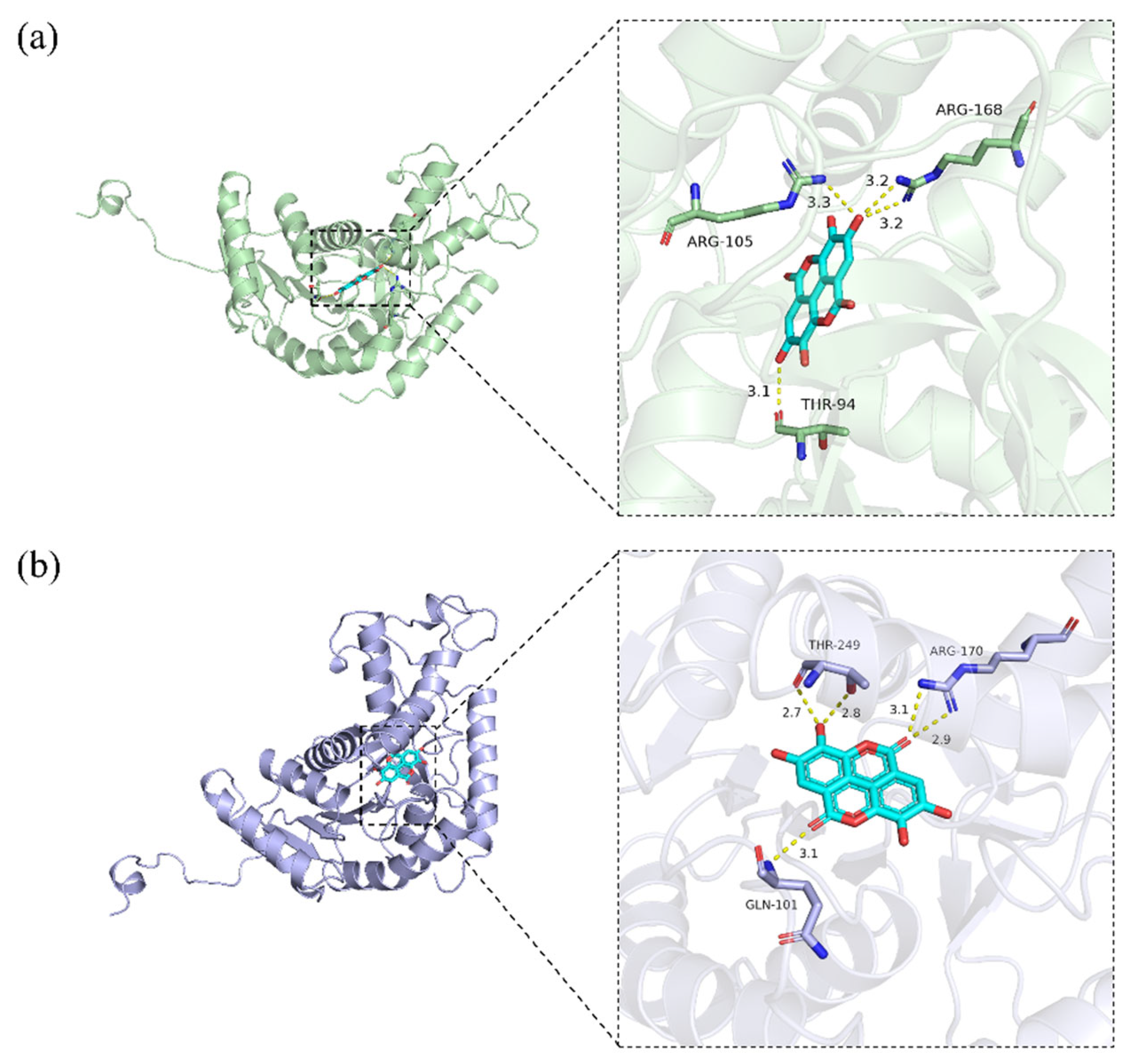
These findings align with prior reports that certain compounds or their structural analogues can act as effective LDH inhibitors. For instance, flavonoids such as quercetin, luteolin, and kaempferol share a common core structure that facilitates multi-modal binding through π–π stacking and hydrogen bonding, resulting in effective LDH inhibition [56]. Similarly, epicatechin gallate features a catechin skeleton, a structure known for its antioxidant and anticancer properties. This skeleton enables the formation of multiple hydrogen-bonding interactions. In addition, the compound’s galloyl moiety may provide additional binding sites that enhance enzyme affinity [57]. Although ellagic acid’s LDH inhibitory activity has been previously reported [58], the molecular docking of its core scaffold to LDH has not been elucidated. Featuring multiple phenolic hydroxyl groups, two lactone groups, and a biphenyl scaffold, it is proposed to bind within the substrate-binding sites of both LDHA and LDHB, as illustrated in Figure 7. Comparative analysis revealed that ellagic acid forms hydrogen bonds with Arg168 in LDHA and the corresponding conserved residue Arg170 in LDHB—both critical for substrate recognition in each subunit. Furthermore, LDHA establishes specific interactions with Thr94 and Arg105, while LDHB forms unique contacts with Gln101 and Thr249. These structural observations indicate that although ellagic acid targets a conserved arginine critical for substrate recognition in both subunits, the distinct microenvironments of the LDHA and LDHB substrate-binding sites likely underlie its differential binding and inhibitory effects. Although ellagic acid does not exhibit high isoenzyme specificity, it provides a valuable structural foundation for the rational design of more selective LDH inhibitors.

2.7. Anti-Proliferative Effects of R. crenulata Bioactives on MDA-MB-231 and HepG2 Cells

To evaluate the anti-proliferative efficacy of the five R. crenulata components, we employed two human carcinoma cell lines, MDA-MB-231 (triple-negative breast adenocarcinoma) and HepG2 (hepatocellular carcinoma). These models were selected based on the reported divergent roles of LDHB, which exhibits tumor-promoting effects in breast cancer but demonstrates tumor-suppressive activity in liver cancer.
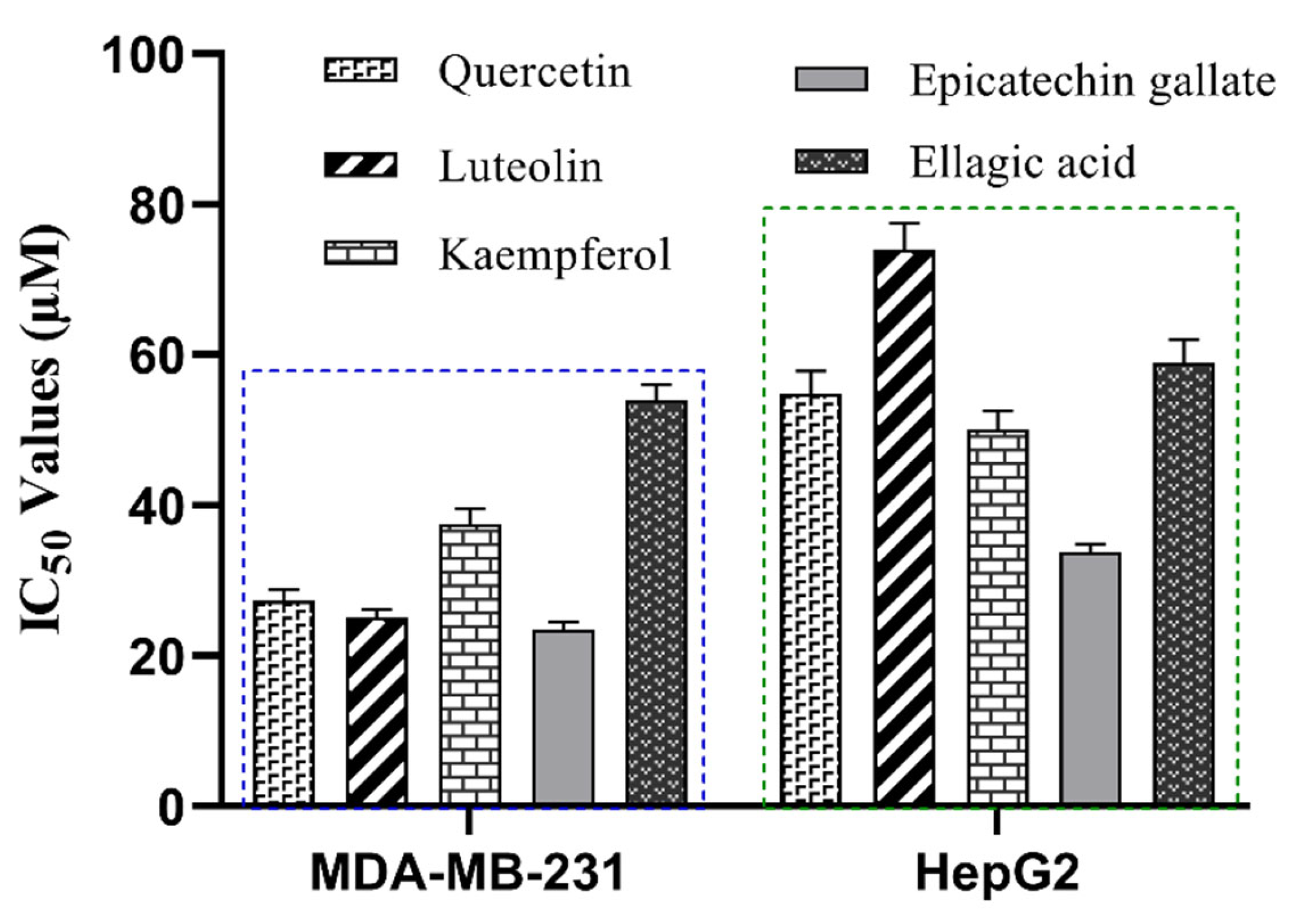
As shown in Figure 8, all five compounds demonstrated significantly stronger anti-proliferative effects in MDA-MB-231 cells than in HepG2 cells. The IC50 values against MDA-MB-231 for luteolin, quercetin, kaempferol, epicatechin gallate, and ellagic acid were 25.1 ± 1.0 µM, 27.3 ± 1.5 µM, 37.5 ± 2.0 µM, 23.5 ± 1.0 µM, and 54.3 ± 2.5 µM, respectively. The corresponding IC50 values against HepG2 were 74.0 ± 3.5 µM, 54.8 ± 3.0 µM, 50.0 ± 2.5 µM, 33.8 ± 1.0 µM, and 59.0 ± 3.0 µM. We propose that this differential efficacy may stem from the distinct LDHB dependencies of the two cell lines. MDA-MB-231 cells are known to be dependent on both LDHA and LDHB for proliferation [59,60]. As our enzyme-level assays confirmed the dual inhibition of both isoforms, the superior anti-proliferative activity in MDA-MB-231 is likely attributable to this concurrent targeting. In contrast, the reduced potency in HepG2 cells is consistent with reports that LDHB expression has no significant impact on their proliferative [21]. Therefore, these cell-based assays functionally validate the anti-proliferative potential of the inhibitors, and the results are consistent with their established role as dual LDHA/LDHB inhibitors.

3. Materials and Methods

3.1. Reagents and Materials

Referring to the mRNA sequence of homo sapiens LDHA (NCBI RefSeq NM_005566.4) and LDHB (NCBI RefSeq NM_001174097.3), the LDHA_pET-28a (+) and LDHB_pET-28a (+) plasmids were synthesized by GenScript Biotech Corporation (Piscataway, NJ, USA). BL21(DE3) competent cells, nickel-nitrilotriacetic acid (Ni-NTA) Sepharose 6FF (His-Tag), kanamycin sulfate, isopropyl β-D-thiogalactoside (IPTG), β-Nicotinamide adenine dinucleotide disodium salt (β-NADH Disodium Salt), and sodium pyruvate were purchased from Sangon Biotech (Shanghai) Co., Ltd. (Shanghai, China). Salidroside, kaempferol, and mycophenolic acid were purchased from Adamas Reagent Co., Ltd. (Shanghai, China). Protocatechuic acid, caffeic acid, (−)-epicatechin, ellagic acid, epicatechin gallate, luteolin, quercetin, tyrosol, and rhodiosin were purchased from Shanghai Standard Technology Co., Ltd. (Shanghai, China). Crenulatin was purchased from Chengdu Push Bio-technology Co., Ltd. (Chengdu, China). Citric acid, gossypol, gallic acid, and azelaic acid were purchased from Shanghai yuanye Bio-Technology Co., Ltd. (Shanghai, China). Astragalin and afzelin were purchased from Shanghai Macklin Biochemical Technology Co., Ltd. (Shanghai, China). Rhodionin was purchased from Shanghai TOP Bio-Pharma Technology Co., Ltd. (Shanghai, China). The MDA-MB-231 and HepG2 cell lines were obtained from Shanghai Institute of Biochemistry and Cell Biology, Chinese Academy of Sciences (Shanghai, China). The Cell Counting Kit-8 (CCK-8) were purchased from Beyotime Biotech Inc. (Shanghai, China). Dulbecco’s Modified Eagle Medium (DMEM), 0.25% trypsin-EDTA, and fetal bovine serum (FBS) were purchased from Thermo Fisher Scientific Inc. (Waltham, MA, USA). The herb R. crenulata (Hook. f. et Thoms) H. Ohba was purchased from Yinfeng Chinese Medicine Port (Yulin, China) and authenticated by Professor Lihong Wu.

3.2. Instruments and Software

An Agilent 1290 high-performance liquid chromatography system coupled with an Agilent 6545 UHD quadrupole time-of-flight mass spectrometer (HPLC-QTOF MS; Agilent Technologies Co., Santa Clara, CA, USA) was used to analyze the components in R. crenulata. Chromatographic separation was performed on a Waters ACQUITY HSS T3 column (2.1 mm × 100 mm, 1.8 μm) with a mobile phase consisting of (A) 0.1% formic acid in deionized water and (B) acetonitrile, delivered at 0.3 mL/min. The gradient elution program was as follows: 0–2 min, 5% B; 2–14 min, 5–25% B; 14–21 min, 25–40% B; 21–28 min, 40–75% B; 28–33 min, 75–95% B; 33–35 min, 95% B. The column temperature was maintained at 40 °C, and the injection volume was 1 µL. The QTOF MS was equipped with a Dual AJS ESI source. And data were acquired in both positive and negative ionization modes. The ESI parameters were optimized as follows: nitrogen was used as the sheath gas at 12 L/min and 325 °C, nebulizer gas at 40 psig, and drying gas at 10 L/min and 325 °C. The capillary voltage, nozzle voltage, and fragment voltage were 4, 0.5, and 0.12 kV, respectively. MS and MS/MS spectra were acquired in the range of m/z 50–1500. For MS/MS analysis, nitrogen was used as the collision gas with collision energies of 10, 20, and 35 eV. Mass calibration was performed with Agilent ESI-L Low Concentration Tuning Mix (m/z 100–3200, Santa Clara, CA, USA), achieving a mass resolution of ~25,000 at m/z 322.
All data were collected and processed using MassHunter LC/MS Data Acquisition B.08.00 (Agilent Technologies Co., Santa Clara, CA, USA) and MassHunter Qualitative Analysis B.07.00 (Agilent Technologies Co., Santa Clara, CA, USA).

3.3. Preparation of R. crenulata Extract

The dried roots of R. crenulata were ground into powder. And 0.5 g of weighed powder was mixed with 10 mL of methanol in a 15 mL centrifuge tube. The mixture was vortexed vigorously and then allowed to sonicate for 30 min. After centrifugation, the supernatant was carefully collected as the extract and stored at −20 °C for subsequent analysis.

3.4. Expression and Purification of Recombinant LDHA and LDHB

The recombinant LDHA-pET-28a (+) and LDHB-pET-28a (+) plasmids were transformed into BL21(DE3) competent cells using the heat shock method. Transformed cells were plated on kanamycin-containing solid medium and incubated overnight at 37 °C. Selected single colonies were inoculated into 5 mL of LB medium with kanamycin and grown overnight at 37 °C with shaking, then expanded to 1 L. When the optical density at 600 nm (OD600) reached 0.6–0.8, protein expression was induced by adding IPTG to a final concentration of 0.2 mM. Induction was carried out at 28 °C for 6 h with continuous shaking. Cells were harvested by centrifugation and lysed using a high-pressure homogenizer. The lysate was subjected to Ni-NTA affinity chromatography for initial purification. The eluted protein was subsequently desalted using a desalting column. Further purification was performed using the ÄKTA FPLC system to obtain high-purity recombinant protein. Finally, the purified LDHA and LDHB proteins were aliquoted and stored at −80 °C for future use.

3.5. Establishment and Optimization of the LDHA and LDHB Inhibitor Screening Platform

Based on comprehensive literature evidence [41,61,62,63,64] and systematic experimental validation, we established optimized enzymatic assay platforms for LDHA and LDHB inhibitor screening. In the platforms, 60 µL of purified enzyme solution (1.95 nM final concentration) was loaded into 96-well plates, followed by 140 µL of a 1:1 (v/v) pyruvate/NADH substrate mixture. Reactions were conducted in 0.1 M Tris-HCl buffer (with 10% glycerol, pH 7.4) at 30 °C, and NADH oxidation was kinetically monitored at 340 nm for 20 min. To determine the kinetic parameters, the optimization protocol for LDHA was performed as follows: with NADH fixed at 0.6 mM while varying sodium pyruvate (0.02–2.5 mM) to determine the optimal concentration; and with sodium pyruvate fixed at 1 mM while varying NADH (0.01–0.7 mM) to determine the optimal concentration. Similarly, the optimization protocol for LDHB was performed as follows: with NADH fixed at 0.6 mM while varying sodium pyruvate (0.01–1.05 mM) to determine the optimal concentration; and with sodium pyruvate fixed at 1 mM while varying NADH (0.01–0.7 mM) to determine the optimal concentration. The initial reaction rates were derived by analyzing the progress curves with OriginPro 2023 (OriginLab Co., Northampton, MA, USA). Kinetic parameters (Km and Vmax) were derived by nonlinear regression analysis of the Michaelis-Menten equation using GraphPad Prism 8.0 (GraphPad Software, San Diego, CA, USA).

3.6. Evaluation of the LDHA and LDHB Inhibitor Screening Platform

The Z’ factor (Z’ = 1 − [3 × (σₚ + σₙ)]/|μₚ − μₙ|) was used to evaluate the robustness of the screening platform, where σ represents the standard deviation, μ represents the mean value, p denotes enzyme activity, and n denotes the background signal. Gossypol was employed as a reference inhibitor for further validation. The detailed steps were as follows: (1) 59 µL of LDHA/LDHB was added into a 96-well plate, followed by 1 µL of serially diluted gossypol solution (11-point concentration gradient, 0.001–60 µM). (2) The enzyme-inhibitor mixture was pre-incubated for 10 min at room temperature. (3)140 µL of the 1:1 (v/v) substrate mixture was added (for LDHA: 0.16 mM sodium pyruvate and 0.04 mM NADH; for LDHB: 0.08 mM sodium pyruvate and 0.04 mM NADH). (4) At 30 °C, the enzymatic reaction was monitored at 340 nm for 20 min. The half maximal inhibitory concentration (IC50), defined as the inhibitor concentration required to reduce enzyme activity (or cell growth) by 50%, was calculated using nonlinear regression analysis of the dose-response data.

3.7. Screening of Inhibitory Components from R. crenulata Against LDHA and LDHB

For the enzyme activity assay, the concentrations of the R. crenulata extract were 20 µg/mL, while the 19 isolated components was assayed at 10 µM and 40 µM. DMSO served as the vehicle control, while the assay procedure was identical to that described above for gossypol. The inhibition rate (I%) of R. crenulata (or each component) was calculated as I% = (1 − Vi/V0) × 100%, where Vi represents the initial enzyme activity rate after treatment with R. crenulata (or each component), and V0 denotes the initial enzyme activity rate of the control group. Components showing inhibition rates greater than 80% were selected for dose-response analysis.

3.8. Molecular Docking Analysis of LDHA and LDHB Inhibitors

The crystal structures of homo sapiens LDHA (PDB ID: 1I10) and LDHB (PDB ID: 7DBK) were retrieved from the RCSB Protein Data Bank (https://www.rcsb.org/, accessed on 13 May 2025). Protein preparation, including hydrogen addition and charge assignment, was performed using PyMOL 3.0 (Schrödinger, LLC, New York, NY, USA) and AutoDock Tools 1.5.7 (the Scripps Research Institute, La Jolla, CA, USA). Inhibitory components were obtained from the PubChem database (https://pubchem.ncbi.nlm.nih.gov/, accessed on 13 May 2025) and prepared for docking using AutoDock Tools 1.5.7. Molecular docking simulations were carried out using AutoDock Vina 1.1.2 (the Scripps Research Institute, La Jolla, CA, USA). The docking active site was defined based on literature [65,66,67] to encompass the NADH and substrate-binding pockets. For each inhibitor, 20 independent docking runs were performed, and the optimal pose was selected based on the most favorable binding affinity (lowest score). The protein-ligand interactions were visualized in three dimensions using PyMOL 3.0.

3.9. Cell Viability Assays of LDHA and LDHB Inhibitors

The MDA-MB-231 and HepG2 cell lines, representing triple-negative breast cancer and hepatocellular carcinoma, were selected to evaluate the inhibitory effects of LDHA and LDHB inhibitors. Cells in the logarithmic growth phase were seeded into 96-well flat-bottom plates at densities of 5000 cells/well for MDA-MB-231 and 10,000 cells/well for HepG2. After 24 h attachment period, the medium was replaced with 100 µL of fresh medium containing test compounds (8-point concentration gradient, 1.5625–100 µM), or vehicle control (0.5% DMSO). Following 72 h of incubation, 10 µL of CCK-8 reagent was added to each well, and cells were further incubated for 1–4 h. Absorbance was measured at 450 nm with a reference wavelength of 600 nm for background correction. Data from three independent experiments (each with three technical replicates) were normalized to the vehicle control (100% viability) and medium-only wells (0% viability). Dose-response curves were fitted using a four-parameter logistic (4PL) model in GraphPad Prism, from which the IC50 values for each compound-cell line combination were derived by nonlinear regression.

4. Conclusions

Systematically elucidating the anticancer mechanisms and identifying the active constituents of R. crenulata are essential for enhancing its clinical applicability and promoting broader acceptance. In this study, we observed differential inhibition of R. crenulata extract against LDH isoenzymes, which prompted further profiling of its inhibitory components. By integrating target-centric enzymology, untargeted metabolomics, structure-guided modeling and functional cellular assays, we established R. crenulata as a rich source of LDH inhibitors. The results showed that quercetin, luteolin, kaempferol, epicatechin gallate, and ellagic acid inhibited both LDHA and LDHB, with stronger activity observed against LDHA. We acknowledge that the in vitro nature of this work limits insights into bioavailability, metabolism, and in vivo toxicity, and that the current preferential inhibition does not equate to clinical selectivity. Despite these limitations, this study provides a comprehensive inhibition profile of R. crenulata targeting LDH isoenzymes. The potent in vitro activity and structure-activity relationships established here provide a critical foundational basis for future in vivo evaluation. Moreover, the preferential inhibition exhibited by five compounds offers a pharmacological starting point for further optimization toward achieving greater isoform specificity for therapeutic applications.

Author Contributions

Y.-X.W.: methodology, experiment, analysis; M.A.: Conceptualization, methodology, analysis, project administration, supervision, writing—review and editing; Q.-F.Z. and S.-S.Z.: Conceptualization, methodology, analysis, resources, supervision; Z.A.: experiment; F.Z.: Conceptualization, methodology, analysis, project administration, resources, funding acquisition, supervision, writing—review and editing; X.-P.C.: Conceptualization, methodology, analysis, project administration, resources, supervision, writing—review and editing; C.-G.F.: Supervision, project administration, resources, and funding acquisition. All authors have read and agreed to the published version of the manuscript.

Funding

This research was funded by National Key R&D Program of China [2024YFC3506700], the Shanghai Municipal Committee of Science and Technology (Grant No. 23ZR1460900), Shanghai Science and Technology Development Fund from Central Leading Local Government (YDZX20223100001004), Shanghai Frontiers Science Center for TCM Chemical Biology, and Organizational Key R&D Program of SHUTCM (2023YZZ01).

Institutional Review Board Statement

Not applicable.

Informed Consent Statement

Not applicable.

Data Availability Statement

Data will be made available upon request.

Acknowledgments

The authors acknowledge Lihong Wu from the Institute of Chinese Materia Medica, Shanghai University of Traditional Chinese Medicine for authenticating the herb R. crenulata.

Conflicts of Interest

The authors have no conflicts of interest to declare.

Abbreviations

The following abbreviations are used in this manuscript:
HPLC-QTOF MSHigh-Performance Liquid Chromatography System Coupled with Quadrupole Time-of-Flight Mass Spectrometer
LDHLactate dehydrogenase
LDHALactate dehydrogenase A
LDHBLactate dehydrogenase B
NAD+Oxidized nicotinamide adenine dinucleotide
NADHReduced nicotinamide adenine dinucleotide
BPCBase Peak Chromatogram
RTRetention Time
IC50The half maximal inhibitory concentration
IPTGIsopropyl β-D-thiogalactoside
DMSODimethyl sulfoxide

References

  1. Faubert, B.; Solmonson, A.; DeBerardinis, R.J. Metabolic reprogramming and cancer progression. Science 2020, 368, eaaw5473. [Google Scholar] [CrossRef] [PubMed]
  2. Xia, L.; Oyang, L.; Lin, J.; Tan, S.; Han, Y.; Wu, N.; Yi, P.; Tang, L.; Pan, Q.; Rao, S.; et al. The cancer metabolic reprogramming and immune response. Mol. Cancer 2021, 20, 28. [Google Scholar] [CrossRef]
  3. Heiden, M.G.V.; Cantley, L.C.; Thompson, C.B. Understanding the Warburg Effect: The Metabolic Requirements of Cell Proliferation. Science 2009, 324, 1029–1033. [Google Scholar] [CrossRef]
  4. Khan, A.A.; Allemailem, K.S.; Alhumaydhi, F.A.; Gowder, S.J.T.; Rahmani, A.H. The Biochemical and Clinical Perspectives of Lactate Dehydrogenase: An Enzyme of Active Metabolism. Endocr. Metab. Immune Disord.-Drug Targets 2020, 20, 855–868. [Google Scholar] [CrossRef]
  5. Mishra, D.; Banerjee, D. Lactate Dehydrogenases as Metabolic Links between Tumor and Stroma in the Tumor Microenvironment. Cancers 2019, 11, 750. [Google Scholar] [CrossRef]
  6. Forkasiewicz, A.; Dorociak, M.; Stach, K.; Szelachowski, P.; Tabola, R.; Augoff, K. The usefulness of lactate dehydrogenase measurements in current oncological practice. Cell. Mol. Biol. Lett. 2020, 25, 35. [Google Scholar] [CrossRef]
  7. Kocianova, E.; Piatrikova, V.; Golias, T. Revisiting the Warburg effect with focus on lactate. Cancers 2022, 14, 6028. [Google Scholar] [CrossRef]
  8. Comandatore, A.; Franczak, M.; Smolenski, R.T.; Morelli, L.; Peters, G.J.; Giovannetti, E. Lactate Dehydrogenase and its clinical significance in pancreatic and thoracic cancers. Semin Cancer Biol. 2022, 86, 93–100. [Google Scholar] [CrossRef] [PubMed]
  9. Feng, Y.; Xiong, Y.; Qiao, T.; Li, X.; Jia, L.; Han, Y. Lactate dehydrogenase A: A key player in carcinogenesis and potential target in cancer therapy. Cancer Med. 2018, 7, 6124–6136. [Google Scholar] [CrossRef] [PubMed]
  10. Claps, G.; Faouzi, S.; Quidville, V.; Chehade, F.; Shen, S.; Vagner, S.; Robert, C. The multiple roles of LDH in cancer. Nat. Rev. Clin. Oncol. 2022, 19, 749–762. [Google Scholar] [CrossRef]
  11. Urbańska, K.; Orzechowski, A. Unappreciated role of LDHA and LDHB to control apoptosis and autophagy in tumor cells. Int. J. Mol. Sci. 2019, 20, 2085. [Google Scholar] [CrossRef]
  12. Wang, R.; Li, J.; Zhang, C.; Guan, X.; Qin, B.; Jin, R.; Qin, L.; Xu, S.; Zhang, X.; Liu, R. Lactate dehydrogenase B is required for pancreatic cancer cell immortalization through activation of telomerase activity. Front. Oncol. 2022, 12, 821620. [Google Scholar] [CrossRef] [PubMed]
  13. Feng, M.; Xiong, G.; Cao, Z.; Yang, G.; Zheng, S.; Qiu, J.; You, L.; Zheng, L.; Zhang, T.; Zhao, Y. LAT2 regulates glutamine-dependent mTOR activation to promote glycolysis and chemoresistance in pancreatic cancer. J. Exp. Clin. Cancer Res. 2018, 37, 274. [Google Scholar] [CrossRef]
  14. Arundhathi, J.D.; Mathur, S.R.; Gogia, A.; Deo, S.; Mohapatra, P.; Prasad, C.P. Metabolic changes in triple negative breast cancer-focus on aerobic glycolysis. Mol. Biol. Rep. 2021, 48, 4733–4745. [Google Scholar] [CrossRef] [PubMed]
  15. Khajah, M.A.; Khushaish, S.; Luqmani, Y.A. Lactate dehydrogenase A or B knockdown reduces lactate production and inhibits breast cancer cell motility in vitro. Front. Pharmacol. 2021, 12, 747001. [Google Scholar] [CrossRef]
  16. Marti, T.M.; Deng, H.; Gao, Y.; Trappetti, V.; Hertig, D.; Karatkevich, D.; Losmanova, T.; Urzi, C.; Ge, H.; Geest, G.A. Targeting Lactate Dehydrogenase B-dependent Mitochondrial Metabolism Affects Tumor Initiating Cells and Inhibits Tumorigenesis of Non-small Cell Lung Cancer by Inducing MtDNA Damage. Cell. Mol. Life Sci. 2022, 79, 445. [Google Scholar]
  17. Cai, S.; Xia, Q.; Duan, D.; Fu, J.; Wu, Z.; Yang, Z.; Yu, C. Creatine kinase mitochondrial 2 promotes the growth and progression of colorectal cancer via enhancing Warburg effect through lactate dehydrogenase B. PeerJ 2024, 12, e17672. [Google Scholar] [CrossRef]
  18. Liu, S.; Zhao, H.; Hu, Y.; Yan, C.; Mi, Y.; Li, X.; Tao, D.; Qin, J. Lactate promotes metastasis of normoxic colorectal cancer stem cells through PGC-1α-mediated oxidative phosphorylation. Cell Death Dis. 2022, 13, 651. [Google Scholar] [CrossRef] [PubMed]
  19. Liu, J.; Chen, G.; Liu, Z.; Liu, S.; Cai, Z.; You, P.; Ke, Y.; Lai, L.; Huang, Y.; Gao, H. Aberrant FGFR tyrosine kinase signaling enhances the warburg effect by reprogramming LDH isoform expression and activity in prostate cancer. Cancer Res. 2018, 78, 4459–4470. [Google Scholar] [CrossRef] [PubMed]
  20. Ye, Y.; Yang, F.; Gu, Z.; Li, W.; Yuan, Y.; Liu, S.; Zhou, L.; Han, B.; Zheng, R.; Cao, Z. Fibroblast growth factor pathway promotes glycolysis by activating LDHA and suppressing LDHB in a STAT1-dependent manner in prostate cancer. J. Transl. Med. 2024, 22, 474. [Google Scholar] [CrossRef]
  21. Zhang, P.; Wan, Y.; Ma, J.; Gong, J.; Zhong, Z.; Cui, Y.; Zhang, H.; Da, Y.; Ma, J.; Li, C. Epigenetic silencing of LDHB promotes hepatocellular carcinoma by remodeling the tumor microenvironment. Cancer Immunol. Immunother. 2024, 73, 127. [Google Scholar] [CrossRef]
  22. Jin, H.; Liu, Q.; Li, J.; Zhao, S.; Tuo, B. Multifaceted roles of lactate dehydrogenase in liver cancer. Int. J. Oncol. 2025, 66, 50. [Google Scholar] [CrossRef] [PubMed]
  23. Bononi, G.; Di Bussolo, V.; Tuccinardi, T.; Minutolo, F.; Granchi, C. A patent review of lactate dehydrogenase inhibitors (2014–present). Expert Opin. Ther. Pat. 2024, 34, 1121–1135. [Google Scholar] [CrossRef]
  24. Balkrishna, A.; Sharma, N.; Srivastava, D.; Kukreti, A.; Srivastava, S.; Arya, V. Exploring the safety, efficacy, and bioactivity of herbal medicines: Bridging traditional wisdom and modern science in healthcare. Future Integr. Med. 2024, 3, 35–49. [Google Scholar] [CrossRef]
  25. Misra, R. Modern drug development from traditional medicinal plants using radioligand receptor-binding assays. Med. Res. Rev. 1998, 18, 383–402. [Google Scholar] [CrossRef]
  26. Zhang, B.; Zhao, S.; Yang, D.; Wu, Y.; Xin, Y.; Cao, H.; Huang, X.-P.; Cai, X.; Sun, W.; Ye, N. A Novel G Protein-Biased and Subtype-Selective Agonist for a G Protein-Coupled Receptor Discovered from Screening Herbal Extracts. ACS Cent. Sci. 2020, 6, 823–824. [Google Scholar] [CrossRef] [PubMed]
  27. Ahmad, I.; Tassawer, A.; Khan, M.U.; Shehroz, M.; Nishan, U.; Sheheryar, S.; Dib, H.; Abdelfattah, M.A.; Shah, M. Identification of novel amides and alkaloids as putative inhibitors of dopamine transporter for schizophrenia using computer-aided virtual screening. Front. Pharmacol. 2025, 16, 1509263. [Google Scholar] [CrossRef]
  28. Nada, H.; Meanwell, N.A.; Gabr, M.T. Virtual screening: Hope, hype, and the fine line in between. Expert Opin. Drug Discov. 2025, 20, 145–162. [Google Scholar] [CrossRef]
  29. Tao, Y.; Yan, J.; Cai, B. Label-free bio-affinity mass spectrometry for screening and locating bioactive molecules. Mass Spectrom. Rev. 2021, 40, 53–71. [Google Scholar] [CrossRef]
  30. Hsu, S.-W.; Chang, T.-C.; Wu, Y.-K.; Lin, K.-T.; Shi, L.-S.; Lee, S.-Y. Rhodiola crenulata extract counteracts the effect of hypobaric hypoxia in rat heart via redirection of the nitric oxide and arginase 1 pathway. BMC Complement. Altern. Med. 2017, 17, 29. [Google Scholar] [CrossRef]
  31. Chang, P.-K.; Yen, I.-C.; Tsai, W.-C.; Chang, T.-C.; Lee, S.-Y. Protective Effects of Rhodiola crenulata Extract on Hypoxia-Induced Endothelial Damage via Regulation of AMPK and ERK Pathways. Int. J. Mol. Sci. 2018, 19, 2286. [Google Scholar] [CrossRef]
  32. Li, J.; Cui, Y.; Yao, J.; Guo, C.; Jiao, M. Mechanistic insights of Rhodiola crenulata in treating diabetic kidney disease via network pharmacology. J. Mol. Endocrinol. 2025, 75, e250006. [Google Scholar] [CrossRef] [PubMed]
  33. Li, X.; Wang, Y.; Su, M.; Chu, X.; Li, S.; Yue, Y.; Zhang, X.; Wang, J.; Han, F. Brain metabolomics study for the protective effects of Rhodiola crenulata extract on Alzheimer’s disease by HPLC coupled with Fourier transform-ion cyclotron resonance mass spectrometry. J. Sep. Sci. 2020, 43, 3216–3223. [Google Scholar] [CrossRef]
  34. Zhu, R.; Fang, C.-F.; Zhang, S.-J.; Han, Z.; Zhu, G.-H.; Cai, S.-Z.; Zheng, C.; Tang, Y.; Wang, Y. Comprehensive Review on Rhodiola crenulata: Ethnopharmacology, Phytochemistry, Pharmacological Properties and Clinical Applications. Chin. J. Integr. Med. 2025, 31, 752–759. [Google Scholar] [CrossRef]
  35. Yuan, C.; Jin, Y.; Yao, L.; Liu, L.; Li, J.; Li, H.; Lai, Y.; Chen, Z.; Pan, Z.; Han, T. Rhodiola crenulata root extract ameliorates fructose-induced hepatic steatosis in rats: Association with activating autophagy. Biomed. Pharmacother. 2020, 125, 109836. [Google Scholar] [CrossRef]
  36. Recio, M.-C.; Giner, R.-M.; Máñez, S. Immunmodulatory and antiproliferative properties of Rhodiola species. Planta Medica 2016, 82, 952–960. [Google Scholar] [CrossRef]
  37. Ravi, P.; Jasuja, H.; Sarkar, D.; Vahidi Pashaki, B.; Gaikwad, H.K.; Vahidi Pashaki, P.; Katti, D.R.; Shetty, K.; Katti, K.S. Rhodiola crenulata induces apoptosis in bone metastatic breast cancer cells via activation of caspase-9 and downregulation of MtMP activity. Sci. Rep. 2025, 15, 9341. [Google Scholar] [CrossRef] [PubMed]
  38. Dudek, M.C.; Wong, K.E.; Bassa, L.M.; Mora, M.C.; Ser-Dolansky, J.; Henneberry, J.M.; Crisi, G.M.; Arenas, R.B.; Schneider, S.S. Antineoplastic effects of Rhodiola crenulata treatment on B16-F10 melanoma. Tumor Biol. 2015, 36, 9795–9805. [Google Scholar] [CrossRef]
  39. Mora, M.C.; Bassa, L.M.; Wong, K.E.; Tirabassi, M.V.; Arenas, R.B.; Schneider, S.S. Rhodiola crenulata inhibits Wnt/β-catenin signaling in glioblastoma. J. Surg. Res. 2015, 197, 247–255. [Google Scholar] [CrossRef]
  40. Sun, A.-Q.; Ju, X.-L. Advances in research on anticancer properties of salidroside. Chin. J. Integr. Med. 2021, 27, 153–160. [Google Scholar] [CrossRef] [PubMed]
  41. Li, W.; Cui, X.; Chen, Z. Screening of lactate dehydrogenase inhibitor from bioactive compounds in natural products by electrophoretically mediated microanalysis. J. Chromatogr. A 2021, 1656, 462554. [Google Scholar] [CrossRef]
  42. Rogatzki, M.J.; Ferguson, B.S.; Goodwin, M.L.; Gladden, L.B. Lactate is always the end product of glycolysis. Front. Neurosci. 2015, 9, 22. [Google Scholar] [CrossRef]
  43. Zhang, J.H.; Chung, T.D.Y.; Oldenburg, K.R. A simple statistical parameter for use in evaluation and validation of high throughput screening assays. J. Biomol. Screen. 1999, 4, 67–73. [Google Scholar] [CrossRef]
  44. Tao, H.; Wu, X.; Cao, J.; Peng, Y.; Wang, A.; Pei, J.; Xiao, J.; Wang, S.; Wang, Y. Rhodiola species: A comprehensive review of traditional use, phytochemistry, pharmacology, toxicity, and clinical study. Med. Res. Rev. 2019, 39, 1779–1850. [Google Scholar] [CrossRef] [PubMed]
  45. Dong, T.; Liu, H.; Sha, Y.; Sun, L. A comparative study of phytochemical metabolites and antioxidant properties of Rhodiola. Arab. J. Chem. 2023, 16, 104420. [Google Scholar] [CrossRef]
  46. Dong, T.; Sha, Y.; Liu, H.; Sun, L. Altitudinal variation of metabolites, mineral elements and antioxidant activities of Rhodiola crenulata (Hook. f. & Thomson) H. Ohba. Molecules 2021, 26, 7383. [Google Scholar]
  47. Yang, Y.-N.; Liu, Z.-Z.; Feng, Z.-M.; Jiang, J.-S.; Zhang, P.-C. Lignans from the root of Rhodiola crenulata. J. Agric. Food Chem. 2012, 60, 964–972. [Google Scholar] [CrossRef]
  48. Ma, D.; Wang, L.; Jin, Y.; Gu, L.; Yin, G.; Wang, J.; Yu, X.-A.; Huang, H.; Zhang, Z.; Wang, B. Chemical characteristics of Rhodiola crenulata and its mechanism in acute mountain sickness using UHPLC-Q-TOF-MS/MS combined with network pharmacology analysis. J. Ethnopharmacol. 2022, 294, 115345. [Google Scholar] [CrossRef]
  49. Chen, K.; Liu, J.; Ma, Z.; Duan, F.; Guo, Z.; Xiao, H. Rapid identification of chemical constituents of Rhodiola crenulata using liquid chromatography-mass spectrometry pseudotargeted analysis. J. Sep. Sci. 2021, 44, 3747–3776. [Google Scholar] [CrossRef]
  50. Han, F.; Li, Y.; Mao, X.; Xu, R.; Yin, R. Characterization of chemical constituents in Rhodiola Crenulate by high-performance liquid chromatography coupled with Fourier-transform ion cyclotron resonance mass spectrometer (HPLC-FT-ICR MS). J. Mass Spectrom. 2016, 51, 363–368. [Google Scholar] [CrossRef] [PubMed]
  51. Han, F.; Li, Y.; Ma, L.; Liu, T.; Wu, Y.; Xu, R.; Song, A.; Yin, R. A rapid and sensitive UHPLC-FT-ICR MS/MS method for identification of chemical constituents in Rhodiola crenulata extract, rat plasma and rat brain after oral administration. Talanta 2016, 160, 183–193. [Google Scholar] [CrossRef]
  52. Svoboda, P.; Vlčková, H.; Nováková, L. Development and validation of UHPLC-MS/MS method for determination of eight naturally occurring catechin derivatives in various tea samples and the role of matrix effects. J. Pharm. Biomed. Anal. 2015, 114, 62–70. [Google Scholar] [CrossRef]
  53. Thabault, L.; Liberelle, M.; Koruza, K.; Yildiz, E.; Joudiou, N.; Messens, J.; Brisson, L.; Wouters, J.; Sonveaux, P.; Frédérick, R. Discovery of a novel lactate dehydrogenase tetramerization domain using epitope mapping and peptides. J. Biol. Chem. 2021, 296, 100422. [Google Scholar] [CrossRef]
  54. Xu, B.; Zhang, H.L.; Shen, B.; Wu, J.M.; Shi, M.T.; Li, X.D.; Guo, Q. Identification biomarkers and therapeutic targets of disulfidptosis-related in rheumatoid arthritis via bioinformatics, molecular dynamics simulation, and experimental validation. Sci. Rep. 2025, 15, 8779. [Google Scholar] [CrossRef]
  55. Bissantz, C.; Kuhn, B.; Stahl, M. A Medicinal Chemist’s Guide to Molecular Interactions. J. Med. Chem. 2010, 53, 5061–5084. [Google Scholar] [CrossRef]
  56. Yırtıcı, Ü. Natural flavonoids as promising lactate dehydrogenase A inhibitors: Comprehensive in vitro and in silico analysis. Arch. Der Pharm. 2024, 357, 2400455. [Google Scholar] [CrossRef]
  57. Bae, J.; Kim, N.; Shin, Y.; Kim, S.-Y.; Kim, Y.-J. Activity of catechins and their applications. Biomed. Dermatol. 2020, 4, 8. [Google Scholar] [CrossRef]
  58. Mishra, S.; Vinayak, M. Role of ellagic acid in regulation of apoptosis by modulating novel and atypical PKC in lymphoma bearing mice. BMC Complement. Altern. Med. 2015, 15, 281. [Google Scholar] [CrossRef]
  59. McCleland, M.L.; Adler, A.S.; Shang, Y.; Hunsaker, T.; Truong, T.; Peterson, D.; Torres, E.; Li, L.; Haley, B.; Stephan, J.-P. An integrated genomic screen identifies LDHB as an essential gene for triple-negative breast cancer. Cancer Res. 2012, 72, 5812–5823. [Google Scholar] [CrossRef]
  60. Huang, X.; Xie, X.; Wang, H.; Xiao, X.; Yang, L.; Tian, Z.; Guo, X.; Zhang, L.; Tang, H.; Xie, X. PDL1 And LDHA act as ceRNAs in triple negative breast cancer by regulating miR-34a. J. Exp. Clin. Cancer Res. 2017, 36, 129.61. [Google Scholar] [CrossRef]
  61. Wu, S.; Liu, J.; Jin, K. Expression of human LDHB in prokaryote and establishment of its inhibitor screening model. J. Shanxi Med. Univ. 2018, 49, 591–595. [Google Scholar] [CrossRef]
  62. Bisswanger, H. Practical Enzymology, 2nd ed.; Wiley-VCH Verlag GmbH & Co. KGaA: Weinheim, Germany, 2011. [Google Scholar]
  63. Zhou, Y.; Tao, P.; Wang, M.; Xu, P.; Lu, W.; Lei, P.; You, Q. Development of novel human lactate dehydrogenase A inhibitors: High-throughput screening, synthesis, and biological evaluations. Eur. J. Med. Chem. 2019, 177, 105–115. [Google Scholar] [CrossRef] [PubMed]
  64. Shibata, S.; Sogabe, S.; Miwa, M.; Fujimoto, T.; Takakura, N.; Naotsuka, A.; Kitamura, S.; Kawamoto, T.; Soga, T. Identification of the first highly selective inhibitor of human lactate dehydrogenase B. Sci. Rep. 2021, 11, 21353. [Google Scholar] [CrossRef] [PubMed]
  65. Sun, R.; Li, X.; Li, Y.; Zhang, X.; Li, X.; Li, X.; Shi, Z.; Bao, J. Screening of novel inhibitors targeting lactate dehydrogenase A via four molecular docking strategies and dynamics simulations. J. Mol. Model. 2015, 21, 133. [Google Scholar] [CrossRef] [PubMed]
  66. Fiume, L.; Manerba, M.; Vettraino, M.; Di Stefano, G. Inhibition of lactate dehydrogenase activity as an approach to cancer therapy. Future Med. Chem. 2014, 6, 429–445. [Google Scholar] [CrossRef]
  67. Vlasiou, M.; Nicolaidou, V.; Papaneophytou, C. Targeting lactate dehydrogenase-B as a strategy to fight cancer: Identification of potential inhibitors by in silico analysis and in vitro screening. Pharmaceutics 2023, 15, 2411. [Google Scholar] [CrossRef]
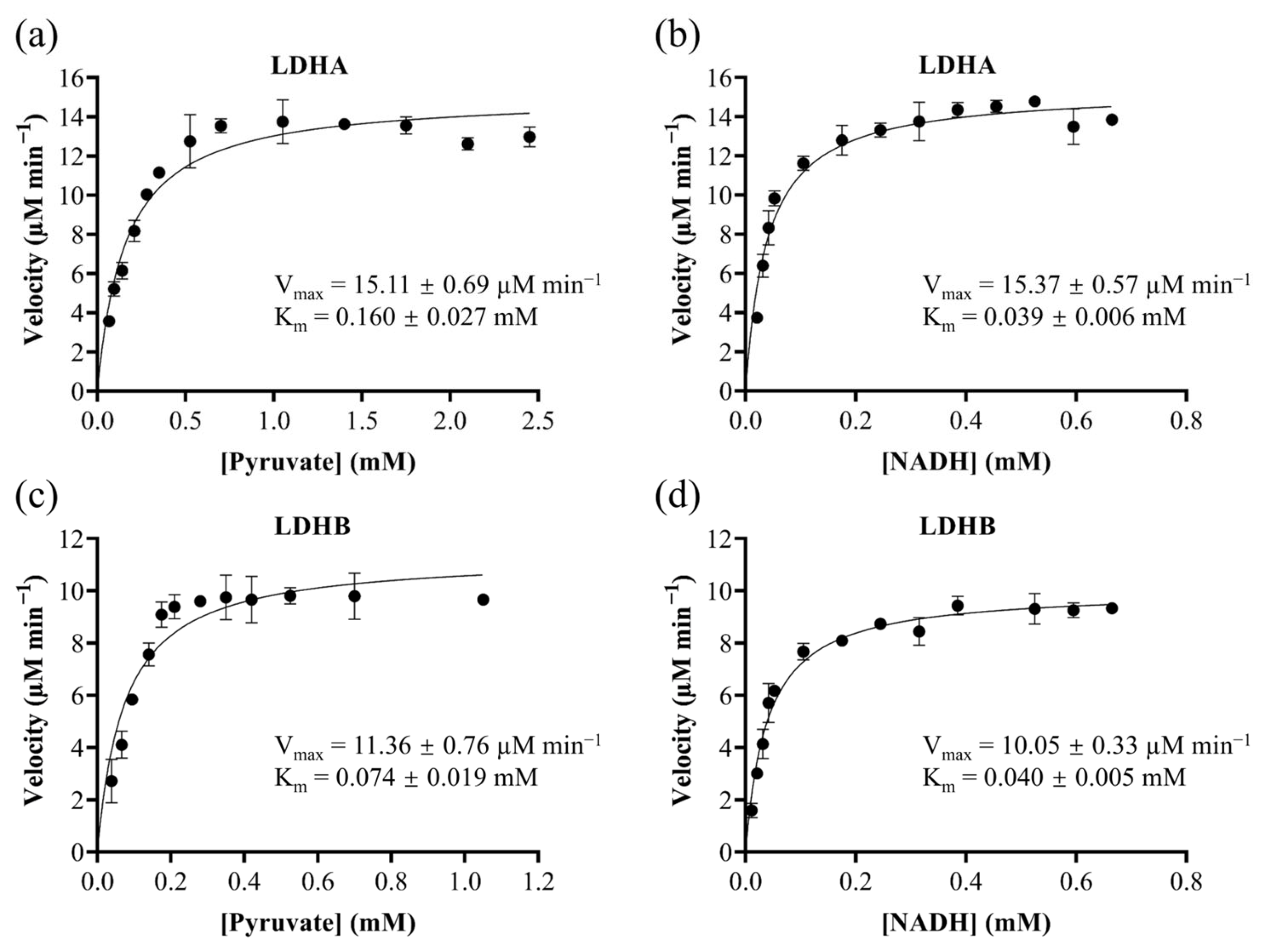
Figure 1. Michaelis-Menten kinetics of recombinant LDHA and LDHB. Kinetics of LDHA toward pyruvate (a) and NADH (b); kinetics of LDHB toward pyruvate (c) and NADH (d).
Molecules 30 04199 g001
Figure 2. Dose-response curves of gossypol against LDHA (a) and LDHB (b).
Molecules 30 04199 g002
Figure 3. Enzymatic progress curves of R. crenulata extract against LDHA and LDHB. Progress curves of NADH consumption demonstrate significant, time-dependent inhibition of both isoforms (p < 0.0001) relative to the DMSO control.
Molecules 30 04199 g003
Figure 4. BPCs of R. crenulata extract in (a) negative and (b) positive ionization modes.
Molecules 30 04199 g004
Figure 5. Structures and inhibitory activity of five bioactive components from R. crenulate. (a) Chemical structures of the five bioactive components. (b) Inhibitory effects of the five bioactive components against LDHA and LDHB.
Molecules 30 04199 g005
Figure 6. Dose-response curves for quercetin, luteolin, kaempferol, and epicatechin gallate against LDHA (a,c,e,g) and for quercetin, luteolin, and kaempferol against LDHB (b,d,f); (h) comparison of their IC50 values.
Molecules 30 04199 g006
Figure 7. Binding modes of ellagic acid with LDHA (a) and LDHB (b).
Molecules 30 04199 g007
Figure 8. Anti-proliferative activity of five inhibitory compounds against MDA-MB-231 and HepG2 cell lines. The IC50 values were consistently lower in MDA-MB-231 cells (blue) than in HepG2 cells (green), demonstrating greater potency against the breast cancer cell line.
Molecules 30 04199 g008
Table 1. 36 identified bioactive components in R. crenulata.
No.RT
(min)
Precursor IonMS/MS (Da)FormulaIdentificationReference
Observed Mass (Da)Error (ppm)
11.349191.0194 [M−H]1.7(−): 129.0188, 111.0086, 87.0084, 85.0292, 67.0187, 57.0346C6H8O7Citric acidstandard
22.19169.0145 [M−H]/171.0291 [M+H]+−1.49/−1.77(−): 125.0243, 79.0191, 51.0244; (+): 153.0181, 127.0389, 125.0229, 109.0285, 107.0126, 81.0335, 53.0388C7H6O5Gallic acidstandard
34.296153.0190 [M−H]/155.0341 [M+H]+2.16/−0.75(−): 108.0212, 109.0294, 110.0325, 91.0189, 81.0345, 53.0398; (+): 137.0227, 93.0335, 111.0440, 81.0333, 65.0387, 53.0383C7H6O4Protocatechuic acidstandard
44.596313.0924 [M−H]1.56(−): 151.0394, 161.0448, 107.0501C14H18O82-(β-D-glucopyranosyloxy)-1-(4-hydroxyphenyl)ethanone[46]
55.570461.1660 [M−H]0.97(−): 299.1124, 179.0554, 119.0345, 101.0243, 89.0244C20H30O122-(4-hydroxyphenyl)ethyl-6-0-β-D-glucopyranosyl-β-D-glucopyranoside[47]
65.927345.1199 [M+HCOO]−/318.1556 [M+NH4]+−2.65/−2.9(−): 299.1132, 89.0245, 119.0496, 179.0559, 59.0141, 71.0138, 101.0244, 113.0240; (+):69.0335, 85.0285, 121.0649, 145.0496, 187.0751, 205.0856, 229.0858, 247.0961, 265.1070C14H20O7Salidrosidestandard
76.169293.1252 [M+HCOO]/271.1156 [M+Na]+−4.07/−1.57(−): 247.1174, 161.0448, 101.0234, 71.0134, 59.0147; (+): 201.0367, 203.0514C11H20O6Crenulatinstandard
86.468137.0597 [M−H]7.99(−): 119.0501, 106.0420C8H10O2Tyrosolstandard
97.667322.1866 [M+NH4]+−1.88(+): 125.0962, 107.0856C14H24O7Creoside I or Isomer[48]
108.050179.0348 [M−H]1.01(−): 134.0368, 135.0445, 107.0501C9H8O4caffeic acidstandard
118.100322.1866 [M+NH4]+−1.88(+): 125.0962, 107.0856C14H24O7Creoside I or Isomer[48]
128.333577.1347 [M−H]0.78(−): 407.0768, 289.0705, 125.0241C30H26O12Procyanidin B3[49]
139.073289.0715 [M−H]/291.0868 [M+H]+0.9/−1.67(−): 245.0811, 221.0818, 203.0702, 151.0391, 123.0446, 109.0290; (+): 207.0648, 165.0545, 147.0438, 139.0387, 123.0440, 111.0437C15H14O6(−)-epicatechinstandard
149.615635.0885 [M−H]0.77(−): 483.0785, 423.0574, 313.0560, 169.0139, 125.0242C27H24O18trigalloyl-β-D-glucopyranose/isomer[50]
1510.239729.1455 [M−H]0.83(−): 577.1325, 451.1040, 407.0774, 289.0718, 169.0143, 125.0248C37H30O16catechin-(4α-8)-catechin-3′0-gallate/epicatechin-(4β-8)epicatechingallate[49]
1610.489609.1461 [M−H]0.01(−): 446.0854, 283.0242, 151.0031C27H30O16kaempferol 3,7-digalactoside/Isomer[50]
1710.722399.1296 [M−H]0.18(−): 329.0515, 271.0453, 169.0141, 125.0241C18H24O103-O-galloyl quinic acid butyl ester/Isomer[51]
1811.201787.0993/[M−H]0.82(−): 635.0906, 617.0788, 465.0676, 169.0139, 295.0453C34H28O22tetragalloyl-β-D-glucopyranose/isomer[50]
1911.604881.1566/[M−H]0.53(−): 729.1447, 711.1360, 577.1328, 559.1244, 541.1143, 407.0768, 289.0715, 169.0136C44H34O20epicatechingallate-(4β-8)-epicatechingallate[50]
2011.962300.9989/[M−H]0.3(−):283.9956, 257.0090, 245.0088, 228.0048, 201.0182, 185.0236, 161.0231, 145.0289, 129.0347, 117.0342C14H6O8Ellagic acidstandard
2111.970441.0825 [M−H]/443.0979 [M+H]+0.5/−1.42(−): 331.0459, 289.0708, 271.0605, 245.0817, 169.0140, 125.0243; (+): 291.0857, 273.0755, 207.0646, 165.0548, 153.0179, 139.0390, 123.0442, 111.0437C22H18O10epicatechin gallatestandard
2213.702447.0931 [M−H]/449.1085 [M+H]+0.41/−1.48(−): 284.0318, 255.0292, 227.0339; (+): 287.0553, 153.0174, 85.0282C21H20O11Astragalinstandard
2314.084187.0976 [M−H]−0.09(−): 169.0872, 143.1072, 125.0969, 102.9481, 97.0658, 57.0341C9H16O4Azelaic acidstandard
2415.025881.1566 [M−H]0.53(−): 729.1447, 711.1348, 577.0993, 559.1236, 541.1141, 433.0921, 407.0768C44H34O20epicatechingallate-(4β-8)-epicatechingallate[50]
2515.158491.2131 [M+HCOO]0.67(−): 313.1649, 161.0460, 149.0454, 131.0346, 101.0243C21H34O10sachaloside II[44]
2615.383431.0983 [M−H]/433.1136 [M+H]+0.16/−1.57(−): 284.0322, 285.0390, 255.0289, 227.0339; (+): 287.0550, 147.0649, 129.0546, 111.0434, 85.0282, 71.0492, 57.0333C21H20O10Afzelinstandard
2715.708609.1459 [M−H]/611.1613 [M+H]+0.34/−1.05(−): 301.0351; (+): 449.1076, 303.0502, 145.0489, 127.0381, 97.0282, 85.0281C27H30O16rhodiosinstandard
2816.182447.0933 [M−H]/449.1086 [M+H]+−0.03/−1.7(−): 301.0346, 255.0289, 229.0505, 166.9975; (+): 303.0503, 169.0124, 121.0276C21H20O11Rhodioninstandard
2917.422285.0403 [M−H]/287.0555 [M+H]+0.56/−1.7(−): 199.0391, 133.0294; (+): 269.0429, 241.0494, 213.0539, 153.0179, 135.0438, 117.0328, 89.0389, 67.0179C15H10O6luteolinstandard
3017.439301.0350 [M−H]/303.0502 [M+H]+1.25/−0.9(−): 178.9978, 151.0034, 121.0288, 107.0136, 83.0136, 65.0034; (+): 257.0453, 229.0498, 201.0548, 153.0182, 137.0238C15H10O7Quercetinstandard
3117.572431.0984 [M−H]−0.07(−): 285.0394, 257.0457, 151.0039C21H20O10kaempferol-7-0-α-L-rhamnoside[52]
3217.788469.2291 [M+HCOO]−0.12(−): 423.2232, 291.1811, 161.0430, 101.0243C19H36O10rhodiooctanoside[44]
3319.828285.0410 [M−H]/287.0554 [M+H]+−1.88/−1.35(−): 257.0431, 239.0342, 211.0396, 185.0606, 171.0445, 159.0449, 107.0137, 93.0347, 63.0245; (+): 258.0527, 213.0547, 185.0595, 153.0182, 137.0231, 121.0281, 93.0335, 68.9973C15H10O6kaempferolstandard
3422.733319.1188 [M−H]/321.1338 [M+H]+−0.28/−1.67(−): 287.0915, 275.1281, 245.0814, 205.0503, 191.0348, 179.0340, 161.0594, 148.0522, 135.0427; (+): 303.1229, 275.1275, 223.0597, 207.0650, 177.0542, 159.0442C17H20O6Mycophenolic acidstandard
3523.165479.0986 [M−H]−0.48(−): 299.0193, 271.0246, 165.9900C25H20O10crenulatin A/crenulatin B/rhodiolin[51]
3628.085295.2278 [M−H]0.23(−): 277.2168, 195.1385, 59.0137C18H32O39-hydroxy-10,12-octadecadienoic acidGNPS
Table 2. Binding energies of five inhibitory compounds against LDHA and LDHB.
ComponentBinding Energy (kcal/mol)
LDHA (1I10)LDHB (7DBK)
Quercetin−8.7−6.8
Luteolin−8.8−7.1
Kaempferol−8.6−7.2
Epicatechin gallate−7.7−7.0
Ellagic acid−8.5−6.8
Disclaimer/Publisher’s Note: The statements, opinions and data contained in all publications are solely those of the individual author(s) and contributor(s) and not of MDPI and/or the editor(s). MDPI and/or the editor(s) disclaim responsibility for any injury to people or property resulting from any ideas, methods, instructions or products referred to in the content.