Article

Targeted Enrichment and Characterization of Diester Diterpenoid Alkaloids in Aconitum Herbs Using Gas–Liquid Microextraction Coupled with High-Resolution Mass Spectrometry

1
State Key Laboratory for Quality Ensurance and Sustainable Use of Dao-di Herbs, National Resource Center for Chinese Materia Medica, China Academy of Chinese Medical Sciences, Beijing 100700, China
2
Graduate School, Tianjin University of Traditional Chinese Medicine, Tianjin 301617, China
3
Shandong Center for Food and Drug Evaluation & Inspection, Jinan 250014, China
*
Authors to whom correspondence should be addressed.
Molecules 2025, 30(19), 4029; https://doi.org/10.3390/molecules30194029
Submission received: 1 September 2025 / Revised: 4 October 2025 / Accepted: 6 October 2025 / Published: 9 October 2025
(This article belongs to the Section Analytical Chemistry)

Abstract

Diterpenoid diester alkaloids (DDAs) are the primary toxic constituents in aconite herbs, while also being the key pharmacologically active components. Consequently, establishing rapid enrichment and highly sensitive analytical methods for DDAs is of critical importance. Herein, we developed and constructed a gas–liquid microextraction (GLME) device, which enables the rapid and selective enrichment of DDAs from complex matrices. The enriched extract can be directly analyzed by high-resolution Orbitrap mass spectrometry without requiring any further pretreatment. A comparative analysis of six commonly used Aconitum herbs medicines and their processed derivatives was conducted. Notably, GLME enhanced the mass spectrometric signals of DDAs by 3–4 orders of magnitude, facilitating the identification of 27 alkaloids, including 3 potential new compounds (15-Ethyl-13-deoxyanhydroaconitine, 13-Hydroxy-15-ethylanhydroaconitine and 8-eicosapentaenoic-benzoylmesaconine). It was found that among the tested samples, the DDAs response intensity of raw Caowu was the highest, and the DDA signals decreased significantly after processing. This result chemically validates the detoxification efficacy of traditional methods. The proposed GLME-MS strategy has the advantages of being green, economical, easy to operate, and highly selective (>1000-fold), which provides a technical reference for the rapid detection, safety assessment, and quality control of Aconitum herbs.

1. Introduction

Aconite-based traditional Chinese medicine (TCM) has a long history of application within China’s TCM practice [1,2,3]. Representative herbs such as Chuanwu (the mother root of Aconitum carmichaelii), Caowu (the root tuber of Aconitum kusnezoffii), and Fuzi (the processed daughter root of Aconitum carmichaelii) are widely used clinically [4]. Modern pharmacological studies have shown that these herbs possess significant pharmacological activities, including anti-inflammatory, analgesic, and cardiotonic effects, among others [5,6,7,8]. Their pharmacodynamic material basis is primarily attributed to various alkaloid components [9]. Aconitum alkaloids are primarily classified into diester, monoester, and non-ester aconitine types based on their structural characteristics. Among these, diester diterpenoid alkaloids (DDAs) can be further categorized into acetates and fatty acid esters. Acetates represent the most common and typical DDAs, whereas fatty acid esters are often termed lipo-alkaloids. Toxicity varies significantly among these classes, with DDAs demonstrating the most acute toxicity. Their toxicity is estimated to be 200–500 times greater than that of monoester diterpenoid alkaloids and 2000–4000 times greater than that of non-ester alkaloids [10]. This high toxicity is closely related to the ester bonds at the C8 and C14 positions of their molecular structures [11]. Although clinically processed Aconitum herbs are commonly used to reduce toxicity, existing processing techniques still struggle to completely eliminate DDA residues. Notably, even trace amounts of DDAs can cause severe toxic reactions that are potentially life-threatening [12]. However, current pharmacopeia standards only use the total content of aconitine, mesaconitine, and hypaconitine as quality control indicators, failing to cover other highly toxic DDAs like yunaconitine. In fact, the wide variety of DDAs with differing toxicities means that relying on only a few components for evaluation may lead to an underestimation of potential safety risks. Therefore, establishing a rapid and sensitive method for the targeted enrichment and detection analysis of DDAs in aconite-based TCM is crucial for improving clinical medication safety standards and enhancing the quality control level of these herbs.
The efficient separation, enrichment, and detection of DDAs in aconite drugs have consistently posed a key technical challenge in the field of traditional Chinese medicine quality control. Current enrichment and pretreatment methods for these toxic components primarily rely on techniques such as solid-phase extraction (SPE) [13] and liquid–liquid extraction (LLE) [14]. SPE operates on interactions between liquid and stationary phases, achieving selective separation through specific hydrophobic, polar, and/or ion-exchange properties between the target analytes and the sorbent [15]. However, it is limited by cumbersome operational steps, time consumption, and high consumable costs [16]. Conversely, LLE separates and enriches analytes based on their differential partition coefficients between two immiscible liquids, yet it suffers from high organic solvent consumption and poor selectivity [17]. Recently, GLME technology [18,19] has garnered significant interest owing to its exceptional selective enrichment capabilities and cost-effectiveness. This technique integrates sample extraction, purification, and enrichment into a single efficient pretreatment step, markedly enhancing detection sensitivity through the selective concentration of trace target substances [20]. Its distinctive advantages include superior impurity removal capacity, effectively eliminating interferents such as proteins, polysaccharides, and salts [21]; operational simplicity; and economic–environmental benefits, as the process requires only water and an auxiliary gas [22,23], thereby substantially reducing organic solvent consumption.
Preliminary studies by our group revealed that GLME demonstrates a specificity enrichment factor exceeding 1000 for toxic DDAs, with negligible enrichment observed for monoester diterpenoid and non-ester alkaloids [20,24]. Building on this foundation, the present study developed a novel analytical approach by coupling GLME with direct infusion electrospray ionization mass spectrometry (DI-ESI-MS). Six representative Aconitum herbs, encompassing both raw and processed forms of Fuzi, Chuanwu, and Caowu, were selected for investigation. This established an efficient and targeted analytical strategy for the profiling of toxic DDAs in Aconitum herbs. This method successfully achieved the targeted enrichment and highly sensitive detection of numerous trace DDAs. A total of 27 alkaloids were identified, comprising 2 potentially novel traditional DDAs and 1 lipo-alkaloid. The remaining compounds included twelve known DDAs, ten lipo-alkaloids, and two other alkaloids. A systematic analysis was conducted to elucidate the distribution patterns and potential risks associated with these toxic components across the different herbs. This work provides a reliable technical framework that supports the development, utilization, and enhanced quality control of Aconitum-based TCMs and their toxic DDAs.

2. Results and Discussion

2.1. Construction and Optimization Conditions of GLME–Mass Spectrometry Device

The main structure of the GLME device comprised a quartz glass tube (i.d. 25 mm; length 1020 mm) filled with the sample solution for analysis. A constant-temperature heating tape was wrapped around the external wall of the tube to maintain a stable temperature. An aperture (i.d. 7 mm) was fabricated at the bottom of the tube and fitted with a porous membrane (i.d. 100 μm). This membrane was connected to a nitrogen gas cylinder to generate micro-bubbles. The nitrogen flow rate was precisely controlled using a gas flow meter. A glass coverslip (24 mm × 32 mm) was positioned at an angle at the top of the quartz tube to collect micro-droplets generated by bursting bubbles. The device has the advantages of being highly efficient, economical, simple, and easy to operation. The use of green, cheap nitrogen and ultrapure water as the extraction carrier gas and solvent minimizes the chemical pollution of the environment or samples by traditional pretreatment methods. In addition, complex matrix solutions such as herbal extracts can be directly analyzed by DI-ESI-MS without pretreatment such as filtration and centrifugation after GLME (see the analytical workflow in Figure 1).
Subsequently, the developed GLME technique was applied to enrich trace DDAs in six distinct Aconitum materials. Prior to direct DI-ESI-MS analysis, key mass spectrometric parameters were systematically optimized to achieve optimal detection sensitivity, using the signal intensity of the target analytes as the indicator (Figure 2A). The results demonstrated that the ion spray voltage considerably influenced the signal intensity. As shown in Figure 2B, the optimal peak response was observed at 3.5 kV (NL = 1.94 × 10⁸), with deviations from this value resulting in signal attenuation. For the sheath gas, the optimal flow rate was 10 arbitrary units (arb) (NL = 2.36 × 10⁸), with further increases leading to a decline in signal intensity (Figure 2C). Optimization of the auxiliary gas flow rate yielded the highest signal intensity at 7 arb (NL = 2.51 × 10⁸), representing a 10.6% enhancement over the intensity at 6 arb (NL = 2.27 × 10⁸). However, exceeding this optimal flow rate resulted in signal reduction (Figure 2D). The ion transfer tube temperature also exerted a notable effect. The signal intensity increased with temperature from 300 °C to 330 °C, reaching a maximum (NL = 3.32 × 10⁸) at the latter temperature (Figure 2E). Finally, optimization of the sample flow rate revealed a relatively stable signal within the 2–4 μL/min range (NL = 2.83 × 10⁸–3.16 × 10⁸), peaking at 4 μL/min before undergoing a sharp decline at higher flow rates (Figure 2F).

2.2. Difference Analysis of Mass Spectrometry Before/After GLME

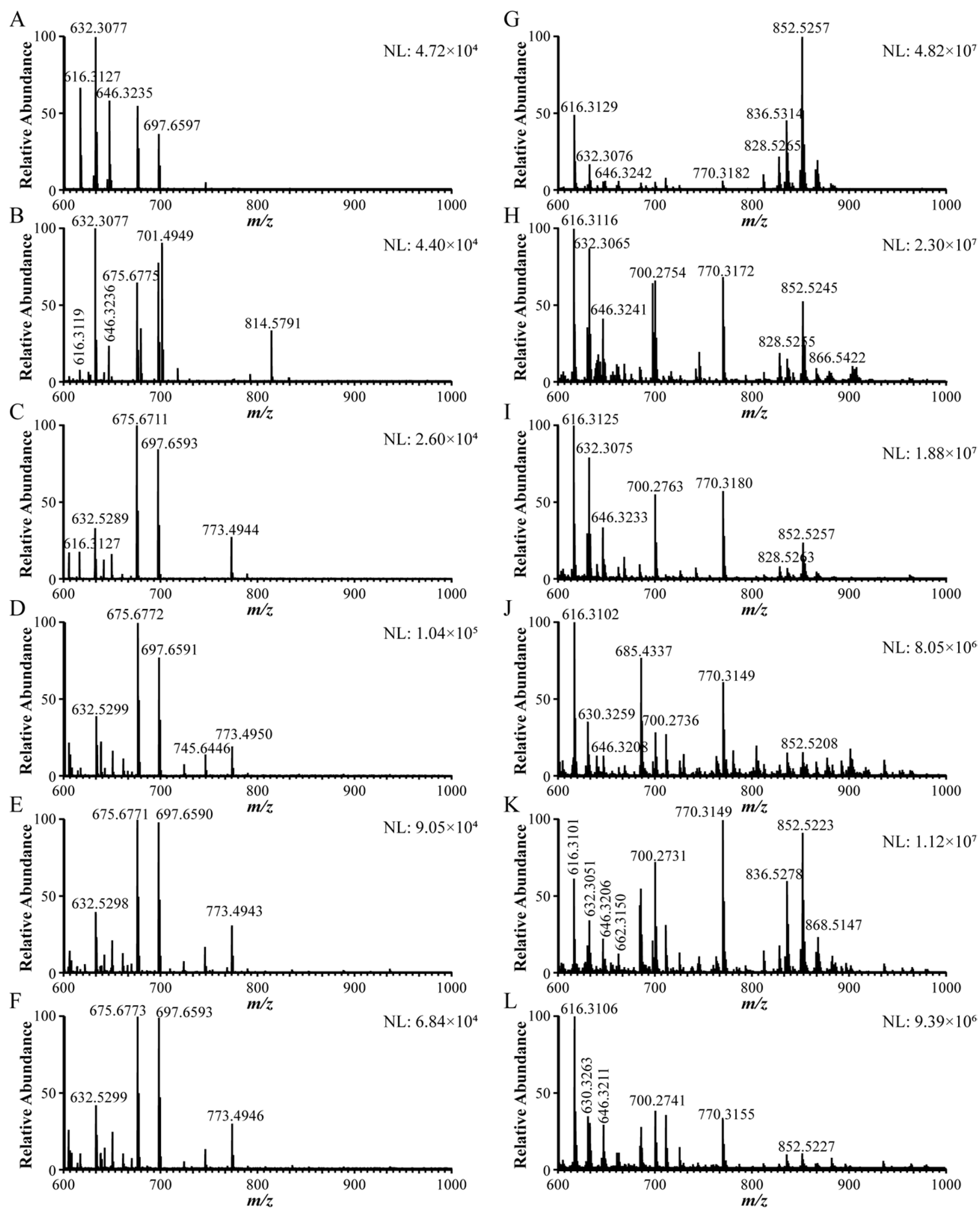
Under the optimized MS conditions, the mass spectrometric fingerprints of Fuzi, Chuanwu, Caowu, and their processed products were compared before and after GLME treatment (Figure 3). The results demonstrated that GLME exhibited remarkable selectivity and enrichment capability for toxic DDAs within complex sample matrices. As shown in Figure 3A–F, the unenriched raw samples displayed complex background interference within the m/z 600–700 range (all signal intensities <1.04 × 10⁵) after blank subtraction, which completely obscured the characteristic peaks of the target alkaloids. In contrast, the GLME-treated samples (Figure 3G–L) showed a dramatic simplification of the spectra. Non-specific signals (e.g., at m/z 675, 697, 745, and 773) were effectively eliminated, whereas the [M+H]+ ions of target compounds, such as aconitine, became distinctly visible with significantly enhanced intensities (>106 to 107). This stark contrast conclusively demonstrates the high selectivity and powerful enrichment capacity of GLME for trace DDAs in complex matrices. Table S1 provides a detailed comparison of the MS response intensities for three highly toxic DDAs (hypaconitine, mesaconitine, and aconitine) across the six Aconitum herbs before and after GLME. The signals for the target compounds were enhanced by 2 to 4 orders of magnitude following GLME; for instance, the signal for hypaconitine in Caowu increased by approximately 7800-fold. Notably, the signal for aconitine, which was very weak or undetectable prior to enrichment, was dramatically enhanced after enrichment, elevating its concentration from undetectable levels to, for example, 2.61 × 10⁶ in processed Chuanwu. Collectively, these findings robustly validate the highly selective enrichment capability of GLME for trace-level, highly toxic DDAs in Aconitum herbal medicines.
Currently, the mechanism of selective enrichment and background interference removal of GLME can be mainly attributed to the surface activity-dominated molecular competition mechanism [25,26]. Specifically, during the bubble rising phase, the enriched components undergo dynamic distribution at the gas–liquid interface. In this process, organic molecules with high surface activity, due to their strong interfacial affinity, preferentially adsorb onto the bubble surface, thereby significantly enriching the interfacial phase; meanwhile, substances with low surface activity (such as most inorganic salt ions) are excluded in the interfacial competition, gradually moving away from the interfacial area and re-entering the solution, ultimately achieving efficient exclusion of background interferences [27]. In addition, based on the previous work of our research group [20], it was found that bubbles had directional and efficient enrichment of DDAs, but had almost no enrichment effect on monoester and non-ester aconitines, indicating that their enrichment tendency may also be related to the polarity of the compounds.

2.3. Identification of Chemical Constituents

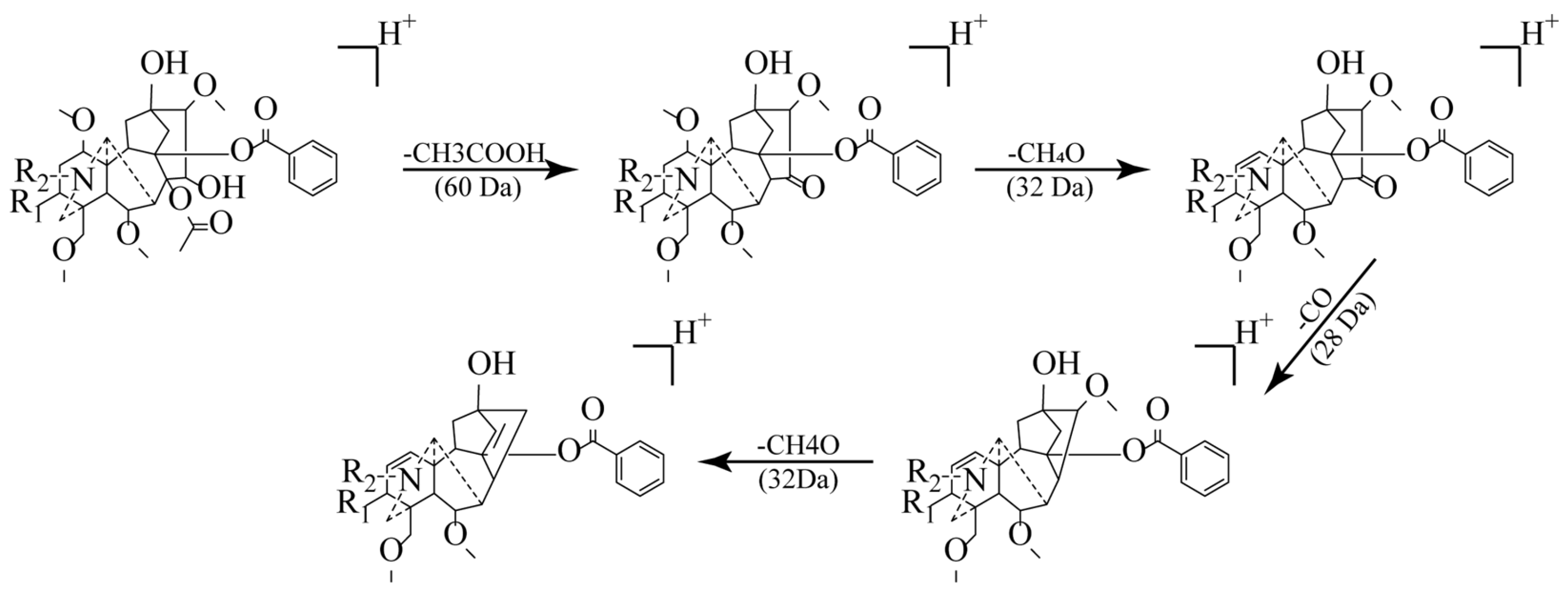
Utilizing the GLME-enriched samples, characteristic ion peaks within the m/z 600–1000 range were systematically identified via an MS2 scan mode using a high-resolution mass spectrometer (Table 1). A total of 27 compounds were successfully identified based on accurate mass measurements (mass error < 5 ppm) and the interpretation of characteristic MS/MS fragmentation patterns. The identified compounds comprised twelve known DDAs (including key toxic constituents, e.g., hypaconitine, mesaconitine, and aconitine), two potentially novel DDAs, eleven lipo-alkaloids, and two other alkaloids. During structural elucidation, the DDAs exhibited characteristic fragmentation pathways (Figure 4): initially, the loss of acetic acid (CH3COOH, 60 Da) to yield the distinctive [M+H-60]+ ion, followed by subsequent losses of methanol (CH3OH, 32 Da) and carbon monoxide (CO, 28 Da), generating fragment ions such as [M+H-60-32]+ and [M+H-60-32-28]+ [22,23]. The structures of the 27 compounds are shown in Figure S1.
High-resolution mass spectrometry (HRMS) analysis of compound 1 revealed a quasi-molecular ion peak at m/z 600.3181 [M+H]+. The MS/MS spectrum exhibited characteristic fragment ions at m/z 540.2971 (corresponding to [M+H-C2H4O2]+, loss of acetic acid), 522.2844 ([M+H-C2H4O2-H2O]+), and 490.2584 ([M+H-C2H4O2-CH4O-H2O]+). Based on these fragmentation patterns and comparison with literature data [28,29], compound 1 was identified as delphinine. The [M+H]+ ions for compounds 4, 6, and 8 were observed at m/z 616.3132, 632.3082, and 646.3242, respectively. The MS/MS spectra of all three compounds displayed analogous fragmentation pathways: initial loss of acetic acid (C2H4O2, 60 Da) to form the [M+H-60]+ ion, followed by successive losses of 32 Da (CH3OH) and 28 Da (CO), yielding the [M+H-60-32]+ and [M+H-60-32-28]+ fragments, respectively. Subsequent direct comparison of the sample MS/MS spectra with those of authentic standards (Figure 5) confirmed identical fragmentation patterns and excellent agreement in fragment ion masses. Consequently, compounds 4, 6, and 8 were unambiguously identified as hypaconitine [30,31], mesaconitine [32], and aconitine [33], respectively. Compound 5 exhibited a quasi-molecular ion peak at m/z 630.3287 [M+H]+. Its MS/MS spectrum showed a fragmentation pattern analogous to the DDAs described above, generating characteristic ions at m/z 570.3072 ([M+H-60]+), 538.2811 ([M+H-60-32]+), and 510.2863 ([M+H-60-32-28]+). Based on comparison with literature data, compound 5 was characterized as 3-deoxyaconitine [34,35]. The MS/MS spectra of 11 known alkaloids identified by reference and online databases can be seen in Figure S2.
Compounds 13 and 14 exhibited a quasi-molecular ion peak at m/z 624.3541 and 640.3494, respectively (Figure 6A,B). Their MS/MS spectra displayed characteristic fragment ions corresponding to the loss of acetic acid at m/z 564.3335 and 580.3279 ([M+H-60]+). This observed fragmentation behavior is highly characteristic of DDAs and aligns with patterns previously reported [36]. Based on the established fragmentation mechanisms of DDAs [37], this neutral loss of 60 Da is rationalized by the elimination of acetic acid, likely involving the C8 acetoxyl group and a proton from the C15 position. Further fragmentation yielded successive losses consistent with methanol (32 Da) and carbon monoxide (28 Da), generating ions at [M+H-60-32]+ and [M+H-60-32-28]+, followed by an additional loss suggestive of a second methanol molecule ([M+H-60-32-28-32]+). These collective fragmentation patterns strongly support the classification of compounds 13 and 14 as diester diterpenoid alkaloids. A notable observation was the exact mass difference of 12.03 Da between compound 14 and compound 12, which corresponds precisely to the mass difference expected for the replacement of a hydroxyl group by an ethyl group (12.03 Da). This suggests that compound 14 could be an ethyl-substituted analogue of compound 12. Extensive searching of the literature and mass spectral databases (e.g., PubChem, HMDB) yielded no matches, preliminarily indicating that compounds 13 and 14 may be previously unreported novel DDAs. However, MS analysis provides only preliminary identification. Subsequent work will therefore focus on the targeted isolation of compounds 13 and 14, followed by structural elucidation via techniques such as NMR spectroscopy and toxicity evaluation, to confirm their chemical structures and biological activities.
Compounds 1727 were tentatively characterized as lipo-alkaloids. Their MS/MS spectra were dominated by neutral losses characteristic of fatty acid moieties. As summarized in Table 1, the predominant neutral losses observed corresponded to palmitic acid, linolenic acid, linoleic acid, and eicosapentaenoic acid. Notably, based on the literature and database comparisons, compound 27 is proposed to be a potentially novel lipo-alkaloid. It exhibited a protonated molecule [M+H]+ at m/z 874.5108 (Figure 6C). The MS/MS spectrum acquired in scan mode revealed a base peak at m/z 572.2865, resulting from a neutral loss of 302.2243 Da. This mass loss corresponds to the exact mass of eicosapentaenoic acid. Although lipo-alkaloids are typically present at low abundance [33], GLME enrichment dramatically enhanced their MS response, thereby providing a robust foundation for the detection and structural characterization of these trace constituents. The MS/MS spectra of 11 known lipid alkaloids identified by references and online databases can be seen in Figure S3. The follow-up study will carry out the targeted separation of compound 27 and investigate the impact of its unique fatty acid chain on the toxicity and activity of the compound.
Table 1. The mass spectrometry data of the 27 main compounds [M+H]+ identified in 6 types of Aconitum traditional Chinese medicines.
TypeNo.Formulam/zError (ppm)MS/MSCompound NameReference
DDAs1C33H45NO9600.3181−2.3540.2971, 522.2844, 490.2584, 480.2760, 458.2321, 448.2493Delphinine[28]
2C32H43NO10602.2963−0.7542.2750, 510.2487, 482.2559, 450.2281N-demethylhypaconitine[38]
3C34H47NO9614.3335−1.8554.3127, 522.2862, 494.2910, 462.2650Chasmaconitine[38]
4C33H45NO10616.3132−2.6556.2919, 524.2656, 496.2707, 464.2443Hypaconitine *[30]
5C34H47NO10630.3287−2.2570.3072, 538.2811, 510.2863, 478.25963-Deoxyaconitine[35]
6C33H45NO11632.3082−2.7572.2867, 540.2607, 512.2655, 480.2393Mesaconitine *[30,32]
7C35H49NO10644.3447−2.8584.2872, 552.2608, 524.2659, 492.2394Crassicauline A[32]
8C34H47NO11646.3242−3.1586.3019, 554.2763, 526.2813, 494.2551Aconitine *[33,39]
9C33H45NO12648.3026−1.7588.2816, 556.2555, 528.2607, 496.2358Beiwutine[35]
10C35H49NO11660.3391−1.9600.3167, 568.2913, 540.2607, 508.2346Yunaconitine[40]
11C34H47NO12662.3187−2.4602.2976, 570.2712, 542.2758, 510.249910-Hydroxy aconitine[39]
12C34H45NO10628.31041.9568.2889, 536.2631, 508.2680, 476.2421Anhydroaconitine[41]
13C36H49NO8624.3541−1.6564.3349, 532.3078, 504.2972, 472.270515-Ethyl-13-deoxyanhydroaconitine -
14C36H49NO9640.3494−2.2580.3279, 548.3018, 520.3072, 488.280513-Hydroxy-15-ethylanhydroaconitine-
Other15C39H41NO11700.2765−1.9640.2556, 578.2397Trifoliolasine E[39,42]
16C43H47NO12770.3180−1.4648.2811, 500.2074, 378.1707, 318.1491(-)-1β,11a-diacetoxy-2α,13α-dibenzoyloxy-7β-hydroxy-15α-isobutanoyloxy-N-methyl-N,19-secohetisan-19-al[39]
lipo-alkaloids17C48H75NO10826.5501−4.6570.3065, 538,2805, 510.28568-palmitic-benzoyldeoxyaconitine[43]
18C48H76NO10828.5258−0.2572.2861, 540.2601, 512.26488-palmitic-benzoylmesaconine[43]
19C47H73NO11834.5154−0.4556.2901, 524.2646, 496.27038-linolenic-benzoylhypaconine[43]
20C49H71NO10836.5308−0.1556.2911, 524.2647, 496.27008-linoleic-benzoylhypaconine[44]
21C49H73NO10842.5416−0.4586.3015, 554.2753, 526.28078-palmitic-benzoylaconine[43]
22C48H75NO11848.5314−0.8570.3069, 538.2806, 510.28578-linolenic-benzoyldeoxyaconitine[43]
23C50H73NO10850.5478−1.8570.3065, 538.2806, 510.28578-linoleic-benzoyldeoxyaconitine[43]
24C50H75NO10852.52500.7572.2859, 540.2600, 512.26508-palmitic-benzoylmesaconine[44]
25C49H73NO11866.5428−1.8586.3016, 554.2753, 526.28058-linoleic-benzoylaconine[44]
26C47H73NO10812.5318−1.4556.2913, 524.2653, 496.27018-palmitic-benzoylaconine[44]
27C51H72NO11874.5108−1.0572.2865, 540.2606, 512.26548-eicosapentaenoic-benzoylmesaconine-
* Compared with reference standards.

2.4. Difference Analysis of Six Kinds of Aconitum Herbs

2.4.1. DDAs

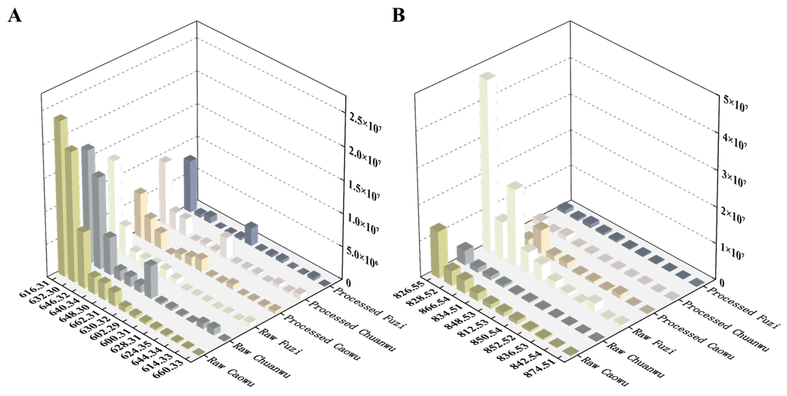
DDAs constitute the primary toxic and pharmacologically active components in Aconitum herbs [45]. A comparison of MS response intensities (Figure 7) revealed substantial variations in DDA levels across the different herbs, with raw Caowu exhibiting a significantly higher response than the other samples. This distribution pattern aligns with the reported order of toxicity for these herbs [46]. As shown in Figure 6A, the processed Chuanwu, processed Caowu, and processed Fuzi exhibited a substantial decrease in DDA response intensity compared to their raw counterparts, with an average reduction of approximately 65%. This confirms the definitive detoxification effect of the traditional processing protocol. Furthermore, the DDA composition was notably altered by processing. While all three raw herbs contained 14 detectable DDAs, the versions showed a reduction: processed Caowu and processed Chuanwu contained 13 and processed Fuzi contained only 11. Despite this drastic decline in abundance, residual DDAs remained detectable at significant levels in the processed products. This finding carries significant implications for clinical practice: the use of processed products still mandates strict adherence to the traditional “decocting first and for a prolonged duration” (>1 h) protocol [47,48], complemented by necessary soaking procedures, to mitigate the potential risk posed by residual toxicity.

2.4.2. Lipo-Alkaloids

Lipo-alkaloids represent a distinct class of diester diterpenoid alkaloid derivatives, characterized by the substitution of the C8 acetyl group in typical DDAs with various long-chain fatty acids (e.g., palmitic acid, linoleic acid). This structural modification is postulated to attenuate toxicity while preserving or potentially enhancing pharmacological activity [49,50]. As shown in Figure 6B, the MS response intensities for lipo-alkaloids were markedly higher in the raw herbs (Fuzi, Caowu, Chuanwu) than in their processed counterparts. This discrepancy is likely attributable to the hydrolysis of the C8 ester bond during processing, resulting in the degradation of lipo-alkaloids and their subsequent conversion to the corresponding monoester diterpenoid or non-ester alkaloids [44], thereby accounting for their reduced abundance. Based on these findings, future studies should prioritize systematically elucidating the contribution of lipo-alkaloids to the overall pharmacodynamic effects and their interactions with DDAs; establishing comprehensive quality control standards that include specific monitoring of lipo-alkaloids; and rigorously evaluating the potential toxicity risks of these compounds.

3. Materials and Methods

3.1. Chemicals and Materials

Reference standards for aconitine, mesaconitine, and hypaconitine (Table S2) were obtained from Beijing Beiterenkang Biotechnology Co., Ltd. (Beijing, China). The ultrapure water was sourced from Guangzhou Watsons Food and Beverage Co., Ltd., in Guangzhou, China. The raw and processed products of Aconiti Lateralis Radix Praeparata (Fuzi), Aconiti Kusnezoffii Radix (Caowu), and Aconiti Radix (Chuanwu) were sourced from Jiangyou Shenqi Chinese Herbal Medicine Planting Co., Ltd. (Sichuan, China). Among them, Fuzi, Chuanwu, and Caowu were the lateral roots of Aconitum carmichaelii Debx., the mother roots of Aconitum carmichaelii Debx., and the tuberous roots of Aconitum kusnezoffii Reichb., respectively, identified by Prof. Zhilai Zhan, and all the samples were stored in the National Resource Center for Chinese Materia Medica, China Academy of Chinese Medical Sciences, with the code WT2025001–WT2025018.

3.2. Parameters of GLME Device

Following a previously optimized protocol from our group [18], 5.0 mg of each sample powder was accurately weighed and transferred into 660 mL of distilled water. Subsequently, the mixtures underwent ultrasonic extraction for 20 min to yield extracts for each of the different samples. These extracts were then processed using a self-built gas–liquid microextraction (GLME) device. Nitrogen served as the carrier gas, with its flow rate precisely controlled at 6 mL/min by a mass flow controller. The extraction temperature was maintained at 20 °C. Following GLME, the extracts were collected in 1.5 mL centrifuge tubes. Without any further pretreatment, these extracts were directly analyzed via DI-ESI-MS.

3.3. Optimization of Mass Spectrometry Conditions

A constant flow infusion system was assembled using a precision syringe (100 μL; Hamilton, Bonaduz, Switzerland) coupled to a syringe pump (LSP-01-3A; Longer Pump, Baoding, China). The extraction solution was continuously infused into an Orbitrap Exploris 240 mass spectrometer (Thermo Fisher Scientific, Waltham, MA, USA) equipped with an electrospray ionization (ESI) source for analysis. To achieve optimal ionization and signal response, key mass spectrometry parameters were optimized by monitoring the signal intensity of characteristic marker compounds. The parameters subjected to optimization included the injection pump flow rate (2–7 μL/min), the ion spray voltage (2.5–5 kV), the sheath gas (5–50 arb), the aux gas (6–11 arb), and the ion transfer tube temperature (300–350 °C).

3.4. Orbitrap MS Analysis

Mass spectrometric analysis was conducted with an Orbitrap Exploris 240 mass spectrometer (Thermo Fisher Scientific) equipped with an ESI source operated in positive ion mode (+3500 V). The ion transfer tube temperature was set to 330 °C, the m/z scan range was 600–1000 Da, and the RF lens was set to 70 [51]. Each sample was analyzed in triplicate by direct infusion via an ESI source, using a sample volume of 100 μL and a precise syringe pump flow rate of 4.0 μL/min. Data acquisition was performed using both full MS and data-dependent MS2 scan modes. The full MS resolution was set to 60,000, and the collision energy was 40 eV. All raw mass spectral data were processed using Xcalibur 4.2 software. The putative identification of MS/MS fragments was based on comparison with reference data from the literature and online databases (e.g., PubChem, HMDB) [52]. The matching was performed with a mass accuracy threshold of 5 ppm.

4. Conclusions

This study developed an integrated strategy combining GLME with mass spectrometry for the targeted enrichment and detection of DDAs in Aconitum herbs. The proposed method enables efficient and selective extraction of trace DDAs from complex matrices, permitting direct analysis via DI-ESI-MS without requiring additional sample pretreatment. More importantly, the signal intensities for trace toxic DDAs were enhanced by over 1000-fold in complex Aconitum herbs. Furthermore, this approach significantly reduces organic solvent consumption, offering notable advantages in simplicity, efficiency, cost-effectiveness, and environmental friendliness.
The results indicated that raw Caowu exhibited the highest DDA response intensity among the tested samples. Concurrently, considerable DDA signals were still detectable in the three processed products, suggesting that current processing protocols require further optimization to maximize toxicity reduction while preserving pharmacological efficacy. Furthermore, lipo-alkaloids, characterized by their lower toxicity and higher bioactivity, warrant further investigation. Future processing strategies should aim to more efficiently degrade toxic DDAs to achieve the objective of “reducing toxicity while preserving efficacy” or even “reducing toxicity while enhancing efficacy.” In summary, this study establishes an efficient, green, and economical strategy for the analysis of trace toxic DDAs in Aconitum herbs and demonstrates the potential of GLME for the selective enrichment of trace target analytes.
It should be noted that the lack of full quantitative validation of the method renders the methodology unsuitable for routine use in risk assessment. This is partly because the focus of this study was on qualitative structural elucidation and partly because commercial reference standards are difficult to obtain for most of the 27 DDAs. Therefore, if systematic quantitative analysis and accurate toxicity assessment of the observed DDAs are required, systematic phytochemical separation and comprehensive quantitative method validation are recommended first. Additionally, the direct infusion MS (DI-ESI-MS) used in this study inevitably introduces matrix interference. Non-target compounds may cause ion suppression by competing for charges with the analytes, ultimately reducing the MS signal and affecting the quantitative accuracy of the target analytes. Fortunately, this study focused on the qualitative analysis of compounds. The results indicate that the changes in relative signal intensity potentially caused by matrix interference do not significantly affect the acquisition of MS/MS data for target analytes and do not hinder structural identification. Thus, considering multiple factors such as analysis time, DI-ESI-MS was employed to characterize samples collected via GLME. Finally, current international research on GLME primarily remains at the level of phenomenological description and application, with its specific mechanisms of action still subject to debate. The 27 DDAs identified in this study as being efficiently enriched by GLME provide highly representative examples for delving into the intrinsic relationship between GLME enrichment efficiency and compound structure and physicochemical properties. This will be a very important and interesting study to carry out in future research.

Supplementary Materials

The following supporting information can be downloaded at https://www.mdpi.com/article/10.3390/molecules30194029/s1: Figure S1: The structures of 27 compounds were identified by compound standard, reference, and online compound databases; Figure S2: The MS/MS spectra of 11 known alkaloids identified by references and online databases; Figure S3: The MS/MS spectra of 10 known lipid alkaloids identified by references and online databases; Table S1: Comparison of response intensity of three main diester diterpenoid alkaloids before and after GLME; Table S2: Structures and main information properties of the 3 key compounds.

Author Contributions

Conceptualization, Y.W. and J.W.; methodology, X.L.; software, Y.W.; validation, J.W., Y.H. and X.L.; data curation, Y.W. and C.M.; writing—original draft preparation, Y.W.; writing—review and editing, Z.Q.; visualization, L.K.; supervision, Z.Q.; project administration, Z.Q.; funding acquisition, Z.Q. and L.K. All authors have read and agreed to the published version of the manuscript.

Funding

This research was funded by the Beijing Natural Science Foundation (No. 7252249), the National Natural Science Foundation of China (No. 82104380), and the Scientific and Technological Innovation Project of China Academy of Chinese Medical Sciences (Nos. CI2023E002, CI2023C039YGL).

Institutional Review Board Statement

Not applicable.

Informed Consent Statement

Not applicable.

Data Availability Statement

The original contributions presented in this study are included in the article/Supplementary Materials. Further inquiries can be directed to the corresponding authors.

Conflicts of Interest

The authors declare no conflicts of interest.

References

  1. Huang, L.; Guo, L.; Zhang, X.; Yu, L.; Sun, J. Species of Chinese materia medica resources based on the fourth national survey of Chinese materia medica resources. Sci. Tradit. Chin. Med. 2024, 49, 3409–3413. [Google Scholar]
  2. Zheng, Y.; Wei, N.; Lu, C.; Li, W.; Jia, X.; Chen, L.; Chen, R.; Chen, Z. Mechanistic insights into honey-boiled detoxification of ChuanWu: A study on alkaloid transformation and supramolecular aggregation. J. Pharm. Anal. 2025, 15, 101205. [Google Scholar]
  3. He, Y.; Wei, Z.; Xie, Y.; Yi, X.; Zeng, Y.; Li, Y.; Liu, C. Potential synergic mechanism of Wutou-Gancao herb-pair by inhibiting efflux transporter P-glycoprotein. J. Pharm. Anal. 2020, 10, 178–186. [Google Scholar]
  4. He, G.; Wang, X.; Liu, W.; Li, Y.; Shao, Y.; Liu, W.; Liang, X.; Bao, X. Chemical constituents, pharmacological effects, toxicology, processing and compatibility of Fuzi (lateral root of Aconitum carmichaelii Debx): A review. J. Ethnopharmacol. 2023, 307, 116160. [Google Scholar] [CrossRef]
  5. Fu, Y.-P.; Li, C.-Y.; Peng, X.; Zou, Y.-F.; Rise, F.; Paulsen, B.S.; Wangensteen, H.; Inngjerdingen, K.T. Polysaccharides from Aconitum carmichaelii leaves: Structure, immunomodulatory and anti-inflammatory activities. Carbohydr. Polym. 2022, 291, 119655. [Google Scholar] [CrossRef] [PubMed]
  6. Wu, Y.; Shao, S.; Guo, Q.; Xu, C.; Xia, H.; Zhang, T.; Shi, J. Aconicatisulfonines A and B, Analgesic Zwitterionic C20-Diterpenoid Alkaloids with a Rearranged Atisane Skeleton from Aconitum carmichaelii. Org. Lett. 2019, 21, 6850–6854. [Google Scholar] [CrossRef] [PubMed]
  7. Zhou, W.; Liu, H.; Qiu, L.-Z.; Yue, L.-X.; Zhang, G.-J.; Deng, H.-F.; Ni, Y.-H.; Gao, Y. Cardiac efficacy and toxicity of aconitine: A new frontier for the ancient poison. Med. Res. Rev. 2021, 41, 1798–1811. [Google Scholar] [CrossRef] [PubMed]
  8. Zhang, W.; Cai, S.; Qin, L.; Feng, Y.; Ding, M.; Luo, Z.; Shan, J.; Di, L. Alkaloids of Aconiti Lateralis Radix Praeparata inhibit growth of non-small cell lung cancer by regulating PI3K/Akt-mTOR signaling and glycolysis. Commun. Biol. 2024, 7, 1118. [Google Scholar]
  9. Qiu, Z.-D.; Wei, X.-Y.; Sun, R.-Q.; Chen, J.-L.; Tan, T.; Xu, J.-Q.; Cui, G.-H.; Chen, T.; Guo, J.; Lai, C.-J.-S.; et al. Limitation standard of toxic aconitines in Aconitum proprietary Chinese medicines using on-line extraction electrospray ionization mass spectrometry. Acta Pharm. Sin. B 2020, 10, 1511–1520. [Google Scholar] [CrossRef]
  10. Liu, S.; Li, F.; Li, Y.; Li, W.; Xu, J.; Du, H. A review of traditional and current methods used to potentially reduce toxicity of Aconitum roots in Traditional Chinese Medicine. J. Ethnopharmacol. 2017, 207, 237–250. [Google Scholar] [CrossRef]
  11. Zhao, F.; Li, J. Recent Progress in Research of Alkaloid in Aconitum Plants. Chin. J. Mod. Appl. Pharm. 2010, 27, 1177–1182. [Google Scholar]
  12. Chunbei, Z.; Shusheng, L.; Jiang, T.; Linda, Q.; Huihui, L.; Yue, Z. Poisoning Associated with Consumption of a Homemade Medicinal Liquor—Chongqing, China, 2018. Morb. Mortal. Wkly. Rep. 2022, 71, 569–573. [Google Scholar]
  13. Gong, X.; Lu, Y.; Chen, X.; Qiu, F.; Chen, Q. Determination of Diester Alkaloid Limit in Mugua Pills by SPE-HPLC. China Pharm. 2017, 26, 17–19. [Google Scholar]
  14. Liu, Z. Study on Extraction Process and Hydrolysis Reaction of Aconite Diester Diterpene Alkaloids. Master’s Thesis, Southwest University of Science and Technology, Mianyang, China, 2012. [Google Scholar]
  15. Ali, N.; Rampazzo, R.d.C.P.; Costa, A.D.T.; Krieger, M.A. Current Nucleic Acid Extraction Methods and Their Implications to Point-of-Care Diagnostics. BioMed Res. Int. 2017, 2017, 9306564. [Google Scholar] [CrossRef]
  16. Liu, P.; Zhou, J.; An, J.; Li, P. Application of turbulent flow chromatography in the analysis of biological samples. Chin. J. Chromatogr. 2010, 28, 168–174. [Google Scholar] [CrossRef]
  17. Wang, X.; Qi, W.; Zhao, X.; Lv, T.; Wang, X.; Zheng, L.; Yan, Y.; You, J. Determination of four phenolic endocrine disruptors in environmental water samples by high performance liquid chromatography-fluorescence detection using dispersive liquid-liquid microextraction coupled with derivatization. Chin. J. Chromatogr. 2014, 32, 623–628. [Google Scholar] [CrossRef]
  18. Gao, Y.; He, Q.; Guo, C.; Chen, W.; Pan, Y. Carbon Dioxide Microbubble Bursting Ionization Mass Spectrometry. Anal. Chem. 2022, 94, 17360–17364. [Google Scholar] [CrossRef]
  19. Gao, Y.; Feng, H.; Xia, B.; He, L.; Yang, C.; Zhao, L.; Pan, Y. Ultrasensitive and Green Bubbling Extraction Strategies: An Extensible Solvent-Free Re-Enrichment Approach for Ultratrace Pollutants in Aqueous Samples. Anal. Chem. 2023, 95, 13683–13689. [Google Scholar] [CrossRef]
  20. Qiu, Z.; Wei, C.; Kang, L.; Zhou, L.; Lai, C.; Li, X.; Yan, B.; Xu, J.; Wang, S.; Huang, L. Sensitive quantitation of ultra-trace toxic aconitines in complex matrices by perfusion nano-electrospray ionization mass spectrometry combined with gas-liquid microextraction. Talanta 2024, 269, 125402. [Google Scholar] [CrossRef]
  21. Mu, Z.; Wang, Y.; Guo, Y.; Jin, X.; Li, D.; Zhao, J. Determination of 16 polycyclic aromatic hydrocarbons in seaweed by ultrasound-assisted gas-liquid microextraction coupled with gas chromatography-mass spectrometry. Chin. J. Chromatogr. 2025, 43, 950–958. [Google Scholar] [CrossRef]
  22. An, S.; Ranaweera, R.; Luo, L. Harnessing bubble behaviors for developing new analytical strategies. Analyst 2020, 145, 7782–7795. [Google Scholar] [CrossRef] [PubMed]
  23. Kou, W.; Zhang, H.; Konstantin, C.; Chen, H.-W. Charged Bubble Extractive Ionization Mass Spectrometry for Protein Analysis. Chin. J. Anal. Chem. 2017, 45, 1937–1943. [Google Scholar] [CrossRef]
  24. Wei, C.; Kang, L.; Zhao, Y.; Zhou, L.; Li, X.; Wang, S.; Huang, L.; Qiu, Z. Rapid toxicity characterization of Aconitum herbal medicines using perfusion nano-electrospray ionization mass spectrometry. J. Pharm. Anal. 2024, 14, 101016. [Google Scholar] [CrossRef] [PubMed]
  25. Cochran, R.E.; Jayarathne, T.; Stone, E.A.; Grassian, V.H. Selectivity Across the Interface: A Test of Surface Activity in the Composition of Organic-Enriched Aerosols from Bubble Bursting. J. Phys. Chem. Lett. 2016, 7, 1692–1696. [Google Scholar] [CrossRef]
  26. Chingin, K.; Yan, R.; Zhong, D.; Chen, H. Enrichment of Surface-Active Compounds in Bursting Bubble Aerosols. ACS Omega 2018, 3, 8709–8717. [Google Scholar] [CrossRef]
  27. Chingin, K.; Cai, Y.; Liang, J.; Chen, H. Simultaneous Preconcentration and Desalting of Organic Solutes in Aqueous Solutions by Bubble Bursting. Anal. Chem. 2016, 88, 5033–5036. [Google Scholar] [CrossRef]
  28. Hu, R.; Zhao, J.; Qi, L.-W.; Li, P.; Jing, S.-L.; Li, H.-J. Structural characterization and identification of C19- and C20-diterpenoid alkaloids in roots of Aconitum carmichaeli by rapid-resolution liquid chromatography coupled with time-of-flight mass spectrometry. Rapid Commun. Mass Spectrom. 2009, 23, 1619–1635. [Google Scholar] [CrossRef]
  29. Yu, J.; Xia, J.; Xu, J.; Chen, S.; Zhang, Y.; Yin, F.; Fang, J.; Cai, L.; Zhang, B.; Zhan, Y.; et al. Chemical profile, anti-hepatoma activity, anti-acetylcholinesterase and antioxidant activity of aerial part of Aconitum carmichaeli Debx. Nat. Prod. Res. 2023, 37, 3884–3888. [Google Scholar] [CrossRef]
  30. Sun, L.; Wang, S.; Sun, M.; Liu, J. Fragmentation pathways of six aconitine alkaloids using HPLC-Q-TOF-MS technology. Chin. Tradit. Herb. Drugs 2016, 47, 2827–2831. [Google Scholar]
  31. Song, S.; Lan, X.; Xu, J.; Cui, Y.; Zhou, H.; Zheng, J.; Dai, S.; Zhang, J. Systematic analysis and identification of diterpenoid alkaloids from Aconitum carmichaeli Debx. by UHPLC-Q-Exactive Orbitrap MS. Chin. J. Pharm. Anal. 2023, 43, 918–929. [Google Scholar]
  32. Jiayu, Z.; Xu, J.; Cui, Y.; Wang, B.; Zhou, H.; Lan, X.; Song, S.; Dai, L.; Zhang, J. Comparative analysis of alkaloids in Aconiti Radix and Aconiti kusnezoffii Radix by UHPLC-Q-Exactive Orbitrap MS/MS. China J. Tradit. Chin. Med. Pharm. 2023, 38, 3285–3295. [Google Scholar]
  33. Bu, R.; Gao, C.; Ma, G.; Lian, H.; Bai, W. Rapid identification of chemical components in Aconiti kusnezoffii Radix and processed products based on UHPLC-Q-Exactive Orbitrap MS/MS combined technology. Chem. Res. Appl. 2025, 37, 854–863. [Google Scholar]
  34. Sun, H.; Wang, M.; Zhang, A.; Ni, B.; Dong, H.; Wang, X. UPLC–Q-TOF–HDMS Analysis of Constituents in the Root of Two Kinds of Aconitum Using a Metabolomics Approach. Phytochem. Anal. 2013, 24, 263–276. [Google Scholar] [CrossRef] [PubMed]
  35. Jaiswal, Y.; Liang, Z.; Ho, A.; Wong, L.; Yong, P.; Chen, H.; Zhao, Z. Distribution of toxic alkaloids in tissues from three herbal medicine Aconitum species using laser micro-dissection, UHPLC–QTOF MS and LC–MS/MS techniques. Phytochemistry 2014, 107, 155–174. [Google Scholar] [CrossRef] [PubMed]
  36. Zhang, C.; Pan, D.; Zhu, Y.; Hao, L. Study on the Fragmentation Behavior and Alcoholysates of Aconitum Alkaloids in Aconitum carmichaeli Debx. J. Emerg. Tradit. Chin. Med. 2016, 25, 2278–2282. [Google Scholar]
  37. Chen, Y.; Wang, H.; Fan, L. Mass spectrometry fragmentation behavior analysis of aconitine. Sci. Technol. Innov. Her. 2010, 21, 7–8. [Google Scholar] [CrossRef]
  38. Yang, Y.; Yin, X.-J.; Guo, H.-M.; Wang, R.-L.; Song, R.; Tian, Y.; Zhang, Z.-J. Identification and comparative analysis of the major chemical constituents in the extracts of single Fuzi herb and Fuzi-Gancao herb-pair by UFLC-IT-TOF/MS. Chin. J. Nat. Med. 2014, 12, 542–553. [Google Scholar] [CrossRef]
  39. Gao, W.; Liu, X.-G.; Liu, L.; Li, P.; Yang, H. Targeted profiling and relative quantification of benzoyl diterpene alkaloids in Aconitum roots by using LC–MS/MS with precursor ion scan. J. Sep. Sci. 2018, 41, 3515–3526. [Google Scholar] [CrossRef]
  40. Song, L.; Zhang, H.; Liu, X.; Zhao, Z.-L.; Chen, S.-L.; Wang, Z.-T.; Xu, H.-X. Rapid determination of yunaconitine and related alkaloids in aconites and aconite-containing drugs by ultra high-performance liquid chromatography–tandem mass spectrometry. Biomed. Chromatogr. 2012, 26, 1567–1574. [Google Scholar] [CrossRef]
  41. Dunstan, W.R.; Carr, F.H. LXIX.—Contributions to our knowledge of the aconite alkaloïds. Part VI. Conversion of aconitine into isaconitine. J. Chem. Soc. Trans. 1893, 63, 991–994. [Google Scholar] [CrossRef]
  42. Zhou, X.-L.; Chen, D.-L.; Chen, Q.-H.; Wang, F.-P. C20-Diterpenoid Alkaloids from Delphinium trifoliolatum. J. Nat. Prod. 2005, 68, 1076–1079. [Google Scholar] [CrossRef]
  43. Yue, H.; Pi, Z.; Song, F.; Liu, Z.; Cai, Z.; Liu, S. Studies on the aconitine-type alkaloids in the roots of Aconitum carmichaeli Debx. by HPLC/ESIMS/MSn. Talanta 2009, 77, 1800–1807. [Google Scholar] [CrossRef] [PubMed]
  44. Qiu, Z.-D.; Zhang, X.; Wei, X.-Y.; Chingin, K.; Xu, J.-Q.; Gao, W.; Yang, B.; Wang, S.-L.; Tan, T.; Liu, E.H.; et al. Online discovery of the molecular mechanism for directionally detoxification of Fuzi using real-time extractive electrospray ionization mass spectrometry. J. Ethnopharmacol. 2021, 277, 114216. [Google Scholar] [CrossRef] [PubMed]
  45. Wei, X.; Qiu, Z.; Chen, J.; Sun, R.; Huang, L.; Lai, C. Research advancement in mechanisms of processing and compatibility for detoxication of Aconitums. China J. Chin. Mater. Medica 2019, 44, 3695–3704. [Google Scholar]
  46. Tian, Y. Research status of toxicity of Aconiti kusnezoffii Radix. J. Med. Inf. 2015, 1, 371. [Google Scholar] [CrossRef]
  47. Sun, W.; Liu, F.; Yuan, Q.; Shan, L.; Xiao, L.; Huang, J. Effect of Decocting Time on Toxicity of Aconite. J. Emerg. Tradit. Chin. Med. 2018, 27, 761–764+768. [Google Scholar]
  48. Wu, P.; Zhou, Y.; Liu, Y.; Yang, L.; Kuai, M.; Zhang, Z. Pre-decoction and long time-decoction of toxic traditional Chinese medicine in 2020 Edition of Chinese Pharmacopeia. Cent. South Pharm. 2021, 19, 762–768. [Google Scholar]
  49. Luan, S.; Gao, Y.; Liang, X.; Zhang, L.; Wu, Q.; Hu, Y.; Yin, L.; He, C.; Liu, S. Aconitine linoleate, a natural lipo-diterpenoid alkaloid, stimulates anti-proliferative activity reversing doxorubicin resistance in MCF-7/ADR breast cancer cells as a selective topoisomerase IIα inhibitor. Naunyn-Schmiedeberg’s Arch. Pharmacol. 2022, 395, 65–76. [Google Scholar] [CrossRef]
  50. Lin, J.; Yan, J.; Xu, Q. Study of rapid extraction and chemical properties of lipo-alkaloids from processed Aconiti lateralis Radix Praeparata. China J. Tradit. Chin. Med. Pharm. 2020, 35, 5148–5152. [Google Scholar]
  51. Qiu, Z.; Wei, C.; Li, X.; Lai, C.; Zhan, Z.; Jin, Y.; Zhou, L.; Hao, Q.; Yang, J.; Wang, S.; et al. Rapid authentication of different herbal medicines by heating online extraction electrospray ionization mass spectrometry. J. Pharm. Anal. 2023, 13, 296–304. [Google Scholar] [CrossRef]
  52. Wang, Y.; Yang, Z.; Wu, J.; Ma, X.; Zhou, L.; Li, X.; Ma, B.; Qiu, Z.; Kang, L. Development and challenges of mass spectrometry database for traditional Chinese medicine: A review. Sci. Tradit. Chin. Med. 2025, 3, 210–221. [Google Scholar] [CrossRef]
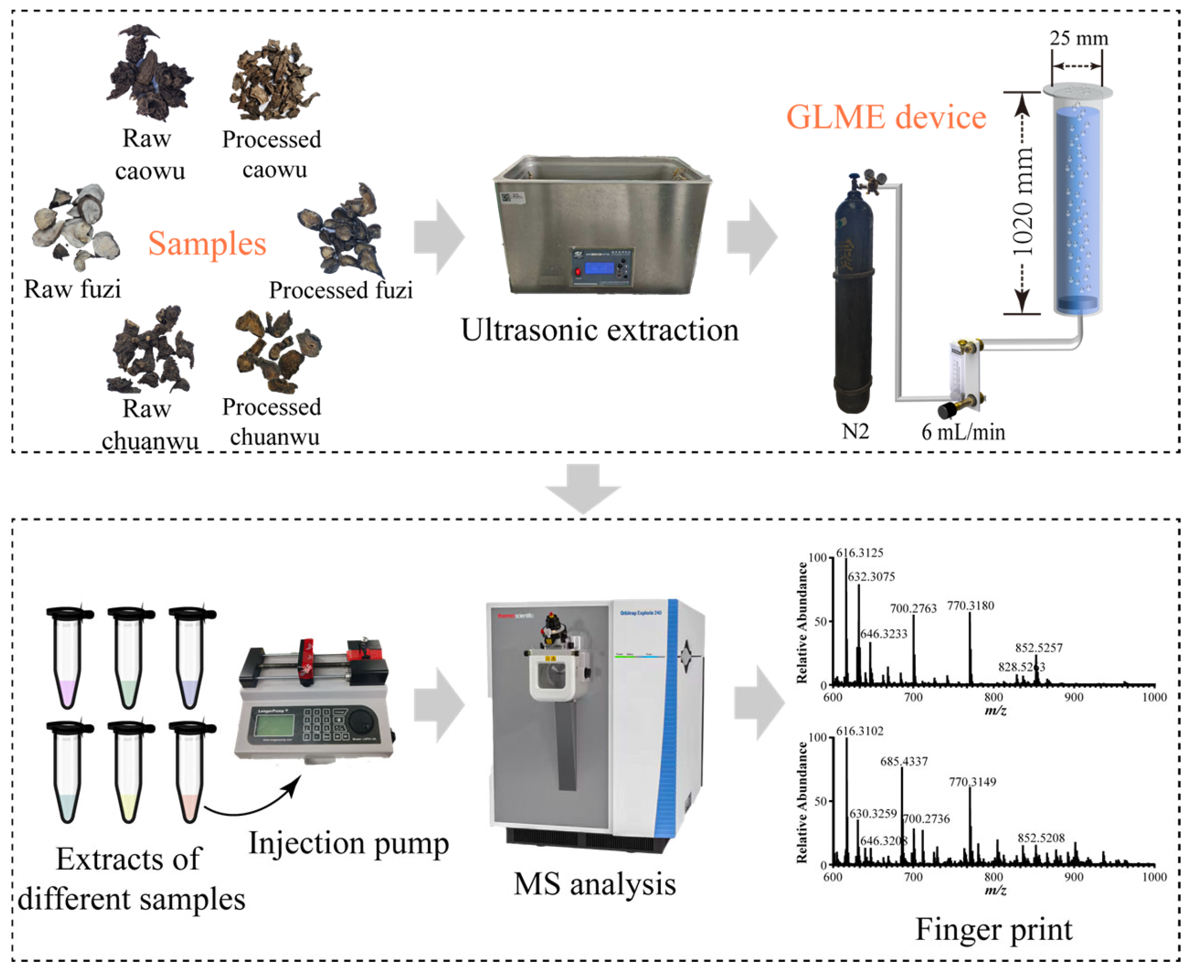
Figure 1. Gas–liquid microextraction–direct perfusion electrospray ionization mass spectrometry analysis process diagram (the pictures of medicinal materials and instruments were all taken by ourselves, and the final pictures were generated by WPS Office 12.1.0).
Molecules 30 04029 g001
Figure 2. Optimization results of key mass spectrometry parameters (n = 3). (A) Finger print, (B) ion spray voltage, (C) sheath gas, (D) aux gas, (E) ion transfer tube temperature, (F) injection pump flow rate. (The key target analytes are marked with ★).
Molecules 30 04029 g002
Figure 3. Comparison of mass spectra of 6 kinds of Aconitum herbs before and after gas–liquid microextraction. (AF) Mass spectrometry results before enrichment of GLME, (GL) mass spectrometry results after enrichment by GLME ((AL) are raw Fuzi, raw Caowu, raw Chuanwu, processed Fuzi, processed Caowu, and processed Chuanwu).
Molecules 30 04029 g003
Figure 4. Elucidation of characteristic fragmentation pathways for DDAs.
Molecules 30 04029 g004
Figure 5. MS/MS comparison results of hypaconitine, mesaconitine, and aconitine in samples and reference standard. (A,B) Hypaconitine, (C,D) mesaconitine, (E,F) aconitine ((A,C,E) are the secondary mass spectra of the standard compound; (B,D,F) are the secondary mass spectra of the samples).
Molecules 30 04029 g005
Figure 6. MS/MS spectra of three potential new compounds: (A) m/z 624.3535, (B) m/z 640.3493, (C) m/z 874.5108.
Molecules 30 04029 g006
Figure 7. Mass spectrometry response difference analysis of characteristic components in raw/processed products of Fuzi, Caowu, and Chuanwu. (A) DDAs, (B) lipo-alkaloids.
Molecules 30 04029 g007
Disclaimer/Publisher’s Note: The statements, opinions and data contained in all publications are solely those of the individual author(s) and contributor(s) and not of MDPI and/or the editor(s). MDPI and/or the editor(s) disclaim responsibility for any injury to people or property resulting from any ideas, methods, instructions or products referred to in the content.