Article

A Turn-On Fluorescence Sensor Based on Guest-Induced Luminescence Ru(bpy)32+@UiO-66 for the Detection of Organophosphorus Pesticides

1
Hunan Provincial Key Laboratory of Materials Protection for Electric Power and Transportation, Hunan Provincial Key Laboratory of Cytochemistry, School of Chemistry and Pharmaceutical Engineering, Changsha University of Science and Technology, Changsha 410114, China
2
Hunan Province Key Laboratory for Advanced Carbon Materials and Applied Technology, College of Materials Science and Engineering, Hunan University, Changsha 410082, China
*
Author to whom correspondence should be addressed.
Molecules 2025, 30(15), 3130; https://doi.org/10.3390/molecules30153130
Submission received: 18 June 2025 / Revised: 20 July 2025 / Accepted: 23 July 2025 / Published: 25 July 2025

Abstract

Luminescent metal–organic frameworks (MOFs) are used for the detection of organophosphorus pesticides (OPs) due to their large surface area and pore volume as well as their special optical properties. However, most self-luminescent MOFs are not only complex to synthesize and unstable in water but also feature a “turn-off” sensing system, which has highly restricted their practical applications in OP detection. Herein, a “turn-on” fluorescence sensor based on the guest-induced luminescence MOF Ru(bpy)32+@UiO-66 was constructed, which realized the sensitive detection of OPs through a dual-enzyme system for the first time. Compared with self-luminescent MOFs, Ru(bpy)32+@UiO-66 was not only more easily synthesized but also had higher chemical and photostability in water. In this strategy, by means of the hydrolysis of AChE and ChOx, H2O2 will be produced, which can oxidize Fe2+ to Fe3+, thereby quenching the fluorescence of Ru(bpy)32+@UiO-66. In the presence of OPs, the activity of AChE can be inhibited, resulting in the inability to generate H2O2 and Fe3+, which will turn on the fluorescence signal of Ru(bpy)32+@UiO-66. As a result, the Ru(bpy)32+@UiO-66 sensing system not only had high sensitivity for OPs detection but also possessed a satisfactory detection recovery rate for parathion-methyl in real samples, which provides a new approach for OP detection in food safety as well as environmental monitoring.

Graphical Abstract

1. Introduction

Organophosphorus pesticides (OPs), a kind of organic pesticide containing phosphorus, are used to prevent and control plant diseases and insect pests due to their high efficiency [1]. However, the heavy use and improper treatment of OPs will lead to excessive residues in the environment, thus making water, soil and various crops polluted. In the meantime, they will be enriched in organisms through the food chain, causing pesticide poisoning [2,3]. It is universally recognized that the main reason for OP poisoning is that OPs can inhibit the activity of acetylcholinesterase (AChE) in the nervous tissues of organisms, which leads to the accumulation of the important neurotransmitter acetylcholine (ACh) in synapses and over-stimulation, resulting in central nervous injury [4,5]. Therefore, a serious threat is presented to human health by OP residues, which a variety of methods for OP detection have been developed to solve, such as high performance liquid chromatography (HPLC) [6,7], gas chromatography (GC) [8,9], enzyme-linked immunosorbent assay (ELISA) [10,11], electrochemical methods [12,13] and so forth. However, although HPLC and GC have good sensitivity, not only are expensive instruments but also complicated operations required. ELISA is susceptible to external environmental conditions and its required antibodies are difficult to prepare. In addition, electrochemical methods require complex treatment of the electrodes. Considering what is mentioned above, it is urgent to develop a sensitive and simple method to achieve efficient detection of OPs.
Currently, more and more attention is being attracted to fluorescence analysis, owing to its advantages of high sensitivity, quick response and simple operation. Meanwhile, many fluorescence assays based on enzyme inhibition have been developed to detect OPs [14,15,16,17,18,19,20,21,22]. Taking acetylcholinesterase (AChE) as an example, the main detection principle is that OPs can irreversibly inhibit the activity of AChE, leading to the failure of hydrolysis of the substrate ACh, so as to cause a change in the fluorescence intensity of probes for OP detection. For example, Li et al. [21] constructed a fluorescent sensor by using the FRET effect between NaY/GdF4:Yb, Er upconversion nanoparticles (UCNPs) and MnO2 nanosheets. Considering the effect of the inhibition of OPs on the activity of AChE, OP detection was realized by the change in the fluorescence intensity of UCNPs. However, the most common fluorescent probes used for detecting OPs have been mainly based on organic fluorescent dyes, UCNPs, quantum dots and so on. Owing to their complicated preparation process, instability and photobleaching, their practical applications are greatly limited. Therefore, research into a kind of highly efficient fluorescent probe for the determination of OPs is highly desirable.
Metal–organic frameworks (MOFs), a class of porous hybrid materials, self-assembled by metal nodes and organic ligands [23], have been used in many fields, including gas storage and separation [24,25], catalysis [26,27], electrochemistry [28,29], biomedicine [30,31] and fluorescence sensing [32,33] due to their advantages of high stability, easy surface modification, large surface area and pore volume. So far, some fluorescence sensors based on self-luminescent MOFs for OP detection have been developed. For instance, Xu et al. [34] synthesized a luminescent ZnPO-MOF. Through the photoinduced electron transfer (PET) effect between it and nitroaromatic compounds, the fluorescence of ZnPO-MOF would be suppressed by parathion-methyl, realizing the detection of parathion-methyl. However, the most self-luminescent MOFs were not only complex to synthesize and unstable in water but also featured a “turn-off” sensing system, which has highly restricted the practical application of MOFs in the detection of OPs. Meanwhile, many MOFs have a large specific surface area and pore volume as well as a stable crystal structure, and therefore, they can be used as excellent carriers of fluorescent guest molecules to construct novel fluorescent probes [35,36] and achieve sensitive detection of OPs in practice.
In this work, a “turn-on” fluorescence sensor based on Ru(bpy)32+@UiO-66 was designed for the detection of OPs, in which the fluorescence intensity of the Ru(bpy)32+ molecule was greatly improved by the encapsulation of it into the UiO-66 framework. Furthermore, compared with self-luminescent MOFs, Ru(bpy)32+@UiO-66 was not only synthesized more easily but also had higher chemical and photostability. In this sensing system, by means of the hydrolysis of the dual enzymes (AChE and ChOx), the generated choline can be transformed into H2O2, which can oxidize Fe2+ to Fe3+, quenching the fluorescence of Ru(bpy)32+@UiO-66. When OPs appear, they can inhibit the activity of AChE, resulting in the inability to generate H2O2 and Fe3+, thus turning on the fluorescence of Ru(bpy)32+@UiO-66 to achieve detection of OPs (Scheme 1), which provides a new strategy for the sensitive detection of OPs.

2. Results and Discussion

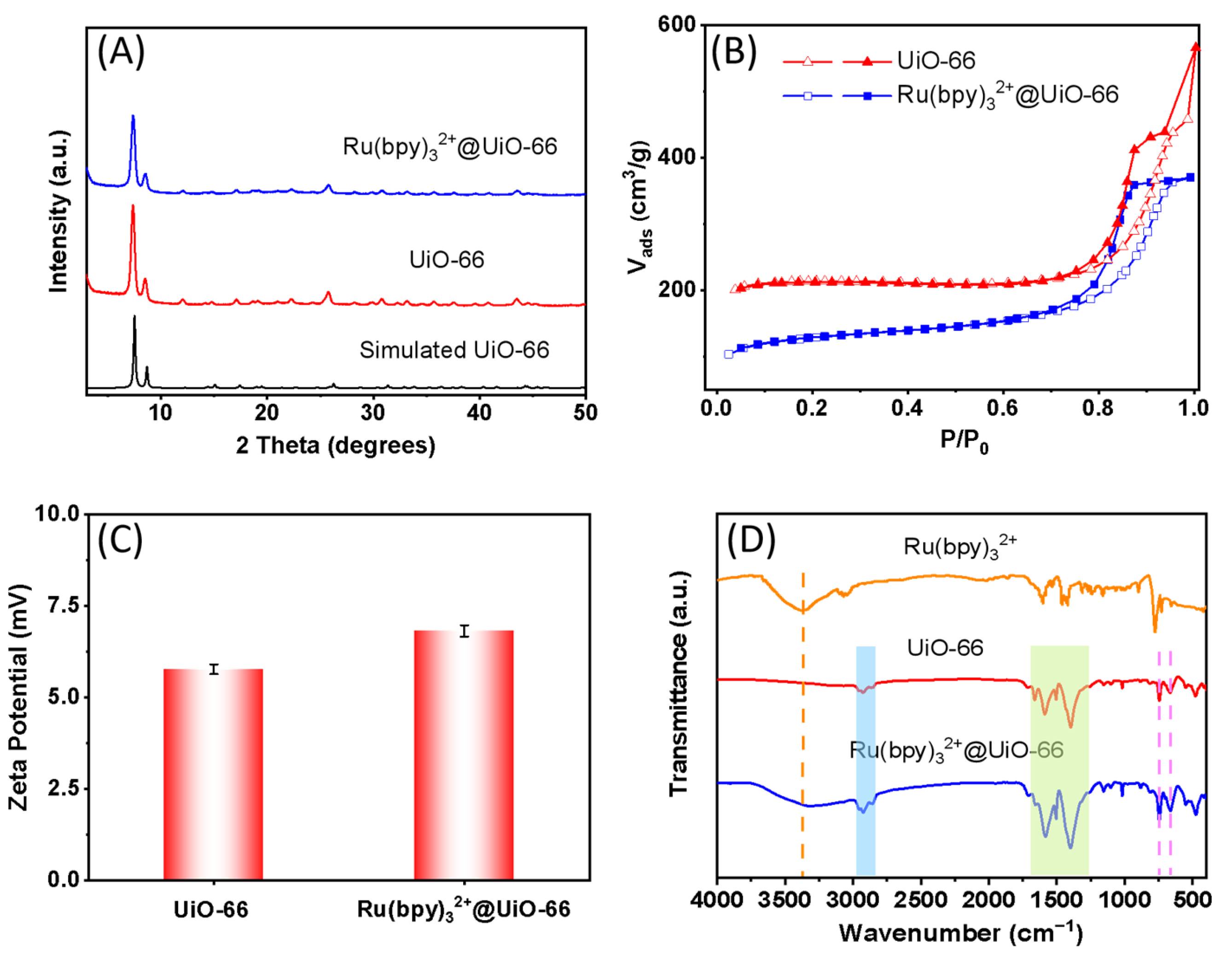
2.1. Characterization of UiO-66 and Ru(bpy)32+@UiO-66

As shown in the following SEM (Figure 1B) and TEM (Figure 1C) images, UiO-66, self-assembled from Zr4+ and terephthalic acid (Figure 1A), is an octahedral crystal with good dispersion and an average diameter of about 400 nm. By encapsulating the luminescent molecule Ru(bpy)32+ into UiO-66, Ru(bpy)32+@UiO-66 was successfully prepared (Figure 1A). Compared with UiO-66, the size and morphology of Ru(bpy)32+@UiO-66 changed slightly, and its color changed from white to yellow (Figure 1D). In addition, the C, N, O, Zr and Ru were uniformly distributed within the Ru(bpy)32+@UiO-66 framework, which was clearly demonstrated by the EDX spectrum (Figure 1E) and element mapping (Figure 1F).
Figure 2A shows that the PXRD patterns of the synthesized UiO-66 and Ru(bpy)32+@UiO-66 are consistent with that of the simulated sample, implying the synthesized UiO-66 had bulky phase purity and the encapsulation of Ru(bpy)32+ into it did not affect its crystallinity and structure. Additionally, compared with UiO-66, the decreased amount of N2 sorption (Figure 2B) and the increase in the ζ-potential (Figure 2C) of Ru(bpy)32+@UiO-66 demonstrated that the Ru(bpy)32+ luminophore was successfully introduced into the UiO-66 framework. FT-IR was also used to analyze the chemical functional groups of Ru(bpy)32+@UiO-66. As illustrated in Figure 2D, the FT-IR spectrum of UiO-66 showed vibrational bands at 2970–2835 cm−1, ascribed to the C-H bonds, and strong bands at 1700–1250 cm−1, arisen from the carboxylate groups and phenyl ring deformations. Meanwhile, the bands at 745 and 665 cm−1 correspond to the Zr-O chemical bond [37]. Typically, the positions of these bands had hardly any change in the FT-IR spectrum of Ru(bpy)32+@UiO-66, indicating that the structure of UiO-66 was not changed in Ru(bpy)32+@UiO-66. In addition, there was a strong and wide band centered at 3370 cm−1 attributed to the O-H stretching mode of the physisorbed water in the spectrum of Ru(bpy)32+, which also existed in the spectrum of Ru(bpy)32+@UiO-66 [38]. All of these results further indicate that the Ru(bpy)32+ molecule was successfully encapsulated by UiO-66. Then, ICP-AES was utilized to determine the loading capacity of the Ru complex in Ru(bpy)32+@UiO-66. It was found that a high loading capacity of 10.35 wt% was obtained.

2.2. Optical Properties of Ru(bpy)32+@UiO-66

The basic optical properties of the probe were investigated. Compared with UiO-66, Ru(bpy)32+ and Ru(bpy)32+@UiO-66 exhibited obvious absorption peaks at 450 nm (Figure 3A) and strong fluorescence emission peaks at about 600 nm (Figure 3B), which indicated that the fluorescence signal of Ru(bpy)32+@UiO-66 originated from the luminescent molecule Ru(bpy)32+ rather than UiO-66, and further demonstrated that Ru(bpy)32+ was encapsulated into the UiO-66 framework. In addition, since the porous structure of UiO-66 can not only separate the Ru(bpy)32+ molecules to prevent aggregation-induced quenching but can also limit their motion or rotation to increase the rigidity of the Ru complex, the fluorescence intensity of Ru(bpy)32+@UiO-66 was much stronger than that of the free Ru(bpy)32+. It is well known that the chemical stability and photostability of the fluorescent probes are crucial for their practical applications. As displayed in Figure 3C, the fluorescence intensity of Ru(bpy)32+@UiO-66 was almost constant in a wide pH range (5.0~10.5), implying that it has high chemical stability. Furthermore, it can be seen from Figure 3D that the fluorescence intensity of Ru(bpy)32+@UiO-66 hardly decreased with the increasing time of UV lamp irradiation. The above results indicate that Ru(bpy)32+@UiO-66 has good chemical and photostability, which provides a reliable guarantee for the construction of sensitive OP sensors.
Subsequently, the effects of different metal ions (Ba2+, Ca2+, Al3+, Mg2+, Mn2+, Cu2+, Zn2+, Fe2+ and Fe3+) on the fluorescence intensity of Ru(bpy)32+@UiO-66 were explored. Figure 4A implies that the fluorescence quenching rate of Ru(bpy)32+@UiO-66 by Fe3+ was as high as 87.28% after incubation for 10 min, while those of other metal ions were very low, suggesting Fe3+ was able to quench the fluorescence signal of Ru(bpy)32+@UiO-66, which may be because Fe3+ has strong oxidizability and simple coordination in the environment of an aqueous solution, which enables it to efficiently quench the fluorescence of Ru(bpy)32+ through the photoinduced electron transfer (PET) effect. Specifically, compared with other common transition metal ions, Fe3+ is one of the strongest oxidants because the Fe3+/Fe2+ pair has a very positive standard reduction potential (+0.77 V). In addition, Fe3+, existing in the form of [Fe(H2O)6]3+ in aqueous solution, is a relatively “hard” Lewis acid, which means that its dynamic electron transfer does not require coordination layer recombination, and its rate is fast. Therefore, compared to other transition metal ions, Fe3+ can efficiently quench the fluorescence signal of Ru(bpy)32+@UiO-66 in a short time. Furthermore, in order to further investigate the recognition behavior of Ru(bpy)32+@UiO-66 on Fe3+, the UV-vis absorption spectra of this system were measured. Figure 4B shows that the intensity of the absorption peak of Ru(bpy)32+@UiO-66 obviously decreased when Fe3+ was introduced, which is mainly due to the ground state interaction between Fe3+ and Ru(bpy)32+, forming a ground state complex that has almost no absorption at 450 nm. Subsequently, the quenching effect of different concentrations of Fe3+ on the fluorescence intensity of Ru(bpy)32+@UiO-66 was studied. As shown in Figure 4C, with the increase of Fe3+ concentration, the fluorescence intensity of Ru(bpy)32+@UiO-66 gradually decreased, and there is a good linear relationship between the fluorescence quenching rate ((F0 − F)/F0) and the concentration of Fe3+ in the range of 0~1.0 mM [(F0 − F)/F0 = 0.50635 [Fe3+] + 3.7444, R2 = 0.9993], with a detection limit of 0.038 mM (Figure 4D).

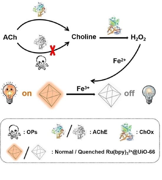
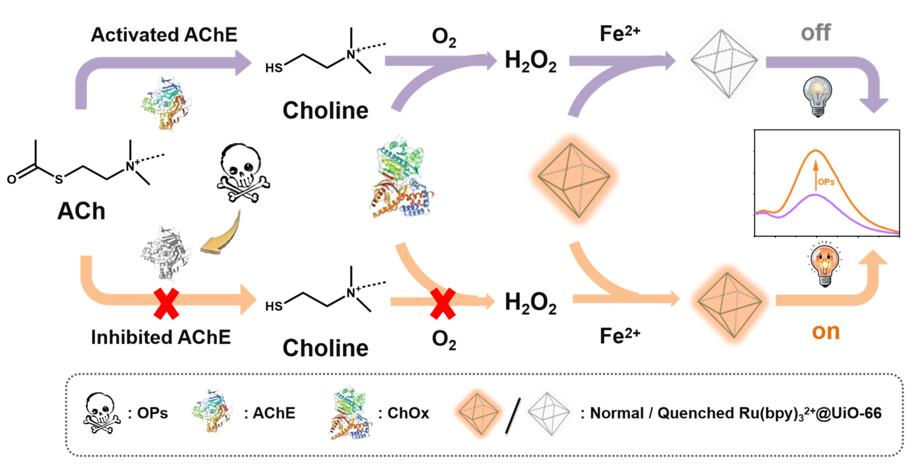
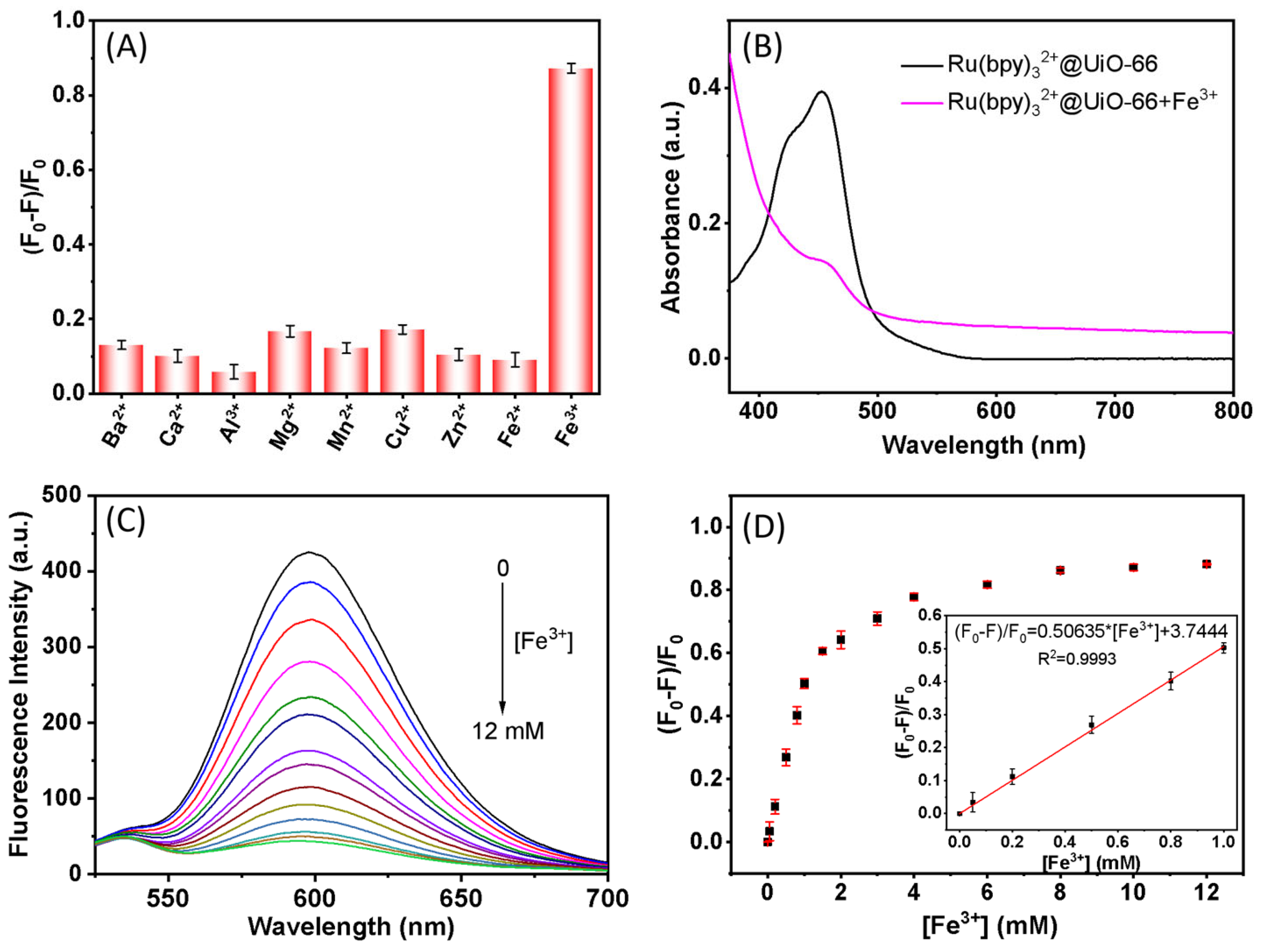
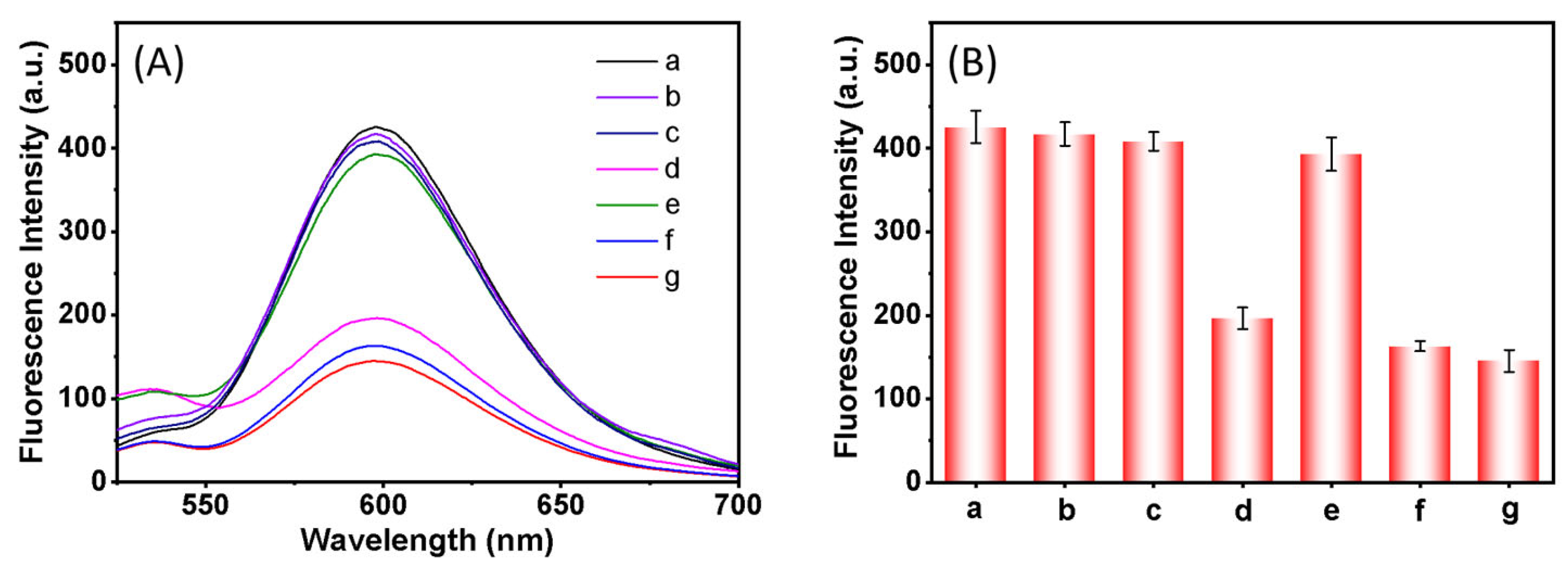
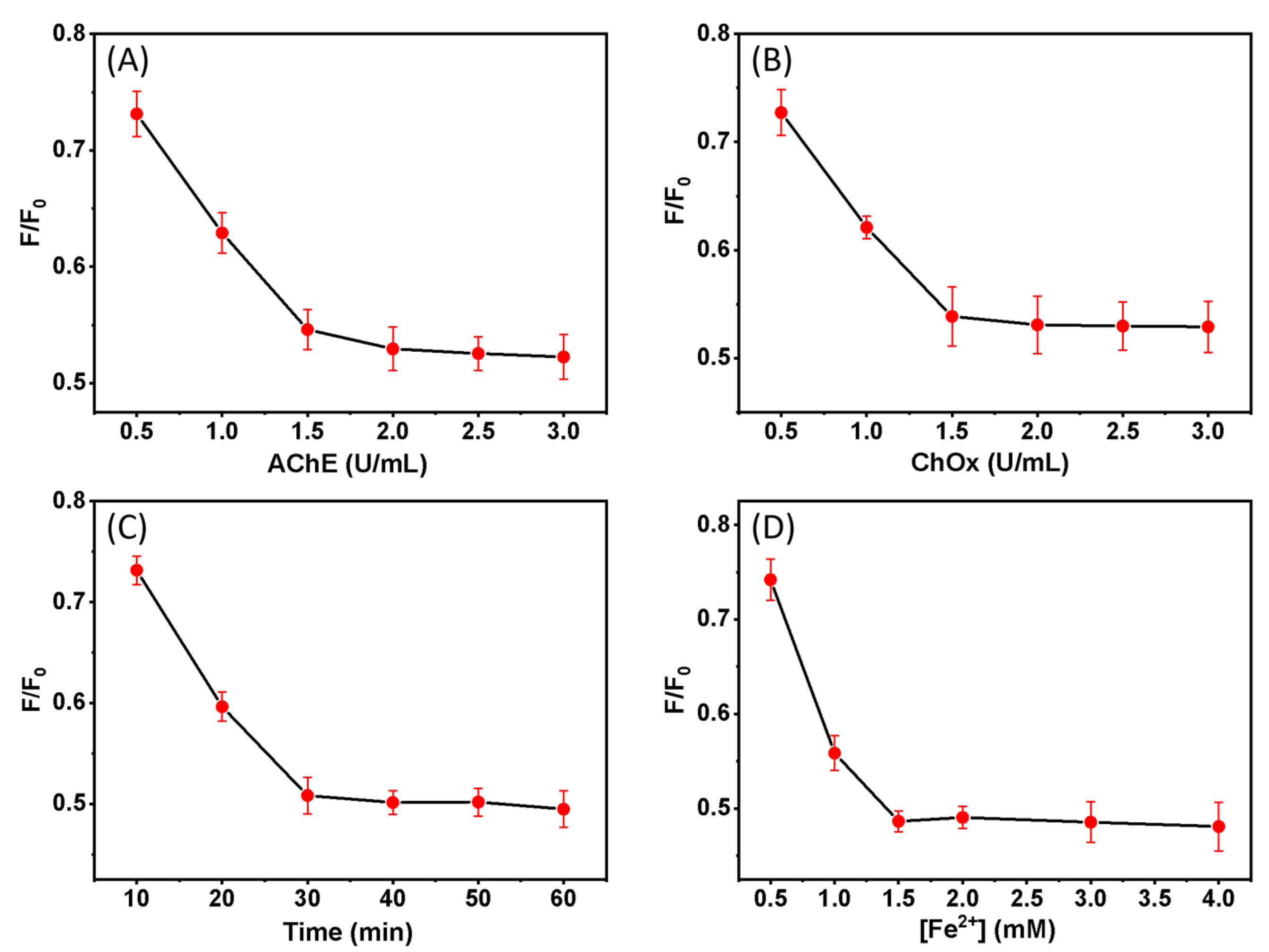
2.3. Feasibility of Ru(bpy)32+@UiO-66 Sensor for OP Sensing

A “turn-on” fluorescence sensor based on the guest-induced luminescent Ru(bpy)32+@UiO-66 was developed to detect OPs, the basic mechanism of which includes three parts, as follows. (1) AChE catalyzes the hydrolysis of substrate ACh to produce choline (Equation (1)), and then choline and O2 are hydrolyzed by ChOx to betaine and H2O2 (Equation (2)) [39,40]. (2) Fe2+ is oxidized to Fe3+ by H2O2 (Equation (3)) [41]. (3) The generated Fe3+ can quench the fluorescence of Ru(bpy)32+@UiO-66, while, in the presence of OPs, the activity of AChE is inhibited by it. Therefore, the choline, H2O2 and final Fe3+ will not be able to generate, thus triggering the fluorescence of Ru(bpy)32+@UiO-66 to achieve detection of OPs (Scheme 1).
A C h + H 2 O A C h E   choline   +   acetic   acid  
  choline   + O 2 C h O x   betaine   + H 2 O 2
F e 2 + + H 2 O 2 H + F e 3 + + H 2 O
Figure 5 shows the effects of different substances on the fluorescence intensity of Ru(bpy)32+@UiO-66, which indicate that the fluorescence was significantly quenched only when Ru(bpy)32+@UiO-66 coexisted with AChE, ChOx, ACh and Fe2+ (d), H2O2 and Fe2+ (f) and Fe3+ (g). In comparison, other conditions had little effect on it. The results demonstrated the above mechanism of this strategy, in which only in the presence of double enzymes (AChE and ChOx) would H2O2 be produced and further oxidize Fe2+ into Fe3+, thus quenching the fluorescence of Ru(bpy)32+@UiO-66 (Equations (1)–(3)). However, in the presence of OPs, the activity of AChE was inhibited, and H2O2 could not be generated to convert Fe2+ to Fe3+, resulting in the turning-on of this fluorescence system.

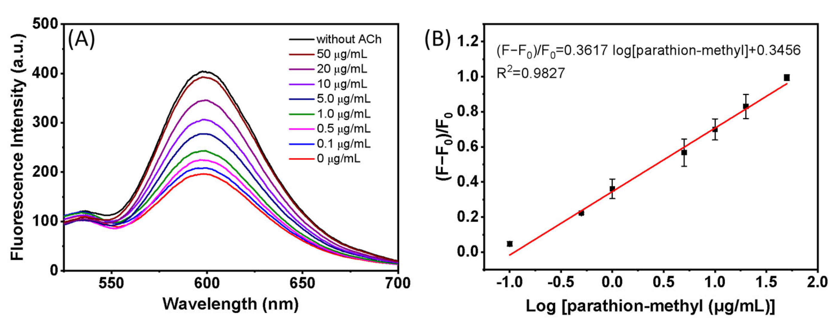
2.4. Detection of OPs

In order to realize the sensitive detection of OPs, the concentrations of ATCh, ChOx and Fe2+, as well as the incubation time, were systematically optimized. As displayed in Figure 6A,B, the fluorescence intensity of Ru(bpy)32+@UiO-66 basically remained constant when the concentrations of AChE and ChOx reached 2.0 U/mL and 1.5 U/mL, respectively. Thus, 2.0 U/mL and 1.5 U/mL were chosen as the optimal concentrations of AChE and ChOx, respectively. Subsequently, the incubation times of AChE, ACh and ChOx were studied. Figure 6C shows that the fluorescence intensity of this system reached a balance after incubation for 30 min. Furthermore, the concentration of Fe2+ in the system was investigated. It can be seen from Figure 6D that with the increasing concentration of Fe2+, the relative fluorescence intensity attained a plateau at 1.5 mM; therefore, the optimum concentration of Fe2+ was 1.5 mM in this sensing system.
Under the optimum conditions, the detection of parathion-methyl, which is one of the widely used OPs, by the Ru(bpy)32+@UiO-66 sensing system was investigated. From Figure 7A, we can see that with the increase in the concentration of parathion-methyl, the fluorescence intensity of Ru(bpy)32+@UiO-66 gradually increased, which indicates that the inhibition of the activity of AChE by parathion-methyl suppresses the generation of Fe3+, thus turning on the fluorescence of Ru(bpy)32+@UiO-66. In addition, the inhibition rate ((F−F0)/F0, F0 and F, corresponding to the fluorescence intensity of Ru(bpy)32+@UiO-66 in the absence and presence of parathion-methyl, respectively) had a linear relationship with the logarithm of parathion-methyl concentration from 0.1 to around 50.0 μg/mL, and the corresponding linear regression equation was (F−F0)/F0 = 0.3617 log [parathion-methyl] + 0.3456, with the limit of detection (LOD) at 10.6 ng/mL, which was lower than those of most other reported fluorescence sensors (Table 1).

2.5. Anti-Interference Ability, Selectivity and Recyclability

In order to explore the anti-interference ability of the proposed sensor, the effects of common metal ions and bioelectrolytes in food samples on this sensor were investigated. It can be seen from Figure 8A that Ca2+, K+, Mg2+, Na+, Zn2+, glycine (Gly), ascorbic acid (AA), bovine serum albumin (BSA), catalase (CAT), glucose oxidase (GOx) and L-glutamic acid (L-Glu) had little effect on the detection of parathion-methyl. In addition, to prove the selectivity of the Ru(bpy)32+@UiO-66 fluorescence sensing system for the detection of parathion-methyl, the effects of five common OPs (chlorpyrifos, dichlorvos, omethoate, paraoxon and malathion) and four other kinds of pesticides (paraquat, nitenpyram, hexazolalcohol and imidacloprid) on the system were studied. As can be seen from Figure 8B, all of the OPs displayed an inhibition effect on the system, with parathion-methyl displaying the highest inhibitory rate, while the other kinds of pesticides exerted a negligible effect to the system, which is principally because the Ops, owning similar chemical structures (Figure 8C), can trigger the irreversible inhibition of AChE. Therefore, the proposed Ru(bpy)32+@UiO-66 fluorescence sensing system exhibits excellent selectivity for OPs, and can thus be used as a universal sensing platform for identification and detection of OPs. Furthermore, the eight cycles of detection of 10 μg/mL parathion-methyl by the Ru(bpy)32+@UiO-66 sensing system showed no significant fluorescent variation, and the relative standard deviation (RSD) was 4.3%, indicating the excellent recyclability of the Ru(bpy)32+@UiO-66 system.

2.6. Detection of OPs in Actual Samples

Parathion-methyl in environmental and agricultural samples (tap water, lake water and cabbage) was detected via the standard addition method. As shown in Table 2, the recovery was between 92.80% and 106.8%, and the RSD was less than 5.6%. The results indicated that the Ru(bpy)32+@UiO-66 sensing system possessed an excellent ability to detect OPs in actual samples.

3. Materials and Methods

3.1. Reagents and Instruments

Acetylcholinesterase (AChE, 500 units, derived from electric eel) and choline oxidase (ChOx, 100 units, derived from Alcaligenes) were purchased from Sigma-Aldrich Co., Ltd. (St. Louis, MO, USA). Ammonium ferrous sulfate [(NH4)2Fe(SO4)2·6H2O], terephthalic acid (PTA), zirconium oxychloride (ZrOCl2·8H2O), acetylcholine chloride (ACh), ruthenium chloride hexahydrate (Ru(bpy)3Cl2·6H2O, >98%), parathion-methyl, dichlorvos, chlorpyrifos, imidacloprid and hexazole alcohol were obtained from Aladdin Biochemical Technology Co., Ltd. (Shanghai, China). Malathion, omethoate, paraquat, nitenpyram and paraoxon were purchased from Walbo Technology Co., Ltd. (Beijing, China). N, N-dimethylformamide (DMF) and glacial acetic acid were provided by Sinopharm Chemical Reagent Co., Ltd. (Shanghai, China). All reagents were used without any further purification.
Scanning electron microscopy (SEM, SU8010, Hitachi, Tokyo, Japan), transmission electron microscopy (TEM, TalosF200X) and high-angle annular dark-field scanning TEM (HAADF-STEM) images with an energy-dispersive X-ray detector (EDX) were used for the morphological characterization and detailed composition analysis of the prepared samples. Powder X-ray diffraction (PXRD) measurements were performed on a D8-ADVANCE powder diffractometer (Brooke, Karlsruhe, Germany). The nitrogen adsorption–desorption isotherms were obtained using a Micromeritics ASAP 2460 Sorptometer (Mike Instruments, Cincinnati, OH, USA). A Zeta potentiometer (Zetasizer Nano ZS90, Malvern Instruments, Malvern, UK) was used to measure the surface potential of the samples. Nicolet iS10 Fourier transform infrared spectroscopy (FT-IR, Thermo Fisher Scientific, Waltham, MA, USA) was employed to characterize the chemical functional groups of the samples. Inductively coupled plasma atomic emission spectroscopy (ICP-AES) was carried out on a PerkinElmer ICP-AES system (PE 2100DV, Waltham, MA, USA). The UV-Vis spectra were collected by using a UV-2700 spectrophotometer (Shimadzu, Kyoto, Japan). The fluorescence emission spectra were recorded via an F-7000 fluorescence spectrophotometer (Hitachi, Japan). Ultrapure water (18.2 MΩ·cm−1) for the sample preparation and detection was prepared with the Milli-Q system (Millipore, Bedford, MA, USA).

3.2. Synthesis of UiO-66

UiO-66 was synthesized according to the reported literature [51]. First, ZrOCl2 (21 mg, 0.066 mM) and terephthalic acid (50 mg, 0.24 mM) were dissolved into 3 mL and 1 mL DMF, respectively. Then, the above two solutions were added into 400 μL glacial acetic acid and mixed and heated at 90 °C for 18 h. After centrifugation, the mixture was washed with DMF and ultrapure water, respectively. Finally, the obtained solid was dried under vacuum for 12 h at 60 °C for future use.

3.3. Preparation of Ru(bpy)32+@UiO-66

Ru(bpy)32+@UiO-66 was prepared as follows. First, 20 mg of the synthesized UiO-66 was dispersed into 10 mL DMF. Then, the above suspension was mixed with a 10 mL DMF solution containing Ru(bpy)3Cl2·6H2O (0.5, 1 or 2 mg/mL). Subsequently, the mixture was stirred for 12 h at 90 °C and washed with DMF and ethanol, respectively, after centrifugation. Finally, the yellow solid was dried under vacuum at 60 °C for 12 h to obtain Ru(bpy)32+@UiO-66.
ICP-AES was utilized to determine the loading capacity of the Ru complex in Ru(bpy)32+@UiO-66. Since the results indicated that 1 mg/mL of Ru(bpy)3Cl2·6H2O achieved the highest loading capacity (10.35 wt%), all subsequent experiments were conducted using Ru(bpy)32+@UiO-66 prepared at this concentration.

3.4. Fluorometric Detection of OPs

The Ru(bpy)32+-UiO-66 was dispersed in deionized water as a stock solution (1 mg/mL), and acetonitrile was used as the solvent for the standard stock solution of the parathion-methyl. The solutions of various biological species (such as AChE, ChOx and so on) were dissolved or diluted with PBS buffer solution (10 mM, pH 7.5). Firstly, different concentrations of parathion-methyl (0, 0.1, 0.5, 1, 5, 10, 20 and 50 μg/mL) were incubated with 200 μL AChE (2 U/mL) for 15 min. Next, 500 μL ACh (40 mM) and 500 μL ChOx (1.5 U/mL) were added into the above solution and incubated at 37 °C for another 30 min. Then, 300 μL the above solution, 10 μL Ru(bpy)32+-UiO-66 (1 mg/mL), 200 μL HCl (3%), 365 μL PBS (10 mM, pH 7.5) and 125 μL Fe2+ (1.5 mM) were mixed thoroughly for 10 min before the fluorescence spectra were collected at an excitation wavelength of 450 nm.

3.5. OP Detection in Real Samples

This sensor was mainly used for the detection of parathion-methyl in environmental and food samples, including lake water, tap water and cabbage samples. The insoluble materials in the lake water sample (10 mL) were removed by the 0.22 μm filter membrane and the chlorine was evaporated from the tap water sample (10 mL) by boiling. Then, the treated water samples were mixed with 10 mL PBS (10 mM, pH 7.4) containing 20% methanol, respectively. As for the cabbage, it (10 g) was ground and mixed with 50 mL acetonitrile under sonication for 10 min. Then, the mixture was centrifuged to obtain the supernatant. The extraction process was repeated three times, and the extraction solutions were combined and evaporated to dry, then dissolved by PBS (10 mM, pH 7.4) containing 20% methanol. Subsequently, the standard solution of 5, 10 and 15 μg/mL parathion-methyl was spiked into the three samples. The recovery (%) and relative standard deviation (RSD) (n = 3, %) were calculated to evaluate the practical value of the sensor.

4. Conclusions

To sum up, a “turn on” fluorescence sensor based on the guest-induced luminescence Ru(bpy)32+@UiO-66 was constructed and it realized the sensitive detection of OPs for the first time. In this strategy, the fluorescence of Ru(bpy)32+@UiO-66 was quenched by Fe3+ which was generated by the dual enzymes and Fe2+ system. In the presence of OPs, they can inhibit the activity of AChE, thereby lighting the fluorescence signal of Ru(bpy)32+@UiO-66 and achieving OPs detection. The experimental results implied a good linear relationship with the logarithm of the parathion-methyl concentration from 0.1 μg/mL to 50.0 μg/mL, and the LOD at 10.6 ng/mL. Furthermore, the recovery was between 92.80% and 106.8% for the detection of actual samples. Therefore, this method opened up a new avenue for the detection of OPs.

Author Contributions

Conceptualization, J.L. and Z.C.; formal analysis, J.L. and J.D.; investigation, J.D., Q.T., C.Y. and Y.L.; methodology, J.L., J.Y. and Z.C.; project administration, J.L. and Z.C.; funding acquisition, J.L. and Z.C.; resources, J.L., J.Y. and Z.C.; supervision, J.L. and Z.C.; writing—original draft, J.L. and J.D.; writing—review and editing, J.L., J.Y. and Z.C. All authors have read and agreed to the published version of the manuscript.

Funding

This research was funded by the National Natural Science Foundation of China (Nos. 21605009, 21275022), the Natural Science Foundation of Hunan Province (Nos. 2019JJ50651, 2025JJ50092) and the Scientific Research Fund of the Hunan Provincial Education Department (Nos. 21B0302, 24A0236).

Institutional Review Board Statement

Not applicable.

Informed Consent Statement

Not applicable.

Data Availability Statement

The data are contained within the article.

Conflicts of Interest

The authors declare no conflicts of interest.

References

  1. Pundir, C.S.; Malik, A. Bio-sensing of organophosphorus pesticides: A review. Biosens. Bioelectron. 2019, 140, 111348. [Google Scholar] [CrossRef]
  2. Farag, A.; Gamila, A.; Kotb, G.; Hamza, A.; Hussein, R.; Elhalwagy, M. Impact of Organophosphorus insecticide Triazophos on Liver, Kidneys and Thyroid in Albino Rats. Int. J. Adv. Res. Biol. Sci. 2016, 3, 199–208. [Google Scholar]
  3. Pehkonen, S.O.; Zhang, Q. The degradation of organophosphorus pesticides in natural waters: A critical review. Crit. Rev. Environ. Sci. Technol. 2002, 32, 17–72. [Google Scholar] [CrossRef]
  4. Legierse, K.C.H.M.; Verhaar, H.J.M.; Vaes, W.H.J.; De Bruijn, J.H.M.; Hermens, J.L.M. Analysis of the time-dependent acute aquatic toxicity of organophosphorus pesticides: The critical target occupation model. Environ. Sci. Technol. 1999, 33, 917–925. [Google Scholar] [CrossRef]
  5. Sun, J.F.; Guo, L.; Bao, Y.; Xie, J.W. A simple, label-free AuNPs-based colorimetric ultrasensitive detection of nerve agents and highly toxic organophosphate pesticide. Biosens. Bioelectron. 2011, 28, 152–157. [Google Scholar] [CrossRef] [PubMed]
  6. Yu, C.C.; Hao, D.Y.; Chu, Q.; Wang, T.; Liu, S.N.; Lan, T.; Wang, F.H.; Pan, C.P. A one adsorbent QuEChERS method coupled with LC-MS/MS for simultaneous determination of 10 organophosphorus pesticide residues in tea. Food Chem. 2020, 321, 126657. [Google Scholar] [CrossRef]
  7. Peyrovi, M.; Hadjmohammadi, M. Alkanol-based supramolecular solvent microextraction of organophosphorus pesticides and their determination using high-performance liquid chromatography. J. Iran. Chem. Soc. 2017, 14, 995–1004. [Google Scholar] [CrossRef]
  8. Manav, Ö.G.; Dinç-Zor, Ş.; Alpdoğan, G. Optimization of a modified QuEChERS method by means of experimental design for multiresidue determination of pesticides in milk and dairy products by GC-MS. Microchem. J. 2019, 144, 124–129. [Google Scholar] [CrossRef]
  9. Cheng, Z.P.; Dong, F.S.; Xu, J.; Liu, X.G.; Wu, X.H.; Chen, Z.L.; Pan, X.L.; Gan, J.; Zheng, Y.Q. Simultaneous determination of organophosphorus pesticides in fruits and vegetables using atmospheric pressure gas chromatography quadrupole-time-of-flight mass spectrometry. Food Chem. 2017, 231, 365–373. [Google Scholar] [CrossRef]
  10. Liu, B.; Ge, Y.; Zhang, Y.; Song, Y.; Lv, Y.Y.; Wang, X.X.; Wang, S. Production of the class-specific antibody and development of direct competitive ELISA for multi-residue detection of organophosphorus pesticides. Food Agric. Immunol. 2012, 23, 157–168. [Google Scholar] [CrossRef]
  11. Hua, X.D.; Liu, X.F.; Shi, H.Y.; Wang, Y.R.; Kim, H.J.; Gee, S.J.; Wang, M.H.; Liu, F.Q.; Hammock, B.D. Development of a heterologous enzyme-linked immunosorbent assay for organophosphorus pesticides with phage-borne peptide. RSC Adv. 2014, 4, 42445–42453. [Google Scholar] [CrossRef]
  12. Soulis, D.; Trigazi, M.; Tsekenis, G.; Chandrinou, C.; Klinakis, A.; Zergioti, I. Facile and Low-Cost SPE Modification Towards Ultra-Sensitive Organophosphorus and Carbamate Pesticide Detection in Olive Oil. Molecules 2020, 25, 4988. [Google Scholar] [CrossRef]
  13. Guo, W.; Chen, Z.Y.; Zhao, Y.X.; Zhao, C.Y.; Lu, X.; Gao, F.M. Hierarchical Cu3V2O8/Cu6Mo5O18/MXene nanostructures by ultrathin nanosheets for highly sensitive electrochemical detection of organophosphorus pesticides. Microchem. J. 2024, 201, 110601. [Google Scholar] [CrossRef]
  14. Tian, J.X.; Gong, S.; Gu, Y.; Li, Z.N.; Liang, Y.Y.; Meng, Z.Y.; Wang, Z.L.; Wang, S.F. A novel nopinone-based fluorescent probe for sensitive detection of pesticide parathion-methyl in soil and its applications in biological imaging. J. Environ. Chem. Eng. 2025, 13, 116193. [Google Scholar] [CrossRef]
  15. Dong, J.J.; Yang, H.T.; Li, Y.; Liu, A.R.; Wei, W.; Liu, S.Q. Fluorescence sensor for organophosphorus pesticide detection based on the alkaline phosphatase-triggered reaction. Anal. Chim. Acta 2020, 1131, 102–108. [Google Scholar] [CrossRef]
  16. Cai, Y.; Qiu, Z.Y.; Lin, X.B.; Zeng, W.; Cao, Y.R.; Lin, W.P.; Liu, Y.J. Self-assembled nanomaterials based on aggregation-induced emission of AuNCs: Fluorescence and colorimetric dual-mode biosensing of organophosphorus pesticides. Sens. Actuators B Chem. 2020, 321, 128281. [Google Scholar] [CrossRef]
  17. Wu, X.L.; Wang, P.S.; Hou, S.Y.; Wu, P.L.; Xue, J. Fluorescence sensor for facile and visual detection of organophosphorus pesticides using AIE fluorogens-SiO2-MnO2 sandwich nanocomposites. Talanta 2019, 198, 8–14. [Google Scholar] [CrossRef] [PubMed]
  18. Wu, X.L.; Song, Y.; Yan, X.; Zhu, C.Z.; Ma, Y.Q.; Du, D.; Lin, Y.H. Carbon quantum dots as fluorescence resonance energy transfer sensors for organophosphate pesticides determination. Biosens. Bioelectron. 2017, 94, 292–297. [Google Scholar] [CrossRef] [PubMed]
  19. Yan, X.; Song, Y.; Zhu, C.Z.; Li, H.X.; Du, D.; Su, X.G.; Lin, Y.H. MnO2 nanosheet-carbon dots sensing platform for sensitive detection of organophosphorus pesticides. Anal. Chem. 2018, 90, 2618–2624. [Google Scholar] [CrossRef] [PubMed]
  20. Chen, J.L.; Chen, X.J.; Wang, P.; Liu, S.W.; Chi, Z.G. Aggregation-induced emission luminogen@manganese dioxide core-shell nanomaterial-based paper analytical device for equipment-free and visual detection of organophosphorus pesticide. J. Hazard. Mater. 2021, 413, 125306. [Google Scholar] [CrossRef]
  21. Li, H.H.; Ali, S.; Wei, W.Y.; Xu, Y.; Lu, H.W.; Mehedi Hassan, M.; Wu, X.Y.; Zuo, M.; Ouyang, Q.; Chen, Q.S. Rapid detection of organophosphorus in tea using NaY/GdF4: Yb, Er-based fluorescence sensor. Microchem. J. 2020, 159, 105462. [Google Scholar] [CrossRef]
  22. Lin, X.F.; Yu, Q.R.; Yang, W.; He, C.X.; Zhou, Y.; Duan, N.; Wu, S.J. Double-enzymes-mediated fluorescent assay for sensitive determination of organophosphorus pesticides based on the quenching of upconversion nanoparticles by Fe3+. Food Chem. 2021, 345, 128809. [Google Scholar] [CrossRef] [PubMed]
  23. Furukawa, H.; Cordova, K.E.; OâTMKeeffe, M.; Yaghi, Q.M. The chemistry and applications of metal-organic frameworks. Science 2013, 341, 974. [Google Scholar] [CrossRef] [PubMed]
  24. Li, J.R.; Kuppler, R.J.; Zhou, H.C. Selective gas adsorption and separation in metal-organic frameworks. Chem. Soc. Rev. 2009, 38, 1477–1504. [Google Scholar] [CrossRef]
  25. Daglar, H.; Gulbalkan, H.C.; Avci, G.; Aksu, G.O.; Altundal, O.F.; Altintas, C.; Erucar, I.; Keskin, S. Effect of metal-organic framework (MOF) database selection on the assessment of gas storage and separation potentials of MOFs. Angew. Chem. Int. Ed. 2021, 60, 7828–7837. [Google Scholar] [CrossRef]
  26. Lv, X.L.; Wang, K.C.; Wang, B.; Su, J.; Zou, X.D.; Xie, Y.B.; Li, J.B.; Zhu, H.C. A base-resistant metalloporphyrin metal-organic framework for C-H bond halogenation. J. Am. Chem. Soc. 2017, 139, 211–217. [Google Scholar] [CrossRef]
  27. Lee, J.Y.; Farha, O.K.; Roberts, J.; Scheidt, K.A.; Nguyen, J.T. Metal-organic framework materials as catalysts. Chem. Soc. Rev. 2009, 38, 1450–1459. [Google Scholar] [CrossRef]
  28. Ji, L.D.; Wang, J.; Wu, K.B.; Yang, N.J. Tunable Electrochemistry of Electrosynthesized Copper Metal-Organic Frameworks. Adv. Funct. Mater. 2018, 28, 1706961. [Google Scholar] [CrossRef]
  29. Hu, A.Q.; Pang, Q.Q.; Tang, C.; Bao, J.X.; Liu, H.Q.; Ba, K.; Xie, S.H.; Chen, J.; Chen, J.H.; Yue, Y.W.; et al. Epitaxial growth and integration of insulating metal-organic frameworks in electrochemistry. Chem. Soc. 2019, 141, 11322–11327. [Google Scholar] [CrossRef]
  30. Horcajada, P.; Gref, R.; Baati, T.; Allan, P.K.; Maurin, G.; Couvreur, P.; Férey, G.; Morris, R.E.; Serre, C. Metal-organic frameworks in biomedicine. Chem. Rev. 2012, 112, 1232. [Google Scholar] [CrossRef]
  31. Zhang, Y.; Wang, F.M.; Liu, C.Q.; Wang, Z.Z.; Kang, L.H.; Huang, Y.Y.; Dong, K.; Ren, J.S.; Qu, X.G. Nanozyme Decorated Metal-Organic Frameworks for Enhanced Photodynamic Therapy. ACS Nano 2018, 12, 651–661. [Google Scholar] [CrossRef]
  32. Gu, Z.Y.; Yang, C.X.; Chang, N.; Yan, X.D. Metal-Organic Frameworks for Analytical Chemistry: From Sample Collection to Chromatographic Separation. Acc. Chem. Res. 2012, 45, 734–745. [Google Scholar] [CrossRef]
  33. Yin, H.Q.; Yang, J.C.; Yin, X.B. Ratiometric fluorescence sensing and real-time detection of water in organic solvents with one-pot synthesis of Ru@MIL-101(Al)-NH2. Anal. Chem. 2017, 89, 13434–13440. [Google Scholar] [CrossRef] [PubMed]
  34. Xu, X.H.; Guo, Y.N.; Wang, X.Y.; Li, W.; Qi, P.P.; Wang, Z.W.; Wang, X.Q.; Gunasekaram, S.; Wang, Q. Sensitive detection of pesticides by a highly luminescent metal-organic framework. Sens. Actuators B Chem. 2018, 260, 339–345. [Google Scholar] [CrossRef]
  35. Yang, W.K.; Yang, Y.H.; Li, H.H.; Lin, D.Y.; Yang, W.T.; Guo, D.Y.; Pan, Q.H. Integration of Cd: ZnS QDs into ZIF-8 for enhanced selectivity toward Cu2+ detection. Inorg. Chem. Front. 2020, 7, 3718–3726. [Google Scholar] [CrossRef]
  36. Zhou, H.C.; Long, J.R.; Yaghi, O.M. Introduction to metal-organic frameworks. Chem. Rev. 2012, 112, 673–674. [Google Scholar] [CrossRef]
  37. Abdelhamid, H.N. UiO-66 as a catalyst for hydrogen production via the hydrolysis of sodium borohydride. Dalton Trans. 2020, 49, 10851–10857. [Google Scholar] [CrossRef]
  38. Zheng, L.; Li, Q.; Deng, X.K.; Guo, Q.F.; Liu, D.D.; Nie, G.M. A novel electrochemiluminescence biosensor based on Ru(bpy)32+-functionalized MOF composites and cycle amplification technology of DNAzyme walker for ultrasensitive detection of kanamycin. J. Colloid Interf. Sci. 2024, 659, 859–867. [Google Scholar] [CrossRef]
  39. Fukuto, T.R. Mechanism of Action of Organophosphorus and Carbamate Insecticides. Environ. Health Persp. 1990, 87, 245–254. [Google Scholar] [CrossRef]
  40. Fatma, N.K.; Faruk, B.; Vasif, H. Construction of an acetylcholinesterase-choline oxidase biosensor for aldicarb determination. Biosens. Bioelectron. 2002, 17, 531–539. [Google Scholar]
  41. Wu, L.; Zhou, M.; Liu, C.; Chen, X.; Chen, Y. Double-enzymes-mediated Fe2+/Fe3+ conversion as magnetic relaxation switch for pesticide residues sensing. J. Hazard. Mater. 2021, 403, 123619. [Google Scholar] [CrossRef]
  42. Xiao, Z.C.; Li, Y.; Nie, Y.L.; Lu, L.Q.; Yang, C. A Cu2+-triggered turn-on fluorescence non-enzymatic probe based on covalent organic framework for the detection of methyl parathion. Anal. Chim. Acta 2025, 1346, 343775. [Google Scholar] [CrossRef]
  43. Zhang, H.Y.; Li, J.; Xiang, Q.; Liu, S.N.; Chen, Z.H.; Zhu, S.W.; Wu, Y.Z.; Qian, Y.; Yang, D.Z.; Ma, Y.S. A ratiometric fluorescence probe based on dual quantum dots for methyl parathion detection in agricultural products. Microchem. J. 2024, 197, 109733. [Google Scholar] [CrossRef]
  44. Yan, X.; Li, H.X.; Wang, X.Y.; Su, X.G. A novel fluorescence probing strategy for the determination of parathion-methyl. Talanta 2015, 131, 88–94. [Google Scholar] [CrossRef]
  45. Yang, Y.Y.; Tong, X.; Chen, Y.X.; Zhou, R.R.; Cai, G.H.; Wang, T.T.; Zhang, S.H.; Shi, S.Y.; Guo, Y. A dual-emission carbon dots-based nonenzymatic fluorescent sensing platform for simultaneous detection of parathion-methyl and glyphosate. Food Chem. 2023, 403, 134346. [Google Scholar] [CrossRef]
  46. Zhang, R.T.; Zhang, L.Y.; Yu, R.Z.; Wang, C.Y. Rapid and sensitive detection of methyl parathion in rice based on carbon quantum dots nano-fluorescence probe and inner filter effect. Food Chem. 2023, 413, 135679. [Google Scholar] [CrossRef] [PubMed]
  47. Li, Y.; Wan, M.; Yan, G.; Qiu, P.; Wang, X.L. A dual-signal sensor for the analysis of parathion-methyl using silver nanoparticles modified with graphitic carbon nitride. J. Pharm. Anal. 2021, 11, 183–190. [Google Scholar] [CrossRef] [PubMed]
  48. Song, W.; Zhang, H.J.; Liu, Y.H.; Ren, C.L.; Chen, H.L. A new fluorescence probing strategy for the detection of parathion-methyl based on N-doped carbon dots and methyl parathion hydrolase. Chin. Chem. Lett. 2017, 28, 1675–1680. [Google Scholar] [CrossRef]
  49. Vyas, T.; Jaiswal, S.; Choudhary, S.; Kodgire, P.; Joshi, A. Recombinant Organophosphorus acid anhydrolase (OPAA) enzyme-carbon quantum dot (CQDs)-immobilized thin film biosensors for the specific detection of Ethyl Paraoxon and Methyl Parathion in water resources. Environ. Res. 2024, 243, 117855. [Google Scholar] [CrossRef]
  50. Huang, S.; Yao, J.; Chu, X.; Liu, Y.; Xiao, Q.; Zhang, Y. One-step facile synthesis of nitrogen-doped carbon dots: A ratiometric fluorescent probe for evaluation of acetylcholinesterase activity and detection of organophosphorus pesticides in tap water and food. J. Agr. Food Chem. 2019, 67, 11244–11255. [Google Scholar] [CrossRef]
  51. Wang, S.Z.; McGuirk, C.M.; Ross, M.B.; Wang, S.Y.; Chen, P.C.; Xing, H.; Liu, Y.; Mirkin, C.A. General and direct method for preparing oligonucleotide-functionalized metal-organic framework nanoparticles. J. Am. Chem. Soc. 2017, 139, 9827–9830. [Google Scholar] [CrossRef]
Scheme 1. Schematic illustration of the principle of OP detection by using Ru(bpy)32+@UiO-66.
Molecules 30 03130 sch001
Figure 1. Synthesis of Ru(bpy)32+@UiO-66 (A), SEM image of UiO-66 (B), TEM images of UiO-66 (C) and Ru(bpy)32+@UiO-66 (D), EDX spectrum of Ru(bpy)32+@UiO-66 (E) and HAADF-STEM image and elemental mappings of Ru(bpy)32+@UiO-66 under high resolution, revealing elemental distribution (F). (Insets of (C,D) are photographs of corresponding powder taken under sunlight. Signals of Cu are from TEM grid in (E).)
Molecules 30 03130 g001
Figure 2. Powder X-ray diffraction (PXRD) profiles of the simulated UiO-66, synthesized UiO-66 and Ru(bpy)32+@UiO-66 (A), N2 sorption isotherms (B) and zeta potentials (C) of UiO-66 and Ru(bpy)32+@UiO-66, as well as the FT-IR spectra of Ru(bpy)32+, UiO-66 and Ru(bpy)32+@UiO-66 (D). (The BET surface areas and average pore sizes of UiO-66 and Ru(bpy)32+@UiO-66 are 727.2 and 448.1 m2/g, as well as 1.6 and 1.4 nm, respectively. Filled and open symbols represent desorption and adsorption branches, respectively, in (B).)
Molecules 30 03130 g002
Figure 3. UV-vis absorption spectrum (A) and fluorescence spectra (λex = 450 nm) (B) of Ru(bpy)32+, Ru(bpy)32+@UiO-66 and UiO-66. Fluorescence intensity of Ru(bpy)32+@UiO-66 at different pH values of PBS (C) and stability of photoluminescence of Ru(bpy)32+@UiO-66 under UV lamp (D) (F0 and F represent the fluorescence intensity under UV lamp for 0 min and different times, respectively).
Molecules 30 03130 g003
Figure 4. Fluorescence quenching rate of Ru(bpy)32+@UiO-66 in the presence of different metal ions. (The concentration of the metal ions was 10 mM. F0 and F represent the fluorescence intensity of Ru(bpy)32+@UiO-66 before and after adding metal ions, respectively.) (A) The UV-vis absorption spectrum of Ru(bpy)32+@UiO-66 and Ru(bpy)32+@UiO-66+Fe3+ (B), fluorescence spectra (λex = 450 nm) of Ru(bpy)32+@UiO-66 (10 μg/mL) in the presence of various concentrations of Fe3+ (0, 0.05, 0.2, 0.5, 0.8, 1, 1.5, 2, 3, 4, 6, 8, 10 and 12 mM) (C) and the corresponding linear plot of (F0 − F)/F0 versus the concentration of Fe3+ (D).
Molecules 30 03130 g004
Figure 5. The effects of different substances on the fluorescence intensity of Ru(bpy)32+@UiO-66. The fluorescence emission spectra (λex = 450 nm) (A) and the corresponding fluorescence intensity (B) of Ru(bpy)32+@UiO-66 in different substances. [Ru(bpy)32+@UiO-66 (a), Ru(bpy)32+@UiO-66 + AChE + ChOx (b), Ru(bpy)32+@UiO-66 + AChE + ChOx + ACh (c), Ru(bpy)32+@UiO-66 + AChE + ChOx + ACh + Fe2+ (d), Ru(bpy)32+@UiO-66 + AChE + ChOx + ACh + Fe2+ + parathion-methyl (e), Ru(bpy)32+@UiO-66 + Fe2+ + H2O2 (f) and Ru(bpy)32+@UiO-66 + Fe3+ (g)].
Molecules 30 03130 g005
Figure 6. The relative fluorescence intensity (F/F0) under different concentrations of AChE (0.5, 1.0, 1.5, 2.0, 2.5 and 3.0 U/mL) (A), ChOx (0.5, 1.0, 1.5, 2.0, 2.5 and 3.0 U/mL) (B) and Fe2+ (0.5, 1.0, 1.5, 2.0, 3.0 and 4.0 mM) (D), and under different incubate times (10, 20, 30, 40, 50 and 60 min) of the AChE + ACh + ChOx system (C).
Molecules 30 03130 g006
Figure 7. The fluorescence spectra (λex = 450 nm) of AChE + ACh + ChOx + Ru(bpy)32+@UiO-66 + Fe2+ in the presence of various concentrations of parathion-methyl (0, 0.1, 0.5, 1.0, 5.0, 10, 20, 50 μg/mL) (A) and the linear plot of (F−F0)/F0 versus the logarithm of parathion-methyl concentration (B) (F and F0 are the fluorescence intensity of the Ru(bpy)32+@UiO-66 system in the presence and absence of parathion-methyl.) Ru(bpy)32+@UiO-66: 10 μg/mL; [ACh]: 40 mM; [AChE]: 2 U/mL; [ChOx]: 1.5 U/mL; [Fe2+]: 1.5 mM; PBS (10 mM, pH 7.5).
Molecules 30 03130 g007
Figure 8. The inhibition rate ((F−F0)/F0) of the parathion-methyl + AChE + ACh + ChOx + Ru(bpy)32+@UiO-66 + Fe2+ system with different interfering substances (A) and 50 μg/mL different pesticides on the AChE+ACh+ChOx+Ru(bpy)32+@UiO-66 + Fe2+ system (B), as well as the chemical structures of the various pesticides (C).
Molecules 30 03130 g008
Table 1. Comparison of various fluorescence probes for detecting parathion-methyl.
ProbeLinear Range
(μg/mL)
Detection Limit (ng/mL)Reference
THIP-OCP0–450.0790.0[14]
Cu2+/TAPB-DMTA-COF0.1474–1.4378.080[42]
β-CD-MoS2 QDs0.05000–25.0032.00[43]
CdTe QDs0.02500–3.00018.00[44]
bCDs0.07896–17.1136.85[45]
CQDs-NFP0.005000–14.00--[46]
g-C3N4/AgNPs100.0–10003.240 × 104[47]
N-doped carbon dots626.4–1.942 × 1048.900 × 104[48]
OPAA-CQDs0–26.30684.4[49]
N-CDs-DAP--13.00[50]
Ru(bpy)32+@UiO-660.1000–50.0010.60This work
Table 2. Determination of parathion-methyl from water and agricultural samples by this strategy.
SampleSpiked
(μg/mL)
Found
(μg/mL)
Recovery
(%)
RSD
(n = 3, %)
Lake water0Not found
5.0005.340106.83.1
10.0010.34103.44.3
15.0015.07100.54.7
Tap water0Not found
5.0004.64092.802.9
10.0010.54105.42.4
15.0014.7898.534.3
Cabbage0Not found
5.0004.68093.605.6
10.0010.06100.61.0
15.0015.04100.34.2
Disclaimer/Publisher’s Note: The statements, opinions and data contained in all publications are solely those of the individual author(s) and contributor(s) and not of MDPI and/or the editor(s). MDPI and/or the editor(s) disclaim responsibility for any injury to people or property resulting from any ideas, methods, instructions or products referred to in the content.

