Article

Enhancing the Pharmacological Properties of Triterpenes Through Acetylation: An Anticancer and Antioxidant Perspective

by
Barbara Bednarczyk-Cwynar
1,2,*,
Piotr Ruszkowski
3,
Andrzej Günther
1,
Szymon Sip
4,
Katarzyna Bednarek-Rajewska
5 and
Przemysław Zalewski
4
1
Department of Organic Chemistry, Faculty of Pharmacy, Poznan University of Medical Sciences, Collegium Pharmaceuticum 2, (CP.2), Rokietnicka Str. 3, 60-806 Poznan, Poland
2
Center of Innovative Pharmaceutical Technology (CITF), Rokietnicka Str. 3, 60-806 Poznan, Poland
3
Department of Pharmacology, Faculty of Pharmacy, Poznan University of Medical Sciences, Collegium Pharmaceuticum 1 (CP.1), Rokietnicka Str. 3, 60-806 Poznan, Poland
4
Department of Pharmacognosy and Biomaterials, Faculty of Pharmacy, Poznan University of Medical Sciences, Collegium Pharmaceuticum 1 (CP.1), Rokietnicka Str. 3, 60-806 Poznan, Poland
5
Department of Clinical Pathology, Faculty of Medical Sciences, Poznan University of Medical Sciences, Przybyszewskiego Str. 49, 60-355 Poznan, Poland
*
Author to whom correspondence should be addressed.
Molecules 2025, 30(12), 2661; https://doi.org/10.3390/molecules30122661
Submission received: 21 March 2025 / Revised: 7 June 2025 / Accepted: 16 June 2025 / Published: 19 June 2025
(This article belongs to the Special Issue Phytochemistry, Antioxidants, and Anti-Diabetes)

Abstract

:
This paper presents the influence of acetylation on the cytotoxic and antioxidant activity of natural triterpenes. Oleanolic acid, betulin, betulinic acid and other triterpenes have been modified to improve their pharmacological properties. Acylation of the hydroxyl group at the C-3 position showed significant changes in biological activity, in particular against cancer cell lines such as HeLa, A-549, MCF-7, PC-3 and SKOV-3, with the highest IC50 results for acetyloleanolic acid (1b) and acetylbetulinic acid (4b). Docking results showed that all compounds tested demonstrated the ability to bind to pockets (C1–C5) of the p53 Y220 protein, obtaining different Vina score values. The strongest binding was observed for compound 2b in the C3 pocket (−10.1 kcal × mol−1), while in the largest C1 pocket, the best result was achieved by compound 5b (−9.1 kcal × mol−1). Moreover, antioxidant studies using the CUPRAC and DPPH tests showed significant differences in the mechanisms of action of the compounds depending on the structure. The analyses of ADMETox confirmed the favorable pharmacokinetic profile and low toxicity of most of the tested derivatives. The results suggest that acetylated triterpenes, especially 1b and 4b, have great potential as anticancer drug candidates, requiring further research on their cytotoxic activity and structural modifications.

Graphical Abstract

1. Introduction

Thanks to their unique biological and pharmacological properties, triterpenes have great research potential in the therapy and prevention of cancer and other diseases. Being a large group of natural compounds, they have a broad spectrum of chemical structures, which allow for diverse biological effects. Most triterpenes have low toxicity to normal cells, which makes them potentially safe for the body. Chemical modifications may additionally reduce toxicity and/or increase specificity of action on cancer cells. Using triterpenes creates proper opportunities for the synergistic effect of these compounds with other therapies (especially supporting anticancer therapies) and reducing side effects. In turn, the presence of triterpenes in numerous natural sources makes them easily available. For example, oleanolic acid, one of the most common triterpene acids, is isolated from more than 1600 species of plants [1] and certain fungi, such as Ganoderma lucidum [2] and Inonotus obliquus [3].
Triterpenes exhibit a wide range of chemical diversity. From a chemical point of view, this large group of natural compounds can be divided into several subgroups, with oleananes and lupanes as the most important and the most widely distributed groups. One of the most typical functional groups of all the known triterpenes is the hydroxyl group at the C-3 position. This hydroxyl group can be subjected to the reaction of ester synthesis, e.g., with the use of carboxylic acids [4], their anhydrides [5,6] or acyl chlorides [7]. Such transformations give, e.g., acetates [4,6], succinates [4], phthalates [5] or nicotinates [7] and other 3-O-esters.
Pharmacological tests have demonstrated various pharmacological activities of oleanolic acid, erythrodiol, allobetulin, betulinic acid, betulin and lupeol. Examples of pharmacological activity of the mentioned triterpenes are presented in Table 1. Table 1 also includes example results of antibacterial, antiviral and anticancer activity tests for the mentioned six triterpenes.
Cancer diseases, as well as many other diseases, e.g., autoimmune disorders, aging, cataracts, rheumatoid arthritis, cardiovascular and neurodegenerative diseases, are caused by, among other factors, oxidative stress [57]. It is caused by excessive production of Reactive Oxygen Species (ROS, free radicals) or their improper use by the body [58]. Under normal physiological conditions, free radicals benefit cellular responses and immune function [57]. They generate oxidative stress at high concentrations, a deleterious process that can damage cell structures, such as DNA, lipids and proteins [59]. Antioxidant and free radicals scavenging activity of triterpenes are the subject of many works, e.g., [60,61].
To improve the solubility of triterpenes in cytotoxic assay media, various modifications of triterpenes are performed and tested. Acetylation seems to be one such modification, as 3-O-acetyl derivatives are more soluble in organic solvents and those of polar character. By comparing the cytotoxic activities of betulinic acid derivatives with various acyl groups at the C-3 position, Ahmad et al. found that the cytotoxic potency may be dependent on the length of the alkyl chain on the acyl group at the above position [62].
The numerous data available in the scientific literature concern complicated derivatives of the mentioned triterpenes, obtained due to multi-stage, complicated chemical transformations. The basic hypothesis in our research was the answer to the question: does acylation of triterpenes with selected acylating agents improve their ADMETox (absorption, distribution, metabolism, excretion and toxicity) profile and specific effect on cancer cells? We decided to perform the acylation reaction of compounds 1a6a to test the cytotoxic and antioxidant activity of basic and acylated triterpenes (1a6a and 1b6b, respectively), to calculate the ADMETox parameters and to conduct molecular docking for the most active derivatives. In this way, we have expanded the data library regarding the pharmacological activity of six basic triterpenes (1a6a) and their simple derivatives (1b6b). Thus, our work brings many novelties to the body of data regarding the diverse properties of the six triterpenes mentioned (1a6a and 1b6b).
While triterpenes such as oleanolic acid, betulin and their derivatives have been extensively studied for their anticancer and antioxidant properties, existing research predominantly focuses on complex, multi-step chemical modifications to enhance their bioactivity. In contrast, our study introduces a simplified yet innovative approach: the direct acetylation of six foundational triterpenes (1a6a) using acetic anhydride without toxic pyridine as an acetylating agent. Furthermore, while prior works often explore isolated compounds or complex derivatives, our research provides the first systematic comparison of acetylated vs. non-acetylated triterpenes across six cancer cell lines, including understudied derivatives such as compounds 3b and 6b.
The urgency of this work lies in the critical need to optimize natural triterpenes—compounds with inherently low toxicity—into clinically viable agents. Despite their promise, poor solubility and variable efficacy limit their therapeutic application. We address these limitations by focusing on simple structural modifications while prioritizing safety and scalability, offering a pragmatic pathway to develop potent, selective anticancer candidates.

2. Results

2.1. Acylation of Triterpenes

For our experiments, six triterpenes were applied: oleanolic acid (1a), erythrodiol (2a), allobetulin (3a), betulinic acid (4a), betulin (5a) and lupeol (6a) (Figure 1). Erythrodiol (2a) was obtained from oleanolic acid (1a) or its methyl ester (structure not given in Figure 1) via lithium aluminum hydride (LiAlH4) reduction. Allobetulin (3a) was synthesized from betulin based on a method from the literature [6].
The triterpenes 1a6a with the C-3 free OH group (Figure 1) were acylated in boiling acetic anhydride (Figure 2), without using toxic pyridine as a solvent. Spectral data of the isolated acylated triterpenes 1b6b are given in Supplementary Materials.

2.2. SAR Analysis

The highest results of structure–activity relationship (SAR) analysis unsubstituted and acetylated triterpenes (1a6a and 1b6b) determined by the PASS method (prediction of activity spectra for substances) are given in Table 2.

2.3. In Vivo Assay

2.3.1. MTT Results

The cytotoxic activity of 1a6a and 1b6b was tested with MTT (3-[4,5-dimethylthiazol-2-yl]-2,5 diphenyl tetrazolium bromide) assay for the following cancer cell lines: HeLa (human cervical epithelioid carcinoma), KB (human oral squamous carcinoma), MCF-7 (human breast carcinoma), A-549 (human lung carcinoma), PC-3 (human prostate carcinoma) and SKOV-3 (human ovarian carcinoma) and for comparison to normal cell line HDF (human dermal fibroblasts). The results are given as IC50 (half-maximal inhibitory concentration) and presented in Table 3.

2.3.2. Selectivity Index

The selectivity index (SI) values for triterpenes 1a6a and their acetyl derivatives 1b6b, determined in the MTT assay, are given in Table 4.

2.3.3. The Apoptosis Assay

The apoptotic index (AI) was calculated for the four most active triterpenes (1a, 5a, 1b, 4b) and one example of a weakly active triterpene (3a) and for two cell lines the most susceptible to the action of the tested triterpenes are presented in Table 5.

2.4. Molecular Docking

2.4.1. Detecting Cavities

The CB-Dock web server searches concave surfaces for cavities (a method called CurPocket).
Below are the results for the crystal structure of p53 Y220 covalently bound to carbazole KG3 (PBD ID: 8DC4), the cavities of which are highlighted in Figure 3. We have selected the top five cavities as candidates for blind docking. The cavities were ranked by size, with the largest C1 and the smallest C5 (Table 6).

2.4.2. Molecular Docking

Results were obtained for 12 compounds, of which 1a6a contained free OH groups at the C3 atom in the molecule, and their acyl derivatives 1b6b. Based on the data presented in Table 7 and Table S2 (attached in Supplementary Materials), it can be concluded that the docking process occurs in all cavities (C1–C5) of the p53 Y220 protein molecule (PDB: 8DC4) and that all compounds tested show a good ability to interact with this structure. Details are shown in Table 7.
The highest result values were obtained for pocket C1, which is the largest, with a calculated volume of 3942 Å3; compound 5b achieved the best docking result (−9.1 kcal × mol−1). Then, in pocket C2 with a volume of 2320 Å3, the highest result was achieved for compound 1b (−9.0 kcal × mol−1). In pocket C3, compound 2b showed the best fit, achieving a score of −10.1 kcal × mol−1, while in pocket C4, compound 6a achieved the best result (−9.6 kcal × mol−1). For the C5 pocket, compound 4b achieved a result of −9.0 kcal × mol−1.
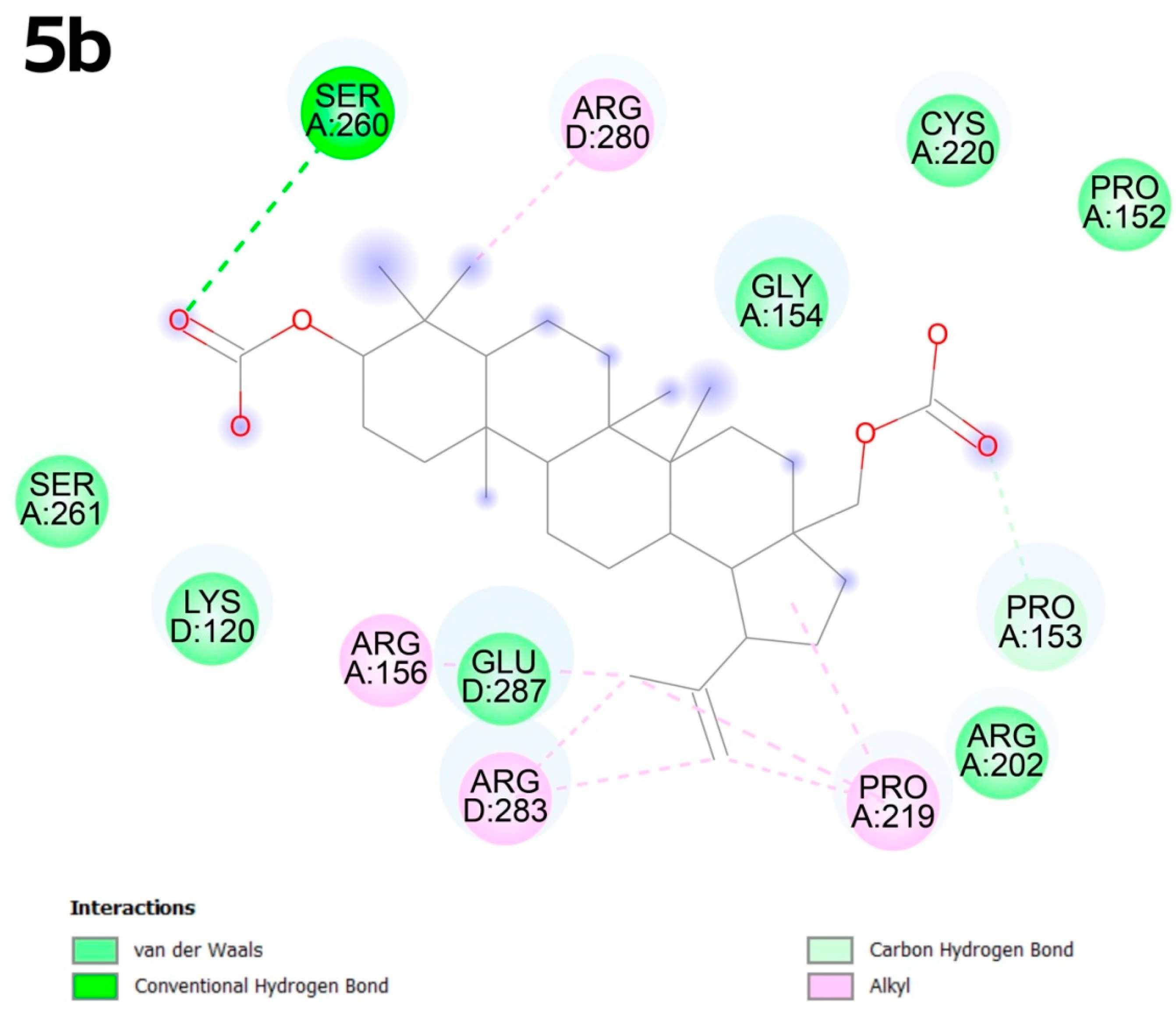
Compound 5b exhibits alkyl interactions with amino acids such as arginine at positions D:280, A:156 and D:283. The electro-negative oxygen atom in the carboxyl group promotes the formation of a conventional hydrogen bond with serine A:260, while the oxygen atom in the acyl group promotes the formation of van der Waals-type bonds (Figure 4).
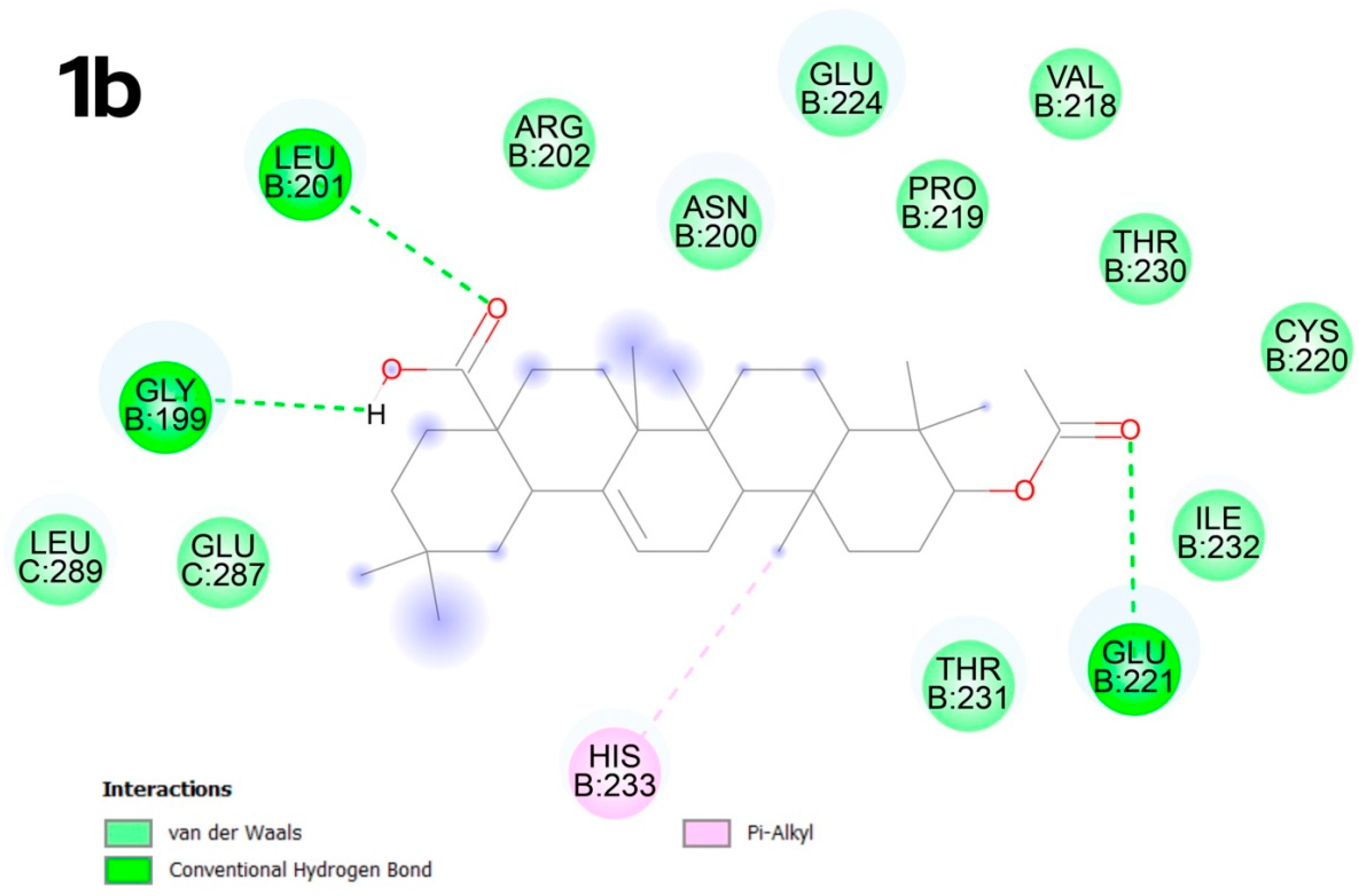
In contrast, compound 1b in the C2 pocket forms numerous van der Waals bonds with the surrounding amino acids, as well as conventional hydrogen bonds between the acyl group and the amino acids GLY B:199, LEU B:201 and between the oxygen atom in the carboxyl group and GLU B:221. An unsaturated bond promotes the formation of π-type alkyl interactions with histidine B:233 (Figure 5).
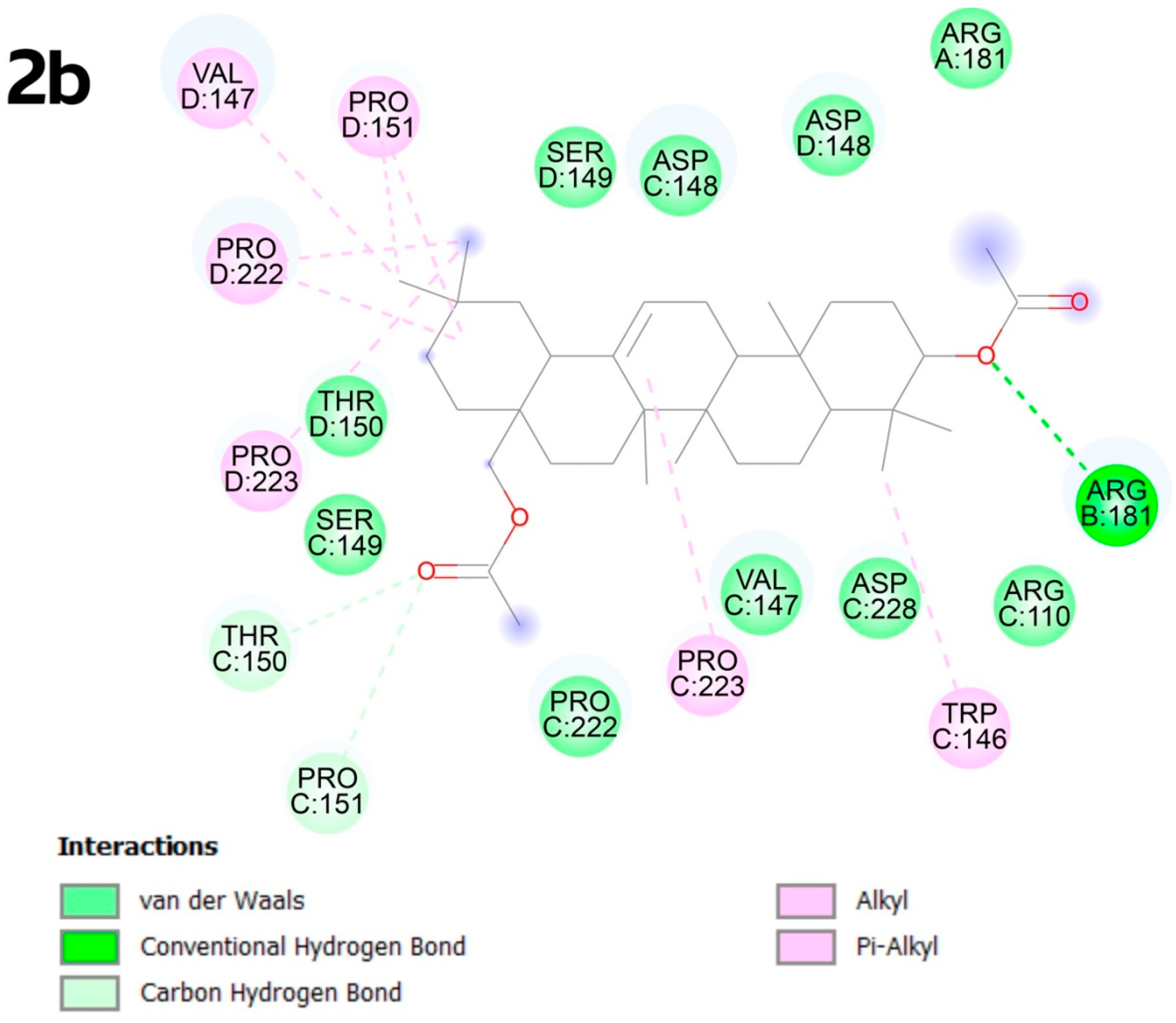
In the C3 pocket, the best result was obtained for compound 2b, which exhibits multiple alkyl bonds with proline amino acids at positions D:151, D:222, D:223, as well as with VAL D:147 and TRP C:146. Additionally, the unsaturated bond of molecule 2b forms a π-type bond with PRO C:223. Carbon–hydrogen bonds occur due to the presence of oxygen atoms in the functional groups, forming interactions with THR C:150, PRO C:151 and ARG B:181 (Figure 6).
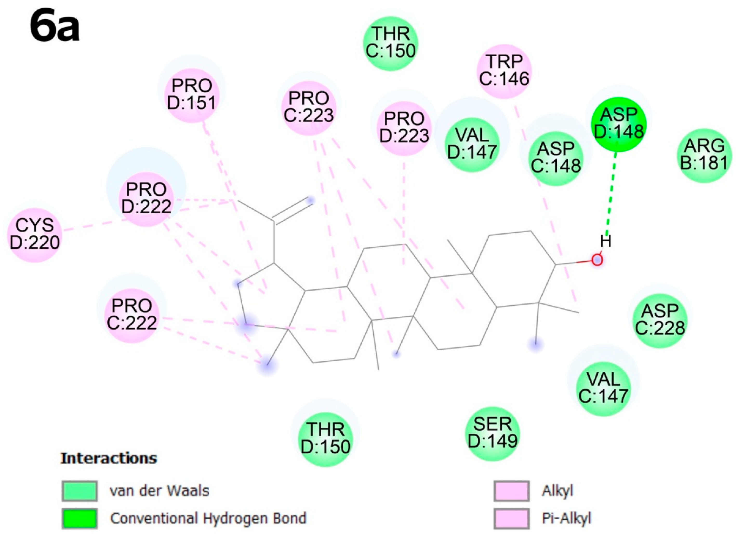
In pocket C4, compound 6a shows predominantly alkyl interactions with proline at positions D:151, C:222, D:222, D:223, and CYS D:220 and TRP C:146. The hydroxyl group at the C-3 atom favors the formation of a conventional hydrogen bond with ASP D:148 (Figure 7).
In the C5 pocket, the best results were obtained for compound 4b, in which van der Waals-type bonds predominate. The presence of oxygen atoms in the functional groups enables the formation of conventional hydrogen bonds with SER D:106, ARG B:181, GLN B:192, a carbon–hydrogen bond with HIS C:115 and an alkyl interaction with PRO B:177 (Figure 8).

2.5. Antioxidant Assay

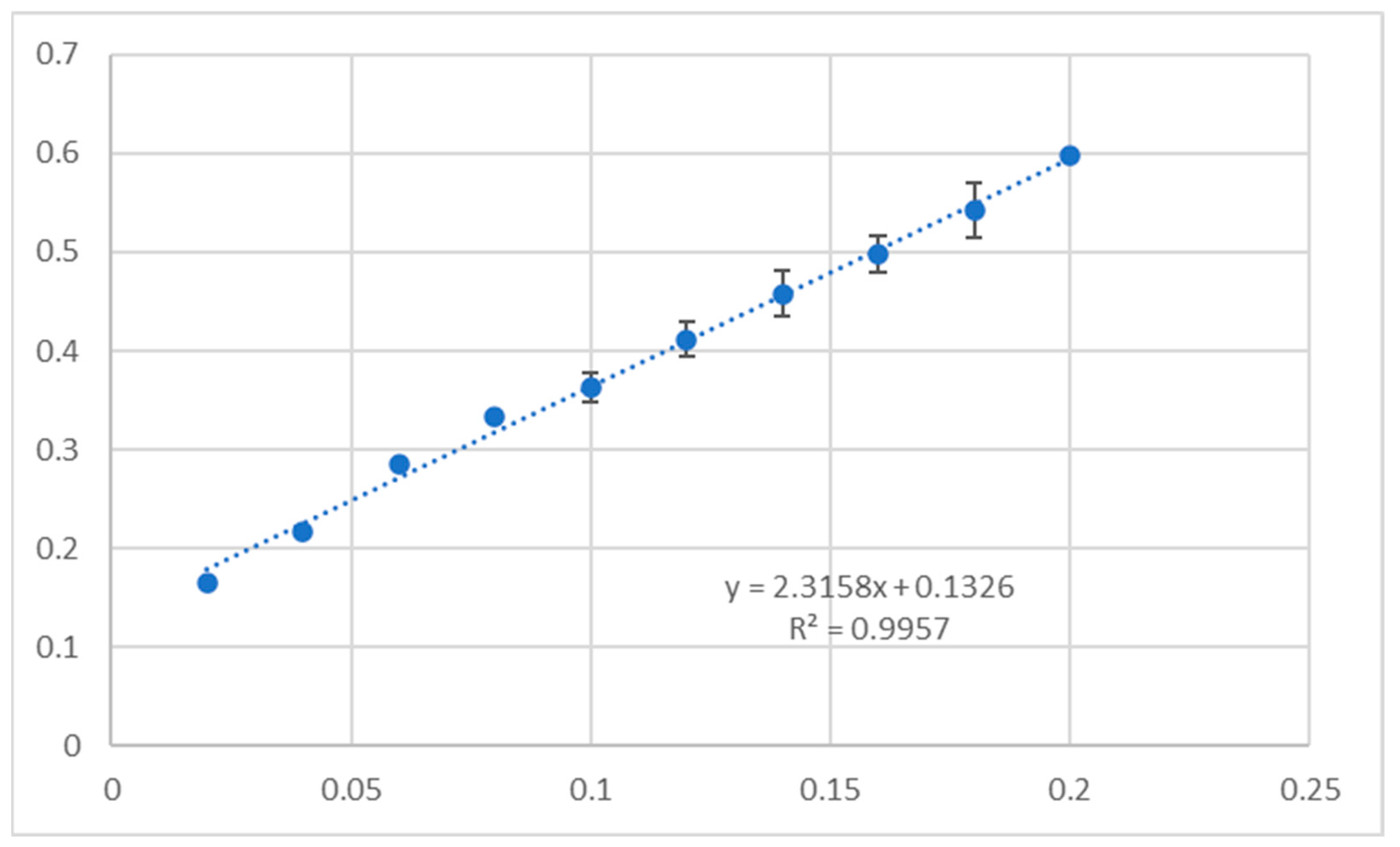
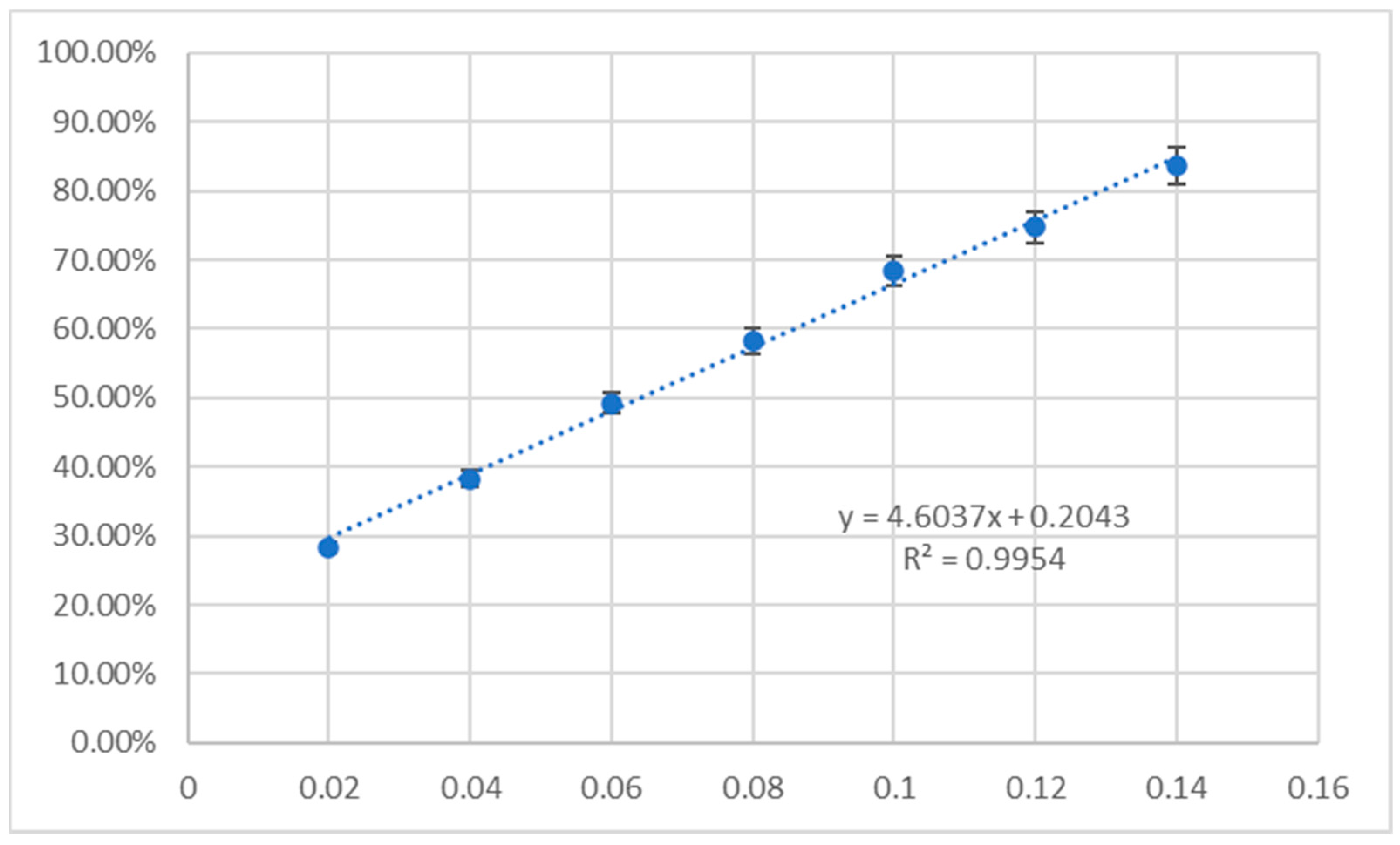
The results of the antioxidant activity of unsubstituted triterpenes 1a6a and their acetylated derivatives 1b6b evaluated with CUPRAC (cupric reducing antioxidant capacity) and DPPH (2,2-diphenyl-1-picrylhydrazyl) assays are given in Table 8. The results are presented as % inhibition of the copper(II) ions and Trolox equivalent calculated from the standard curve (Figure 9) and as % inhibition of the DPPH radical and Trolox equivalent, calculated from the standard curve (Figure 10).

2.6. ADMETox Analysis

The physicochemical properties, pharmacokinetics and ADMETox (adsorption, distribution, metabolism, excretion and toxicity) activity of compounds 1a6a and 1b6b are given in Table 9.
Additional information concerning the above tests can be found in Supplementary Materials of our earlier publication [66].

3. Discussion

3.1. Acylation of Triterpenes 1a6a

Acylation of the C-3 hydroxyl function of triterpene with carboxylic acid anhydrides gives the appropriate 3-O-acyl derivatives. Such a reaction is usually conducted using dried pyridine at room temperature [67] or in refluxed acetic anhydride [68]. In order to avoid using toxic pyridine, we decided to use the second of the methods we mentioned in another publication [68]. This reaction consisted of heating the triterpene in boiling acetic anhydride, and after the reaction was completed (TLC control), the reaction mixture was poured into water, and the separated white precipitate was filtered, washed with water, dried and crystallized.

3.2. Structure–Activity Analysis

Among the tested unsubstituted and acylated triterpenes (1a6a, Figure 1, and 1b6b, respectively), most of the substances showed antineoplastic activity, with Pa values exceeding 0.900 for almost all tested compounds (1a6a and 1b6b, Table 2). Allobetulin (3a) and its acetyl derivative (3b) showed the highest probability of high efficacy in the case of treatment of colon cancer (Pa > 0.900, Table 2) and only slightly lower in the case of treatment of lung cancer (Pa > 0.800).
The expected cytotoxic activity against cancer cells may be based on various mechanisms, e.g., enhancement of the process of apoptosis, stimulation of caspase 3 and caspase 8 or stimulation of TF NF kappa B/TF factor (Table 2).
Apoptosis, or programmed cell death, plays a vital role in cancer treatment by enabling the selective elimination of malignant cells while minimizing damage to surrounding healthy tissue. This highly regulated process is essential for maintaining cellular homeostasis and acts as a natural barrier against tumorigenesis. As a result, restoring the apoptotic machinery in cancer cells has become a central goal of modern oncology [69]. Substances that initiate or enhance this process play a key role in the apoptosis process. Such substances, called apoptosis agonists, activate the molecular pathways that lead to programmed cell death, restoring or intensifying the cell’s ability to undergo apoptosis when it is no longer viable or safe for the organism [69]. As shown by the prediction results using the PASS method (Table 2), oleanolic acid (1a) has the greatest chance of effective pro-apoptotic activity. Numerous publications in the scientific literature confirm these prediction results and demonstrate the proapoptotic effect of oleanolic acid (e.g., [70]).
Central to the execution of apoptosis is a family of proteolytic enzymes known as caspases (cysteine–aspartic proteases), which function as key molecular effectors of the apoptotic cascade. Caspases are generally classified into two groups: initiator caspases (e.g., caspase-8 and caspase-9), which respond to apoptotic signals and activate downstream pathways, and executioner caspases (e.g., caspase-3, -6 and -7), which carry out the dismantling of cellular components such as DNA, cytoskeletal proteins and nuclear structures. Activation of caspases can occur via two main apoptotic pathways: the extrinsic pathway, triggered by death receptors like Fas and TRAIL-R that activate caspase-8, and the intrinsic (mitochondrial) pathway, which involves mitochondrial outer membrane permeabilization and activation of caspase-9 via the apoptosome. Both pathways converge on executioner caspases, leading to systematic and non-inflammatory cell death. The essential role of caspases in apoptosis makes them critical targets for therapeutic intervention in diseases characterized by either excessive cell death (e.g., neurodegenerative diseases) or impaired apoptosis (e.g., cancers) [71,72,73].
Of the tested triterpenes 1a6a and 1b6b, the vast majority show a very high probability of caspase stimulant activity (Pa > 0.900, Table 2), in particular caspase 3 stimulant activity, with oleanolic acid again being the particularly active compound. This direction of pharmacological activity has also been confirmed in experimental studies and published [74].
Interestingly, betulinic acid (4a), as the only one among the twelve tested triterpenes (1a6a and 1b6b), showed a moderate probability of caspase stimulant activity (Table 2). Betulinic acid (4a) has been widely studied for its anticancer potential, particularly its ability to induce apoptosis in various cancer cell types, e.g., [75,76]. However, its effectiveness as a direct caspase stimulant is limited. While triterpene 4a can activate caspases downstream in the apoptotic cascade, especially caspase-3, it does so indirectly through mitochondrial dysfunction rather than direct activation of the caspase system. Specifically, betulinic acid primarily targets the intrinsic (mitochondrial) pathway, where it induces mitochondrial outer membrane permeabilization (MOMP), leading to cytochrome c release and apoptosome formation, which in turn activates caspase-9 and subsequently caspase-3 [75,76]. However, betulinic acid does not efficiently engage the extrinsic (receptor-mediated) pathway, nor does it directly bind or activate caspases themselves. Furthermore, its pro-apoptotic effects are often cell-type specific, and in some resistant tumor models, betulinic acid fails to induce strong caspase activation due to compensatory anti-apoptotic mechanisms such as upregulation of Bcl-2 or survival [77]. As a result, although betulinic acid is a promising compound for apoptosis induction, its role as a direct caspase stimulant is limited, and its efficacy may require combination with other agents that enhance or bypass caspase activation [77].
Nuclear factor kappa B (NF-κB) plays a key role in cancer by regulating inflammation, cell survival and proliferation genes. In many cancers, NF-κB is constitutively activated, enabling tumor cells to evade apoptosis through upregulation of anti-apoptotic genes like Bcl-2 and IAPs [78,79]. It also promotes angiogenesis, metastasis and therapy resistance by inducing cytokines, growth factors and survival proteins. Moreover, NF-κB shapes the tumor microenvironment by promoting chronic inflammation and immune evasion [80]). These functions make NF-κB a critical target in cancer therapy, though its role in normal immune function complicates systemic inhibition. As PASS prediction revealed (Table 2), both unsubstituted triterpenes 1a6a (Figure 1) and their acetyl derivatives (1b6b, Figure 2) showed a high probability of transcription factor (TF)-stimulating activity, in particular TF NF kappa B, with a Pa value exceeding 0.900 for the vast majority of compounds tested (1a6a and 1b6b, Figure 1 and Figure 2).
Triterpenes are known to modulate the transcription factor NF-κB due to their ability to influence key signaling pathways involved in inflammation and immune responses [81]. Many triterpenes, such as betulinic acid and oleanolic acid, can either activate or inhibit NF-κB depending on the cellular context and structure of the compound. Their lipophilic nature allows them to interact with membrane-bound receptors or intracellular signaling proteins like IKK (IκB kinase), influencing the degradation of IκB and subsequent translocation of NF-κB to the nucleus [81]. In some cancer or immune cells, triterpenes can stimulate NF-κB activity, potentially enhancing immune surveillance or inflammatory responses, while in other contexts they may inhibit chronic inflammation by blocking NF-κB activation [81]. This dual capacity makes them valuable pharmacological tools for modulating NF-κB signaling in both cancer and inflammatory diseases.
High Pa (probability of activity) values predicted by the PASS software for antitumor activity indicated a strong likelihood that the tested compounds possess cancer-inhibiting properties. This computational evidence supported the rationale for selecting the mutant p53 Y220C protein as a molecular target, as it plays a key role in tumor suppression and is frequently mutated in cancers. Targeting p53 Y220C aligns with the predicted bioactivity of the compounds, offering a focused approach to exploring their potential as reactivators or stabilizers of this dysfunctional tumor suppressor [82].

3.3. Cytotoxic Activity of Triterpenes 1a6a and 1b6b

3.3.1. In Vivo Assay

Six triterpenes with free hydroxyl function at the C-3 position, oleanolic acid (1a), erythrodiol (2a), allobetulin (3a), betulinic acid (4a), betulin (5a) and lupeol (6a) and their 3-O-acetyl derivatives (1b6b, respectively), were subjected to the MTT assay. Four of these compounds have a carboxyl function at the C-17 position (1a, 4a, 1b, and 4b), four triterpenes had a primary hydroxyl group at the C-17 position (2a, 5a, 2b, and 5b) and four compounds had other functionalities at the C-17 position (3a, 6a, 3b and 6b).
Among the six triterpenes, 1a6a that were tested against six human cancer cell lines—HeLa (cervical carcinoma), KB (nasopharynx carcinoma), MCF-7 (breast carcinoma), A-549 (lung carcinoma), PC-3 (prostate carcinoma) and SKOV-3 (ovarian carcinoma)—the most active turned out to be oleanolic acid (1a), with IC50 values in the range of 8.79 µM (for A-549 cells) to 18.63 µM (for PC-3 cells), betulinic acid (4a), with IC50 values from 9.52 to 35.21 µM (for SKOV-3 and KB cells, respectively) and betulin (5a), with IC50 values from 6.22 to 19.74 µM (PC-3 and KB cells, respectively) (Table 3). Reduction of the C-17 group of oleanolic acid (1a) leading to primary hydroxyl (as in a molecule of erythrodiol, 2a) caused the obtained compound to be 4.4–7.2 times less active for HeLa, KB, MCF-7 and A-459 cell lines than oleanolic acid 1a. The transformation of betulin 5a into allobetulin 3a caused a worsening of anticancer activity of the triterpene 3a, with the IC50 values from about 20 to 70 µM (Table 3).
Acetylation of erythrodiol (2a), allobetulin (3a), betulin (5a) and lupeol (6a) led to derivatives with low or very low levels of cytotoxic activity, with IC50 values mainly in a range from 40 to > 100 µM. At the same time, the acetylation of oleanolic acid (1a) and betulinic acid (4b) led to a significant improvement in cytotoxic activity. The IC50 value for 3-O-acetyloleanolic acid (1b) varied from 0.09 µM (for the SKOV-3 cell line) to 1.86 µM (for MCF-7 cells); for 3-O-acetylbetulinic acid (4b) the IC50 value varied from 0.93 µM (for PC-3 cells) to 1.62 µM (for HeLa cells) (Table 3).
According to the literature data [83], chemical compounds present good cytotoxicity for IC50 ≤ 10 μM and moderate cytotoxicity for 10 μM < IC50 ≤ 30 μM. Taking into account the above statements and the IC50 values for six unsubstituted triterpenes, 1a6a, and their acetyl derivatives, 1b6b, the compounds with very high cytotoxic activity are acetyloleanolic acid (1b) and acetylbetulinic acid (4b), while their parent compounds, i.e., oleanolic acid (1a) and betulinic acid (4a), as well as betulin (5a) can be classified as compounds of high or moderately high activity (depending on the cancer cell line these compounds act on).

3.3.2. Selectivity Index

The selectivity index for a given cancer cell line is the quotient of the IC50 value of a normal cell line (e.g., HDF, as in our experiments) and the IC50 value of a cancer cell line. This quotient is an important indication in research on the anticancer activity of both extracts and other preparations from materials of natural origin, individual substances isolated from these extracts and substances of natural origin subjected to chemical modifications. Sometimes, the SI value is a factor that determines whether tests of the cytotoxic activity of a preparation or substance will be continued. As Pena-Moran [84] states, the limit value of the selectivity index determining the validity of further research is at least 10. According to Valderrama, for individual substances that would become potential anticancer agents, the selectivity index limit value is at least 2 [85].
As Table 4 shows, the best selectivity index, approximately 2, for almost all tested triterpenes was obtained for two cancer cell lines: PC-3 and SKOV-3. Among all tested triterpenes, triterpene 1a (oleanolic acid) and 4b (diacetylbetulin) showed the highest results. For the first of the mentioned triterpenes (1a), the SI for the two cancer cell lines most susceptible to the action of triterpenes (PC-3 and SKOV-3) was much lower than for the remaining lines (HeLa, KB, MCF-7 and A-549) and was slightly above 1 (1.33 and 1.32, respectively; Table 4). In turn, the second triterpene with the most favorable selectivity index, i.e., diacetylbetulin (4b), showed the highest selectivity index value of approximately 3 for the PC-3 and SKOV-3 lines (Table 4).

3.3.3. Apoptosis

The ability of all tested compounds to induce apoptosis was investigated with two cancer cell lines (SKOV-3 and PC-3). All twelve compounds (1a6a and 1b6b) induced apoptosis to a similar extent in both cancer cell lines. Betulin (5a) was the most effective in inducing apoptosis as an internal standard. The lower concentration of 0.1 µg/mL showed a mean apoptotic index of 5.54 (Table 5). Oleanolic acid (1a), the second internal standard for investigated triterpenoids, showed an apoptotic index of 5.16 and 5.27, respectively, for the SKOV-3 and PC-3 cell lines (Table 5).
For other compounds, it was found that increased concentration also increased apoptotic indices. The investigation has shown that compounds 1b (acetyloleanolic acid) and 4b (acetylbetulinic acid) were the most effective in inducing apoptosis.

3.4. Molecular Docking

CB-Dock2 is an advanced molecular docking program distinguished by its high accuracy and efficiency in predicting binding positions and affinities between small molecules and target proteins. By considering ligand and protein flexibility and using a multiple protein conformation approach, CB-Dock2 better represents dynamic interactions than other rigid docking models. The program also uses advanced scoring functions and optimization algorithms, significantly improving predictions’ accuracy. CB-Dock2’s effectiveness has been repeatedly validated, making it a valuable tool in drug discovery and development [86,87,88].
The CB-Dock2 is not just a rough binding site determination tool but an advanced structure-based docking platform that also uses machine learning elements. It was designed as a blind docking method that integrates several docking approaches, including classical structural algorithms based on the detection of caverns on the protein surface and template-assisted docking of homologous protein–ligand complexes. This combination of structural and template algorithms allows CB-Dock2 to identify the ligand binding site precisely. According to the literature, the improved CB-Dock2 algorithm achieves about 85% accuracy in predicting the correct ligand binding pose (RMSD < 2.0 Å) in validation tests, outperforming the original CB-Dock and other popular blind docking tools. This means that CB-Dock2 provides accuracy comparable to traditional docking platforms and has internal validation mechanisms (e.g., via optional matching to homologous structures) that increase the results’ reliability [89].
In order to translate the PASS results into a specific molecular target, we considered proteins central to the regulation of cancer cell survival. Ultimately, we selected the p53 tumor suppressor protein (mutant Y220C) as a docking target, as its reactivation/stabilization is a known pro-apoptotic mechanism in cancer cells. The p53 Y220C variant has an allosteric pocket to which small molecules can bind to restore the function of this protein—making it an attractive target in the context of PASS indications with anticancer potential.
As a validation step, we redocked the co-crystallized ligand carbazole KG3 into the p53 Y220C structure (PDB ID: 8DC4]) using AutoDock Vina (version 1.2.0) integrated within the CB-Dock2 framework. This validation approach is consistent with standard docking protocols, where redocking of known ligands helps confirm the accuracy and reliability of the docking parameters [90,91]. Comparative analysis of binding energies relative to the co-crystallized ligand allowed for identifying compounds with potential to stabilize the p53 Y220C mutant conformation and restore its tumor suppressor function [92].
The tumor suppressor protein p53, which has long been the subject of intensive research, was chosen for docking, with its different isoforms and interactions playing a key role in cellular processes and disease pathogenesis. The p53 Y220 protein is a particular variant and has attracted considerable attention due to its potential in molecular docking applications. The p53 Y220 is a mutated form of the p53 protein found in the Protein Data Bank under ID 8DC4. This specific mutation has been linked to several oncogenic properties, including the ability to confer chemo-resistance and promote the growth and spread of malignant tumors [93,94].
A crystal structure study of p53 Y220, covalently bound to carbazole KG3 (PDB ID: 8DC4), was carried out to identify potential pockets on the protein’s surface, using the CB-Dock2 server. This enabled molecular docking to assess the interactions of compounds 1a6a and 1b6b with these pockets.
Using the CurPocket method, the CB-Dock2 server extracted five key pockets in the 8DC4 structure. The pockets were ordered by volume, from largest (C1) to smallest (C5). The largest pocket (C1) had a volume of 3942 Å3, while the smallest (C5) had a volume of 370 Å3. Details of the location and size of these pockets are shown in Table 7 and Figure 3.
Docking into multiple cavities (C1–C5) was conducted to identify each compound’s most favorable binding sites, as p53 Y220 is a mutant with altered surface topology. While energies are provided for all pockets, biological interpretation focuses on the most energetically favorable interactions—e.g., compound 2b in pocket C3 (−10.1 kcal × mol−1), likely corresponding to the KG3 binding region. This approach ensures comprehensive exploration and prevents oversight of high-affinity noncanonical sites.
Molecular docking analysis showed that all compounds tested (1a6b) could bind to each of the pockets (C1–C5), achieving different Vina score values, which reflect the strength of the ligand–protein interaction. The highest binding value was observed for compound 5b in the C1 pocket, achieving a Vina score of −9.1 kcal × mol−1. Another favorable interaction was shown by compound 1b in the C2 pocket with a value of −9.0 kcal × mol−1. In the C3 pocket, the highest Vina score value was achieved by compound 2b (−10.1 kcal × mol−1), while for the C4 pocket the best result was obtained for compound 6a (−9.6 kcal × mol−1). In the last pocket C5, the highest Vina score value was achieved by compound 4b, with a result of −9.0 kcal × mol−1.
A lower affinity value (Vina score) indicates a stronger binding interaction between the molecules, indicating that all compounds tested showed good docking results and could inhibit interactions between the 8DC4 protein and cellular proteins. In the largest pocket, C1, the highest binding value was achieved for compound 5b, while compound 1b stood out in the C2 pocket, compound 6a in the C4 pocket and compound 4b in the smallest pocket, C5.
Molecular docking to the pockets identified on the surface of the p53 protein revealed various strong interactions between the compounds tested and the protein, suggesting that some of these compounds may represent potential inhibitors of p53. The best result was achieved by compound 2b in the C3 pocket, which is about 4.8 times smaller than the largest pocket (C1).
Although larger pockets on the surface of the protein may offer more stable and potent binding sites for ligands, appropriate modification of the molecule—in this case, the addition of two acyl groups—gave better results than the variant with free hydroxyl groups. This result may suggest that the pocket prefers alkyl interactions, increasing binding stability.

3.5. Antioxidant Activity

The antioxidant activity results for the tested triterpenes 1a6a and 1b6b, measured using the CUPRAC and DPPH assays (Table 8), reveal significant differences in the antioxidant mechanisms of individual compounds. Analyzing the data for derivatives 1a6a and 1b6b, we can observe substantial variability in activity across both assays, reflecting the diverse antioxidant capacities of these compounds due to their structural properties and mechanisms of action.
The CUPRAC assay results show that compounds such as 5a, 2b and 1b exhibit relatively high reducing activity (0.29900, 0.24016 and 0.21986, respectively; Table 8), suggesting that they have an efficient electron transfer capacity, which is a crucial factor in this assay. On the other hand, derivatives like 3b and 3a show significantly lower activity (0.03212 and 0.08248, respectively; Table 8), indicating a reduced ability to participate in redox reactions involving the reduction of copper(II) ions to copper(I). In contrast, the DPPH assay, which measures the ability of compounds to donate hydrogen atoms to neutralize free radicals, shows a different activity profile. For instance, derivative 5a, which displayed the highest activity in the CUPRAC assay (0.29900; Table 8), exhibited one of the lowest activities in the DPPH assay (0.01077; Table 8). This suggests that the antioxidant mechanism of compound 5a is primarily based on electron transfer rather than hydrogen atom donation.
Meanwhile, compound 2a demonstrated relatively similar, albeit lower, activity in both assays (0.09414 in CUPRAC and 0.02092 in DPPH; Table 8), indicating a more versatile antioxidant mechanism encompassing electron transfer and hydrogen atom donation. These results underscore the crucial influence of a compound’s structure on its antioxidant properties. For example, derivative 6a, which scored 0.13602 in the CUPRAC assay (Table 8), exhibited moderate activity in the DPPH assay (0.01570; Table 8), suggesting it effectively participates in both antioxidant mechanisms. Conversely, compounds such as 3b, which showed the lowest activity in both assays (0.03212 in CUPRAC and 0.00182 in DPPH; Table 8), have limited antioxidant capacity, possibly due to an unfavorable structural configuration that impedes electron transfer and hydrogen atom donation. Further analysis of derivatives 1b, 2b and 4b, which scored relatively high in the CUPRAC assay (0.21986, 0.24016 and 0.19986, respectively; Table 8) but much lower in the DPPH assay, highlights the importance of electron transfer as the dominant antioxidant mechanism for these compounds. Their intense activity in the CUPRAC assay suggests the presence of functional groups that favor redox reactions, which are less involved in hydrogen atom transfer mechanisms, explaining their lower DPPH activity—meanwhile, compounds 5b and 5a present an intriguing contrast. While 5a showed the highest CUPRAC activity (0.29900; Table 8), it had relatively low DPPH activity (0.01077; Table 8). In contrast, 5b, although demonstrating lower CUPRAC activity (0.17609; Table 8), displayed a significantly higher DPPH result (0.00786; Table 8), suggesting structural differences that influence their antioxidant mechanisms differently in the two assays.
In conclusion, the results indicate that the tested triterpenes exhibit varied antioxidant properties depending on the evaluation method. In particular, derivatives with high CUPRAC activity, such as 1b, 2b and 4b, may have promising potential for therapeutic applications in oxidative stress-related conditions that require strong reducing properties.

3.6. ADMETox Analysis

Among the tested compounds, unsubstituted and acetylated triterpenes (1a6a and 1b6b, respectively), most of the above-mentioned triterpenes had a favorable molecular weight and also showed favorable values of most parameters determining the physicochemical properties, e.g., nHA, nHD, nRot, nRing, nHet, fChar, nRig, flexibility and TPSA (Table 9). Due to the very low solubility of the above compounds in water, the values of logP and logD were outside the optimal range (Table 9).
The QED (quantitative estimation of drug-likeness) test showed that all the tested compounds (1a6a and 1b6b) are similar to known drugs (QED ≥ 0.67). The number of sp3 hybridized carbons in the above 12 triterpenes is ≥0.42, which is a favorable value. The MCF18 value for all tested triterpenes (1a6a and 1b6b, respectively) exceeds 45, which means a high level of novelty, following the trends currently observed in medicinal chemistry. In turn, the NP value (natural product-likeness, Npscore) within the range of 3.0–3.3 (Table 9) confirms the high similarity to compounds of natural origin (from which compounds 1b6b were obtained).
PAINS, BMS and Chelator tests are negative for almost all tested triterpenes, which means that there are no unfavorable elements of the structure of the molecules of these substances, which can be potentially responsible for toxicity or may, for example, enter into chemical interaction with other chemical substances present in the body (Table 9).
The Caco-2 test showed excellent or very good permeability of all of the tested triterpenes (1a6a and 1b6b). The excellent or outstanding absorption and permeation profile has been confirmed by subsequent ADMET parameters, e.g., HIA, F20% (Table 9).
Theoretical predictions indicate that almost all tested triterpenes (1a6a and 1b6b) will probably bind well to plasma proteins and perfectly penetrate the blood-brain barrier, showing excellent volume distribution (about 1 L × kg−1) and proper percentage of the fraction unbound to plasma proteins (Table 9).
The excretion of the tested triterpenes is predicted by applying CL and T1/2 tests. The clearance of a drug (CL) is an important pharmacokinetic parameter that defines, together with the volume of distribution, the half-life and thus the frequency of dosing of a drug. The clearance of the tested triterpenes (1a6a and 1b6b) was in a range of 2–6 mL × min−1 × kg−1 (apart from triterpene 2a, with CL about 10.5 mL × min−1 × kg−1), with a high probability of being short half-life compounds (T1/2 ≤ 0.300) (Table 9).
Almost all triterpenes showed a very low probability of toxicity and biotoxicity. This means that these compounds can be used as non-toxic drugs (Table 9).

4. Materials and Methods

4.1. NMR

The 1H- and 13C-NMR spectra of compounds 1a6a and 1b6b were recorded using a Bruker Advance 600 MHz spectrometer (Billerica, MA, USA) with CDCl3 as a solvent, with tetramethylsilane (TMS) as an internal standard (Sigma-Aldrich®, Darmstadt, Germany). Chemical shifts (δ) were expressed in parts per million (ppm) relative to tetramethylsilane (TMS) as an internal standard, using CDCl3 as a solvent. Coupling constants (J) were expressed in Hertz (Hz).

4.2. Acetylation of Triterpenes 1a6a

4.2.1. General Information

Oleanolic acid (1a) was purchased from Bio-Tech®, Beijing (China). Erythrodiol (2a) and allobetulin (3a) were synthetized from oleanolic acid (1a) and betulin (5a), respectively. Betulinic acid (4a), betulin (5a) and lupeol (6a) were purchased from Natchem® (Kraków, Poland). The purity of the substrates and the obtained products was confirmed by HP TLC and NMR methods.
All commercially available solvents and reagents used in our experiments were graded “pure for analysis” (Sigma-Aldrich®, Darmstadt Germany; Fluka®, Charlotte, NC, USA; Chempur®, Piekary Śląskie, Poland; and POCh®, Gliwice, Poland). The solvents were dried according to the usual procedures.
The melting points were measured with the Büchi apparatus in an open capillary and are uncorrected.

4.2.2. Syntheses

Oleanolic acid or methyl oleanolate reduction with LiAlH4: A solution of oleanolic acid (1a) or its methyl ester (1 mmol) in 5 mL of dried THF was added dropwise to a suspension of LiAlH4 (10 mmol) in 5 mL of THF, and the resulting mixture was stirred for 1 h. Next, EtOH was added dropwise, and the mixture was filtered off and evaporated until dry. The obtained white solid was crystallized from EtOH. Yield: 93.0%, m.p. 230–232 °C (lit. m.p. 232.5–234 °C [95]).
General procedure of triterpene acylation: A solution of 1 mmol of triterpene 1a6a in 5 mL of dried acetic anhydride was refluxed for 30 min. After cooling, the mixture was poured into the 5-fold volume of water, and the resulting precipitate was filtered off, washed with water, dried and crystallized.
  • 3-O-Acetyloleanolic acid (1b): m.p. 267–268 °C (lit. m.p. 268–269 °C [96]).
  • 3,28-di-O-Acetylerythrodiol (2b): m.p. 185–187 °C (lit. m.p. 185–185.5 °C [95]).
  • 3-O-Acetylallobetulin (3b): m.p. 285–285 °C (lit. m.p. 287–287.5 °C [97], lit. m.p. 280–281 °C [98]).
  • 3-O-Acetylbetulinic acid (4b): m.p. 290–291 °C (lit. m.p. 285–290 °C [99]).
  • 3,28-di-O-Acetylbetulin (5b): m.p. 215–217 °C (lit. m.p. 213–218 °C [98]).
  • 3-O-Acetylolupeol (6b): m.p. 216–218 °C (lit. m.p. 217–218 °C [98]).

4.3. SAR Analysis

The structure–activity relationship analysis was performed using the PASS (prediction of activity spectra for substances) computer system [100]. It is a program that predicts many types of pharmacological activities and their mechanisms based on the structure of the analyzed chemical substance. Such mathematical analysis results in a list of probable pharmacological activities or their mechanisms. The probability of a given activity occurring (marked with the Pa symbol) or the non-occurrence of a given activity (marked with the Pi symbol) is assessed on a scale from 0.000 to 1.000.

4.4. MTT Assay

All the cell lines and mediums were obtained from the American Type Culture Collection (ATCC) supplied by LGC-Standards (Lomianki, Poland). Human cancer cell lines were cultured as follows: HeLa (human cervix carcinoma) and KB (human oral squamous carcinoma) lines were cultured in DMEM (Dulbecco’s Modified Eagle’s Medium); MCF-7 (human breast carcinoma) and A-549 (human lung carcinoma) were cultured in RPMI 1640 medium; PC-3 (human prostate carcinoma) was cultured in F-12K medium; SKOV-3 (human ovarian cystadenocarcinoma) was cultured in McCoy’s modified medium; HDF (human normal dermal fibroblasts) was cultured in Fibroblast Basal Medium. Each medium was supplemented with 10% fetal bovine serum, 1% L- glutamine and 1% penicillin/streptomycin solution. The cell lines were kept in an incubator at 37 °C.
All the tested cell lines (HeLa, KB, MCF-7, A-549, PC-3, SKOV-3 and HDF) were subjected to MTT assay. In short, this assay is based on the reaction between mitochondria enzymes (dehydrogenases) of tested cell lines and 3-(4,5-dimethylthiazol-2-yl)-2,5-diphenyltetrazolium bromide (MTT). The final product of this reaction gives violet formazan crystals. The number of live cells is proportional to the amount of formazan and is represented by the violet dye reagent. The details of the experiments are given in [19].

4.5. Apoptosis

The induction of apoptosis by tested compounds was determined with Cell Death Detection ELISA Plus from Roche Diagnostics GmbH (Mannheim, Germany).
Cell Death Detection ELISA Plus is an enzyme immunoassay for determining cytoplasmic histone-associated DNA fragments (mono- and oligonucleotides) after induced cell death. Cells (104) were pipetted into wells of 96-well plates, and test compounds were added at final concentrations of 0.1, 1.0 and 10.0 µg × mL−1. Cells incubated in the absence of test compounds served as negative controls. After 4 h of incubation, cells were harvested by centrifugation of the plates for 10 min at 200× g. Supernatants were removed carefully, and cell pellets were resuspended in 200 µL of cell lysis buffer and incubated for 30 min at room temperature. After centrifugation of the plates (10 min at 200× g), 20 µL of cell lysates was transferred in duplicate into wells of provided streptavidin-coated microplates. Then, 80 µL of the immunoreagent containing a mixture of anti-histone-biotin monoclonal antibody and anti-DNA-peroxidase monoclonal antibody was added to each well. The microplates were covered with a provided adhesive foil and incubated for 2 h at room temperature. Thereafter, the solution in the wells was removed by tapping the microplate. The wells were rinsed three times with incubation buffer, and 100 µL of substrate (2,2′-azino-bis [3-ethylbenzothiazoline-6-sulphonic acid], ABTS) was added to each well. After 10–20 min, 100 µL of ABTS stop solution was pipetted to each well, and the absorbance was read at 405 nm (test wavelength) and 490 nm (reference wavelength). The apoptotic index was calculated based on the enrichment of mono- and oligonucleosomes according to the following equation:
apoptotic index = [{A405(sample) − A490(sample)}/{A405(negative control) − A490(negative control)}]

4.6. Molecular Docking

Ligand preparation: The ligands were prepared by first drawing the two-dimensional (2D) structures of compounds 1a6a and 1b6b using ChemDraw 22.0.0. These structures were then converted into three-dimensional (3D) representations in OpenBabel format [101] to obtain the coordinates representing the most energetically favorable conformation. Geometry optimization was performed using Avogadro version 1.2.0 software. The Steepest Descent algorithm applied the Universal Force Field (UFF). The optimized 3D structures of compounds 1a6a and 1b6b were saved as SDF files. They were used as input files for docking analysis on the CB-Dock2 server.
Protein preparation: The crystallographic data for the p53 protein (cancer mutation Y220C) with the 8DC4 structure was sourced from the RCSB Protein Data Bank (PDB ID: 8DC4) at a resolution of 2.40 Å. P53 is covalently bound to the carbazole KG3 in this X-ray crystal structure. The downloaded 8DC4.pdb protein molecule was not manually prepared, as steps such as removing ligands and crystal water molecules and adding missing hydrogen atoms were automatically handled by the CB-Dock2 server.
Detecting cavities and uploading ligands: After the p53 molecule was imported into the CB2-Dock server, the number of cavities for docking was set to 5 under the “more parameters” option. The email address to receive the output files was specified, and the Search Cavities function was selected. Ligand structures (compounds 1a6a and 1b6b) were uploaded to the CB-Dock2 server [101], and docking was initiated through the platform’s web interface using default parameters. The tool automatically identified the top five potential binding cavities based on cavity detection algorithms.

4.7. Antioxidant Activity

The CUPRAC reagent (7.5 mM ethanolic 96% neocuproine solution, 10 mM CuCl2 × H2O solution and an ammonium acetate buffer of pH 7.0) and DPPH reagent (2,2-diphenyl-1-picrylhydrazyl 0.2 mM solution) were applied.
The CUPRAC method measures the reducing power of a sample by quantifying its ability to convert the Cu-neocuproine reagent complex to the Cu(I) form. In contrast, the DPPH assay relies on the ability of an antioxidant to donate a hydrogen atom or an electron to the stable DPPH radical, resulting in its reduction and subsequent color change. The results are presented as % inhibition of the copper(II) ions and Trolox equivalent calculated from the standard curve (Figure 9) and as % inhibition of the DPPH radical and Trolox equivalent, calculated from the standard curve (Figure 10). The spectroscopic detection in CUPRAC and DPPH assays was conducted with Synergy H1 Multi-Mode Microplate Reader (BioTek Instruments, Inc., Winooski, VT, USA). The details of the experiments are given in [102].

4.8. ADMETox Profile

The physicochemical properties, pharmacokinetics and ADMETox (adsorption, distribution, metabolism, excretion and toxicity) activity of compounds 1a6a and 1b6b were estimated based on the comprehensive database ADMETlab Manual (2.0) [103]. First, the structures of the analyzed compounds were prepared using the JSME editor.

5. Conclusions

The conclusions from the research suggest that the acetylation of natural triterpenes, particularly oleanolic triterpenic acid and betulinic acid, significantly affects their cytotoxic and antioxidant properties. The most significant increase in activity was observed for acetyloleanolic acid (1b) and acetylbetulinic acid (4b), which showed very high cytotoxicity towards the tested cancer cell lines, with IC50 values in the micromolar or submicromolar range. This clearly exceeds the effect of their unmodified forms (1a, 4a), suggesting that the modification at the C-3 position is crucial for anticancer activity.
Antioxidant tests using the CUPRAC and DPPH methods showed that acetylated triterpenes have different abilities to neutralize free radicals, which may result from differences in the mechanisms of action, depending on the chemical structure of the compounds. The results of ADMETox analyses also confirmed that most of the tested derivatives are characterized by a good pharmacokinetic profile and low toxicity, which makes them promising candidates for potential anticancer drugs. Docking results showed that all compounds tested (1a6a and 1b6b) could bind to pockets (C1-C5) of the p53 Y220 protein, obtaining different Vina score values. The strongest binding was observed for compound 2b in the C3 pocket (−10.1 kcal × mol−1), while in the largest C1 pocket, the best result was achieved by compound 5b (−9.1 kcal × mol−1). The results suggest that larger pockets may offer more stable binding sites, but that appropriate structural modification, such as the presence of acyl groups, increases binding efficiency. The analysis indicates potential inhibitory properties of the selected compounds towards the p53 protein.
The acetylation reaction has essential functions. First of all, it is a simple reaction that allows for the reversible “blocking” of the hydroxyl (-OH), amino (-NH2) or thiol (-SH) group to prevent undesirable reactions. In addition, acetylation increases the molecule’s hydrophobicity, which can be beneficial when isolating or crystallizing compounds. The third reason for performing the acetylation reaction is to change the pharmacological activity of the molecules—chemically modified molecules can react in a more controlled, more selective way, or acetyl derivatives simply become more active than their unsubstituted parent compounds.
Table 10 compares anticancer activity between oleanolic acid and its acetylated derivative, acetyloleanolic acid.
The data in Table 10 clearly demonstrate that substituting the hydroxyl group (-OH) at the C-3 position of the oleanolic acid molecule with an acetoxy group (-OCOCH3) consistently results in a noticeable improvement in anticancer activity across various cell lines. This observation strongly supports the conclusion that acetylation of triterpenes is not merely a method for protecting or blocking the reactive hydroxyl group at C-3, but it also serves as a simple, cost-effective and highly efficient approach to enhancing the pharmacological properties of triterpenes—particularly their cytotoxic or anticancer potential. These findings reinforce the value of such chemical modifications in medicinal chemistry and drug development, especially in the search for more potent and selective anticancer agents.
To sum up, the obtained results suggest that the acetylation of natural triterpenes, especially those with carboxyl groups at the C-17 position, may lead to compounds with high biological activity. Further research on structural modifications and mechanisms of action of acetylated triterpenes is recommended to exploit their potential fully in anticancer therapy.

6. Future Research Directions

While some triterpenes (e.g., oleanolic acid, betulinic acid) have been studied, their acetylated derivatives’ comparative pharmacological profiles—especially against multiple cancer lines (HeLa, PC-3, SKOV-3) and their antioxidant mechanisms—are underexplored. Our work systematically evaluates these aspects, highlighting structure–activity trends that were not previously quantified (e.g., IC50 values for compounds 3b, 5b).
The presented work brings significant novelty in the following ways:
  • First comparative evaluation: Our study is the first to juxtapose acetylated and non-acetylated triterpenes across six cancer cell lines (HeLa, KB, MCF-7, A-549, PC-3, SKOV-3), including rarely investigated derivatives like 3b and 6b. This broad-spectrum analysis reveals stark contrasts in cytotoxicity, with acetylated derivatives 1b and 4b exhibiting submicromolar IC50 values, underscoring the transformative impact of C-3 acylation.
  • Systematic acylation effects: By systematically modifying six triterpenes, we identify structure–activity relationships (SARs) that highlight the critical role of the C-17 carboxyl group. For instance, acetylation of oleanolic acid (1a) and betulinic acid (4a) enhanced activity by 40–100-fold, whereas analogous modifications in erythrodiol (2a) or lupeol (6a) reduced efficacy, emphasizing the necessity of tailored functionalization.
  • Mechanistic insights via novel docking: Unlike prior studies, we explore interactions with the p53 Y220C mutant (PDB: 8DC4), a high-priority oncogenic target. Molecular docking using CB-Dock2—a machine learning–enhanced tool—revealed unique binding modes, such as 2b’s strong affinity for the C3 pocket (−10.1 kcal × mol−1), suggesting potential inhibition of p53-driven tumorigenesis.
  • CB-Dock2 advancements: Our use of CB-Dock2, which incorporates ligand and protein flexibility, outperformed traditional rigid docking methods. This approach provided unprecedented accuracy in predicting binding poses, as evidenced by 5b’s alkyl interactions with arginine residues in the C1 pocket, validating its utility in drug discovery.
  • Integrated methodology: By combining cytotoxicity, apoptosis, antioxidant assays and ADMETox profiling with computational analyses, we provide a holistic pharmacological evaluation. For example, 1b’s high CUPRAC activity (0.21986 mg × mL−1 Trolox equivalent) contrasted with its low DPPH response, highlighting electron transfer as its primary antioxidant mechanism—a distinction critical for therapeutic applications.
Future directions of our research will focus on four aspects:
  • Structural modifications: An example of planned chemical transformations is the introduction of additional functional groups (e.g., nitro, halogens) to increase molecular interactions with target proteins. More branched acyl groups will be introduced into the triterpene molecule to optimize lipophilicity and penetration of cell membranes.
  • Synergistic studies: The combination of unsubstituted/acetylated triterpenes with other active substances (e.g., kinase inhibitors) may lead to synergistic anticancer activity.
  • Exploration of mechanisms of action: the moderate activity of some triterpene derivatives may result from mechanisms of internal cellular resistance (e.g., expression of MDR pumps). Studying these mechanisms may indicate ways to improve pharmacological activity.
  • Application of nanocarriers: Research on encapsulating compounds in lipid or polymer nanoparticles may increase their bioavailability and specificity towards cancer cells.
It is also worth expanding research on the antioxidant properties of these compounds, using other biological models, to better understand their potential in neutralizing oxidative stress associated with cancer. An essential aspect will also be optimizing the pharmacokinetic profile, including increasing the bioavailability and stability of compounds in the body, which may require further chemical modifications.

Supplementary Materials

The following supporting information can be downloaded at https://www.mdpi.com/article/10.3390/molecules30122661/s1: Supplementary Materials, Table S1.1. 1H NMR data for mother triterpenes 1a6a and their derivatives 1b6b were obtained via acetic anhydride and aspirin acylation. Table S1.2. 13C NMR data for mother triterpenes 1a6a and their derivatives 1b6b were obtained via acetic anhydride and aspirin acylation. Table S2. Docking result for compounds 1a6a and 1b6b with p53 protein Y220.

Author Contributions

Conceptualization, B.B.-C., P.R. and P.Z.; methodology, B.B.-C., P.R. and P.Z.; investigation, B.B.-C., P.R., P.Z., S.S., K.B.-R. and A.G.; writing—original draft preparation, B.B.-C., P.R. and P.Z.; writing—review and editing, B.B.-C., P.R., P.Z., S.S., K.B.-R. and A.G.; visualization, B.B.-C., S.S., K.B.-R. and A.G.; supervision, B.B.-C. All authors have read and agreed to the published version of the manuscript.

Funding

This research was supported by funds from Poznan University of Medical Sciences.

Institutional Review Board Statement

Not applicable.

Informed Consent Statement

Not applicable.

Data Availability Statement

All data concerning this paper are available in the manuscript body or in the Supplementary Data.

Acknowledgments

We would like to thank Lucjusz Zaprutko for valuable substantive assistance during the synthesis and analysis of their results.

Conflicts of Interest

The authors declare no conflicts of interest.

References

  1. Yeung, M.F.; Che, C.T. A review of presence of oleanolic acid in natural products. Nat. Proda Med. 2009, 2, 77–290. [Google Scholar]
  2. Smina, T.P.; Mathew, J.; Janardhanan, K.K.; Devasagayam, T.P.A. Antioxidant activity and toxicity profile of total triterpenes isolated from Ganoderma lucidum (Fr.) P. Karst occurring in South India. Environ. Toxicol. Pharmacol. 2011, 32, 438–446. [Google Scholar] [CrossRef] [PubMed]
  3. Yin, Y.; Cui, Y.; Ding, H. Optimization of betulin extraction process from Inonotus obliguus with pulsed electric fields. Innov. Food Sci. Emerg. Technol. 2008, 9, 306–310. [Google Scholar] [CrossRef]
  4. Chen, J.; Liu, J.; Zhang, L.; Wu, G.; Hua, W.; Wu, X.; Sun, H. Pentacyclic triterpenes. Part 3: Synthesis and biological evaluation of oleanolic acid derivatives as novel inhibitors of glycogen phosphorylase. Bioorg. Med. Chem. Lett. 2006, 16, 2915–2919. [Google Scholar] [CrossRef]
  5. Kvasnica, M.; Sarek, J.; Klinotova, E.; Dzubak, P.; Hajduch, M. Synthesis of phthalates of betulinic acid and betulin with cytotoxic activity. Bioorg. Med. Chem. 2005, 13, 3447–3454. [Google Scholar] [CrossRef]
  6. Kazakova, O.B.; Khusnutdinova, E.F.; Tolstikov, G.A.; Suponitsky, K.Y. Synthesis of new olean-18(19)-ene derivatives from allobetulin. Russ. J. Bioorg. Chem. 2010, 36, 512–515. [Google Scholar] [CrossRef]
  7. Kazakova, O.B.; Giniyatullina, G.V.; Yamansarov, E.Y.; Tolstikov, G.A. Betulin and ursolic acid synthetic derivatives as inhibitors of Papilloma virus. Bioorg. Med. Chem. Lett. 2010, 20, 4088–4090. [Google Scholar] [CrossRef]
  8. Errichiello, F.; D’Amato, M.; Gambuti, A.; Moio, L.; Pastore, A.; AL-Hmadi, H.; Stornaiuolo, M.; Serino, E.; Taglialatela-Scafati, O.; Forino, M. Oleanolic acid: A promising antidiabetic metabolite detected in Aglianico grape pomace. J. Funct. Foods 2023, 104, 105548. [Google Scholar] [CrossRef]
  9. Castellano, J.M.; Garcia-Rodriguez, S.; Espinosa, J.M.; Millan-Linares, M.C.; Rada, M.; Perona, J.S. Oleanolic acid exerts a neuroprotective effect against microglial cell activation by modulating cytokine release and antioxidant defense systems. Biomolecules 2019, 9, 683. [Google Scholar] [CrossRef]
  10. Hong, W.; Fu, W.; Zhao, Q.; Xue, C.; Cai, W.; Dong, N.; Shan, A. Effects of oleanolic acid on acute liver injury triggered by lipopolysaccharide in broiler chickens. Br. Poult. Sci. 2023, 64, 697–709. [Google Scholar] [CrossRef]
  11. Bai, X.; Lai, T.; Zhou, T.; Li, Y.; Li, X.; Zhang, H. In vitro antioxidant activities of phenols and oleanolic acid from mango peel and their cytotoxic effect on A549 cell line. Molecules 2018, 23, 1395. [Google Scholar] [CrossRef] [PubMed]
  12. Salman, I.; Fakhoury, M.; Fouani, M.; Lawand, N. Peripheral anti-nociceptive and anti-inflammatory effect of oleanolic acid in a rat model of osteoarthritis. Antiinflamm. Antiallergy Agents Med. Chem. 2021, 20, 239–249. [Google Scholar] [CrossRef] [PubMed]
  13. Wang, A.H.; Ma, H.Y.; Yi, Y.L.; Zhu, S.J.; Yu, Z.W.; Zhu, J.; Mei, S.; Bahetibike, S.; Lu, Y.Q.; Huang, L.T.; et al. Oleanolic acid derivative alleviates cardiac fibrosis through inhibiting PTP1B activity and regulating AMPK/TGF-β/Smads pathway. Eur. J. Pharmacol. 2023, 960, 176116. [Google Scholar] [CrossRef]
  14. Zhang, S.; Liu, Y.; Wang, X.; Tian, Z.; Qi, D.; Li, Y.; Jiang, H. Antihypertensive activity of oleanolic acid is mediated via downregulation of secretory phospholipase A2 and fatty acid synthase in spontaneously hypertensive rats. Int. J. Mol. Med. 2020, 46, 2019–2034. [Google Scholar] [CrossRef] [PubMed]
  15. Shan, T.; Ye, J.; Jia, J.; Wang, Z.; Jiang, Y.; Wang, Y.; Wang, Y.; Zheng, K.; Ren, Z. Viral UL8 is involved in the antiviral activity of oleanolic acid against HSV-1 infection. Front. Microbiol. 2021, 12, 689607. [Google Scholar] [CrossRef]
  16. Hichri, F.; Ben Jannet, H.; Cheriaa, J.; Jegham, S.; Mighri, Z. Antibacterial activities of a few prepared derivatives of oleanolic acid and of other natural triterpenic compounds. Comptes Rendus Chim. 2003, 6, 473–483. [Google Scholar] [CrossRef]
  17. Pan, Y.; Zhou, F.; Song, Z.; Huang, H.; Chen, Y.; Shen, Y.; Jia, Y.; Chen, J. Oleanolic acid protects against pathogenesis of atherosclerosis, possibly via FXR-mediated angiotensin (Ang)-(1-7) upregulation. Biomed. Pharmacother. 2018, 97, 1694–1700. [Google Scholar] [CrossRef]
  18. Bednarczyk-Cwynar, B.; Zaprutko, L.; Ruszkowski, P.; Hładoń. Synthesis and in vitro cytotoxic activity of A- or/and C-ring modified oleanolic acid derivatives. Org. Biomol. Chem. 2012, 10, 2201–2205. [Google Scholar] [CrossRef]
  19. Bednarczyk-Cwynar, B.; Ruszkowski, P.; Bobkiewicz-Kozlowska, T.; Zaprutko, L. Oleanolic Acid A-lactams inhibit the growth of HeLa, KB, MCF-7 and Hep-G2 cancer cell lines at micromolar concentrations. Anticancer Agents Med. Chem. 2016, 16, 579–592. [Google Scholar] [CrossRef]
  20. Sasikumar, K.; Dubey, V.; Ghosh, A.R. Oleanolic acid from black raisins, Vitis vinifera with antioxidant and antiproliferative potentials on HCT 116 colon cancer cell line. Braz. J. Pharm. Sci. 2020, 56, 17158. [Google Scholar] [CrossRef]
  21. Allouche, Y.; Warleta, F.; Campos, M.; Sánchez-Quesada, C.; Uceda, M.; Beltrán, G.; Gaforio, J.J. Antioxidant, antiproliferative, and pro-apoptotic capacities of pentacyclic triterpenes found in the skin of olives on MCF-7 human breast cancer cells and their effects on DNA damage. J. Agric. Food Chem. 2011, 59, 121–130. [Google Scholar] [CrossRef]
  22. Allouche, Y.; Beltrán, G.; Gaforio, J.J.; Uceda, M.; Mesa, M.D. Antioxidant and antiatherogenic activities of pentacyclic triterpenic diols and acids. Food Chem. Toxicol. 2010, 48, 2885–2890. [Google Scholar] [CrossRef]
  23. Martín, R.; Miana, M.; Jurado-López, R.; Martínez-Martínez, E.; Gómez-Hurtado, N.; Delgado, C.; Bartolomé, M.V.; San Román, J.A.; Cordova, C.; Lahera, V.; et al. Diol triterpenes block profibrotic effects of angiotensin II and protect from cardiac hypertrophy. PLoS ONE 2012, 7, e41545. [Google Scholar] [CrossRef]
  24. Peñas-Fuentes, J.L.; Siles, E.; Rufino-Palomares, E.E.; Pérez-Jiménez, A.; Reyes-Zurita, F.J.; Lupiáñez, J.A.; Fuentes-Almagro, C.; Peragón-Sánchez, J. Effects of erythrodiol on the antioxidant response and proteome of HepG2 cells. Antioxidants 2021, 11, 73. [Google Scholar] [CrossRef]
  25. Juan, M.E.; Wenzel, U.; Daniel, H.; Planas, J.M. Erythrodiol, a natural triterpenoid from olives, has antiproliferative and apoptotic activity in HT-29 human adenocarcinoma cells. Mol. Nutr. Food Res. 2008, 52, 595–599. [Google Scholar] [CrossRef]
  26. Urban, M.; Vlk, M.; Dzubak, P.; Hajduch, M.; Sarek, J. Cytotoxic heterocyclic triterpenoids derived from betulin and betulinic acid. Bioorg. Med. Chem. 2012, 20, 3666–3674. [Google Scholar] [CrossRef]
  27. Wang, Y.; Huang, X.; Zhang, X.; Wang, J.; Li, K.; Liu, G.; Lu, K.; Zhang, X.; Xie, C.; Zheng, T.; et al. Synthesis and biological evaluation of novel allobetulon/allobetulin–nucleoside conjugates as antitumor agents. Molecules 2021, 27, 4738. [Google Scholar] [CrossRef]
  28. Skurydina, E.S.; Vasil’eva, N.Y.; Kuznetsova, S.A.; Titova, N.M.; Kuznetsov, B.N. Development of a one-step method for producing allobetulin from birch outer bark and study of its antioxidant activity. Russ. J. Bioorg. Chem. 2024, 7, 2866–2873. [Google Scholar] [CrossRef]
  29. Machado, D.G.; Cunha, M.P.; Neis, V.B.; Balen, G.O.; Colla, A.; Bettio, L.E.B.; Oliveira, A.; Pazini, F.L.; Dalmarco, J.B.; Simionatto, E.L.; et al. Antidepressant-like effects of fractions, essential oil, carnosol and betulinic acid isolated from Rosmarinus officinalis L. Food Chem. 2013, 136, 999–1005. [Google Scholar] [CrossRef]
  30. Adesanwo, J.K.; Makinde, O.O.; Obafemi, C.A. Phytochemical analysis and antioxidant activity of methanol extract and betulinic acid isolated from the roots of Tetracera potatoria. J. Pharm. Res. 2013, 6, 903–907. [Google Scholar] [CrossRef]
  31. Puniani, E.; Cayer, C.; Kent, P.; Mullally, M.; Sánchez-Vindas, P.; Álvarez, L.P.; Cal, V.; Merali, Z.; Arnason, J.T.; Durst, T. Ethnopharmacology of Souroubea sympetala and Souroubea gilgii (Marcgraviaceae) and identification of betulinic acid as an anxiolytic principle. Phytochem 2015, 113, 73–78. [Google Scholar] [CrossRef]
  32. Lin, C.K.; Tseng, C.K.; Chen, K.H.; Wu, S.H.; Liaw, C.C.; Lee, J.C. Betulinic acid exerts anti-hepatitis C virus activity via the suppression of NF-κB- and MAPK-ERK1/2-mediated COX-2 expression. Br. J. Pharmacol. 2015, 172, 4481–4492. [Google Scholar] [CrossRef]
  33. Oliveira Costa, J.F.; Barbosa-Filho, J.M.; De Azevedo Maia, G.L.; Guimarães, E.T.; Meira, C.S.; Ribeiro-dos-Santos, R.; Pontes de Carvalho, L.C.; Soares, M.B.P. Potent anti-inflammatory activity of betulinic acid treatment in a model of lethal endotoxemia. Int. Immunopharmacol. 2014, 23, 469–474. [Google Scholar] [CrossRef]
  34. Ryu, S.Y.; Oak, M.H.; Yoon, S.K.; Cho, D.I.; Yoo, G.S.; Kim, T.S.; Kim, K.M. Anti-allergic and anti-inflammatory triterpenes from the herb of Prunella vulgaris. Planta Med. 2000, 66, 358–360. [Google Scholar] [CrossRef]
  35. Kim, S.J.; Quan, H.Y.; Jeong, K.J.; Kim, D.Y.; Kim, G.W.; Jo, H.K.; Chung, S.H. Beneficial effect of betulinic acid on hyperglycemia via suppression of hepatic glucose production. J. Agric. Food Chem. 2014, 62, 434–442. [Google Scholar] [CrossRef]
  36. Xie, R.; Zhang, H.; Wang, X.Z.; Yang, X.Z.; Wu, S.N.; Wang, H.G.; Shen, P.; Ma, T.H. The protective effect of betulinic acid (BA) diabetic nephropathy on streptozotocin (STZ)-induced diabetic rats. Food Funct. 2017, 8, 299–306. [Google Scholar] [CrossRef]
  37. Jain, M.; Kapadia, R.; Jadeja, R.N.; Thounaojam, M.C.; Devkar, R.V.; Mishra, S.H. Hepatoprotective potential of Tecomella undulata stem bark is partially due to the presence of betulinic acid. J. Ethnopharmacol. 2012, 143, 194–200. [Google Scholar] [CrossRef]
  38. Wang, S.; Wang, K.; Zhang, C.; Zhang, W.; Xu, Q.; Wang, Y.; Zhang, Y.; Li, Y.; Zhang, Y.; Zhu, H.; et al. Overaccumulation of p53-mediated autophagy protects against betulinic acid-induced apoptotic cell death in colorectal cancer cells. Cell Death Dis. 2017, 8, e3087. [Google Scholar] [CrossRef]
  39. Wang, H.; Dong, F.; Wang, Y.; Wang, X.; Hong, D.; Liu, Y.; Zhou, J. Betulinic acid induces apoptosis of gallbladder cancer cells via repressing SCD1. Acta Biochim. Biophys. 2020, 52, 200–206. [Google Scholar] [CrossRef] [PubMed]
  40. Lee, D.; Lee, S.R.; Kang, K.S.; Ko, Y.; Pang, C.; Yamabe, N.; Kim, K.H. Betulinic acid suppresses ovarian cancer cell proliferation through induction of apoptosis. Biomolecules 2019, 9, 257. [Google Scholar] [CrossRef]
  41. Oliveira-Costa, J.F.; Meira, C.S.; Gomes das Neves, M.V.; Claro Dos Reis, B.P.Z.; Soares, M.B.P. Anti-inflammatory activities of betulinic acid: A review. Front. Pharmacol. 2022, 13, 883857. [Google Scholar] [CrossRef] [PubMed]
  42. Pârvănescu Pană, R.D.; Watz, C.G.; Moacă, E.A.; Vlaia, L.; Marcovici, I.; Macașoi, I.G.; Borcan, F.; Olariu, I.; Coneac, G.; Drăghici, G.A.; et al. Oleogel Formulations for the topical delivery of betulin and lupeol in skin injuries-preparation, physicochemical characterization, and pharmaco-toxicological evaluation. Molecules 2021, 26, 4174. [Google Scholar] [CrossRef] [PubMed]
  43. Pohjala, L.; Alakurtti, S.; Ahola, T.; Yli-Kauhaluoma, J.; Tammela, P. Betulin-derived compounds as inhibitors of alphavirus replication. J. Nat. Prod. 2009, 72, 1917–1926. [Google Scholar] [CrossRef] [PubMed]
  44. Zhao, H.; Liu, Z.; Liu, W.; Han, X.; Zhao, M. Betulin attenuates lung and liver injuries in sepsis. Int. Immunopharmacol. 2016, 30, 50–56. [Google Scholar] [CrossRef]
  45. Tang, J.J.; Li, J.G.; Qi, W.; Qiu, W.W.; Li, P.S.; Li, B.L.; Song, B.L. Inhibition of SREBP by a small molecule, betulin, improves hyperlipidemia and insulin resistance and reduces atherosclerotic plaques. Cell Metab. 2011, 13, 44–56. [Google Scholar] [CrossRef] [PubMed]
  46. de Melo, C.L.; Queiroz, M.G.; Filho, A.C.; Rodrigues, A.M.; de Sousa, D.F.; Almeida, J.G.; Pessoa, O.D.; Silveira, E.R.; Menezes, D.B.; Melo, T.S.; et al. Betulinic acid, a natural pentacyclic triterpenoid, prevents abdominal fat accumulation in mice fed a high-fat diet. J. Agric. Food Chem. 2009, 57, 8776–8781. [Google Scholar] [CrossRef]
  47. Rzeski, W.; Stepulak, A.; Szymański, M.; Juszczak, M.; Grabarska, A.; Sifringer, M.; Kaczor, J.; Kandefer-Szerszeń, M. Betulin elicits anti-cancer effects in tumour primary cultures and cell lines in vitro. Basic Clin. Pharmacol. Toxicol. 2009, 105, 425–432. [Google Scholar] [CrossRef]
  48. Li, Y.; Liu, X.; Jiang, D.; Lin, Y.; Wang, Y.; Li, Q.; Liu, L.; Jin, Y.H. Betulin induces reactive oxygen species-dependent apoptosis in human gastric cancer SGC7901 cells. Arch. Pharm. Res. 2016, 39, 1257–1265. [Google Scholar] [CrossRef]
  49. Boryczka, S.; Bębenek, E.; Wietrzyk, J.; Kempińska, K.; Jastrzębska, M.; Kusz, J.; Nowak, M. Synthesis, structure and cytotoxic activity of new acetylenic derivatives of betulin. Molecules 2013, 18, 4526–4543. [Google Scholar] [CrossRef]
  50. Fernández, M.A.; de las Heras, B.; García, M.D.; Sáenz, M.T.; Villar, A. New insights into the mechanism of action of the anti-inflammatory triterpene lupeol. J. Pharm. Pharmacol. 2001, 53, 1533–1539. [Google Scholar] [CrossRef]
  51. Sudhahar, V.; Ashok Kumar, S.; Varalakshmi, P.; Sujatha, V. Protective effect of lupeol and lupeol linoleate in hypercholesterolemia associated renal damage. Mol. Cell Biochem. 2008, 317, 11–20. [Google Scholar] [CrossRef] [PubMed]
  52. Vasconcelos, J.F.; Teixeira, M.M.; Barbosa-Filho, J.M.; Lúcio, A.S.; Almeida, J.R.; de Queiroz, L.P. The triterpenoid lupeol attenuates allergic airway inflammation in a murine model. Int. Immunopharmacol. 2008, 8, 1216–1221. [Google Scholar] [CrossRef]
  53. Sultana, S.; Saleem, M.; Sharma, S.; Khan, N. Lupeol, a triterpene, prevents free radical mediated macromolecular damage and alleviates benzoyl peroxide induced biochemical alterations in murine skin. Indian J. Exp. Biol. 2003, 41, 827–831. [Google Scholar] [PubMed]
  54. You, Y.J.; Nam, N.H.; Kim, Y.; Bae, K.H.; Ahn, B.Z. Antiangiogenic activity of lupeol from Bombax ceiba. Phytother. Res. 2003, 17, 341–344. [Google Scholar] [CrossRef] [PubMed]
  55. Pitchai, D.; Roy, A.; Ignatius, C. In vitro evaluation of anticancer potentials of lupeol isolated from Elephantopus scaber L. on MCF-7 cell line. J. Adv. Pharm. Technol. Res. 2014, 5, 179–184. [Google Scholar] [CrossRef]
  56. Saini, M.; Khan, M.F.; Sangwan, R.; Khan, M.A.; Kumar, A.; Verma, R.; Ahamad, T.; Jain, S. Design, Synthesis and in-vitro antitumor activity of lupeol derivatives via modification at C-3 and C-30 positions. Chem. Sel. 2019, 4, 1800–1805. [Google Scholar] [CrossRef]
  57. Lien, A.P.H.; Hua, H.; Chuong, P.H. Free radicals, antioxidants in disease and health. Int. J. Biomed. Sci. 2008, 4, 89–96. [Google Scholar]
  58. Gülçin, I.; Elias, R.; Gepdiremen, A.; Boyer, L. Antioxidant activity of lignans from fringe tree (Chionanthus virginicus L.). Eur. Food Res. Technol. 2006, 223, 759–767. [Google Scholar] [CrossRef]
  59. Lobo, V.; Patil, A.; Phatak, A.; Chandra, N. Free radicals, antioxidants and functional foods: Impact on human health. Pharmacogn. Rev. 2010, 4, 118–126. [Google Scholar] [CrossRef]
  60. Ling, T.; Boyd, L.; Riva, F. Triterpenoids as reactive oxygen species modulators of cell fate. Chem. Res. Toxicol. 2022, 35, 569–584. [Google Scholar] [CrossRef]
  61. Günther, A.; Makuch, E.; Nowak, A.; Duchnik, W.; Kucharski, Ł.; Pełech, R.; Klimowicz, A. Enhancement of the antioxidant and skin permeation properties of betulin and its derivatives. Molecules 2021, 26, 3435. [Google Scholar] [CrossRef] [PubMed]
  62. Ahmad, F.B.H.; Moghaddam, M.G.; Basri, M.; Rahman, M.B.A. Anticancer activity of 3-O-acylated betulinic acid derivatives obtained by enzymatic synthesis. Biosci. Biotechnol. Biochem. 2010, 74, 1025–1029. [Google Scholar] [CrossRef]
  63. Bednarczyk-Cwynar, B.; Ruszkowski, P.; Atamanyuk, D.; Lesyk, R.; Zaprutko, L. Hybrids of oleanolic acid with norbornene-2,3-dicarboximide-N-carboxylic acids as potential anticancer agents. Acta Pol. Pharm. Drug Res. 2017, 74, 827–835. [Google Scholar]
  64. Bednarczyk-Cwynar, B.; Więcaszek, T.; Ruszkowski, P. Cytotoxic activity of some lupeol derivatives. Nat. Prod. Commun. 2016, 11, 1237–1238. [Google Scholar] [CrossRef]
  65. Günther, A.; Zalewski, P.; Sip, S.; Ruszkowski, P.; Bednarczyk-Cwynar, B. Oleanolic acid dimers with potential application in medicine—Design, synthesis, physico-chemical characteristics, cytotoxic and antioxidant activity. Int. J. Mol. Sci. 2024, 25, 6989. [Google Scholar] [CrossRef] [PubMed]
  66. Bednarczyk-Cwynar, B.; Ruszkowski, P. Acylation of oleanolic acid oximes effectively improves cytotoxic activity in in vitro studies. Pharmaceutics 2024, 16, 86. [Google Scholar] [CrossRef] [PubMed]
  67. Rali, S.; Oyedeji, O.O.; Aremu, O.O.; Oyedeji, A.O.; Nkeh-Chungag, B.N. Semisynthesis of derivatives of oleanolic acid from Syzygium aromaticum and their antinociceptive and anti-inflammatory properties. Mediators Inflamm. 2016, 2016, 8401843. [Google Scholar] [CrossRef]
  68. Gunther, A.; Zalewski, P.; Sip, S.; Ruszkowski, P.; Bednarczyk-Cwynar, B. Acetylation of oleanolic acid dimers as a method of synthesis of powerful cytotoxic agents. Molecules 2024, 29, 4291. [Google Scholar] [CrossRef]
  69. Pistritto, G.; Trisciuoglio, D.; Ceci, C.; Garufi, A.; D’Orazi, G. Apoptosis as anticancer mechanism: Function and dysfunction of its modulators and targeted therapeutic strategies. Aging 2016, 8, 603–619. [Google Scholar] [CrossRef]
  70. Hu, C.; Cao, Y.; Li, P.; Tang, X.; Yang, M.; Gu, S.; Xiong, K.; Li, T.; Xiao, T. Oleanolic acid induces autophagy and apoptosis via the AMPK-mTOR signaling pathway in colon cancer. J. Oncol. 2021, 2021, 8281718. [Google Scholar] [CrossRef]
  71. Thornberry, N.A.; Lazebnik, Y. Caspases: Enemies within. Science 1998, 281, 1312–1316. [Google Scholar] [CrossRef] [PubMed]
  72. Elmore, S. Apoptosis: A review of programmed cell death. Toxicol. Pathol. 2007, 35, 495–516. [Google Scholar] [CrossRef] [PubMed]
  73. Taylor, R.C.; Cullen, S.P.; Martin, S.J. Apoptosis: Controlled demolition at the cellular level. Nat. Rev. Mol. Cell Biol. 2008, 9, 231–241. [Google Scholar] [CrossRef]
  74. Kim, G.J.; Jo, H.J.; Lee, K.J.; Choi, J.W.; An, J.H. Oleanolic acid induces p53-dependent apoptosis via the ERK/JNK/AKT pathway in cancer cell lines in prostatic cancer xenografts in mice. Oncotarget 2018, 9, 26370–26386. [Google Scholar] [CrossRef]
  75. Fulda, S.; Friesen, C.; Los, M.; Scaffidi, C.; Mier, W.; Benedict, M.; Nuñez, G.; Krammer, P.H.; Peter, M.E.; Debatin, K.M. Betulinic acid triggers CD95 (APO-1/Fas)- and p53-independent apoptosis via activation of caspases in neuroectodermal tumors. Cancer Res. 1997, 57, 4956–4964. [Google Scholar]
  76. Selzer, E.; Pimentel, E.; Wacheck, V.; Schlegel, W.; Pehamberger, H.; Jansen, B.; Kodym, R. Effects of betulinic acid alone and in combination with irradiation in human melanoma cells. J. Investig. Dermatol. 2000, 114, 935–940. [Google Scholar] [CrossRef]
  77. Fulda, S.; Scaffidi, C.; Susin, S.A.; Krammer, P.H.; Kroemer, G.; Peter, M.E.; Debatin, K. Activation of mitochondria and release of mitochondrial apoptogenic factors by btulinic acid. J. Biol. Chem. 1998, 273, 33942–33948. [Google Scholar] [CrossRef]
  78. Karin, M. Nuclear factor-κB in cancer development and progression. Nature 2006, 441, 431–436. [Google Scholar] [CrossRef] [PubMed]
  79. Baud, V.; Karin, M. Is NF-kappaB a good target for cancer therapy? Hopes and pitfalls. Nat. Rev. Drug Discov. 2009, 8, 33–40. [Google Scholar] [CrossRef]
  80. DiDonato, J.A.; Mercurio, F.; Karin, M. NF-κB and the link between inflammation and cancer. Immunol. Rev. 2012, 246, 379–400. [Google Scholar] [CrossRef]
  81. Yadav, V.R.; Prasad, S.; Sung, B.; Kannappan, R.; Aggarwal, B.B. Targeting inflammatory pathways by triterpenoids for prevention and treatment of cancer. Toxins 2010, 2, 2428–2466. [Google Scholar] [CrossRef] [PubMed]
  82. Chasov, V.; Davletshin, D.; Gilyazova, E.; Mirgayazova, R.; Kudriaeva, A.; Khadiullina, R.; Yuan, Y.; Bulatov, E. Anticancer therapeutic strategies for targeting mutant p53-Y220C. J. Biomed. Res. 2024, 38, 222–232. [Google Scholar] [CrossRef] [PubMed]
  83. Sukandar, E.R.; Kaennakam, S.; Raab, P.; Nost, X.; Rassamee, K.; Bauer, R.; Siripong, P.; Ersam, T.; Tip-pyang, S.; Chavasiri, W. Cytotoxic and anti-inflammatory Activities of dihydroisocoumarin and xanthone derivatives from Garcinia picrorhiza. Molecules 2021, 26, 6626. [Google Scholar] [CrossRef]
  84. Peńa-Morán, O.A.; Villarreal, M.L.; Álvarez-Berber, L.; Meneses-Acosta, A.; Rodríguez-López, V. Cytotoxicity, post-treatment recovery, and selectivity analysis of naturally occurring podophyllotoxins from Bursera fagaroides var. fagaroides on breast cancer cell lines. Molecules 2016, 21, 1013. [Google Scholar] [CrossRef]
  85. Valderrama, J.A.; Delgado, V.; Sepúlveda, S.; Benites, J.; Theoduloz, C.; Calderon, P.B.; Muccioli, G.G. Synthesis and cytotoxic activity on human cancer cells of novel isoquinolinequinone-amino acid derivatives. Molecules 2016, 21, 1199. [Google Scholar] [CrossRef]
  86. Pagadala, N.S.; Syed, K.; Tuszynski, J. Software for molecular docking: A review. Biophys. Rev. 2017, 9, 91–102. [Google Scholar] [CrossRef]
  87. Nguyen, N.T.; Nguyen, T.H.; Pham, T.N.H.; Huy, N.T.; Bay, M.V.; Pham, M.Q.; Nam, P.C.; Vu, V.V.; Ngo, S.T. Autodock Vina adopts more accurate binding poses but autodock4 forms better binding affinity. J. Chem. Inf. Model. 2020, 60, 204–211. [Google Scholar] [CrossRef]
  88. Pinzi, L.; Rastelli, G. Molecular docking: Shifting paradigms in drug discovery. Int. J. Mol. Sci. 2019, 20, 4331. [Google Scholar] [CrossRef]
  89. Liu, Y.; Yang, X.; Gan, J.; Chen, S.; Xiao, Z.X.; Cao, Y. CB-Dock2: Improved protein-ligand blind docking by integrating cavity detection, docking and homologous template fitting. Nucleic Acids Res. 2022, 50, W159–W164. [Google Scholar] [CrossRef]
  90. Trott, O.; Olson, A.J. AutoDock Vina: Improving the speed and accuracy of docking with a new scoring function, efficient optimization, and multithreading. J. Comput. Chem. 2010, 31, 455–461. [Google Scholar] [CrossRef]
  91. Forli, S.; Huey, R.; Pique, M.E.; Sanner, M.F.; Goodsell, D.S.; Olson, A.J. Computational protein–ligand docking and virtual drug screening with the AutoDock suite. Nat. Protoc. 2016, 11, 905–919. [Google Scholar] [CrossRef] [PubMed]
  92. Basse, N.; Kaar, J.L.; Settanni, G.; Joerger, A.C.; Rutherford, T.J.; Fersht, A.R. Toward the rational design of p53-stabilizing drugs: Probing the surface of the oncogenic Y220C mutant. Chem. Biol. 2010, 17, 46–56. [Google Scholar] [CrossRef]
  93. Strano, S.; Dell’Orso, S.; Mongiovi, A.M.; Monti, O.; Lapi, E.; Agostino, S.D.; Fontemaggi, G.; Blandino, G. Mutant p53 proteins: Between loss and gain of function. Head Neck 2006, 29, 488–496. [Google Scholar] [CrossRef] [PubMed]
  94. Cadwell, C.; Zambetti, G.P. The effects of wild-type p53 tumor suppressor activity and mutant p53 gain-of-function on cell growth. Gene 2002, 77, 15–30. [Google Scholar] [CrossRef]
  95. Wu, B.Y.; Parks, L.M. Reduction of triterpene acids with lithium aluminum hydride. J. Am. Pharm. Assoc. 1950, 39, 475–476. [Google Scholar] [CrossRef]
  96. Wrzeciono, U.; Malecki, I.; Budzianowski, J.; Kierylowicz, H.; Zaprutko, L.; Beimcik, E.; Kostępska, H. Nitrogenous triterpene derivatives. 10. Hemisuccinates of some oleanolic acid derivatives and their antiulcer effect. Pharmazie 1985, 40, 542–544. [Google Scholar] [PubMed]
  97. Klinot, J.; Vystrčil, A. Nebenprodukte bei der umsetzung von allobetulin zu heterobetulin. Collect. Czechoslov. Chem. Commun. 1964, 29, 516–530. [Google Scholar] [CrossRef]
  98. Yagishita, K. Isolation and identification of betulin, lupeol, and β-amyrin from the bird-lime of Trochodendron aralioides Siebold et Zuccarini. J. Agric. Chem. Soc. Jap. 1957, 21, 77–81. [Google Scholar] [CrossRef]
  99. Kozubek, M.; Höhlich, L.; Hoenke, S.; Deigner, H.; Al-Harrasi, A.; Csuk, R. Apoptotic activity of substituted 3-O-acetyl-betulinic acid benzylamides. Eur. J. Med. Chem. Rep. 2021, 3, 100016. [Google Scholar] [CrossRef]
  100. Available online: www.way2drug.com (accessed on 2 October 2024).
  101. Available online: https://cadd.labshare.cn/cb-dock2/index.php (accessed on 8 October 2024).
  102. Garbiec, E.; Rosiak, N.; Tykarska, E.; Zalewski, P.; Cielecka-Piontek, J. Sinapic acid co-amorphous systems with amino acids for improved solubility and antioxidant activity. Int. J. Mol. Sci. 2023, 24, 5533. [Google Scholar] [CrossRef]
  103. Available online: https://admetmesh.scbdd.com (accessed on 2 October 2024).
  104. Chen, Z.; Huang, K.Y.; Ling, Y.; Goto, M.; Duan, H.Q.; Tong, X.H.; Liu, Y.L.; Cheng, Y.Y.; Morris-Natschke, S.L.; Yang, P.C.; et al. Discovery of an oleanolic acid/hederagenin-nitric oxide donor hybrid as an EGFR tyrosine kinase inhibitor for non-small-cell lung cancer. J. Nat. Prod. 2019, 82, 3065–3073. [Google Scholar] [CrossRef] [PubMed]
  105. Medina-O’Donnell, M.; Rivas, F.; Reyes-Zurita, F.J.; Martinez, A.; Martin-Fonseca, S.; Garcia-Granados, A.; Ferrer-Martín, R.M.; Lupiañez, J.A.; Parra, A. Semi-synthesis and antiproliferative evaluation of PEGylated pentacyclic triterpenes. Eur. J. Med. Chem. 2016, 118, 64–78. [Google Scholar] [CrossRef] [PubMed]
  106. Bhandari, P.; Patel, N.K.; Gangwal, R.P.; Sangamwar, A.T.; Bhutani, K.K. Oleanolic acid analogs as NO, TNF-α and IL-1β inhibitors: Synthesis, biological evaluation and docking studies. Bioorg. Med. Chem. Lett. 2014, 24, 4114–4119. [Google Scholar] [CrossRef] [PubMed]
  107. Parra, A.; Martin-Fonseca, S.; Rivas, F.; Reyes-Zurita, F.J.; Medina-O’Donnell, M.; Martinez, A.; Garcia-Granados, A.; Lupiañez, J.A.; Albericio, F. Semi-synthesis of acylated triterpenes from olive-oil industry wastes for the development of anticancer and anti-HIV agents. Eur. J. Med. Chem. 2014, 74, 278–301. [Google Scholar] [CrossRef]
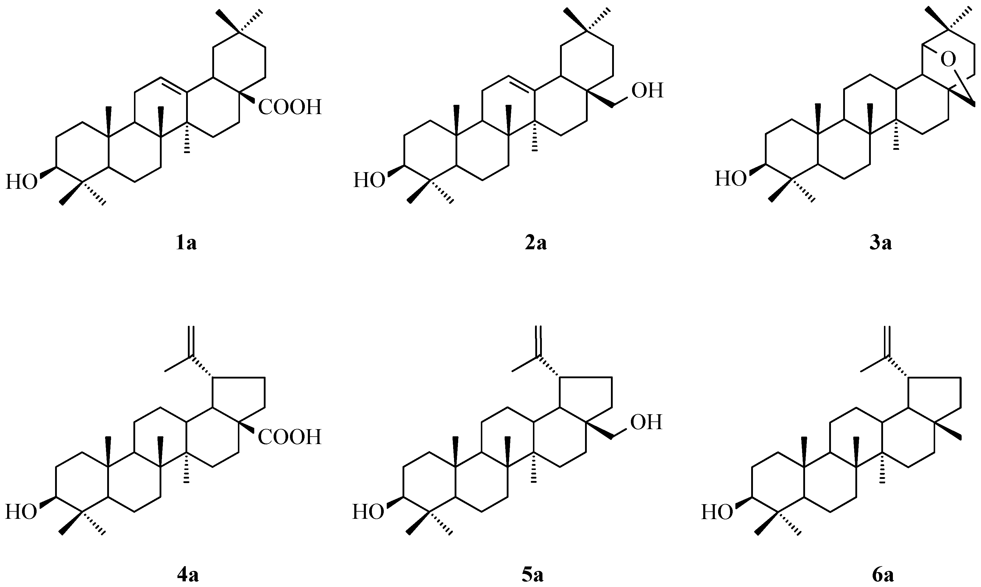
Figure 1. The structure of used triterpenes: oleanolic acid (1a), erythrodiol (2a), allobetulin (3a), betulinic acid (4a), betulin (5a) and lupeol (6a).
Molecules 30 02661 g001
Figure 2. Transformation of triterpenes (1a6a) into their acetyl derivatives (1b6b).
Molecules 30 02661 g002
Figure 3. (A). Graphical result of searching the top 5 cavities for p53 Y220 (PBD ID: 8DC4), where the largest is C1 and the smallest is C5; (B). whole 8DC4 molecule, the dark pink color indicates the cavity of the molecule; (C). a different angle is shown to show the cavities better.
Molecules 30 02661 g003
Figure 4. Docking score for compound 5b with p53 Y220 (PBD ID: 8DC4), Vina score for pocket C1 is −9.1 kcal × mol−1.
Molecules 30 02661 g004
Figure 5. Docking score for compound 1b with p53 Y220 (PBD ID: 8DC4). Vina score for pocket C2 is −9.0 kcal × mol−1.
Molecules 30 02661 g005
Figure 6. Docking score for compound 2b with p53 Y220 (PBD ID: 8DC4). Vina score for pocket C3 is −9.3 kcal × mol−1.
Molecules 30 02661 g006
Figure 7. Docking score for compound 6a with p53 Y220 (PBD ID: 8DC4). Vina score for pocket C4 is −9.6 kcal × mol−1.
Molecules 30 02661 g007
Figure 8. Docking score for compound 4b with p53 Y220 (PBD ID: 8DC4). Vina score for pocket C5 is −9.0 kcal × mol−1.
Molecules 30 02661 g008
Figure 9. Standard curve for CUPRAC radical inhibition by Trolox.
Molecules 30 02661 g009
Figure 10. Standard curve for DPPH radical inhibition by Trolox.
Molecules 30 02661 g010
Table 1. Examples of pharmacological activity of oleanolic acid, erythrodiol, allobetulin, betulinic acid, betulin and lupeol.
TriterpeneExamples of Pharmacological ActivityRef.
oleanolic acidantidiabetic[8]
neuroprotective[9]
hepatoprotective[10]
antioxidant[11]
antinociceptive and anti-inflammatory[12]
cardioprotective[13]
antihypertensive[14]
antiviral: EC50 = 13.07 µM (HSV-1/153), EC50 = 12.89 µM (HSV-1/106), EC50 = 13.09 µM (HSV-1/Blue)[15]
antibacterial: MBC = 700 µg × mL−1 (Staphylococcus aureus), MIC = 95 µg × mL−1 (S. aureus), MBC = 900 µg × mL−1 (Escherichia coli), MIC = 95 µg × mL−1 (E. coli), MBC = 300 µg × mL−1 (Salmonella typhi), MIC = 65 µg × mL−1 (S. typhi)[16]
antiatherosclerotic[17]
anticancer: IC50 = 14.93 µM (KB), IC50 = 13.95 µM (MCF-7), IC50 = 11.82 µM (HeLa), IC50 = 16.20 µM (Hep-G2)[18,19]
anticancer: IC50 = 40 µg × mL−1 (HCT-116)[20]
antioxidant, antiproliferative[21]
erythrodiolantioxidant, antiproliferative[21]
antioxidant, antiatherogenic[22]
Anti-inflammatory, vasorelaxing and cardioprotective[23]
antioxidant and anticancer: IC50 = 27.3 µM (Hep-G2)[24]
anticancer: EC50 = 48.8 µM (HT-29)[25]
anticancer: IC50 = 250 µM (CCRF-CEM, CEM)[26]
anticancer: IC50 = 64.96 µM (SMMC-7721), IC50 = 87.73 µM (Hep-G2), IC50 = 62.96 µM (A-459)[27]
antioxidant[28]
betulinic acidantidepressant[29]
antioxidant[30]
anxiolytic[31]
antiviral: EC50 = 11.2 μM (HCV)[32]
anti-inflammatory[33]
antiallergic and anti-inflammatory[34]
antihyperglycemic[35]
nephroprotective[36]
hepatoprotective[37]
anticancer: IC50 = 30 µM (CCRF-CEM, CEM)[26]
anticancer: IC50 = 125 µM (HT-29); IC50 = 58 µM (SW-480); IC50 = 178 µM (HCT-116)[38]
anticancer: IC50 = 30 µM (NOZ)[39]
anticancer: IC50 = 44.47 µM (A-2780)[40]
betulinanti-inflammatory[41]
wound healing[42]
antiviral: IC50 = 45.5 µM (SFV)[43]
antiseptic[44]
antihyperlipidemic[45]
antiobesity[46]
anticancer: IC50 = 250 µM (CCRF-CEM, CEM) [26]
anticancer: IC50 = 2.5 µM (SK-N-AS); IC50 = 5.9 µM (C-6); IC50 = 10.3 µM (TE-671); IC50 = 4.3 µM (HT-29); IC50 = 5.2 µM (T-47D); IC50 = 6.8 µM (FTC-238); IC50 = 7.4 µM (A-549); IC50 = 6.4 µM (RPMI-8226); IC50 = 6.7 µM (Jurkat 1E.6); IC50 = 2.8 µM (HPOC); IC50 = 3.4 µM (HPCC); IC50 = 3.4 µM (HPGBM)[47]
antioxidant; anticancer: IC50 = 29.4 µM (13 µg/mL; SGC7901)[48]
anticancer: IC50 = 73.2 µM (32.4 µg/mL; T-47D); IC50 = 24.6 µM (10.9 µg/mL; CCRF/CEM); IC50 = 51.7 µM (22.9 µg/mL; SW-707); IC50 = 12.4 µM (5.5 µg/mL; P-388)[49]
lupeolwound healing[42]
anti-inflammatory[50]
nephroprotective[51]
antiallergic[52]
antioxidant[53]
antiangiogenic[54]
anticancer: IC50 = 80 µM (MCF-7)[55]
anticancer: IC50 = 46.06 µM (MDA MB-231); IC50 = 31.910 µM (HeLa); IC50 = 64.82 µM (A-549)[56]
Legend: IC50 = half maximal inhibitory concentration; EC50 = half maximal effective concentration; BMC = minimum bactericidal concentration; MIC = minimum inhibitory concentration; HSV-1/153, HSV-1/106 and HSV-1/Blue = Herpes simplex virus type 1 ACV-resistant strains; KB = human oral squamous carcinoma; MCF-7 = human breast carcinoma; HeLa = human cervical epithelioid carcinoma; Hep-G2 = human hepatocellular carcinoma; HCT-116 = human colorectal carcinoma; HT-29 = human colorectal adenocarcinoma; CCRF-CEM, CEM = human T lymphoblastic leukemia; SMMC-7721 = human hepatocarcinoma; A-459 = human lung carcinoma; HCV = Hepatitis C virus; SW-480 = human colon adenocarcinoma; NOZ = human gall bladder carcinoma; A-2780 = human ovarian carcinoma; SFV = Semliki Forest virus; SK-N-AS = human neuroblastoma; C-6 = human glioma; TE-671 = human rhabdomyosarcoma–medulloblastoma; HT-29 = human colon adenocarcinoma; T-47D = human breast carcinoma; FTC-238 = human thyroid carcinoma; RPMI-8226 = human multiple myeloma; Jurkat 1E.6 = human T cell leukemia; HPOC = human ovarian carcinoma; HPCC = human cervical carcinoma; HPGBM = human glioblastoma multiforme; SGC-7901 = human gastric carcinoma; SW-707 = human colon carcinoma; P-388 = murine lymphocytic leukemia; MDA MB-231 = human epithelial adenocarcinoma.
Table 2. The PASS method calculated the predicted activity of unsubstituted and acetylated triterpenes (1a6a and 1b6b, respectively).
ActivityPa Factor (and Pi Factor) of Compounds 1a–6a and 1b–6b
1a2a3a4a5a6a1b2b3b4b5b6b
Antineoplastic0.876
(0.005)
0.920
(0.005)
0.950
(0.004)
0.925
(0.005)
0.948
(0.004)
0.950
(0.004)
0.890
(0.005)
0.923
(0.005)
0.954
(0.004)
0.934
(0.004)
0.952
(0.004)
0.954
(0.004)
Antineoplastic (colon c.)<0.7000.734
(0.005)
0.917
(0.003)
0.789
(0.005)
0.853
(0.004)
0.831
(0.004)
<0.7000.790
(0.005)
0.925
(0.003)
0.836
(0.004)
0.876
(0.004)
0.863
(0.004)
Antineoplastic (colorectal c.)<0.7000.736
(0.005
0.920
(0.003)
0.794
(0.005)
0.858
(0.004)
0.836
(0.004)
<0.7000.791
(0.005)
0.927
(0.003)
0.840
(0.004)
0.879
(0.004)
0.867
(0.004)
Antineoplastic (lung c.)0.766
(0.005)
0.802
(0.004)
0.883
(0.003)
0.815
(0.004)
0.833
(0.004)
0.850
(0.004)
0.792
(0.004)
0.823
(0.004)
0.899
(0.003)
0.831
(0.004)
0.859
(0.003)
0.869
(0.003)
Antiprotozoal (Leishmania)0.721
(0.008)
<0.700<0.7000.923
(0.003)
0.881
(0.003)
0.891
(0.003)
0.821
(0.004)
0.848
(0.004)
0.790
(0.005)
0.954
(0.002)
0.961
(0.002)
0.940
(0.002)
Apoptosis
agonist
0.901
(0.004)
0.892
(0.004)
0.759
(0.010)
0.822
(0.007)
0.837
(0.005)
0.883
(0.005)
0.891
(0.004)
0.878
(0.005)
0.747
(0.011)
0.850
(0.005)
0.825
(0.006)
0.874
(0.005)
Caspase 3 stim.0.984
(0.002)
0.971
(0.002)
0.820
(0.005)
<0.7000.974
(0.002)
0.978
(0.002)
0.974
(0.002)
0.870
(0.004)
0.720
(0.010)
0.976
(0.002)
0.880
(0.004)
0.954
(0.003)
Caspase 8 stim.0.914
(0.001)
0.878
(0.001)
0.808
(0.002)
<0.7000.869
(0.001)
0.865
(0.001)
0.910
(0.001)
0.846
(0.001)
0.804
(0.002)
0.900
(0.001)
0.835
(0.001)
0.864
(0.001)
Chemopre-
ventive
0.937
(0.002)
0.852
(0.003)
0.707
(0.006)
0.835
(0.003)
0.733
(0.005)
0.792
(0.004)
0.948
(0.002)
0.912
(0.002)
0.717
(0.006)
0.855
(0.003)
0.802
(0.004)
0.806
(0.004)
Mucomem-
branous prot.
0.894
(0.005)
0.824
(0.013)
0.732
(0.042)
0.786
(0.022)
<0.7000.847
(0.009)
0.935
(0.004)
0.892
(0.005)
0.795
(0.020)
<0.7000.778
(0.025)
0.895
(0.005)
Oxidoreductase inh.0.904
(0.002)
0.888
(0.003)
0.823
(0.005)
0.809
(0.006)
0.745
(0.010)
0.834
(0.005)
0.915
(0.002)
0.885
(0.003)
0.848
(0.004)
<0.7000.724
(0.013)
0.855
(0.004)
TF NF kappa B stim.0.954
(0.001)
0.931
(0.001)
0.864
(0.002)
0.804
(0.003)
0.935
(0.001)
0.947
(0.001)
0.936
(0.001)
0.893
(0.002)
0.788
(0.003)
0.941
(0.001)
0.904
(0.001)
0.924
(0.001)
TF stim.0.954
(0.001)
0.931
(0.001)
0.864
(0.002)
0.804
(0.003)
0.935
(0.001)
0.947
(0.001)
0.936
(0.001)
0.893
(0.002)
0.788
(0.003)
0.941
(0.001)
0.904
(0.001)
0.924
(0.001)
Legend: Pa = probability of activity, Pi = probability of inactivity; antag. = antagonist, inh. = inhibitor, prom. = promoter, prot. = protectant, stim. = stimulant, treatm. = treatment; c. = cancer, TF = transcription factor.
Table 3. Results of MTT assay for unsubstituted and acetylated triterpenes (1a6a and 1b6b, respectively) determined in the MTT assay.
Comp. No.Cell Line, IC50 [µM] (±SD)
HeLaKBMCF-7A-549PC-3SKOV-3HDF
1a11.82
(±0.19) *
14.93
(±0.07) *
13.95
(±0.11) *
8.79
(±0.20) *
18.63
(±0.05) ***
18.81
(±0.09) ***
24.87
(±0.04) ***
2a54.32
(±0.02)
66.04
(±0.44)
65.52
(±0.18)
63.67
(±0.12)
21.09
(±0.18)
18.33
(±0.05)
27.99
(±0.66)
3a49.44
(±0.21)
45.33
(±0.03)
71.06
(±0.13)
51.74
(±0.18)
20.17
(±0.04)
20.93
(±0.02)
48.22
(±0.19)
4a27.50
(±0.12)
35.21
(±0.06)
27.89
(±0.12)
26.23
(±0.19)
10.61
(±0.09)
9.52
(±0.21)
19.92
(±0.49)
5a18.32
(±0.02)
19.74
(±0.13)
18.09
(±0.02)
19.56
(±0.17)
6.22
(±0.06)
6.95
(±0.08)
15.81
(±0.66)
6a37.74
(±0.12) **
51.17
(±1.92) **
51.82
(±0.15) **
45.70
(±0.12) **
14.52
(±0.03)
14.57
(±0.09)
29.04
(±0.06)
1b0.24
(±0.19)
0.36
(±0.07)
1.86
(±0.16)
0.24
(±0.12)
0.11
(±0.02)
0.09
(±0.01)
0.19
(±0.07)
2b70.30
(±0.15)
49.96
(±0.31)
61.58
(±0.17)
51.61
(±0.43)
25.13
(±0.09)
25.51
(±0.04)
53.11
(±0.52)
3b>100>100>100>10052.22
(±0.14)
53.09
(±0.55)
62.02
(±0.18)
4b1.62
(±0.18)
1.50
(±0.07)
1.50
(±0.11)
1.23
(±0.13)
0.93
(±0.02)
1.03
(±0.01)
2.88
(±0.09)
5b>100>100>100>10075.91
(±0.37)
75.96
(±0.84)
81.73
(±0.49)
6b72.64
(±0.14)
72.52
(±0.44)
49.97
(±0.31)
56.22
(±0.59)
22.16
(±0.15)
21.09
(±0.11)
34.97
(±0.39)
Legend: HeLa = cervix carcinoma; KB = nasopharynx carcinoma; MCF-7 = breast carcinoma; A-549 = lung carcinoma; PC-3 = prostate carcinoma; SKOV-3 = ovarian carcinoma; HDF = normal dermal fibroblasts; ±SD = standard deviation; * = data published in [63]; ** = data published in [64]; *** = data published in [65].
Table 4. Selectivity index for unsubstituted and acetylated triterpenes (1a6a and 1b6b, respectively) determined in the MTT assay.
Comp. No.Cell Line, SI
HeLaKBMCF-7A-549PC-3SKOV-3
1a2.101.661.782.831.33 **1.32 **
2a0.870.420.430.441.321.53
3a0.971.060.680.932.392.30
4a0.720.560.710.761.882.09
5a0.860.800.870.812.542.27
6a0.770.570.560.632.001.99
1b0.790.530.100.791.732.11
2b0.751.060.861.032.112.08
3b- - - *- - - *- - - *- - - *1.191.17
4b1.771.921.922.343.102.80
5b- - - *- - - *- - - *- - - *1.081.07
6b0.480.480.700.621.581.66
Legend: SI = selectivity index; HeLa = cervix carcinoma; KB = nasopharynx carcinoma; MCF-7 = breast carcinoma; A-549 = lung carcinoma; PC-3 = prostate carcinoma; SKOV-3 = ovarian carcinoma; * = selectivity index was not calculated because the IC50 value exceeded 100 µM; ** = published in [65].
Table 5. Apoptotic index for the selected 1a, 3a, 5a, 1b and 4b determined in the MTT assay for SKOV-3 and PC-3 cell lines.
Comp. No.Cell Line, AI
SKOV-3PC-3
1a5.16 (0.01)5.27 (0.04)
3a5.78 (0.01)5.27 (0.01)
5a5.26 (0.03)5.82 (0.03)
1b6.79 (0.16)6.90 (0.04)
4b7.81 (0.02)7.09 (0.01)
Legend: AI = Apoptosis index; SKOV-3 = ovarian carcinoma; PC-3 = prostate carcinoma.
Table 6. The results from the CB-Dock2 web server include the five largest cavities, from largest (C1) to smallest (C5), with their assigned calculated volumes (Å3), as well as the coordinates of their centers and dimensions expressed in angstroms (Å).
CurPocket IDCavity Volume (Å3)Center (x, y, z)Cavity Size (x, y, z)
C13942−16, 49, 6130, 18, 27
C22320−6, 46, 918, 23, 18
C3828−33, 65, 3411, 20, 8
C4430−40, 55, 4019, 7, 8
C5370−30, 44, 248, 14, 15
Table 7. Docking results presented as Vina score values (kcal × mol−1).
Pocket IDCompound Number
1a1b2a2b3a3b4a4b5a5b6a6b
C1−6.4−9.0−7.6−7.1−9.3−8.3−6.0−7.8−7.1−9.1−8.5−9.1
C2−6.3−9.0−6.5−6.4−6.5−6.4−6.2−5.7−5.8−6.0−6.0−6.1
C3−8.6−7.4−9.3−10.1−9.9−9.3−8.6−9.3−8.2−8.1−9.3−9.6
C4−8.2−8.5−8.0−8.8−9.4−9.1−8.6−8.4−8.0−8.2−9.6−8.6
C5−7.8−7.8−7.5−8.9−8.3−7.9−6.8−9.0−7.1−7.5−7.5−7.2
Table 8. The results of the antioxidant activity of unsubstituted and acetylated triterpenes (1a6a and 1b6b, respectively) evaluated with CUPRAC and DPPH assays.
Comp. No.Trolox Equivalent [mg/mL]
CUPRAC AssayDPPH Assay
1a0.10312 ± 0.006840.02633 ± 0.001090
2a0.09414 ± 0.009500.02092 ± 0.000866
3a0.08248 ± 0.005000.01677 ± 0.000694
4a0.07632 ± 0.007270.01526 ± 0.000632
5a0.29900 ± 0.006630.01077 ± 0.000446
6a0.13602 ± 0.005930.00065 ± 00.01570
1b0.21986 ± 0.006570.01430 ± 0.000592
2b0.24016 ± 0.009950.00313 ± 0.000130
3b0.03212 ± 0.001660.00182 ± 0.000755
4b0.19986 ± 0.001500.00376 ± 0.000156
5b0.17609 ± 0.004870.00786 ± 0.000325
6b0.13490 ± 0.005140.00348 ± 0.000144
Table 9. ADMETox data calculated for unsubstituted and acetylated triterpenes (1a6a and 1b6b, respectively).
Properties
(Optimal Values)
Compound Number
1a2a3a4a5a6a1b2b3b4b5b6b
Mol. Weight (100~600)456.360442.380442.380456.360442.380426.390498.370526.400484.390498.370526.400468.400
Volume505.750499.598493.678505.751499.598490.807546.497581.089534.423546.497581.089531.553
Density0.9020.8850.8960.9020.8850.8690.9120.9060.9060.9120.9060.881
NHA (0~12)322321443442
nHD (0~7)221221100100
nRot (0~11)110221352463
nRing (0~6)556555556555
maxRing (0~18)2222721212122227212121
nHet (1~15)322321443442
fChar (−4~+4)000000000000
nRig (0~30)272629272626282830282827
Flexibility (2)0.0370.0380.0000.0740.0770.0380.1070.1790.0670.1430.2140.111
Stereo Centers (2)88101010108810101010
TPSA (0~140)57.53040.46029.46057.53040.46020.23063.60052.60035.53063.60052.60026.300
LogS (−4~0.5)−5.036−5.769−6.590−5.148−5.681−6.667−5.982−6.964−7.152−5.866−6.772−7.189
LogP (0~3)6.1136.5386.5155.5745.7476.6896.8897.6746.9946.1496.8437.170
LogD (1~3)4.8434.7055.1454.8734.6825.3134.8734.9755.2144.9454.9715.417
QED (0.67)0.4090.4240.4280.4360.4520.4210.3110.2740.3560.3200.2740.299
Sascore (6)4.5894.7025.5244.6894.7614.6634.6244.7545.5424.7254.8234.692
Fsp3 (0.420)0.9000.9331.0000.9000.9330.9330.8750.8820.9690.8750.8820.906
MCE-18 (45)105.368102.207116.200104.000100.793100.793106.667105.000117.397105.300103.594102.164
Npscore (−5~5)3.2723.3263.1463.0723.2333.0543.2173.0733.0613.0122.9752.956
Lipinski RuleAAAAAAARAARA
Pfizer RuleRRRRRRRRRRRR
GSK RuleRRRRRRRRRRRR
Golden TriangleAARAARARRARR
PAINS (alerts)000000000000
ALARM NMR (alerts)000000000000
BMS (alerts)000000000000
Chelator Rule (alerts)000000000000
Caco-2 perm. (≥5.15)−5.198−4.867−5.115−6.283−4.942−5.042−5.166−4.989−5.040−5.203−5.029−4.946
MDCK perm.
(≤2 × 10⁻⁶ cm/s)
2.00 × 10−58.83 × 10−61.91 × 10−51.80 × 10−51.30 × 10−51.00 × 10−51.60 × 10−51.30 × 10−52.22 × 10−52.46 × 10−51.97 × 10−51.47 × 10−5
Pgp-inh. (0.300)0.0000.0020.0020.0020.0080.0290.0010.1710.0280.0140.8130.174
Pgp-sub. (0.300)0.0000.0000.0000.0000.0000.0000.0000.0000.0000.0000.0000.000
HIA (0.300)0.0120.0110.0050.0070.0040.0030.0140.0190.0050.0070.0050.004
F20% (0.300)0.0740.8780.2500.3400.8800.7420.0310.4550.0180.0120.0210.014
F30% (0.300)0.7560.9520.9560.8980.9390.8910.8280.9290.8650.8990.9620.833
PPB (90%)98.13098.23097.48096.51597.76298.99897.01397.24796.05696.84195.96798.268
VD (0.04–20 L/kg)0.7181.1941.4410.6141.1031.6500.6861.1961.2640.6871.3031.681
BBB penetr. (0.300)0.6740.5620.3760.4920.4710.6270.3290.2540.1760.2060.2620.353
Fu (5.000%)3.5242.3652.0722.5791.6251.7133.4611.9272.1512.1231.7141.646
CYP1A2 inh. (0.700)0.0120.0380.0320.0210.0470.0370.0110.0220.0280.0160.0290.033
CYP1A2 sub. (0.700)0.3230.1940.3360.5490.3330.5980.1660.1190.1520.2430.1300.455
CYP2C19 inh. (0.700)0.0280.0540.0630.0270.0540.0730.0290.0700.0680.0260.0650.080
CYP2C19 sub. (0.700)0.9160.9240.9320.9280.9390.9470.8930.9140.9310.9310.9300.948
CYP2C9 inh. (0.700)0.1570.1870.1000.1290.1420.0930.2180.1960.1010.1380.1250.090
CYP2C9 sub. (0.700)0.8130.1240.1750.7030.1600.5690.7230.0800.1430.6780.1100.596
CYP2D6 inh. (0.700)0.0120.0490.0500.0050.0310.0570.0120.1110.0330.0050.0790.029
CYP2D6 sub. (0.700)0.5280.2450.8370.7640.8070.8980.1770.0960.6750.6000.4750.879
CYP3A4 inh. (0.700)0.1720.7170.2520.1270.5190.2230.2460.6050.2780.1550.5300.250
CYP3A4 sub. (0.700)0.2080.4550.3350.2230.4640.4330.3770.6670.5040.3250.5800.525
CL (15 mL/min/kg)3.09410.5094.7013.0396.4865.3722.1193.0913.4542.4543.1653.552
T1/2< 3h (0.300)0.0230.0170.0430.0630.0510.0370.0130.0090.0270.0390.0256.486
hERG Blockers (0.300)0.0040.0210.5110.0310.0560.1240.0030.0080.3260.0210.0440.102
H-HT (0.300)0.2960.4020.2540.2750.2620.0850.2780.3130.2710.2980.2580.126
DILI (0.300)0.0100.0070.0310.0090.0110.0240.0240.2020.4370.0460.4920.341
AMES Tox. (0.300)0.0080.0030.0050.0030.0020.0020.0050.0020.0030.0020.0020.002
ROAT (0.300)0.2280.1620.1680.2210.1290.1850.0800.0210.0380.0450.0090.032
FDAMDD (0.300)0.9090.9250.9390.9280.9280.9190.6230.7530.7270.6280.7260.515
Skin Sensit. (0.300)0.0280.0760.8930.3290.6240.7270.0360.0350.5320.1950.1290.680
Carcinogen. (0.300)0.0630.0670.0050.0180.0140.0060.0800.0590.0050.0160.0130.005
Eye Corrosion (0.300)0.0120.0060.1690.0220.0190.8820.1950.0080.1700.0300.0070.877
Eye Irritation (0.300)0.0840.0220.2630.0390.0470.5500.0500.0360.1960.0320.0680.458
Respir. Tox. (0.300)0.9680.9800.9340.9450.8410.5800.9680.9580.8020.9280.6030.412
Bioconc. Factors1.9443.0752.4762.1172.8412.5602.4222.7842.3232.5532.4442.410
IGC505.0215.1765.7265.1765.3575.7105.0485.3115.7895.2405.4915.773
LC50FM5.9376.1266.8416.2896.4956.9516.0346.3546.8686.3658.6116.977
LC50DM6.3376.6766.9246.5046.8686.9906.3596.7316.8526.4666.9106.904
NR-AR (0.300)0.3690.0550.0020.0690.0210.0070.6650.1670.0190.4590.0750.116
NR-AR-LBD (0.300)0.2730.3550.0420.4950.2520.0650.7330.7880.1120.7400.4840.233
NR-AhR (0.300)0.0010.0000.0000.0000.0000.0000.0010.0010.0000.0010.0010.000
NR-Aromatase (0.300)0.7590.7020.3810.4630.5470.2670.7950.6750.4480.4260.5020.311
NR-ER (0.300)0.4120.2490.3090.1150.1160.2720.7210.1780.2860.1350.0690.229
NR-ER-LBD (0.300)0.5930.7650.6960.4320.2370.5460.7850.6330.8050.6570.7370.786
NR-PPAR γ (0.300)0.9650.2970.0220.7630.0210.0170.9640.1960.0200.8260.0190.021
SR-ARE (0.300)0.5560.2750.0640.2000.0810.0490.6370.2160.0490.1630.0580.038
SR-ATAD5 (0.300)0.0520.1180.0080.0780.0400.0090.4280.3850.0150.1900.1480.016
SR-HSE (0.300)0.7470.1280.0270.5670.0500.0300.7700.2110.0310.5070.0910.041
SR-MMP (0.300)0.9710.9420.6470.9400.8760.6200.9560.8080.2610.8060.4690.277
SR-p53 (0.300)0.2710.1300.0100.4430.1120.0120.6470.4320.0110.5370.3530.015
Acute Toxicity Rule A.000000000000
Gen. Carcin. Rule A.000000000000
Non Gen. Carcin. Rule A.000000000000
Skin Sensit. Rule A.000000000000
Aquatic Tox. Rule A.112111001000
Non Biodegr. Rule A.001000001000
SureChEMBL Rule000000000000
FAF-Drugs4 Rule000000000000
Toxicophores000000000000
Legend: A. = alerts; Bioconc. = bioconcentration; Biodegr. = biodegradation; carcin. = carcinogenicity; Gen. Carcin. = genotoxic carcinogenicity; inh. = inhibitor; Mol. = molecular; penetr. = penetration; perm. = permeability; Respir. Tox. = respiratory toxicity; ROA = rat oral acute toxicity; sensit. = sensitization; sub. = substrate; Tox. = toxicity; for values given in a range 0.0001.000 (unless stated otherwise): 0.0000.300 = low probability, 0.301–0.699 = moderate probability, 0.7001.000 = high probability.
Table 10. Acetylation of triterpenes (on the example of oleanolic acid) as a method of improving cytotoxic activity.
ResultsRef.
Oleanolic acid: IC50 = 6.4 µM (A-549)Acetyloleanolic acid: IC50 = 5.8 µM (A-549)[104]
Oleanolic acid: IC50 > 10 µM (KB)Acetyloleanolic acid: IC50 = 7.6 µM (KB)
Oleanolic acid: IC50 > 10 µM (KB-VIN)Acetyloleanolic acid: IC50 = 7.6 µM (KB-VIN)
Oleanolic acid: IC50 = 106.4 µM
(B16-F10)
Acetyloleanolic acid: IC50 = 64.7 µM
(B16-F10)
[105]
Oleanolic acid: IC50 = 429.9 µM
(HT-29)
Acetyloleanolic acid: IC50 = 148.5 µM
(HT-29)
Oleanolic acid: IC50 = 211.8 µM
(Hep-G2)
Acetyloleanolic acid: IC50 = 103.75 µM (Hep-G2)
Oleanolic acid: IC50 = 41.7 µM
(RAW 264.7)
Acetyloleanolic acid: IC50 = 13.8 µM
(RAW 264.7)
[106]
Oleanolic acid: IC50 = 35.2 µM (J774A.1)Acetyloleanolic acid: IC50 = 16.8 µM (J774A.1)
Oleanolic acid: IC50 = 106.4 µM
(B16-F10)
Acetyloleanolic acid: IC50 = 64.7 µM
(B16-F10)
[107]
Legend: IC50 = half maximal inhibitory concentration; A-459 = human lung carcinoma; KB = human oral squamous carcinoma; KB-VIN = multidrug resistant human epidermoid carcinomaB16-F10 = murine melanoma; HT-29 = human colorectal adenocarcinomaHep-G2 = human hepatocellular carcinoma; RAW 264.1 = adherent cell line isolated from a mouse tumor that was induced by Abelson murine leukemia virus; J774A.1 = macrophage-like cell line derived from a BALB/c mouse.
Disclaimer/Publisher’s Note: The statements, opinions and data contained in all publications are solely those of the individual author(s) and contributor(s) and not of MDPI and/or the editor(s). MDPI and/or the editor(s) disclaim responsibility for any injury to people or property resulting from any ideas, methods, instructions or products referred to in the content.