Article

The Inhibitory Effect of Agastache rugosa Essential Oil on the Dental Biofilm

1
Institute of Biomaterials and Implant, College of Dentistry, Wonkwang University, Iksan 54538, Republic of Korea
2
Department of Oral Biochemistry, School of Dentistry, Wonkwang University, Iksan 54538, Republic of Korea
3
Department of Food and Nutrition, School of Food, Kunsan National University, Kunsan 54150, Republic of Korea
4
Department of Food Science and Biotechnology, Jeonbuk National University, Jeonju 54896, Republic of Korea
5
Wonkwang Dental Research Institute, Wonkwang University, Iksan 54538, Republic of Korea
*
Author to whom correspondence should be addressed.
These authors contributed equally to this study.
Molecules 2024, 29(20), 4907; https://doi.org/10.3390/molecules29204907
Submission received: 2 September 2024 / Revised: 5 October 2024 / Accepted: 12 October 2024 / Published: 16 October 2024
(This article belongs to the Special Issue Essential Oils: From Extraction to Application)

Abstract

:
This study aimed to identify the inhibitory effect of Agastache rugosa essential oil (AREO) on the cariogenic properties of Streptococcus mutans, which causes dental caries and dental plaque formation. After extracting the AREO, their effects on the growth and acid production of S. mutans were examined. Furthermore, S. mutans biofilm formation was observed on the resin teeth surface. The effect on the expression of biofilm-related genes of S. mutans was measured using real-time PCR. AREO components were analyzed using gas chromatography (GC) and GC-mass spectrometry (MS). The growth and acid production of S. mutans were significantly inhibited at concentrations of 0.02 mg/mL or higher of AREO. At 0.04 mg/mL, inhibition was similar to that of the positive control, 0.1% NaF. AREO suppressed the expression of virulence factors such as gtfB, gtfC, gtfD, gbpB, SpaP, brpA, relA, and vicR at concentrations of 0.02 mg/mL or higher. As a result of GC and GC-MS analyses, the main components of AREO included estragole, limonene, and β-caryophyllene. These results suggest that A. rugosa may be a useful agent for inhibiting the cariogenic properties of S. mutans.

1. Introduction

Dental caries is a disease that gradually and irreversibly destroys the hard tissues of teeth, such as the enamel, cementum, and dentin [1,2]. Dental caries are prevalent worldwide and are a representative cause of tooth loss in humans. As the incidence tends to increase with increasing sugar consumption and longer life expectancy, effective management measures are urgently needed [3,4].
Dental caries is caused by dental plaque, a type of dental biofilm formed by oral bacteria. A large number of microorganisms are known to exist in dental biofilm. These microorganisms induce dental caries by dissolving the hard tissues of teeth and forming organic acids by metabolizing sugars.
Streptococcus mutans is known as one of the causative bacteria that play the most important role in the formation of dental plaque and the induction of dental caries. S. mutans is a Gram-positive facultative anaerobic bacterium that grows well even under aerobic conditions, which resides on the tooth surface in the oral cavity and uses carbohydrates, especially glucose and fructose contained in ingested food, to produce organic acids (mainly lactic acid) in the process of metabolism and discharges them outside of the cells. Extracellular organic acids demineralize hard tissues, such as tooth enamel, dentin, and cementum [5,6,7]. Furthermore, glucosyltransferase (GTFase) produced by S. mutans catalyzes the synthesis of fructan by fructosyltransferase (FTFase) and glucan, an extracellular polysaccharide, using sucrose as a substrate [1,3]. These adhesive and insoluble glucans and fructans act to firmly adhere S. mutans to the tooth surface together with other bacteria and increase the formation of dental plaque. In addition, extracellular polysaccharide reduces the permeability of the dental plaque, preventing the diffusion of acids produced by bacteria in the dental plaque, acting as a barrier against the penetration of saliva, which has a buffering action, and promoting demineralization of the tooth surface by concentrating the acid produced by oral bacteria inside the dental plaque [4,8,9,10].
Fluoride compounds and chlorhexidine are used to inhibit the formation of dental microfilm, while vaccine development is also being carried out. However, fluoride compounds are toxic to oral cells at more than 80 ppm [11]. Chlorhexidine has an unpleasant taste, increases the acquired pellicle, and discolors teeth yellow when used for a long time [12,13].
Recently, studies have been conducted to suppress the formation of dental plaque using vaccines, but they are not sufficiently effective due to high development costs and low efficacy. The fact that dental caries due to dental plaque is still a major cause of tooth loss is evidence that the existing methods are not sufficiently effective. Therefore, there is a demand for the development of a more effective and practical method for inhibiting dental plaque.
Many studies have analyzed Agastache rugosa (Fisch. & C.A.Mey.) O. Kuntze essential oil (AREO) and reported the presence of various volatile organic compounds. Estragole, also known as methyl chavicol, is the main component of AREO [14,15,16]. The components were also analyzed, and nine compounds were identified as follows: menthone, isomenthone, dihydrocarbone, anethole, vanillin, eugenol, methyl eugenol, β-caryophyllene, and β-caryophyllene oxide [17]. Physiological activities include antibacterial, antifungal, antiviral, antimutagenic, anticancer, nematicidal, insecticidal, antioxidant, and Alzheimer’s alleviating activities [18].
In particular, AREO has shown strong antibacterial activity against Staphylococcus aureus [17]. The essential oil extracted from A. rugosa leaves has been reported to have an antibacterial effect against E. coli (MIC, 9.4 µg/mL). The essential oil of A. rugosa flowers inhibits biofilm activity and has shown similar or stronger activity against S. aureus (MIC, 21 μg/mL) than penicillin [19]. AREO also inhibits Escherichia coli O157:H7 ATCC 43895, which causes hemorrhagic nephrotic syndrome and enteritis, and Salmonella typhimurium ATCC 7988, which causes intestinal diseases, such as typhoid in humans, in a concentration-dependent manner. AREO also exhibited antifungal activity against Aspergillus niger, Candida albicans, and Cryptococcus neoformans [16].
The chloroform and ethyl acetate solvent fractions of A. rugosa extracted with hot water have been reported to have strong antibacterial activity against 15 types of microorganisms. As a result of gas chromatography-mass spectrometry (GC-MS) analysis, eugenol and its isomers, which have structural similarities with estragole, which is a strong antibacterial substance with a large number of phenolic compounds, were found to be predominant. The chloroform fraction contained 18.1% alkanes and 13.5% ketones, as well as terpenes, acids, and phenols [20].
Although many studies have reported that AREO exhibits antibacterial activity, a few studies have investigated the effects of A. rugosa on S. mutans, the causative agent of dental plaque formation. Therefore, this study aimed to observe the inhibition of S. mutans growth, acid production, and virulence gene expression by AREO, as well as its biofilm formation inhibitory effect on S. mutans.

2. Results

2.1. Inhibitory Effect on the Growth of S. mutans

The antibacterial activity of AREO against S. mutans was observed. AREO was added at concentrations of 0.01, 0.02, 0.03, and 0.04 mg/mL, and S. mutans was inoculated and cultured. The growth rates were 91.1%, 65.4%, 38.2%, and 9.16%, respectively, compared to the 100% growth rate of the control group. At a concentration of 0.04 mg/mL, the growth of S. mutans was inhibited similarly to the positive control with the addition of 0.1% NaF (Figure 1).

2.2. Inhibitory Effect on Adhesion to Saliva-Coated Hydroxyapatite (S-HA)

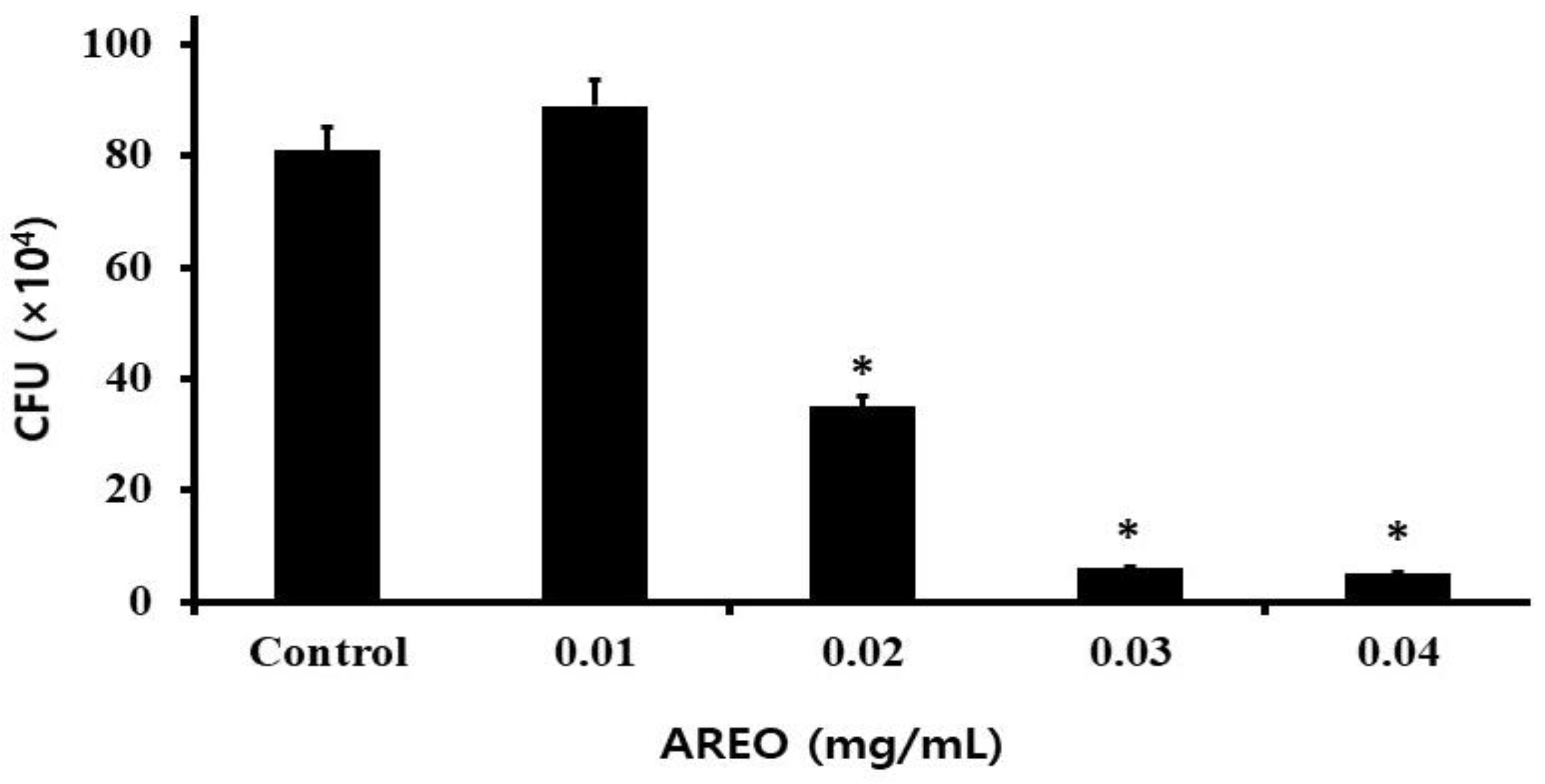
The adhesion to S-HA of S. mutans by AREO was studied, and S. mutans was found to be significantly suppressed in a dose-dependent manner. After confirming the effect of inhibiting adhesion to S-HA at an AREO concentration of 0.01–0.04 mg/mL, it was found that 81 ± 4.24 (×104) CFU/mL adhered in the control group. At each concentration and with 89 ± 9.57 (×104) CFU/mL, 35 ± 1.22 (×104) CFU/mL, 6 ± 2.12 (×104) CFU/mL, and 5 ± 1.22 (×104) CFU/mL adhered, respectively, showing 109.9%, 46.2%, 7.5%, and 6.2%, adhesion inhibition rates, respectively, compared to the control group (Figure 2).
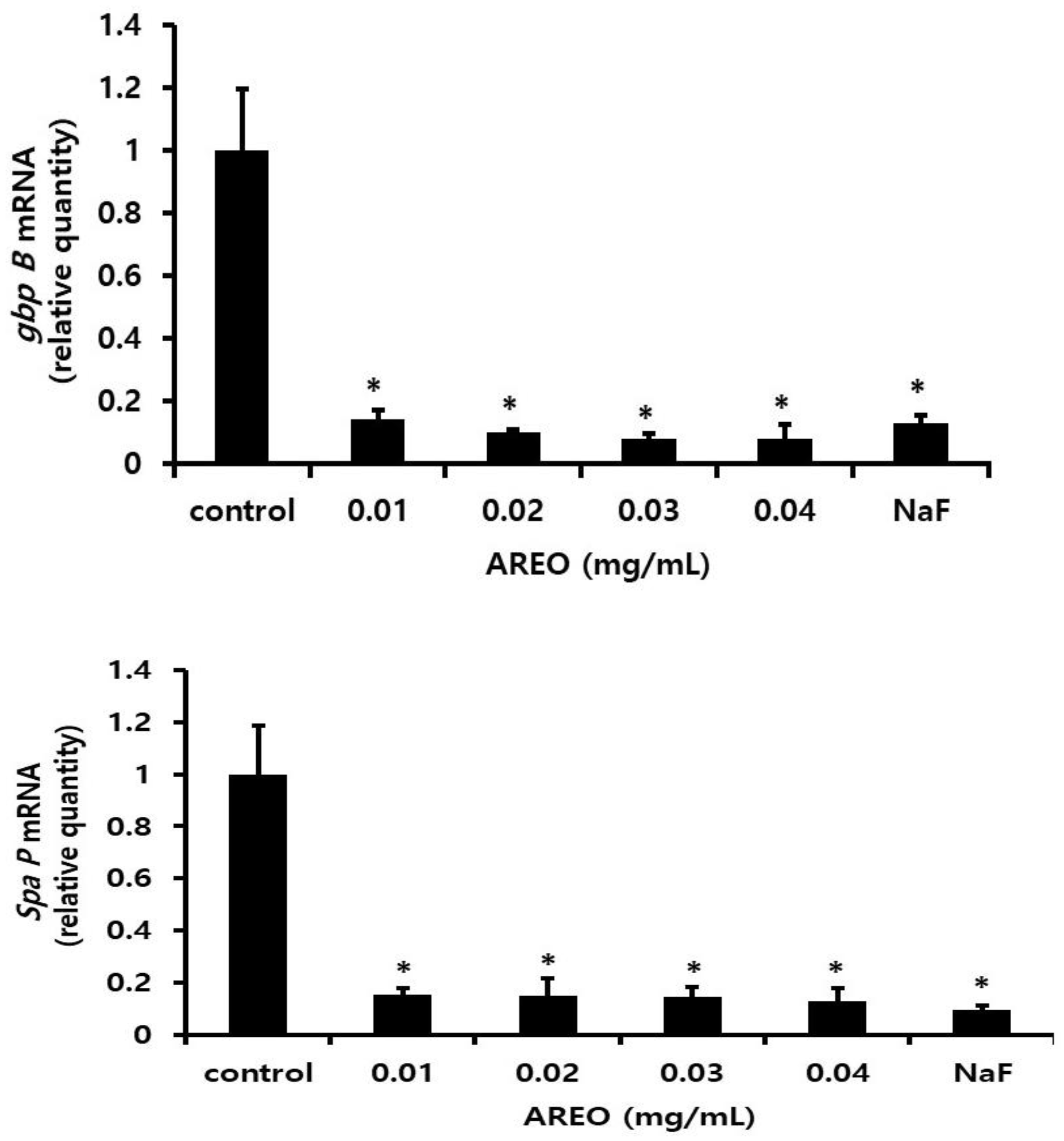
As a result of observing the mRNA expression of gbpB and spaP to investigate the effect of AREO on the expression of adherence-related genes, real-time PCR analysis showed that gbpB and spaP mRNA expression was suppressed at concentrations higher than 0.01 mg/mL (Figure 3).

2.3. The Inhibitory Effect on S. mutans Biofilm Formation

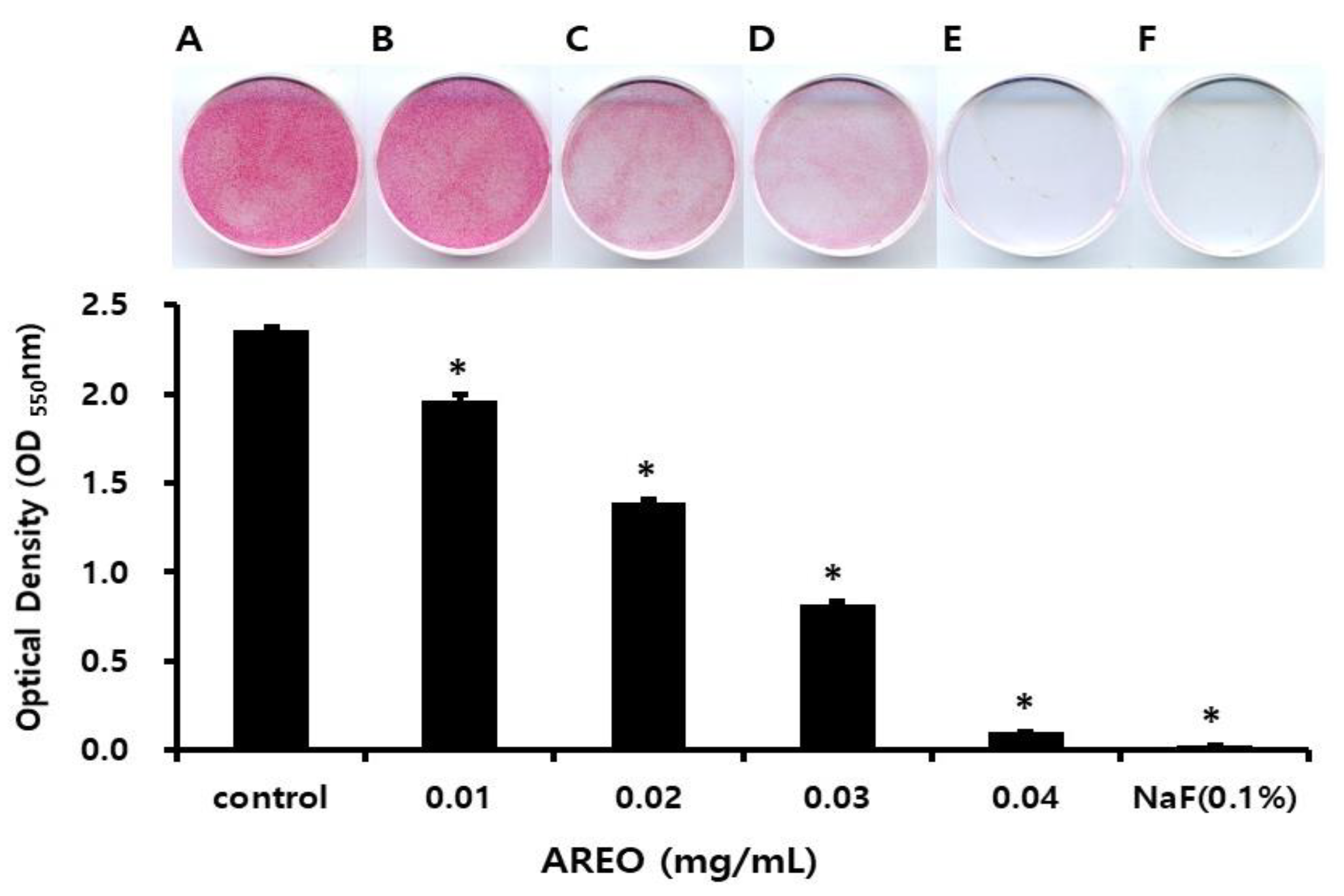
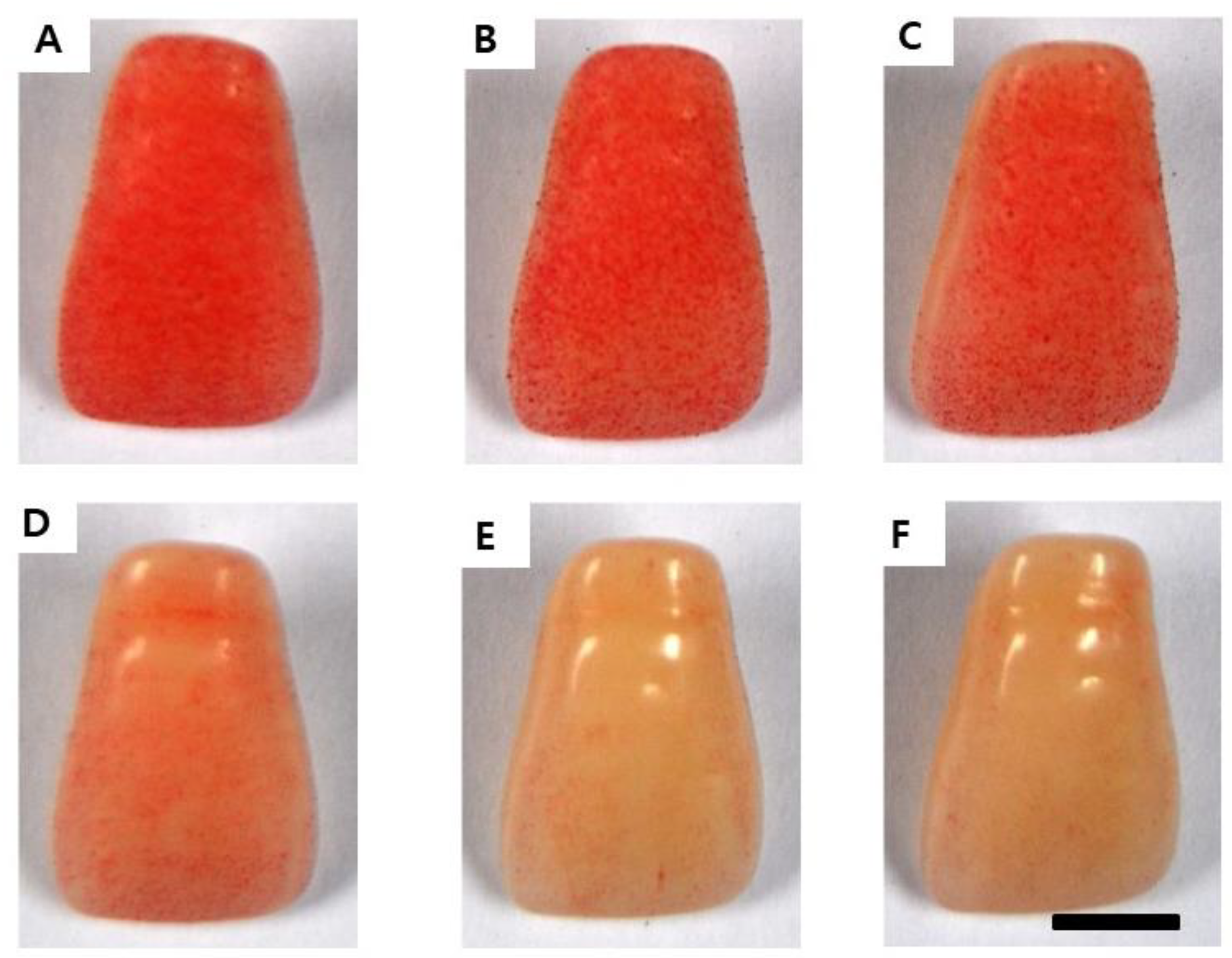
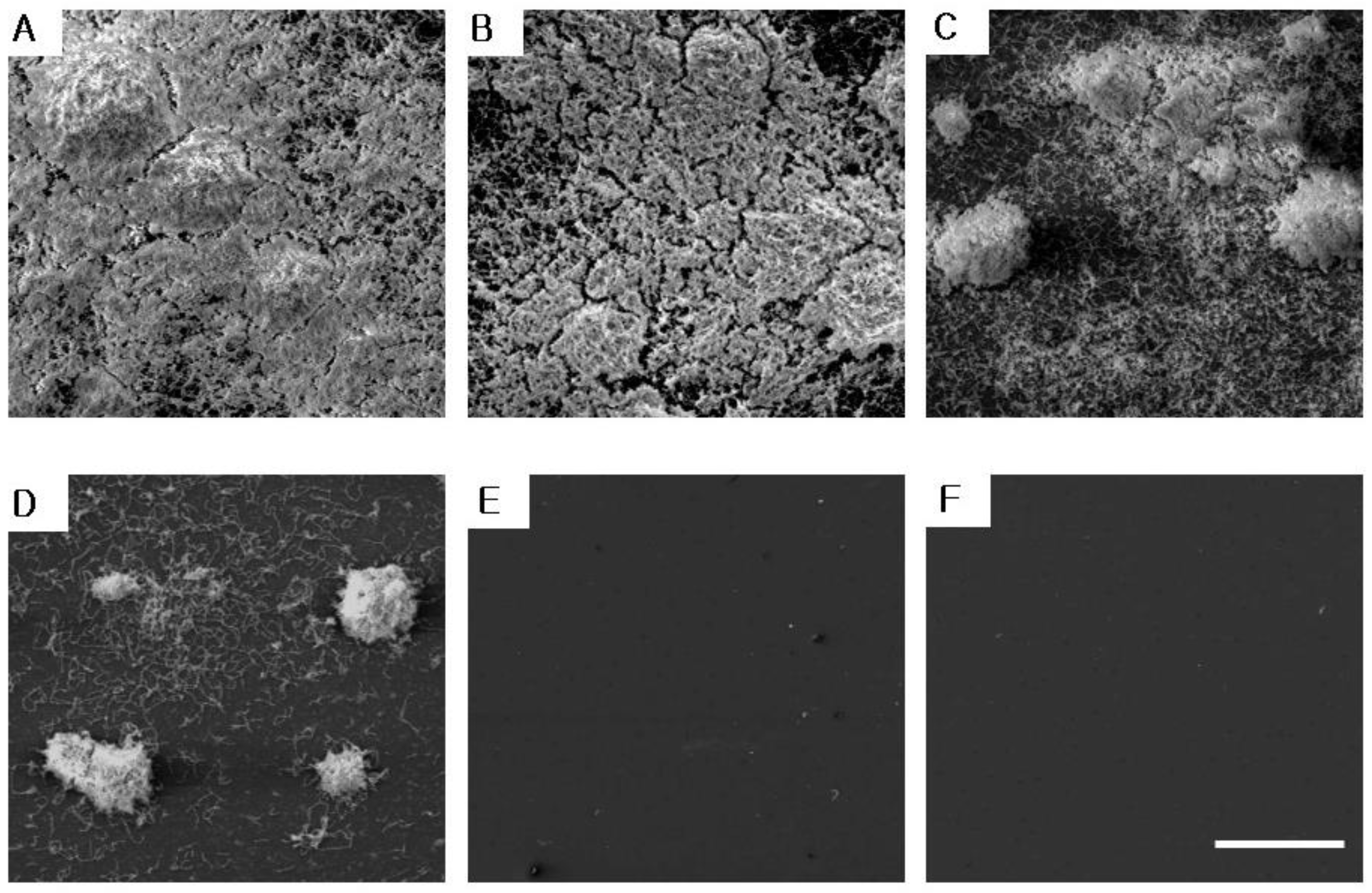
Figure 4 and Figure 5 shows the experimental results of the inhibitory effect of AREO on S. mutans biofilm formation. A large amount of S. mutans biofilm was formed in the control group without AREO, and as the AREO concentration increased, S. mutans biofilm formation was suppressed. These experimental results were similar to those observed using an electron microscope (Figure 6). In the control group, a large amount of S. mutans biofilm was formed, and as the AREO concentration increased, the formation of the S. mutans biofilm was clearly suppressed, even under an electron microscope. In particular, inhibition similar to that of 0.1% NaF, a positive control, was observed at 0.04 mg/mL. In addition, biofilm formation by AREO was suppressed on the surface of artificial teeth, and an excellent effect was observed at a concentration of 0.04 mg/mL or more (Figure 5).
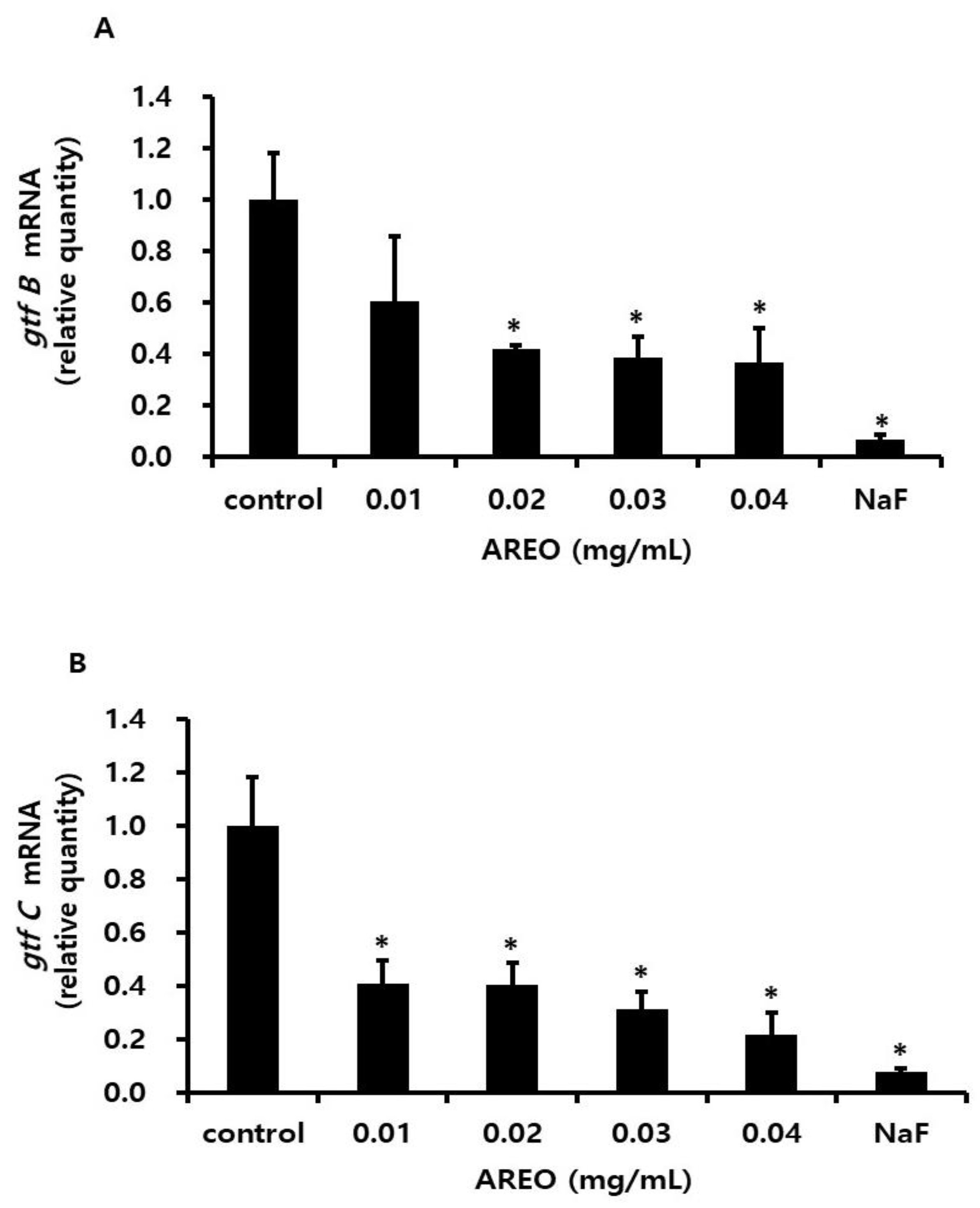
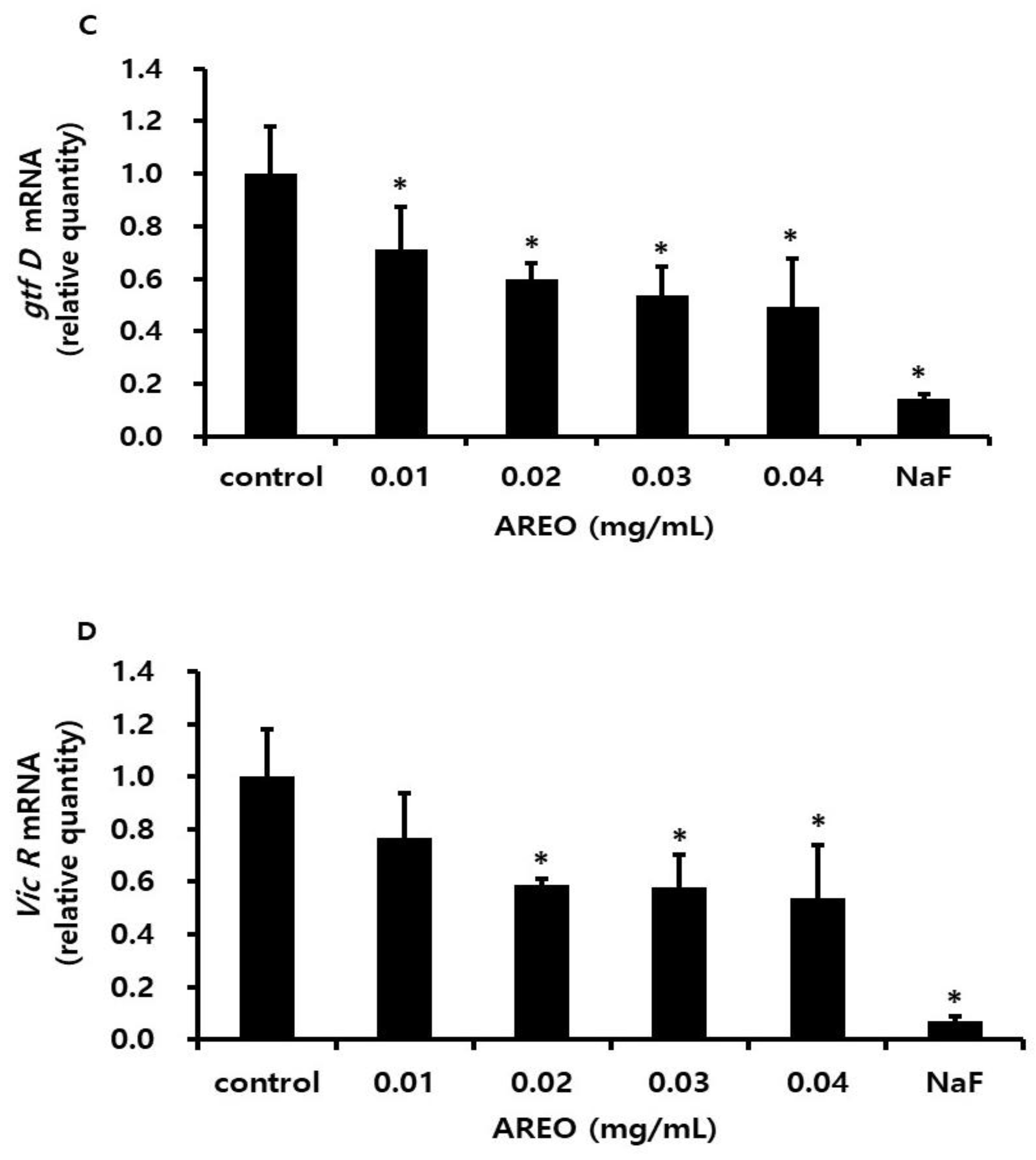
It was confirmed using real-time PCR to observe gene expression associated with biofilm formation by AREO. By treatment with AREO concentrations of 0.01–0.04 mg/mL, mRNA levels of gtfC and gtfB genes were decreased at concentrations of 0.01 mg/mL or higher, and mRNA levels of gtfB and vicR genes were decreased at concentrations of 0.02 mg/mL or higher (Figure 7).
Regarding the biofilm formation, Figure 4 clearly shows the constant decrease. However, mRNA levels are kept constant over 0.02 mg/mL in Figure 7. These two data are not consistent with each other. Although real-time RT PCR was used to monitor the mRNA levels of key biofilm-related genes, it is well known that there can be a delay between mRNA expression and the corresponding biological activity. This may partially explain the possibility of discrepancy between biofilm formation and mRNA levels. Furthermore, there is a possibility that AREO contributes to biofilm degradation, which requires further investigation in future studies.

2.4. Inhibitory Effect on the Acid Production of S. mutans

In order to examine the effect of AREO on the organic acid production of S. mutans, S. mutans was inoculated into the experimental group, to which AREO was added at concentrations of 0.01, 0.02, 0.03, and 0.04 mg/mL. The mixtures were then cultured for 24 h, followed by measuring the pH with a pH meter. Table 1 shows the results. In the control, the pH before inoculation with bacteria was about 7.38 ± 0.05, but after inoculation and incubation with bacteria, the pH dropped to 5.41 ± 0.05. In the experimental group to which AREO was added, a statistically significant difference was shown at a concentration of 0.02 mg/mL compared to the control group.
We have performed additional experiments to find out whether AREO can neutralize acids. In our study, AREO has no ability to neutralize acid. The pH of the control group cultured with S. mutans was 5.31, and the addition of AREO did not result in significant pH changes, indicating that AREO does not have acid-neutralizing properties in this system.
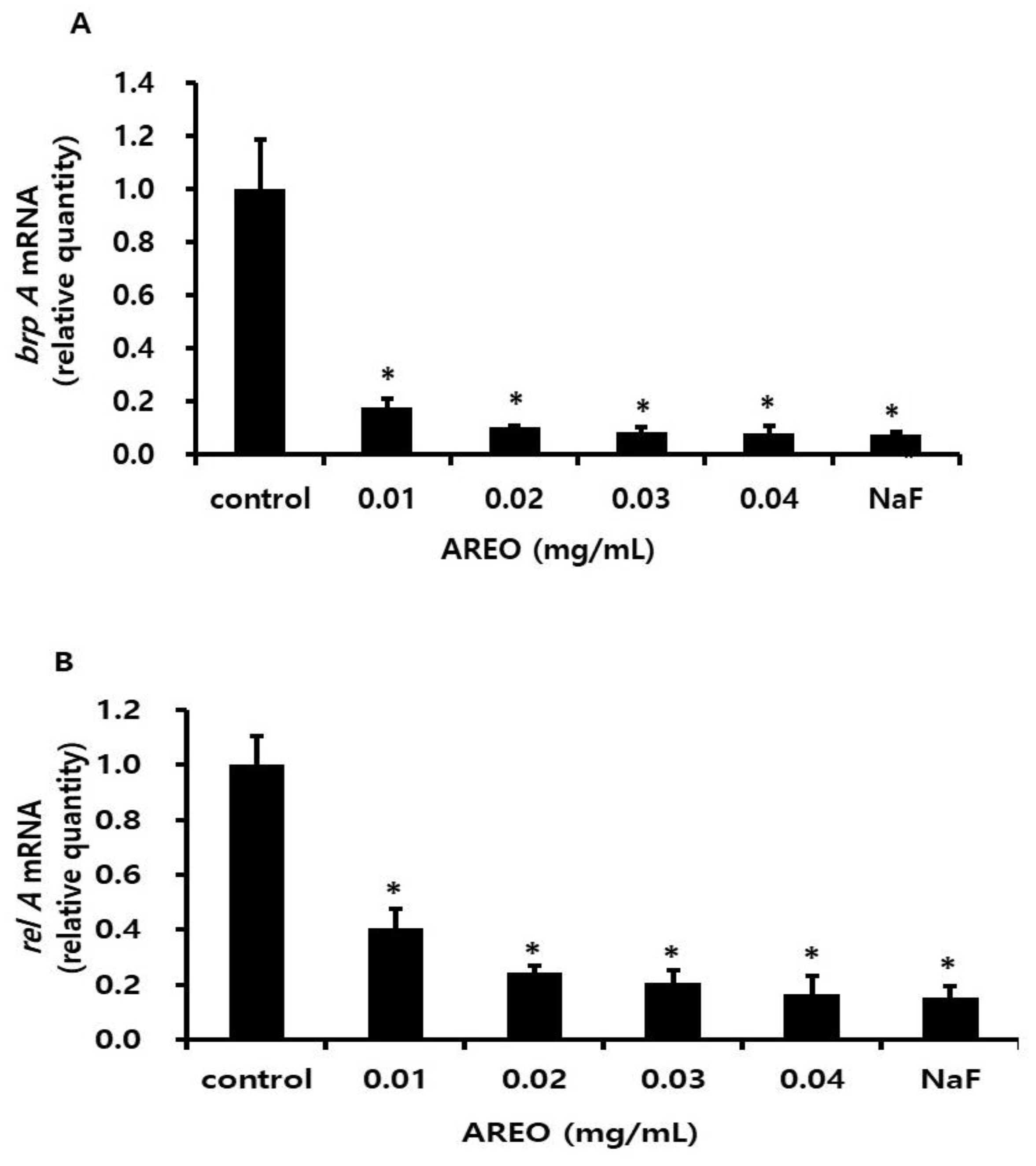
Gene expression of brpA and relA, which contribute to acid resistance regulation, was also reduced (Figure 8).

2.5. GC and GC-MS Analysis Results of AREO

As a result of analyzing AREO using GC and GC-MS, 43 constituents were identified, accounting for 98.59% of the total essential oil. The major components were estragole (88.69%), β-Caryophyllene (2.56%), and limonene (2.29%) (Table 2). Estragole, β-caryophyllene, and limonene are thought to be responsible for the anti-cariogenic properties of AREO. Future research will explore specific roles of major compounds in anti-cariogenic activities.

3. Discussion

Research into the prevention and treatment of dental caries, which is the main cause of tooth loss, is ongoing. Nevertheless, the rate of dental caries is increasing, requiring more financial investment and the establishment of appropriate prevention methods. For this reason, continued efforts have been made to develop several natural products using substances that help prevent and treat dental caries and periodontal disease. Many studies have reported that some natural substances inhibit the growth of S. mutans, the causative agent of dental caries in the oral cavity, but their mechanisms of action have been reported to be different. Plagiorhegama dubium and Terminalia chebula extracts have been reported to inhibit the growth and acid production of S. mutans [22,23]. Schisandra chinensis and Cinnamomum cassia Blume extracts have been reported to inhibit S. mutans growth and reduce adhesion to S-HA beads [18,24,25]. In addition, propolis [17], Phellodendron amurense extract [26], and seaweed extracts, such as funoran [27] and berberine, have also been reported to have antibacterial activity against S. mutans [28]. The polyphenols extracted from green tea leaves and the ethyl acetate extract of Sophora flavescens inhibit the synthesis of water-insoluble glucan by inhibiting the GTFase of S. mutans [29,30], and oolong tea leaf extract inhibits the cell-free GTFase of S. mutans, which synthesizes water-insoluble glucan [31]. Salvia miltiorrhiza, Dryopteris crassirhizoma, and Platycodon grandiflorus extracts have been reported to have antibacterial activity against S. mutans [32,33,34]. In addition, Artemisia capillaris, Artemisia argyi, and Artemisia herba essential oil extracts have antibacterial effects against S. mutans and Streptococcus sanguinis [35], and Galla Rhois extract containing tannic acid inhibits dental caries with antibacterial and bactericidal action [36,37].
Therefore, this study aimed to observe the biofilm formation inhibitory effect of AREO, traditionally used for food and medicine. The inhibitory effect of AREO on the growth of S. mutans, the causative bacterium of dental caries, was observed at a concentration of 0.02 mg/mL or higher. Chamaecyparis obtusa oil at concentrations above 0.05 mg/mL [38] and Brazilian Piperaceae essential oil at concentrations of 0.05–0.5 mg/mL have shown growth-inhibitory effects [39]. ɑ-Pinene, the main component of Chrysanthemum boreale, has shown an inhibitory effect at 0.25 mg/mL or higher [40], and rosemary (Rosemary-R), lemon grass (LG), floral petal (Floral-FR), and orange (Orange-O) essential oils have shown antibacterial effects at 0.156 mg/mL or higher [41]. The inhibition of biofilm formation by S. mutans was confirmed here by adhesion inhibition on dishes, resin-based teeth, and S-HA. As a result of SEM and confocal observation, a similar inhibitory activity with the 0.1% NaF positive control could be observed with AREO at 0.04 mg/mL. The antibacterial activity was stronger than that of ɑ-pinene from C. boreale [40] at a concentration of 0.5 mg/mL or higher, of propolis at a concentration of 0.2 mg/mL or higher [17], and of cypress oil at a concentration of 0.1 mg/mL or higher [38]. Similar to 0.1% NaF (the main component of toothpaste), at a concentration of 2 g/mL or higher of continentalic acid, a single component of Aralia elata, and at a concentration of 4 µg/mL or higher of kaurenoic acid from A. elata, the activity was lower than the biofilm formation inhibitory effect, which seemed to be the result of a single active ingredient [42,43]. The biofilm formation inhibitory effect was observed as an antibacterial effect of AREO on S. mutans. In terms of the genetic effects, considering that the expression of genes related to bacterial adhesion (gbpB and spaP) was significantly reduced compared to that of genes related to extracellular polysaccharide synthesis (gtfB, gtfC, and gtfD), AREO appeared to have an inhibitory effect on biofilm formation by inhibiting the formation of S. mutans attachment to the tooth surface. Propolis, which has a biofilm inhibitory effect, has also been reported to affect the expression of bacterial adhesion-related genes (gbpB and spaP) [17], and Chamaecyparis obtusa oil has been reported to affect the synthesis of extracellular polysaccharides and the expression of genes related to bacterial adhesion [38]. AREO seems to suppress bacterial growth and biofilm formation by inhibiting the expression of genes related to tooth surface adhesion.
The GC and GC-MS analyses in this study confirmed that estragole (88.69%), β-caryophyllene (2.56%), and limonene (2.29%) were present in AREO. The component analysis of AREO was analyzed and showed a similar trend to those reported in previous studies, and the antibacterial activity of estragole, the main component, has been reported in several studies. The antibacterial activity of S. mutans, the causative agent of dental caries, was confirmed, and the effect of inhibiting dental caries seemed to be due to the inhibition of biofilm formation, which was presumed to be due to the effect of suppressing the expression of genes related to adhesion to tooth surfaces. In the future, the efficacy of the components of AREO may require further research through a pure refining process.

4. Materials and Methods

4.1. Plant Materials and Essential Oils

Fresh leaves of A. rugosa were collected from a local farm in Yusong-Gu, Daejeon Metropolitan City, Republic of Korea, in late August 2021. The samples were authenticated by Professor Byung-Kil Choo (Department of Crop Agriculture and Life Science, Jeonbuk National University, Jeonbuk, Republic of Korea). Voucher specimen (AR-202105) was stored in the Laboratory of Fermentation Technology (Professor Myung-Kon Kim, Jeonbuk National University, Jeonbuk, Republic of Korea). The collected sample was kept in an airtight plastic container and stored in a cold room (4 °C) for 2 days until use. The leaves (1 kg) of A. rugosa were mechanically ground and extracted by distillation for 3 h using a Clevenger-type apparatus. The AREO used was a pale yellow oil with a yield of 1.02% based on the fresh weight of the plant. The AREO was stored in a cryogenic freezer (−70 °C) to minimize the loss of volatile compounds.

4.2. Strains and Culture

The strain used in this experiment was inoculated with S. mutans ATCC 25175 at a concentration of 1 × 10⁸ CFU/mL in brain heart infusion (BHI, BD, Sparks, MD, USA) broth, and after adding samples by concentration, it was then incubated at 37 °C for 24 h. Then, the optical density was measured at 550 nm using a spectrophotometer (Spectra Max 250, Molecular Devices Co., Menlo Park, CA, USA). Moreover, 0.1% NaF was used as a positive control. The experiment was repeated three times.

4.3. Adhesion to Saliva-Coated Hydroxyapatite Beads

The saliva secreted by stimulation with paraffin wax from a healthy adult male was collected in a cooled beaker. The collected saliva was centrifuged (12,000 rpm, 4 °C, 15 min), the supernatant was collected and treated at 60 °C for 30 min to inactivate the enzymes, and then used while storing at −20 °C. Hydroxyapatite beads (Bio-Rad Lab., Hercules, CA, USA) were washed five times with distilled water to remove small particles and dried at 37 °C before use. The saliva was coated on the beads by treatment with 30 mg of the dried hydroxyapatite beads with 1 mL of saliva at 37 °C for 60 min. Then, after washing the beads three times with 0.1 M potassium phosphate buffer (KPB, pH 7.0), AREO was added at each concentration. S. mutans was added at a concentration of 1 × 10⁷ CFU/mL and adhered to saliva-coated hydroxyapatite (S-HA) beads for 90 min in a shaking incubator at 37 °C. Then, after washing three times with 0.1 M KPB (pH 7.0), bacteria adhered to the S-HA were removed using an ultrasonic device (50 W, 30 s). Then, the bacterial solution was diluted and spread on a Mitis Salivarius Agar plate (BD, Sparks, MD, USA) and cultured for 24 h at 37 °C. Next, the number of colonies was counted [44]. AREO was not added to the control group.

4.4. S. mutans Biofilm Formation

After adding BHI liquid medium and AREO to a 35 mm dish, the bacteria were inoculated at a concentration of 5 × 10⁵ CFU/mL. After 24 h of culture in a 37 °C incubator, all of the supernatant was removed. Each dish was washed with 1.5 mL of distilled water. After staining with 0.1% safranin for 30 s, they were washed twice with distilled water, dried, and photographed [44].

4.5. S. mutans Biofilm Formed on the Surface of the Artificial Teeth

After adding BHI liquid medium and AREO to artificial teeth (Endura, Shofu Inc., Kyoto, Japan), the bacteria were inoculated at a concentration of 5 × 10⁵ CFU/mL. After culturing for 24 h in a 37 °C incubator, all the filtrate was removed. Each artificial tooth was washed with 1.5 mL of distilled water. After staining with 0.1% safranin for 30 s, it was washed twice with distilled water, dried, and photographed [45].

4.6. Measurement of S. mutans Biofilm Using a Scanning Electronic Microscope

After adding BHI liquid medium and AREO to a 35 mm dish, the bacteria were inoculated at a concentration of 5 × 10⁵ CFU/mL. After 24 h of culture in a 37 °C incubator, all the supernatant was removed. Each dish was washed with 1.5 mL of distilled water. The bacteria were then fixed for 24 h in a 2.5% glutaraldehyde solution (in a 0.1 M sodium cacodylate buffer, pH 7.2, 4 °C). Starting with 70% ethanol, washing and dehydration were performed by increasing the concentration to 80%, 95%, and 100%. After freeze-drying, the bacteria were coated with gold and photographed by scanning electron microscopy (SEM) [45].

4.7. Acid Production of S. mutans

After adding the sample to the BHI liquid medium containing 1% glucose, the bacteria were inoculated to a concentration of 1 × 10⁸ CFU/mL. After culturing for 24 h in an incubator at 37 °C, absorbance was measured at 550 nm using an ELISA reader (Molecular Devices Co., San Jose, CA, USA) based on the BHI broth. The pH was measured using a pH meter (Mettler-Toledo, Schwerzenbach, Switzerland) to observe the inhibitory effect on acid production. The control underwent the same process without adding the sample [46].

4.8. Real-Time PCR Analysis

Real-time PCR was performed to evaluate the effect of A. rugosa on S. mutans gene expression. cDNA was synthesized by isolating total RNA from S. mutans cultured in samples treated with different concentrations of the extract. Amplification was performed using a StepOnePlus Real-time PCR system with QPCR SYBR Green Mixes (Applied Bio system, Foster City, CA, USA). In addition, 16S rRNA was used as a control. Primer pairs have been described in a previous study [42]. Primer pairs are listed in Table 3. Assays were performed in duplicate on at least two independent RNA samples.

4.9. GC and GC-MS Analyses

GC analysis was performed on a Hewlett–Packard model 6890 series gas chromatograph with a flame ionization detector and a split ratio of 1:30 using a DB-5HT-fused silica capillary column (30 m × 0.32 mm, i.d., 0.25 μm film thickness). The column temperature was programmed from 40 °C to 230 °C at 2 °C/min and then kept constant at 230 °C for 20 min. The injector and detector temperatures were 230 °C and 250 °C, respectively. The carrier gas was nitrogen, with a flow rate of 0.80 mL/min. Peak areas were measured by electronic integration, and the relative amounts of the individual components were based on the peak areas.
GC-MS analysis was performed on an Agilent Technologies 7890A GC and 5975C mass selective detector operating in the EI mode at 70 eV and fitted with a DB-5MS-fused silica capillary column (30 m × 0.25, i.d., 0.25 μm film thickness). The column temperature was programmed from 40 °C to 230 °C at 2 °C/min and then kept constant at 230 °C for 20 min. The injector and ion source temperatures were both 250 °C. The carrier gas was helium at a flow rate of 1.0 mL/min. The identification of individual components was based on comparisons of mass spectra with the NIST/NBS mass spectral database and retention indices with data from previous studies [21]. Linear retention indices were calculated against those of an n-paraffin (C6∼C26) series [47].

4.10. Statistical Processing

The experiment was repeated three times, and the obtained results were presented as the average and standard deviation (SD) using the statistical program SPSS (ver. 10.0). At the p = 0.05 level, the average value of the experimental group and the control group was verified by an independent sample t-test.

5. Conclusions

This study demonstrated the antibacterial and antibiofilm effects of AREO on S. mutans. In addition, the effects on the expression of genes related to biofilm formation of S. mutans were investigated. Therefore, AREO is expected to be a promising agent to prevent dental caries. AREO is mainly composed of estragole, β-caryophyllene, and limonene, which may be responsible to be an antibacterial component, and inhibit growth and acid secretion in planktonic cultures. In this study, we focused on the overall anti-cariogenic potential of AREO and identified its major components. Although the individual inhibitory effects of the compounds such as limonene, estragole, and beta-caryophyllene were not tested in this study, future research will explore their specific roles in growth inhibition, adhesion, biofilm formation, and acid production. This initial study is valuable in providing a holistic understanding of AREO’s anti-cariogenic properties and its potential as a natural agent against dental biofilm. To our knowledge, this is the first report that AREO shows anti-cariogenic properties. However, despite the novelty and significance of this study, there are some limitations. One limitation is that the biofilm formation and the gene expression levels do not correspond, and another is that the antimicrobial effects of the major compounds need to be further investigated in future studies.

Author Contributions

Conceptualization, Y.-O.Y.; data curation, Y.-O.Y. and Y.-H.K.; formal analysis, E.S.K., Y.-H.K. and J.K.; investigation, E.S.K. and J.K.; methodology, B.-I.P.; resources, B.-I.P.; writing—original draft, E.S.K.; writing—review and editing, Y.-O.Y. All authors have read and agreed to the published version of the manuscript.

Funding

This research received no external funding.

Institutional Review Board Statement

Not applicable.

Informed Consent Statement

Not applicable.

Data Availability Statement

The data that support the findings of this study are available from the corresponding author upon reasonable request.

Acknowledgments

This paper was supported by Wonkwang University in 2023.

Conflicts of Interest

The authors declare no conflicts of interest.

References

  1. Hamada, S.; Torii, M. Interaction of glucosyltransferase from Streptococcus mutans with various glucans. J. Gen. Microbiol. 1980, 116, 51–59. [Google Scholar] [CrossRef] [PubMed]
  2. Wang, X.Y.; Zhang, Q.; Chen, Z. A possible role of LIM mineralization protein 1 in tertiary dentinogenesis of dental caries treatment. Med. Hypothese 2007, 69, 584–586. [Google Scholar] [CrossRef] [PubMed]
  3. Wiater, A.; Choma, A.; Szczodrak, J. Insoluble glucans synthesized by cariogenic streptococci: A structural study. J. Basic Microbiol. 1999, 39, 265–273. [Google Scholar] [CrossRef]
  4. Koga, T.; Asakawa, H.; Okahashi, N.; Hamada, S. Sucrose-dependent cell adherence and cariogenicity of serotype C Streptococcus mutans. J. Gen. Microbiol. 1986, 10, 2873–2883. [Google Scholar] [CrossRef]
  5. Loeshe, W.J. Role of Streptococcus mutans in human dental decay. Microbiol. Rev. 1986, 50, 353–380. [Google Scholar] [CrossRef]
  6. Marsh, P.D. Are dental diseases examples of ecological catastrophes? Microbiology 2003, 149, 279–294. [Google Scholar] [CrossRef]
  7. Marsh, P.D. Microbial ecology of dental plaque and its significance in health and disease. Adv. Dent. Res. 1994, 8, 263–271. [Google Scholar] [CrossRef]
  8. Inoue, M.; Koga, T.; Sato, S.; Hamada, S. Synthesis adherent insoluble glucan by the concerted action of the two glucosyltranferase components of Streptococcus mutans. FEBS Lett. 1982, 143, 101–104. [Google Scholar] [CrossRef]
  9. Koga, T.; Hamada, S.; Murakawa, S.; Endo, A. Effect of a glucosyltransferase inhibitor on glucan synthesis and cellular adherence of Streptococcus mutans. Infect. Immun. 1982, 38, 882–886. [Google Scholar] [CrossRef]
  10. Wenham, D.G.; Davies, R.M.; Cole, J.A. Insoluble glucan synthesis by mutansucrase as determinant of the cariogenicity of Streptococcus mutans. J. Gen. Microbiol. 1981, 127, 407–415. [Google Scholar] [CrossRef]
  11. Jeng, J.H.; Hsieh, C.C.; Lan, W.H.; Chang, M.C.; Lin, S.K.; Hahn, L.J.; Kuo, M.Y. Cytotoxicity of sodium fluoride on human oral mucosal fibroblasts and its mechanisms. Cell Biol. Toxicol. 1998, 14, 383–389. [Google Scholar] [CrossRef] [PubMed]
  12. Fardal, O.; Tumbull, R.S. A review of the literature on use of chlorhexidine in dentistry. J. Am. Dent. Assoc. 1986, 112, 863–869. [Google Scholar] [CrossRef] [PubMed]
  13. Lee, H.J.; Kim, M.Y.; Yang, D.N. The Effect of Oral Rinsing Solution on the Color Stability, Surface Microhardness and Surface Roughness Change of Composite Resin. J. Converg. Inf. Technol. 2019, 9, 159–167. [Google Scholar] [CrossRef]
  14. Charles, D.J.; Simon, J.E.; Widrlechner, M.P. Characterization of the essential oil of Agastache species. J. Agric. Food Chem. 1991, 39, 1946–1949. [Google Scholar] [CrossRef]
  15. Baik, J.A. Analysis, antibacterial, and insecticide effects on domestic native fragrant plants Elsholtzia ciliata and Agastache rugosa. J. People Plants Environ. 2016, 19, 79–83. [Google Scholar] [CrossRef]
  16. Lee, S.E.; Park, C.K.; Cha, M.S.; Kim, J.K.; Seong, N.S.; Bang, K.H.; Bang, J.K. Antimicrobial activity of essential oils from Mentha arvensis L. var. piperascens Malivaud and Agastache rugosa O. Kuntze on Escherichia coli and Salmonella typhimurium. Korean J. Med. Crop Sci. 2002, 10, 206–211. [Google Scholar]
  17. Park, B.I.; Jung, Y.W.; Kim, Y.H.; Lee, S.M.; Kwon, L.S.; Kim, K.J.; An, S.Y.; Choi, N.Y.; You, Y.O. Effect of the ethanol extract of propolis on formation of Streptococcus mutans biofilm. Int. J. Oral Biol. 2016, 41, 253–262. [Google Scholar] [CrossRef]
  18. Hong, M.J.; Kim, J.H.; Kim, H.Y.; Kim, M.J.; Kim, S.M. Chemical composition and biological activity of essential oil of Agastache rugosa (Fisch. & C. A. Mey.) O. Kuntze. Korean J. Med. Crop Sci. 2020, 28, 95–110. [Google Scholar] [CrossRef]
  19. Liang, J.; Huang, X.; Ma, G. Antimicrobial activities and mechanisms of extract and components of herbs in East Asia. R. Soc. Chem. Adv. 2022, 12, 29197–29213. [Google Scholar] [CrossRef]
  20. Song, J.H.; Kim, M.J.; Kwon, H.D.; Park, I.H. Antimicrobial activity and components of extracts from Agastache rugosa during growth period. J. Food Sci. Nutr. 2001, 6, 10–15. [Google Scholar]
  21. Adams, R.P. Identification of Essential Oil Components by Gas Chromatography/Mass Spectroscopy; Allured Publishing Corp.: Carol Stream, IL, USA, 2007. [Google Scholar]
  22. Park, S.N.; Lim, Y.K.; Kook, J.K. Antimicrobial effect of coptidis rhizome extract against mutans streptococci and periodontopathogens. Int. J. Oral Biol. 2015, 40, 79–83. [Google Scholar] [CrossRef]
  23. Nayak, S.S.; Ankola, A.V.; Metgud, S.C.; Bolmal, U.K. An in vitro study to determine the effect of Terminalia chebula extract and its formulation on Streptococcus mutans. J. Contemp. Dent. Pract. 2014, 15, 278–282. [Google Scholar] [CrossRef] [PubMed]
  24. Min, Y.K.; Jeon, J.K.; Kim, S.G.; Chang, K.W. Inhibitory effects of Schizandra chinensis extracts on the growth and adsorption to saliva coated HA beads of some oral bacteria. J. Korean Acad. Oral Health 2001, 25, 165–183. [Google Scholar]
  25. Lee, K.W.; Lee, S.K.; Chang, K.W. Effects of the crude Cinnamomi cortex extract on the growth and the adherence to hydroxyapatite beads of mutans streptococci. J. Korean Acad. Oral Health 1999, 23, 25–34. [Google Scholar]
  26. Tsujii, T.; Kawada-Matsuo, M.; Migita, H.; Ohta, K.; Oogai, Y.; Yamasaki, Y.; Komatsuzawa, H. Antibacterial activity of phellodendron bark against Streptococcus mutans. Microbiol. Immunol. 2020, 64, 424–434. [Google Scholar] [CrossRef]
  27. Saeki, Y.; Kato, T.; Naito, Y.; Takazoe, I.; Okuda, K. Inhibitory effects of funoran on the adherence and colonization of mutans streptococci. Caries Res. 1996, 30, 119–125. [Google Scholar] [CrossRef]
  28. Kazemipoor, M.; Fadaei Tehrani, P.; Zandi, H.; Golvardi Yazdi, R. Chemical composition and antibacterial activity of Berberis vulgaris (barberry) against bacteria associated with caries. Clin. Exp. Dent. Res. 2021, 7, 601–608. [Google Scholar] [CrossRef]
  29. Otake, S.; Makimura, M.; Kuroki, T.; Nishihara, Y.; Hirasawa, M. Anticaries effects of polyphenolic compounds from Japanese green tea. Caries Res. 1991, 25, 438–443. [Google Scholar] [CrossRef]
  30. Lee, H.O.; Lee, K.H.; Park, N.K.; Jeong, S.I.; Baek, S.H.; Han, D.M. Antibacterial effects of Sophora flavescens on Streptococcus mutans. Korean J. Food Nutr. 2000, 13, 539–546. [Google Scholar]
  31. Nakahara, K.; Kawabata, S.; Ono, H.; Tanaka, T.; Ooshima, T.; Hamada, S. Inhibitory effect of oolong tea polyphenols on glycosyltransferases of mutans Streptococci. Appl. Environ. Microbiol. 1993, 59, 968–973. [Google Scholar] [CrossRef]
  32. Lee, J.H.; Yim, D.Y.; Choi, S.S. Antibacterial activity and anti-inflammatory effect of methanol extracts of Saliva miltiorrhiza against oral pathogenic bacteria. Korean J. Pharmacogn. 2021, 52, 41–48. [Google Scholar] [CrossRef]
  33. Ban, S.H.; Kim, J.E.; Pandit, S.; Jeon, J.G. Influences of Dryopteris crassirhizoma extract on the viability, growth and virulence properties of Streptococcus mutans. Molecules 2012, 17, 9231–9244. [Google Scholar] [CrossRef] [PubMed]
  34. Kim, J. Antibacterial and anti-inflammatory effects of Platycodon grandiflorum extracts. J. Digit. Converg. 2014, 12, 359–366. [Google Scholar] [CrossRef]
  35. Cho, Y.H.; Chiang, M.H. Essential oil composition and antibacterial activity of Artemisia capillaris, Artemisia argyi, and Artemisia princeps. J. Korean Soc. Int. Agric. 2001, 13, 313–320. [Google Scholar]
  36. Doh, E.S.; Yoo, J.H. Antibacterial activity of bio-fermented Galla Rhios extract. Korea J. Herbol. 2014, 29, 21–27. [Google Scholar] [CrossRef]
  37. Shin, A.R.; Ohk, S.H.; Choi, C.H.; Hong, S.J. Identification and partial purification of antibacterial compounds against Streptococcus mutans from Galla Rhois. J. Korean Acad. Oral Health 2016, 40, 3–8. [Google Scholar] [CrossRef]
  38. Kim, E.H.; Kang, S.Y.; Park, B.I.; Kim, Y.H.; Lee, Y.R.; Hoe, J.H.; Choi, N.Y.; Ra, J.Y.; An, S.Y.; You, Y.O. Chamaecyparis obtusa suppresses virulence genes in Streptococcus mutans. Evid. Based Complement. Altern. Med. 2016, 2396404. [Google Scholar] [CrossRef]
  39. Carvalho Êni, S.; Ayres, V.F.S.; Oliveira, M.R.; Corrêa, G.M.; Takeara, R.; Guimarães, A.C.; Santiago, M.B.; Oliveira, T.A.S.; Martins, C.H.G.; Crotti, A.E.M.; et al. Anticariogenic activity of three essential oils from Brazilian Piperaceae. Pharmaceuticals 2022, 15, 972. [Google Scholar] [CrossRef]
  40. Park, B.I.; You, Y.O.; Mo, J.S.; An, S.Y.; Choi, N.Y.; Kim, K.J. Anti-cariogenic properties of ɑ-pinene, a monoterpene in plant essential oil. Int. J. Oral Biol. 2017, 42, 25–31. [Google Scholar] [CrossRef]
  41. Lee, S.Y.; Kim, J.G.; Baik, B.J.; Yang, Y.M.; Lee, K.Y.; Lee, H.Y.; Kim, M.A. Antimicrobial effect of essential oils on oral bacteria. J. Korean Acad. Pediatr. Dent. 2009, 36, 1–11. [Google Scholar]
  42. Jeong, S.I.; Kim, B.S.; Keum, K.S.; Lee, K.H.; Kang, S.H.; Park, B.I.; Lee, Y.R.; You, Y.O. Kaurenoic acid from Aralia continentalis inhibits biofilm formation of Streptococcus mutans. Evid.-Based Complement. Altern. Med. 2013, 160592. [Google Scholar] [CrossRef]
  43. Hay, D.I.; Gibbons, R.J.; Spinell, D.M. Characteristics ofsome high molecular weight constituents with bacterial aggregating activity from whole saliva and dentalplaque. Caries. Res. 1971, 5, 111–123. [Google Scholar] [CrossRef] [PubMed]
  44. O’Neill, E.; Hilary, H.; James, P.O. Carriage of both the fnbA and fnbB genes and growth at 37 °C promote FnBP mediated biofilm development in meticillin-resistant Staphylococcus aureus clinical isolates. J. Med. Microbiol. 2009, 58, 399–402. [Google Scholar] [CrossRef]
  45. Kwang, H.L.; Kim, B.S.; Keum, K.S.; Yu, H.H.; Kim, Y.H.; Chang, B.S.; Ra, J.Y.; Moon, H.D.; Seo, B.R.; Choi, N.Y.; et al. Essential oil of Curcuma longa inhibits Streptococcus mutans biofilm formation. J. Food Sci. 2011, 76, H226–H230. [Google Scholar] [CrossRef]
  46. Ooshimaa, T.; Osakaa, Y.; Sasakia, H.; Osawab, K.; Yasudab, H.; Matsumuraa, M.; Sobuea, S.; Matsumotoa, M. Caries inhibitory activity of cacao bean husk extract in in-vitro and animal experiments. Arch. Oral Biol. 2000, 45, 639–645. [Google Scholar] [CrossRef]
  47. VanDenDool, H.; Kratz, P.D. A generalization of the retention index system including linear temperature programmed gas-liquid partition chromatography. J. Chromatogr. A 1963, 11, 463–471. [Google Scholar] [CrossRef]
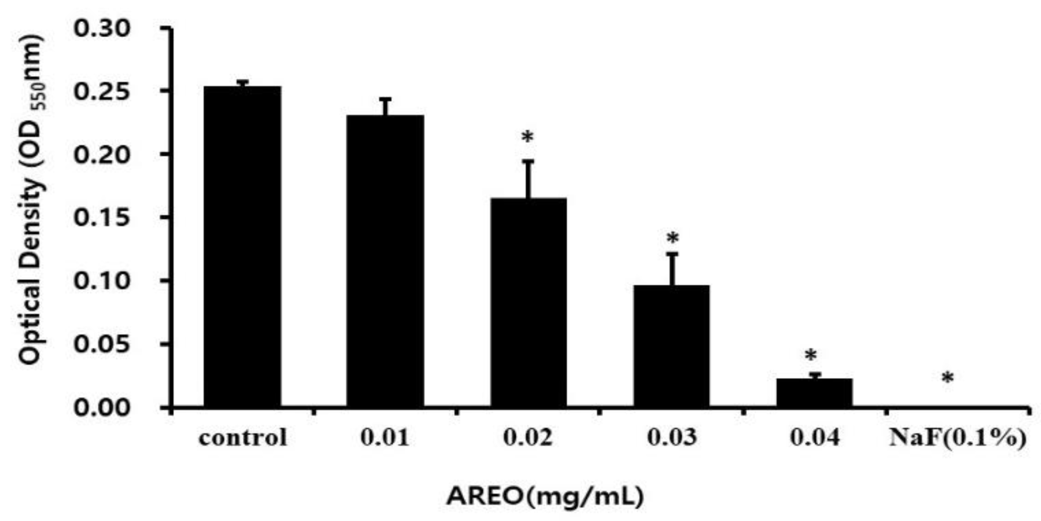
Figure 1. The growth of S. mutans inhibitory effect of AREO. The degree of inhibitory activity was observed in the presence of AREO at concentrations of 0.01, 0.02, 0.03, and 0.04 mg/mL. In addition, 0.1% NaF was used as a positive control. Significance was determined at * p < 0.05 compared to the control group.
Molecules 29 04907 g001
Figure 2. Effect of AREO on the levels of S. mutans (CFU). S. mutans was inoculated into BHI broth with various concentrations of AREO and incubated for 24 h. The amounts of S. mutans that adhered to S-HA beads that were treated with various concentrations of AREO are shown. When treated with 0.01–0.04 mg/mL of AREO, adherence was significantly repressed. Each value is expressed as the mean ± SD. Significance was determined at * p < 0.05 compared to the control group.
Molecules 29 04907 g002
Figure 3. Real-time PCR analysis of multiple genes encoding biofilm formation-associated virulence factors. After culturing S. mutans with various concentrations of the AREO component, real-time PCR analysis was performed. Each value is expressed as mean ± standard deviation. Significance was determined at * p < 0.05 compared to the control group.
Molecules 29 04907 g003
Figure 4. Inhibitory effect of AREO on biofilm formation in S. mutans. Safranin staining of S. mutans biofilms after treatment with AREO. (A) Control, (B) 0.01 mg/mL, (C) 0.02 mg/mL, (D) 0.03 mg/mL, (E) 0.04 mg/mL, and (F) positive control (0.1% NaF). Significance was determined at * p < 0.05 compared to the control group.
Molecules 29 04907 g004
Figure 5. Inhibitory effect of AREO on S. mutans biofilm formation on the surface of a resin-based tooth. (A) Control, (B) 0.01 mg/mL, (C) 0.02 mg/mL, (D) 0.03 mg/mL, (E) 0.04 mg/mL, and (F) positive control (0.1% NaF). Bar = 3.5 mm.
Molecules 29 04907 g005
Figure 6. SEM of S. mutans biofilms grown in AREO. Biofilm formation was decreased in the presence of AREO at concentrations greater than 0.04 mg/mL. (A) Control, (B) 0.01 mg/mL, (C) 0.02 mg/mL, (D) 0.03 mg/mL, (E) 0.04 mg/mL, and (F) positive control (0.1% NaF). Bar = 50 μm.
Molecules 29 04907 g006
Figure 7. Real-time PCR analysis of multiple genes encoding biofilm formation-associated virulence factors. Relative quantity of gtfB (A), gtfC (B), gtfD (C) and VicR (D). After culturing S. mutans with various concentrations of AREO, real-time PCR analysis was performed. Each value is expressed as the mean ± SD. Significance was determined at * p < 0.05 compared to the control group.
Molecules 29 04907 g007aMolecules 29 04907 g007b
Figure 8. Real-time PCR analysis of multiple genes encoding acid tolerance-related virulence factors. Relative quantity of brpA (A) and relA (B). After culturing S. mutans with various concentrations of AREO, real-time PCR analysis was performed. Each value is expressed as the mean ± SD. Significance was determined at * p < 0.05 compared to the control group.
Molecules 29 04907 g008
Table 1. The pH changes in S. mutans cultures incubated with different concentrations of AREO.
Concentration
(mg/mL)
pH
(Before Incubation)
pH
(After Incubation)
Control7.38 ± 0.055.41 ± 0.05
0.017.38 ± 0.055.48 ± 0.10
0.027.38 ± 0.055.74 ± 0.00 *
0.037.38 ± 0.056.54 ± 0.11 *
0.047.38 ± 0.007.19 ± 0.05 *
NaF (0.1%)7.38 ± 0.007.37 ± 0.00 *
Data (pH) are represented as the mean ± SD. * p < 0.05 as compared with the control group after incubation.
Table 2. AREO constituents.
CompoundRetention IndexPeak Area (%)
Calculated 1)Literature 2)
2-Ethyl furan712713t
n-Hexanal799800t
trans-2-Hexenal8628620.06
cis-3-Hexen-1-ol867858t
α-Pinene930939t
Camphene9509530.08
Benzaldehyde9589610.12
Sabinene967971 3)t
1-Octen-3-ol9859780.45
3-Octanone9879860.08
Myrcene9919910.05
Limonene103110312.29
Benzyl alcohol103710320.06
Phenylacetaldehyde103910430.07
Linalool11021098t
1-Octen-3-yl acetate112111190.24
trans-Isopulegone118111750.13
Estragole1219119688.69
Chavicol12871265 3)1.05
Isopiperitenone12921282 3)0.08
trans-Anethole13001285t
p-Vinylguaiacol13231324t
Eugenol136613560.22
Unidentified1379-0.24
β-Bourbonene138313840.05
cis-Jasmone139513940.05
Methyl eugenol140414010.06
β-Caryophyllene142414182.56
α-Humulene144714540.12
Germacrene D147914800.80
Bicyclogermacrene149314940.35
trans-Methyl isoeugenol15051495 3)0.07
trans,trans-α-Farnesene15081508t
δ-Cadinene152115240.08
Spathulenol157015760.07
Caryophyllene oxide157315810.06
τ-Cadinol16271625t
τ-Muurolol163516410.05
α-Cadinol164816530.07
6,10,14-Trimethylpentadecan-2-one18421843t
Benzyl salicylate18831876 3)t
Oleic acid21132115 3)0.27
Total 98.58
1) Retention indices on nonpolar DB-5HT fused silica capillary column. 2) Adams, Identification of essential oil components by gas chromatography/mass spectroscopy [21]. 3) NIST (National Institute of Standards and Technology) Chemistry webbook, SRD (Standard Reference Database) no. 69, 2023. t: trace (peak area less than 0.05%).
Table 3. Oligonucleotide primers used in this study.
Genes *Genes DescriptionPrimer Sequences (5′-3′)
16S rRNANormalizing internal standardsForwardCCTACGGGAGGCAGCAGTAG
ReverseCAACAGAGCTTTACGATCCGAAA
gtfBGlucosyltransferase B (gtfB)ForwardAGCAATGCAGCCAATCTACAAAT
ReverseACGAACTTTGCCGTTATTGTCA
gtfCGlucosyltransferase SI (gtfC)ForwardGGTTTAACGTCAAAATTAGCTGTATTAGC
ReverseCTCAACCAACCGCCACTGTT
gtfDGlucosyltransferase-I (gtfD)ForwardACAGCAGACAGCAGCCAAGA
ReverseACTGGGTTTGCTGCGTTTG
brpABiofilm-regulation proteinForwardGGAGGAGCTGCATCAGGATTC
ReverseAACTCCAGCACATCCAGCAAG
gbpBGlucan-binding proteinForwardATGGCGGTTATGGACACGTT
ReverseTTTGGCCACCTTGAACACCT
relAGuanosine tetra (penta)-phosphate synthetaseForwardACAAAAAGGGTATCGTCCGTACAT
ReverseAATCACGCTTGGTATTGCTAATTG
spaPCell surface antigenForwardGACTTTGGTAATGGTTATGCATCAA
ReverseTTTGTATCAGCCGGATCAAGTG
vicRTwo-component system regulatoryForwardTGACACGATTACAGCCTTTGATG
ReverseCGTCTAGTTCTGGTAACATTAAGTCCAATA
* Based on the NCBI S. mutans genome database.
Disclaimer/Publisher’s Note: The statements, opinions and data contained in all publications are solely those of the individual author(s) and contributor(s) and not of MDPI and/or the editor(s). MDPI and/or the editor(s) disclaim responsibility for any injury to people or property resulting from any ideas, methods, instructions or products referred to in the content.