Abstract
Thyroid transcription factor 1 (TTF1) is an important cancer-related biomarker for clinical diagnosis, especially for carcinomas of lung and thyroid origin. Herein, a novel label-free electrochemical immunosensor was prepared for TTF1 detection based on nanohybrids of ribbon-like tungsten disulfide-reduced graphene oxide (WS2-rGO) and gold nanoparticles (AuNPs). The proposed immunosensor employed H2O2 as the electrochemical probe because of the excellent peroxidase-like activity of ribbon-like WS2-rGO. The introduction of AuNPs not only enhanced the electrocatalytic activity of the immunosensor, but also provided immobilization sites for binding TTF1 antibodies. The electrochemical signals can be greatly amplified due to their excellent electrochemical performance, which realized the sensitive determination of TTF1 with a wide linear range of 0.025–50 ng mL−1 and a lower detection limit of 0.016 ng mL−1 (S/N = 3). Moreover, the immunosensor exhibited high selectivity, good reproducibility, and robust stability, as well as the ability to detect TTF1 in human serum with satisfactory results. These observed properties of the immunosensor enhance its potential practicability in clinical applications. This method can also be used for the detection of other tumor biomarkers by using the corresponding antigen–antibody complex.
1. Introduction
Thyroid transcription factor 1 (TTF1) is a tissue-specific transcription factor expressed mainly in the epithelial cells of the lungs and thyroid [1]. TTF1 immunopositivity has been used as a biomarker for the diagnosis of carcinomas of lung or thyroid origin by pathologists [2]. It has been reported that TTF1 is expressed in ∼90% of small cell lung cancers, ∼60–90% of lung adenocarcinomas and the majority of thyroid carcinomas, but is rarely expressed in other carcinomas [3]. Moreover, TTF1 is one of the best immunohistochemical biomarkers in the diagnosis of carcinomas of lung origin, especially lung adenocarcinoma [4]. Given its role in diagnosis, the detection of TTF1 is of vital importance. There have been many techniques developed for biomarker detection. However, the assays for TTF1 have rarely been reported, except for enzyme-linked immunoassay (ELISA) [5]. Although ELISA is common, it has some inherent disadvantages, such as its complex chromogenic procedure and strict time limitation [6]. Therefore, it is highly desirable to develop other simple and sensitive methods for TTF1 detection.
Over the past few years, electrochemical methods have been widely used in immunosensors due to their advantages, such as excellent specificity, good sensitivity and low cost [7,8,9]. Label-free electrochemical immunosensors in particular have attracted more attention, due to the ease involved with their operation, saving time and eliminating complex labeling processes when compared to sandwich-type immunosensors [10,11,12]. However, protein biomarkers cannot be used directly as an electrochemical indicator, because of the poor electrochemical properties of antigens and antibodies, as well as their complexity. Therefore, electrochemical probes were introduced to achieve label-free electrochemical immunosensing. Hydrogen peroxide (H2O2) is a common and important reactive oxygen species (ROS) involved in various biological and industrial processes. Monitoring its levels is essential for applications ranging from healthcare to environmental monitoring. In recent years, various electrochemical sensors have been developed to monitor H2O2 with high sensitivity and specificity [13,14]. Due to its excellent electrochemical response, H2O2 is widely employed as a probe in label-free electrochemical immunosensors [15,16].
In addition, to ensure sufficient sensitivity of electrochemical immunosensors, many efforts have been made to improve the electrochemical response towards the electrochemical probe by modifying various nanomaterials on the electrodes, such as carbon nanomaterials [17], metal and nonmetallic nano-oxides [18,19] and noble metal nanoparticles [20]. Amongst them, metal sulfides display great potential for signal amplification, due to their properties of good catalytic activity and good biocompatibility [21,22]. Tungsten disulfide (WS2) is a 2D-layered graphene-like material, which, due to its electrochemical properties, sheet-like architecture and high surface area, has attained much attention from researchers. Recently, WS2 has attracted significant attention due to its catalytic activity and superior thermal and oxidative stability, which make it a suitable candidate for electrochemical sensor applications [23,24]. However, the direct application of WS2 was limited due to its poor conductivity. To overcome this challenge, WS2 is usually incorporated with other materials such as graphene (Gr) which is known to exhibit excellent electrical conductivity and robust stability [25,26]. For instance, Yildirim et al. developed a molecularly imprinted electrochemical electrode based on WS2 nanoflowers on N,B-doped graphene, which demonstrated high selectivity and high sensitivity towards l-phenylalanine and made important contributions to the diagnosis of phenylketonuria [27]. Chen et al. reported a facile electrode based on mesoporous WS2 nanorods decorated with nitrogen-doped reduced graphene oxide networks. The modified electrode showed an excellent sensing ability towards a toxic antibiotic drug (roxarsone) [28]. In other studies, WS2-Gr nanohybrids were also utilized in Li-ion batteries [29], Na-ion batteries [30], UV photodetectors [31] and hydrogen evolution reactions [32]. To date, little has been reported about the application of WS2-Gr in electrochemical immunosensors.
In this work, a novel H2O2-based label-free electrochemical immunosensor for TTF1 determination was proposed utilizing ribbon-like WS2-rGO nanohybrids with intrinsic peroxidase-like activity (Scheme 1). The introduction of AuNPs in the fabrication of the immunosensor not only provided active sites to immobilize the antigens, but also further enhanced its electrochemical performance. The quantitative detection of TTF1 biomarkers could be achieved through the change in electrochemical current caused by the attachment of antigens and antibodies. Finally, the proposed electrochemical immunosensor was successfully used for the detection of TTF1 in a serum sample. This immunosensor has potential for the detection of other biomarkers and has great potential for practical applications.
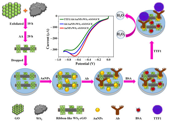
Scheme 1.
Schematic illustration of label-free electrochemical immunosensor for TTF1 detection.
2. Results and Discussion
2.1. Characterization of the WS2-rGO Nanohybrids
An SEM was used to observe the morphological features of the nanohybrids. Prior to mixing with GO, the natural WS2 showed an aggregation state with neat edges (Figure 1A). Moreover, the pure rGO, that was obtained through the reduction of GO without the presence of WS2, presented a film-like appearance with many random wrinkles and rugged edges (Figure 1B). When the mixture of WS2 and GO was exfoliated together using ultrasound and reduced by AA, WS2 presented a ribbon-like structure with a number of rGO nanofilms coated (Figure 1C). At a higher resolution, the structures of the ribbon-like WS2 and the adherent rGO nanofilms can be observed more clearly (Figure 1D,E). EDS analysis with C (9.23%), O (3.77%), W (13.58%) and S (22.52%) further confirmed the successful synthesis of WS2-rGO nanohybrids (Figure 1E). The Si element (50.91%) can also be observed, because the sample was placed on a silicon wafer for EDS analysis.
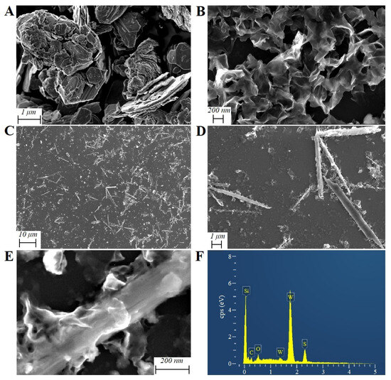
Figure 1.
SEM images of bulk WS2 (A), rGO (B), WS2-rGO nanohybrids (C–E) and corresponding EDS analysis (F).
The obtained ribbon-like WS2-rGO nanohybrids were also analyzed using UV-vis spectra (Figure 2A). In the spectra of GO, a typical UV-vis absorption peak at about 230 nm and a shoulder peak at about 300 nm were observed, which corresponded to the π-π* transitions of the aromatic C=C band and the n-π* transitions of the C=O band, respectively (Figure 2A, curve a). After reacting with AA, the two peaks disappeared and a new absorption peak appeared at 270 nm (Figure 2A, curve b), which illustrated that the GO was reduced to rGO with AA. In the spectra of WS2-rGO nanohybrids, in addition to the absorption of rGO, new absorption peaks were observed in the region from 400 nm to 700 nm (Figure 2A, curve c). It has been reported that bulk WS2 has an absorption edge of 920 nm [33]. However, the UV-vis absorption exhibits a significant blue shift when the size decreases from bulk WS2 to nanoscale [34]. The UV-vis absorption in this work further proved the successful synthesis of WS2-rGO nanohybrids.
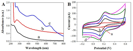
Figure 2.
(A) UV-vis absorption spectra of GO (a), rGO (b) and WS2-rGO (c). (B) Cyclic voltammograms of K[Fe3(CN)6] (2.0 × 10⁻³ mol L−1) at the electrodes of GO/GCE (a), bare GCE (b), rGO/GCE (c), WS2-rGO/GCE (d) and AuNPs/WS2-rGO/GCE (e). Scan rate: 0.1 V s−1.
FTIR analysis was used to confirm the effective non-covalent modification of functional groups and estimate the vibrational mutations of the molecules. Figure S1 illustrates the FTIR spectrum of WS2 and the WS2-rGO nanocomposite. For WS2, the vibrational band at 3516 cm−1 can be assigned to atmospheric –OH, and the 2921 cm−1 band can be associated with W-S bending vibrations. The bands found at 1639 cm−1 and 1576 cm−1 can be ascribed to the stretching vibrations of the hydroxyl groups. The bands positioned at 1096 cm−1 and 583 cm−1 are attributed to S-S and W-S bonds, respectively. For WS2-rGO, the bands appearing at 615 cm−1 and 490 cm−1 can be ascribed to C-S and S-S stretching vibrations. Most of the characteristic bands associated with WS2 are observed in the WS2-rGO composite, with a partial shift in the band positions. The 2921 cm−1 and 2919 cm−1 bands assigned to W-S in WS2 and the WS2-rGO composite, and the 615 cm−1 band attributed to C-S in WS2-rGO detected thus confirm the formation of the composite. Significantly, the FTIR spectrum results established the composition and interaction of ribbon-like WS2 on the surface of the rGO sheets.
To evaluate the electrochemical performance of the nanohybrids, the responses of various electrodes were analyzed using cyclic voltammetry (CV) in 1 × 10⁻³ mol L−1 [Fe(CN)6]3−/4− containing a 0.1 mol L−1 KCl solution. Before modification, a pair of typical reversible redox peaks was observed on bare GCE (Figure 2B, curve b). The redox peaks almost disappeared on GO/GCE, due to the poor conductivity of GO (Figure 2B, curve a). After being modified with rGO, the redox peaks on rGO/GCE increased sharply (Figure 2B, curve c) due to the good conductivity of rGO, which further illustrated that GO was successfully reduced with AA. When GCE was modified with the WS2-rGO nanohybrids, the redox peaks and charging current all increased further (Figure 2B, curve d), benefitting from the excellent electrical conductivity and the large specific area of WS2-rGO. Finally, the redox currents of [Fe(CN)6]3−/4− reached their maximum on AuNPs/WS2-rGO/GCE because of the introduction of AuNPs (Figure 2B, curve e), which confirmed that the electrode was assembled successfully and could be used for further analysis.
2.2. Electrochemical Behavior of the Immunosensor
In general, the sensitivity of the label-free electrochemical immunosensor depends on the electrochemical activity of the biosensor towards the electrochemical probe. We therefore investigated the electrocatalytic activity of the ribbon-like WS2-rGO and AuNPs nanohybrids towards H2O2. In the absence of H2O2, there were no peaks on all of the modified electrodes (Figure S2A–F). When 5 mM of H2O2 was added into the N2-saturated PBS, there was still no obvious response on bare GCE (Figure S2G, curve a), whereas a slight reduction in peak was observed on GO/GCE (Figure S2G, curve b). After coating GCE with rGO, the electrochemical response towards H2O2 was greatly increased, due to the good conductivity of rGO (Figure S2G, curve c). Additionally, when WS2 was doped on GCE, the electrochemical response towards H2O2 increased more sharply than that of bare GCE (Figure S2H). Moreover, with the introduction of nano-WS2, the reduction current was further increased compared to that of rGO/GCE (Figure 3A, curves a and b), indicating that the WS2-rGO nanohybrids had good electrocatalytic activity towards H2O2. The electrochemical properties reached optimum results on AuNPs/WS2-rGO/GCE (Figure 3A, curve c), which was a result of the comprehensive effect of nano-WS2, rGO and AuNPs. The above result further confirmed that the catalytic activity of AuNPs/WS2-rGO nanocomposites was exceptional in the electrochemical reduction of H2O2, which made it possible to construct a highly sensitive immunosensor.
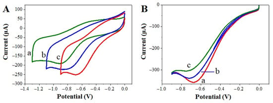
Figure 3.
(A) CVs of rGO/GCE (a), WS2-rGO/GCE (b) and AuNPs/WS2-rGO/GCE (c) in the presence of 5 mM of H2O2 in N2-saturated pH 7.0 PBS. Scan rate: 0.1 V s−1. (B) LSVs of AuNPs/WS2-rGO/GCE (a), Ab/AuNPs/WS2-rGO/GCE (b), and TTF1/Ab/AuNPs/WS2-rGO/GCE (c) in the presence of 5 mM of H2O2 in N2-saturated pH 7.0 PBS. Scan rate: 0.1 V s−1.
The fabrication process of the label-free electrochemical immunosensor was investigated using linear sweep voltammetry (LSV). Before the immobilization of anti-TTF1, a well-defined reduced peak was observed in AuNPs/WS2-rGO/GCE (Figure 3B, curve a). After the modified electrode was immobilized with anti-TTF1 and blocked using BSA, the electrochemical response towards H2O2 decreased gradually (Figure 3B, curve b), which suggested that the non-conductive substances of anti-TTF1 and BSA can hinder electron transfer and disturb the electrocatalytic activity toward H2O2. The peak current of H2O2 was further decreased when TTF1 was captured with Ab/AuNPs/WS2-rGO/GCE (Figure 3B, curve c), which indicated that the complex of antibodies and antigens can further disturb the reduction of H2O2 on the surface of the immunosensor. The gradual decrease in the response current indicated that the label-free electrochemical immunosensor was prepared successfully and can be used for TTF1 detection with H2O2 acting as the electrochemical probe.
2.3. Optimization of Detection Conditions
In order to achieve an optimal electrochemical signal response, the optimization of experimental conditions is necessary. The pH value can influence the electrocatalytic process of AuNPs/WS2-rGO/GCE. Figure S3 shows the electrocatalytic current responses of AuNPs/WS2-rGO/GCE in PBS at different pH values in 5 mM of H2O2. As shown in this figure, the optimal electrochemical signal response was achieved at a pH of 7.0. Moreover, the antibodies and antigens can maintain their biological activity in this approximately neutral condition. Therefore, PBS at pH 7.0 was selected for use throughout this study.
In order to achieve the optimal electrochemical result, parameters such as the incubation time of anti-TTF1 and the incubation temperature of TTF1 antigens were also studied. As shown in Figure 4A, when the incubation time of anti-TTF1 was varied from 0 to 4 h, the response currents of H2O2 decreased gradually, as a result of more anti-TTF1 molecules being conjugated to the surface of AuNPs/WS2-rGO/GCE. The reduction current of H2O2 was observed to be constant as the incubation time was increased from 4 h to 8 h, indicating that the anti-TTF1 had reached saturation on the electrode. Therefore, 4 h was used as the optimum incubation time for anti-TTF1 conjugation. For more efficient binding of TTF1 antigens with anti-TTF1, the incubation temperature of TTF1 on the surface of Ab/AuNPs/WS2-rGO/GCE was also investigated. As shown in Figure 4B, the greatest reduction in electrochemical response was observed at 37 °C, demonstrating that 37 °C is more suitable for the combination of TTF1 antigens with anti-TTF1.
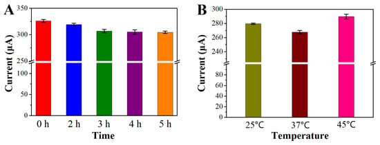
Figure 4.
Optimization of (A) incubation time of anti-TTF1 on AuNPs/WS2-rGO/GCE and (B) incubation temperature of TTF1 antigens on BSA/Ab/AuNPs/WS2-rGO/GCE. LSV responses were performed in a N2-saturated pH 7.0 PBS containing 5 mM of H2O2. Error bars represent the standard deviation (SD) from three independent determinations.
2.4. Electrochemical Detection of TTF1 Antigens
Under optimal conditions, the electrochemical immunosensing system developed was used for the detection of TTF1 antigens using LSV. As the concentration of TTF1 was increased from 25 pg mL−1 to 50 ng mL−1, the amount of TTF1 antigens conjugated to the surface of Ab/AuNPs/WS2-rGO/GCE also increased, leading to the gradual decrease in electrochemical responses (Figure 5A). Moreover, a good linear relationship was obtained between the concentration of TTF1 and the reduction current of H2O2 (iP (μA) = −1857.63 c (μg mL−1) + 314.32 (R = 0.9940)) with a detection limit of 0.016 ng mL−1 (Figure 5B). Compared with the reported ELISA methods [5], a wider linear range was obtained in this work. The good sensitivity observed benefitted from the signal amplification strategy of the AuNPs/WS2-rGO nanocomposites. The results indicated that the electrochemical immunosensor developed could be employed for the ultra-sensitive detection of TTF1 antigens.
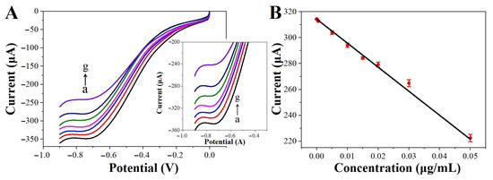
Figure 5.
(A) LSV responses of the immunosensor in N2-saturated pH 7.0 PBS containing 5 mM of H2O2 for TTF1 detection with different concentrations. From a to g: 0.025 ng mL−1, 0.05 ng mL−1, 5 ng mL−1, 10 ng mL−1, 20 ng mL−1, 30 ng mL−1 and 50 ng mL−1. Scan rate: 0.1 V s−1. (B) The corresponding calibration curve. Error bars represent the SD from three independent determinations.
2.5. Specificity, Reproducibility and Stability of the Immunosensor
Specificity is one of the most important indexes of the label-free electrochemical immunosensor. The selectivity analysis was performed using thrombospondin-1 (TSP1) [35], extracellular signal-regulated kinase (ERK) [36], BSA and glucose (GLU) as interference. A 15 ng mL−1 of TTF1 solution, or a mixture of 15 ng mL−1 of TTF1 with 100 ng mL−1 of interference solution, was detected using the developed immunosensor of Ab/AuNPs/WS2-rGO/GCE. As shown in Figure S4A, the electrochemical response of H2O2 on the immunosensors that were incubated in the mixtures was almost the same as that on TTF1/Ab/AuNPs/WS2-rGO/GCE, suggesting that the interference of TSP1, ERK, BSA and GLU did not disturb the detection of TTF1. This result proved the high specificity of the developed immunosensor.
To test the reproducibility of this assay, the H2O2 response was studied on five independent immunosensors. The reduction currents of H2O2 were almost the same, with a relative standard deviation (RSD) of 1.02% (Figure S4B). To assess the assay’s repeatability, identical samples of H2O2 were tested using the same immunosensor. The reduction currents of H2O2 exhibited remarkable consistency, with a low RSD of 1.06%, affirming the high reproducibility of this measurement. Additionally, the response current of the electrochemical immunosensors after storage at 4 °C for 7 days was observed to have only decreased by 4.30%, as compared to that of the freshly prepared immunosensor. The above results indicated good reproducibility and stability of the developed immunosensor.
2.6. Determination of TTF1 in Human Serum Sample
In order to evaluate the viability of the developed method, the label-free electrochemical immunosensor of Ab/AuNPs/WS2-rGO/GCE was used for the detection of TTF1 in a human serum sample. The measurement method of TTF1 in serum was the same as that used in the standard TTF1 solution. Subsequently, a certain concentration of TTF1 was added into the serum sample, and the final content of TTF1 in the samples was measured afterwards. As shown in Table S1, the concentration of TTF1 in the serum sample was obtained with a concentration of 0.76 ng mL−1, which was found to be within the range of reported values [37]. Moreover, the results were observed to range from 98.7% to 103.1%, with an RSD lower than 5.2%, suggesting the acceptable accuracy of the developed immunosensor. Finally, we compared the proposed electrochemical immunosensor with the ELISA method to measure TTF1 content in serum samples. The results showed that the TTF1 levels measured with these two methods are essentially consistent (Table S1), indicating the reliability of the method proposed in this work.
3. Materials and Methods
3.1. Apparatus and Reagents
A CHI650B electrochemical platform (Chenhua Instrument Co., Ltd., Shanghai, China) was employed to observe the electrochemical behaviors. A traditional three-electrode system was used for the electrochemical measurements, with a bare glassy carbon electrode (GCE, 4 mm in diameter) or a modified GCE as the working electrode, a platinum wire as the auxiliary electrode and a saturated calomel electrode (SCE) as the reference electrode. A Scientz-IID ultrasonic cell crusher (Xinzhi biotechnology co., Ltd., Ningbo, China) was used to exfoliate the GO or the mixture of GO and WS2. The characterization of the materials was performed using a scanning electron microscope (SEM, Zeiss, sigma 500, Jena, Germany), UV-2550 spectrophotometer (Shimadzu, Kyoto, Japan) and Fourier-transform Infra-Red spectra (FTIR, PerkinElmer, Waltham, MA, USA).
TTF1 antibodies (anti-TTF1 or Ab) and TTF1 antigens were purchased from Hunan Jingke biotechnology Co., Ltd., Changsha, China. The commercial ELISA kits were purchased from Cusabio (Wuhan, China). Bulk WS2, graphite powers and HAuCl4·3H2O were purchased from Shanghai Macklin Biochemical Co., Ltd., China. Ascorbic acid (AA) was purchased from Shanghai Aladdin Chemistry Co., Ltd., China. NaOH, N,N-Dimethylformamide (DMF) and hydrogen peroxide (H2O2, 30%) were purchased from Tianjin Fuchen chemical reagent Co., Ltd., China. The nitrogen-saturated phosphate buffer solution (N2-saturated PBS, pH 7.0) was prepared by mixing a 0.1 mol L−1 NaH2PO4 and Na2HPO4 solution and removing the dissolved oxygen by purging nitrogen gas for 20 min. The water used was ultra-pure deionized water (18.2 MΩ·cm).
The venous serum samples were obtained from a clinical laboratory and approved by the Ethics Committee of Zhengzhou Hospital of Traditional Chinese Medicine (Ethics ID: AF/SC-08/03.0). All sample donors provided voluntary and informed consent.
3.2. Synthesis of Ribbon-like WS2-rGO Nanohybrids
Graphene oxide (GO) was synthesized from graphite powder using Hummers’ method, as described in the previous report [38]. In order to synthesize WS2-rGO nanohybrids, 10 mg of bulk WS2 and 10 mg of GO powder were dispersed in 10 mL of DMF. The mixture was then exfoliated using an ultrasonic cell crusher with a power of 855 W. After ultra-sonicating for 10 h, the homogeneous solution was set for 24 h at room temperature to remove the WS2 and GO crowds that were not exfoliated. Then, the supernatant was centrifuged at 1000 g to further remove the bulk WS2 and GO. The reduction of GO was performed by adding 100 mg of reductant AA to the supernatant mixture of GO and WS2 that was obtained through centrifugation [39]. After heating for 20 h at 95 °C with vigorous stirring, the rGO was generated with doped nano-WS2. Then, the obtained WS2-rGO nanohybrids were washed several times using alcohol and water, until the pH reached 7.0. Finally, the WS2-rGO nanohybrids were freeze-dried and stored at 4 °C for further use. For comparison, the pure rGO was also synthesized this way, but without the presence of WS2.
3.3. Preparation of Electrochemical Immunosensor
Firstly, 1 mg of WS2-rGO nanohybrids was dispersed in 1 mL of DMF and ultra-sonicated for 30 min. Before fabrication, the bare GCE was polished to a mirror-like finish using 0.05 μm of alumina powder. As shown in Scheme 1, the electrochemical immunosensor was fabricated by doping 10 μL of WS2-rGO suspension onto the GCE surface and then air drying to prepare a WS2-rGO-modified GCE (WS2-rGO/GCE). In order to conjugate anti-TTF1, AuNPs were coated on the surface of WS2-rGO/GCE using electrodeposition for 30 s at a potential of −0.2 V in a 1% HAuCl4 solution (noted as AuNPs/WS2-rGO/GCE). After washing with PBS (pH = 7.0), 6 μL of 10 ng mL−1 anti-TTF1 was dropped on the AuNPs/WS2-rGO/GCE surface and incubated at 4 °C for 4 h. This was then followed by incubation with 6 μL of 1% BSA for 1 h to eliminate the non-specific adsorption (noted as Ab/AuNPs/WS2-rGO/GCE). After each incubation, the immunosensor was washed with PBS (pH = 7.0) to remove the unbound substances. Thereafter, the prepared immunosensor Ab/AuNPs/WS2-rGO/GCE was ready for the detection of TTF1 antigens.
3.4. Electrochemical Measurements of TTF1
In order to achieve the optimal electrochemical result, parameters such as the incubation time of anti-TTF1, the incubation temperature of TTF1 antigens and the optimal reaction pH were studied. To bond the TTF1 antigens, the prepared Ab/AuNPs/WS2-rGO/GCE was incubated with 6 μL of TTF1 antigens (with varying concentration in pH 7.0 PBS) for 1 h at 37 °C. Subsequently, the proposed electrochemical immunosensor (noted as TTF1/Ab/ AuNPs/WS2-rGO/GCE) was washed extensively to remove unbounded TTF1 molecules. Electrochemical measurement of the TTF1 biomarker was performed in a N2-saturated PBS (pH = 7.0) solution by adding 5 mM of H2O2 as a probe. After covering with TTF1 antigens, the resistance would increase and the response current of H2O2 would be decreased, which can be used as the signal to quantify TTF1 antigens.
4. Conclusions
In this work, we have developed a novel label-free electrochemical immunosensor for TTF1 detection using AuNPs/WS2-rGO nanocomposites as the electrode materials and H2O2 as the electrochemical probe, respectively. The ribbon-like WS2-rGO nanohybrids showed excellent electrocatalytic activity towards the electrochemical reduction of H2O2. The introduction of AuNPs not only enhanced the electrochemical performance of the immunosensor, but also provided active sites for anti-TTF1 immobilization. Through using UV-vis, FTIR and SEM characterizations, the structure and morphology of the ribbon-like WS2-rGO composite were analyzed. Taking advantage of the synergistic effect of nano-WS2, rGO and AuNPs, the label-free electrochemical immunosensor developed high sensitivity for TTF1 detection, with a wide linear range of 25 pg mL−1 to 50 ng mL−1 and a lower detection limit of 16 pg mL−1. Moreover, the immunosensor exhibited high selectivity, good reproducibility and robust stability, which enable it to be applied in human serum with satisfactory results. This method provided a potential application in clinical diagnosis and can be used for the detection of other biomarkers by using the corresponding antigen-antibody complex.
Supplementary Materials
The following supporting information can be downloaded at: https://www.mdpi.com/article/10.3390/molecules29020552/s1.
Author Contributions
Conceptualization, Z.L.; formal analysis, W.W.; investigation, H.T.; resources, H.T.; writing—original draft preparation, W.W.; writing—review and editing, H.T., L.Z. and Z.L.; visualization, H.T. and W.W.; supervision, L.Z. and Z.L.; funding acquisition, W.W. and Z.L. All authors have read and agreed to the published version of the manuscript.
Funding
This research was funded by the National Natural Science Foundation of China (22274143 to Z.L., 32301892 to W.W.), the Program for Innovative Research Teams (in Science and Technology) in the University of Henan Province (22TRTSTHN002 to Z.L.), the Key Projects of the Joint Fund for Science and Technology of Henan Province (222301420007 to Z.L.), the Excellent Youth Foundation of Henan Scientific Committee (232300421021 to Z.L.), the Key Science and Technology Research of Henan Province (222102310043 to W.W.), the Chinese Postdoctoral Science Foundation (2023M733257 to W.W.) and the Fundamental Research Funds for the Central Universities from Hunan University.
Institutional Review Board Statement
The venous serum samples were obtained from a clinical laboratory and approved by the Ethics Committee of Zhengzhou Hospital of Traditional Chinese Medicine (Ethics ID: AF/SC-08/03.0, 10 June 2022).
Informed Consent Statement
Not applicable.
Data Availability Statement
Data are contained within the article and Supplementary Materials.
Acknowledgments
We thank Qiyong Cai for supporting the venous serum samples. The authors thank the teachers and students in our research team for their help and support.
Conflicts of Interest
The authors declare no conflicts of interest.
References
- Sun, J.M.; Han, J.; Ahn, J.S.; Park, K.; Ahn, M.J. Significance of Thymidylate Synthase and Thyroid Transcription Factor 1 Expression in Patients with Nonsquamous Non-small Cell Lung Cancer Treated with Pemetrexed-Based Chemotherapy. J. Thorac. Oncol. 2011, 6, 1392–1399. [Google Scholar] [CrossRef] [PubMed]
- La Salvia, A.; Siciliani, A.; Rinzivillo, M.; Verrico, M.; Baldelli, R.; Puliani, G.; Modica, R.; Zanata, I.; Persano, I.; Fanciulli, G.; et al. Thyroid transcription factor-1 expression in lung neuroendocrine tumours: A gender-related biomarker? Endocrine 2023. [Google Scholar] [CrossRef] [PubMed]
- Schilsky, J.B.; Ni, A.; Ahn, L.; Datta, S.; Travis, W.D.; Kris, M.G.; Chaft, J.E.; Rekhtman, N.; Hellmann, M.D. Prognostic impact of TTF-1 expression in patients with stage IV lung adenocarcinomas. Lung Cancer 2017, 108, 205–211. [Google Scholar] [CrossRef] [PubMed]
- Mukhopadhyay, S.; Katzenstein, A.L.A. Comparison of Monoclonal Napsin A, Polyclonal Napsin A, and TTF-1 for Determining Lung Origin in Metastatic Adenocarcinomas. Am. J. Clin. Pathol. 2012, 138, 703–711. [Google Scholar] [CrossRef] [PubMed]
- Porcel, J.M.; Palma, R.; Bielsa, S.; Esquerda, A.; Gatius, S.; Matias-Guiu, X.; Salud, A. TTF-1 and napsin A on cell blocks and supernatants of pleural fluids for labeling malignant effusions. Respirology 2015, 20, 831–833. [Google Scholar] [CrossRef] [PubMed]
- Zhang, Y.C.; Gao, Y.; Zhang, X.L.; Wang, H.M.; Xia, T.; Bian, C.C.; Liang, S.; Tang, X.Z.; Wang, X. Electrochemical immunosensor for HBe antigen detection based on a signal amplification strategy: The co-catalysis of horseradish peroxidase and nanoporous gold. Sens. Actuator B-Chem. 2019, 284, 296–304. [Google Scholar] [CrossRef]
- Kiio, L.K.; Onyatta, J.O.; Ndangili, P.M.; Oloo, F.; Santamaria, C.; Montuenga, L.M.; Mbui, D.N. Ultrasensitive immunosensor for multiplex detection of cancer biomarkers carcinoembryonic antigen (CEA) and yamaguchi sarcoma viral oncogene homolog 1 (YES1) based on eco-friendly synthesized gold nanoparticles. Talanta 2024, 266, 124934. [Google Scholar] [CrossRef]
- Braz, B.A.; Hospinal-Santiani, M.; Martins, G.; Gogola, J.L.; Valenga, M.G.P.; Beirao, B.C.B.; Bergamini, M.F.; Marcolino-Junior, L.H.; Thomaz-Soccol, V.; Soccol, C.R. Gold-binding peptide as a selective layer for electrochemical detection of SARS-CoV-2 antibodies. Talanta 2023, 257, 124348. [Google Scholar] [CrossRef]
- Zhu, X.D.; Shan, J.K.; Dai, L.; Shi, F.F.; Wang, J.S.; Wang, H.; Li, Y.Y.; Wu, D.; Ma, H.M.; Wei, Q.; et al. PB@PDA nanocomposites as nanolabels and signal reporters for separate-type cathodic photoelectrochemical immunosensors in the detection of carcinoembryonic antigens. Talanta 2023, 254, 124134. [Google Scholar] [CrossRef]
- Cancelliere, R.; Di Tinno, A.; Di Lellis, A.M.; Contini, G.; Micheli, L.; Signori, E. Cost-effective and disposable label-free voltammetric immunosensor for sensitive detection of interleukin-6. Biosens. Bioelectron. 2022, 213, 114467. [Google Scholar] [CrossRef]
- Yukird, J.; Chailapakul, O.; Rodthongkum, N. Label-free anti-Mullerian hormone sensor based on polyaniline micellar modified electrode. Talanta 2021, 222, 121561. [Google Scholar] [CrossRef]
- Filik, H.; Avan, A.A. Electrochemical immunosensors for the detection of cytokine tumor necrosis factor alpha: A review. Talanta 2020, 211, 120758. [Google Scholar] [CrossRef] [PubMed]
- Ullah, R.; Rasheed, M.A.; Abbas, S.; Rehman, K.-U.; Shah, A.; Ullah, K.; Khan, Y.; Bibi, M.; Ahmad, M.; Ali, G. Electrochemical sensing of H2O2 using cobalt oxide modified TiO2 nanotubes. Curr. Appl. Phys. 2022, 38, 40–48. [Google Scholar] [CrossRef]
- Zribi, R.; Ferlazzo, A.; Fazio, E.; Condorelli, M.; D’Urso, L.; Neri, G.; Corsaro, C.; Neri, F.; Compagnini, G.; Neri, G. Ag Nanoplates Modified-Screen Printed Carbon Electrode to Improve Electrochemical Performances Toward a Selective H2O2 Detection. IEEE Trans. Instrum. Meas. 2023, 72, 1–8. [Google Scholar] [CrossRef]
- Dong, Y.F.; Qin, X.L.; Wang, M.H.; Gu, C.Y.; Zhu, Z.W.; Yang, D.; Shao, Y.H. Electrochemiluminescent Detection of Proteins Based on Fullerenols Modified Gold Nanoparticles and Triple Amplification Approaches. Anal. Chem. 2020, 92, 1890–1897. [Google Scholar] [CrossRef]
- Yan, Q.; Cao, L.L.; Dong, H.; Tan, Z.L.; Hu, Y.T.; Liu, Q.; Liu, H.; Zhao, P.P.; Chen, L.; Liu, Y.Y.; et al. Label-free immunosensors based on a novel multi-amplification signal strategy of TiO2-NGO/Au@Pd hetero-nanostructures. Biosens. Bioelectron. 2019, 127, 174–180. [Google Scholar] [CrossRef] [PubMed]
- Suwannachat, J.; Saenchoopa, A.; Tun, W.S.T.; Patramanon, R.; Daduang, S.; Daduang, J.; Kulchat, S. An electrochemical AChE-based biosensor for organophosphate pesticides using a modified CuNWs/rGO nanocomposite on a screen-printed carbon electrode. Food Chem. 2024, 434, 137431. [Google Scholar] [CrossRef] [PubMed]
- Yang, Z.Y.; Cao, B.Y.; An, X.W.; Yu, Z.L.; Zhao, W.D.; Su, F.C.; Guan, G.Q.; Zhang, Y.H.; Xie, Z.K.; Ye, B.X. Fabrication of nitrogen-carbon mediated γ-Mo2N nanocomposite based electrochemical sensor for rapid and sensitive determination of antioxidant 6-PPD in the environment. Talanta 2024, 266, 125072. [Google Scholar] [CrossRef] [PubMed]
- Sweety; Kumar, D. Electrochemical immunosensor based on titanium dioxide grafted MXene for EpCAM antigen detection. J. Colloid Interface Sci. 2023, 652, 549–556. [Google Scholar] [CrossRef]
- Zhu, J.; He, B.; Liu, Y.; Wang, Y.; Wang, J.; Liang, Y.; Jin, H.; Wei, M.; Ren, W.; Suo, Z.; et al. A novel magneto-mediated electrochemical biosensor integrated DNAzyme motor and hollow nanobox-like Pt@Ni-Co electrocatalyst as dual signal amplifiers for vanilla detection. Biosens. Bioelectron. 2023, 241, 115690. [Google Scholar] [CrossRef]
- Yan, J.H.; Wang, K.D.; Liu, H.J.; Wang, L.W.; Li, Y.X.; Zhang, G.Q.; Deng, L. Construction of electrochemical biosensors based on MoSe2@1T-MoS2 heterojunction for the sensitive and rapid detection of miRNA-155 biomarker in breast cancer. Bioelectrochemistry 2023, 154, 108541. [Google Scholar] [CrossRef]
- Hua, H.F.; Chen, B.B.; Ji, Z.H.; Wang, J. Sulfur-vacancy-enriched MoS2-CNTs with highly dispersed Au particles for sensitive dopamine detection. Appl. Surf. Sci. 2023, 639, 158244. [Google Scholar] [CrossRef]
- Yeh, Y.S.; Yen, Y.K.; Shanmugam, R. Tungsten disulfide nanotubes enhanced nanocomposite paper-based aptasensor for label-free electrochemical detection of interferon-gamma. Microchem. J. 2023, 193, 109081. [Google Scholar] [CrossRef]
- Kumar, S.R.S.; Rakhi, R.B.; Haritha, V.S. WS2-Nanosheet-Modified Electrodes as an Efficient Electrochemical Sensing Platform for the Nonenzymatic Detection of the Insecticide Imidacloprid. ACS Omega 2023, 8, 8695–8702. [Google Scholar]
- Zhou, L.Y.; Yan, S.C.; Lin, Z.X.; Shi, Y. In situ reduction of WS2 nanosheets for WS2/reduced graphene oxide composite with superior Li-ion storage. Mater. Chem. Phys. 2016, 171, 16–21. [Google Scholar] [CrossRef]
- Ashraf, M.A.; Liu, Z.L.; Peng, W.X.; Parsaee, Z. Design, preparation and evaluation of a high performance sensor for formaldehyde based on a novel hybride nonocomposite ZnWO3/rGO. Anal. Chim. Acta 2019, 1051, 120–128. [Google Scholar] [CrossRef]
- Yildirim, M.; Boelukbas, O.S.; Ozer, Z.P.; Polat, I.; Atar, N.; Yola, M.L. L-Phenylalanine-Imprinted Electrochemical Sensor Based on WS2 Nanoflowers on N,B-Doped Graphene and Its Application to Milk Samples. Ind. Eng. Chem. Res. 2022, 62, 4587–4594. [Google Scholar]
- Chen, T.-W.; Rajaji, U.; Chen, S.-M.; Chinnapaiyan, S.; Ramalingam, R.J. Facile synthesis of mesoporous WS2 nanorods decorated N-doped RGO network modified electrode as portable electrochemical sensing platform for sensitive detection of toxic antibiotic in biological and pharmaceutical samples. Ultrason. Sonochem. 2019, 56, 430–436. [Google Scholar] [CrossRef] [PubMed]
- Wu, Y.L.; Hong, J.B.; Zhong, W.X.; Wang, C.X.; Li, Z.F.; Dmytro, S. Auxiliary ball milling to prepare WS2/graphene nanosheets composite for lithium-ion battery anode materials. Tungsten 2023, 1–10. [Google Scholar] [CrossRef]
- Su, D.W.; Dou, S.X.; Wang, G.X. WS2@graphene nanocomposites as anode materials for Na-ion batteries with enhanced electrochemical performances. Chem. Commun. 2014, 50, 4192–4195. [Google Scholar] [CrossRef] [PubMed]
- Shelke, N.T.; Karche, B.R. Hydrothermal synthesis of WS2/RGO sheet and their application in UV photodetector. J. Alloys Compd. 2015, 653, 298–303. [Google Scholar] [CrossRef]
- Hussain, S.; Vikraman, D.; Sheikh, Z.A.; Mehran, M.T.; Shahzad, F.; Batoo, K.M.; Kim, H.S.; Kim, D.K.; Ali, M.; Jung, J.W. WS2-embedded MXene/GO hybrid nanosheets as electrodes for asymmetric supercapacitors and hydrogen evolution reactions. Chem. Eng. J. 2023, 452, 139523. [Google Scholar] [CrossRef]
- Ho, W.K.; Yu, J.C.; Lin, J.; Yu, J.G.; Li, P.S. Preparation and photocatalytic behavior of MoS2 and WS2 nanocluster sensitized TiO2. Langmuir 2004, 20, 5865–5869. [Google Scholar] [CrossRef] [PubMed]
- Cao, S.X.; Liu, T.M.; Zeng, W.; Hussain, S.; Peng, X.H.; Pan, F.S. Synthesis and characterization of flower-like WS2 nanospheres via a facile hydrothermal route. J. Mater. Sci.-Mater. Electron. 2014, 25, 4300–4305. [Google Scholar] [CrossRef]
- Guillon, J.; Petit, C.; Moreau, M.; Toutain, B.; Henry, C.; Roche, H.; Bonichon-Lamichhane, N.; Salmon, J.P.; Lemonnier, J.; Campone, M.; et al. Regulation of senescence escape by TSP1 and CD47 following chemotherapy treatment. Cell Death Dis. 2019, 10, 199. [Google Scholar] [CrossRef] [PubMed]
- Ahmed, M.; Carrascosa, L.G.; Wuethrich, A.; Mainwaring, P.; Trau, M. An exosomal- and interfacial-biosensing based strategy for remote monitoring of aberrantly phosphorylated proteins in lung cancer cells. Biomater. Sci. 2018, 6, 2336. [Google Scholar] [CrossRef] [PubMed]
- Jiang, R.; Mo, X. Expression and clinical significance of TTF-1 in the serum of lung adenocarcinoma patients. China Trop. Med. 2013, 13, 1525. [Google Scholar]
- Hummers, W.S., Jr.; Offeman, R.E. Preparation of Graphitic Oxide. J. Am. Chem. Soc. 1958, 80, 1339. [Google Scholar] [CrossRef]
- Wang, W.; Tang, H.; Wu, Y.; Zhang, Y.; Li, Z. Highly electrocatalytic biosensor based on Hemin@AuNPs/reduced graphene oxide/chitosan nanohybrids for non-enzymatic ultrasensitive detection of hydrogen peroxide in living cells. Biosens. Bioelectron. 2019, 132, 217–223. [Google Scholar] [CrossRef] [PubMed]
Disclaimer/Publisher’s Note: The statements, opinions and data contained in all publications are solely those of the individual author(s) and contributor(s) and not of MDPI and/or the editor(s). MDPI and/or the editor(s) disclaim responsibility for any injury to people or property resulting from any ideas, methods, instructions or products referred to in the content. |
© 2024 by the authors. Licensee MDPI, Basel, Switzerland. This article is an open access article distributed under the terms and conditions of the Creative Commons Attribution (CC BY) license (https://creativecommons.org/licenses/by/4.0/).