Article

Constituents from Dolichos lablab L. Flowers and Their Anti-Inflammatory Effects via Inhibition of IL-1β Release

1
State Key Laboratory of Component-based Chinese Medicine, Tianjin University of Traditional Chinese Medicine, 10 Poyanghu Road, West Area, Tuanbo New Town, Jinghai District, Tianjin 301617, China
2
Tianjin Key Laboratory of TCM Chemistry and Analysis, Tianjin University of Traditional Chinese Medicine, 10 Poyanghu Road, West Area, Tuanbo New Town, Jinghai District, Tianjin 301617, China
*
Authors to whom correspondence should be addressed.
These authors contributed equally to this work.
Molecules 2024, 29(16), 3751; https://doi.org/10.3390/molecules29163751
Submission received: 12 July 2024 / Revised: 30 July 2024 / Accepted: 6 August 2024 / Published: 7 August 2024

Abstract

:
The occurrence of inflammation is closely related to the activation of the NLRP3 inflammasome. IL-1β produced during the activation of the NLRP3 inflammasome has strong pro-inflammatory activity and can also promote the release of inflammatory factors by other immune cells, exacerbating inflammatory damage to tissues. Utilizing IL-1β as the detection index to find small-molecule inhibitors targeting NLRP3 from natural products will benefit the search for drugs for inflammation-related diseases. During the exploration of anti-inflammatory active components derived from the flowers of Dolichos lablab L., an ingredient in traditional Chinese medicine with dual applications in both medicinal treatment and dietary consumption, fourteen compounds (114), including seven previously unreported ones, named flosdolilabnitrogenousols A–D (14) and flosdolilabsaponins A–C (57), were found. Their structures were established through extensive NMR spectra determination, HR-ESI-MS analysis, ECD calculations, and chemical reactions. Flosdolilabsaponin A (5) stands out as an exceptionally rare tetracyclic lactone oleane-type saponin. Additionally, the inhibitory activity on IL-1β release of all compounds, without cytotoxicity, was evaluated using BMDMs stimulated with LPS/Nigericin. An Elisa assay revealed that compounds 1, 8, 9, and 1114 exhibited significant inhibition of IL-1β release at a concentration of 30 μM. Structure–activity relationships were also discussed. This study indicates that D. lablab flowers possess anti-inflammatory activity, which might exert its effect by suppressing the activation of the NLRP3 inflammasome.

Graphical Abstract

1. Introduction

Inflammation is the primary immune response to alert the body to an infection or tissue damage. However, an excessive inflammatory response can lead to ongoing damage and the possibility of developing chronic inflammation [1]. Diseases associated with chronic inflammation have become a major health burden across the world [2]. The occurrence of inflammation is closely related to the activation of the NOD-like receptor family pyrin domain containing 3 (NLRP3) inflammasome. Dysfunction of the NLRP3 inflammasome constitutes an important driving force for inflammation-related diseases such as ulcerative colitis, gout arthritis, atherosclerosis, and type 2 diabetes [3]. Thus, the discovery of NLRP3 inflammasome inhibitors is a very attractive therapeutic strategy for the treatment of inflammation-related diseases.
The activation of the NLRP3 inflammasome mainly includes the priming stage and the assembly stage. Pathogen-associated molecular patterns such as lipopolysaccharides (LPSs) can promote the expression of NLRP3 and Pro-interleukin (IL)-1β through nuclear factor-κB signalling during the priming step [4]. Under the stimulation of damage-associated molecular patterns such as nigericin or adenosine-triphosphate (ATP), the NLRP3 inflammasome activates caspase-1, which subsequently mediates the mutation of IL-1β release and, finally, is involved in the inflammatory response cascade [5]. IL-1β production has strong pro-inflammatory activity and can also encourage other immune cells to release inflammatory factors, exacerbating inflammatory damage to tissues [6]. Therefore, IL-1β, being a key pro-inflammatory cytokine, serves as an essential indicator for the determination of inflammatory activity in vitro. Utilizing IL-1β as the detection index to find small-molecule inhibitors targeting NLRP3 from natural products will benefit the search for drugs for inflammation-related diseases. The NLRP3 inflammasome is predominantly expressed in the cytoplasm of innate immune cells like macrophages [7]. Primary cells such as bone marrow-derived macrophages (BMDMs) exhibit properties and functions that closely resemble those of endogenous macrophages. Thus, LPS/ATP- or LPS/Nigericin-stimulated BMDMs are commonly employed as in vitro models for the preliminary screening of NLRP3 inflammasome inhibitors, taking the production of IL-1β as an indicator.
As an ingredient of traditional Chinese medicine (TCM) with dual applications in both medicinal treatment and dietary consumption, the flowers of Dolichos lablab L. (Fabaceae family) are characterized by their neutral, naturally sweet taste and their effects, such as their ability to strengthen the spleen and the stomach, clearing away heat, reducing dampness, and promoting diuresis. They are commonly used to treat dysentery, diarrhea, redness, and leucorrhea, as well as summer heat-related dampness [8]. Pharmacological research shows that a 90% ethanol extract of D. lablab flowers can also significantly suppress the expression of inflammatory cytokines such as IL-6, TNF-α, and IL-1β in a mice model of ulcerative colitis and promote the recovery of intestinal epithelial tight junctions [9]. Some reports in the literature suggest that this plant contains volatile components [10], flavonoids [11], and phenolic acids [12]. However, the components responsible for exerting an anti-inflammatory effect are still unclear, hindering the elucidation of this plant’s medicinal effects and mechanism. In this study, a phytochemical investigation was undertaken to elucidate the constituents of D. lablab flowers. Furthermore, an Elisa assay on IL-1β was utilized to determine the anti-inflammatory inhibitory effect of the obtained compounds using an LPS/Nigericin-induced BMDM model. Herein, we report the compounds’ isolation, structure elucidation, bioactivities, and structure–activity relationships.

2. Results

Our comprehensive phytochemical investigation of D. lablab flowers was conducted using silica gel column chromatography (CC), ODS CC, and preparative HPLC (pHPLC) to gain purified compounds. Then, their structures were elucidated by [α]D, UV, IR, NMR, and MS spectra, as well as chemical reactions. This effort led to the isolation of seven previously undescribed compounds, flosdolilabnitrogenousols A–D (14) and flosdolilabsaponins A–C (57), along with seven known isolates (814) (Figure 1).
Flosdolilabnitrogenousol A (1) was obtained as a white powder exhibiting positive optical rotation ([α]D25 +20.8, MeOH). The high-resolution electrospray ionization–mass spectrometry (HR-ESI-MS) analysis revealed an ion peak at m/z 303.09854 [M − H] (calculated for C15H15O5N2, 303.09755) corresponding to the molecular formula C15H16O5N2. Its 1H, 13C NMR (Table 1), and HSQC spectral determination suggested the presence of one ortho-disubstituted benzene ring [δH 6.99 (1H, t like, ca. J = 8 Hz, H-5), 7.07 (1H, t like, ca. J = 8 Hz, H-6), 7.34 (1H, br. d, ca. J = 8 Hz, H-7), 7.48 (1H, br. d, ca. J = 8 Hz, H-4)], one trisubstituted olefinic bond [δH 7.16 (1H, d, J = 2.5 Hz, H-2)], two methylene {δH [3.07 (1H, dd, J = 7.5, 14.5 Hz), 3.14 (1H, dd, J = 5.5, 14.5 Hz), H2-10], 3.15 (2H, s, H2-15)}, one methine [δH 4.55 (1H, ddd, J = 5.5, 7.5, 7.5 Hz, H-11)], one methoxy [δH 3.57 (3H, s, 12-COOCH3)], two active hydrogen signals [δH 8.57 (1H, d, J = 7.5 Hz, H-13), 10.89 (1H, d, J = 2.5 Hz, H-1)], and three ester carbonyl or amide groups [δC 165.8 (C-14), 169.2 (C-16), 172.0 (C-12)]. The three moieties indicated by bold lines were consolidated by the proton–proton correlations found in its 1H 1H COSY spectrum (Figure 2). Furthermore, its planar structure was elucidated through the HMBC correlations observed from δH 10.89 (H-1) to δC 111.3 (C-7), 127.0 (C-9), 136.0 (C-8); from δH 7.16 (H-2) to δC 27.1 (C-10), 127.0 (C-9), 136.0 (C-8); from δH 7.48 (H-4) to δC 109.0 (C-3), 136.0 (C-8); from δH 6.99 (H-5) to δC 127.0 (C-9); from δH 7.07 (H-6) to δC 136.0 (C-8); from δH 3.07, 3.14 (H2-10) to δC 109.0 (C-3), 123.7 (C-2), 127.0 (C-9), 172.0 (C-12); from δH 4.55 (H-11) to δC 109.0 (C-3), 165.8 (C-14), 172.0 (C-12); from δH 8.57 (H-13) to δC 53.1 (C-11), 165.8 (C-14); from δH 3.15 (H2-15) to δC 165.8 (C-14), 169.2 (C-16); and from δH 3.57 (12-COOCH3) to δC 172.0 (C-12) (Figure 2). Its chemical shifts (Table 1) and optical rotation were closely resembled those of N-malonyl-l-tryptophan ([α]D25 +47.4, MeOH) [13], suggesting that the absolute configuration of C-11 was S. This conclusion was supported by the agreement between its calculated and experimental electronic circular dichroism (ECD) curves (Figure 3) [14].
Flosdolilabnitrogenousol B (2) was obtained as a white powder with positive optical rotation ([α]D25 +17.1, MeOH). Its molecular formula, C15H16O5N2 (m/z 303.09842 [M − H]; calculated for C15H15O5N2, 303.09755), determined by HR-ESI-MS, was identical to that of compound 1. Its 1H and 13C NMR spectra (Table 1) closely resembled those of flosdolilabnitrogenousol A (1), with significant differences found in the chemical shifts of C-12, C-14, and C-16 [compound 1: δC 165.8 (C-14), 169.2 (C-16), 172.0 (C-12); compound 2: δC 164.9 (C-14), 168.1 (C-16), 172.9 (C-12)], indicating the change in the location of methoxy. It was confirmed by the HMBC correlation observed from δH 3.58 (16-COOCH3) to δC 168.1 (C-16) (Figure 2). Ultimately, the absolute configuration of flosdolilabnitrogenousol B (2) was elucidated to be 11S through a comparison of its experimental ECD spectra with those of compound 1 (Figure 3).
Flosdolilabnitrogenousol C (3) was isolated as a yellow powder and showed a pseudomolecular ion peak m/z 287.06720 [M − H] (calculated for C14H11O5N2, 287.06625) in the HR-ESI-MS analysis. In combination with the 13C NMR data, the molecular formula of C14H12O5N2 and ten degrees of unsaturation were proposed. Similarly to compound 1, the 1H and 13C NMR (Table 2), 1H 1H COSY, and HSQC spectra suggested the presence of one ortho-disubstituted benzene ring [δH 7.10 (1H, t like, ca. J = 8 Hz, H-9), 7.31 (1H, t like, ca. J = 8 Hz, H-10), 7.45 (1H, br. d, ca. J = 8 Hz, H-8), 7.68 (1H, br. d, ca. J = 8 Hz, H-11)], one “–CH2–CH–” moiety {δH [2.58 (1H, dd, J = 2.5, 14.0 Hz), 2.94 (1H, dd, J = 10.0, 14.0 Hz), H2-5], 4.58 (1H, dd, J = 2.5, 10.0 Hz, H-4)}, no other proton-coupled methylene [δH 2.93, 4.32 (1H each, both d, J = 16.0 Hz, H2-2′)], and three ester carbonyl or amide groups [δC 165.9 (C-1′), 167.3 (C-3′), 170.6 (C-13)] in compound 3. However, comparing its 1H and 13C NMR spectra with those of 1, the disappearance of two NH and one olefinic proton signals and the appearance one hydroxyl signal at δH 6.47 (1H, br. s, 6-OH) and one heteroatom-replaced methine signal at δH 5.74 (1H, s, H-2), as well as one oxygenated quaternary carbon at δC 84.4 (C-6), were found. In its HMBC spectrum, the following correlations were observed: from δH 5.74 (H-2) to δC 42.7 (C-5), 60.0 (C-4), 133.9 (C-7), 142.1 (C-12), 165.8 (C-1′), 167.3 (C-3′); from δH 4.58 (H-4) to δC 83.2 (C-2), 84.4 (C-6), 167.3 (C-3′), 170.6 (C-13); from δH 2.58, 2.94 (H2-5) to δC 83.2 (C-2), 133.9 (C-7), 170.6 (C-13); from δH 7.45 (H-8) to δC 84.4 (C-6), 142.1 (C-12); from δH 7.10 (H-9) to δC 133.9 (C-7), 113.9 (C-11); and from δH 2.93, 4.35 (H2-2′) to δC 165.8 (C-1′), 167.3 (C-3′) (Figure 2). Then, its planar structure was identified, which was the same as that of the reported compound, equisetinine B, with a configuration of 2S,4S,6S [15].
However, their NMR data and optical rotation ([α]D25 +50.2 for compound 3; [α]D20 –89 for equisetinine B, both in MeOH) differed significantly, suggesting distinct configurations between them. Compound 3 possessed three chiral centres—C-2, C-4, and C-6—resulting in eight possible isomers, namely, 2S,4S,6S, 2R,4S,6S, 2S,4S,6R, 2R,4S,6R, and their enantiomers. To elucidate the absolute configuration of compound 3, the ECD spectra of the 2S,4S,6S, 2R,4S,6S, 2S,4S,6R, 2R,4S,6R isomers were calculated, individually. As a result, the calculated ECD curve of the 2S,4S,6R isomer matched well with the experimental one of compound 3 (Figure 3), suggesting that the absolute configuration of compound 3 was 2S,4S,6R [14]. Consequently, the structure of flosdolilabnitrogenousol C (3) was established.
Flosdolilabnitrogenousol D (4) was obtained as a white powder with negative optical rotation ([α]D25 –41.7, MeOH). The HR-ESI-MS analysis revealed its molecular formula as C14H14O6N (m/z 292.08252 [M − H]; calculated for C14H13O6N, 292.08156). The presence of one cis-p-coumaroyl [δH 7.45 (2H, d, J = 8.4 Hz, H-2′,6′), 6.72 (2H, d, J = 8.4 Hz, H-3′,5′), 6.67 (1H, d, J = 12.6 Hz, H-7′), 5.84 (1H, d, J = 12.6 Hz, H-8′); δC 169.9 (C-9′)], one methylene [δH 2.79 (1H, dd, J = 7.2, 17.4 Hz), 2.84 (1H, dd, J = 5.4, 17.4 Hz), H2-3], one methine [δH 4.83 (1H, dd, J = 5.4, 7.2 Hz, H-2)], one methoxy [δH 3.73 (3H, s, 1-COOCH3), and two ester carbonyls [δC 172.9 (C-1), 174.2 (C-4)] was suggested by its 1H, 13C NMR (Table 3), and HSQC spectral determination. The above-mentioned fragments were combined to form its planar structure according to the HMBC correlations observed: from δH 4.83 (H-2) to δC 169.9 (C-9′), 172.9 (C-1), 174.2 (C-4); from δH 2.79, 2.84 (H2-3) to δC 172.9 (C-1), 174.2 (C-4); and from δH 3.73 (1-COOCH3) to δC 172.9 (1-COOCH3) (Figure 2). Its experimental ECD spectra were consistent with the calculated one (Figure 3), suggesting that its absolute configuration was 3S [14]. In summary, the structure of flosdolilabnitrogenousol E (4) was thus identified.
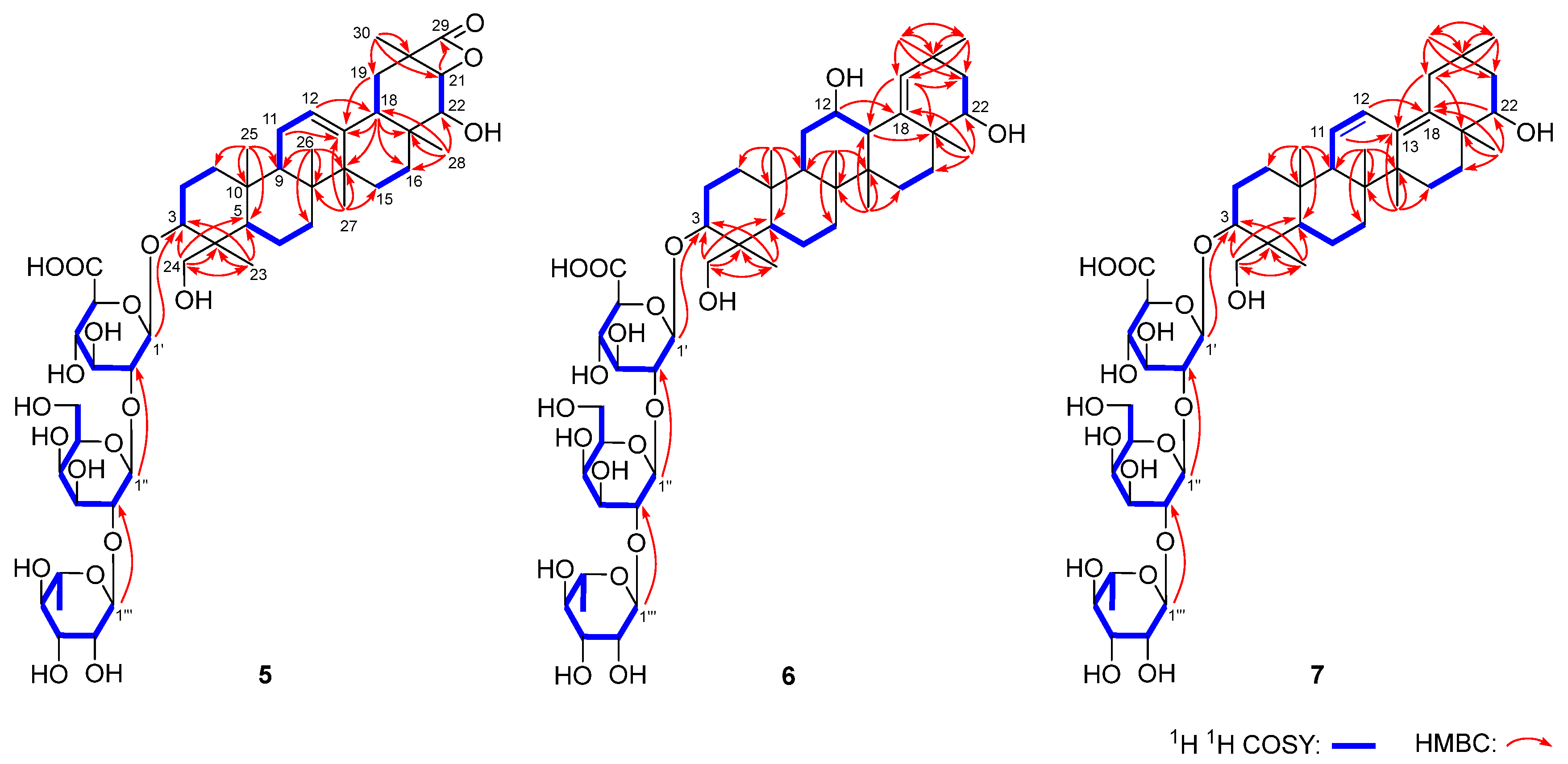
Flosdolilabsaponin A (5) was obtained as a white powder with negative optical rotation ([α]D25 –5.8, MeOH). Its molecular formula was determined to be C48H74O20 (m/z 969.47211 [M − H]; calculated for C48H73O20, 969.46897) according to the HR-ESI-MS analysis. The characteristic absorptions of the hydroxyl group (3367 cm–1), carboxyl (1716 cm–1), tetracyclic lactone (1822 cm–1), and ether bond (1075 cm–1) appeared in the IR spectrum. Flosdolilabsaponin A (5) was firstly hydrolyzed with HCl, followed by l-cysteine methyl ester hydrochloride and O-toluene isothiocyanate derivatization to obtain its derivative. The comparison of the HPLC retention time of the obtained derivative with that of the derivatives of standard sugar samples indicated the existence of d-glucuronic acid, d-galactose, and l-rhamnose in it [16]. The anomeric proton signals at δH 5.00 (1H, d, J = 7.2 Hz, H-1′), 5.80 (1H, d, J = 7.2 Hz, H-1″), and 6.30 (1H, br. s, H-1‴) displayed in its 1H NMR spectrum (Table 4) suggested that the three glycosyls were β-d-glucuronopyranosyl, β-d-galactopyranosyl, and α-l-rhamnopyranosyl, respectively. Forty-eight signals were shown in its 13C NMR spectrum (Table 4). Except for the eighteen signals which could be assigned to the above three glycosyls, most of the remaining thirty were in the range of δC 10–60, suggesting that it was terpenoid saponin. Its 1H NMR spectrum suggested the presence of six methyl [δH 0.72, 0.87, 1.13, 1.16, 1.46, 1.57 (3H each, all s, H3-25, 26, 27, 28, 23, 29)], one hydroxymethyl [δH 3.27, 4.28 (1H each, both d, J = 11.4 Hz, H2-24)], three oxygenated methine [δH 3.43 (1H, dd, J = 3.0, 10.8 Hz, H-3), 4.15 (1H, d, J = 7.8 Hz, H-22), 4.57 (1H, d, J = 7.8 Hz, H-21)], one olefinic proton [δH 5.32 (1H, t like, ca. J = 3 Hz, H-12)], and one carboxyl group [δC 175.7 (C-29)], indicating that it was an oleanic acid type of saponin. The nine fragments shown with bold lines were determined by the proton–proton cross-peaks observed in its 1H 1H COSY spectrum (Figure 4). Furthermore, its planar structure was identified according to the found HMBC correlations: from δH 1.79 (H-11) to δC 141.8 (C-13); from δH 5.32 (H-12) to δC 47.8 (C-18), 141.8 (C-13); from δH 1.12, 1.70 (H2-15) to δC 141.8 (C-13); from δH 2.10 (H-18) to δC 42.3 (C-14); from δH 1.13, 2.60 (H2-19) to δC 37.8 (C-17), 141.8 (C-13); from δH 1.46 (H3-23) to δC 43.9 (C-4), 56.2 (C-5), 63.7 (C-24), 91.2 (C-3); from δH 3.27, 4.28 (H2-24) to δC 23.1 (C-23), 43.9 (C-4), 56.2 (C-5), 91.2 (C-3); from δH 0.72 (H3-25) to δC 36.5 (C-10), 38.7 (C-1), 47.8 (C-9), 56.2 (C-5); from δH 0.87 (H3-26) to δC 33.7 (C-7), 37.8 (C-8), 42.3 (C-14), 47.8 (C-9); from δH 1.13 (H3-27) to δC 25.8 (C-15), 39.6 (C-8), 42.3 (C-14), 141.8 (C-13); from δH 1.16 (H3-28) to δC 31.9 (C-16), 37.8 (C-17), 47.8 (C-18), 79.4 (C-22); from δH 1.57 (H3-30) to δC 35.7 (C-19), 55.4 (C-20), 83.1 (C-21), 175.7 (C-29); from δH 4.57 (H-21) to δC 175.7 (C-29); from δH 5.00 (H-1′) to δC 91.2 (C-3); from δH 5.80 (H-1″) to δC 76.8 (C-2′); and from δH 6.30 (H-1‴) to δC 77.8 (C-2″) (Figure 4). Its chemical shifts, C-1–C11 and C-23–C-27, coincided with those of a known compound, soyasaponin Ⅱ [17], indicating that their configurations at C-3–C-5, C-8–C-10, and C-14 were identical. Furthermore, the NOE correlation between δH 5.32 (H-11) and δH 2.10 (H-18) suggested that H-18 was β-orientated. Meanwhile, H-21, H3-28, and H3-30 were also determined to be in a β orientation through the NOE correlations between δH 2.10 (H-18) and δH 1.16 (H3-28), 1.57 (H3-30), and 4.57 (H-21) (Figure 5). Moreover, the coupling constant between δH 2.60 (H-19α) and δH 2.10 (H-18) was J = 13.2 Hz, suggesting that both H-18 and H-19α were in an axial bond. Furthermore, 22-OH was clarified as having a β orientation according to the NOE cross-peak between δH 2.60 (H-19α) and δH 4.15 (H-22). Thus, the structure of flosdolilabsaponin A (5) was identified. It is a very rare tetracyclic lactone oleane-type triterpene saponin.
The molecular composition of flosdolilabsaponin B (6) was determined to be C48H78O19 (m/z 957.50568 [M − H]; calculated for C48H75O19, 957.50536). Its 1H, 13C NMR (Table 4), and 2D NMR spectra, including 1H 1H COSY, HSQC, and HMBC, suggested that compound 6 owned the same glycosyl, α-l-rhamnopyranosyl(1→2)-β-d-galactopyranosyl(1→2)-β-d-glucuronopyranosyl [δH 4.97 (1H, d, J = 6.6 Hz, H-1′), 5.77 (1H, d, J = 7.2 Hz, H-1″), 6.28 (1H, br. s, H-1‴)] as compound 5. Most of the thirty unattributed carbon signals in the 13C NMR spectrum were also in the high field region. Additionally, the signals that could be assigned to seven methyl [δH 0.70, 1.05, 1.06, 1.17, 1.35, 1.35, 1.41 (3H each, all s, H3-25, 26, 29, 30, 27, 28, 23)], one hydroxymethyl [δH 3.25, 4.26 (1H each, both d, J = 11.4 Hz, H2-24)], three methine substituted by oxygen [δH 3.27 (1H, dd, J = 4.2, 11.4 Hz, H-3), 4.08 (1H, m, H-22), 4.43 (1H, m, overlapped, H-12)], and one olefinic bond [δH 5.97 (1H, s, H-19)], present in its 1H NMR spectrum, suggested that compound 6 was also an oleanic acid type of saponin. The following HMBC correlations were observed in its HMBC spectrum (Figure 4): from H-12 to C-18; from H-13 to C-17; from H-19 to C-13, C-17, C-21; from H3-23 to C-3–C-5, C-24; from H2-24 to C-3–C-5, C-23; from H3-25 to C-1, C-5, C-9, C-10; from H3-26 to C-7–C-9, C-14; from H3-27 to C-8, C-13–C-15; from H3-28 to C-16–C-18, C-22; from H3-29 to C-19–C-21, C-30; and from H3-30 to C-19–C-21, C-29. Based on this determination, its planar structure was clarified. By comparing it with compound 5, it was found that the NMR data of the A ring and A ring substituents of the two compounds were basically same, suggesting that they were consistent in the configuration of the C-3–C-5 and C-8–C-10 positions. The coupling constant between H-12 and H-13 was about zero, indicating that the two were in a cis-configuration. Furthermore, the following NOE correlations indicated that H-12, H-13, H3-25, H3-26, H3-28, and 22-OH were all in a β orientation (Figure 5): between δH 0.70 (H3-25) and δH 1.05 (H3-26); between δH 1.05 (H3-26) and δH 2.40 (H-13); between δH 2.40 (H-13) and δH 1.35 (H3-28), 4.43 (H-12); between δH 1.35 (H3-28) and δH 1.97 (Hβ-21); between δH 1.97 (Hβ-21) and δH 1.06 (H3-30); and between δH 1.17 (H3-29) and δH 4.08 (H-22). Finally, the position of α-l-rhamnopyranosyl(1→2)-β-d-galactopyranosyl(1→2)-β-d-glucuronopyranosyl’s linkage with aglycon was determined to be position-3 through the HMBC correlation observed from δH 4.97 (H-1′) to δC 91.3 (C-3).
Flosdolilabsaponin C (7) with the molecular formula of C48H76O18 (m/z 941.50995 [M + H]; calculated for C48H77O18, 941.51044) was also obtained as a white powder. The comparison of the NMR data of 7 (Table 4) with those of 5 and 6 indicated that all of them had a trisaccharide moiety, α-l-rhamnopyranosyl(1→2)-β-d-galactopyranosyl(1→2)-β-d-glucuronopyranosyl. In addition, the 1H NMR spectrum of compound 7 suggested the existence of seven methyl [δH 0.71, 0.76, 0.90, 1.03, 1.10, 1.35, 1.46 (3H each, all s, H3-25, 26, 30, 29, 27, 28, 23)], one hydroxymethyl [δH 3.27, 4.24 (1H each, both d, J = 11.4 Hz, H2-24)], two oxygenated methin [δH 3.42 (1H, dd, J = 4.2, 11.4 Hz, H-3), 3.80 (1H, dd, J = 3.6, 11.4 Hz, H-22), and a pair of olefinic protons coupled to one another [δH 5.56 (1H, br. d, ca. J = 10 Hz, H-11), 6.49 (1H, d, J = 10.2 Hz, H-12)]. Combined with its 13C NMR spectrum (Table 4), flosdolilabsaponin C (7) was also deduced to be an oleanic acid type of saponin. Through the proton–proton cross-peaks displayed in its 1H 1H COSY spectrum, as well as the long-range correlations found in its HMBC spectrum (Figure 4), the planar structure of 7 was concluded. The chemical shifts in its aglycon were highly consistent with those of astraolesaponin F [18], suggesting that the aglycon of flosdolilabsaponin C (7) was (3β,4β,22β)-3,22,24-triol oleana-11,13(18)-diene.
Meanwhile, the structures of the known compounds were identified as N-malonyl-l-tryptophan (8) [13], equisetinine A (9) [15], N-benzoylaspartate (10) [19], nicotinic acid (11) [20], adenine (12) [21], β-adenosine (13) [22], and guanosine (14) [23] (Figure 1) via 1/2D-NMR spectroscopy, HR-ESI-MS, and/or [α]D, ECD, as well as a comparison with the literature.
In addition, an LPS/Nigericin-induced BMDM model was used to investigate the inhibitory effects of all the obtained compounds on IL-1β release using inflammasome inhibitor MCC950 as the control group. 3-(4,5-Dimethylthiazol-2-yl)-2,5-diphenyltetrazolium bromide (MTT) experiments were initially conducted to determine the non-cytotoxic concentrations for the bioactivity assay. As shown in Figure 6A, MCC950 and all the compounds, apart from 2 and 4, exhibited no cytotoxicity on BMDMs at a concentration of 30 μM; when the concentration was reduced to 20 μM, compounds 2 and 4 were eventually safe for the cells. Based on the MTT results, a further Elisa assay for screening the inhibitory effect on IL-1β release suggested that MCC950 as well as compounds 1, 8, 9, and 1114 could effectively reduce the increase in IL-1β secretion in the cell supernatant induced by LPS/Nigericin at the measured concentration. Among the compounds identified from D. lablab flowers, 14 was the most effective, and it was stronger than that of the inflammasome inhibitor MCC950 (Figure 6B).

3. Discussion

Chronic inflammation plays a significant role in the development of various diseases, such as cancer, cardiovascular disease, diabetes, obesity, autoimmune diseases, and inflammatory bowel disease. Its occurrence is closely related to the activation of the NLRP3 inflammasome [3]. The IL-1β produced during the activation of the NLRP3 inflammasome has strong pro-inflammatory activity and can also promote the release of inflammatory factors by other immune cells, exacerbating inflammatory damage to tissues. Therefore, IL-1β, as a key pro-inflammatory cytokine, is an important indicator for the determination of inflammatory activity in vitro. Currently, the direct or indirect blocking of IL-1β using drugs like anakinra or canakinumab is employed in treating inflammatory diseases [24]. However, canakinumab may lead to side effects such as swelling, diarrhea, and gastroenteritis, while anakinra can also increase infection risk. Medicinal and edible plants with potent therapeutic effects and non-toxic characteristics have attracted more and more researcher attention. As an ingredient in TCM with dual applications in both medicinal treatment and dietary consumption, D. lablab flowers have been reported to significantly suppress the expression of inflammatory cytokines such as IL-6, TNF-α, and IL-1β in a mice model of ulcerative colitis [9]. However, the components responsible for exerting this anti-inflammatory effect are still unclear.
Therefore, a combination of various CC and spectral techniques were initially employed to isolate and identify the components from a 70% EtOH extract of D. lablab flowers. As results, seven previously undescribed compounds, flosdolilabnitrogenousols A−D (14) and flosdolilabsaponins A−C (57), along with seven known isolates (814) were obtained. Flosdolilabsaponin A (5) is an exceptionally rare tetracyclic lactone oleane-type saponin. All the known compounds were first isolated from Lablab genus, and equisetinine A (9), N-benzoylaspartate (10), and guanosine (14) were first found in the Leguminosae family, as retrieved by Scifinder. This investigation significantly enriches both the chemical composition information database as well as the material database pertaining to Lablab genus within the Leguminosae family.
Subsequently, to identify anti-inflammatory active compounds from D. lablab flowers, the release of IL-1β from LPS/Nigericin-stimulated mouse BMDM cells was measured using the Elisa method. Notably, flosdolilabnitrogenousol A (1), N-malonyl-l-tryptophan (8), equisetinine A (9), nicotinic acid (11), adenine (12), β-adenosine (13), and guanosine (14) exhibited significant inhibition of IL-1β release at a concentration of 30 μM. Furthermore, a comparative analysis of nitrogen-containing compounds 1 and 8 suggested that the methyl esterification of 12-carboxyl reduced their IL-1β release inhibitory activity (1 > 8). The evaluation of compounds 1, 2, and 8 demonstrated that 16-carboxyl was essential for suppressing IL-1β release (1, 8 > 2). Meanwhile, compound configuration greatly influenced the IL-1β release activity of compounds 3 and 9; notably, the S-configured compound exhibited superior activity over the R-configured one (9 > 3). In addition, the structure–activity relationships (SARs) suggested that the adenine unit was a critical functional group responsible for exerting the inhibitory effect on IL-1β release from LPS/Nigericin-induced BMDMs for 12 and 13, while d-ribose displayed a negligible impact. These findings suggest that these compounds may exert an anti-inflammatory effect by suppressing the activation of the NLRP3 inflammasome. However, this mechanism remains to be studied.
Moreover, among the obtained compounds, nicotinic acid (11) has been previously reported to have anti-inflammatory activity by regulating Sirtuin 1 (SIRT1) expression in LPS/ATP-stimulated human umbilical vein endothelial cells (HUVECs) and reducing reactive oxygen species (ROS) production in HUVECs, thereby inhibiting NLRP3 inflammasome activation and the subsequent caspase-1 cleavage as well as interleukin IL-1β secretion [25]. Additionally, as a key regulator of inflammation, β-adenosine (13) has been reported to provide help for many anti-inflammatory drugs such as sulfasalazine and methotrexate, having an anti-inflammatory effect [26]. Meanwhile, guanosine (14) has been shown to significantly inhibit the increase in inflammatory factors IL-1, IL-6, TNF-α, and IFN-γ and the decrease in anti-inflammatory factor IL-10 in cerebrospinal fluid after ischemic injury [27]. All the reported information coincides with the experimental results of our research.
Some currently used drugs have no obvious medicinal activity themselves, but they can be used as drug precursors to produce pharmacological components after metabolization in the body. In this study, flosdolilabnitrogenousols B−D (24) and N-benzoylaspartate (10) showed no significant bioactivity in BMDMs. However, compounds 2 and 3 as well as bioactive compounds 1, 8, and 9 might produce malonic acid and the essential amino acid l-tryptophan in vivo. Tryptophan has been reported to enhance the ability of the microbial collective to protect the host against enteric pathogens by activating recombinant dopamine receptor D2 (DRD2) and its downstream pathways, thereby improving gut health and treating severe gastroenteritis, enterocolitis, hemorrhagic diarrhea, and acute kidney failure caused by certain intestinal pathogens [28]. In addition, kynurenine, a metabolite of tryptophan, can reduce acute pancreatitis mortality by inhibiting the production of inflammatory factor TNF-α [29]. Meanwhile, both compounds 4 and 10 might be metabolized to l-aspartic acid in vivo. Aspartate can regulate the activity of inflammasomes through N-methyl-d-aspartate (NMDA) receptors and reduce the expression level of inflammasome components and the production of IL-1β [30]. Moreover, as the possible metabolite of compound 4, coumaric acid, when combined with other anti-inflammatory compounds, could enhance the inhibitory effect on IL-1β release by NLRP3, thereby enhancing the drug’s ability to facilitate recovery from DSS-induced colitis in mice [31].
The above results suggest that D. lablab flowers make a very good drug and food dual-use substance, which will play an important role in the prevention and improvement of inflammatory diseases and can be properly consumed in one’s daily diet.

4. Experiment

4.1. Plant Material

The dried flowers of D. lablab were ordered from the Bozhou medicinal material market, Anhui province, China, identified by Professor Lin Ma (Traditional Chinese Medicine of Tianjin University). The specimen (20210920001) was deposited at the Academy of Traditional Chinese Medicine of Tianjin University.

4.2. General Experimental Procedures

The NMR spectra were determined on a Bruker ascend 600 MHz and/or 500 MHz NMR spectrometer (Bruker BioSpin AG Industriestrasse 26 CH-8117, Fällanden, Switzerland). The HR-ESI-MS data were acquired on a Thermo HR-ESI-MS mass spectrometer connected to the UltiMate 3000 UHPLC instrument via an ESI interface (Thermo Scientific, Waltham, MA, USA). Optical rotation, UV, IR, and ECD spectral analyses were conducted on a Rudolph Autopol® IV automatic polarimeter (l = 50 mm) (Rudolph Research Analytical, Hackettstown, NJ, USA), a Agilent Cary 60 UV-Vis (Agilent Technologies Inc., Santa Clara, CA, USA), a Varian 640-IR FT-IR spectrophotometer (Varian Australia Pty Ltd., Mulgrave, Australia), and a Circular dichroism spectrum (J-815, JASCO, Tokyo, Japan), respectively. CC isolation was achieved with Macroporous resin D101 (Haiguang Chemical Co., Ltd., Tianjin, China), silica gel (48–75 μm, Qingdao Haiyang Chemical Co., Ltd., Qingdao, China), and ODS (S-50 μm, YMC Co., Ltd., Tokyo, Japan). The HPLC column analysis was performed with Cosmosil 5C18-MS-II and a Cosmosil PBr column (4.6 mm i.d. × 250 mm, 5 µm, Nakalai Tesque, Inc., Tokyo, Japan). The compounds were purified with Cosmosil 5C18-MS-II and a Cosmosil PBr column (20 mm i.d. × 250 mm, 5 µm, Nakalai Tesque, Inc., Tokyo, Japan). Dichloromethane (CH2Cl2), methanol (MeOH), acetonitrile (CH3CN), acetic acid (HAc), and other reagents (chromatographically pure or analytically pure) were ordered from Tianjin Concord Technology Co., Ltd., Tianjin, China.

4.3. Extraction and Isolation

The dried D. lablab flowers (10.0 kg) were sequentially extracted with 14 L, 10 L, and 10 L of 70% EtOH for 3 h, 2 h, and 2 h, respectively. After filtration, the solvent was removed under reduced pressure using a rotary evaporator to yield a 70% ethanol extract (4.5 kg), 4.0 kg of which was suspended in H2O (6 L) and then partitioned in EtOAc-H2O (3 × 6.0 L) to obtain a H2O extract (3.6 kg). The H2O extract (3.2 kg) was loaded onto D101 macroporous resin CC (H2O → 95% EtOH) to gain a H2O eluate (2.9 kg) and a 95% EtOH eluate (DLFC, 224.0 g), respectively.
DLFC (150.0 g) was fractioned by silica gel CC [CH2Cl2-MeOH (100:1 → 20:1 → 100:7 → 10:1 → 7:1 → 5:1 → 4:1 → 3:1 → 2:1 → 0:100, v/v)] to yield DLFC 1–DLFC 13. DLFC 3 (6.9 g) was further loaded onto ODS CC [MeOH-H2O (25:75 → 30:70 → 40:60 → 50: 50 → 60:40 → 70:30 → 80:20 → 90:10 → 100:0, v/v) to give DLFC 3-1–DLFC 3-10. DLFC 3-1 (515.3 mg) was separated by pHPLC [MeOH-1% HAc (18:82, v/v), Cosmosil PBr column] to gain DLFC3-1-1–DLFC3-1-4. DLFC 3-1-1 (8.9 mg) was further purified by pHPLC [MeOH-1% HAc (10:90, v/v), Cosmosil PBr column] to obtain nicotinic acid (11, 3.2 mg, tR 25.9 min). DLFC 4 (5.5 g) was subjected to ODS CC [MeOH-H2O (20:80 → 30:70 → 40:60 → 50:50 → 60:40 → 70:30 → 80:20 → 90:10 → 100:0, v/v)] to produce DLFC 4-1–DLFC 4-11. DLFC 4-2 (508.2 mg) was prepared by pHPLC [CH3CN-1% HAc (8:92, v/v), Cosmosil 5C18-MS-II column] to obtain DLFC 4-2-1–DLFC 4-2-4. Among them, DLFC 4-2-4 was identified as N-benzoyl-l-aspartic acid (10, 25.1 mg, tR 43.2 min). DLFC 4-2-3 (58.1 mg) was purified by pHPLC [MeOH-1% HAc (24:76, v/v), Cosmosil PBr column] to obtain flosdolilabnitrogenousol C (3, 25.8 mg, tR 42.1 min). DLFC 4-4 (1.0 g) was separated by pHPLC with CH3CN-1% HAc (16:84, v/v) on a Cosmosil 5C18-MS-II column to obtain DLFC 4-4-1–DLFC 4-4-7. DLFC 4-4-5 (121.9 mg) was further prepared by pHPLC [MeOH-CH3CN-1% HAc (26:10:64, v/v/v), Cosmosil PBr column] to yield equisetinine A (9, 50.2 mg, tR 48.4 min). DLFC 5 (3.5 g) was fractioned by ODS CC [MeOH-H2O (20:80 → 30:70 → 40:60 → 50: 50 → 60:40 → 70:30 → 80:20 → 90:10 → 100:0, v/v)] to produce DLFC 5-1–DLFC 5-8. DLFC 5-2 (700.0 mg) was isolated by pHPLC [CH3CN-1% HAc (10:90, v/v), Cosmosil 5C18-MS-II column] to obtain DLFC 5-2-1–DLFC 5-2-5. DLFC 5-2-5 (12.2 mg) was further purified by pHPLC [CH3CN-1% HAc (15:85, v/v), Cosmosil PBr column] to gain flosdolilabnitrogenousol D (4, 2.3 mg, tR 53.8 min). DLFC 5-4 (790.4 mg) was separated by pHPLC [CH3CN-1% HAc (22:78, v/v), Cosmosil 5C18-MS-II column] to give DLFC 5-4-1–DLFC 5-4-4. DLFC 5-4-4 was identified as flosdolilabnitrogenousol A (1, 37.6 mg, tR 48.9 min). DLFC 5-4-3 (81.3 mg) was further purified by pHPLC [CH3CN-1% HAc (26:74, v/v), Cosmosil PBr column] to yield flosdolilabnitrogenousol B (2, 50.7 mg, tR 40.3 min). DLFC 6 (7.4 g) was loaded onto ODS CC [MeOH-H2O (20:80 → 30:70 → 40:60 → 50: 50 → 60:40 → 70:30 → 80:20 → 90:10 → 100:0, v/v)] to give DLFC 6-1–DLFC 6-12. DLFC 6-5 (624.0 mg) was purified by pHPLC [CH3CN-1% HAc (24:76, v/v), Cosmosil PBr column] to produce N-malonyl-l-tryptophan (8, 44.8 mg, tR 29.0 min). DLFC 7 (11.2 g) was subjected to ODS CC [MeOH-H2O (20:80 → 30:70 → 40:60 → 50: 50 → 60:40 → 70:30 → 80:20 → 90:10 → 100:0, v/v)] to yield DLFC 7-1–DLFC 7-11. DLFC 7-2 (473.3 mg) was prepared by pHPLC [MeOH-1% HAc (10:90, v/v), Cosmosil PBr column] to gain adenine (12, 37.8 mg, tR 14.3 min) and β-adenosine (13, 157.2 mg, tR 52.0 min). DLFC 9 (21.7 g) was loaded onto ODS CC [MeOH-H2O (20:80 → 30:70 → 40:60 → 50: 50 → 60:40 → 70:30 → 80:20 → 90:10 → 100:0, v/v)] to gain DLFC 9-1–DLFC 9-13. DLFC 9-2 (293.4 mg) was prepared by pHPLC [CH3CN-1% HAc (5:95, v/v), Cosmosil PBr column] to yield guanosine (14, 10.6 mg, tR 30.4 min). DLFC 9-9 (535.9 mg) was separated by pHPLC [CH3CN-1% HAc (32:68, v/v), Cosmosil 5C18-MS-II column] to gain DLFC 9-9-1–DLFC 9-9-4. DLFC 9-9-2 (29.3 mg) was further purified by pHPLC [CH3CN-1% HAc (38:62, v/v), Cosmosil PBr column] to obtain flosdolilabsaponin A (5, 9.1 mg, tR 33.4 min). DLFC 9-9-4 (30.3 mg) was prepared by pHPLC [CH3CN-1% HAc (38:62, v/v), Cosmosil PBr column] to obtain flosdolilabsaponin B (6, 15.1 mg, tR 35.7 min). DLFC 10 (19.8 g) was subjected to ODS CC [MeOH-H2O (20:80 → 30:70 → 40:60 → 50: 50 → 60:40 → 70:30 → 80:20 → 90:10 → 100:0, v/v)] and pHPLC [MeOH-1% HAc (73:27, v/v), Cosmosil 5C18-MS-II column] to yield DLFC 10-9-1–DLFC 10-9-5. DLFC 10-9-1 (55.8 mg) was purified by pHPLC [CH3CN-1% HAc (40:60, v/v), Cosmosil PBr column] to yield flosdolilabsaponin C (7, 6.3 mg, tR 44.6 min). The schematic representation of the extraction and separation of compounds 114 is shown in Figure S52.

4.4. Spectral Data of 17

4.4.1. Flosdolilabnitrogenousol A (1)

White powder; [α]D25 +20.8 (conc 1.49, MeOH); UV λmax (MeOH) nm (log ε): 221 (4.26), 282 (3.54), 290 (3.49); CD (conc 0.001 M, MeOH) mdeg (λnm): –0.53 (246), +1.49 (226); IR νmax (KBr) cm–1: 3272, 3057, 2952, 1736, 1655, 1542, 1437, 1342, 1024; 1H NMR (DMSO-d6, 500 MHz) and 13C NMR (DMSO-d6, 125 MHz) δ: Table 1; and ESI-Q-Orbitrap MS: m/z 303.09891 [M − H] (calculated for C15H15O5N2, 303.09755).

4.4.2. Flosdolilabnitrogenousol B (2)

White powder; [α]D25 +17.1 (conc 1.50, MeOH); UV λmax (MeOH) nm (log ε): 221 (4.17), 283 (3.39), 290 (3.32); CD (conc 0.001 M, MeOH) mdeg (λnm): +8.16 (224), +2.84 (201); IR νmax (KBr) cm–1: 3319, 3056, 1733, 1660, 1539, 1437, 1343, 1022; 1H NMR (DMSO-d6, 500 MHz) and 13C NMR (DMSO-d6, 125 MHz) δ: Table 1; and ESI-Q-Orbitrap MS: m/z 303.09842 [M − H] (calculated for C15H15O5N2, 303.09755).

4.4.3. Flosdolilabnitrogenousol C (3)

White powder; [α]D25 +50.2 (conc 1.29, MeOH); UV λmax (MeOH) nm (log ε): 205 (4.24), 250 (3.67), 280 (3.08), 288 (2.94); CD (conc 0.001 M, MeOH) mdeg (λnm): –1.78 (282), +4.60 (257), –7.83 (237), +22.88 (206). IR νmax (KBr) cm–1: 3266, 2901, 1704, 1684, 1603, 1481, 1419, 1317, 1024; 1H NMR (DMSO-d6, 500 MHz) and 13C NMR (DMSO-d6, 125 MHz) δ: Table 2; and ESI-Q-Orbitrap MS: m/z 287.06720 [M − H] (calculated for C14H11O5N2, 287.06625).

4.4.4. Flosdolilabnitrogenousol D (4)

White powder; [α]D25 –41.7 (conc 0.23, MeOH); UV λmax (MeOH) nm (log ε): 203 (4.01), 226 (3.79, sh), 292 (3.81); CD (conc 0.001 M, MeOH) mdeg (λnm): +0.46 (278), –1.85 (237), +5.10 (206); IR νmax (KBr) cm–1: 3259, 2953, 1734, 1647, 1606, 1515, 1437, 1020; 1H NMR (CD3OD, 600 MHz) and 13C NMR (CD3OD, 150 MHz) δ: Table 3; and ESI-Q-Orbitrap MS: m/z 292.08252 [M − H] (calculated for C14H13O6N, 292.08156).

4.4.5. Flosdolilabsaponin A (5)

White powder; [α]D25 –5.8 (conc 0.45, MeOH); IR νmax (KBr) cm–1: 3367, 2935, 1822, 1716, 1609, 1452, 1415, 1348, 1306, 1075, 1047; 1H NMR (C5D5N, 600 MHz) and 13C NMR (C5D5N, 150 MHz) δ: Table 4; and ESI-Q-Orbitrap MS: m/z 969.47211 [M − H] (calculated for C48H73O19, 969.46897).

4.4.6. Flosdolilabsaponin B (6)

White powder; [α]D25 –13.6 (conc 0.47, MeOH); IR νmax (KBr) cm–1: 3367, 2945, 1724, 1607, 1449, 1417, 1359, 1305, 1074, 1047; 1H NMR (C5D5N, 600 MHz) and 13C NMR (C5D5N, 150 MHz) δ: Table 4; and ESI-Q-Orbitrap MS: m/z 957.50568 [M − H] (calculated for C48H77O19, 957.50536).

4.4.7. Flosdolilabsaponin C (7)

White powder; [α]D25 –29.4 (conc 0.16, MeOH); IR νmax (KBr) cm–1: 3368, 2941, 1728, 1609, 1455, 1412, 1384, 1305, 1047; 1H NMR (C5D5N, 600 MHz) and 13C NMR (C5D5N, 150 MHz) δ: Table 4; and ESI-Q-Orbitrap MS: 941.50995 [M + H]+ (calculated for C48H77O18, 941.51044).

4.5. Determination of the Absolute Configuration of Sugars in Compound 5

Compound 5 (3.0 mg) was hydrolyzed with HCl and derived with l-cysteine methyl ester hydrochloride and O-toluene isothiocyanate sequentially to obtain derivatives, by referring to the literature [16]. The obtained derivatives were then analyzed using the same HPLC analysis conditions reported in the literature. Afterwards, by comparing their retention times with those of the authentic sample, d-glucuronic (tR: 20.0 min), d-galactose (tR: 16.5 min), and l-rhamnose (tR: 31.2 min) were determined to be present in compound 5.

4.6. ECD Calculation

The ECD spectra for the optimized conformers were calculated at the APFD/6-311+g(2d,p) level with a CPCM solvent model in CH3OH. The conformational geometry was optimized using density functional theory, followed by time-dependent density functional theory calculations. The calculated ECD spectra of different conformers were simulated with a half-bandwidth of 0.2 eV calculated using the Gaussian 16 software (Gaussian, Inc., Wallingford, CT, USA). The ECD curves were extracted using the GaussView 6.0 software (Gaussian, Inc., Wallingford, CT, USA). Compound-specific ECD curves were weighted according to the Boltzmann distribution after UV correction. Finally, the calculated data were extracted from GaussView and then treated by the OriginPro 2021 software to compare them with the experimental spectral data.

4.7. BMDM Preparation

Male C57BL/6 mice aged 6–8 weeks (Beijing vital river laboratory animal technology, Beijing, China) underwent euthanasia via cervical dislocation. The bone marrow from the femurs and the tibiae was flushed three times using a syringe filled with RPMI-1640 medium (Gibco BRL, Santa Clara, CA, USA). The obtained cells were filtered through a sieve with a pore size of 70 μm, and the erythrocytes were removed using a red blood cell lysing buffer. RPMI-1640 complete medium containing 20 ng/mL recombinant mouse macrophage colony-stimulating factor (M-CSF) obtained from Med Chem Express was used to resuspend the cells gently. The cells were then seeded in 24-well plates at a density of 2 × 10⁵ cells/mL and in 96-well plates at a density of 1 × 10⁵ cells/mL. The cultured cells were then placed in an incubator with 5% CO2 at 37 °C to continue culturing until the seventh day, and the culture medium was changed every two days.

4.8. MTT Assay

The experimental design encompassed two groups: the normal control and the drug treatment cohorts. After the supernatant of BMDM cells cultured for 7 days was discarded, the drug treatment groups were treated with the tested compounds for 18 h, except for the normal group, where it was changed with the complete culture medium. Then, the supernatant was removed, and 100 μL MTT (Sigma-Aldrich, St. Louis, MO, USA) solution (500 μg/mL) was added, further culturing the cells for 4 h. In the end, formazan crystals were dissolved in dimethylsulfoxide and detected in the absorbance of 490 nm through a BioTek Cytation five-cell imaging multi-mode reader (Winooski, VT, USA).

4.9. Inhibitory Effect of the Tested Compounds on LPS/Nigericin-Induced IL-1β Release on BMDMs Measured by Mouse IL-1β Elisa Kit

LPS/Nigericin (Sigma-Aldrich, St. Louis, MO, USA) was used to establish an in vitro cellular inflammation model on BMDMs. A normal group (N), an LPS/Nigericin group (M), the tested compounds’ groups, and MCC950 (Med Chem Express, Monmouth Junction, NJ, USA), known as the Ctrl group, were set. To be more specific, except for the normal group, the LPS/Nigericin group, the Ctrl group, and the tested compounds’ groups were stimulated with 1 μg/mL LPS for 4 h. Then, the culture medium was replaced with RPMI-1640 complete medium, containing different compounds (30 μM or 20 μM) and MCC950 (30 μM) for 4 h. Finally, 10 μM nigericin was added to the cell supernatant for 30 min. The cell supernatant was collected, and the IL-1β concentration was measured according to the instructions of the Elisa kit provided by Elabscience Biotechnology Co., Ltd. (Catalog No: E-EL-M0037; Wuhan, China). Specifically, we added 50 μL of the standard or the collected cell supernatant sample to the microplate and incubated it at 37 °C for 90 min. Then, the liquid was discarded, and 50 μL of biotinylated detection Ab working solution was added to each well. After incubating for 60 min at 37 °C, the liquid was removed, and the plate was washed three times. Next, 50 μL of HRP conjugate working solution was added. After incubating for 30 min at 37 °C, the liquid was discarded, and the plate was washed five times. A total of 50 μL of substrate reagent was added, followed by incubation for 15 min at 37 °C. Finally, 25 μL of stop solution was added, and the plate was read at 450 nm by a BioTek Cytation five-cell imaging multi-mode reader (Winooski, VT, USA).

4.10. Statistical Analysis

The experimental results were analyzed using GraphPad Prism 8.0 (GraphPad Software, Inc., La Jolla, CA, USA). The values were expressed as the mean ± SD. Comparisons between the two groups were made using a t-test, whereas comparisons between multiple groups were performed using a one-way analysis of variance (One-way, ANOVA). A value of * p < 0.05 was considered statistically significant.

5. Conclusions

In summary, seven previously undescribed compounds along with seven known compounds were identified from D. lablab flowers. Among them, compound 5 is a rare tetracyclic lactone oleane-type saponin. This paper enriched the compound library regarding Lablab. Moreover, compounds 1, 8, 9, and 1114 were suggested to effectively suppress inflammation by inhibiting IL-1β release using LPS/Nigericin-induced BMDMs. This study establishes a foundation for exploring this plant’s application in preventing and treating inflammation-related diseases, while clarifying its clinical potential as both a medicinal drug and a food supplement. However, further action mechanisms remain to be explored.

Supplementary Materials

The following supporting information can be downloaded at https://www.mdpi.com/article/10.3390/molecules29163751/s1: supplementary data including the NMR and HRESIMS spectra of compounds 17 and the physical data of compounds 814 can be found in the online version.

Author Contributions

Z.S., H.L., J.C. and W.Z.: carried out the research, performed the analysis and interpretation, and prepared the graphic elements and the illustrative materials. J.R., Q.Z. and Z.D.: collected the data and revised the article for significant intellectual content. Y.Z. and T.W.: conceptualized and designed this study, drafted the article text, and critically revised the article for important intellectual content. All authors have read and agreed to the published version of the manuscript.

Funding

The work was supported by the Tianjin Committee of Science and Technology of China (No. 23ZYJDSS00030) and the National Natural Science Foundation of China (No. 82074118).

Institutional Review Board Statement

Not applicable.

Informed Consent Statement

Not applicable.

Data Availability Statement

Data are contained within the article and Supplementary Materials.

Conflicts of Interest

The authors declare that they have no known competing financial interests or personal relationships that could have appeared to influence the work reported in this paper.

References

  1. Leuti, A.; Fazio, D.; Fava, M.; Piccoli, A.; Oddi, S.; Maccarrone, M. Bioactive lipids, inflammation and chronic diseases. Adv. Drug Deliv. Rev. 2020, 159, 133–169. [Google Scholar] [CrossRef] [PubMed]
  2. Vande Walle, L.; Lamkanfi, M. Drugging the NLRP3 inflammasome: From signalling mechanisms to therapeutic targets. Nat. Rev. Drug Discov. 2024, 23, 43–66. [Google Scholar] [CrossRef] [PubMed]
  3. Wei, Z.; Zhan, X.; Ding, K.; Xu, G.; Shi, W.; Ren, L.; Fang, Z.; Liu, T.; Hou, X.; Zhao, J.; et al. Dihydrotanshinone I specifically inhibits NLRP3 inflammasome activation and protects against septic shock in vivo. Front. Pharmacol. 2021, 12, 750815. [Google Scholar] [CrossRef] [PubMed]
  4. Wang, R.; Wu, W.; Li, W.; Huang, S.; Li, Z.; Liu, R.; Shan, Z.; Zhang, C.; Li, W.; Wang, S. Activation of NLRP3 inflammasome promotes foam cell formation in vascular smooth muscle cells and atherogenesis via HMGB1. J. Am. Heart Assoc. 2018, 7, e008596. [Google Scholar] [CrossRef] [PubMed]
  5. Peng, Y.; Liu, B.; Pei, S.; Zheng, D.; Wang, Z.; Ji, T.; Pan, S.; Shen, H.Y.; Wang, H. Higher CSF levels of NLRP3 inflammasome is associated with poor prognosis of anti-N-methyl-D-aspartate receptor encephalitis. Front. Immunol. 2019, 10, 905. [Google Scholar] [CrossRef] [PubMed]
  6. Wang, S.N.; Guo, X.Y.; Tang, J.; Ding, S.Q.; Shen, L.; Wang, R.; Ma, S.F.; Hu, J.G.; Lü, H.Z. Expression and localization of absent in melanoma 2 in the injured spinal cord. Neural. Regen. Res. 2019, 14, 542–552. [Google Scholar] [PubMed]
  7. Liu, H.; Zhan, X.; Xu, G.; Wang, Z.; Li, R.; Wang, Y.; Qin, Q.; Shi, W.; Hou, X.; Yang, R.; et al. Cryptotanshinone specifically suppresses NLRP3 inflammasome activation and protects against inflammasome-mediated diseases. Pharmacol. Res. 2021, 164, 105384. [Google Scholar] [CrossRef] [PubMed]
  8. The National Health and Family Planning Commission releases the latest list of dual-purpose Chinese medicines. Zhongyi Linchuang Yanjiu 2017, 9, 11–14.
  9. Gao, Y.; Huang, R.; Qiu, Y.; Liu, Y.; Chen, L. Characterization of the chemical composition of different parts of Dolichos lablab L. and revelation of its anti-ulcerative colitis effects by modulating the gut microbiota and host metabolism. J. Ethnopharmacol. 2024, 322, 117629. [Google Scholar] [CrossRef]
  10. Zhang, X.; Yang, X.; Peng, X.; Wang, Y.; Zhou, L.; Liu, X.; Guo, Q. Analysis of volatile components in different parts of Dolichos lablab by GC-MS. Zhongguo Yaoye 2020, 29, 12–14. [Google Scholar]
  11. Yue, D. Study on Chemical Constituents, Antioxidant and Hypoglycemic Activities of Flavonoids from Flos Dolichoris lablab L.; Anhui Ploytechnic University: Wuhu, China, 2021. [Google Scholar]
  12. Shi, Z.W.; Zhang, W.; Liu, W.; Ruan, J.Y.; Wang, T.; Zhang, Y. Isolation and identification of phenolic constituents from the flower bud of Dolichos lablab L. Zhongguo Yaowuhuaxue Zazhi 2023, 33, 857–863. [Google Scholar]
  13. Song, K.S.; Ishikawa, Y.; Kobayashi, S.; Sankawa, U.; Ebizuka, Y. N-Acylamino acids from Ephedra distachya cultures. Phytochemistry 1992, 31, 823–826. [Google Scholar] [CrossRef]
  14. Lu, M.; Ruan, J.; Yu, R.; Zhang, Y.; Zhao, W.; Yang, D.; Wang, W.; Zhang, Y.; Wang, T. Neolignan derivatives from Penthorum chinense with antitumor activity in human colorectal cancer cells by regulating Wnt/β-catenin signaling pathway. Phytochemistry 2023, 214, 113827. [Google Scholar] [CrossRef]
  15. Zhu, D.H.; Zhang, J.K.; Jia, J.F.; Liu, J.J.; Wei, J.J.; Yang, M.; Yang, Y.; Li, M.; Hao, Z.Y.; Zheng, X.K.; et al. Alkaloids from the stem of Ephedra equisetina. J. Asian Nat. Prod. Res. 2023, 25, 238–244. [Google Scholar] [CrossRef] [PubMed]
  16. Li, H.; Cao, H.; Ruan, J.; Wu, Y.; Yang, D.; Gao, Q.; Wang, D.; Chen, Q.; Zhang, Y.; Wang, T. Saponins from Aesculus wilsonii seeds exert anti-inflammatory activity through the suppression of NF-κB and NLRP3 pathway. Arab. J. Chem. 2023, 16, 105077. [Google Scholar] [CrossRef]
  17. Liu, Y.; Zhang, Q.; Chen, H.; Wang, B.; An, D.; Zhao, Y. Structural determination of saponins from Hedysarum polybotrys. Magn. Reason. Chem. 2006, 44, 1128–1130. [Google Scholar] [CrossRef]
  18. Zhang, Y.; Li, X.; Ruan, J.; Wang, T.; Dong, Y.; Hao, J.; Liu, E.; Han, L.; Gao, X.; Wang, T. Oleanane type saponins from the stems of Astragalus membranaceus (Fisch.) Bge. var. mongholicus (Bge.) Hsiao. Fitoterapia 2016, 109, 99–105. [Google Scholar] [CrossRef]
  19. Uchida, K.; Kawakishi, S. Ascorbate-mediated specific oxidation of the imidazole ring in a histidine derivative. Bioorg. Chem. 1989, 17, 330–343. [Google Scholar] [CrossRef]
  20. Tang, L.C.; Wang, N.; Yao, H.P.; Yang, X.H.; Deng, S.M.; Zhang, C.Y. Chemical components of the Hevea brasiliensis skim. Linye Huaxue Yu Gongye 2013, 33, 125–129. [Google Scholar]
  21. Lim, S.S.; Jung, Y.J.; Hyun, S.K.; Lee, Y.S.; Choi, J.S. Rat lens aldose reductase inhibitory constituents of Nelumbo nucifera stamens. Phytother. Res. 2006, 20, 825–830. [Google Scholar] [CrossRef]
  22. Domondon, D.L.; He, W.; De Kimpe, N.; Hofte, M.; Poppe, J. β-Adenosine, a bioactive compound in grass chaff stimulating mushroom production. Phytochemistry 2004, 65, 181–187. [Google Scholar] [CrossRef] [PubMed]
  23. Li, Y.; Wang, C.; Wang, F.; Dong, H.; Guo, S.; Yang, J.; Xiao, P. Chemical constituents of Dendrobium candidum. Zhongguo Zhongyao Zazhi 2010, 35, 1715–1719. [Google Scholar] [PubMed]
  24. Diaz-Del-Olmo, I.; Worboys, J.; Martin-Sanchez, F.; Gritsenko, A.; Ambrose, A.R.; Tannahill, G.M.; Nichols, E.M.; Lopez-Castejon, G.; Davis, D.M. Internalization of the membrane attack complex triggers NLRP3 inflammasome activation and IL-1β secretion in human macrophages. Front. Immunol. 2021, 12, 720655. [Google Scholar] [CrossRef] [PubMed]
  25. Li, Y.; Yang, G.; Yang, X.; Wang, W.; Zhang, J.; He, Y.; Zhang, W.; Jing, T.; Lin, R. Nicotinic acid inhibits NLRP3 inflammasome activation via SIRT1 in vascular endothelial cells. Int. Immunopharmacol. 2016, 40, 211–218. [Google Scholar] [CrossRef] [PubMed]
  26. Cronstein, B.N. Adenosine, an endogenous anti-inflammatory agent. J. Appl. Physiol. 1994, 76, 5–13. [Google Scholar] [CrossRef] [PubMed]
  27. Hansel, G.; Tonon, A.C.; Guella, F.L.; Pettenuzzo, L.F.; Duarte, T.; Duarte, M.M.M.F.; Oses, J.P.; Achaval, M.; Souza, D.O. Guanosine protects against cortical focal ischemia. Involvement of inflammatory response. Mol. Neurobiol. 2015, 52, 1791–1803. [Google Scholar] [CrossRef]
  28. Scott, S.A.; Fu, J.; Chang, P.V. Dopamine receptor D2 confers colonization resistance via microbial metabolites. Nature 2024, 628, 180–185. [Google Scholar] [CrossRef] [PubMed]
  29. Cervenka, I.; Agudelo, L.Z.; Ruas, J.L. Kynurenines: Tryptophan’s metabolites in exercise, inflammation, and mental health. Science 2017, 357, eaaf9794. [Google Scholar] [CrossRef] [PubMed]
  30. Farooq, A.; Hoque, R.; Ouyang, X.; Farooq, A.; Ghani, A.; Ahsan, K.; Guerra, M.; Mehal, W.Z. Activation of N-methyl-D-aspartate receptor downregulates inflammasome activity and liver inflammation via a β-arrestin-2 pathway. Am. J. Physiol. Gastrointest. Liver Physiol. 2014, 307, 732–740. [Google Scholar] [CrossRef]
  31. Sun, S.; Li, Z.; Huang, C.; Liu, J.; Yu, Q.; Jiang, X.; Yue, K.; Zhao, J.; Xu, T.; Liu, Y.; et al. Discovery of novel 2,3-dihydro-1H-indene-5-sulfonamide NLRP3 inflammasome inhibitors targeting colon as a potential therapy for colitis. J. Med. Chem. 2023, 66, 16141–16167. [Google Scholar] [CrossRef]
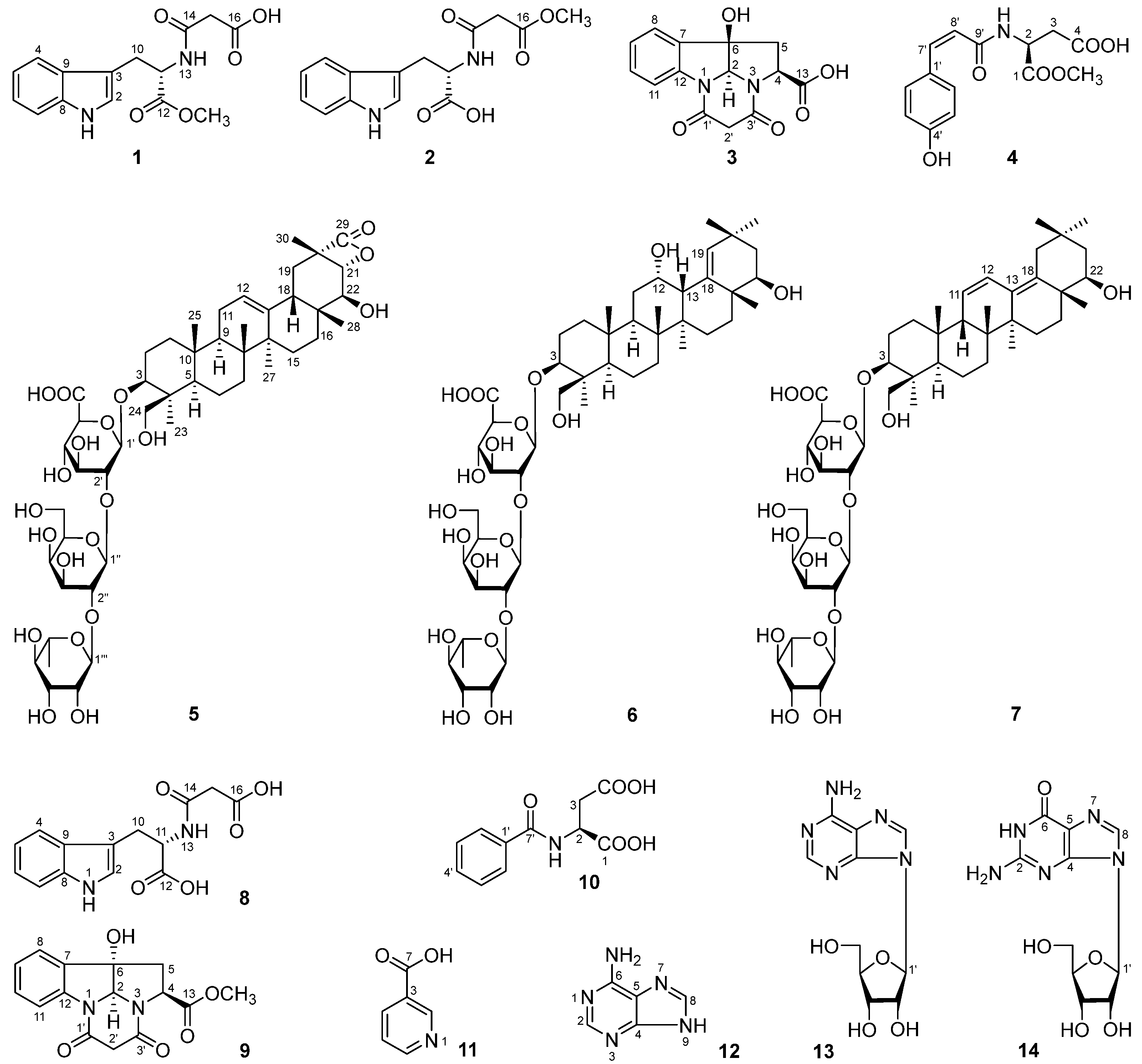
Figure 1. Chemical structures of compounds 114.
Molecules 29 03751 g001
Figure 2. The main 1H 1H COSY and HMBC correlations of compounds 14.
Molecules 29 03751 g002
Figure 3. ECD spectra of compounds 14.
Molecules 29 03751 g003
Figure 4. The main 1H 1H COSY and HMBC correlations of compounds 57.
Molecules 29 03751 g004
Figure 5. The main NOE correlations of compounds 5 and 6.
Molecules 29 03751 g005
Figure 6. Inhibitory effect of compounds 1–14 on IL-1β release from mice BMDMs. 6A: MTT assay of compounds 114 on BMDMs from mice. The final administrated concentration of MTT was 500 μg/mL. N: normal group. The values represent the mean ± SD. The drug-treated group vs. N: *** p < 0.001. n = 6. 6B: Inhibitory effect of compounds 114 on IL-1β release from mice BMDMs cells stimulated by LPS/Nigericin. N: normal group; M: model group, 1 μg/mL LPS + 10 μM Nigericin; and Ctrl: MCC950 group. The final administrated concentration of compounds 1, 3, and 514, as well as MCC950, was 30 μM, while 2 and 4 were 20 μM. The values represent the mean ± SD. M vs. N; ### p < 0.001; the drug-treated group vs. M: *** p < 0.001. n = 3.
Molecules 29 03751 g006
Table 1. 1H (500 MHz) and 13C (125 MHz) NMR data for compounds 1 and 2 in DMSO-d6.
No.12
δCδH (J in Hz)δCδH (J in Hz)
110.89 (d, 2.5, 1H)10.88 (d, 2.0, 1H)
2123.77.16 (d, 2.5, 1H)123.57.14 (d, 2.0, 1H)
3109.0109.5
4117.97.48 (br. d, ca. 8, 1H)118.17.53 (dd, 1.0, 8.0, 1H)
5118.36.99 (t like, ca. 8, 1H)118.36.98 (dt like, ca. 1, 8, 1H)
6120.97.07 (t like, ca. 8, 1H)120.87.08 (dt like, ca. 1, 8, 1H)
7111.37.34 (br. d, ca. 8, 1H)111.27.34 (br. d, ca. 8, 1H)
8136.0135.9
9127.0127.2
1027.13.07 (dd, 7.5, 14.5, 1H)27.03.05 (dd, 7.5, 15.0, 1H)
3.14 (dd, 5.5, 14.5, 1H) 3.18 (dd, 5.5, 15.0, 1H)
1153.14.55 (ddd, 5.5, 7.5, 7.5, 1H)53.14.50 (ddd, 5.5, 7.5, 8.0, 1H)
12172.0172.9
138.57 (d, 7.5, 1H)8.43 (d, 8.0, 1H)
14165.8164.9
1542.13.15 (s, 2H)41.83.30 (s, 2H)
16169.2168.1
12-COOCH351.73.57 (s, 1H)
16-COOCH3 51.63.58 (s, 3H)
Table 2. 1H (500 MHz) and 13C (125 MHz) NMR for compound 3 in DMSO-d6.
No.δCδH (J in Hz)No.δCδH (J in Hz)
283.25.74 (s, 1H)11113.97.68 (br. d, ca. 8, 1H)
460.04.58 (dd, 2.5, 10.0, 1H)12142.1
542.72.58 (dd, 2.5, 14.0, 1H)13170.6
2.94 (dd, 10.0, 14.0, 1H)1′165.8
684.42′46.92.93 (d, 16.0, 1H)
7133.9 4.35 (d, 16.0, 1H)
8124.47.45 (br. d, ca. 8, 1H)3′167.3
9124.27.10 (t like, ca. 8, 1H)6-OH6.47 (br. s, 1H)
10129.67.31 (t like, ca. 8, 1H)13-COOH 12.72 (br. s, 1H)
Table 3. The 1H and 13C NMR data for compound 4 in CD3OD.
No.δCδH (J in Hz)No.δCδH (J in Hz)
1172.93′,5′116.06.72 (d, 8.4, 2H)
250.44.83 (dd, 5.4, 7.2, 1H)4′159.4
337.02.79 (dd, 7.2, 17.4, 1H)7′139.46.67 (d, 12.6, 1H)
2.84 (dd, 5.4, 17.4, 1H)8′120.55.84 (d, 12.6, 1H)
4174.29′169.9
1′127.81-COOCH353.03.73 (s, 3H)
2′,6′132.67.45 (d, 8.4, 2H)
Table 4. 1H and 13C NMR data for compounds 57 in C5D5N.
No.567
δCδH (J in Hz)δCδH (J in Hz)δCδH (J in Hz)
138.70.83 (m, 1H)38.70.80 (m, 1H)37.90.85 (m, o, 1H)
1.39 (m, o, 1H) 1.41 (m, o, 1H) 1.62 (m, o, 1H)
226.71.88 (m, 1H)26.71.86 (m, o, 1H)26.61.94 (m, o, 1H)
2.23 (m, 1H) 2.11 (m, 1H) 2.26 (m, o, 1H)
391.23.43 (dd, 3.0, 10.8)91.33.27 (dd, 4.2, 11.4, 1H)91.23.42 (dd, 4.2, 11.4, 1H)
443.944.043.9
556.20.86 (br. d, ca. 11, 1H)56.50.87 (br. d, ca. 11, 1H)55.40.87 (m, o, 1H)
618.51.25 (m, 1H)18.51.25 (m, 1H)18.61.29 (m, o, 1H)
1.55 (m, o, 1H) 1.55 (m, o, 1H) 1.62 (m, o, 1H)
733.71.40 (m, o, 1H)35.21.52 (m, 1H)32.81.32 (m, 1H)
1.44 (m, 1H)
839.641.140.5
947.81.51 (dd, 7.2, 10.2, 1H)45.11.99 (dd, 7.2, 12.0, 1H)54.31.94 (br. s, 1H)
1036.536.436.2
1123.91.79 (m, o, 1H)31.71.42 (m, 1H)126.25.56 (br. d, ca. 10, 1H)
1.64 (m, 1H)
12124.65.32 (t like, ca. 3, 1H)67.94.43 (m, o, 1H)126.56.49 (d, 10.2, 1H)
13141.843.12.40 (br. s, 1H)135.2
1442.343.142.3
1525.81.12 (m, o, 1H)28.91.20 (m, 1H)24.61.13 (m, 1H)
1.70 (m, 1H) 1.87 (m, 1H) 1.68 (m, 1H)
1631.91.84 (m, 2H)34.31.70 (m, 1H)33.71.85 (m, o, 1H)
2.24 (m, 1H) 2.26 (m, o, 1H)
1737.840.441.2
1847.82.10 (br. d, ca. 13, 1H)140.3137.9
1935.71.13 (m, o, 1H)130.85.97 (s, 1H)37.81.88 (d, 14.4, 1H)
2.60 (dd, 13.2, 13.2, 1H) 2.49 (d, 14.4, 1H)
2055.433.932.3
2183.14.57 (d, 7.8, 1H)42.31.87 (m, o, 1H)44.81.76 (dd, 3.6, 11.4, 1H)
1.97 (dd, 12.6, 12.6, 1H) 1.84 (m, o, 1H)
2279.44.15 (d, 7.8, 1H)75.34.08 (m, 1H)76.63.80 (dd, 3.6, 11.4, 1H)
2323.11.46 (s, 3H)22.91.41 (s, 3H)22.91.46 (s, 3H)
2463.73.27 (d, 11.4, 1H)63.63.25 (d, 11.4, 1H)63.33.27 (d, 11.4, 1H)
4.28 (d, 11.4, 1H) 4.26 (d, 11.4, 1H) 4.24 (d, 11.4, 1H)
2516.10.72 (s, 3H)17.40.70 (s, 3H)18.20.71 (s, 3H)
2617.30.87 (s, 3H)16.41.05 (s, 3H)16.60.76 (s, 3H)
2724.31.13 (s, 3H)20.01.35 (s, 3H)20.31.10 (s, 3H)
2822.81.16 (s, 3H)18.91.35 (s, 3H)18.71.35 (s, 3H)
29175.730.81.17 (s, 3H)32.61.03 (s, 3H)
3019.41.57 (s, 3H)32.21.06 (s, 3H)25.50.90 (s, 3H)
1′105.55.00 (d, 7.2, 1H)105.54.97 (d, 6.6, 1H)105.65.03 (d, 7.2, 1H)
2′76.84.60 (m, o, 1H)76.74.58 (dd, 6.6, 9.0, 1H)76.84.46 (dd, 7.2, 8.4, 1H)
3′78.64.62 (m, o, 1H)78.54.61 (dd, 9.0, 9.6, 1H)78.64.65 (dd, 8.4, 8.4, 1H)
4′73.94.46 (dd, 9.6, 9.6, 1H)74.04.43 (m, o, 1H)73.94.48 (dd, 8.4, 9.6, 1H)
5′77.64.62 (m, o, 1H)77.64.69 (d, 8.4, 1H)77.64.66 (d, 9.6, 1H)
6′172.7172.8172.6
1″101.85.80 (d, 7.2, 1H)101.85.77 (d, 6.6, 1H)101.85.81 (d, 6.6, 1H)
2″77.84.57 (m, o, 1H)77.94.55 (dd, 6.6, 9.0, 1H)77.94.58 (dd, 6.6, 8.4, 1H)
3″76.74.11 (dd, 3.0, 9.0, 1H)76.74.09 (br. d, ca. 9, 1H)76.74.12 (br. d, ca. 8, 1H)
4″71.24.40 (br. d, ca. 3, 1H)71.24.39 (br. s, 1H)71.24.41 (br. s, 1H)
5″76.53.94 (t like, ca. 6, 1H)76.53.93 (t like, ca. 5, 1H)76.53.94 (t like, ca. 5, 1H)
6″61.64.31 (dd, 6.0, 10.8, 1H)61.64.30 (dd, 4.8, 10.2, 1H)61.64.32 (dd, 5.4, 10.8, 1H)
4.43 (dd, 6.0, 10.8, 1H) 4.41 (dd, 6.0, 10.2, 1H) 4.42 (dd, 5.4, 10.8, 1H)
1‴102.56.30 (br. s, 1H)102.56.28 (br. s, 1H)102.66.31 (br. s, 1H)
2‴72.54.81 (br. s, 1H)72.24.81 (br. s, 1H)72.54.83 (br. s, 1H)
3‴72.84.69 (br. d, ca. 8, 1H)72.54.68 (br. d, ca. 8, 1H)72.94.70 (br. d, ca. 9, 1H)
4‴74.44.35 (dd, 9.0, 9.6, 1H)74.44.32 (dd, 8.4, 9.0, 1H)74.44.36 (dd, 9.0, 10.8, 1H)
5‴69.55.00 (m, 1H)69.44.99 (m, 1H)69.55.02 (m, 1H)
6‴19.01.79 (d, 6.0, 3H)19.01.78 (d, 6.0, 3H)19.01.82 (d, 6.0, 3H)
o: overlapped.
Disclaimer/Publisher’s Note: The statements, opinions and data contained in all publications are solely those of the individual author(s) and contributor(s) and not of MDPI and/or the editor(s). MDPI and/or the editor(s) disclaim responsibility for any injury to people or property resulting from any ideas, methods, instructions or products referred to in the content.