Article

Inhibition of DNA Topoisomerase Ι by Flavonoids and Polyacetylenes Isolated from Bidens pilosa L.

1
Key Laboratory of Chemistry in Ethnic Medicinal Resources, State Ethnic Affairs Commission and Ministry of Education, Yunnan Minzu University, Kunming 650504, China
2
Yunnan Key Laboratory of Chiral Functional Substance Research and Application, Yunnan Minzu University, Kunming 650504, China
3
School of Chemistry and Environmental Engineering, Qujing Normal University, Qujing 655011, China
*
Authors to whom correspondence should be addressed.
These authors contributed equally to this work.
Molecules 2024, 29(15), 3547; https://doi.org/10.3390/molecules29153547
Submission received: 13 June 2024 / Revised: 17 July 2024 / Accepted: 25 July 2024 / Published: 27 July 2024

Abstract

:
Human DNA topoisomerase I (Topo I) is an essential enzyme in regulating DNA supercoiling during transcription and replication, and it is an important therapeutic target for anti-tumor agents. Bidens pilosa L. is a medicinal herb that is used as a folk medicine for cancers in China. A new flavonoid (1) and a new polyacetylene (20), along with eighteen flavonoids (219) and nine polyacetylenes (2129), were isolated and identified from the methanol extract of the whole plant of B. pilosa, and some of the compounds (4, 5, 6 and 7) exhibited potent cytotoxicity against a panel of five human cancer cell lines. The DNA relaxation assay revealed that some flavonoids and polyacetylenes exerted inhibitory activities on human DNA Topo I, among them compounds 1, 2, 5, 6, 7, 8, 15, 19, 20, 22, and 24 were the most active ones, with IC50 values of 393.5, 328.98, 145.57, 239.27, 224.38, 189.84, 89.91, 47.5, 301.32, 178.03, and 218.27 μM, respectively. The structure–activity analysis of flavonoids was performed according to the results from the Topo I inhibition assay. The DNA content analysis revealed that 5, 6, and 7 potently arrested cell cycle at the G1/S and G2/M phases in human colon cancer cell DLD-1 depending on the concentration of the inhibitors. The levels of protein expression related to the G1/S and G2/M cell cycle checkpoints were in accordance with the results from the DNA content analysis. These findings suggest that flavonoids are one of the key active ingredients accounting for the anti-tumor effect of B. pilosa.

Graphical Abstract

1. Introduction

Bidens pilosa L. is an annual herb of the Asteraceae; it is edible and has been traditionally used for the treatment of many diseases in many countries. For its anti-hyperglycemic, antihypertensive, and anti-tumor effects, B. pilosa has been widely used as a folk herbal medicine in China for treating various disorders, and in Brazil, it has been included in the official list of medicinal plants by the public health system [1]. Modern phytochemical studies revealed that B. pilosa contains flavonoids, polyacetylenes, phenolic acids, terpenoids, steroids, and other components [2], of which flavonoids and polyacetylenes are the main constituents [3]. A wide range of biological activities has been reported about B. pilosa and the compounds isolated from it, including antioxidative, anti-inflammatory, antibacterial, anticancer, antidiabetic, and immunomodulatory activities, etc. [4,5]. Flavonoids are a kind of polyphenols that broadly exist in plants and exhibit a wide range of bioactivities; to date, more than one hundred flavonoids have been isolated from B. pilosa, which were reported to have antioxidant, hepatoprotective, and cytotoxic activities, etc. [6]. Acetylenes are a kind of compounds containing one or more carbon–carbon triple bond(s) in their backbones and were found regularly in only five families, namely Campanulaceae, Asteraceae, Araliaceae, Pittosporaceae, and Umbelliferae. To date, more than fifty acetylenes have been isolated from B. pilosa, which are reported to play an important role in the plant with anti-cancer, anti-malaria, and anti-diabetes activities [7,8].
DNA topoisomerases (Topos) are the key enzymes that control DNA topology through the passage of DNA strands [9], which is important for DNA replication and transcription. According to whether they make transient single- or double-stranded breaks in the DNA, Topos are classified as Topo I and II, respectively [10]. Because of the overexpression and higher activity in tumor cells compared to normal cells, Topos have become important therapeutic targets for anti-tumor drugs [11]. Many natural compounds isolated from plants, such as camptothecin (CPT) and podophyllotoxin, exhibit potent inhibitory activities in Topos [12], and their derivatives are used as important anti-tumor drugs in the clinic, such as topotecan, irinotecan, and etoposide [13].
Currently, many studies have been conducted on the anti-tumor potential of natural products against Topos. As far as we know, no description of the inhibitory effect of polyacetylenes on Topos has been found, and the mechanism of the B. pilosa compounds’ activity has not been elucidated. Regarding the good anticancer activity of B. pilosa, the chemical and pharmacological constituents of the plant were investigated by us [14]. In this study, a new flavonoid and a new polyacetylene, together with 27 known flavonoids and polyacetylenes, were isolated from B. pilosa, of which some showed potent cytotoxicity and inhibitory activities on DNA Topo I. In this paper, we report the identification of the new compounds, the cytotoxicity, the inhibitory activities on Topo I, and the cytotoxic mechanism in cancer cells. To the best of our knowledge, this is the first time that the inhibition of flavonoids and polyacetylenes isolated from B. pilosa against DNA Topo I is reported.

2. Results and Discussion

2.1. Identification of Two New Compounds

Compound 1, obtained as a yellow powder, had the molecular formula of C28H34O16 by the positive HR-ESI-MS (m/z 649.1746 [M + Na]+, calcd. 649.1739), requiring 12 degrees of unsaturation (Figure S1). The UV spectrum (Figure S9) exhibited a maximum absorption peak at 372 nm, which was typical of chalcones. The 1H NMR spectrum in DMSO-d6 (Figure S2 and Table 1) showed one hydrogen-bonded phenolic hydroxy at δH 12.90 (1H, br s, 2′-OH); one normal phenolic hydroxy at δH 9.25 (1H, s, 3-OH); a pair of mutually coupled aromatic doublets at δH 8.02 (1H, d, J = 9.3 Hz, H-6′) and 6.84 (1H, d, J = 9.3 Hz, H-5′) due to a 1,2,3,4-tetrasubstituted benzene ring; a set of 1,3,4-trisubstituted benzene ring signals at δH 7.34 (1H, d, J = 1.9 Hz, H-2), 7.32 (1H, dd, J = 8.4, 1.9 Hz, H-6), and 7.01 (1H, d, J = 8.4 Hz, H-5); a pair of characteristic trans-olefinic doublets at δH 7.70 and 7.77 (each 1H, d, J = 15.4 Hz); and two β-glucopyranosyl anomeric protons at δH 4.98 (1H, d, J = 7.4 Hz, H-1‴) and 4.85 (1H, d, J = 7.6 Hz, H-1″), together with a series of other overlapped signals from the sugar moieties, as well as a methoxy signal at δH 3.84 (3H, s). The 13C NMR spectrum (Figure S3 and Table 1) showed a total of 28 carbon resonances, including a conjugated ketone carbonyl carbon at δC 192.1 (s), 14 aromatic or olefinic carbons due to two benzene rings and a double bond group, two sugar anomeric carbons at δC 103.8 (d, C-1″) and 100.7 (d, C-1‴), combined with five pairs of characteristic oxygenated carbons (8 × CH, 2 × CH2) assignable to two β-glucopyranosyl moieties, as well as a methoxy carbon at δC 55.8 (q). The existence of two benzene rings, one carbonyl, one double bond, and two sugar rings was exactly matched with its degrees of unsaturation. The above NMR features are generally similar to those of 4-O-methylokanin 4′-O-β-d-glucopyranoside, a chalcone glycoside isolated from a plant of the same genus [15]. The careful comparison of their NMR data revealed that Compound 1 had an extra β-glucopyranosyl moiety. A detailed analysis of the HMBC correlations (Figure 1 and Figure S6) confirmed that the aglycone structure was the same as that of 4-O-methylokanin 4′-O-β-d-glucopyranoside. It should be pointed out that the position of the methoxy group was also verified by the ROESY correlation (Supplemental Figure S8; Figure 1) of OCH3↔H-5. Furthermore, the HMBC correlations from H-6′ [δH 8.02 (1H, d, J = 9.3 Hz)] and H-1‴ [δH 4.98 (1H, d, J = 7.4 Hz)] to C-4′ [δC 155.6 (s)] indicated the presence of a β-glucopyranosyl at C-4′, which was also supported by the ROESY correlation of H-1‴ ↔ H-5′. The HMBC correlations from H-5′ [δH 6.84 (1H, d, J = 9.3 Hz)] and H-1″ [δH 4.85 (1H, d, J = 7.6 Hz)] to C-3′ [δC 133.6 (s)] positioned the other β-glucopyranosyl at C-3′. Therefore, the structure of Compound 1 was established as 4-O-methylokanin 3′,4′-di-O-β-d-glucopyranoside, shown in Figure 1.
Compound 20, obtained as a white powder, had a molecular formula of C13H12O3 by the positive HR-ESI-MS (m/z 217.0865 [M + H]+, calcd. 217.0859), requiring eight degrees of unsaturation (Figure S11). In the IR spectrum, absorption bands attributable to the acetylene (2146 cm−1) and hydroxyl (3269, 3396 cm−1) groups were observed (Figure S19). The 1H NMR spectrum (Figure S12 and Table 2) in CD3OD showed a pair of mutually coupled aromatic doublets at δH 7.28 (2H, d, J = 8.6 Hz, H-2′, H-6′) and 6.72 (2H, d, J = 8.6 Hz, H-3′, H-5′), corresponding to a p-substituted benzene ring, an oxygenated methine signal at δH 3.77 (1H, dddd, J = 6.2, 6.0, 5.8, 4.9 Hz, H-2), an oxygenated methylene signal at δH 3.54 (1H, dd, J = 11.2, 5.8 Hz, H-1a) and 3.59 (1H, dd, J = 11.2, 4.9 Hz, H-1b), as well as an aliphatic methylene signal at δH 2.50 (1H, dd, J = 17.3, 6.2 Hz, H-3a) and 2.60 (1H, dd, J = 17.3, 6.0 Hz, H-3b), which means the existence of three exchangeable protons in the structure. The 13C NMR spectrum (Figure S13 and Table 2) showed a total of 11 carbon signals, including 4 aromatic carbons assignable to a p-substituted benzene ring, 4 characteristic quaternary carbons at δC 80.8 (s, C-4), 76.2 (s, C-7), 73.2 (s, C-6) and 67.6 (s, C-5) due to two acetylenic groups, an oxygenated methine carbon at δC 71.7 (d, C-2), an oxygenated methylene carbon at δC 66.0 (t, C-1), as well as an aliphatic methylene carbon at δC 25.2 (t, C-3). The analysis of the degrees of unsaturation also supported the presence of two acetylenic groups. The above NMR features were generally similar to those of 7-phenylhepta-4,6-diyne-1,2-diol, a polyacetylene isolated from the same plant [3]. The comparison of their NMR data revealed that the obvious difference only came from the substitution pattern of the benzene ring. The HMBC correlations (Figure S16 and Figure 2) from H-2′ and H-6′ [δH 7.28 (2H, d, J = 8.6 Hz)] to C-4′ [δC 160.0 (s)] indicated the presence of a phenolic hydroxy group at C-4′. The resulting structure was further verified by the 2D NMR analysis. The absolute configuration of C-2 was tentatively deduced to be R-form by the comparison of the specific rotatory values with synthetic analogues [16]. Finally, the structure of Compound 20 was established as (R)-7-(4-hydroxyphenyl) hepta-4,6-diyne-1,2-diol, shown in Figure 2.

2.2. Structures of Compounds 129

In addition to 1 and 20, eighteen flavonoids (219) [17,18,19,20,21,22,23,24,25,26,27,28,29,30,31,32,33,34] and nine polyacetylenes (2129) [3,35,36,37,38,39,40,41,42] (Figure 3) were isolated from the methanol extract of the whole plant of B. pilosa; among them, compounds 8, 11, 17, 28, and 29 were isolated from this plant for the first time. LC-MS and 1D NMR were used to establish the structures of all the known compounds, and the spectra data for compounds 219 and 2129 are described in the supporting information.

2.3. Compounds 129 Showed Cytotoxicities and Topo I Inhibitory Activities

The cytotoxicities of 129 were tested by an SRB assay on a panel of five human cancer cell lines and one human hepatocyte cell line. The results from Table 3 showed that compounds 4, 5, 6, and 7 exhibited potent cytotoxicities against A549, HCT116, and DLD-1 cells, and their IC50 values were at the same levels compared to those of CPT, while, in MDA-MB-231 and HepG2 cells, they are less active than CPT. Compared to 4, 5, 6, and 7, compounds 3, 810, 13, 15, 2023, and 2528 exhibited weak cytotoxicities with IC50 values varying from 50.50 μM to over 200 μM in different cell lines. Compounds 1, 2, 11, 12, 14, 1619, 24, and 29 exhibited no cytotoxicity in all cell lines at the tested concentration of 200 μM. In general, all the compounds were less active in HL-7702 than in other cancer cell lines.
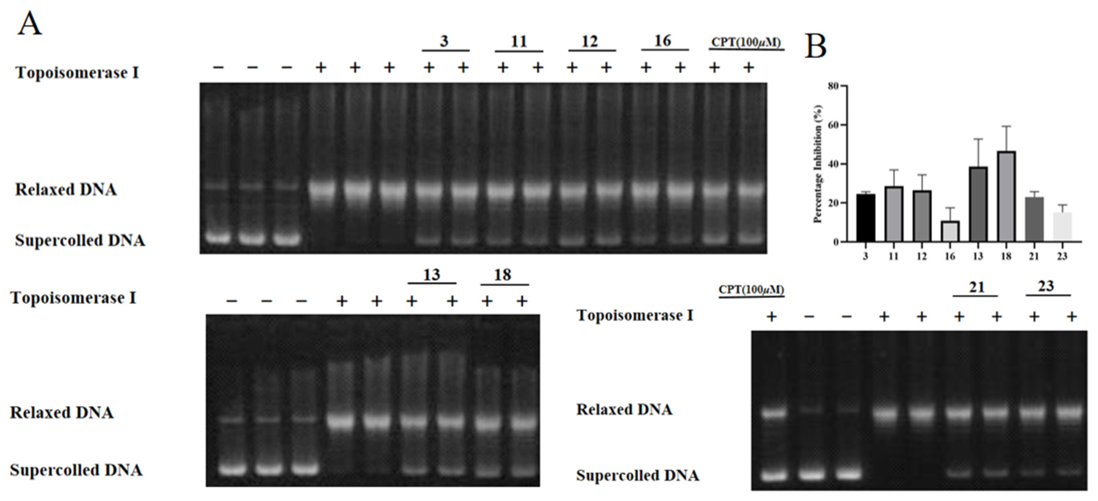
A DNA relaxation assay was performed to determine the inhibitory effect of compounds 129 on Topo I. To remove false positive results, a preliminary assay of DNA cleavage was performed, and no cleavage effect was detected in any of the compounds (Figure S20). As the results show in Table 3, compounds 1, 2, 5, 6, 7, 8, 15, 19, 20, 22, and 24 were the active ones, with IC50 values of 393.5, 328.98, 145.57, 239.27, 224.38, 189.84, 89.91, 47.5, 301.32, 178.03, and 218.27 μM, respectively. As for the other compounds, though the IC50 values were over the maximum tested concentration, the percentage inhibition values of these compounds on Topo I activity were different at 400 μM. The results from Figure 4 show that 3, 11, 12, 16, 13, 18, 21, and 23 exhibited inhibitory activities on Topo I at the concentration of 400 μM and the percentage inhibition values were 24.63%, 28.56%, 26.41%, 10.92%, 38.61%, 46.66%, 23.04%, and 15.18%, respectively, which indicated that these compounds showed weak inhibitory activity against DNA Topo I in vitro. For compounds 4, 9, 10, 14, 17, and 25~29, no inhibition at 400 μM was detected.

2.4. Compounds 57 Arrested Cell Cycle in Cancer Cells

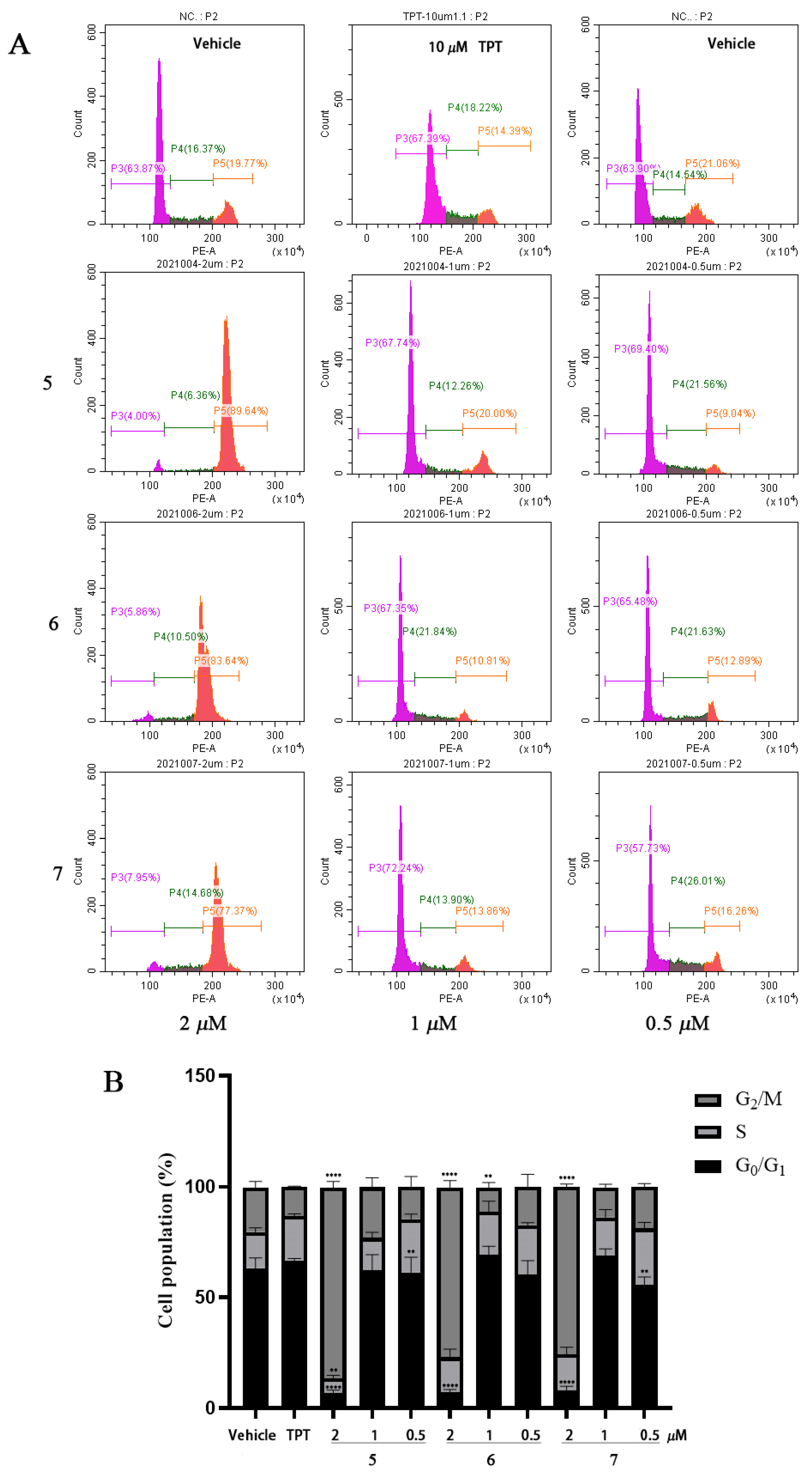
The effect of flavonoids 5, 6, and 7 on the DLD-1 cell cycle was examined by DNA content analysis. The results show that all the compounds inhibited the cell growth in a cell cycle-nonspecific way, in which a potent G2/M-phase arrest was observed after treatment with a higher concentration, and the obvious cell cycle arrest in the G1 or S phase was monitored after treatment with a lower concentration. After treatment with 2 μM of compounds 5, 6, and 7 for 24 h, the percentages of the cell population in the G2/M phase significantly increased from 20.12% to 86.05%, 76.49%, and 75.75%, and the cells in the G1/S (combined) phase remarkably decreased from 79.53% to 13.46%, 22.91%, and 24.26%, respectively, which means a potent G2/M cell cycle arrest caused by the treatment (p < 0.0001). In addition, after treatment with 1 and 0.5 μM of compounds 5, 6, and 7 for 24 h, the percentages of cells in the G1/S phase increased from 79.53% to 76.83%, 88.65% (p < 0.0001), and 86.07% (p < 0.001) and from 79.53% to 85.30% (p < 0.01), 82.61%, and 81.16% (p < 0.01), respectively, which indicates that an apparent G1- or S-phase arrest was induced after the treatment with a lower compound concentration (Figure 5).

2.5. Compound 5 Regulated Cell Cycle-Related Protein Expression in Cancer Cells

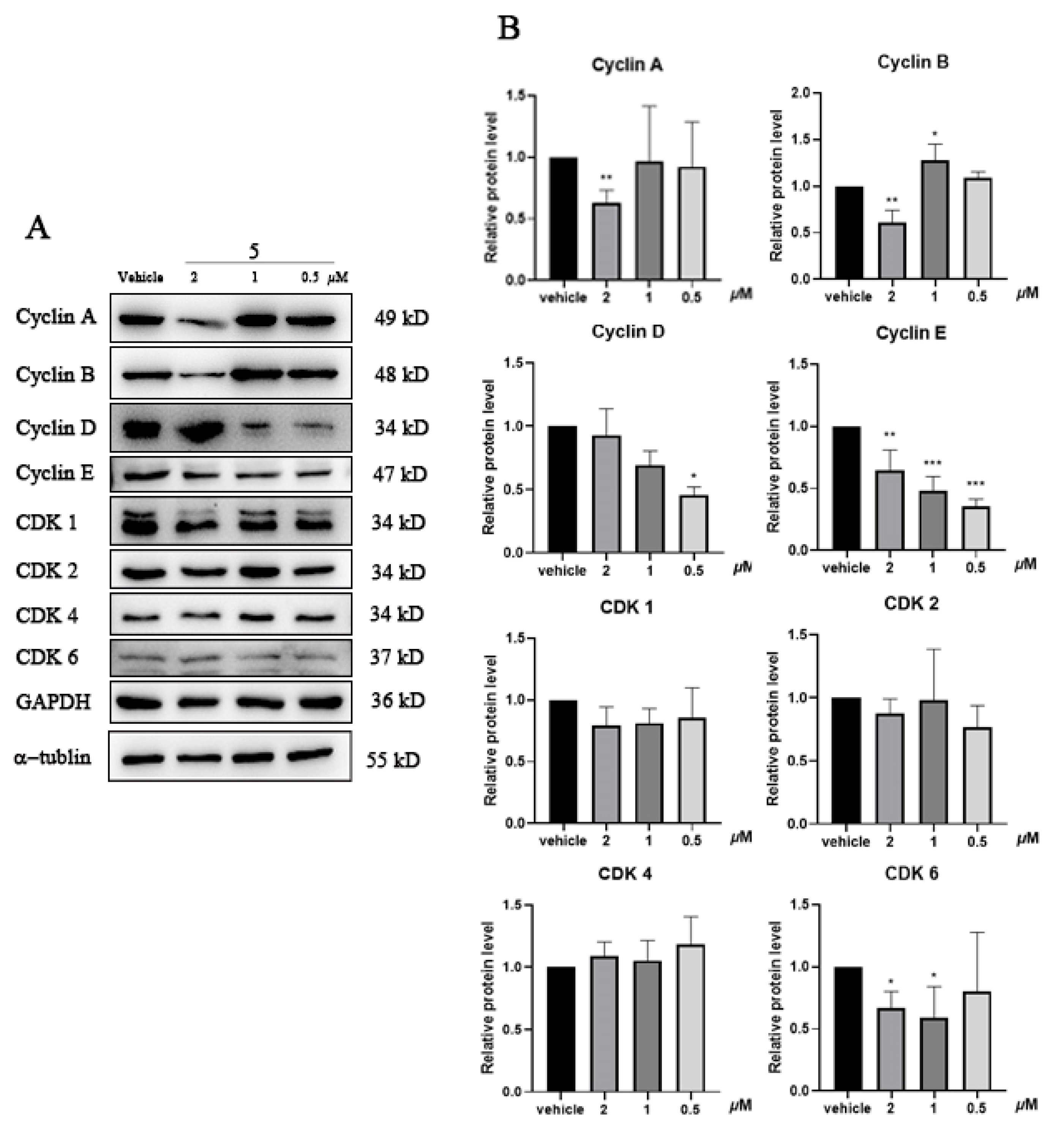
As a compound that showed potent inhibitory activity on DNA Topo I and cytotoxicity on cancer cell lines, the effect of 5 on cell cycle-related protein expression levels was investigated by Western blot in DLD-1 cells after treatment for 24 h. The results show that compound 5 potently decreased the expression levels of cyclin A, cyclin B, and CDK6 at 2 μM; meanwhile, 5 caused a potent downregulation of cylcin D, cyclin E, and CDK6 at 1 and 0.5 μM (Figure 6). The results indicate that, at different concentration of the inhibitor treated, flavonoid 5 caused G1/S- or G2/M-phase cell cycle arrests by regulating the expression of different cell cycle-related proteins.

3. Materials and Methods

3.1. Plant Material

The samples of B. pilosa L. were collected from the Liangwangshan mountain, Kunming, Yunnan province, China, in June 2020 and authenticated by Kunming Plant Branch Biotechnology Co., Ltd. (Zhang Jun) (Kunming, China), and a voucher specimen (YMU-ZF20200624) was deposited at the Key Laboratory of Chemistry in Ethnic Medicinal Resources, State Ethnic Affairs Commission and Ministry of Education, Yunnan Minzu University.

3.2. General Experimental Procedures

NMR experiments were conducted on a Bruker DRX-400 spectrometer operating at 400 MHz (1H) and 100 MHz (13C) at 300 K (chemical shifts δ in ppm, coupling constants J in Hz) (Bruker, Ettlingen, Germany). LC-MS data were obtained with an Agilent liquid chromatography-G6400 series triple quadrupole mass spectrometer (Agilent Technologies, Santa Clara, CA, USA). High-performance liquid chromatography (HPLC) separation was performed on an Agilent 1260 series with Agilent ZORBAX SB (21.2 × 250 mm) preparative column packed with C18 (7 µm) (Agilent Technologies, Santa Clara, CA, USA), Agilent ZORBAX XDB (9.4 × 250 mm) semi-preparative column packed with C18 (5 µm), and Agilent ZORBAX XDB (4.6 × 250 mm) analytical columns packed with C18 (5 µm). The thin-layer chromatography (TLC) analysis were performed with Merck silica gel 60 GF254 aluminum sheet (Merck, Darmstadt, Germany), and the spots were first viewed under UV light at λ 254 nm and 365 nm, and then stained with benzaldehyde (prepared from 135 mL ethanol + 5 mL sulfuric acid + 1.5 mL acetic acid + 3.7 mL CH3OC6H4CHO) followed by heating. Column chromatography (CC) was performed on Sephadex LH-20 (GE Healthcare, Waukesha, WI, USA), silica gel (100, 200, 200–300, or 300–400 mesh) (Qingdao Marine Chemical Inc., Qingdao, China), C18 reversed-phase silica gel (S-50 μm) (YMC, Kyoto, Japan), and MCI GEL (CHP20/P120) (Mitsubishi Chemical, Tokyo, Japan). All solvents used for chromatographic separations were distilled before use.

3.3. Extraction and Isolation

Powdered and air-dried whole plant of B. pilosa (20 kg) were extracted 7 times with 95% methanol (MeOH) by maceration for 24 h at room temperature. The MeOH extracts (2 kg) were combined and concentrated under reduced pressure. A portion of this extract was suspended in water (H2O) and successively extracted with light petroleum (PE), ethyl acetate (EtOAc), and n-butanol (n-BuOH) to obtain PE (310 g), EtOAc (165 g), and n-BuOH (360 g) fractions, respectively. The PE extract (310 g) was fractionated by CC (2500 g) on silica gel (60–100 mesh, 15 cm × 100 cm) and eluted with PE/DCM (100:1 to 1:1, v/v), PE/EtOAc (5:1 to 1:1, v/v) to obtain 8 fractions (A-H). Fr.A (1.3 g) was analyzed using TLC, and the same components were merged to obtain 3 fractions (FrA.1-FrA.3). FrA.1 (810.2 mg) was separated using silica gel CC (100–200 mesh) and eluted with PE/DCM (100:1 to 1:1, v/v) to yield 3 fractions (FrA.1.1-FrA.1.3); FrA.1.1 was further separated using silica gel CC (200–300 mesh) and eluted with PE/DCM (100:1, v/v) and Sephadex LH-20 column (MeOH) to yield compound 3 (50.6 mg). FrA.1.2 was separated using Sephadex LH-20 column (DCM:MeOH = 1:1) and silica gel to obtain compound 23 (32.4 mg). FrA.1.3 was fractionated by Sephadex LH-20 column (DCM:MeOH = 1:1) and silica gel to obtain compound 22 (20.1 mg). FrA.2 was further separated by silica gel CC, eluted with PE/EtOAc (100:1, v/v) and HPTLC, and eluted with PE/EtOAc (15:1, v/v) to obtain compound 24 (12.3 mg). The EtOAc extract (165 g) was fractionated by CC (2500 g) on silica gel (60–100 mesh, 15 × 100 cm) and eluted with PE and DCM/MeOH (1:0 to 0:1, v/v) to obtain 7 fractions (Fr.I-Fr.O). Fr.K (DCM: MeOH = 50:1) was analyzed using TLC, and the same components were merged to obtain 6 fractions (FrK.1-FrK.6). FrK.1 was subjected to middle chromatogram isolated (MCI) gel and eluted with MeOH/H2O (50:50 to 1:0, v/v) to yield 7 fractions (FrK.1.1-FrK.1.7); FrK.1.4 was fractionated by Sephadex LH-20 column (MeOH), silica gel, and ODS-C18 column and eluted with MeOH/H2O (40:60 to 1:0, v/v) to yield compound 21 (25 mg). FrK.1.6 was separated using a Sephadex LH-20 column (MeOH) to obtain 3 fractions (FrK.1.6.1-FrK.1.6.3); FrK.1.6.2 was further separated using silica gel CC (200–300 mesh), eluted with DCM/MeOH (1:0 to 0:1, v/v), and then depurated by Sephadex LH-20 column (MeOH) to obtain compound 4 (25 mg) and compound 5 (30 mg). FrK.1.7 was separated using a Sephadex LH-20 column (MeOH) to obtain 4 fractions (FrK.1.7.1-FrK.1.7.4); FrK.1.7.3 was subjected to the ODS-C18 column and eluted with MeOH/H2O (30:70 to 1:0, v/v) to yield compounds 6 (15 mg) and 7 (15 mg). Fr.L (DCM: MeOH = 25:1) was analyzed using TLC and 3 fractions were obtained (FrL.1-FrL.3); FrL.2 was depurated by MCI, silica gel, Sephadex LH-20 (MeOH), and C18 column and eluted with MeOH/H2O (30:70 to 1:0, v/v) to yield compound 20 (10 mg). Fr.M (DCM: MeOH = 10:1) was analyzed using TLC, and the same components were merged to obtain 6 fractions (FrM.1-FrM.6); FrM.6 was depurated by MCI and eluted with MeOH/H2O (50:50 to 1:0, v/v) to yield 3 fractions (FrM.6.1-FrM.6.3). FrM.6.1 was separated using a Sephadex LH-20 column (MeOH) to obtain 6 fractions (Fr.6.1.1-Fr.6.1.6); Fr.6.1.3 was fractionated by reverse-phase chromatography (RP-18) and then purified with chromatography silica gel and Sephadex LH-20 (MeOH) to obtain compounds 25 (5 mg) 27 (3.5 mg), 28 (7.1 mg), and 29 (10.4 mg). Fr.6.1.4 was depurated by repeated preparative HPLC and eluted with MeOH/H2O (70:30, v/v, ν = 4 mL/min) to obtain 4 fractions (FrM.6.1.4.1-FrM.6.1.4.4). FrM.6.1.4.2 was further separated by recrystallization to obtain compound 9 (15 mg). FrM.6.1.4.1 was fractionated by RP-18 and then purified with chromatography silica gel, Sephadex LH-20 (MeOH), iterative semi-preparative HPLC, and recrystallization to obtain compounds 1 (13 mg), 10 (18 mg), 11 (18 mg), 12 (20 mg), 13 (15 mg), 16 (9 mg), and 17 (4 mg). FrM.6.1.4.3 was separated by iterative semi-preparative HPLC to obtain compound 26 (50 mg). Fr.6.1.6 was depurated by LH-20 and iterative semi-preparative HPLC and eluted with MeOH/H2O (50:50, v/v, ν = 1.5 mL/min) to obtain compound 15 (3 mg). Fr.6.1.5 was further separated by repeated preparative HPLC and eluted with MeOH/H2O (53:47, v/v, ν = 4 mL/min), iterative semi-preparative HPLC and eluted with ACN/H2O (19:81, v/v, ν = 2.5 mL/min), MeOH/H2O (37:63, v/v, ν = 2 mL/min), and recrystallization to obtain compounds 2 (22 mg), 8 (7 mg), 18 (25 mg), and 19 (2.8 mg). The n-BuOH extract (360 g) was fractionated by macroporous absorption resin and eluted with EtOH/H2O (50:50 to 1:0, v/v) to yield 3 fractions (Fr.P-Fr.R). Fr.P was separated using a Sephadex LH-20 column (MeOH), then depurated by repeated preparative HPLC, and eluted with MeOH/H2O (70:30, v/v, ν = 4 mL/min) to obtain 4 fractions (Fr.P.1-Fr.P.4). Fr.P.3 was further separated using semi-preparative HPLC and eluted with MeOH/H2O (42:58, v/v, ν = 2 mL/min) to obtain compound 14 (7 mg).

3.4. Cell Culture

The cell lines MDA-MB-231 (No. TCHu227), A549 (No. TCHu150), and HepG2 (No. TCHu72) were obtained from the Cell Bank/Stem Cell Bank, Chinese Academy of Sciences (Shanghai, China). The cell lines DLD-1 (No. CC0507) and HCT116 (No. CC0506) were obtained from the Cell Bank of Cellcook Biotech Co., Ltd. (Guangzhou, China). Cell line HL-7702 (No. BNCC338358) was obtained from Bena Culture Collection (Zhengzhou, China).
The human cancer cell lines MDA-MB-231 (breast adenocarcinoma), DLD-1 (colorectal adenocarcinoma), A549 (lung epithelial carcinoma), HCT116 (colorectal adenocarcinoma), HepG2 (hepatocellular carcinoma), and human hepatocyte HL-7702 were cultured in Dulbecco’s modified eagle medium (DMEM, Biological Industries, Beit Haemek, Israel) containing 10% (v/v) fetal bovine serum (FBS, Biological Industries, Beit Haemek, Israel) at 37 °C in a humidified incubator (ThermoFisher, Carlsbad, CA, USA) with a 5% CO2 atmosphere.

3.5. Cytotoxicity Assay

The cytotoxic activities of the compounds against cancer cells were examined with a sulforhodamine B (SRB) colorimetric assay. Cells were seeded at a density of 5 × 10³/well in 96-well plates for 24 h; various concentrations of the tested compounds, dissolved in DMSO and diluted with the complete medium, were added in the wells in triplicate and incubated for 48 h. After that, the cell viability was assessed with the SRB assay as we described before [43]. The IC50 values were determined using the Reed and Muench method and are expressed as the mean ± SD of at least three independent measurements. Camptothecin (CPT) (Energy Chemical, Shanghai, China) was used as a reference compound for the positive control.

3.6. Topo I Inhibition Assay

The DNA relaxation assay was adopted to investigate the compounds’ inhibitory activities on Topo I. Different concentrations of the compounds, dissolved and diluted in DMSO, were added into the relaxation buffer (50 mM Tris, pH 7.5, 50 mM KCl, 10 mM MgCl2, 0.1 mM EDTA, 0.5 Mm dithiothreitol, and 0.05 mg/mL bovine serum albumin) containing 0.2 U Topo I (ThermoFisher, Waltham, MA, USA) and incubated at room temperature for 10 min. After that, 500 ng supercoiled pBR322 plasmid DNA (ThermoFisher, Waltham, MA, USA) was added and then incubated at 37 °C for another 30 min. Finally, the reactions were stopped with a stopping solution (5% sodium dodecyl sulfate, 0.0025% bromophenol blue, and 25% glycerol), and the mixtures were applied onto 0.8% agarose gel and subjected to electrophoresis for 1.5 h in Tris-Acetate-EDTA buffer. After being stained with GelRed (Biotium, Fremont, CA, USA), the gel was imagined with the imaging system (Biotop, Shanghai, China). CPT (Energy Chemical, Shanghai, China) was used as a reference compound for the positive control. The band density of the supercoiled DNA was quantified by the ImageJ software (version 1.53a, National Institutes of Health, Bethesda, MD, USA), and the inhibition activity is expressed as the percentage inhibition and calculated by formula: Inhibition % = [(A − B)/(C − B)] × 100%, where A is the band density of the tested samples, B is the band density of the negative control, and C is the band density of the blank control. IC50 values were determined using the Reed and Muench method and are expressed as the mean ± SD of at least three independent measurements in duplicate.

3.7. DNA Content Analysis

Cells were seeded in 6-well plates and allowed to adhere for 24 h at 37 °C. After treatment with different concentrations of the samples for another 24 h, the cells were harvested and washed with PBS, and then, the DNA content analysis was conducted with a cell cycle staining kit (Multi sciences, Hangzhou, China), according to the manufacturer’s instructions. The fluorescence intensity of the cells was measured by flow cytometry (Beckman CytoFlex, Miami, FL, USA). Topotecan (TPT) (Solarbio, Beijing, China) was used as a reference compound for the positive control.

3.8. Western Blot Analysis

Cells treated with the compounds were harvested, and the total protein was isolated with lysis buffer (72 mM Tris pH 6.8, 2% SDS, 10% glycerol, and 0.1% bromophenol blue). The proteins were subjected to SDS-PAGE and transferred to a 0.45 μm PVDF membrane (Millipore, Burlington, USA). After being blocked with 5% non-fat milk in TBST, the membranes were washed and then incubated with specific primary antibodies (Cdk2 and Cdk6 were purchased from BBI Life Sciences Corporation, Shanghai, China; cyclin A, cyclin B, cyclin D, cyclin E, Cdk1, and Cdk4 were purchased from Proteintech, Rosemont, IL, USA) in TBST containing 2% non-fat milk overnight at 4 °C. After washing, the membranes were incubated with secondary horseradish peroxidase (HRP)-conjugated antibodies at room temperature for 1 h. The blot was visualized using an enhanced chemiluminescence assay kit (Meilunbio, Dalian, China) under a chemiluminescence detector (Tanon, Shanghai, China). The band density of the blot was quantified by the ImageJ software with at least three independent experiments.

3.9. Statistical Analysis

All the results were calculated by at least three independent experiments and are presented as mean ± S.D. values. A one-way ANOVA or the Student’s t-test was used for the statistical analysis. A p value < 0.05 was considered statistically significant.

4. Discussion

Cancer is commonly characterized by uncontrolled cell division and proliferation, which are directly related to DNA replication. Targeting Topos disrupts DNA replication and transcription, which leads to the inhibition of cell division, thereby stopping the growth of cancer cells [12]. In a wide range of human solid tumors, the intracellular level of Topo I is higher than that in normal tissues, especially in metastatic ovarian cancer, cervical cancer, small-cell lung cancer, metastatic colon cancer, and pancreatic cancer [11], suggesting that controlling the Topo I level is essential in treating cancers. Several studies have revealed that specific flavonoids in natural products inhibit the activity of DNA Topo I [44]. Licochalcones A, a chalcone isolated from Glycyrrhiza inflata, and luteolin, a flavone isolated from the leaves of Vitex negundo, were reported to show cytotoxicity by inhibiting DNA Topo I [45,46]; therefore, flavonoids hold therapeutic prospects in the treatment of cancers.
B. pilosa L. is a herb that is used as a folk medicine for cancer treatment in China. In this study, nineteen flavonoids and ten polyacetylenes (Figure 3) were isolated from the plant, including one flavone (15), eleven flavonols (414), two flavanones (1617), three chalcones (13), two aurone (18, 19), and ten polyacetylenes (2029), and their inhibitory activities on DNA Topo I and cytotoxicities on cancer cell lines were tested (Table 3). The new compounds 1 and 20 and the polyacetylenes 22 and 24 were found to be the inhibitors of DNA Topo I for the first time. The structure–activity relationship of flavonoids against Topo I was analyzed. Based on the results from Table 3 and Figure 4, it was demonstrated that, compared to chalcones, flavones, flavonols, and aurones, flavonoids showed a strong inhibitory activity against DNA Topo I in vitro. Compared to flavanones, flavones and flavonols are more active, indicating that the double bond between C2 and C3 is important to the inhibitory activity, which was verified by the observation of the decreased inhibitory activities of flavanones (16 and 17), which lack the C2-C3 unsaturation bond on the C-ring. By comparing the activity of 4 with those of 5, 6, and 7, we deduced that the substitution of the C-6 position on the A-ring of flavonols was essential for Topo I inhibition, and if there was no substitution, the activity potently decreased. Additionally, if the substitution at the C-6 position was glycosylated, the activities of the compounds also decreased, which was verified by comparing 5, 6, 7, and 8 with 9. The C-7 position is also important for the inhibitory activity, which was enhanced by the substitution of small groups (-OH and -OCH3), and if the substitution was glycosylated, the activities of the compounds (10, 12, 13, 14, and 17) potently decreased, which was also confirmed by comparing 14 with its aglycone 5. As for the B-ring of flavonols, substitutions at C-3′ and C-4′ are important for the inhibitory activity (5, 6, and 7), and comparing 5 with 7, the activity of the hydroxy substitution at the C-3′ position is better than that of the methoxy substitution. Furthermore, the inhibition results from compounds 12 and 14 indicates that the hydroxyl group substitution at C-3′ and C-4′ is superior to that of methoxy group, which is correlated with what has been described in previous publications [47,48]. As for the cytotoxicity of compounds, only 5, 6, 7, 15, and 22 showed good activity in cancer cell lines, as well as in the DNA Topo I assay, which indicates that their cytotoxicity was partly caused by DNA Topo I inhibition. As for compound 4, though inactive against Topo I, a good cytotoxicity on cancer cells was observed (not as good as those of 5, 6, and 7), indicating that DNA Topo I is only one of the cytotoxic targets of flavonoids, which intervenes in the characteristics of multi-targets for natural products [44]. As the most active inhibitor of Topo I, 15, a more hydrophilic compound compared to 5, 6, and 7, shows a weaker cytotoxicity, which is attributed to the lipid bilayer of cell membranes that prevents the entrance of 15 into the cytosol, leading to less cytotoxic activity. Comparing aurones 18 and 19, the esterification of the glucose of 19 enhanced the inhibitory effect on Topo I. Although 19 showed a good inhibitory activity, its cytotoxicity was weak, which may be attributed to the metabolized difference between cellular experiments and non-cellular ones.
Topoisomerase I-targeting drugs exert a cytotoxic effect by producing enzyme-mediated DNA damage, which leads to cell cycle arrest. Thus, the cell cycle analysis of DLD-1 cells treated with 5, 6, and 7 was conducted by flow cytometry. The results from the DNA content (Figure 5) analysis show that all flavonoids caused potent G2/M arrests at a higher concentration and significant G1/S arrests at a lower concentration, which indicates that flavonoids 5, 6, and 7 induced double-strand DNA breaks by the inhibition of DNA Topo I; then, the activated DNA damage response accumulated cells in different phases depending on the concentration of the inhibitor used. The cell cycle-nonspecific arrest by flavonoids is in agreement with what was reported for DNA damaging agents in previous publications [49,50]. The eucaryotic cell cycle is regulated by the periodic synthesis and destruction of cyclins that complex with cyclin-dependent kinases (Cdks); thus, cyclin/Cdk complexes are the key regulators for governing the checkpoints in a cell division cycle. In order to verify the results of the DNA content analysis, the levels of different cyclins and Cdks in the cell cycle were investigated by Western blot (Figure 6). The results exhibit that compound 5 caused cell cycle arrest by downregulating the expression of different cyclins or Cdks at different concentrations. As the main regulator for controlling the G2/M checkpoint, the cyclin B/Cdk1 complex is the key regulator of cell mitosis, and treatment with 2 μM of compound 5 caused a significant decrease in cyclin B, and because the periodic synthesis and destruction of cyclins control the activation of the Cdks and the cyclin/Cdk complexes [51], it suggests that the reduction in cyclin B induced by 5 blocked the cells’ entry into mitosis and led to the potent G2/M arrest in DLD-1 cells. In the normal eucaryotic cell cycle process, the activity of the cyclin D/Cdk4/6 and cyclin E/Cdk2 complexes controls G1-phase progression, and the cyclin A/Cdk2 complex controls the S phase. In this study, the results from the Western blot show that the treatment with lower concentrations of compound 5 decreased the expression of cyclin D, cyclin E, and Cdk6 (Figure 6), which indicates that 5 inhibits the activity of the cyclin D/Cdk4/6, cyclin A/Cdk2, and cyclin E/Cdk2 complexes at a lower concentration and arrests the G1/S cell cycle checkpoint in DLD-1 cells. The results from the Western blot are consistent with that from the DNA content analysis. Additionally, as it was shown in DLD-1 cells, compound 5 also caused potent G2/M arrests at 2 μM and significant G1/S arrests at 0.5 μM and regulated the expression of cell cycle-related proteins in the HCT116 cell line (Figure S21). All the results indicate that the flavonoids and polyacetylenes that exist in B. pilosa are the cytotoxic components responsible for its anti-tumor activity by inhibiting DNA Topo I.

5. Conclusions

Flavonoids broadly exist in plants and exhibit a wide range of bioactivities. B. Pilosa is a traditional herb that is used as anti-tumor folk medicine in China and many other countries. However, its main constituents with anti-tumor activities have not been identified. In this study, a new chalcone and a new polyacetylene, together with twenty-seven known flavonoids and polyacetylenes, were isolated from B. pilosa, and their inhibitory activities against DNA Topo I were reported for the first time, except for compound 15, which was isolated from the leaves of Vitex negundo L. and has been reported to show good activity [46]. The study of the structure–activity relationship analysis and cytotoxicity mechanism indicated that Topo I-targeting flavonoids and polyacetylenes are two kinds of active ingredients that lead to the anti-tumor effect of B. pilosa, which is of great importance for the R&D of flavonoids and the traditional herb B. pilosa.

Supplementary Materials

The following supporting information can be downloaded at: https://www.mdpi.com/article/10.3390/molecules29153547/s1. Figure S1: HRESI-MS spectrum of compound 1. Figure S2: 1H-NMR (DMSO-d6) spectrum of compound 1. Figure S3: 13C-NMR (DMSO-d6) spectrum of compound 1. Figure S4: DEPT (90°, 135°) spectrum of compound 1. Figure S5: HSQC (DMSO-d6) spectrum of compound 1. Figure S6: HMBC (DMSO-d6) spectrum of compound 1. Figure S7: 1H-1H COSY (DMSO-d6) spectrum of compound 1. Figure S8: ROESY (DMSO-d6) spectrum of compound 1. Figure S9: UV spectrum of compound 1. Figure S10: IR spectrum of compound 1. Figure S11: HRESI-MS spectrum of compound 20. Figure S12: 1H-NMR (CD3OD) spectrum of compound 20. Figure S13: 13C-NMR (CD3OD) spectrum of compound 20. Figure S14: DEPT (90°,135°) spectrum of compound 20. Figure S15: HSQC (CD3OD) spectrum of compound 20. Figure S16: HMBC (CD3OD) spectrum of compound 20. Figure S17: 1H-1H COSY (CD3OD) spectrum of compound 20. Figure S18: UV spectrum of compound 20. Figure S19: IR spectrum of compound 20. Figure S20: The effects of compounds on DNA cleavage. Figure S21: Results of flavonoid 5 induced cell cycle arrests in HCT116 cells. Characterization data of compounds 219 and 2129.

Author Contributions

G.Z. (Guiyuan Zeng): conceptualization; formal analysis; investigation; writing—original draft. Y.W.: formal analysis; investigation. M.Z.: formal analysis; investigation. J.Y. (Jumei Yi): investigation. J.M.: validation. B.Y.: validation. W.S.: formal analysis. F.D.: formal analysis. J.Y. (Junlin Yin): supervision. G.Z. (Guangzhi Zeng): conceptualization; funding acquisition; supervision; writing—review and editing. All authors have read and agreed to the published version of the manuscript.

Funding

This work was supported by the National Natural Science Foundation of China (81960639) and the Project of Yunnan Provincial Department of Science and Technology (202205AC160084, 202401BC070018, 202402AN360010).

Institutional Review Board Statement

Not applicable.

Informed Consent Statement

Not applicable.

Data Availability Statement

The original contributions presented in the study are included in the article (and Supplementary Material), further inquiries can be directed to the corresponding authors.

Conflicts of Interest

The authors declare no conflicts of interest.

References

  1. Silva, F.L.; Fischer, D.C.; Tavares, J.F.; Silva, M.S.; de Athayde-Filho, P.F.; Barbosa-Filho, J.M. Compilation of secondary metabolites from Bidens pilosa L. Molecules 2011, 16, 1070–1102. [Google Scholar] [CrossRef] [PubMed]
  2. Pozharitskaya, O.N.; Shikov, A.N.; Makarova, M.N.; Kosman, V.M.; Faustova, N.M.; Tesakova, S.V.; Makarov, V.G.; Galambosi, B. Anti-inflammatory activity of a HPLC-fingerprinted aqueous infusion of aerial part of Bidens tripartita L. Phytomedicine 2010, 17, 463–468. [Google Scholar] [CrossRef] [PubMed]
  3. Wang, R.; Wu, Q.X.; Shi, Y.P. Polyacetylenes and flavonoids from the aerial parts of Bidens pilosa. Planta Med. 2010, 76, 893–896. [Google Scholar] [CrossRef] [PubMed]
  4. Wu, J.; Wan, Z.; Yi, J.; Wu, Y.; Peng, W.; Wu, J. Investigation of the extracts from Bidens pilosa Linn. var. radiata Sch. Bip. for antioxidant activities and cytotoxicity against human tumor cells. J. Nat. Med. 2013, 67, 17–26. [Google Scholar] [CrossRef] [PubMed]
  5. Tobinaga, S.; Sharma, M.K.; Aalbersberg, W.G.; Watanabe, K.; Iguchi, K.; Narui, K.; Sasatsu, M.; Waki, S. Isolation and identification of a potent antimalarial and antibacterial polyacetylene from Bidens pilosa. Planta Med. 2009, 75, 624–628. [Google Scholar] [CrossRef] [PubMed]
  6. Yuan, L.P.; Chen, F.H.; Ling, L.; Dou, P.F.; Bo, H.; Zhong, M.M.; Xia, L.J. Protective effects of total flavonoids of Bidens pilosa L. (TFB) on animal liver injury and liver fibrosis. J. Ethnopharmacol. 2008, 116, 539–546. [Google Scholar] [CrossRef] [PubMed]
  7. Kumari, P.; Misra, K.; Sisodia, B.S.; Faridi, U.; Srivastava, S.; Luqman, S.; Darokar, M.P.; Negi, A.S.; Gupta, M.M.; Singh, S.C.; et al. A promising anticancer and antimalarial component from the leaves of Bidens pilosa. Planta Med. 2009, 75, 59–61. [Google Scholar] [CrossRef] [PubMed]
  8. Chien, S.C.; Young, P.H.; Hsu, Y.J.; Chen, C.H.; Tien, Y.J.; Shiu, S.Y.; Li, T.H.; Yang, C.W.; Marimuthu, P.; Tsai, L.F.; et al. Anti-diabetic properties of three common Bidens pilosa variants in Taiwan. Phytochemistry 2009, 70, 1246–1254. [Google Scholar] [CrossRef]
  9. Delgado, J.L.; Hsieh, C.M.; Chan, N.L.; Hiasa, H. Topoisomerases as anticancer targets. Biochem. J. 2018, 475, 373–398. [Google Scholar] [CrossRef]
  10. Liang, X.; Wu, Q.; Luan, S.; Yin, Z.; He, C.; Yin, L.; Zou, Y.; Yuan, Z.; Li, L.; Song, X.; et al. A comprehensive review of topoisomerase inhibitors as anticancer agents in the past decade. Eur. J. Med. Chem. 2019, 171, 129–168. [Google Scholar] [CrossRef]
  11. Gu, L.; Hickey, R.J.; Malkas, L.H. Therapeutic Targeting of DNA Replication Stress in Cancer. Genes 2023, 14, 1346. [Google Scholar] [CrossRef] [PubMed]
  12. Singh, S.; Pandey, V.P.; Yadav, K.; Yadav, A.; Dwivedi, U.N. Natural Products as Anti-Cancerous Therapeutic Molecules Targeted towards Topoisomerases. Curr. Protein Pept. Sci. 2020, 21, 1103–1142. [Google Scholar] [CrossRef] [PubMed]
  13. Jain, C.K.; Majumder, H.K.; Roychoudhury, S. Natural Compounds as Anticancer Agents Targeting DNA Topoisomerases. Curr. Genom. 2017, 18, 75–92. [Google Scholar] [CrossRef] [PubMed]
  14. Zhao, Z.Y.; Ma, Q.T.; Wu, Y.H.; Duan, W.L.; Zeng, G.Z.; Yin, J.L. Chemical constituents of the whole plant of Bidens pilosa L. J. Yunnan Minzu Univ. Nat. Sci. Ed. 2022, 31, 627–632. [Google Scholar]
  15. Redl, K.; Davis, B.; Bauer, R. Chalcone glycosides from Bidens campylotheca. Phytochemistry 1992, 32, 218–220. [Google Scholar] [CrossRef]
  16. Cui, S.; Zou, Y.; Wu, Y.; Gao, P. Synthesis and Absolute Configuration of the 7-Phenylhepta-4,6-diyne-1,2-diol Isolated from Bidens pilosa. Synthesis 2012, 44, 3108. [Google Scholar]
  17. Li, C.H.; Zhu, Y.J.; Yu, S.H.; Jiang, H.Q.; Zhou, H.L. A new polyacetylene and a new isonicotinic acid glucoside from Bidens parviflora Willd. Acta Pharm. Sin. 2020, 55, 489–494. [Google Scholar]
  18. Tian, X.; Zhou, S.X.; Wei, H.L.; Hu, N.; Dai, Z.; Liu, Z.G.; Han, Z.Z.; Tu, P.F. Flavonoids from the herb of Bidens pilosa L. J. Chin. Pharm. Sci. 2011, 20, 518–522. [Google Scholar] [CrossRef]
  19. Liu, J.; Chen, R.; Nie, Y.; Feng, L.; Li, H.D.; Liang, J.Y. A new carbamate with cytotoxic activity from the aerial parts of Siegesbeckia pubecens. Chin. J. Nat. Med. 2012, 10, 13–15. [Google Scholar] [CrossRef]
  20. Glasl, S.; Mucaji, P.; Werner, I.; Presser, A.; Jurenitsch, J. Sesquiterpenes and flavonoid aglycones from a Hungarian taxon of the Achillea millefolium group. Z. Naturforsch C J. Biosci. 2002, 57, 976–982. [Google Scholar] [CrossRef]
  21. Yuan, K.; Xu, R.; Chen, C. A new flavonol glycoside from Tridax procumbens Linn. J. Chem. Res. 2009, 2009, 165–166. [Google Scholar] [CrossRef]
  22. Lee, H.J.; Sim, M.O.; Woo, K.W.; Jeong, D.E.; Jung, H.K.; An, B.; Cho, H.W. Antioxidant and Antimelanogenic Activities of Compounds Isolated from the Aerial Parts of Achillea alpina L. Chem. Biodivers. 2019, 16, e1900033. [Google Scholar] [CrossRef] [PubMed]
  23. Qin, J.J.; Zhu, J.X.; Zhu, Y.; Yan, L.; Jin, H.Z.; Zhang, W.D. Flavonoids from the Aerial Parts of Inula japonica. Chin. J. Nat. Med. 2010, 8, 257–259. [Google Scholar] [CrossRef]
  24. Xu, R.; Zhang, J.; Yuan, K. Two new flavones from Tridax procumbens Linn. Molecules 2010, 15, 6357–6364. [Google Scholar] [CrossRef] [PubMed]
  25. Yang, X.W.; Hang, M.Z.; Zhao, W.Q.; Jin, Y.S.; Chen, H.S. Studies on Chemical Constituents of Bidens bipinnata. Pharm. J. Chin. People’s Lib. Army 2009, 25, 283–286. [Google Scholar]
  26. Kim, G.R.; Kim, E.N.; Jeong, G.S. Simultaneous quantitative analysis of flavonoids isolated from the leaves of Diospyros kaki. Korean J. Pharmacogn. 2020, 51, 139–145. [Google Scholar]
  27. Shafiq, N.; Riaz, N.; Saleem, M. Isolation, characterization of flavonoids from Seriphidium oliverianum and their antioxidant and anti-urease activities. J. Chem. Soc. Pak. 2014, 36, 517–523. [Google Scholar]
  28. Laraoui, H.; Long, C.; Haba, H.; Benkhaled, M. New methylated flavonol glucosides from Fumana montana Pomel. Nat. Prod. Res. 2013, 27, 1770–1775. [Google Scholar] [CrossRef] [PubMed]
  29. Wang, X.Y.; Chen, G.R.; Deng, Z.Y.; Zhao, J.; Ge, J.F.; Li, N.; Chen, F.H. Chemical constituents from Bidens bipinnata. China J. Chin. Mater. Med. 2014, 39, 1838–1844. [Google Scholar]
  30. Li, Y.L.; Li, J.; Wang, N.L.; Yao, X.S. Flavonoids and a new polyacetylene from Bidens parviflora Willd. Molecules 2008, 13, 1931–1941. [Google Scholar] [CrossRef]
  31. Cai, F.J.; Li, C.H.; Sun, X.H.; Wang, L.; Tian, J.L.; Zhao, W.; Kong, D.G.; Liu, Q.; Zhou, H.L. A new dihydroflavone and a new polyacetylene glucoside from Bidens parviflora. J. Asian Nat. Prod. Res. 2022, 24, 963–970. [Google Scholar] [CrossRef]
  32. Deng, K.Z.; Liu, C.; Chang, M.Y.; Zong, Q.; Xiong, Y. Isolation and Identification of Phenols Compounds in Aurantii Fructus Immaturus. China Pharm. 2020, 31, 1040–1043. [Google Scholar]
  33. Li, P.; Wang, Y.L.; Tang, L.; Guo, Z.Y.; Long, C.L. Studies on flavonoids from Bidens pilosa. Chin. Tradit. Herb. Drugs 2023, 44, 2498–2501. [Google Scholar]
  34. Okuyama, T.; Li, S.; Kuang, H.X.; Okada, Y. A New Aurone Glucoside and a New Chalcone Glucoside from Bidens bipinnata Linne. Heterocycles 2003, 61, 557–561. [Google Scholar] [CrossRef]
  35. Chang, M.H.; Wang, G.J.; Kuo, Y.H.; Lee, C.K. The Low Polar Constituents from Bidens pilosa L. var. Minor (Blume) Sherff. J. Chin. Chem. Soc. 2013, 47, 1131–1136. [Google Scholar] [CrossRef]
  36. Kimura, Y.; Hiraoka, K.; Kawano, T.; Fujioka, S.; Shimada, A. Nematicidal Activities of Acetylene Compounds from Coreopsis lanceolata L. Z. Naturforschung C 2015, 63, 1121–1124. [Google Scholar] [CrossRef] [PubMed]
  37. Xie, J.D.; Chen, Z.K.; Xie, F.D.; Hu, J.J.; Liang, H.J.; Li, Q.R.; Yuan, J. Studies on Isolation of Polyacetylenes from Bidens pilosa var.radiata and their biological activities. J. Trop. Subtrop. Bot. 2021, 29, 216–220. [Google Scholar]
  38. Zhao, A.H.; Zhao, Q.S.; Peng, L.Y.; Zhang, J.X.; Lin, Z.W.; Sun, H.D. A New Chalcone Glycoside from Bidens pilosa. Acta Bot. Yunnanica 2004, 26, 121–126. [Google Scholar]
  39. Rücker, G.; Kehrbaum, S.; Sakulas, H.; Lawong, B.; Goeltenboth, F. Acetylenic glucosides from Microglossa pyrifolia. Planta Med. 1992, 58, 266–269. [Google Scholar] [CrossRef]
  40. Chiang, Y.M.; Chang, C.L.; Chang, S.L.; Yang, W.C.; Shyur, L.F. Cytopiloyne, a novel polyacetylenic glucoside from Bidens pilosa, functions as a T helper cell modulator. J. Ethnopharmacol. 2007, 110, 532–538. [Google Scholar] [CrossRef]
  41. Xi, F.M.; Li, C.T.; Han, J.; Yu, S.S.; Wu, Z.J.; Chen, W.S. Thiophenes, polyacetylenes and terpenes from the aerial parts of Eclipata prostrate. Bioorganic Med. Chem. 2014, 22, 6515–6522. [Google Scholar] [CrossRef]
  42. Lee, S.H.; Kim, J.G.; Le TP, L.; Han, J.S.; Cho, Y.B.; Lee, M.K.; Lee, D.; Hwang, B.Y. Polyacetylenes from the roots of Cirsium japonicum var. ussuriense. Phytochemistry 2022, 202, 113319. [Google Scholar] [CrossRef] [PubMed]
  43. Wu, Y.; Liu, Z.; He, Z.; Yi, J.; Qiao, X.; Tan, C.; Xing, Y.; Zeng, Y.; Yang, D.; Yin, J.; et al. Cantharidin analogue alleviates dextran sulfate sodium-induced colitis in mice by inhibiting the activation of NF-κB signaling. Eur. J. Med. Chem. 2023, 260, 115731. [Google Scholar] [CrossRef] [PubMed]
  44. Scotti, L.; Bezerra Mendonca, F.J.; Ribeiro, F.F.; Tavares, J.F.; da Silva, M.S.; Barbosa Filho, J.M.; Scotti, M.T. Natural Product Inhibitors of Topoisomerases: Review and Docking Study. Curr. Protein Pept. Sci. 2018, 19, 275–291. [Google Scholar] [CrossRef]
  45. Yoon, G.; Kang, B.Y.; Cheon, S.H. Topoisomerase I inhibition and cytotoxicity of licochalcones A and E from Glycyrrhiza inflata. Arch. Pharm. Res. 2007, 30, 313–316. [Google Scholar] [CrossRef] [PubMed]
  46. Chowdhury, A.R.; Sharma, S.; Mandal, S.; Goswami, A.; Mukhopadhyay, S.; Majumder, H.K. Luteolin, an emerging anti-cancer flavonoid, poisons eukaryotic DNA topoisomerase I. Biochem. J. 2002, 366, 653–661. [Google Scholar] [CrossRef] [PubMed]
  47. Constantinou, A.; Mehta, R.; Runyan, C.; Rao, K.; Vaughan, A.; Moon, R. Flavonoids as DNA topoisomerase antagonists and poisons: Structure-activity relationships. J. Nat. Prod. 1995, 58, 217–225. [Google Scholar] [CrossRef]
  48. Müller, L.; Schütte, L.R.F.; Bücksteeg, D.; Alfke, J.; Uebel, T.; Esselen, M. Topoisomerase poisoning by the flavonoid nevadensin triggers DNA damage and apoptosis in human colon carcinoma HT29 cells. Arch. Toxicol. 2021, 95, 3787–3802. [Google Scholar] [CrossRef]
  49. Warren, N.J.H.; Eastman, A. Comparison of the different mechanisms of cytotoxicity induced by checkpoint kinase I inhibitors when used as single agents or in combination with DNA damage. Oncogene 2020, 39, 1389–1401. [Google Scholar] [CrossRef]
  50. Singh, B.N.; Mudgil, Y.; John, R.; Achary, V.M.; Tripathy, M.K.; Sopory, S.K.; Reddy, M.K.; Kaul, T. Cell cycle stage-specific differential expression of topoisomerase I in tobacco BY-2 cells and its ectopic overexpression and knockdown unravels its crucial role in plant morphogenesis and development. Plant Sci. 2015, 240, 182–192. [Google Scholar] [CrossRef]
  51. Johnson, D.G.; Walker, C.L. Cyclins and cell cycle checkpoints. Annu. Rev. Pharmacol. Toxicol. 1999, 39, 295–312. [Google Scholar] [CrossRef]
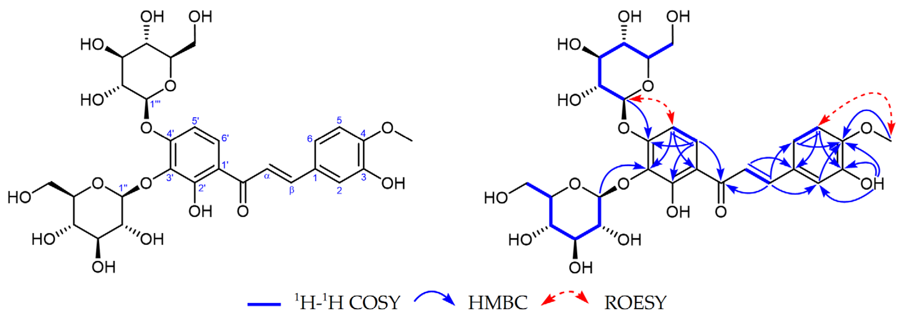
Figure 1. Key 1H–1H COSY, HMBC, and ROESY correlations of compound 1.
Molecules 29 03547 g001
Figure 2. Key 1H–1H COSY and HMBC correlations of compound 20.
Molecules 29 03547 g002
Figure 3. Chemical structures of compounds 129 isolated from B. pilosa.
Molecules 29 03547 g003
Figure 4. The inhibitory effect of flavonoids 3, 11, 12, 16, 13, 18, 21, and 23 on DNA Topo I. (A) Representative images of the Topo I inhibition assay; the tested concentration of the flavonoids is 400 μM. (B) The percentage inhibition of the compounds on Topo I activity at the concentration of 400 μM.
Molecules 29 03547 g004
Figure 5. Results of flavonoid (5, 6, and 7)-induced cell cycle arrests in DLD-1 cells. (A) Representative images of flow cytometry analysis of the DNA content. (B) Statistic analysis of cells in the G1, S, and G2/M phases after treatment with 5, 6, and 7. ** p < 0.01 and **** p < 0.0001 mean significant differences from the vehicle control. Data are expressed as mean ± SD values from three independent experiments.
Molecules 29 03547 g005
Figure 6. Effects of flavonoid 5 on cell cycle-relevant proteins in DLD-1 cells. (A) Representative immunoblots of the protein expression. (B) Histograms for the quantified results of protein levels, which were adjusted with corresponding α-tubulin/GAPDH protein levels and expressed as folds of the control. Experiments were independently repeated at least three times. * p < 0.05, ** p < 0.01 and *** p < 0.001 mean significant differences from the vehicle group.
Molecules 29 03547 g006
Table 1. 1H NMR and 13C NMR of compound 1 (DMSO-d6, δppm).
No.13C NMR (δ ppm)1H NMR (δ ppm)No.13C NMR (δ ppm)1H NMR (δ ppm)
C=O192.1 1″103.84.85 (1H, d, J = 7.6)
α119.37.77 (1H, d, J = 15.4)2″74.13.29 (1H, overlapped)
β144.97.70 (1H, d, J = 15.4)3″76.33.22 (1H, overlapped)
1127.5-4″69.73.13 (1H, overlapped)
2115.17.34 (1H, d, J = 1.9)5″77.23.10 (1H, overlapped)
3146.7-6″60.83.44 (1H, overlapped)
3.62 (1H, m)
4150.6-1‴100.74.98 (1H, d, J = 7.4)
5112.07.01 (1H, d, J = 8.4)2‴73.43.35 (1H, overlapped)
6122.67.32 (1H, dd, J = 8.4, 1.9)3‴76.03.30 (1H, overlapped)
1′116.8-4‴69.83.17 (1H, overlapped)
2′156.5-5‴77.43.40 (1H, overlapped)
3′133.6-6‴60.73.47 (1H, overlapped)
3.71 (1H, m)
4′155.6-4-OCH355.83.84 (3H, s)
5′106.96.84 (1H, d, J = 9.3)3-OH 9.25 (1H, s)
6′127.18.02 (1H, d, J = 9.3)2′-OH 12.90 (1H, br s)
Table 2. 1H NMR and 13C NMR of compound 20 (CD3OD, δ ppm).
No.13C NMR (δ ppm)1H NMR (δ ppm)No.13C NMR (δ ppm)1H NMR (δ ppm)
166.03.54 (1H, dd, J = 11.2, 5.8)
3.59 (1H, dd, J = 11.2, 4.9)
1′113.5-
271.73.77 (1H, dddd, J = 6.2, 6.0, 5.8, 4.9)2′, 6′135.17.28 (2H, d, J = 8.6)
325.22.50 (1H, dd, J = 17.3, 6.2)
2.60 (1H, dd, J = 17.3, 6.0)
3′, 5′116.66.72 (2H, d, J = 8.6)
480.8-4′160.0-
567.6-
673.2-
776.2
Table 3. The cytotoxicities against five cancer cell lines and Topo I inhibitory activities of compounds 129.
Comp.IC50 (μM)
CytotoxicityTopo I
A549HCT116MDA-MB-231HepG2DLD-1HL-7702
1>200>200>200>200>200>200393.50 ± 38.99
2>200>200>200>200>200>200328.98 ± 58.77
3135.56 ± 25.04177.96 ± 6.11166.71 ± 5.63>200117.49 ± 11.60>200>400
48.16 ± 4.044.52 ± 0.04>2003.05 ± 1.317.14 ± 0.3519.61 ± 0.62>400
52.16 ± 0.300.79 ± 0.1339.67 ± 4.5840.3 ± 11.140.60 ± 0.0514.36 ± 0.45145.57 ± 7.88
63.09 ± 0.280.59 ± 0.10>20034.59 ± 8.011.58 ± 0.0723.91 ± 5.73239.27 ± 31.35
70.86 ± 0.030.43 ± 0.0381.57 ± 7.4328.38 ± 13.050.85 ± 0.0211.44 ± 4.42224.38 ± 27.18
8177.84 ± 4.38>200>200>200186.79 ± 48.65>200189.84 ± 22.09
9135.31 ± 32.2987.68 ± 15.27>200>20050.50 ± 0.31125.54 ± 7.08>400
10>20059.29 ± 4.56>200>20063.00 ± 4.08114.75 ± 0.55>400
11>200>200>200>200>200>200>400
12>200>200>200>200>200>200>400
13>200139.10 ± 11.31>200>200178.68 ± 5.94>200>400
14>200>200>200>200>200>200>400
1593.92 ± 2.9231.70 ± 1.5494.91 ± 0.50174.80 ± 16.6469.17 ± 4.04>20089.91 ± 28.08
16>200>200>200>200>200>200>400
17>200>200>200>200>200>200>400
18>200>200>200>200>200>200>400
19>200>200>200>200>200>20047.50 ± 15.3
20194.01 ± 12.77173.81 ± 2.47>200>200>200>200301.32 ± 34.94
21186.68 ± 6.63>200>200>200>200>200>400
22124.84 ± 23.42117.63 ± 5.70146.00 ± 7.17178.53 ± 3.62141.56 ± 10.15160.56 ± 2.18178.03 ± 45.72
23129.78 ± 4.9370.91 ± 3.73139.89 ± 5.53167.79 ± 10.78122.54 ± 8.76198.81 ± 0.86>400
24>200>200>200>200>200>200218.27 ± 50.82
25173.81 ± 12.3162.38 ± 15.77>200>200187.21 ± 17.4>200>400
26187.84 ± 15.83>200>200>200>200>200>400
27187.87 ± 14.88112.23 ± 6.59164.29 ± 11.74>200144.41 ± 7.36>200>400
28190.39 ± 12.09>200>200>200>200>200>400
29>200>200>200>200>200>200>400
a CPT4.41 ± 1.1112.07 ± 5.7420.41 ± 3.701.71 ± 0.4716.96 ± 3.822.11 + 2.0511.11 ± 3.43
a the reference compounds of the positive control.
Disclaimer/Publisher’s Note: The statements, opinions and data contained in all publications are solely those of the individual author(s) and contributor(s) and not of MDPI and/or the editor(s). MDPI and/or the editor(s) disclaim responsibility for any injury to people or property resulting from any ideas, methods, instructions or products referred to in the content.