Review

Long Non-Coding RNA AGAP2-AS1: A Comprehensive Overview on Its Biological Functions and Clinical Significances in Human Cancers

School of Pharmaceutical Sciences, Zhejiang Chinese Medical University, Hangzhou 310053, China
*
Authors to whom correspondence should be addressed.
Molecules 2024, 29(15), 3461; https://doi.org/10.3390/molecules29153461
Submission received: 7 June 2024 / Revised: 18 July 2024 / Accepted: 19 July 2024 / Published: 24 July 2024

Abstract

:
Long non-coding RNAs (lncRNAs) are well known for their oncogenic or anti-oncogenic roles in cancer development. AGAP2-AS1, a new lncRNA, has been extensively demonstrated as an oncogenic lncRNA in various cancers. Abundant experimental results have proved the aberrantly high level of AGAP2-AS1 in a great number of malignancies, such as glioma, colorectal, lung, ovarian, prostate, breast, cholangiocarcinoma, bladder, colon and pancreatic cancers. Importantly, the biological functions of AGAP2-AS1 have been extensively demonstrated. It could promote the proliferation, migration and invasion of cancer cells. Simultaneously, the clinical significances of AGAP2-AS1 were also illustrated. AGAP2-AS1 was exceptionally overexpressed in various cancer tissues. Clinical studies disclosed that the abnormal overexpression of AGAP2-AS1 was tightly connected with overall survival (OS), lymph nodes metastasis (LNM), clinical stage, tumor infiltration, high histological grade (HG), serous subtype and PFI times. However, to date, the biological actions and clinical significances of AGAP2-AS1 have not been systematically reviewed in human cancers. In the present review, the authors overviewed the biological actions, potential mechanisms and clinical features of AGAP2-AS1 according to the previous studies. In summary, AGAP2-AS1, as a vital oncogenic gene, is a promising biomarker and potential target for carcinoma prognosis and therapy.

1. Introduction

Nowadays, carcinoma is a leading cause of fatality in a great number of countries worldwide. The cancer occurrences and mortalities were greatly increasing year by year [1,2]. In 2020, there were around 19.3 million novel cases and 10.0 million deaths in the world [1]. Notably, breast, lung, colorectal, prostate and stomach cancers were the most commonly diagnosed carcinomas. Although great efforts have been made to investigate the possible cancerogenic mechanisms and develop novel anticancer agents, the number of deaths are still increasing.
LncRNAs are specific RNA molecules longer than 200 nucleotides, which do not encode proteins [3]. Theoretically, intergenic region, 5′/3′-untranslational regions, intron and exon could transcribe lncRNAs. Then, the intricate second structures were formed, which further interacted with RNA, DNA and proteins [4]. The mechanisms of lncRNAs regulating gene expression were quite complicated, including preventing the degradation of mRNA, modulating transcription factors, binding to promoters to regulate targeted gene expression and regulating macromolecule–protein interactions [5,6]. Momentously, the biological activities of lncRNAs were extensively verified, such as being involved in cell differentiation, cell cycle regulation and epigenetic control. Functionally, abundant evidence has confirmed that lncRNAs could display both oncogenic or anti-oncogenic roles in cancers. They could promote tumorigenesis at different stages via interacting with cancer-associated signaling pathways [7,8]. Notably, some lncRNAs were even verified as hallmarks in human cancers [9,10,11].
AGAP2-AS1, a novel lncRNA (1567 nt in length), is transcribed by a gene situated in 12q14.1 (Figure 1A) [12]. The secondary and three-dimensional structures of AGAP2-AS1 are illustrated in Figure 1B,C. Motif analysis indicated that 10 different motifs were identified in AGAP2-AS1, which were sequentially named as motifs 1–10 (Figure 1D). As expected, the motifs were conserved in composition. Moreover, motif 1, motif 4, motif 7 and motif 8 were found to be the core motifs. Notably, all core motifs appeared twice. Currently, studies regarding the biological functions of AGAP2-AS1 are mainly focusing on cancer, psoriasis pathogenesis, pre-eclampsia and systemic sclerosis [12,13,14,15]. Interestingly, most of the studies illustrated the biological roles of AGAP2-AS1 in cancers. As an antisense lncRNA transcribed from AGAP2, AGAP2-AS1 was widely involved in the progression of cancers [12,16]. According to the reports, AGAP2-AS1 was demonstrated to be abnormally overexpressed in cancers [12]. The AGAP2-AS1 level in tumors was much higher than that in normal tissues (Figure 1E). It was conventionally correlated with poor OS and DFS [17]. Importantly, the oncogenic functions of AGAP2-AS1 were also extensively documented, including promotion of proliferation, migration, invasion and tumorigenesis [18,19,20]. However, to date, there have been no systematical and comprehensive reviews regarding AGAP2-AS1 in human cancers. Therefore, the aim of the present overview was to summarize the biological roles and potential molecular mechanisms of AGAP2-AS1 according to the previous studies. Meanwhile, the clinical significances of AGAP2-AS1 were also illustrated. In summary, as a vital oncogene, AGAP2-AS1 is a promising biomarker and potential target for cancer therapy.

2. Biological Functions

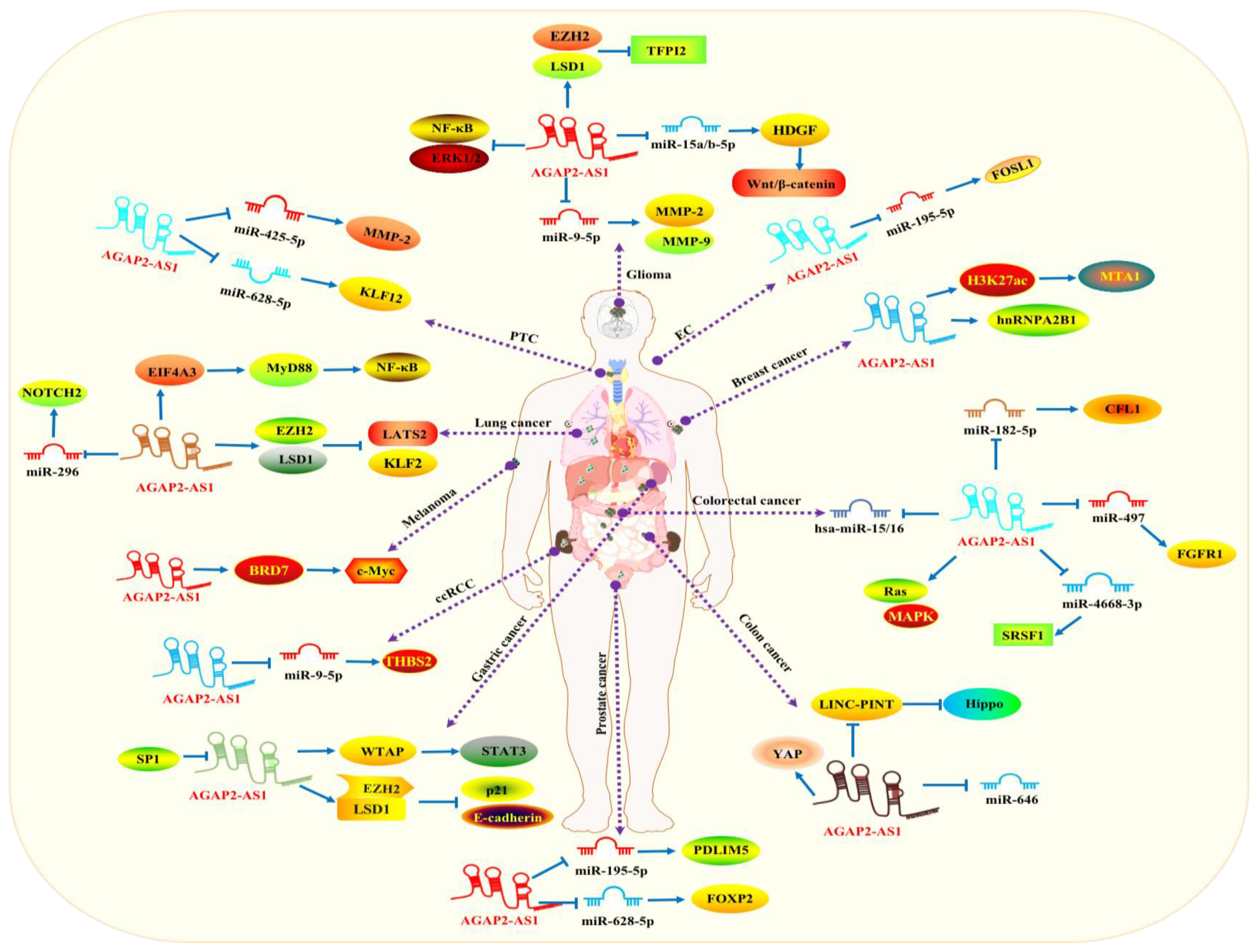
A large quantity of studies have testified to the aberrant expression of AGAP2-AS1 in human malignancies, including glioma, colorectal, lung, ovarian, prostate, breast, cholangiocarcinoma, bladder, colon and pancreatic cancers. Results indicated that AGAP2-AS1 could promote cancer progression mainly via facilitating cancer proliferation, migration and invasion. Meanwhile, the related molecular mechanisms were also documented (Figure 2 and Table 1).

2.1. Glioma

Glioma, the most universally arising neuroepithelial cancer, principally occurs in the brain and arises in the glia [21,22]. Importantly, the biological actions of AGAP2-AS1 in neuroglioma were demonstrated in some studies [23,24]. Experimental results illustrated that the aberrant expression of AGAP2-AS1 was tightly linked with cancer invasion, multiplication and migration. Wang et al. verified that high expression of AGAP2-AS1 was notably detected in LN229 and U87MG cells. Inhibition of AGAP2-AS1 remarkably decreased the proliferation, migration and invasion of cancer cells. Meanwhile, Annexin-V staining showed that the number of apoptotic cells were obviously increasing [23]. Similar results were also demonstrated in some other cell lines, such as U87, U251, A172, LN229 and SHG44 cells [19,25,26]. Importantly, the potential mechanisms were also reported. Luo et al. illustrated that AGAP2-AS1 interacted with EZH2 and LSD1, and recruited them to the TFPI2 promoter region to restrain its transcription, thereby exerting oncogenic functions [25]. Moreover, Luo et al. constructed a ceRNA network of AGAP2-AS1-miR-9-5p-MMP-2/MMP-9. Enrichment analysis predicted that AGAP2-AS1 could facilitate the migration, proliferation and differentiation of glioma cells via sponging miR-9-5p [27]. In U87, U251 and LN229 cells, AGAP2-AS1 could advance cancer proliferation via sponging miR-15a/b-5p to increase the HDGF level [26]. Furthermore, suppression of AGAP2-AS1 obviously restrained the progression of cancer cells via downregulating the levels of NF-κB and Erk1/2 [28]. The above results indicated that AGAP2-AS1 promoted the proliferation, migration and invasion of glioma cells mainly through regulating miR-15a/b-5p/HDGF/Wnt/β-catenin, EZH2/LSD1/TFPI2, miR-9-5p-MMP-2/MMP-9 and NF-κB/Erk1/2 pathways.
Table 1. The biological functions and related molecular mechanisms of AGAP2-AS1 in human carcinomas.
Cell LinesExpression StatusRoleFunctionRegulatory MechanismRef.
Glioma
U87 and U251 cellsUpregulationOncogene↑ Proliferation
↑ Metastasis
↑ Invasion
↓ Apoptosis
/[19]
LN229 and U87MG cellsUpregulationOncogene↑ Proliferation
↑ Metastasis
↑ Invasion
↓ Apoptosis
/[23]
U87, U251 and LN229 cell linesUpregulationOncogene↑ Proliferation
↓ Apoptosis
miR-15a/b-5p/HDGF/Wnt/β-catenin axis[26]
U87/MG and U251/MG cells UpregulationOncogene↑ Proliferation
↑ Invasion
↓ Apoptosis
↑ Tumor growth
EZH2/LSD1/TFPI2[25]
/UpregulationOncogene↑ Proliferation
↑ Metastasis
↑ Invasion
miR-9-5p/MMP-2/MMP-9 axis[27]
T98G, U251 and LN229 cellsUpregulationOncogene↑ Proliferation
↑ Migration
↑ Invasion
NF-κB/Erk1/2 [28]
LATS2 and KLF2 cellsUpregulationOncogene↑ Proliferation
↑ Metastasis
↑ Drug resistance
↓ Apoptosis
NOTCH, ERBB, RIG, NOD and JAK/STATA pathways[29]
LC
A549, ltp-2, SPCA1, H157 and NIH-H358 cell linesUpregulationOncogene↑ Proliferation
↑ Migration
↑ Drug resistance
miR-296/NOTCH2 [30]
MRC-5 cellsUpregulationOncogene↑ Proliferation
↑ Metastasis
EIF4A3/MyD88/NF-κB pathway[31]
H1299 and H1975 cellsUpregulationOncogene↑ Proliferation
↑ Invasion
↑ Migration
↓ Apoptosis
LATS2/KLF2/EZH2/ LSD1[32]
CRC
LoVo and SW480 cellsUpregulationOncogene↑ Growth
↑ Migration
↑ Invasion
↑ EMT
E2F4/miR-182-5p/CFL1 axis[20]
DLD-1 and RKO cellsUpregulationOncogene↑ Proliferation
↓ Apoptosis
↑ Migration
↑ Invasion
miR-497/FGFR1 axis[33]
SW480 and HT29 cellsUpregulationOncogene↑ Proliferation
↑ Migration
hsa-miR-15/16 family[34]
DLD-1 and HT29 cellsUpregulationOncogene↑ ProliferationRas/MAPK pathway[35]
SW620 and HT-29 cellsUpregulationOncogene↑ Proliferation
↑ Migration
↑ Invasion
↑ EMT
miR-4668-3p/SRSF1 axis [36]
CLC
RKO and HCT116 cellsUpregulationOncogene↑ Proliferation
↑ Invasion
↑ Migration
LINC-PINT/Hippo signaling[37]
SW480 and HCT-116 cellsUpregulationOncogene↑ Proliferation
↓ Apoptosis
/[38]
HCT116 cellsUpregulationOncogene↑ Proliferation
↓ Apoptosis
miR-646[39]
SW480 and HCT-116 cellsUpregulationOncogene↑ Proliferation
↑ Migration
↓ Apoptosis
YAP pathway[40]
OC
OVCAR3 and A2780 cellsUpregulationOncogene↑ ProliferationMEG3[15]
SKOV3-ip, OVCAR3, HO8910, HEY and ES2 cell linesDownregulationAnti-oncogene↓ Proliferation
↓ Migration
↓ Invasion
KRAS/FGFR4/CTSK/EMT[16]
PCa
VCaP, 22Rv1, CRL-1740, CRL-2422 and PC3M cell linesUpregulationOncogene ↑ Proliferation
↑ Migration
↑ Invasion
↑ Tumor growth
miR-195-5p/PDLIM5 axis[41]
PCa cellsUpregulationOncogene↑ Proliferation
↑ Migration
↑ Invasion
↑ EMT
↓ Apoptosis
miR-628-5p/FOXP2/WNT axis[42]
BC
MCF-7, BT-474, SK-BR-3 and MDA-MB-231 cell linesUpregulationOncogene↑ Proliferation
↓ Apoptosis
HuR/H3K27ac/MTA1 [43]
SKBR-3 and BT474 cellsUpregulationOncogene↑ Cell viability
↑ Trastuzumab resistance
hnRNPA2B1[12]
CHOL
RBE and HuCCT1 cellsUpregulationOncogene↑ Proliferation
↓ Apoptosis
EZH2/CDKN1A [44]
RBE and HuCCT1 cellsUpregulationOncogene↑ Proliferation
↑ Migration
↑ Invasion
/[45]
BLCA
UM-UC-3 and T24 cellsUpregulationOncogene↑ Proliferation
↑ Migration
/[46]
5637 and T24 cellsUpregulationOncogene↑ Proliferation
↑ Migration
↑ Invasion
↑ Angiogenesis
↑ Tumor growth
IGF2BP2/LRG1[47]
RCC
786-O and ACHN cellsUpregulationOncogene↑ M2 polarization
↑ Malignant behavior
↑ Proliferation
↑ Tumor growth
miR-9-5p/THBS2/PI3K-AKT axis[48]
PTC
K1 and TPC1 cellsUpregulationOncogene↑ Migration
↑ Invasion
miR-425-5p/MMP-2 axis[49]
BCPAP, KTC-1, NIM1 and TPC1 cell linesUpregulationOncogene↑ Proliferation
↑ Migration
↑ Invasion
miR-628-5p/KLF12 axis[50]
GC
AGS and HGC-27 cellsUpregulationOncogene↑ Proliferation
↑ Migration
WTAP/METTL3/METTL14/STAT3 axis[51]
BGC-823 and AGS cellsUpregulationOncogene↑ Proliferation
↑ Migration
↑ Invasion
↑ Tumor growth
LSD1/EZH2/CDKN1A/E-cadherin transcription[52]
EC
KYSE70, KYSE-510 and EC9706 cell linesUpregulationOncogene↑ Proliferation
↓ Apoptosis
↑ Migration
↑ Invasion
↑ Tumorigenesis
miR-195-5p/FOSL1 axis[53]
Melanoma
A375 and A875 cellsUpregulationOncogene↑ Proliferation
↑ Colony formation
↑ Migration
BRD7/c-Myc pathway[54]
LSCC
AMC-HN-8 and Tu-177 cellsUpregulationOncogene↑ Proliferation
↑ Invasion
miR-193a-3p/LOXL4 axis[55]
PC
AsPC-1 and BxPC-3 cells UpregulationOncogene↑ Invasion
↑ Proliferation
↑ Migration
ANKRD1/ANGPTL4[56]
“↑”: Promotion; “↓”: Suppression.

2.2. Colorectal Carcinoma (CRC)

Colorectal carcinoma is widely diagnosed in the digestive system [57]. Functionally, a large number of experiments have proved the biological roles of AGAP2-AS1 in colorectal carcinoma, including promoting cell growth, migration, invasion and EMT process. Wang et al. circumstantiated that AGAP2-AS1 promoted colorectal cancer cell growth through modulating the Ras/MAPK pathway. In DLD-1 and HT29 cells, downregulation of AGAP2-AS1 resulted in marked cancer cell growth suppression, cell cycle arrest (at G0/G1 phase) and protein level reduction (p-MEK, p-Raf-1, p-Erk and Ras) [35]. In LoVo and SW480 cells, AGAP2-AS1 could be transcriptionally activated by E2F4. AGAP2-AS1 suppression significantly restrained cell growth, migration, invasion and EMT process. Mechanistic studies revealed that AGAP2-AS1 upregulated CFL1 through competitively combining with miR-182-5p. Meanwhile, restoration of CFL1 counteracted the depleted effects of AGAP2-AS1 on cancer progression [20]. Similar results were also observed in SW620 and HT-29 cells. Suppression of AGAP2-AS1 remarkably restrained cancer progression. Mechanistically, AGAP2-AS1 sponged miR-4668-3p to release SRSF1. Downregulation of miR-4668-3p promoted the malignant processes in AGAP2-AS1-knockdown CRC cells, whereas those effects were cancelled out by suppression of SRSF1 [36]. In DLD-1 and RKO cells, Hong et al. demonstrated that AGAP2-AS1 advanced CRC cancer growth and metastasis, restrained apoptosis and promoted chemoresistance to gemcitabine. The results from mechanistic investigation revealed that AGAP2-AS1 modulated FGFR1 expression through sponging miR-497 [33]. Furthermore, Ghasemi et al. suggested that AGAP2-AS1 facilitated CRC progression via sponging the family of hsa-miR-15/16 and upregulating their targets [34]. The above data suggested that AGAP2-AS1 promoted cell growth, migration, invasion and EMT process of CRC via regulating E2F4/miR-182-5p/CFL1, miR-497/FGFR1, hsa-miR-15/16, Ras/MAPK and miR-4668-3p/SRSF1 pathways.

2.3. Lung Carcinoma (LC)

The incidence of LC is over 1.8 million every year, and the death rate is quite high [1,58]. Numerous experiments have confirmed the biological roles of AGAP2-AS1 in LC, including promotion of cancer growth, metastasis and drug resistance [30,31,32]. In H1299 and H1975 cells, silencing of AGAP2-AS1 significantly restrained the differentiation and metastasis of cancer cells. Meanwhile, the number of apoptotic cells were obviously increasing. Importantly, the role of AGAP2-AS1 on promoting tumorigenesis was also verified in in vivo experiments. Mechanistic studies demonstrated that AGAP2-AS1 bound with EZH2 and LSD1, and recruited them to the promoter regions of KLF2 and LATS2, thereby suppressing the transcription [32]. On the contrary, overexpression of AGAP2-AS1 prominently activated the differentiation and metastasis of MRC-5 cells. EIF4A3 promoted its stability via binding with AGAP2-AS1, which could positively modulate MyD88/NF-κB signaling. Consistently, similar results were also observed in in vivo models [31]. Moreover, the effects of AGAP2-AS1 on the progression and radio resistance of lung carcinoma were examined. In H157R24-1 and A549R26-1 cells, molecular studies revealed that miR-296 was negatively regulated by AGAP2-AS1, while NOTCH2 was a downstream target of miR-296. Mechanistically, the exosomal AGAP2-AS1 derived by M2 macrophage could enhance the radiotherapy immunity via suppressing miR-296 and activating NOTCH2 [30]. These studies revealed that AGAP2-AS1 affected the malignant behaviors (promotion of cancer growth, metastasis and drug resistance) of lung cancer via modulating the miR-296/NOTCH2, EIF4A3/MyD88/NF-κB and LATS2/KLF2/EZH2/LSD1 pathways.

2.4. Ovarian Carcinoma (OC)

OC, a lethal malignancy in clinic, is commonly occurring in women [59]. The biological functions of AGAP2-AS1 in OC were investigated. In OVCAR3 and A2780 cell lines, overexpression of AGAP2-AS1 resulted in significant suppression of MEG3 (p < 0.05). Correspondingly, aberrant expression of MEG3 mitigated the function of AGAP2-AS1 on cell growth. Notably, overexpressed AGAP2-AS1 did not show significant effects on cell invasion and migration [15]. However, Zheng et al. verified that AGAP2-AS1 could suppress cell differentiation, migration and invasion in EOC cells. The molecular mechanisms were related to suppression of EMT and downregulation of CTSK, FGFR4 and KRAS [16]. Therefore, the above studies suggested that AGAP2-AS1 exhibited an important function on cancer proliferation in OC via suppressing MEG3. However, the exact biological functions of AGAP2-AS1 in EOC cells still needed to be further examined.

2.5. Prostate Cancer (PCa)

PCa, mainly observed in males worldwide, is one of the leading causes of cancer death [60]. An increasing number of results have proved the momentous characters of lncRNAs in the tumorigenesis of PCa. Notably, AGAP2-AS1 was verified as an oncogene in PCa [41,42]. AGAP2-AS1 was abnormally overexpressed in PCa. Silencing of AGAP2-AS1 significantly restrained cancer proliferation, migration and invasion. Mechanistically, AGAP2-AS1 bound with miR-195-5p, which further downregulated the expression of PDLIM5 to obstruct cancer progression. Accordantly, the above mechanisms were also proved in in vivo models [41]. Moreover, Zhao et al. also demonstrated the biological actions of AGAP2-AS1 in PCa, including restraining apoptotic cell death, promoting proliferation, migration, invasion and EMT process. AGAP2-AS1 sponged miR-628-5p, which could negatively regulate FOXP2 to affect the cancer growth. Upregulation of FOXP2 reversed AGAP2-AS1 knockdown-induced suppression on cancer growth [42]. Furthermore, mechanistic studies also enucleated that AGAP2-AS1 could activate the WNT pathway. Their results suggested that the feedback loop of AGAP2-AS1/miR-628-5p/FOXP2 promoted carcinoma growth through activating the WNT signaling [42]. Collectively, AGAP2-AS1 promoted the proliferation, migration and invasion of PCa through regulating the miR-195-5p/PDLIM5 and miR-628-5p/FOXP2 axes.

2.6. Breast Carcinoma (BC)

BC is widely diagnosed in females. It is known as the main reason of carcinomatous fatality [61,62]. The physiological effects of AGAP2-AS1 in BC were already documented in many studies. Experiments verified that AGAP2-AS1 was significantly overexpressed in various BC cell lines and clinical samples [43]. Silencing of AGAP2-AS1 remarkably restrained cell proliferation and increased apoptotic cell death in MCF-7 (ER+) cells. Mechanistical results revealed that AGAP2-AS1 bound with HuR to upregulate H3K27ac, thereby elevating the activity of MTA1 promoter and upregulating MTA1 expression. High expression of H3K27ac partially offset the function of si-AGAP2-AS1-mediated apoptosis induction. This mechanism was also verified in in vivo models [43]. In addition, the aberrant expression of AGAP2-AS1 was also detected in drug-resistant cells. In trastuzumab-resistant cells (HER2-positive BT474 and SKBR-3 cells), AGAP2-AS1 knockdown promoted the inhibitory effects of trastuzumab on cell growth. Experimental data indicated that the excreted AGAP2-AS1 was assembled into exosomes in an hnRNPA2B1-reliant way. Co-treatment exosomes with trastuzumab remarkably decreased the inhibitory effects of trastuzumab on cell growth [12]. Hence, AGAP2-AS1 could promote the progression (proliferation and drug resistance) of BC via regulation of HuR/H3K27ac/MTA1 and hnRNPA2B1 pathways.

2.7. Cholangiocarcinoma (CHOL)

The biological characters of AGAP2-AS1 in CHOL were also assessed. Ji et al. demonstrated that AGAP2-AS1 could promote the CHOL cell growth. AGAP2-AS1 influenced CDKN1A transcription by interacting with EZH2 in cancer cells. Their results illustrated that AGAP2-AS1 suppression significantly restrained the growth of HUCCT1 and RBE cells. Importantly, SP1 could positively regulate AGAP2-AS1 expression. Suppression of AGAP2-AS1 led to a remarkable increase in apoptotic cells [44]. Moreover, in RBE and HuCCT-1 cells, experiments illustrated that inhibition of GOLGA7B significantly facilitated cell migration and invasion, while knockdown of AGAP2-AS1 exhibited the reverse effects [45]. The above results suggested that AGAP2-AS1 facilitated cell growth, migration and invasion by modulating CDKN1A and GOLGA7B pathways.

2.8. Bladder Cancer (BLCA)

The characters of AGAP2-AS1 in BLCA were also reported in some studies. In UM-UC-3 and T24 cells, Xu et al. demonstrated that silencing of AGAP2-AS1 could obviously restrain cell proliferation and migration [46]. In contrast, overexpression of AGAP2-AS1 significantly facilitated cancer progression in in vitro and in vivo model systems, including promotion of proliferation, invasion, migration and angiogenesis. Suppression of AGAP2-AS1 displayed reverse effects. Mechanistic studies demonstrated that AGAP2-AS1 could directly bind to IGF2BP2 to promote LRG1 stability. Meanwhile, overexpression of LRG1 could reverse the AGAP2-AS1-knockdown mediated effects in cancer cells [47]. These data proved that AGAP2-AS1 advanced cancer proliferation, invasion, migration and angiogenesis through regulating the IGF2BP2/LRG1 axis.

2.9. Colon Cancer (CLC)

Colon cancer is commonly diagnosed in the digestive system. The incidence is second only to lung and liver cancer [1,63]. Ji et al. documented that AGAP2-AS1 and LINC-PINT could form a negative feedback loop to promote CLC progression. Their data indicated that AGAP2-AS1 suppressed LINC-PINT in RKO and HCT116 cells. Meanwhile, suppression of LINC-PINT promoted the expression of AGAP2-AS1. Experiments also proved that AGAP2-AS1 could significantly promote cancer progression via regulating Hippo signaling [37]. In SW480 and HCT-116 cells, highly expressed AGAP2-AS1 was also verified [38,39,40]. Inhibition of AGAP2-AS1 led to a reduction in cell proliferation and migration [38,39,40]. Notably, AGAP2-AS1 knockdown triggered the phosphorylation of YAP and led to a marked decrease in MMP-9 and MMP-2. Further results proved that AGAP2-AS1 upregulated MMPs via activation of the YAP pathway (p < 0.05) [40]. However, Liu et al. indicated that AGAP2-AS1 displayed the biological functions via targeting miR-646 in HCT-116 cells [39]. In summary, AGAP2-AS1 promoted cancer proliferation, migration and invasion of CLC cells via regulating LINC-PINT/Hippo, miR-646 and YAP pathways.

2.10. Pancreatic Cancer (PC)

The characters of AGAP2-AS1 in PC were also assessed. In AsPC-1 and BxPC-3 cells, AGAP2-AS1 significantly affected cancer progression in vitro and in vivo. It was documented that RREB1 could positively regulate the AGAP2-AS1 transcription. Studies indicated that AGAP2-AS1 epigenetically stifled ANKRD1 and ANGPTL4 through recruiting EZH2, thereby facilitating cancer proliferation and metastasis [56]. These data demonstrated that AGAP2-AS1 facilitated PC proliferation and metastasis through the RREB1/ANKRD1/ANGPTL4 pathway.

2.11. Renal Cell Carcinoma (RCC)

AGAP2-AS1 was demonstrated to be highly overexpressed in RCC. In 786-O and ACHN cells, experiments verified that AGAP2-AS1 could be stabilized by IGF2BP3 via m6A modification. Overexpression of AGAP2-AS1 promoted pernicious behaviors of RCC cells and resulted in M2 polarization. Functionally, AGAP2-AS1 directly sponged miR-9-5p to upregulate the expression of THBS2. On the other side, THBS2 could subsequently improve macrophage polarization via regulating the PI3K/AKT pathway [48]. Taken together, AGAP2-AS1 promoted the proliferation and tumor growth of RCC through regulation of the miR-9-5p/THBS2/PI3K-AKT axis.

2.12. Laryngeal Squamous Cell Carcinoma (LSCC)

LSCC is a highly malignant and invasive carcinoma. Experiments demonstrated that AGAP2-AS1 was markedly overexpressed in Tu-177 and AMC-HN-8 cell lines. Suppression of AGAP2-AS1 obviously restrained cell growth and invasion. Mechanistically, AGAP2-AS1 could sponge miR-193a-3p to regulate its expression. In addition, LOXL4 was demonstrated as a direct target of miR-193a-3p. In brief, those results illustrated that AGAP2-AS1 regulated the miR-193a-3p/LOXL4 axis to facilitate cancer growth and invasion [55].

2.13. Melanoma

The biological characters of AGAP2-AS1 in melanoma were also reported. Results indicated that AGAP2-AS1 was overexpressed in SKCM tissues. Its expression was associated with poor prognosis. Silencing of AGAP2-AS1 in A875 and A375 cells evidently suppressed cell growth and migration in various model systems. Experimental data demonstrated that AGAP2-AS1 could interact with BRD7. Silencing of AGAP2-AS1 alleviated the interaction of BRD7 and c-Myc, which could further decrease the expression of c-Myc. Notably, c-Myc overexpression reversed the biological effects in AGAP2-AS1- and BRD7-deficient cells [54]. These results demonstrated that AGAP2-AS1 is involved in oncogenesis (cell growth and migration) through regulating the BRD7/c-Myc pathway.

2.14. Papillary Thyroid Cancer (PTC)

AGAP2-AS1 also showed significant functions on promotion of cell growth, migration and invasion in PTC cells [49,50]. Restraint of AGAP2-AS1 prominently alleviated the migration and invasion of K1 and TPC1 cells. Mechanistic studies revealed that AGAP2-AS1 could competitively bind to miR-425-5p to upregulate the level of MMP-2, thereby promoting cell migration and invasion. Moreover, clinical studies disclosed that miR-424-5p was evidently downregulated in cancerous samples. It was negatively correlated with the expression of AGAP2-AS1 [49]. In addition, Xu et al. verified that AGAP2-AS1 exerted the biological effects via regulation of the miR-628-5p/KLF12 axis. Their results proved that the level of AGAP2-AS1 was negatively correlated with miR-628-5p. Mechanistically, AGAP2-AS1 sponged miR-628-5p to directly regulate KLF12 expression. Importantly, silencing of KLF12 increased the inhibitory functions of miR-628-5p on cancer growth and metastasis [50]. Collectively, AGAP2-AS1 promoted the proliferation, migration and invasion of PTC via regulating miR-425-5p/MMP-2 and miR-628-5p/KLF12 axes.

2.15. Esophageal Cancer (EC)

The effects of AGAP2-AS1 on EC were also examined. Experimental results proved that AGAP2-AS1 suppression or miR-195-5p overexpression could strikingly arrest cell cycle, promote apoptotic cell death, and alleviate proliferation, migration and invasion. Functionally, AGAP2-AS1 could bind to miR-195-5p to regulate FOSL1. Conversely, AGAP2-AS1 knockdown preeminently suppressed the cancer progression via upregulation of miR-195-5p and downregulation of FOSL1 [53]. Moreover, it was well known that the antisense lncRNAs were usually acting as regulators of their sense counterparts. Consistently, Zheng et al. (2020) proved that the expression of AGAP2-AS1 in EOC tissues was negatively correlated with that of AGAP2 [61].

2.16. Gastric Cancer (GC)

The biological roles of AGAP2-AS1 in GC were verified in various studies [51,52]. Functional studies revealed that silencing of AGAP2-AS1 led to suppression of cell proliferation and migration [51,52]. Data from mechanistic experiments uncovered that AGAP2-AS1 bound with WTAP to facilitate the WTAP/METTL3/METTL14 complex. Meanwhile, STAT3 mRNA was stabilized by AGAP2-AS1 in an m6A-reliant way, thereby activating the IL-6/STAT3 pathway. Notably, AGAP2-AS1/WTAP/STAT3 pathway activation memorably facilitated cell proliferation and migration [51]. Moreover, Qi et al. demonstrated that SP1 could upregulate AGAP2-AS1 expression in AGS and BGC-823 cells. Functionally, AGAP2-AS1 interacted with LSD1 and EZH2 to inhibit the transcription of p21 and E-cadherin, resultingly exerting oncogenic functions [52]. In summary, AGAP2-AS1 affected the proliferation and migration of GC by regulating the WTAP/STAT3/METTL3/METTL14 and LSD1/EZH2/p21/E-cadherin pathways.

3. Clinical Significances

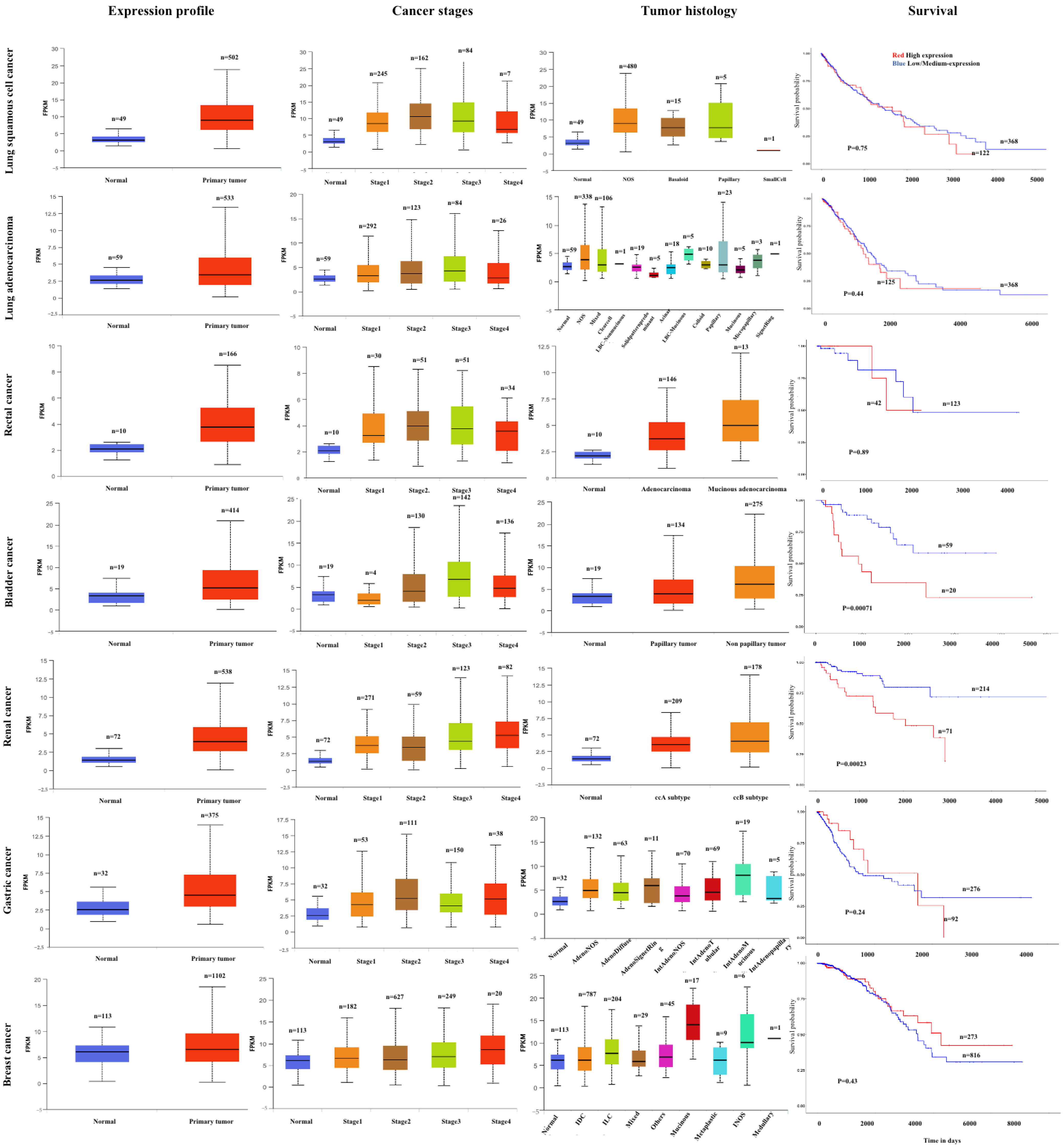
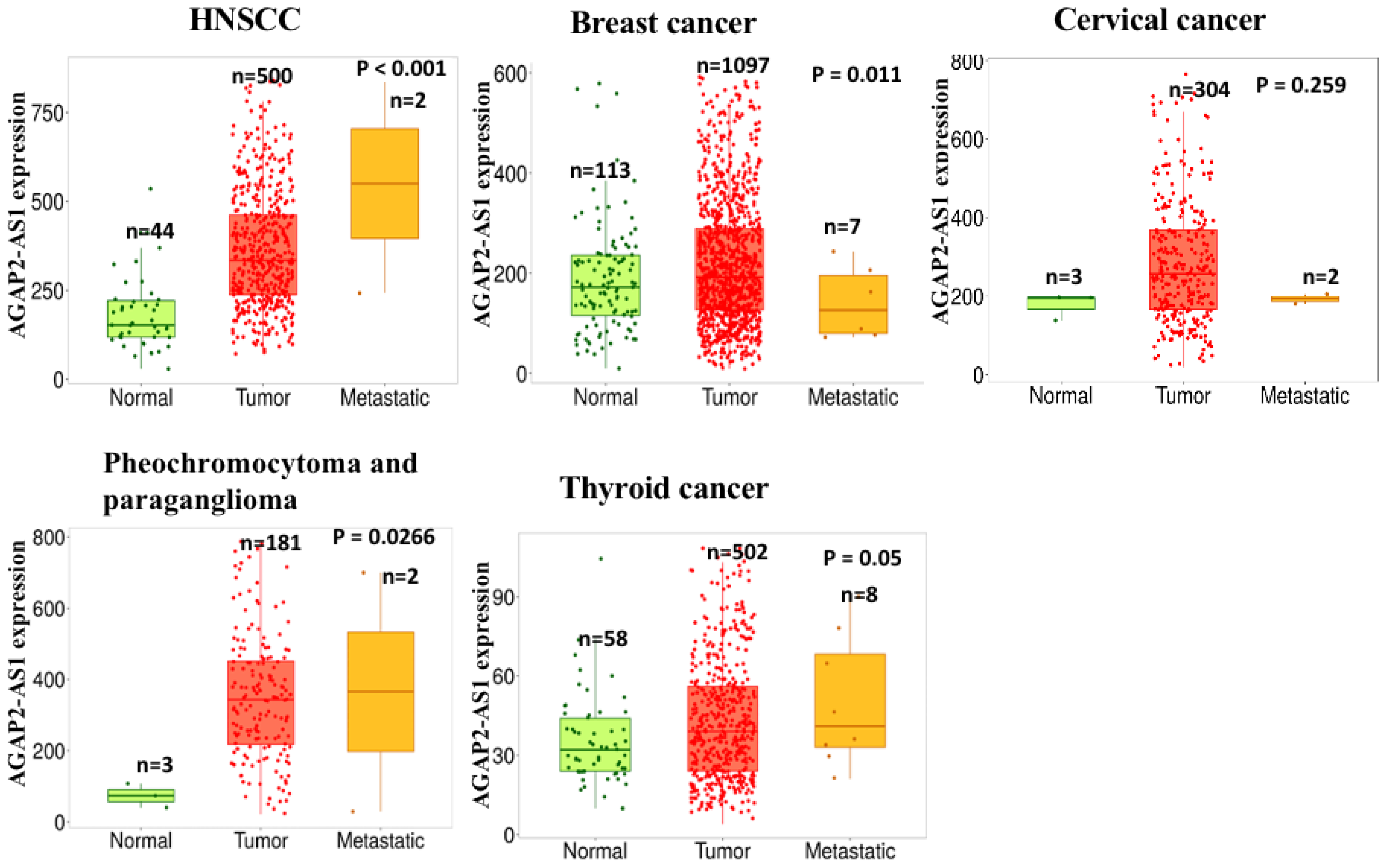
The expression level of AGAP2-AS1 in clinical carcinomatous and non-carcinomatous tissues was analyzed using the UALCAN database (https://ualcan.path.uab.edu/, accessed on 18 May 2024) [64,65]. As shown in Figure 1, AGAP2-AS1 was significantly overexpressed in a large number of carcinomas, including glioma, LC, CLC, OC, PTC, CHOL, BLCA, CRC, PC and ccRCC. Experimental analysis indicated that the expression of AGAP2-AS1 in some tumors was closely correlated with TS, histology, survival and cancer metastasis (Figure 3). Meanwhile, the data from the TNM plot (https://tnmplot.com/analysis/, accessed on 18 May 2024) also proved the high expression of AGAP2-AS1 in tumor and metastatic tissues (Figure 4) [66]. The clinical significances of AGAP2-AS1 in human cancers are indicated in Table 2.

3.1. Gliomas

The clinical significances of AGAP2-AS1 in gliomas were widely documented by dataset analysis. Wang et al. divided anaplastic gliomas into grade II- and grade IV-like groups using datasets. Multivariate analysis indicated that AGAP2-AS1 was evidently raised with TG [23]. Correspondingly, similar results were demonstrated by Wang et al. [67]. Luo et al. constructed a ceRNA network of AGAP2-AS1-miR-9-5p-MMP-2/MMP-9 and identified that AGAP2-AS1 was a promising therapeutic target for GBM [27]. In addition, AGAP2-AS1 was also identified as one of the immune-related lncRNAs in patients with gliomas [29,67]. The signature of AGAP2-AS1 exhibited prognostic values for anaplastic gliomas [67]. Interestingly, AGAP2-AS1 was not only an independent prognostic factor but also could effectively predict the survival rate of patients [68]. However, Yu et al. verified that GBM subtype-A was featured by low expression of AGAP2-AS1 [29].
On the other hand, clinical samples were also applied to assess the clinical relevance of AGAP2-AS1. Abnormal overexpression of AGAP2-AS1 was observed in several studies [19,25,26]. Tian et al. documented that the expression of AGAP2-AS1 was significantly overexpressed in 136 glioma tissue specimens. Kaplan–Meier analysis revealed that the AGAP2-AS1 expression was negatively associated with OS in GBM patients [19]. In a cohort of 91 pairs of glioma tissues, the level of AGAP2-AS1 in carcinomatous samples was higher than that in non-carcinomatous samples. Noticeably, the aberrant expression of AGAP2-AS1 was highly correlated with advanced TG and low OS rate [26]. In addition, Luo et al. also obtained the similar data from 58 paired tissues. GEPIA data analysis disclosed that AGAP2-AS1 overexpression in patients indicated a shorter OS [25]. Collectively, the above studies demonstrated that the expression of AGAP2-AS1 in clinical glioma tissues was highly associated with advanced TG and OS.

3.2. Lung Carcinoma

The abnormal level of AGAP2-AS1 in lung carcinoma was widely verified. In a cohort of 121 pairs of lung carcinoma samples (84 radioresistant and 37 radioresistant cases), AGAP2-AS1 was observably upregulated in carcinomatous samples. Compared with the radiosensitive groups, radioresistant patients showed a much higher level of AGAP2-AS1. Importantly, the AGAP2-AS1 level was closely related to the TNM stage and LNM. However, other factors, such as tumor differentiation, age and pathological pattern, did not exhibit any correlation with the expression of AGAP2-AS1. Moreover, Kaplan–Meier analysis indicated that the high level of AGAP2-AS1 in patients was tightly linked to lower OS and DFS [30,32,69]. Meanwhile, the abnormal expression of AGAP2-AS1 in NSCLC patients was also extensively demonstrated [32,70,71,72,73,74,75]. Experiments revealed that the level of AGAP2-AS1 was highly correlated with TNM stage, LNM and poor prognostic outcomes [32,72,73,74,75]. In another study with 84 patients, the expression of serum AGAP2-AS1 in NSCLC patients was associated with differentiation degree [75]. In a cohort of 80 pairs of NSCLC tissues, the AGAP2-AS1 expression increased 41.5-fold in 72.5% of cancerous tissues. Importantly, increased AGAP2-AS1 level was also correlated with tumor size in NSCLC patients [32]. Ma et al. demonstrated that the survival rate of patients with low expression of AGAP2-AS1 was 63.64% (28/44), which was significantly higher than 34. 21% (13/38) of patients with high expression of AGAP2-AS1 [72]. Moreover, in a cohort of 198 pairs of NSCLC tissues, results indicated that the AGAP2-AS1 level in NSCLC was closely linked with tumor stage (TS) and LNM. Kaplan–Meier analysis illustrated that patients with high expression of AGAP2-AS1 showed a shorter OS time (p < 0.001). Meanwhile, multivariate analysis demonstrated that AGAP2-AS1 was an independent prognostic factor of OS [73]. In addition, Zhang et al. verified that the aberrant level of AGAP2-AS1 in NSCLC was closely associated with clinical stage, tumor infiltration and LNM [74]. In summary, the above data proved that the expression of AGAP2-AS1 in LC tissues was highly correlated with TNM stage, LNM, lower OS, DFS, poor prognostic outcome, tumor size, tumor infiltration and differentiation degree.
Table 2. Clinical significance of AGAP2-AS1 in human carcinomas.
CancerPropertySamplesClinic-Pathological FeaturesRef.
GliomasOncogeneGEO: 9 paired tissues
TCGA: 169 GBM cases and 5 normal samples
Correlated with occurrence and development of glioblastoma.[27]
OncogeneGSE16011, CGGA and REMBRANDT datasetsCorrelated with TG.[23]
OncogeneCGGA dataset: 51 paired tissues Correlated with TG.[67]
Oncogene136 cancer tissues and 20 normal tissuesNegatively correlated with OS.[19]
Oncogene91 paired tissuesAssociated with the advanced TG.[26]
Oncogene58 paired tissuesCorrelated with OS.[25]
LCOncogene121 LC patientsRelated to the TNM stage and LNM. [30]
Oncogene198 paired tissuesCorrelated with TG and LNM.[73]
Oncogene150 patients with NSCLC, including 86 ADCs and 64 SCCs
150 healthy controls
Correlated with LNM and TNM stage.[71]
Oncogene80 NSCLC patientsServed as a potential independent prognostic value in NSCLC.[32]
Oncogene120 patients with NSCLC
60 healthy controls
Correlated with clinical stage, tumor infiltration and LNM.[74]
Oncogene82 paired tissuesCorrelated with occurrence and development of NSCLC.[72]
Oncogene84 NSCLC patients
60 healthy controls
Correlated with LNM, TNM stage and differentiation degree.[75]
Oncogene535 tumor samples and 59 normal samplesCorrelated with total patient survival.[69]
Oncogene14 smokers, 17 NSCLC patients and 14 healthy subjectsCorrelated with the development of NSCLC.[70]
OCOncogene82 paired tissuesCorrelated with clinical stage and the expression levels of MEG3.[15]
Anti-oncogene80 cancerous tissues and 10 normal tissuesAssociated with advanced FIGO stage, high HG, LNM and serous subtype. [16]
PTCOncogene110 paired tissuesAssociated with invasion and migration.[49]
Oncogene31 paired tissuesNot stated.[50]
CHOLOncogeneTCGA and GEO databases (not stated)Correlated with metabolism-related mechanisms in tumorigenesis. [76]
OncogeneTCGA: 36 cancer tissues and 9 normal tissuesCorrelated with survival time.[44]
Oncogene9 paired tissuesCorrelated with survival time.[45]
BLCAOncogene33 paired tissuesAssociated with higher possibility of recurrence.[46]
Oncogene45 paired tissuesPositively correlated with T stage, grade and vascular invasion; negatively correlated with the survival.[47]
RCOncogene50 paired tissuesAbnormally overexpressed.[35]
Oncogene70 paired tissuesAbnormally overexpressed.[20]
Oncogene116 paired tissuesHighly correlated to TG.[33]
Oncogene100 paired tissuesNo association with the clinicopathological characteristics.[34]
CLCOncogeneTCGA: 457 cancer samples and 42 healthy samples
20 paired tissues
Aberrantly overexpressed.[40]
Oncogene66 paired tissuesNot significantly correlated with TG.[37]
ccRCCOncogene539 ccRCC tissues and 72 adjacent healthy tissuesAssociated with poor OS.[77]
Oncogene443 paired tissuesCorrelated with overall unfavorable survival outcome.[18]
Oncogene50 paired tissuesAssociated with poor survival and prognosis.[48]
MelanomaOncogene468 cancer tissues and 555 corresponding normal tissuesAssociated with shorter OS, disease-specific survival and PFI times.[54]
GCOncogeneGSE13911: 69 paired tissues
GSE54129: 27 paired tissues
Associated with adverse OS and PFS in advanced-stage (III–IV) GC.[51]
Oncogene50 paired tissuesAssociated with larger tumors, advanced pathological stage and LNM.[52]
PCOncogene16 paired tissues
46 paired tissues
Associated with poor prognosis.[56]
LSCCOncogene23 paired tissues Associated with the metastasis of LSCC.[55]
BCOncogene78 paired tissuesAssociated with differentiate malignant states.[78]
Oncogene30 paired tissuesAberrantly overexpressed.[43]
PCaOncogene50 cancerous tissues and 20 benign tissuesAberrantly overexpressed.[41]

3.3. OC

OC is a conventional carcinoma in females. In a cohort of 82 paired OC tissues, RT-qPCR analysis indicated that AGAP2-AS1 noticeably increased in cancerous tissues compared with the adjacent non-cancerous tissues. Importantly, the aberrant expression of AGAP2-AS1 was highly correlated with advanced clinical stages [15]. Moreover, studies also revealed that MEG3 was downregulated in tumor tissues. Linear regression analysis demonstrated that the levels of AGAP2-AS1 and MEG3 were negatively associated in tumor samples [15]. In another study, Zheng et al. illustrated that AGAP2-AS1 remarkably decreased in 267 OC tissues in the GEO database. Their further results verified that AGAP2-AS1 was observably reduced in 80 EOC samples. Importantly, the low expression of AGAP2-AS1 was tightly connected with advanced high HG, serous subtype, FIGO stage and LNM [16]. These data indicated that the expression of AGAP2-AS1 in OC was significantly related to clinical stage, high HG, serous subtype, FIGO stage and LNM.

3.4. PTC

In a cohort of 31 paired PTC tumor samples, the AGAP2-AS1 level was assessed by RT-qPCR. Results indicated that AGAP2-AS1 was noticeably upregulated in cancerous tissues [50]. In addition, the level of AGAP2-AS1 in 110 paired PTC tissues was also analyzed. Compared with non-cancerous tissues, the expression of AGAP2-AS1 in cancer tissues was much higher than that in non-cancerous tissues. Importantly, the level of AGAP2-AS1 was significantly correlated with the TNM stage (p < 0.01) and LNM (p < 0.05) [49]. Therefore, those results revealed that the AGAP2-AS1 level in PTC showed obvious correlation with TNM stage and LNM.

3.5. CHOL

The AGAP2-AS1 level in CHOL tissues was also assessed. In a cohort of 32 pairs of CHOL tissues, experimental results indicated that AGAP2-AS1 was markedly overexpressed in tumors [44]. Ji et al. examined the expression of AGAP2-AS1 in 36 cancerous tissues and 9 non-cancerous tissues using the TCGA database. The aberrant expression of AGAP2-AS1 was confirmed. In accordance with the survival analysis, experimental results proved that the elevated AGAP2-AS1 level was associated with short survival time [44]. Similar results were also observed in another study [45]. Hence, AGAP2-AS1 could be applied as an important prognostic signature to independently predict prognosis [76].

3.6. BLCA

The high expression of AGAP2-AS1 was also demonstrated in patients with BLCA [46,47]. The survival analysis of AGAP2-AS1 in 33 BLCA samples illustrated that the overexpression of AGAP2-AS1 was closely associated with the probability of BLCA recrudescency (p < 0.05) [46]. In a cohort of 45 paired BLCA tissues, AGAP2-AS1 was significantly overexpressed in tumors. In addition, the AGAP2-AS1 level in stages III and IV was much higher than that in stage II. Importantly, the expression of AGAP2-AS1 was negatively associated with the patient survival, but positively associated with vascular invasion, grade and T stage [47].

3.7. Rectal Cancer (RC)

The aberrant overexpression of AGAP2-AS1 was markedly observed in RC tissues in several studies [20,35]. In a study of 116 cases of RC, experiments documented that the AGAP2-AS1 expression was negatively correlated with the patient survival. AGAP2-AS1 was noticeably overexpressed in 48.3% (56 of 116) RC tissues. Furthermore, the expression of AGAP2-AS1 was tightly linked to TS. Survival analysis disclosed that the OS and DFS in the overexpressed group were noticeably shorter than that in the low group [33]. Meanwhile, Ghasemi et al. proved that the AGAP2-AS1 level was 4-fold higher in tumor samples than that in the non-tumor samples. However, there was no correlation between the clinical features and the AGAP2-AS1 level [34].

3.8. PC

In a cohort of 46 pairs of PC tissues, RT-qPCR was applied to analyze the AGAP2-AS1 level in tissues. Experiments indicated that AGAP2-AS1 obviously increased in 78.3% (36/46) PC tissues. The level of AGAP2-AS1 in tumors was remarkably connected with LNM, late TNM staging and tumor size. Whereas, AGAP2-AS1 showed no evident correlation with age, HG and gender. Notably, Kaplan–Meier analysis revealed that aberrant expression of AGAP2-AS1 in PC patients usually represented poor prognosis (p < 0.01) [56].

3.9. CLC

The TCGA analysis from 457 CLC samples and 42 healthy samples indicated that the AGAP2-AS1 level was evidently overexpressed in colon tumor tissues. Meanwhile, Jin et al. examined the level of AGAP2-AS1 in 20 matched colon carcinoma and adjacent tissues. Consistently, AGAP2-AS1 remarkably increased in cancer tissues [40]. Homologous results were demonstrated in a study with 66 cases. However, Ji et al. verified that the AGAP2-AS1 level in cancer tissues was not evidently correlated with TS [37].

3.10. ccRCC

In a cohort of 443 ccRCC patients, experimental results verified the overexpression of AGAP2-AS1 in cancer samples. Meanwhile, the level of AGAP2-AS1 was closely associated with unfavorable survival outcomes [18]. Gao et al. detected the AGAP2-AS1 level in 539 tumor tissues and 72 adjacent non-tumor tissues. Their data illustrated that the upregulated AGAP2-AS1 in tumor tissues was observably correlated with distant metastasis, LNM, T3/T4, worse prognosis, male, poor OS and high TG (III/IV) [77]. Moreover, in a cohort of 50 pairs of ccRCC tissues, authors illustrated that the abnormal level of AGAP2-AS1 was highly connected with poor survival and prognosis in RCC patients [48]. Furthermore, Zhang et al. found that the prognostic signature containing AGAP2-AS1 was established as a promising biomarker for ccRCC prognosis [79].

3.11. Melanoma

The clinical significances of AGAP2-AS1 in melanoma were analyzed by the TCGA database. Results demonstrated that AGAP2-AS1 in SKCM tissues prominently increased. Highly expressed AGAP2-AS1 was notably correlated with advanced stage in SKCM. Importantly, SKCM patients with overexpressed AGAP2-AS1 were commonly accompanied with DSS, shorter OS and PFI times [54].

3.12. GC

Nie et al. analyzed the datasets of GSE13911 (n = 69) and GSE54129 (n = 27). Their results documented that patients with higher AGAP2-AS1 level had shorter OS, PFS and PPS. The Kaplan–Meier plotter indicated that overexpression of AGAP2-AS1 was related to adverse OS and PFS in advanced-stage (III-IV) GC, as well as HER2-negative and -positive status [51]. In another study, 50 paired GC tissues were collected. AGAP2-AS1 was obviously overexpressed in cancer tissues. Importantly, high AGAP2-AS1 level indicated a poor prognosis, shorter OS, large tumors, advanced TS and LNM [52].

3.13. BC

Wu et al. assessed the level of AGAP2-AS1 in 30 paired BC tissues. Experimental data demonstrated the high AGAP2-AS1 level in tumor tissues (p < 0.05) [43]. However, Mohebi et al. proved that AGAP2-AS1 in 78 tumoral tissues was evidently lower than that in non-cancerous tissues. They also proved that the expression level of AGAP2-AS1 was lower in patients with menarche age between 10 and 12 years old [78]. In addition, Hisey et al. investigated the RNA expression profiles in 15,741 tumor and non-tumor samples in the TCGA and GTEx databases. Whereas, there were no significant differences of AGAP2-AS1 between tumor and nontumor tissues [80]. Therefore, they concluded that AGAP2-AS1 was not a potential biomarker in breast cancer [80].

3.14. Other Cancers

The investigation of AGAP2-AS1 in other cancers was also reported. In a cohort of 23 paired LSCC tissues, the level of AGAP2-AS1 was markedly higher in LSCC tissues than that in non-cancerous tissues by RT-qPCR analysis [55]. In another study, RT-qPCR also demonstrated a remarkable increase in AGAP2-AS1 in prostate cancerous samples (p < 0.05) [41]. Nevertheless, their clinical significances of AGAP2-AS1 in LSCC and prostate cancer were not fully illustrated.

4. Future Perspectives

To date, a great number of experiments have demonstrated that AGAP2-AS1 plays an important character in cancer progression, such as cancer proliferation, migration, invasion and EMT process. Notably, AGAP2-AS1 was proven as a new oncogene in human cancers. Experimental data proved that AGAP2-AS1 was involved in promoting cancer proliferation, migration and invasion. Importantly, AGAP2-AS1 also showed obvious clinical significances. High level of AGAP2-AS1 was closely associated with poor survival in patients, indicating its potential as a prognostic marker. Clinical results showed that aberrant overexpression of AGAP2-AS1 was significantly connected with OS, LNM, clinical stage, tumor infiltration, high HG, serous subtype and PFI times.
Since AGAP2-AS1 is frequently overexpressed in various human cancers, targeting AGAP2-AS1 could probably achieve therapeutic purposes in the future. The strategies of targeting AGAP2-AS1 are diverse. Currently, compounds such as DAPT and berberine could significantly suppress cancer progression via targeting lncRNAs [81,82]. Therefore, development of small molecules from chemicals could be a potential way to inhibit AGAP2-AS1 in cancers. Secondly, small interfering RNAs (siRNAs) complementary to AGAP2-AS1 could be applied for targeted therapy. siRNAs could remarkedly decrease the expression of targeted lncRNAs, thereby suppressing the proliferation, migration and invasion of cancer cells [83]. Moreover, AGAP2-AS1 was demonstrated as an immune-related lncRNA in patients with gliomas [29,67]. Hence, the approach of immune response targeting could be another way to regulate AGAP2-AS1. Finally, the CRISPR genome editing and targeting of upstream molecules of AGAP2-AS1 are also promising approaches.
Even though the biological activities and clinical features of AGAP2-AS1 have been extensively proved in many cancers, much work is still required to explore the biological role of AGAP2-AS1 in various cancers. Future studies should focus on elucidating the possible molecular mechanisms, studying epigenetic modifications and exploiting AGAP2-AS1-related manners for cancer diagnosis and treatment. Mechanistically, the molecular mechanisms and clinical significances of AGAP2-AS1 in many carcinomas are still not completely clarified. The upstream regulators and downstream targets of AGAP2-AS1 in many cancers are still unclear. Furthermore, studies regarding the role of AGAP2-AS1 in immune response and tumor microenvironment are still limited. Finally, it was reported that lncRNAs could synchronously regulate numerous targets, thereby triggering a wide range of unconversant physiologic and pathologic responses. To avert the possible side effects resulted from off-targeting, it is momentous to clarify the pathophysiological mechanism of AGAP2-AS1 before clinical application.

5. Conclusions

Collectively, the present overview concluded the biological activities, molecular mechanisms and clinical features of AGAP2-AS1 in various carcinomas. AGAP2-AS1 played a critical role in cancer progression. Meanwhile, the clinical significances of AGAP2-AS1 were also verified in a number of studies. Hence, AGAP2-AS1 is a promising biomarker for cancer therapy.

Author Contributions

Conceptualization, Y.W. and C.L.; methodology, C.L.; validation, F.M. and B.Z.; formal analysis, F.M.; investigation, F.M.; data curation, F.M.; writing—original draft preparation, F.M. and B.Z.; writing—review and editing, Y.W. and C.L.; supervision, Y.W. and C.L.; project administration, C.L.; funding acquisition, C.L. All authors have read and agreed to the published version of the manuscript.

Funding

This research was funded by the Zhejiang Chinese Medical University Research Fund Project (2023JKZKTS21).

Institutional Review Board Statement

Not applicable.

Informed Consent Statement

Not applicable.

Data Availability Statement

Not applicable.

Conflicts of Interest

The authors declare no conflicts of interest.

Abbreviations

ANGPTL4: Angiopoietin-like 4; ANKRD1: Ankyrin repeat domain 1; BC: Breast carcinoma; BLCA: Bladder cancer; BRD7: Bromodomain containing 7; CHOL: Cholangiocarcinoma; ccRCC: Clear cell renal cell carcinoma; CDKN1A: cyclin-dependent kinase inhibitor 1A; CFL1: Cofilin-1; CLC: Colon cancer; CRC: Colorectal cancer; CTSK: Cathepsin-K; DFS: Disease-free survival; DSS: Disease-specific survival; E2F4: E2F transcription factor 4; EC: Esophageal cancer; EIF4A3: Eukaryotic initiation factor 4A3; EMT: Epithelial–mesenchymal transition; EOC: Epithelial ovarian cancer; EZH2: Enhancer of zeste homolog 2; FGFR: Fibroblast growth factor receptor; FOXP2: Forkhead box protein 2; Fosl1: Fos-like antigen 1; GC: Gastric cancer; H3K27ac: Histone H3 lysine 27 acetylation; HDGF: Hepatoma-derived growth factor; HG: Histological grade; HNSCC: Head and neck squamous cell carcinoma; HuR: Hu-antigen R; IGF2BP2: Insulin-like growth factor 2 mRNA binding protein 2; KLF: Kruppel-like factor; KRAS: Kirsten rat sarcoma; LATS2: Large tumor suppressor kinase; LC: Lung cancer; LINC-PINT: Long intergenic non-protein-coding RNA p53-induced transcript; LncRNAs: Long non-coding RNAs; LNM: Lymph node metastasis; LOXL4: Lysyl oxidase-like 4; LSCC: Laryngeal squamous cell carcinoma; LSD1: Lysine (K)-specific demethylase 1A; MAPK: Mitogen-activated protein kinases; MEG3: Maternally expressed gene 3; METTL3: Methyltransferase-like 3; MMPs: Matrix metalloproteinases; MTA1: Metastasis-associated 1; MyD88: Myeloid differentiation factor-88; NF-κB: Nuclear factor kappa B; NSCLC: Non-small-cell lung cancer; OC: Ovarian carcinoma; OS: Overall survival; PCa: Prostate cancer; PC: Pancreatic cancer; PDLIM5:PDZ and LIM domain 5 gene; PFS: Progression-free survival; PPS: Post-progression survival; PTC: Papillary thyroid cancer; RC: Rectal cancer; RCC: Renal cell carcinoma; SKCM: Skin cutaneous melanoma; SRSF1: serine/arginine-rich splicing factor 1; TG: Tumor grade; THBS2: Thrombospondin-2; TFPI2: Tissue factor pathway inhibitor 2; TNM: Tumor node metastasis; TS: Tumor stage; WTAP: WT1-associated protein.

References

  1. Sung, H.; Ferlay, J.; Siegel, R.L.; Laversanne, M.; Soerjomataram, I.; Jemal, A.; Bray, F. Global cancer statistics 2020: GLOBOCAN estimates of incidence and mortality worldwide for 36 cancers in 185 countries. CA Cancer J. Clin. 2021, 71, 209–249. [Google Scholar] [CrossRef]
  2. Ferlay, J.; Colombet, M.; Soerjomataram, I.; Parkin, D.M.; Pineros, M.; Znaor, A.; Bray, F. Cancer statistics for the year 2020: An overview. Int. J. Cancer 2021, 149, 778–789. [Google Scholar] [CrossRef] [PubMed]
  3. Guttman, M.; Amit, I.; Garber, M.; French, C.; Lin, M.F.; Feldser, D.; Huarte, M.; Zuk, O.; Carey, B.W.; Cassady, J.P.; et al. Chromatin signature reveals over a thousand highly conserved large non-coding RNAs in mammals. Nature 2009, 458, 223–227. [Google Scholar] [CrossRef]
  4. Wang, N.; Yu, Y.; Xu, B.; Zhang, M.; Li, Q.; Miao, L. Pivotal prognostic and diagnostic role of the long non-coding RNA colon cancer-associated transcript 1 expression in human cancer (Review). Mol. Med. Rep. 2019, 19, 771–782. [Google Scholar] [CrossRef] [PubMed]
  5. Zhao, W.; An, Y.; Liang, Y.; Xie, X.W. Role of HOTAIR long noncoding RNA in metastatic progression of lung cancer. Eur. Rev. Med. Pharmacol. Sci. 2014, 18, 1930–1936. [Google Scholar]
  6. Chen, B.; Li, J.; Chi, D.; Sahnoune, I.; Calin, S.; Girnita, L.; Calin, G.A. Non-coding RNAs in IGF-1R signaling regulation: The underlying pathophysiological link between diabetes and cancer. Cells 2019, 8, 1638. [Google Scholar] [CrossRef]
  7. Zhou, L.; Jiang, J.; Huang, Z.; Jin, P.; Peng, L.; Luo, M.; Zhang, Z.; Chen, Y.; Xie, N.; Gao, W.; et al. Hypoxia-induced lncRNA STEAP3-AS1 activates Wnt/beta-catenin signaling to promote colorectal cancer progression by preventing m(6)A-mediated degradation of STEAP3 mRNA. Mol. Cancer 2022, 21, 168. [Google Scholar] [CrossRef] [PubMed]
  8. Malla, R.R. Long noncoding RNAs: Potential mediators of liver cancer metastasis. Crit. Rev. Oncog. 2021, 26, 21–33. [Google Scholar] [CrossRef]
  9. Zhu, Z.; Li, M.; Weng, J.; Li, S.; Guo, T.; Guo, Y.; Xu, Y. LncRNA GAS6-AS1 contributes to 5-fluorouracil resistance in colorectal cancer by facilitating the binding of PCBP1 with MCM3. Cancer Lett. 2024, 589, 216828. [Google Scholar] [CrossRef]
  10. Jiang, T.; Qi, J.; Xue, Z.; Liu, B.; Liu, J.; Hu, Q.; Li, Y.; Ren, J.; Song, H.; Xu, Y.; et al. The m(6)A modification mediated-lncRNA POU6F2-AS1 reprograms fatty acid metabolism and facilitates the growth of colorectal cancer via upregulation of FASN. Mol. Cancer 2024, 23, 55. [Google Scholar] [CrossRef]
  11. Yang, L.; Wang, M.; Wang, Y.; Zhu, Y.; Wang, J.; Wu, M.; Guo, Q.; Han, X.; Pandey, V.; Wu, Z.; et al. LINC00460-FUS-MYC feedback loop drives breast cancer metastasis and doxorubicin resistance. Oncogene 2024, 43, 1249–1262. [Google Scholar] [CrossRef] [PubMed]
  12. Zheng, Z.; Chen, M.; Xing, P.; Yan, X.; Xie, B. Increased expression of exosomal AGAP2-AS1 (AGAP2 antisense RNA 1) in breast cancer cells inhibits trastuzumab-induced cell cytotoxicity. Med. Sci. Monit. 2019, 25, 2211–2220. [Google Scholar] [CrossRef] [PubMed]
  13. Xian, J.; Shang, M.; Dai, Y.; Wang, Q.; Long, X.; Li, J.; Cai, Y.; Xia, C.; Peng, X. N(6)-methyladenosine-modified long non-coding RNA AGAP2-AS1 promotes psoriasis pathogenesis via miR-424-5p/AKT3 axis. J. Dermatol. Sci. 2022, 105, 27–36. [Google Scholar] [CrossRef] [PubMed]
  14. Xu, Y.; Xia, X.; Jiang, Y.; Wu, D.; Wang, S.; Fu, S.; Yang, N.; Zhang, Y.; Sun, L. Down-regulated lncRNA AGAP2-AS1 contributes to pre-eclampsia as a competing endogenous RNA for JDP2 by impairing trophoblastic phenotype. J. Cell Mol. Med. 2020, 24, 4557–4568. [Google Scholar] [CrossRef] [PubMed]
  15. Chen, J.; Peng, X.; Dai, Y. The Long non-coding RNA (lncRNA) AGAP2-AS1 is upregulated in ovarian carcinoma and negatively regulates lncRNA MEG3. Med. Sci. Monit. 2019, 25, 4699–4704. [Google Scholar] [CrossRef] [PubMed]
  16. Zheng, T.; Lin, X.; Tang, X.; Hua, K.; Qiu, J. The antisense long noncoding RNA AGAP2-AS1 regulates cell proliferation and metastasis in epithelial ovarian cancer. J. Cancer 2020, 11, 5318–5328. [Google Scholar]
  17. Zhong, P.; Hua, H.; Chen, S.; Zhu, Z.; Xie, F. The prognostic value of lncRNA AGAP2-AS1 in cancer patients: A meta-analysis. Medicine 2021, 100, e28425. [Google Scholar] [CrossRef] [PubMed]
  18. Nakken, S.; Eikrem, O.; Marti, H.P.; Beisland, C.; Bostad, L.; Scherer, A.; Flatberg, A.; Beisvag, V.; Skandalou, E.; Furriol, J.; et al. AGAP2-AS1 as a prognostic biomarker in low-risk clear cell renal cell carcinoma patients with progressing disease. Cancer Cell Int. 2021, 21, 690. [Google Scholar] [CrossRef] [PubMed]
  19. Tian, Y.; Zheng, Y.; Dong, X. AGAP2-AS1 serves as an oncogenic lncRNA and prognostic biomarker in glioblastoma multiforme. J. Cell Biochem. 2019, 120, 9056–9062. [Google Scholar] [CrossRef]
  20. Guo, Z.; Liu, X.; Shao, H. E2F4-induced AGAP2-AS1 up-regulation accelerates the progression of colorectal cancer via miR-182-5p/CFL1 axis. Dig. Liver Dis. 2022, 54, 878–889. [Google Scholar] [CrossRef]
  21. Liu, Y.; Ali, H.; Khan, F.; Pang, L.; Chen, P. Epigenetic regulation of tumor-immune symbiosis in glioma. Trends Mol. Med. 2024, 30, 429–442. [Google Scholar] [CrossRef] [PubMed]
  22. Nafari, A.; Ghaffary, E.M.; Shaygannejad, V.; Mirmosayyeb, O. Concurrent glioma and multiple sclerosis: A systematic review of case reports. Mult. Scler. Relat. Disord. 2024, 84, 105455. [Google Scholar] [CrossRef] [PubMed]
  23. Wang, W.; Yang, F.; Zhang, L.; Chen, J.; Zhao, Z.; Wang, H.; Wu, F.; Liang, T.; Yan, X.; Li, J.; et al. LncRNA profile study reveals four-lncRNA signature associated with the prognosis of patients with anaplastic gliomas. Oncotarget 2016, 7, 77225–77236. [Google Scholar] [CrossRef]
  24. Steponaitis, G.; Stakaitis, R.; Valiulyte, I.; Krusnauskas, R.; Dragunaite, R.; Urbanaviciute, R.; Tamasauskas, A.; Skiriute, D. Transcriptome-wide analysis of glioma stem cell specific m6A modifications in long-non-coding RNAs. Sci. Rep. 2022, 12, 5431. [Google Scholar] [CrossRef]
  25. Luo, W.; Li, X.; Song, Z.; Zhu, X.; Zhao, S. Long non-coding RNA AGAP2-AS1 exerts oncogenic properties in glioblastoma by epigenetically silencing TFPI2 through EZH2 and LSD1. Aging 2019, 11, 3811–3823. [Google Scholar] [CrossRef] [PubMed]
  26. Zheng, Y.; Lu, S.; Xu, Y.; Zheng, J. Long non-coding RNA AGAP2-AS1 promotes the proliferation of glioma cells by sponging miR-15a/b-5p to upregulate the expression of HDGF and activating Wnt/beta-catenin signaling pathway. Int. J. Biol. Macromol. 2019, 128, 521–530. [Google Scholar] [CrossRef]
  27. Luo, X.; Tu, T.; Zhong, Y.; Xu, S.; Chen, X.; Chen, L.; Yang, F. AGAP2-AS1 may promote the occurrence and development of glioblastoma by sponging miR-9-5p: Evidence from a ceRNA network. Front. Oncol. 2021, 11, 607989. [Google Scholar] [CrossRef]
  28. Duan, W.W.; Yang, L.T.; Liu, J.; Dai, Z.Y.; Wang, Z.Y.; Zhang, H.; Zhang, X.; Liang, X.S.; Luo, P.; Zhang, J.; et al. A TGF-beta signaling-related lncRNA signature for prediction of glioma prognosis, immune microenvironment, and immunotherapy response. CNS Neurosci. Ther. 2023, 30, e14489. [Google Scholar] [CrossRef]
  29. Yu, W.; Ma, Y.; Hou, W.; Wang, F.; Cheng, W.; Qiu, F.; Wu, P.; Zhang, G. Identification of immune-related lncRNA prognostic signature and molecular subtypes for glioblastoma. Front. Immunol. 2021, 12, 706936. [Google Scholar] [CrossRef]
  30. Zhang, F.; Sang, Y.; Chen, D.; Wu, X.; Wang, X.; Yang, W.; Chen, Y. M2 macrophage-derived exosomal long non-coding RNA AGAP2-AS1 enhances radiotherapy immunity in lung cancer by reducing microRNA-296 and elevating NOTCH2. Cell Death Dis. 2021, 12, 467. [Google Scholar] [CrossRef]
  31. Xu, Q.; Zhao, T.; Han, H.; Fan, J.; Xie, W. EIF4A3 stabilizes the expression of lncRNA AGAP2-AS1 to activate cancer-associated fibroblasts via MyD88/NF-kappaB signaling. Thorac. Cancer 2023, 14, 450–461. [Google Scholar] [CrossRef] [PubMed]
  32. Li, W.; Sun, M.; Zang, C.; Ma, P.; He, J.; Zhang, M.; Huang, Z.; Ding, Y.; Shu, Y. Upregulated long non-coding RNA AGAP2-AS1 represses LATS2 and KLF2 expression through interacting with EZH2 and LSD1 in non-small-cell lung cancer cells. Cell Death Dis. 2016, 7, e2225. [Google Scholar] [CrossRef] [PubMed]
  33. Hong, S.; Yan, Z.; Song, Y.; Bi, M.; Li, S. LncRNA AGAP2-AS1 augments cell viability and mobility, and confers gemcitabine resistance by inhibiting miR-497 in colorectal cancer. Aging 2020, 12, 5183–5194. [Google Scholar] [CrossRef] [PubMed]
  34. Ghasemi, T.; Khalaj-Kondori, M.; Hosseinpour Feizi, M.A.; Asadi, P. Long non-coding RNA AGAP2-AS1 is up regulated in colorectal cancer. Nucleos Nucleot Nucl. 2021, 40, 829–844. [Google Scholar] [CrossRef] [PubMed]
  35. Wang, Q.; Peng, W.; Li, J.; Gu, Q.; Fu, Z. AGAP2-AS1 promotes colorectal cancer proliferation through Ras/MAPK signaling pathway. Acta Univ. Med. Nanjing 2019, 39, 16–20. [Google Scholar]
  36. Li, H.; Guo, S.; Zhang, M.; Li, L.; Wang, F.; Song, B. Long non-coding RNA AGAP2-AS1 accelerates cell proliferation, migration, invasion and the EMT process in colorectal cancer via regulating the miR-4,668-3p/SRSF1 axis. J. Gene Med. 2020, 22, e3250. [Google Scholar] [CrossRef] [PubMed]
  37. Ji, L.; Chen, S.; Gu, L.; Wang, J.; Zhang, X. LncRNA AGAP2-AS1 promotes cancer cell proliferation, migration and invasion in colon cancer by forming a negative feedback loop with LINC-PINT. Cancer Manag. Res. 2021, 13, 2153–2161. [Google Scholar] [CrossRef] [PubMed]
  38. He, L.; Wu, S. Effects of AGAP2 -AS1 on proliferation and apoptosis of colon cancer cells. China Mod. Doc. 2020, 58, 20–23. [Google Scholar]
  39. Liu, J.; Yang, H.; Song, C. Effects of proliferation inhibition and apoptosis induction on colon cancer cells by Tribulus terrestris saponins via lncRNA AGAP2-AS1/miR-646 regulation. Chin. Tradit. Pat. Med. 2021, 43, 356–362. [Google Scholar]
  40. Jin, T.; Lei, X.; Tong, X. Long non-coding RNA AGAP2-AS1 affects biological behavior of colon cancer cells through the YAP signaling pathway. Chin. J. Clin. Oncol. 2020, 47, 601–608. [Google Scholar]
  41. Xie, P.; Liu, M.; Chen, F.; Wu, S.; Shao, T.; Wang, W.; Xu, C.; Zhou, H. Long non-coding RNA AGAP2-AS1 silencing inhibits PDLIM5 expression impeding prostate cancer progression via up-regulation of microRNA-195-5p. Front. Genet. 2020, 11, 1030. [Google Scholar] [CrossRef]
  42. Zhao, X.; Liu, Y.; Luo, C.; Zuo, Y. AGAP2-AS1/miR-628-5p/FOXP2 feedback loop facilitates the growth of prostate cancer via activating WNT pathway. Carcinogenesis 2021, 42, 1270–1280. [Google Scholar] [CrossRef] [PubMed]
  43. Wu, M.; Wen, L.; Zhou, Y.; Wu, W. Role of lncRNA AGAP2-AS1 in breast cancer cell resistance to apoptosis by the regulation of MTA1 promoter activity. Technol. Cancer Res. Treat. 2022, 21, 15330338221085361. [Google Scholar] [CrossRef]
  44. Ji, H.; Wang, J.; Lu, B.; Li, J.; Zhou, J.; Wang, L.; Xu, S.; Peng, P.; Hu, X.; Wang, K. SP1 induced long non-coding RNA AGAP2-AS1 promotes cholangiocarcinoma proliferation via silencing of CDKN1A. Mol. Med. 2021, 27, 10. [Google Scholar] [CrossRef]
  45. Xu, L.; Xiao, T.; Xu, L.; Yao, W. Identification of therapeutic targets and prognostic biomarkers in cholangiocarcinoma via WGCNA. Front. Oncol. 2022, 12, 977992. [Google Scholar] [CrossRef]
  46. Xu, Z.; Chen, H.; Sun, J.; Mao, W.; Chen, S.; Chen, M. Multi-Omics analysis identifies a lncRNA-related prognostic signature to predict bladder cancer recurrence. Bioengineered 2021, 12, 11108–11125. [Google Scholar] [CrossRef] [PubMed]
  47. Zhao, X.; Chen, J.; Zhang, C.; Xie, G.; Othmane, B.; Kuang, X.; Liu, B. LncRNA AGAP2-AS1 interacts with IGF2BP2 to promote bladder cancer progression via regulating LRG1 mRNA stability. Cell. Signal. 2023, 111, 110839. [Google Scholar] [CrossRef]
  48. Xu, P.; Feng, D.X.; Wang, J.; Wang, Y.D.; Xie, G.; Zhang, B.; Li, X.H.; Zeng, J.W.; Feng, J.F. LncRNA AGAP2 antisense RNA 1 stabilized by insulin-like growth factor 2 mRNA binding protein 3 promotes macrophage M2 polarization in clear cell renal cell carcinoma through regulation of the microRNA-9-5p/THBS2/PI3K-Akt pathway. Cancer Cell Int. 2023, 23, 330. [Google Scholar] [CrossRef] [PubMed]
  49. Shao, L.; Sun, W.; Zhang, H.; Zhang, P.; Wang, Z.; Dong, W.; He, L.; Zhang, T.; Qin, Y. Long non-coding RNA AGAP2-AS1 increases the invasiveness of papillary thyroid cancer. Aging 2020, 12, 18019–18032. [Google Scholar] [CrossRef]
  50. Xu, C.; Shao, Y.; Liu, J.; Yao, X.; Quan, F.; Zhao, Q.; Zhao, R.; Kou, B.; Li, H.; Han, P.; et al. Long non-coding RNA AGAP2-AS1 promotes proliferation and metastasis in papillary thyroid cancer by miR-628-5p/KLF12 axis. J. Bioenerg. Biomembr. 2021, 53, 235–245. [Google Scholar] [CrossRef]
  51. Nie, K.; Zheng, Z.; Li, J.; Chang, Y.; Deng, Z.; Huang, W.; Li, X. AGAP2-AS1 promotes the assembly of m6A methyltransferases and activation of the IL6/STAT3 pathway by binding with WTAP in the carcinogenesis of gastric cancer. FASEB J. 2023, 37, e23302. [Google Scholar] [CrossRef] [PubMed]
  52. Qi, F.; Liu, X.; Wu, H.; Yu, X.; Wei, C.; Huang, X.; Ji, G.; Nie, F.; Wang, K. Long noncoding AGAP2-AS1 is activated by SP1 and promotes cell proliferation and invasion in gastric cancer. J. Hematol. Oncol. 2017, 10, 48. [Google Scholar] [CrossRef] [PubMed]
  53. Shen, S.; Li, K.; Liu, Y.; Liu, X.; Liu, B.; Ba, Y.; Xing, W. Silencing lncRNA AGAP2-AS1 upregulates miR-195-5p to repress migration and invasion of EC cells via the decrease of FOSL1 expression. Mol. Ther. Nucleic Acids 2020, 20, 331–344. [Google Scholar] [CrossRef] [PubMed]
  54. Wu, L.; Li, S.; Xu, J.; Shen, C.; Qian, Q. AGAP2-AS1/BRD7/c-Myc signaling axis promotes skin cutaneous melanoma progression. Am. J. Transl. Res. 2023, 15, 350–362. [Google Scholar] [PubMed]
  55. Ren, P.; Niu, X.; Zhao, R.; Liu, J.; Ren, W.; Dai, H.; Chen, J.; Yan, J.; Li, B.; Shao, Y.; et al. Long non-coding RNA AGAP2-AS1 promotes cell proliferation and invasion through regulating miR-193a-3p/LOXL4 axis in laryngeal squamous cell carcinoma. Cell Cycle 2022, 21, 697–707. [Google Scholar] [CrossRef]
  56. Hui, B.; Ji, H.; Xu, Y.; Wang, J.; Ma, Z.; Zhang, C.; Wang, K.; Zhou, Y. RREB1-induced upregulation of the lncRNA AGAP2-AS1 regulates the proliferation and migration of pancreatic cancer partly through suppressing ANKRD1 and ANGPTL4. Cell Death Dis. 2019, 10, 207. [Google Scholar] [CrossRef]
  57. Rastin, F.; Javid, H.; Oryani, M.A.; Rezagholinejad, N.; Afshari, A.R.; Karimi-Shahri, M. Immunotherapy for colorectal cancer: Rational strategies and novel therapeutic progress. Int. Immunopharmacol. 2024, 126, 111055. [Google Scholar] [CrossRef] [PubMed]
  58. Latimer, K.M.; Mott, T.F. Lung cancer: Diagnosis, treatment principles, and screening. Am. Fam. Physician 2015, 91, 250–256. [Google Scholar] [PubMed]
  59. Davidson, B.; Trope, C.G.; Reich, R. The clinical and diagnostic role of microRNAs in ovarian carcinoma. Gynecol. Oncol. 2014, 133, 640–646. [Google Scholar] [CrossRef] [PubMed]
  60. Skotheim, R.I.; Bogaard, M.; Carm, K.T.; Axcrona, U.; Axcrona, K. Prostate cancer: Molecular aspects, consequences, and opportunities of the multifocal nature. Biochim. Biophys. Acta Rev. Cancer 2024, 1879, 189080. [Google Scholar] [CrossRef]
  61. Siegel, R.L.; Miller, K.D.; Wagle, N.S.; Jemal, A. Cancer statistics, 2023. CA Cancer J. Clin. 2023, 73, 17–48. [Google Scholar] [CrossRef] [PubMed]
  62. Canadian Cancer Statistics Advisory Committee. Canadian Cancer Statistics: A 2022 Special Report on Cancer Prevalence; Canadian Cancer Society: Toronto, ON, Canada, 2022. [Google Scholar]
  63. Latenstein, A.E.J.; van der Geest, L.G.M.; Bonsing, B.A.; Groot Koerkamp, B.; Haj Mohammad, N.; de Hingh, I.; de Meijer, V.E.; Molenaar, I.Q.; van Santvoort, H.C.; van Tienhoven, G.; et al. Nationwide trends in incidence, treatment and survival of pancreatic ductal adenocarcinoma. Eur. J. Cancer 2020, 125, 83–93. [Google Scholar] [CrossRef] [PubMed]
  64. Chandrashekar, D.S.; Bashel, B.; Balasubramanya, S.A.H.; Creighton, C.J.; Ponce-Rodriguez, I.; Chakravarthi, B.; Varambally, S. UALCAN: A portal for facilitating tumor subgroup gene expression and survival analyses. Neoplasia 2017, 19, 649–658. [Google Scholar] [CrossRef]
  65. Chandrashekar, D.S.; Karthikeyan, S.K.; Korla, P.K.; Patel, H.; Shovon, A.R.; Athar, M.; Netto, G.J.; Qin, Z.S.; Kumar, S.; Manne, U.; et al. UALCAN: An update to the integrated cancer data analysis platform. Neoplasia 2022, 25, 18–27. [Google Scholar] [CrossRef]
  66. Bartha, A.; Gyorffy, B. TNMplot.com: A web tool for the comparison of gene expression in normal, tumor and metastatic tissues. Int. J. Mol. Sci. 2021, 22, 2622. [Google Scholar] [CrossRef]
  67. Wang, W.; Zhao, Z.; Yang, F.; Wang, H.; Wu, F.; Liang, T.; Yan, X.; Li, J.; Lan, Q.; Wang, J.; et al. An immune-related lncRNA signature for patients with anaplastic gliomas. J. Neurooncol. 2018, 136, 263–271. [Google Scholar] [CrossRef] [PubMed]
  68. Chen, Y.; Pan, Y.; Gao, H.; Yi, Y.; Qin, S.; Ma, F.; Zhou, X.; Guan, M. Mechanistic insights into super-enhancer-driven genes as prognostic signatures in patients with glioblastoma. J. Cancer Res. Clin. Oncol. 2023, 149, 12315–12332. [Google Scholar] [CrossRef] [PubMed]
  69. Zhuang, J.; Chen, Z.; Chen, Z.; Chen, J.; Liu, M.; Xu, X.; Liu, Y.; Yang, S.; Hu, Z.; He, F. Construction of an immune-related lncRNA signature pair for predicting oncologic outcomes and the sensitivity of immunosuppressor in treatment of lung adenocarcinoma. Respir. Res. 2022, 23, 123. [Google Scholar] [CrossRef] [PubMed]
  70. Wu, F.; Yin, Z.; Yang, L.; Fan, J.; Xu, J.; Jin, Y.; Yu, J.; Zhang, D.; Yang, G. Smoking induced extracellular vesicles release and their distinct properties in non-small cell lung cancer. J. Cancer 2019, 10, 3435–3443. [Google Scholar] [CrossRef]
  71. Tao, Y.; Tang, Y.; Yang, Z.; Wu, F.; Wang, L.; Yang, L.; Lei, L.; Jing, Y.; Jiang, X.; Jin, H.; et al. Exploration of serum exosomal lncRNA TBILA and AGAP2-AS1 as promising biomarkers for diagnosis of non-small cell lung cancer. Int. J. Biol. Sci. 2020, 16, 471–482. [Google Scholar] [CrossRef]
  72. Ma, X.; Wu, J.; Zhu, H.; Zhang, Z.; Li, X.; Liu, Z. Relationship between KLF9, PTPRJ, AGAP2-AS1 expression levels in non-small cell lung cancer tissues and clinicopathological features and prognosis. J. Qiqihar Med. Univ. 2021, 42, 1387–1391. [Google Scholar]
  73. Fan, K.J.; Liu, Y.; Yang, B.; Tian, X.D.; Li, C.R.; Wang, B. Prognostic and diagnostic significance of long non-coding RNA AGAP2-AS1 levels in patients with non-small cell lung cancer. Eur. Rev. Med. Pharmacol. Sci. 2017, 21, 2392–2396. [Google Scholar]
  74. Zhang, X.; Wei, H.; Wang, W.; Shi, J.; Zhang, Y. Changes of serum exosomes miR-152-5p and AGAP2-AS1 levels and their evaluation value for prognosis in non-small cell lung cancer. Med. J. West. China 2021, 33, 1844–1853. [Google Scholar]
  75. Wu, C.; Fu, F.; Lin, W.; Guo, N.; Ma, X. Expression of serum lipocalin-2, periodin and agap2-as1 in non-small cell lung cancer and their correlation with pathological features. Prog. Mod. Biomed. 2022, 22, 3577–3581. [Google Scholar]
  76. Zou, W.; Wang, Z.; Wang, F.; Li, L.; Liu, R.; Hu, M. A metabolism-related 4-lncRNA prognostic signature and corresponding mechanisms in intrahepatic cholangiocarcinoma. BMC Cancer 2021, 21, 608. [Google Scholar] [CrossRef]
  77. Gao, L.; Zhao, A.; Wang, X. Upregulation of lncRNA AGAP2-AS1 is an independent predictor of poor survival in patients with clear cell renal carcinoma. Oncol. Lett. 2020, 19, 3993–4001. [Google Scholar] [CrossRef] [PubMed]
  78. Mohebi, M.; Ghafouri-Fard, S.; Modarressi, M.H.; Dashti, S.; Zekri, A.; Kholghi-Oskooei, V.; Taheri, M. Expression analysis of vimentin and the related lncRNA network in breast cancer. Exp. Mol. Pathol. 2020, 115, 104439. [Google Scholar] [CrossRef]
  79. Zhang, L.; Li, Y.; Cai, B.; Chen, J.; Zhao, K.; Li, M.; Lang, J.; Wang, K.; Pan, S.; Zhu, K. A Notch signaling-related lncRNA signature for predicting prognosis and therapeutic response in clear cell renal cell carcinoma. Sci. Rep. 2023, 13, 21141. [Google Scholar] [CrossRef] [PubMed]
  80. Hisey, C.L.; Tomek, P.; Nursalim, Y.N.S.; Chamley, L.W.; Leung, E. Towards establishing extracellular vesicle-associated RNAs as biomarkers for HER2+ breast cancer. F1000Research 2020, 9, 1362. [Google Scholar] [CrossRef]
  81. Liu, F.; Chu, H.X.; Han, J.S.; Sun, X.; Chen, J.; Qiu, X.L.; Zheng, X.H.; Jia, B.; Zhao, J.J. Inhibitory effect of the Notch pathway-inhibitor DAPT on invasion and metastasis of tongue cancer via lncRNA-KAT14 regulation. Eur. Rev. Med. Pharmacol. Sci. 2020, 24, 189–199. [Google Scholar]
  82. Dai, W.; Mu, L.; Cui, Y.; Li, Y.; Chen, P.; Xie, H.; Wang, X. Berberine promotes apoptosis of colorectal cancer via regulation of the long non-coding RNA (lncRNA) cancer susceptibility candidate 2 (CASC2)/AU-binding factor 1 (AUF1)/B-cell CLL/lymphoma 2 (Bcl-2) axis. Med. Sci. Monit. 2019, 25, 730–738. [Google Scholar] [CrossRef] [PubMed]
  83. Wang, R.; Huang, Z.; Qian, C.; Wang, M.; Zheng, Y.; Jiang, R.; Yu, C. LncRNA WEE2-AS1 promotes proliferation and inhibits apoptosis in triple negative breast cancer cells via regulating miR-32-5p/TOB1 axis. Biochem. Biophys. Res. Commun. 2020, 526, 1005–1012. [Google Scholar] [CrossRef] [PubMed]
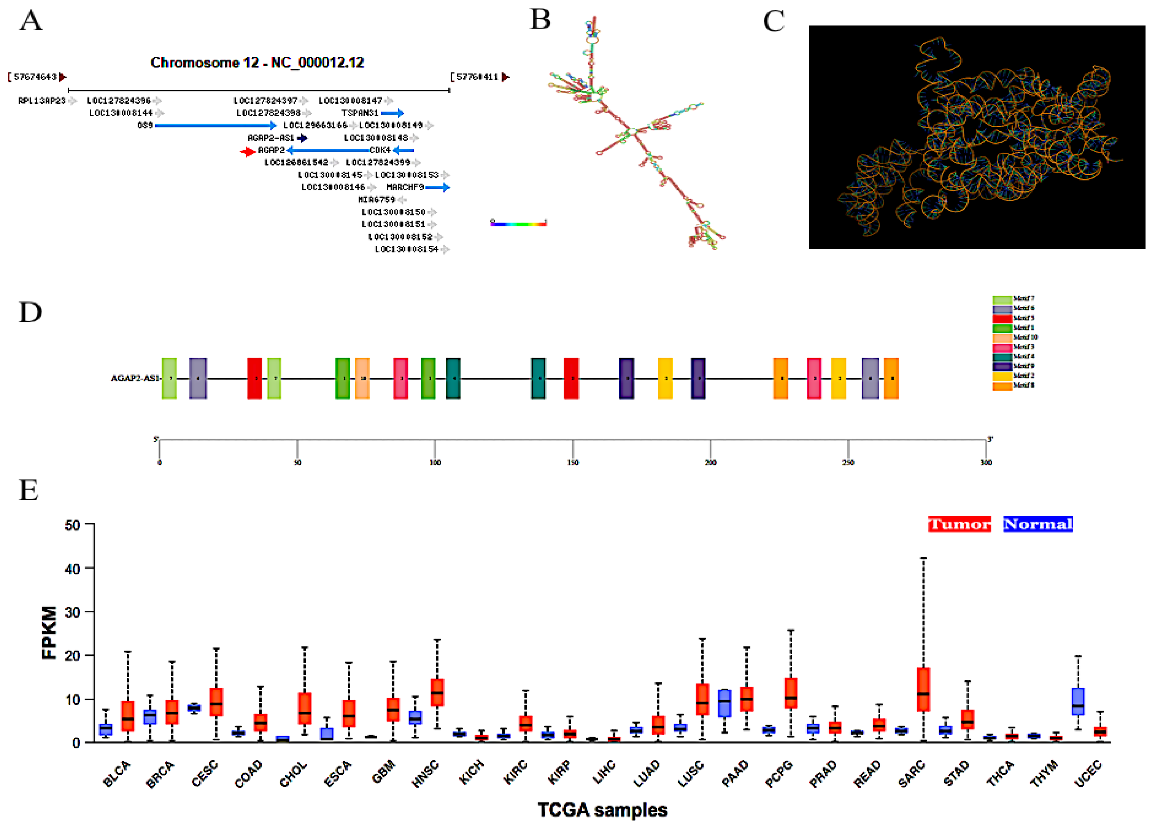
Figure 1. Related information of AGAP2-AS1. (A) The genomic localization of AGAP2-AS1 (https://www.ncbi.nlm.nih.gov, accessed on 6 July 2024). (B) Secondary structure of AGAP2-AS1. (C) Three-dimensional structure of AGAP2-AS1. (D) Motif analysis of AGAP2-AS1. (E) The expression level of AGAP2-AS1 in clinical carcinomatous (red color) and non-carcinomatous (blue color) tissues was analyzed using the UALCAN database (https://ualcan.path.uab.edu/, accessed on 18 May 2024). BLCA: Bladder urothelial carcinoma; BRCA: Breast invasive carcinoma; CESC: Cervical squamous cell carcinoma; CHOL: Cholangiocarcinoma; COAD: Colon adenocarcinoma; ESCA: Esophageal carcinoma; GBM: Glioblastoma multiforme; HNSC: Head and neck squamous cell carcinoma; KICH: Kidney chromophobe; KIRC: Kidney renal clear cell carcinoma; KIRP: Kidney renal papillary cell carcinoma; LIHC: Liver hepatocellular carcinoma; LUAD: Lung adenocarcinoma; LUSC: Lung squamous cell carcinoma; LIHC: Liver hepatocellular carcinoma; LUAD: Lung adenocarcinoma; LUSC: Lung squamous cell carcinoma; PAAD: Pancreatic adenocarcinoma; PCPG: Pheochromocytoma and paraganglioma; PRAD: Prostate adenocarcinoma; READ: Rectum adenocarcinoma; SARC: Sarcoma; THYM: Thymoma; THCA: Thyroid carcinoma; UCEC: Uterine corpus endometrial carcinoma.
Molecules 29 03461 g001
Figure 2. The potential molecular mechanisms of AGAP2-AS1 in human carcinomas. AGAP2-AS1 promoted cancer proliferation, migration and invasion in various cancers, including glioma, PTC, LC, melanoma, ccRCC, GC, PC, EC, BC, CRC and CLC.
Molecules 29 03461 g002
Figure 3. The AGAP2-AS1 expression profile, and its correlation with cancer stage, tumor histology and survival analyzed using the UALCAN database (https://ualcan.path.uab.edu/, accessed on 20 May 2024).
Molecules 29 03461 g003
Figure 4. The AGAP2-AS1 expression in normal, tumor, and metastatic tumor tissues using the TNM plot (https://tnmplot.com/analysis/, accessed on 22 May 2024).
Molecules 29 03461 g004
Disclaimer/Publisher’s Note: The statements, opinions and data contained in all publications are solely those of the individual author(s) and contributor(s) and not of MDPI and/or the editor(s). MDPI and/or the editor(s) disclaim responsibility for any injury to people or property resulting from any ideas, methods, instructions or products referred to in the content.