Abstract
Since the discovery of cisplatin’s antitumoral activity and its approval as an anticancer drug, significant efforts have been made to enhance its physiological stability and anticancer efficacy and to reduce its side effects. With the rapid development of targeted and personalized therapies, and the promising theranostic approach, platinum drugs have found new opportunities in more sophisticated systems. Theranostic agents combine diagnostic and therapeutic moieties in one scaffold, enabling simultaneous disease monitoring, therapy delivery, response tracking, and treatment efficacy evaluation. In these systems, the platinum core serves as the therapeutic agent, while the functionalized ligand provides diagnostic tools using various imaging techniques. This review aims to highlight the significant role of platinum–based complexes in theranostic applications, and, to the best of our knowledge, this is the first focused contribution on this type of platinum compounds. This review presents a brief introduction to the development of platinum chemotherapeutic drugs, their limitations, and resistance mechanisms. It then describes recent advancements in integrating platinum complexes with diagnostic agents for both tumor treatment and monitoring. The main body is organized into three categories based on imaging techniques: fluorescence, positron emission tomography (PET), single–photon emission computed tomography (SPECT), and magnetic resonance imaging (MRI). Finally, this review outlines promising strategies and future perspectives in this evolving field.
1. Introduction
Cis-dichlorodiammine platinum(II), commonly known as cisplatin, is one of the most known and worldwide used chemotherapy agent for the treatment of several kinds of cancers in adults. The fortunate and accidental discovery of anticancer properties in 1965 by B. Rosenberg, followed by the Food and Drug Administration (FDA) approval (1978), triggered extensive research in this field, leading to the development of novel therapeutics based on a metallic scaffold. As a result, platinum(II) complexes have emerged as the most successful category of metal–based anticancer drugs, receiving enormous attention for over half a century [1,2].
Unfortunately, the use of cisplatin is limited due to severe dose-limiting side effects which arise from the drug’s indiscriminate uptake by all rapidly dividing cells, coupled with the body’s attempt to eliminate the exogenous drug through the kidneys. These adverse effects include nephrotoxicity, neurotoxicity, ototoxicity, myelosuppression, intrinsic/acquired therapy resistance, and the inconvenient intravenous administration route [3,4].
1.1. Mechanism of Action of Cisplatin and Resistance Mechanisms
The action mechanism of cisplatin is nowadays well known (Figure 1). Cellular absorption relies on a combination of passive diffusion through the plasma membrane and active transport facilitated by membrane proteins, specifically copper transporter ions and organic cation transporters. Notably, transmembrane copper transport protein 1 (CTR1) is involved in copper homeostasis and cisplatin uptake. Indeed, downregulation of CTR1 is frequently observed in cisplatin-resistant cells, contributing to poor therapeutic responses in non–small cell lung cancer (NSCLC) patients [5].
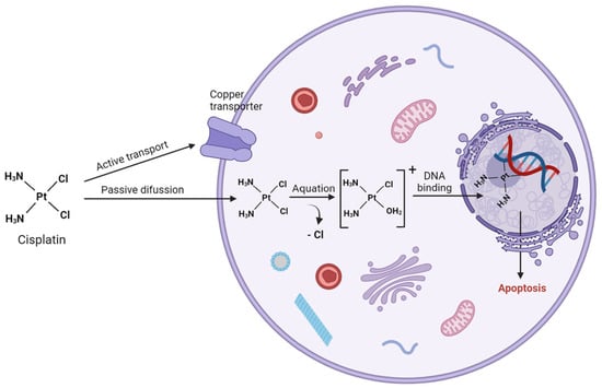
Figure 1.
Mechanism of action of cisplatin (created with BioRender.com).
On the other hand, copper-extruding P-type ATPases (ATP7A and ATP7B) regulate cisplatin export [6]. These transporters are observed to be upregulated in cisplatin-resistant cancer cells and high levels of ATP7A and ATP7B are associated with significantly poorer overall survival in patients with ovarian cancer and ovarian carcinoma, respectively [7,8]. Following intravenous administration of cisplatin, its intracellular activation relies on the drug’s high tendency to undergo aquation where one or both chloride ligand(s) are replaced by one or two molecules of water. This reaction is facilitated by the square planar geometry of cisplatin and is further promoted by the lower chloride concentration in the cytoplasm (4–20 mM) compared to the bloodstream (100 mM). These reactive cisplatin species (mono-aquo and di-aquo complexes) are then prone to bind to cytoplasmic S-containing nucleophiles, such as glutathione (GSH), metallothioneins and other cysteine-rich proteins. Indeed, high levels of glutathione, glutathione-S-transferase or γ-glutamyl cysteine synthetase have been observed in cisplatin-resistant cells [9,10,11]. The activated platinum complexes, carrying positive charges, will also establish interactions with negatively charged DNA. Through this mechanism, cisplatin covalently binds preferentially to the N7 position of guanine nucleobase and to a lesser extent, adenine. Several DNA-Pt adducts have been identified, with the major ones being the cross-links between two adjacent guanines, (intrastrand DNA) and between two guanine bases from different DNA strands (interstrand adducts). These adducts induce a distortion in the DNA structure ranging from 10 to 60 degrees towards the major groove and unwind the helix by up to 23 degrees [12,13], hindering both replication and transcription, ultimately leading to apoptotic cell death. Several protein families are involved in the recognition of Pt-DNA adducts that finally lead to cisplatin-resistant mechanisms, including nucleotide excision repair (NER) proteins, mismatch repair (MMR) proteins and non-histone chromosomal high-mobility group protein 1 (HMGB1). Cisplatin resistance mechanisms can be classified as pre-target (alterations in cisplatin transport before DNA binding), on-target (repair of Pt-DNA lesions), post-target (cellular events occurring after formation of DNA-Pt adducts) and off-target (alterations in signaling pathways indirectly hindering cisplatin-induced proapoptotic events) [14]. Table 1 reports the major resistance mechanisms associated with cisplatin chemotherapy.

Table 1.
Molecular mechanisms of cisplatin resistance.
To circumvent clinical cisplatin resistance and side effects, several platinum analogues with diverse structural motifs have been developed and explored for their antitumor properties. While some exhibit promising in vitro and in vivo biological activity, the translation to clinic for most of these species has been hindered by various challenges [1].
Besides cisplatin, two more platinum(II)-based anticancer drugs received approval by FDA, carboplatin and oxaliplatin, called the “second generation of Pt chemotherapeutics”, which showed a higher physiological stability due to the chelate ligand(s) (Figure 2) [36]. In addition, four more platinum(II) complexes have received approval for human use (Figure 2). Nedaplatin, a second-generation drug, has been approved in Japan for the treatment of NSCLC, small cell lung cancer (SCLC), esophageal cancer, and head and neck cancers. It demonstrates higher activity than carboplatin and comparable efficacy to cisplatin. Heptaplatin is approved in South Korea for the treatment of gastric cancer. More recently, in 2009, Miriplatin was approved in Japan for treating hepatocellular carcinoma [37,38], and Lobaplatin was approved in China for the treatment of chronic myelogenous leukemia, inoperable metastatic breast cancer and SCLC. Most of the previously mentioned drugs offer advantages over cisplatin, such as drug resistance alleviation, toxicity and side effects mitigation, and water solubility improvement.
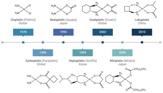
Figure 2.
Platinum drugs in clinical use with their respective approval years.
1.2. Pt(IV) Complexes to Improve Cytotoxicity and to Overcome Resistance
In the last two decades, a lot of scientific interest has moved toward the synthesis and application of Pt(IV) species. These complexes are prodrugs because they must be intracellularly activated by reduction, releasing the active Pt(II) scaffold and restoring their latent cytotoxic activity [39]. Pt(IV) complexes are prepared by oxidative addition of the square planar Pt(II) complexes to yield the octahedral complex while retaining the original Pt(II) equatorial coordination sphere. The major advantage of Pt(IV) species is their higher stability and lower reactivity toward S-containing nucleophiles, as a result of the octahedral geometry (d6 low-spin electronic configuration) compared to the square planar Pt(II) counterparts (d8). The combination of two extra axial ligands to fine-tune chemical properties and high kinetic inertness to substitution reactions have turned Pt(IV) complexes into promising anticancer drug candidates. To date, three Pt(IV) compounds (tetraplatin, iproplatin and satraplatin) have undergone clinical trials, but none have gained clinical approval yet (Figure 3). Tetraplatin was abandoned due to severe neurotoxicity, while iproplatin demonstrated lower activity compared with carboplatin and cisplatin. Satraplatin, the first orally administered platinum(IV) complex, exhibited documented efficacy and acceptable safety in patients with hormone-refractory prostate cancer and small-cell lung cancer. Despite this, it failed to gain FDA approval due to the low convincing benefits in terms of overall survival [4,40].
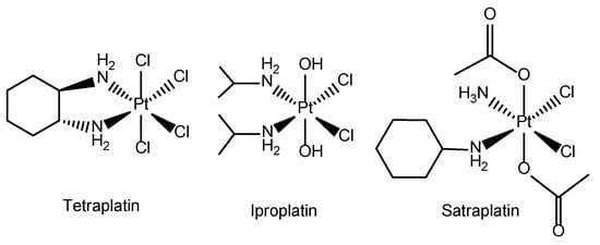
Figure 3.
Structure of Pt(IV) compounds tetraplatin, iproplatin and satraplatin.
The axial ligands of the Pt(IV) complexes can be viewed as cargos that the cytotoxic Pt(II) moiety unloads inside the cell. These ligands can play various roles, serving as passive uptake enhancers (lipophilic moieties) on cancer cells or subcellular targeting agents. Additionally, they can be designed to tether the prodrug to delivery system like polymers or nanoparticles. They can also be bioactive moieties such as other drugs, enzyme inhibitors, pathway activators or suppressor, epigenetic modifiers, and antimetabolites. The integration of these ligands aims to synergistically enhance the pharmacological properties in conjunction with the Pt(II) moiety [2].
1.3. Translation to Theranostics
Besides the therapeutic properties, an appropriate functionalization of the Pt-based species can play an important role in the diagnostic detection of cancer. Through modification with biocompatible nanomaterials or appropriate functional ligands, the therapeutic units may have the potential to evolve into theranostic agents, offering simultaneously therapeutic and diagnostic applications [41].
The term “theranostic” (syn: theragnostic) has been coined to describe platforms that serve dual roles as diagnostic and therapeutic agents. A broader definition has also been proposed, referring to an integrated therapeutic system, which can diagnose, deliver targeted therapy, and track the response to therapy. Indeed, the idea is that theranostic agents could provide the ability to simultaneously monitor the disease and the efficacy of the treatment [42]. This combination promises to create a paradigm shift in individual patient care through an appropriate diagnostic methodology with an optimized treatment strategy to personalize a separate therapeutic intervention [43].
This emerging field in personalized medicine is guided by ligands targeting malfunctioning cells. In the realm of cancer theranostics, diverse strategies involving nanomaterials, metal complexes or small molecules have been developed. As an example, metallic, lipidic and polymer nanoparticles exhibit a variety of biological characteristics that can be used for theranostic purposes. While clinical approval for the use of nanomaterials as transport vehicles has been longstanding, clinical translation of nanomaterial-based theranostic systems from bench to bedside has faced major hurdles [44].
In this review, our primary focus lies in exploring different strategies for the integration of Pt complexes with diagnostic probes to obtain theranostic systems. According to the design goals, we divided them into three categories depending on the main imaging techniques used in combination with the anticancer drug: (i) optical imaging (fluorescence), (ii) positron emission tomography (PET) and single-photon emission computed tomography (SPECT) and (iii) magnetic resonance imaging (MRI). The applications, advantages, and disadvantages of these commonly used imaging techniques are summarized in Table 2. Every imaging modality will be discussed in more detail in the subsequent sections and their application as Pt-based theranostic agents.

Table 2.
Example of imaging techniques used in theranostics [45,46].
2. Illuminating Disease through Fluorescence Imaging
Fluorescence imaging is a non-invasive technique that allows for the monitoring of the dynamics of luminescent molecules within cells or small animals, offering high sensitivity and excellent temporal resolution. A targeted biological compound is exposed to a specific radiation (absorption) and the subsequent emitted light (emission) is captured for analysis. Utilizing standard infrastructure like a charged-couple device (CCD) camera and a light source, fluorescence imaging proves to be a cost-effective method (Figure 4) [47]. Additionally, its remarkable sensitivity capable of detecting concentrations as low as picomolar to femtomolar levels enables the visualization of individual molecules and facilitates the tracking of biological processes. Unlike imaging techniques involving the detection of high-energy gamma rays such as PET and SPECT, fluorescence imaging operates with low-energy photons, ensuring its safety for both samples and operators. However, a significant limitation of this technique lies in its restricted tissue penetration, thereby impeding optical imaging of probes in deep tissues or large subjects, consequently hindering its clinical translation [48].

Figure 4.
Basic principles of optical fluorescence molecular imaging. Following administration of the fluorophore, the subject is illuminated with excitation light (λ1). This excites the fluorophore, causing it to emit light (λ2), which is then captured by a CCD camera within IVIS Lumina LT in vivo imaging system, processed, and converted into an image (created with BioRender.com).
A persisting challenge when studying Pt-based drugs in vitro and/or in vivo is that there are a few spectroscopic techniques available for detecting their faith. The main techniques employed to measure and quantify the cellular internalization of Pt-based samples in cells are Graphite Furnace Atomic Absorption Spectrometry (GFAAS) and Inductively Coupled Plasma Mass Spectrometry (ICP-MS) analysis [49,50] but, unfortunately, no information regarding speciation and cellular localization could be obtained. To address these challenges, several Pt complexes containing tethered fluorophores have been generated to evaluate its pharmacokinetic behavior. This assessment covers organ accumulation, systemic circulation time, intracellular target, and excretion patterns.
Most of these compounds retained the traditional structure–activity requirements for Pt(II) anticancer compounds (square planar complexes with leaving groups and amines in cis orientation) [51]. Initial investigations involved linking cisplatin with fluorophores such as fluorescein (1) [52] carboxyfluorescein diacetate (CFDA), (2), 6-(2,4-dinitrophenyl)aminohexanoic acid (DNP), (3) [53], and Alexa [54] (Figure 5). These complexes have been used to monitor the distribution of cisplatin in human ovarian carcinoma, human osteosarcoma, and human cervical carcinoma cells, respectively. Remarkably, they demonstrated swift accumulation of fluorophore-cisplatin analogues in the nucleus, succeeded by sequestration within Golgi vesicles. More recent works described a series of Pt-based complexes tethered to boron-dipyrromethene (BODIPY) [55,56,57] (Figure 5). This family of fluorophores has gained considerable interest in bioimaging because of their high absorption coefficients, sharp emission, high fluorescence quantum yields and excellent chemical and photostability. Both 4 and 5 showed primarily cytosolic localization, with nuclear distribution observed only at higher concentrations [55]. Regarding compound 6, colocalization experiments with commercially available mitochondria-specific dye (MitoTracker) revealed its predominantly localization in the mitochondria, while nuclear colocalization remained indiscernible [56]. Moreover, in vivo pharmacokinetics studies of these compounds have also been conducted. Using a window chamber tumor model, which enables simultaneous time-course fluorescence microscopy of the fluorescent drug in both mouse vasculature and tumor tissue, 7 showed plasma kinetics comparable to cisplatin. Furthermore, high-resolution intravital imaging was employed to monitor DNA damage in individual tumor cells following administration. The study focused on the fluorescence signal of 53BP1 puncta, which are markers of DNA damage response within the cells. No linear correlation was observed between the fluorescence signal of these 53BP1 puncta and the accumulation of the drug CP11, indicating potential heterogeneity in the cells’ drug response [57].
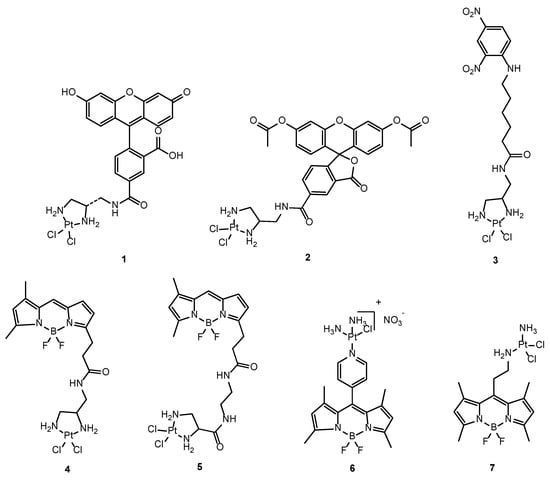
Figure 5.
Chemical structures of the different fluorophore-cisplatin analogues: FDDP (1), CFDA-Pt (2), DNP-Pt (3), BODIPY-Pt (4–6), CP-11 (7).
Nevertheless, incorporating a fluorophore into a small inorganic complex can significantly modify its physicochemical properties and biological activity. Such an approach must consider the organelle selectivity of the fluorophore, which may vary depending on parameters such as molecular weight, partition coefficient, amphiphilic character and pKa values [58]. Thus, it is uncertain if fluorescent analogues accurately model cisplatin’s cellular behavior, and, likewise, bulky fluorophores may interfere with DNA binding mechanisms of platinum. Furthermore, some of them, and in particular BODIPY, can generate ROS (Reactive Oxygen Species) under irradiation that can potentially interfere with the cell damage mechanism. Notably, none of the previous Pt-fluorophore complexes showed significant nuclear localization.
To address these limitations and to allow for a “label-free” study of Pt drug fate, several exogeneous fluorophores have been synthesized to investigate the speciation of platinum anticancer drugs. For example, FCDPt1 (Figure 6) is a fluorescein-based probe linked to dithiocarbamic acid moiety which selectively identifies platinum species [59]. Similarly, RPt1, a rhodamine-based probe incorporating phenyl isothiocyanate, has been developed for studying metabolism of trans-Pt complexes [60]. Furthermore, Rho-DDTC was the first rhodamine-based fluorescent turn-on probe designed to distinguish between Pt(II) and Pt(IV) complexes upon intracellular reduction [61,62]. Following this discovery, numerous analogues were developed to enhance fluorescence intensity and sensitivity of these probe [63,64]. Probe RD640-TC was developed as the first tool for in vivo tracking of cisplatin due to its remarkable characteristics such as high fluorescence quantum yield (Φ = 0.68), two-photon absorption properties (enabling greater penetration depth) and high photostability (Figure 6) [65].

Figure 6.
Chemical structures of different exogeneous fluorescent probes.
More examples of turn-on probes include the work by Lippard et al. [66], who synthesized two analogues of fluorescein conjugated to cisplatin Pt(IV) prodrugs, 8 and 9, and that of Bin Liu et al. [39], who developed a cisplatin Pt(IV) prodrug conjugated to an aggregation-induced emission (AIE) luminogen (10). In both cases, the reduction of Pt(IV) to Pt(II) within the cellular environment was monitored by the release of fluorescein ligands and luminogen, respectively. Additionally, Bin Liu et al. incorporated a short hydrophilic peptide to ensure water solubility and a cyclic arginine-glycine-aspartic acid (cRGD) tripeptide as a targeting ligand (Figure 7).
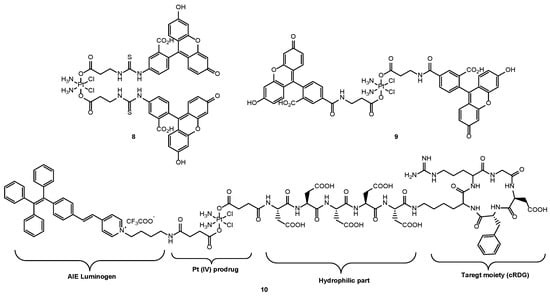
Figure 7.
Chemical structures of the different Pt(IV)-fluorescent scaffolds: Pt(IV) (FITC)2 (8), Pt(IV) FL2 (9), Pt(IV) prodrug-AIE conjugate (10).
To promote the clinical translation of fluorophores, both near infrared NIR-I (700–900 nm) and NIR-II dyes (1000–1700 nm) have emerged as imaging tools to afford precise dynamic actions in vivo with high spatiotemporal resolution, deeper penetration and decreasing light absorption and scattering (Table 3) [67,68].
Among these, gold nanoclusters (AuNCs) stand out for their remarkable properties: excellent biocompatibility, size below the renal excretion threshold, robust photostability, facile modification, and excellent photothermal activity [69]. In a study conducted by Li et al., AuNCs were coupled with Pt(IV)-cisplatin and folic acid (FA), enabling not only fluorescence imaging but also targeted chemotherapy for breast cancer (Figure 8). Biodistribution analysis revealed that FA-modified AuNC-Pt nanoparticles demonstrated high tumor uptake, effectively evaded reticuloendothelial system (RES) capture, and facilitated renal clearance [70].

Figure 8.
Schematic illustration of AuNCs-based theranostic platform. The star symbol corresponds to Pt(IV)-cisplatin based scaffold and the wavy lines to HOOC–PEG–FA compound (created with BioRender.com).
Similar studies were carried out by Zhiqiang Yu et al., who reported a cisplatin delivery platform based on AuNCs. The results showed that not only AuNCs-Pt does effectively bind glutathione (GSH) via Au-S bonds, scavenging intracellular GSH and thereby facilitating increased internalization of cisplatin, leading to enhanced anticancer activity, but also that the unique NIR-II imaging capability of AuNCs-Pt enables precise visualization of Pt transport in both ovarian and hepatocellular carcinoma tumor models. Importantly, in vivo NIR-II imaging revealed that AuNCs-Pt exhibited significantly superior tumor growth inhibition compared to cisplatin while inducing minimal systemic toxicity, underscoring its potential as a highly effective theranostic agent [71].
Another class of NIR-I fluorescent probes are conjugated polymers, which are promising for their high brightness and low toxicity. Bin Liu et al. employed conjugated polyelectrolytes (CPE) grafted with polyethylene glycol (PEG) chains to load cisplatin for targeted in vitro and in vivo NIR imaging. These nanoparticles enable sustained cisplatin release, enhancing internalization. While these nanoparticles hold potential for liver cancer applications due to their high accumulation in RES, further optimization of their physical and chemical properties is needed for broader cancer imaging and chemotherapy applications [72].
It has been well known that Pt(II) metallacycles-based supramolecular coordination complexes can act as anticancer agents; however, poor photostability, low tumor uptake and penetration depth limited the in vivo antitumor applications [73]. Recently, Stang et al. designed a NIR-II theranostic nanoprobe that incorporates a Pt(II) metallacycle and an organic molecular dye (benzobisthiadiazole) into DSPE-mPEG500 to enhance stability and biocompatibility of the theranostic agent in vivo (Figure 9). Compared to cisplatin, this NIR-II theranostic agent displayed better efficiency of tumor growth inhibition and prolonged blood circulation, as well as reduced systemic toxicity [74].
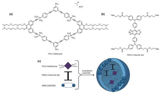
Figure 9.
Structures of (a) Pt(II)metallacycle, (b) NIR(II) molecular dye and (c) representation of the nanoplatform (created with BioRender.com).
In a parallel investigation, Han et al. employed a benzobisthidiazole-based NIR II imaging probe (TQTPA) to synthesize multimodal nanoparticles loaded with both TQTPA and cisplatin using hyaluronic acid, enabling active targeting and enhanced accumulation in tumor tissue. The therapeutic efficacy of this chemo-photothermal synergistic approach was assessed in a mouse model. Upon irradiation, the tumor site temperatures significantly exceeded those of the negative control. Moreover, after 13 days of treatment, tumor volume decreased by 83% compared to the untreated control group [75].
BODIPY fluorophores can also be used for NIR imaging by implementing π-conjugated structures (red-shifted absorbance), (Figure 10). In this way, Zhigang Xie et al. synthesized compound 11 (λabs = 645 nm, λem = 674 nm), demonstrating its internalization in HepG2 cells and localization within mitochondria. Through MTT assays, 11 showed comparable cytotoxicity to cisplatin in HepG2 and HeLa cells. Through NIR imaging, 11 was localized at tumor sites four hours post-injection [76]. Following this work, Minhuan Lan et al. further enhanced NIR imaging capabilities by synthesizing a NIR cisplatin-appended BODIPY fluorophore (12) with greater red-shifted absorption (λabs = 748 nm, λem = 947 nm) through the incorporation of N,N-dimethylaniline into BODIPY (Figure 10) [77].
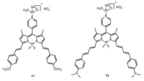
Figure 10.
Chemical structures of BODIPY-based NIR imaging probes (11 and 12).
Quantum dots (QDs) are renowned for their distinct optical features like good photostability, large Stokes shift, and high fluorescence quantum yields. In a study by Rylander et al., a novel hybrid system was developed, comprising self-assembled single-walled carbon nanohorns (SWNHs) loaded with cisplatin and adorned with CdSe/ZnS quantum dots on their exterior. Through fluorescence imaging, SWNHs–QD+cis hybrids were tracked in rat bladder transitional carcinoma cells after 72 h incubation. These images helped to validate the mechanism of continued cell death over 72 h as well as the compound’s internalization and cisplatin delivery on the nucleus over time [78]. However, a significant drawback of metal-based quantum dots lies in their inherent toxicity. Indeed, the previous work assessed cell viability using SWNHs as a negative control but did not include evaluation with SWNHs+QDs. Consequently, graphene quantum dots (GQDs) have emerged as alternative, non-toxic nanocarriers with superior biocompatibility and fluorescent properties. As an example, Sierin Lim et al. designed GQDs functionalized with anti-epidermal growth factor receptor antibodies and loaded with cisplatin, achieving targeted delivery of cisplatin in breast cancer cells, as validated through fluorescence imaging [79]. Moreover, GQDs offer the possibility to fine-tune their optical properties, enabling the development of NIR-II imaging QDs for in vivo NIR imaging-guided photothermal therapy [80]. This platform could also be coupled with cisplatin/Pt(IV) prodrugs delivery for multimodal therapeutic applications. Table 3 highlights the advantages, disadvantages, applications and future research on the NIR-fluorophores mentioned in this section [81,82].

Table 3.
Summary of the advantages, disadvantages, applications and future research on the NIR-fluorophores mentioned in this chapter [81,82].
Table 3.
Summary of the advantages, disadvantages, applications and future research on the NIR-fluorophores mentioned in this chapter [81,82].
| NIR Fluorophores | Advantages | Disadvantages | Applications | Future Research |
|---|---|---|---|---|
| AuNCs | Easy surface modification. High biocompatibility. Large Stokes shift. | Photobleaching. | Bioimaging and therapeutic applications. | Escaping recognition by immune system. |
| Conjugated polymers | High brightness. Low toxicity. | Limitation of excitation and emission wavelengths (<900 nm). | Photoacoustic imaging. | Increase polymer’s conjugation degree to shift spectra towards NIR-II window. |
| Benzobisthiadizole | Large Stokes shifts. High imaging quality. | Photobleaching. | Probes for clinical translation. | Design of new materials for NIR-II imaging. |
| BODIPYs | High quantum yields. Excellent photostability. | Poor water solubility. | In vivo visualization of tumors. | Reducing bandgap of fluorophores to bathochromic shift wavelength enhancing hydrolytic stability. |
| QDs | High quantum yield. Good photostability. | Toxicity. | In vitro evaluation. | Design of non-toxic QDs for in vivo NIR imaging. |
3. Pioneering Platinum-Radiopharmaceuticals for SPECT and PET Imaging
SPECT/PET are radionuclide molecular imaging modalities that allow for the real-time monitoring of drugs’ distribution within the body as well as non-invasive monitoring of their therapeutic efficacy [83]. The fundamental principle of SPECT is the administering of a radiotracer-containing imaging agent (such as a peptide, protein, or nanoparticle) with a gamma-emitting radioisotope (Technetium-99m, Iodine-123, Thallium-201) to the patient. Subsequently, gamma rays emitted by the tracer are captured by a rotating gamma camera, which collects data from various angles to facilitate tomographic reconstruction. On the other hand, PET employs positron-emitting radioisotopes, such as fluorine-18, Gallium-68, Copper-64, Zirconium-89. Positrons emitted by the tracer will then collide with electrons in the surrounding tissues, a phenomenon known as annihilation, which results in the creation of two gamma rays, each with an energy of 511 keV, moving in opposite directions. The PET detector detects these gamma rays which determine the location of annihilation event. The resulting electrical signals are then converted into sinograms, which are further reconstructed into detailed tomographic images (Figure 11) [47]. While both techniques are similar, PET exhibits significantly higher sensitivity, typically ranging from two to three orders of magnitude greater than that of SPECT [84].

Figure 11.
Schematic representation of the basic principles of PET (created with BioRender.com).
The development of smart molecules, possessing both therapeutic and diagnostic properties, holds the potential to revolutionize cancer therapy by enhancing efficiency and safety. The simplest examples of such theranostics are the radioactive forms of cisplatin (CDDP) and carboplatin (CP) such as 191Pt/195mPt-CDDP, 195mPt-CP, 18F-FCP (a derivative of carboplatin) and 13N-CDDP (Figure 12).

Figure 12.
Chemical structures of 191Pt-CDDP (13), 195mPt-CDDP (CISSPECT®), (14), 195mPt-CP (15), 18F-FCP (16) and 13N-CDDP (17).
Among the four radioactive isotopes of Pt (Platinum-191, Platinum-193m, Platinum-195m, Platinum-197), Platinum-195m has gained special attention over the years due to its ease of production, ideal gamma energy spectrum (similar toThallium-201) and appropriate half-life (4 days) for efficient synthesis, quality control and in vivo radio-imaging (Table 4).

Table 4.
Characteristics of main Pt radionuclides.
The synthesis of Platinum-195m has been well established [85,86], and relies on the thermal neutron irradiation of 194Pt with yields ranging from 60 to 90%. More recently, an alternative preparation method has been introduced to enhance the specific activity of Platinum-195m. This method involves irradiating 197Au with breaking radiation. Notably, this novel approach has demonstrated a remarkable 37,000-fold increase in specific activity over a span of 25 years [87].
Platinum-195m is not a pure gamma emitter, as each disintegration also releases 36 auger electrons that deposit large amounts of energy (around 25 keV) within nanometer-micrometer distances in tissues. Therefore, integration of cisplatin to Platinum-195m emerges as a versatile platform, offering the potential for both enhanced therapeutic effects and tumor imaging. For this reason, several studies were conducted with 14, also known as (CISSPECT®). Using 14 and 15, Wolf et al. studied non-invasive in vivo pharmacokinetics of these compounds in Walker 526 solid tumors bearing rats [88]. In a more recent study, 14 proved to be a safe radiopharmaceutical, highlighting its potential for optimizing dosage in patients undergoing cisplatin chemotherapy. Moreover, the radiopharmaceutical presented a favorable biodistribution profile in which the kidneys received the highest dose accounting for a clearance half-life of 40 h [89]. Later, the first preclinical SPECT images of 14 were conducted, showing its potential as a tracer to image the biodistribution of platinum-based compounds in vivo [90]. In addition, 14 has proven to be more effective than regular cisplatin due to its enhanced toxicity after incubation in the Chinese hamster V79 cell line [91]. Its enhanced toxicity needs to be further validated in in vivo models [92].
Given the inherent therapeutic efficacy of the Platinum-195m radioisotope, there is a current trend in the development of radio-therapeutically active compounds. An illustrative example is the development of 195mPt-biphosphonates (BP) complex 18 (Figure 13). The bisphosphonate ligand was chosen to specifically target bone cancers due to high affinity for Ca2+.
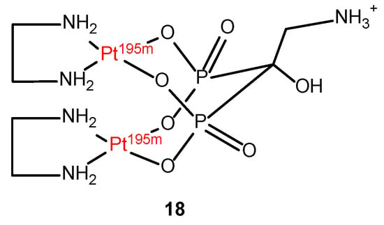
Figure 13.
Chemical structure of 195mPt-biphosphonate (195mPt-BP) compound (18).
This complex (18) has demonstrated a specific accumulation in bone sites exhibiting high metabolic activity, resulting in an impressive 11-fold increase in DNA damage within metastatic tumor cells when compared to non-radioactive Pt-BP. Furthermore, the biodistribution of these compounds can be precisely monitored through SPECT imaging, revealing minimal systemic toxicity. This opens a promising avenue for further research in the field of radiopharmaceuticals, particularly for the treatment of bone cancer [93,94]. On the other hand, earlier studies used 13 to examine biodistribution and kinetics in patients undergoing cisplatin treatment [95,96], following the demonstration of its enhanced antitumor effect in both in vitro and in vivo models compared to non-radioactive cisplatin [97,98,99].
However, the further application of this radioisotope for human imaging studies is hindered by certain drawbacks. Above all, its high energy γ-photons (539 keV) are not ideal for high-contrast radio imaging using modern gamma cameras. Moreover, unlike Platinum-195m, which decays to 195Pt, Platinum-191 decays to 191Ir which is a different element with different pharmacokinetics leading to regulatory challenges in human imaging studies. Little research has been carried out regarding Platinum-193m and 197Platinum-197 radioisotopes [100].
The synthesis of 17 has also been reported through several methods [101,102] to investigate the pharmacokinetics of cisplatin in both mice and brain tumor patients [103]. However, its clinical use is constrained by its very short half-life (t1/2 = 10 min).
A different approach was taken by Zweit et al., who developed compound 16, a feasible PET tracer for assessing the biodistribution of carboplatin [104]. Fluorine-18 is widely used and is the preferred radioisotope for PET imaging due to its favorable characteristics such as half-life of 109 min, high positron emission (97%), suitable positron energy (0.634 MeV) and being easily produced by cyclotron (Table 5) [105].

Table 5.
Selected radionuclides used in PET and SPECT, their production mode and decay properties [106].
Compared to Platinum-195m, Fluorine-18 offers several advantages for clinical use. The routine production and supply of Platinum-195m face significant challenges due to its high cost and logistical difficulties. On the other hand, 18F has high availability, versatile chemistry, and compatibility with a wide range of biomolecules.
In this way, 16 demonstrated its capability to assess the drug distribution across various tumor types, showcasing its potential to enhance the overall efficacy of platinum-based chemotherapy on an individual patient basis. Following this discovery, a versatile platform consisting of 16 encapsulated in 111In-labeled liposomes was synthesized for image-guided drug delivery [107]. This study proves the dual-tracer imaging feasibility of SPECT and PET probes in vivo, aiming to enhance the precision of drug tracing within the body.
In more recent studies, supramolecular structures were developed that can encapsulate Pt drugs while incorporating radionuclides for in vivo PET imaging. An example is the work by Casini et al. [108], who developed a palladium-based metallo-cage scaffold to encapsulate cisplatin while incorporating Fluorine-18. Although very promising outcomes were obtained, further investigation is needed to refine the kinetic stability of these supramolecular structures, ensuring optimal biodistribution profiles for future clinical applications. Despite the synthesis of numerous Pt(II)-metallo-assemblies, their potential as imaging-assisted cancer diagnosis agents is still in an early stage of exploration [109].
Terpyridine (TP) platinum-based complexes have been developed as theranostic platforms with a specific focus on targeting G-quadruplexes to overcome platinum drug resistance (Figure 14). These complexes are conjugated with 64Cu-NOTA (NOTA = 1,4,7-Triazacyclononane-1,4,7-triacetic acid) for targeted radiotherapy. This radioisotope (t1/2 = 12.8 h; EC, [43.1%], β+, 0.653 MeV [18%]; β-, 0.579 MeV [38.4%]) not only possesses PET imaging properties via β+ emission but also holds potential for cancer therapy via auger electron emission. Guérin et al. synthesized two 64Cu-NOTA-TP conjugates (19 and 20), each featuring a unique TP-NOTA linker, with the aim of investigating their cytotoxic effects in both human colorectal tumor cells and a normal fibroblast cell line (Figure 14). Notably, the use of a flexible linker in 20 demonstrated superior selectivity towards cancer cells, a higher percentage of nucleus internalization, and comparable cytotoxicity to cisplatin with an activity of 4.0 MBq/nmol [110,111]. Following the promising results obtained in vitro, the stability and the biodistribution profile of 20 through PET imaging was analyzed in vivo. It showed a promising tumor uptake ranging from 1.8 ± 0.4 to 3.0 ± 0.2% ID/g over 48 h. Furthermore, it demonstrated the ability to retard tumor growth with any observable signs of toxicity [112].
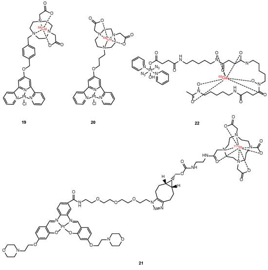
Figure 14.
Chemical structures of [64Cu]Cu-NOTA-TP (19), [64Cu]Cu-NOTA-C3-TP (20), Pt(II)salpen-111In (21) and Pt-succ-DFO-68Ga (22).
Another example is 21, which was developed to target G-quadruplexes. Here, the Pt scaffold was functionalized with a 111In-DOTA moiety to assess its in vivo biodistribution via SPECT imaging [113]. Unfortunately, the complex exhibited limited cell permeability, highlighting the necessity for further research in this field (Figure 14).
Gallium-68 is a promising radioisotope for PET imaging, offering a suitable half-life (68 min) for rapid imaging and reduced patient exposure. Moreover, standard radiolabeling protocols are already established for this radioisotope. Notably, Sadler et al. conducted a pioneering in vivo imaging study using dynamic PET imaging to observe the whole-body distribution of a photoactivable Pt(IV) azido complex (22). The findings demonstrated a promising pharmacokinetic profile, indicating the potential for advancing 68Ga-DFO (DFO = deferoxamine) complexes in the image-guided treatment of therapeutic Pt(IV) prodrugs [114].
As a conclusion of this section, it needs to be highlighted that the design of novel theranostic platforms featuring both therapeutic (Pt-based complexes) and imaging modalities is still in its infancy and therefore further research needs to be carried out.
4. Magnetic Resonance Imaging (MRI) Contrast Agent-Pt Conjugates for Detecting and Treating Solid Tumors
MRI is indeed the most efficient technique for tumor imaging. It is non-invasive with excellent tissue contrast and spatial resolution (10–100 µm), and it is applicable in whole body with precise 3D localization. It employs radiofrequency radiation in the presence of carefully controlled magnetic fields, producing high-quality cross-sectional images of the body [115,116]. While the image resolution of MRI is superior to other tissue-penetrating imaging techniques such as PET or SPECT, its use is still limited in detecting disparities in the magnetic properties of tissues and organs in clinical practice [117].
Contrast-enhanced MRI plays an increasingly key role in diagnostic medicine. Its usefulness originates from its capability to provide more detailed diagnostic information that cannot be obtained with other non-invasive techniques. The contrast agent is administered while the patient is in the scanner, and the diagnostic images appear within minutes (Figure 15).

Figure 15.
Representation of the basic principles of MRI (created with BioRender.com).
The basis for an MRI signal is the precession of water hydrogen nuclei with respect to an applied magnetic field, and MRI contrast agents can be used to shorten the relaxation times of water molecules. They accumulate tissue-specifically and therefore enhance contrast in this specific organ, allowing for faster imaging (higher throughput) and for imaging that is less sensitive to artifacts caused by motion (better-quality images). Two different relaxation times are analyzed: longitudinal (spin–lattice relaxation time, T1) and the transversal (spin–spin relaxation time, T2). The rate constants corresponding to the T1 or T2 relaxation times are defined as 1/T1 and 1/T2. Contrast agents with higher relaxivities [r1 (=1/T1) and r2 (=1/T2)] give stronger contrast enhancement [118].
The contrast agents are small, such as hydrophilic gadolinium(III)-based chelates, but gadolinium is not the only element that can be used to generate MRI contrast. Indeed, iron oxide nanoparticles and manganese(II) complexes have been approved for liver imaging, although they are still not commercially successful. Paramagnetic elements such as gadolinium(III) and high–spin manganese(II) are for T1-weighted because of their shorter T1 time, while superparamagnetic contrast agents such as iron-oxide nanoparticles are for T2-weighted, for the same reason [119,120,121].
Gadolinium may also play an important role in therapeutic techniques, such as synchrotron stereotactic radiotherapy, in which the selective delivery of gadolinium to the cell nucleus would significantly enhance the efficacy of the treatment [122,123]. Gadolinium(III) complexes have also been explored as potential agents in an experimental anti-cancer treatment known as gadolinium neutron-capture therapy, which is closely related to the boron neutron-capture therapy [124,125]. When looking at the design of new small theranostic molecules, one common approach is that the therapeutic component (i.e., Pt-based scaffold) is covalently linked to the diagnostic component (i.e., Gd contrast agent) [117]. More recent studies showed the development of hybrid Gd-Pt theranostic agents incorporated into micelles [126,127] or nanoparticles instead [128,129]. All these approaches aim to mitigate the inherent cytotoxicity of the drug carrier [130].
4.1. Pt-Based Gd (III) Conjugates as Theranostic Contrast Agents
In 2010, Crossley et al. [131] reported the pioneering work of gadolinium transport to a tumor cell nucleus via conjugation with a platinum complex. This innovative approach involved the functionalization of Gd–DTPA (DTPA = diethylenetriaminepentaacetic acid) with two Pt(II) (terpy) (terpy = 2,2′:6′,2″-terpyridine) units 23. It was shown to function as a potent DNA intercalator of lung tumor cells. The tumor cells exhibited an enhanced accumulation capacity of both Pt and Gd with respect to the control, suggesting the presence of a tumor-selective uptake mechanism for the complex and the integrity of the complex in vitro. However, despite these observations, the Gd-Pt complex displayed significant cytotoxicity towards both normal and tumor cells, preventing its use as a potential theranostic agent (Figure 16).

Figure 16.
Structure of Pt (II)-based Gd-DTPA conjugate (23), Pt (II)-based Gd–DOTA conjugate (24) and Pt (II)-based Gd–DTPA complexes 25 and 26.
Fenton et al. [132] presented a next generation of DNA metal-intercalators that addressed a few unfavorable key factors associated with the previous prototype. By replacing the acyclic DTPA ligand with the macrocycle DOTA (DOTA = 2,2′,2″,2‴-(1,4,7,10-tetraazacyclododecane-1,4,7,10-tetrayl)tetraacetic acid), the amide hydrolytic stability was enhanced. Moreover, by employing only a single Pt(II)-terpy unit, the cytotoxicity decreased with a concomitant increase in the cellular uptake while retaining an excellent DNA targeting ability (24).
Zhu et al. [133] developed two bifunctional Pt–Gd complexes (25 and 26) to monitor the temporal distribution of Pt drugs in vivo and to assess the therapeutic response in situ. These complexes partially dissociate in the tumor environment releasing the cytostatic Pt scaffold and the residual Gd moiety for imaging. The incorporation of Pt units significantly enhances the cellular uptake of Gd complexes, while the proton relaxivities of the Pt–Gd complexes surpassed those of Gd–DTPA under physiological conditions. Also, these complexes exhibit prolonged retention in mice kidneys, presenting an opportunity for non-invasive diagnosis of acute renal injury induced by nephrotoxic platinum drugs in mice models (Figure 16).
A different approach was developed through the conjugation of a gadolinium(III) texaphyrin to platinum-based scaffolds as a strategy for selective targeting tumoral tissues and overcoming mechanisms associated with cisplatin resistance. Metallotexaphyrins, a class of expanded porphyrins (e.g., motexafin gadolinium [MGd]), demonstrated notable accumulation in both primary and metastatic tumors [134,135] and possess intrinsic anticancer activity attributed to their macrocyclic ligand-centered redox activity [136,137]. Since 2004, Sessler’s group has focused its interest on the development of texaphyrin drug conjugates as promising candidates for tumor localization [138,139,140,141,142,143] and on the combination of Motexafin gadolinium drug (MGd, Figure 17) to mediate the cancer-specific reduction of Pt(IV) agents [144]. Magda et al. [138] reported the first generation of texaphyrin-type drugs (27 and 28), where established anticancer agents such as cisplatin were covalently conjugated to a tumor-localizing texaphyrin core. Unfortunately, this initial study faced limitations in in vitro evaluation due to poor solubility and hydrolytic instability (Figure 17).
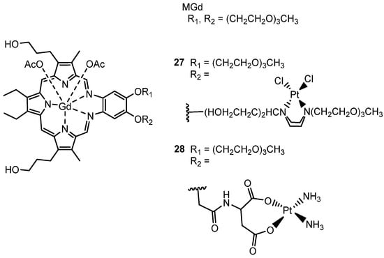
Figure 17.
Structure of Motexafin-Gadolinium MGd and Pt (II)-based Motexafin-Gadolinium conjugates 27 and 28.
More recently, a novel gadolinium texaphyrinmalonate-Pt conjugate 29 was reported by Arambula et al. [139], which showed anti-proliferative activity in vitro comparable to that of carboplatin. The inclusion of the malonate chelating group, analogous to that present in carboplatin, was strategically designed to facilitate Pt release under physiological conditions (Figure 18). Furthermore, working with an ovarian cancer cell model, an enhanced intracellular platinum accumulation was observed, as well as a greater number of Pt–DNA adducts compared to the controls (i.e., methilmalonatoplatinum, carboplatin). Despite these findings, the DNA damage repair induced by the conjugate remained comparable to that of cisplatin, suggesting the persistence of other molecular mechanisms of resistance, such as failure to reactivate the tumor suppressor factor p53 [140].
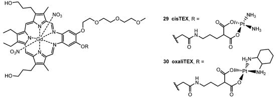
Figure 18.
Structure of Pt (II)-based Texaphyrinmalonate conjugates cisTEX (29) and oxaliTEX (30).
To overcome key platinum pharmacological and molecular resistance mechanisms in vitro, a novel texaphyrin-oxaliplatin-like conjugate 30 was synthesized and biologically screened (Figure 18). While very effective in terms of anticancer activity, it was observed that the Pt moieties were hydrolytically unstable in aqueous environments, resulting in slow, uncontrolled release of the Pt(II) core. Therefore, difficulties were encountered in terms of formulating this generation conjugate for in vivo applications [141].
In order to develop a targeting Pt-texaphyrin system capable of controlled Pt release, a second generation of platinum(IV)-texaphyrin conjugates was synthetized later by the same group. The presence of a Pt(IV) core increased the hydrolytic stability relative to the first-generation Pt(II) systems, and Pt(IV) conjugates were able to release the Pt(II) scaffold in a controlled fashion upon exposure to light or a reducing environment. The asymmetric Pt(IV) based on cisplatin scaffold was chosen as the platinum precursor for 31 and 32 (Figure 19, first-generation). Both conjugates demonstrated a very good anti-proliferative activity in vitro against both wild-type and cisplatin-resistant ovarian cancer cell lines, showing improvements in the IC50 values for both cell lines, as well a decrease in the resistance factor (the resistance factor is calculated as the ratio between the IC50 values of cisplatin-resistant ovarian cancer and wild-type cell lines) [142].
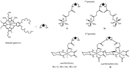
Figure 19.
Structure of first generation of Pt (II)-based texaphyrinmalonate conjugates 31 and 32 and second generation of Pt (IV)-based texaphyrin 33–36.
A combination of both in vitro and in vivo studies on the latest generation of Pt(IV)-based texaphyrin conjugates (33–36 Figure 19) showed promising results in overcoming platinum-resistant ovarian and colon cancers. In this case, the 1,2-diaminocyclohexane (DACH) ligand environment around the platinum center (oxalilplatin scaffold) is believed to be crucial in producing agents capable of overcoming p53-based platinum resistance. Texaphyrin conjugates based on a Pt(IV)–DACH complex were designed to deliver an active Pt(II), analogue to oxaliplatin, in the reducing environments characteristic of many solid tumors and to overcome common p53-related cisplatin resistance mechanisms. Among the four different complexes shown in Figure 19, 34 demonstrated a superior efficacy in retarding/inhibiting tumor growth in mouse models, including those with clinically relevant Pt-resistant mouse models, outperforming the most closely related current platinum-based standards of care. These findings suggest that this complex could play a promising role in clinical applications [143].
Inspired by this promising work of Sessler and coworkers, who demonstrated how Gd(III) complexes can be used synergistically with Pt(IV) prodrugs, Adams et al. took the opportunity to investigate Gd(III)–Pt(IV) mixed-metal complexes as theranostic agents [145,146]. Firstly, two Gd(III)–Pt(IV) conjugates (37 and 38, Figure 20) were synthesized by coupling a Gd(III) MRI contrast agent in the axial position of a cisplatin and carboplatin-based scaffold forming novel Pt(IV) complexes [145]. Unlike the typical Gd(III) contrast agents, which are confined to the extracellular space surrounding tumors, these complexes are tailored for intracellular contrast enhancement of cancer cells due to the presence of the more lipophilic platinum moiety. Among the two agents, 37 emerged as the most promising, displaying greater cellular toxicity, higher intracellular accumulation of Gd(III), and superior MR contrast enhancement in vitro. Cellular accumulation analyses between in vitro and in vivo studies highlighted the variations in MR contrast enhancement, offering a potential strategy for imaging Pt in resistance cell lines. This platform could potentially be applied across various chemotherapeutics, particularly in cases where decreased drug accumulation is a dominant mechanism of chemoresistance [146].

Figure 20.
Structure of Pt (IV)-based Gd (III) conjugates as theranostic agents with Gd(III) MRI contrast agent in axial position of cisplatin 37 and carboplatin 38.
4.2. Multifunctional Nanodevices Combining Platinum Complexes and Magnetic Resonance (MRI)
Nanotechnology is one of the fast-moving areas in the medicinal field and it contributes significantly to the progress of medicinal science, with an increasing role in diagnostics, in vivo imaging, and improved treatment of disease [146,147]. The usefulness of nanoparticles is mainly derived from their small size and large surface area for in vivo drug delivery [148]. Nanoscale carriers as drug delivery systems can maximize the therapeutic efficacy and minimize the side effects of loaded drugs. Indeed, they can be accumulated selectively in solid tumors exploiting the enhanced permeability and retention effect [149,150,151]. Engineered nanodevices can target cancer cells, release their cargo in response to a stimulus, and selectively deliver drugs to the final target, and this approach can enhance the pharmacological activity of the drugs and improve the pharmacokinetics [152,153,154]. In oncology, nanotechnology finds application as nanoscale Trojan horses to bypass drug inactivation pathways in the cytoplasm and deliver drugs efficiently to the target nuclei, overcoming multidrug resistance in cancer cells [155]. In the last decade, several families of nanoparticle therapeutics platforms have been developed to selectively deliver Pt-based anticancer drugs to tumor sites, including inorganic nanoparticles [156,157] and polymeric micelles [118,158].
Recent advances in nanomedicine include the functionalization of the surface of nanodevices with targeting ligands [159], as well as imaging and therapeutic moieties [160], or the loading of drugs with an integration of various imaging elements that allows for multimodal and multifunctional nano-agents.
MRI stands out as one of the most extensively utilized imaging techniques for nanostructures, as MRI contrast agents are mainly paramagnetic complexes or magnetic nanoparticles. As previously mentioned, the direct conjugation of imaging contrast agents to therapeutic entities, such as Pt-complexes, has been found to compromise both the biodistribution and biological activity of the drug [138,139,140,141]. Nevertheless, the integration of imaging functionality into nanoscale drug carriers preserved these important attributes while potentially enhancing the targeting properties towards diseased sites and improving both diagnostic and therapeutic effectiveness [161,162]. Specific examples of how nanodevices have been exploited to enhance T1 and T2 contrast agents are described in Section 4.3 and Section 4.4.
4.3. T1-Enhanced Contrast Agents
Regarding T1-enhanced contrast agents like paramagnetic molecules, such as gadolinium and manganese, higher relaxivity gives a stronger contrast enhancement. A strategy aimed at enhancing the relaxivity of Gd contrast agents involves reducing the mobility of the metal complex. This is achieved by conjugation/loading them onto nanoscale platforms such as micelles, thereby slowing molecular reorientation [163,164]. Polymeric micelles have been considered one of the most promising drug delivery systems in the field of cancer therapy and have revealed a reduction in side effects and high effectiveness against various intractable tumors. The development of micelles with both imaging and therapeutic functions has enabled real-time visualization of their distribution inside the body, facilitating the optimization of treatment protocols [160,165,166].
Kaida et al. [127] developed a self-assembly polymeric micelle that integrates both MRI functionality and cancer therapeutic capacity (Figure 21). This was achieved by incorporating gadolinium-diethylenetriaminepentaacetic acid (Gd–DTPA) as a contrast agent and the complex (1,2-diaminocyclohexane)platinum(II) (DACHPt), based on the DTPA anticancer drug oxaliplatin scaffold. In vitro and in vivo studies on pancreatic tumor models showed an enhanced tumor accumulation of the micelles, along with improved drug release, leading to enhanced efficacy and decreased toxicity. Additionally, real-time monitoring of drug distribution and tracking of tumor size were achieved. Gd–DTPA/DACHPt-loaded micelles also revealed an outstanding contrast enhancement attributed to the accumulation and to the increase in the relaxivity, suggesting a great potential of this modality for the clear detection of the tumor. In a more recent study [167], the same group examined the feasibility of using these Gd–DTPA/DACHPt-loaded micelles to simultaneously diagnose and treat a clinically rat model of hepatocellular carcinoma (HCC) and demonstrated the safety and the enhanced efficacy of these micelles for the selective imaging and treatment of HCC.
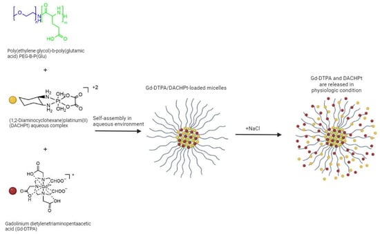
Figure 21.
Synthetic view of the structure of Gd–DTPA/DACHPt-loaded micelles and the consequently releasing of Pt and Gd complexes from the micelles (created with BioRender.com).
An alternative approach was described by Feng et al. [126] with the conjugation of Gd(III) and cisplatin on the surface of DOTA functionalized cross-linked small molecular micelles, as seen in Figure 22. The in vivo evaluation showed that the new theranostic nanoplatform exhibited not only a stronger and longer-lasting contrast enhancement efficacy but also a greater tumor growth inhibition rate with lower side effects. Due to the toxicity associated with the use of paramagnetic chelates for T1-weighted MRI, there is a demanding need to explore alternative transition-metal oxides [168].
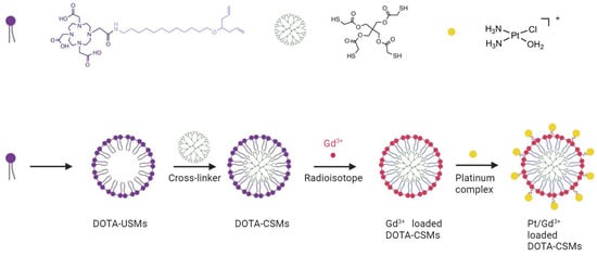
Figure 22.
Schematic procedure of Pt/Gd3+-loaded DOTA micelles (created with BioRender.com).
More recently, manganese dioxide (MnO2) nanostructures have emerged as a novel responsive MRI-T1 contrast agent [169]. In these nanoparticles, manganese presents a 4+ oxidation state, displaying weak paramagnetism. However, in the presence of biologically relevant concentrations of GSH or other redox-active species, MnO2 nanostructures undergo reduction to form free aqueous Mn(II) and O2. Mn in its 2+ oxidation state has two unpaired electrons and exhibits strong paramagnetism, leading to an enhanced MRI signal [170]. In this context, Hao et al. [169] reported a very promising approach by developing a tumor-targeted theranostic delivery system comprising degradable MnO2 nanosheets functionalized with hyaluronic acid (HA) and loaded with cisplatin, as shown in Figure 23. Here, HA was used as a delivery molecule, as overexpression of its receptors has been found in various tumors. Once the tumor is reached, controlled drug release is achieved by the tumor-microenvironment-responsive degradation of MnO2 nanosheets.

Figure 23.
Schematic illustration of preparation of MnO2/HA/cDDP nanosheets (created with BioRender.com).
However, the preparation of these theranostic agents required challenging multistep reactions. A few years later, Brito et al. [171] described a MnO2–Pt (IV) nanostructure synthesized through a simplified one-pot facile ultrasonication reaction, significantly reducing the time and complexity of the materials. These nanoparticles demonstrated a robust off/on MRI behavior both in vitro and in vivo in response to the reducing agents. They also exhibited efficiency in inducing cell death, comparable to that of cisplatin. However, further systemic imaging and therapeutic studies are still required to fully assess their potential.
In the same year, Zhang et al. [172] presented a promising strategy aimed at overcoming the primary limitation associated with polymeric micelles as drug delivery vehicles such as premature release, and drug binding with plasma proteins before reaching tumor cells. They developed poly(glycolic acid)/cisplatin (PGA/CDDP) NPs via electrostatic interaction, which were further coated with MnO2 to prevent premature leakage and augment the therapeutic effect of cisplatin in tumor cells. Additionally, the MnO2 shells can react with high concentration of GSH, H2O2 and H+ for a multi-responsive controlled drug release (Figure 24). This innovative approach led to a significant enhancement in the toxic effect of pure cisplatin of approximately 2–3 times, upon coupling with MnO2. Furthermore, serving as a multi-functional nanoplatform, PGA/CDDP@MnO2 NPs holds considerable promise for imaging and diagnosing various tumors.
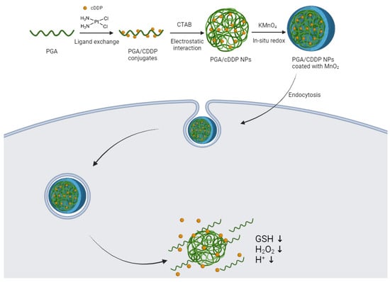
Figure 24.
Schematic diagram of the preparation process and controlled drug release of PGA/CDDP@MnO2 NPs (created with BioRender.com).
4.4. T2-Enhanced Contrast Agents
The most studied T2-enhanced contrast agents are superparamagnetic iron-oxide (Fe3O4) nanoparticles. In this case, the value of T2 is inversely proportional to its cross-sectional area, meaning that the same amount of magnetized material is more effective when dispersed as fewer large aggregates rather than numerous smaller ones [173]. Mn-doped Fe3O4 nanoparticles may be used as simultaneous dual-modal probes for T1/T2 MRI, providing complementary imaging information for an early and precise diagnosis [174].
SPIONs (Super Paramagnetic Iron Oxide Nanoparticles) possess some excellent properties, such as biocompatibility, hydrophilicity, nontoxicity, and no immunogenicity and as a result, they have garnered significant attention in recent years as both drug carriers and diagnostic agents [175,176]. Based on this idea, Xing et al. [177] synthesized superparamagnetic magnetite nanocrystal clusters. Sodium carboxymethylcellulose (Na-CMC SPMNC) was chosen as a biocompatible and biodegradable polymer for external coating. The platinum drug (monochlorinated CDDP, CMDP) was loaded onto the clusters, forming a conjugate of CMDP–CMC–SPMNC magnetic drug carriers that were able to transport platinum drug preferentially to its biological target by making use of a magnetic field (Figure 25). In addition to the benefits of simplicity in preparation and high loading capacity, the system significantly enhances the cellular uptake of platinum drugs. The cytotoxicity towards the human cervical cancer HeLa cells and the human hepatocarcinoma HepG2 cells of the drug carrier conjugate are higher than or at least comparable to those of cisplatin.
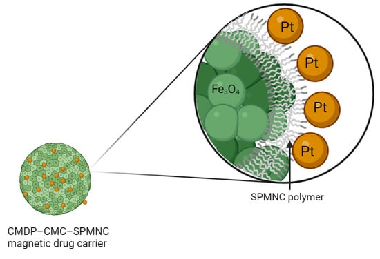
Figure 25.
Structure of conjugate CMDP–CMC–SPMNC magnetic drug carrier (created with BioRender.com).
In subsequent work, Wang et al. [128] reported a study where an analog of cisplatin (CMDP) was tethered to modified maghemite nanoparticles (OTPBA–SPION) for targeted cancer theranostics under the influence of an external magnetic field. This approach offered several advantages, including particle size conducive to tumor cell entry, straight-forward preparation, high drug-loading capacity, nontoxic degradation of the core, and more importantly, and specific accumulation in cancer tissues under an external magnetic field. However, there was a concern that Pt(II) moieties could react during delivery, potentially posing toxic effects on normal tissues.
To overcome these limitations, Zhu et al. [178] proposed a novel strategy involving the substitution of the previous Pt(II) pharmacophore with a Pt(IV) prodrug complex that is more inert and more stable than Pt(II) complexes as described earlier, thereby mitigating undesirable side reactions in the blood plasma. In this study, SPIONs were retained to construct a targeted Pt prodrug system, with polyethylene glycol (PEG) coating applied to enhance in vivo stability. A functionalized prodrug of cisplatin (HSPt) was loaded onto the surface of the PEGylated SPIONs. The system produced a significant negative contrast in MRI, suggesting its potential as a theranostic agent for chemotherapy. Cytotoxicity was enhanced in the presence of GSH, facilitating controlled drug release within the tumor microenvironment.
Cheng et al. [174] designed and developed an innovative Pt-FMO nanoplatform (where FMO stands for manganese deposited iron oxide, Fe–Mg composite oxide) that served as a simultaneous dual-modal probe for T1/T2 MRI while also enabling mutual beneficial cascade reactions to promote ferroptosis and apoptosis in combination with its utilization as an MRI agent. A cisplatin prodrug Pt(IV) containing a carboxylic acid in the axial position was covalently conjugated to the surface of FMO via amide bond formation and was activated through endogenous GSH to generate the Pt(II) drug (Figure 26). In vitro and in vivo essays demonstrate that a remarkably lower Pt dose (8.89%) of Pt-FMO was able to achieve a significant in vivo antitumor effect of cisplatin. The combination effect on tumor cells through cisplatin-induced apoptosis and Fenton-reaction-promoted ferroptosis is crucial to achieve such promising results.
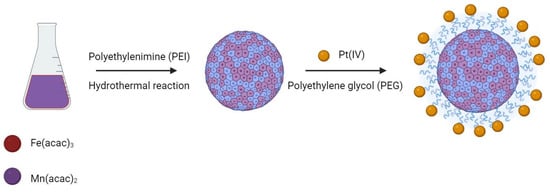
Figure 26.
Scheme of the preparation of magnetic Pt-FMO nanoparticles (created with BioRender.com).
5. Conclusions
The overview provided in this review about recent advancements in the use of Pt-based drugs coupled with various imaging techniques showed a huge potential in future translation to clinical studies. Nevertheless, further studies are needed to overcome the limitations of the different imaging techniques involved and to improve the activity of the anticancer agent.
Regarding fluorescence imaging, coupling Pt compounds with fluorophores to study their spatial-temporal distribution in cells poses a challenge because the fluorophore’s structure can alter the original Pt-based compound’s physicochemical properties. To address this issue, various exogenous turn-on probes have been developed. Among the different NIR-fluorescent probes mentioned before, AuNCs have the highest potential for in vivo tumor imaging due to their small size, tunable emissions from the visible to the NIR range, excellent biocompatibility, easy surface functionalization with peptides/proteins/antibodies for active cell-targeting, and enhanced EPR effect.
Regarding SPECT imaging, CISSPECT® was the most efficient theranostic agent developed to predict chemotherapy treatment efficacy of cisplatin, external radio beam therapy optimization and reduction in side effects such as severe kidney toxicity. While CISSPECT® shows promising results, PET imaging offers distinct advantages, including higher sensitivity, superior resolution, and the use of shorter-half-life radiotracers that reduce radiation exposure. Notably, we highlight the promising outcomes of terpyridine platinum-based complexes conjugated with 64Cu-NOTA as an innovative theranostic platform aimed at overcoming platinum drug resistance. Although PET imaging of Pt-theranostic compounds is still a rare technique, recent developments, such as the 68Ga-Pt(IV)azido complex (22), demonstrate significant potential for image-guided treatment of therapeutic Pt(IV) prodrugs.
In the field of MRI imaging, significant efforts have been made in the development of Pt-Gd complexes. While first-generation Gd-Pt complexes encountered challenges such as cytotoxicity and stability concerns, ongoing research has led to substantial advancements in their design and efficacy. Particularly noteworthy are texaphyrin-based and Pt(IV) conjugates, which hold promise for overcoming platinum resistance through targeted therapeutic action. Notably, complex 34 has demonstrated superior efficacy in inhibiting tumor growth, underscoring its potential for clinical applications. Moreover, nanotechnology has highly promoted MRI contrast agents research, thus offering versatile platforms that decrease the toxicity of Gd-based probes and achieve a stronger contrast enhancement. However, we believe SPIONs have a huge potential as both MRI contrast agents and drug delivery platforms due to their high biocompatibility, low toxicity, decreased immunogenicity, extended imaging windows, and easy surface functionalization with ligands or therapeutic agents.
For the future of molecular medicine, developing dual-modality molecular imaging probes will be pivotal, as no single imaging modality can provide all the necessary information. For example, PET offers excellent sensitivity and metabolic functionality but suffers from poor spatial resolution. Conversely, MRI provides supremely high-resolution anatomic information in the sub-millimeter range. By synergistically combining these modalities, we can achieve comprehensive diagnostic capabilities. Several current strategies are already moving in this direction [179,180].
Therefore, our future contribution in this field will be to develop SPION-based platforms with Pt-derivatives coupled to radio-chelators to create MRI/PET imaging theranostic probes for both cancer diagnosis and treatment. We aim to evaluate these probes both in vitro and in vivo to assess their potential for clinical translation.
In conclusion, the use of Pt-based complexes in a theranostic scenario emerges as a strategy to solve current issues of one of the widest-used anticancer drugs and to design an individual therapeutic intervention in combination with an appropriate diagnostic methodology. We are confident that this direction is a promising approach so that personalized molecular medicine may soon become a clinical reality.
Author Contributions
Conceptualization, D.M. and T.W.; writing—original draft preparation, G.F. and I.L.-M.; writing—review and editing, D.M., T.W. and C.K.; visualization, G.F. and I.L.-M.; supervision, D.M. and T.W.; project administration, D.M. and C.K.; funding acquisition, D.M. and C.K. All authors have read and agreed to the published version of the manuscript.
Funding
This research was funded by the STRIKE project HORIZON-MSCA-2021-DN-01, grant number 101072462.
Institutional Review Board Statement
Not applicable.
Informed Consent Statement
Not applicable.
Data Availability Statement
Data sharing is not applicable.
Conflicts of Interest
The authors declare no conflicts of interest.
References
- Peng, K.; Liang, B.B.; Liu, W.; Mao, Z.W. What Blocks More Anticancer Platinum Complexes from Experiment to Clinic: Major Problems and Potential Strategies from Drug Design Perspectives. Coord. Chem. Rev. 2021, 449, 214210. [Google Scholar] [CrossRef]
- Gibson, D. Multi–Action Pt(IV) Anticancer Agents; Do We Understand How They Work? J. Inorg. Biochem. 2019, 191, 77–84. [Google Scholar] [CrossRef] [PubMed]
- Wheate, N.J.; Walker, S.; Craig, G.E.; Oun, R. The Status of Platinum Anticancer Drugs in the Clinic and in Clinical Trials. Dalton Trans. 2010, 39, 8113–8127. [Google Scholar] [CrossRef]
- Theiner, S.; Varbanov, H.P.; Galanski, M.; Egger, A.E.; Berger, W.; Heffeter, P.; Keppler, B.K. Comparative In Vitro and In Vivo Pharmacological Investigation of Platinum(IV) Complexes as Novel Anticancer Drug Candidates for Oral Application. J. Biol. Inorg. Chem. 2015, 20, 89–99. [Google Scholar] [CrossRef] [PubMed]
- Kim, E.S.; Tang, X.M.; Peterson, D.R.; Kilari, D.; Chow, C.W.; Fujimoto, J.; Kalhor, N.; Swisher, S.G.; Stewart, D.J.; Wistuba, I.I.; et al. Copper Transporter CTR1 Expression and Tissue Platinum Concentration in Non-Small Cell Lung Cancer. Lung Cancer 2014, 85, 88–93. [Google Scholar] [CrossRef]
- Lasorsa, A.; Natile, G.; Rosato, A.; Tadini–Buoninsegni, F.; Arnesano, F. Monitoring Interactions Inside Cells by Advanced Spectroscopies: Overview of Copper Transporters and Cisplatin. Curr. Med. Chem. 2018, 25, 462–477. [Google Scholar] [CrossRef]
- Samimi, G.; Varki, N.M.; Wilczynski, S.; Safaei, R.; Alberts, D.S.; Howell, S.B. Increase in Expression of the Copper Transporter ATP7A during Platinum Drug-Based Treatment Is Associated with Poor Survival in Ovarian Cancer Patients. Clin. Cancer Res. 2003, 9, 5853–5859. [Google Scholar]
- Samimi, G.; Safaei, R.; Katano, K.; Holzer, A.K.; Rochdi, M.; Tomioka, M.; Goodman, M.; Howell, S.B. Increased Expression of the Copper Efflux Transporter ATP7A Mediates Resistance to Cisplatin, Carboplatin, and Oxaliplatin in Ovarian Cancer Cells. Clin. Cancer Res. 2004, 10, 4661–4669. [Google Scholar] [CrossRef]
- Iida, T.; Mori, E.; Mori, K.; Goto, S.; Urata, Y.; Oka, M.; Kohno, S.; Kondo, T. Co-Expression of Gamma-Glutamylcysteine Synthetase Sub-Units in Response to Cisplatin and Doxorubicin in Human Cancer Cells. Int. J. Cancer. 1999, 82, 405–411. [Google Scholar] [CrossRef]
- Goto, S.; Iida, T.; Cho, S.; Oka, M.; Kohno, S.; Kondo, T. Overexpression of Glutathione S-Transferase π Enhances the Adduct Formation of Cisplatin with Glutathione in Human Cancer Cells. Free Rad. Res. 1999, 31, 549–558. [Google Scholar] [CrossRef]
- Chen, G.; Hutter, K.-J.; Zeller, W.J. Positive Correlation between Cellular Glutathione and Acquired Cisplatin Resistance in Human Ovarian Cancer Cells. Cell Biol. Toxicol. 1995, 11, 273–281. [Google Scholar] [CrossRef] [PubMed]
- Wang, D.; Lippard, S.J. Cellular Processing of Platinum Anticancer Drugs. Nat. Rev. Durg Discov. 2005, 4, 307–320. [Google Scholar] [CrossRef] [PubMed]
- Kelland, L. The Resurgence of Platinum-Based Cancer Chemotherapy. Nat. Rev. Cancer 2007, 7, 573–584. [Google Scholar] [CrossRef] [PubMed]
- Dilruba, S.; Kalayda, G.V. Platinum–Based Drugs: Past, Present and Future. Cancer Chemother. Pharmacol. 2016, 77, 1103–1124. [Google Scholar] [CrossRef]
- Holzer, A.K.; Samimi, G.; Katano, K.; Naerdemann, W.; Lin, X.; Safaei, R.; Howell, S.B. The Copper Influx Transporter Human Copper Transport Protein 1 Regulates the Uptake of Cisplatin in Human Ovarian Carcinoma Cells. Mol. Pharmacol. 2004, 66, 817–823. [Google Scholar] [CrossRef] [PubMed]
- Yoshizawa, K.; Nozaki, S.; Kitahara, H.; Ohara, T.; Kato, K.; Kawashiri, S.; Yamamoto, E. Copper Efflux Transporter (ATP7B) Contributes to the acquisition of Cisplatin-Resistance in Human Oral Squamous Cell Lines. Oncol. Rep. 2007, 18, 987–991. [Google Scholar] [CrossRef] [PubMed]
- Nakayama, K.; Kanzaki, A.; Terada, K.; Mutoh, M.; Ogawa, K.; Sugiyama, T.; Takenoshita, S.; Itoh, K.; Yaegashi, N.; Miyazaki, K.; et al. Prognostic Value of the Cu-Transporting ATPase in Ovarian Carcinoma Patients Receiving Cisplatin-Based Chemotherapy. Clin. Cancer Res. 2004, 10, 1804–2811. [Google Scholar] [CrossRef] [PubMed]
- Ueda, S.; Shirabe, K.; Morita, K.; Umeda, K.; Kayashima, H.; Uchiyama, H.; Soejima, Y.; Taketomi, A.; Maehara, Y. Evaluation of ERCC1 Expression for Cisplatin Sensitivity in Human Hepatocellular Carcinoma. Ann. Surg. Oncol. 2011, 18, 1204–1211. [Google Scholar] [CrossRef] [PubMed]
- Britten, R.A.; Liu, D.; Tessier, A.; Hutchison, M.J.; Murray, D. ERCC1 Expression as a Molecular Marker of Cisplatin Resistance in Human Cervical Tumor Cells. Int. J. Cancer 2000, 89, 453–457. [Google Scholar] [CrossRef]
- Steffensen, K.D.; Waldstrøm, M.; Jakobsen, A. The Relationship of Platinum Resistance and ERCC1 Protein Expression in Epithelial Ovarian Cancer. Int. J. Gynecol. Cancer 2009, 19, 820–825. [Google Scholar] [CrossRef]
- Ulker, M.; Duman, B.B.; Sahin, B.; Gumurdulu, D. ERCC1 and RRM1 as a Predictive Parameter for Non-Small Cell Lung, Ovarian or Pancreas Cancer Treated with Cisplatin and/or Gemcitabine. Contemp. Oncol. 2015, 19, 207–213. [Google Scholar] [CrossRef] [PubMed]
- Kishi, K.; Doki, Y.; Yano, M.; Yasuda, T.; Fujiwara, Y.; Takiguchi, S.; Kim, S.; Higuchi, I.; Monden, M. Reduced MLH1 Expression after Chemotherapy Is an Indicator for Poor Prognosis in Esophageal Cancers. Clin. Cancer Res. 2003, 9, 4368–4375. [Google Scholar] [PubMed]
- Kawashima, N.; Yoshida, H.; Miwa, M.; Fujiwara, K. MLH1 Is a Prognostic Biomarker for Serous Ovarian Cancer Treated with Platinum- and Taxane-Based Chemotherapy. Anticancer Res. 2019, 39, 5505–5513. [Google Scholar] [CrossRef] [PubMed]
- Chavez–Dominguez, R.L.; Perez–Medina, M.A.; Lopez–Gonzalez, J.S.; Galicia–Velasco, M.; Matias–Florentino, M.; Avila–Rios, S.; Rumbo–Nava, U.; Salgado–Aguayo, A.; Gonzalez–Gonzalez, C.; Aguilar–Cazares, D. Role of HMGB1 in Cisplatin-Persistent Lung Adenocarcinoma Cell Lines. Front. Oncol. 2021, 11, 750677. [Google Scholar] [CrossRef] [PubMed]
- Xia, J.; Yu, X.; Song, X.; Li, G.; Mao, X.; Zhang, Y. Inhibiting the Cytoplasmic Location of HMGB1 Reverses Cisplatin Resistance in Human Cervical Cancer Cells. Mol. Med. Rep. 2017, 15, 488–494. [Google Scholar] [CrossRef] [PubMed]
- Ma, Y.; Feng, Q.; Han, B.; Yu, R.; Jin, Z. Elevated HMGB1 Promotes the Malignant Progression and Contributes to Cisplatin Resistance of Non-Small Cell Lung Cancer. Hereditas 2023, 160, 33. [Google Scholar] [CrossRef] [PubMed]
- Nishiguchi, Y.; Oue, N.; Fujiwara–Tani, R.; Sasaki, T.; Ohmori, H.; Kishi, S.; Mori, S.; Mori, T.; Ikeda, N.; Matsumoto, S.; et al. Role of Metastasis-Related Genes in Cisplatin Chemoresistance in Gastric Cancer. Int. J. Mol. Sci. 2020, 21, 254. [Google Scholar] [CrossRef] [PubMed]
- Timmerman, D.M.; Eleveld, T.F.; Gillis, A.J.M.; Friedrichs, C.C.; Hillenius, S.; Remmers, T.L.; Sriram, S.; Looijenga, L.H.J. The Role of Tp53 in Cisplatin Resistance in Mediastinal and Testicular Germ Cell Tumors. Int. J. Mol. Sci. 2021, 22, 11774. [Google Scholar] [CrossRef] [PubMed]
- Castedo, M.; Coquelle, A.; Vivet, S.; Vitale, I.; Kauffmann, A.; Dessen, P.; Pequignot, M.O.; Casares, N.; Valent, A.; Mouhamad, S.; et al. Apoptosis Regulation in Tetraploid Cancer Cells. EMBO J. 2006, 25, 2584–2595. [Google Scholar] [CrossRef]
- Ma, X.; Le Teuff, G.; Lacas, B.; Tsao, M.S.; Graziano, S.; Pignon, J.P.; Douillard, J.Y.; Le Chevalier, T.; Seymour, L.; Filipits, M.; et al. Prognostic and Predictive Effect of TP53 Mutations in Patients with Non-Small Cell Lung Cancer from Adjuvant Cisplatin-Based Therapy Randomized Trials: A LACE-Bio Pooled Analysis. J. Thorac. Oncol. 2016, 11, 850–861. [Google Scholar] [CrossRef]
- Wu, H.M.; Jiang, Z.F.; Ding, P.S.; Shao, L.J.; Liu, R.Y. Hypoxia-Induced Autophagy Mediates Cisplatin Resistance in Lung Cancer Cells. Sci. Rep. 2015, 5, 12291. [Google Scholar] [CrossRef] [PubMed]
- Wang, J.; Wu, G.S. Role of Autophagy in Cisplatin Resistance in Ovarian Cancer Cells. J. Biol. Chem. 2014, 289, 17163–17173. [Google Scholar] [CrossRef]
- Ren, J.-H.; He, W.-S.; Nong, L.; Zhu, Q.-Y.; Hu, K.; Zhang, R.-G.; Huang, L.-L.; Zhu, F.; Wu, G. Acquired Cisplatin Resistance in Human Lung Adenocarcinoma Cells Is Associated with Enhanced Autophagy. Cancer Biother. Radiophar. 2010, 25, 75–80. [Google Scholar] [CrossRef]
- Huang, D.; Duan, H.; Huang, H.; Tong, X.; Han, Y.; Ru, G.; Qu, L.; Shou, C.; Zhao, Z. Cisplatin Resistance in Gastric Cancer Cells Is Associated with HER2 Upregulation-Induced Epithelial-Mesenchymal Transition. Sci. Rep. 2016, 6, 20502. [Google Scholar] [CrossRef]
- Calikusu, Z.; Yildirim, Y.; Akcali, Z.; Sakalli, H.; Bal, N.; Unal, I.; Ozyilkan, O. The Effect of HER2 Expression on Cisplatin-Based Chemotherapy in Advanced Non-Small Cell Lung Cancer Patients. J. Exp. Clin. Cancer Res. 2009, 28, 97. [Google Scholar] [CrossRef]
- Gibson, D. Platinum(IV) Anticancer Prodrugs-Hypotheses and Facts. Dalton Trans. 2016, 45, 12983–12991. [Google Scholar] [CrossRef] [PubMed]
- Lee, K.H.; Hyun, M.S.; Kim, H.-K.; Jin, H.M.; Yang, J.; Song, H.S.; Do, Y.R.; Ryoo, H.M.; Chung, J.S.; Zang, D.Y.; et al. Randomized, Multicenter, Phase III Trial of Heptaplatin 1-Hour Infusion and 5-Fluorouracil Combination Chemotherapy Comparing with Cisplatin and 5-Fluorouracil Combination Chemotherapy in Patients with Advanced Gastric Cancer. Cancer Res. Treat. 2009, 41, 12–18. [Google Scholar] [CrossRef]
- McKeage, M.J. Lobaplatin: A New Antitumour Platinum Drug. Expert. Opin. Investig. Drugs 2001, 10, 119–128. [Google Scholar] [CrossRef] [PubMed]
- Yuan, Y.; Chen, Y.; Tang, B.Z.; Liu, B. A Targeted Theranostic Platinum(Iv) Prodrug Containing a Luminogen with Aggregation-Induced Emission (AIE) Characteristics for in Situ Monitoring of Drug Activation. Chem. Commun. 2014, 50, 3868–3870. [Google Scholar] [CrossRef]
- Kelland, L. Broadening the Clinical Use of Platinum Drug-Based Chemotherapy with New Analogues: Satraplatin and Picoplatin. Expert. Opin. Investig. Drugs 2007, 16, 1009–1021. [Google Scholar] [CrossRef]
- Wang, X.; Wang, X.; Guo, Z. Functionalization of Platinum Complexes for Biomedical Applications. Acc. Chem. Res. 2015, 48, 2622–2631. [Google Scholar] [CrossRef] [PubMed]
- Vivero-Escoto, J.L.; Huxford-Phillips, R.C.; Lin, W. Silica-Based Nanoprobes for Biomedical Imaging and Theranostic Applications. Chem. Soc. Rev. 2012, 41, 2673–2685. [Google Scholar] [CrossRef]
- Janib, S.M.; Moses, A.S.; MacKay, J.A. Imaging and Drug Delivery Using Theranostic Nanoparticles. Adv. Drug Deliv. Rev. 2010, 62, 1052–1063. [Google Scholar] [CrossRef] [PubMed]
- Asif, S.; Shahid, S. Emerging Trends in Nano-Theranostics: Platinum-Based Drug Delivery Systems for Cancer Treatment. Glob. Drug Des. Dev. Rev. 2022, 8, 15–28. [Google Scholar] [CrossRef]
- Workman, P.; Aboagye, E.O.; Balkwill, F.; Balmain, A.; Bruder, G.; Chaplin, D.J.; Double, J.A.; Everitt, J.; Farningham, D.A.H.; Glennie, M.J.; et al. Guidelines for the Welfare and Use of Animals in Cancer Research. Br. J. Cancer 2010, 102, 1555–1577. [Google Scholar] [CrossRef] [PubMed]
- Cabral, H.; Nishiyama, N.; Kataoka, K. Supramolecular Nanodevices: From Design Validation to Theranostic Nanomedicine. Acc. Chem. Res. 2011, 44, 999–1008. [Google Scholar] [CrossRef]
- James, M.L.; Gambhir, S.S. A Molecular Imaging Primer: Modalities, Imaging Agents, and Applications. Physiol. Rev. 2012, 92, 897–965. [Google Scholar] [CrossRef] [PubMed]
- Schouw, H.M.; Huisman, L.A.; Janssen, Y.F.; Slart, R.H.J.A.; Borra, R.J.H.; Willemsen, A.T.M.; Brouwers, A.H.; van Dijl, J.M.; Dierckx, R.A.; van Dam, G.M.; et al. Targeted Optical Fluorescence Imaging: A Meta-Narrative Review and Future Perspectives. Eur. J. Nucl. Med. Mol. Imaging 2021, 48, 4272–4292. [Google Scholar] [CrossRef]
- Kirin, S.I.; Ott, I.; Gust, R.; Mier, W.; Weyhermüller, T.; Metzler–Nolte, N. Cellular Uptake Quantification of Metalated Peptide and Peptide Nucleic Acid Bioconjugates by Atomic Absorption Spectroscopy. Angew. Chem. Int. Ed. 2008, 47, 955–959. [Google Scholar] [CrossRef]
- Egger, A.E.; Rappel, C.; Jakupec, M.A.; Hartinger, C.G.; Heffeter, P.; Keppler, B.K. Development of an Experimental Protocol for Uptake Studies of Metal Compounds in Adherent Tumor Cells. J. Anal. At. Spectrom. 2009, 24, 51–61. [Google Scholar] [CrossRef][Green Version]
- Hambley, T.W. The Influence of Structure on the Activity and Toxicity of Pt Anti-Cancer Drugs. Coord. Chem. Rev. 1997, 166, 181–223. [Google Scholar] [CrossRef]
- Safaei, R.; Katano, K.; Larson, B.J.; Samimi, G.; Holzer, A.K.; Naerdemann, W.; Tomioka, M.; Goodman, M.; Howell, S.B. Intracellular Localization and Trafficking of Fluorescein-Labeled Cisplatin in Human Ovarian Carcinoma Cells. Clin. Cancer Res. 2005, 11, 756–767. [Google Scholar] [CrossRef] [PubMed]
- Molenaar, C.; Teuben, J.M.; Heetebrij, R.J.; Tanke, H.J.; Reedijk, J. New Insights in the Cellular Processing of Platinum Antitumor Compounds, Using Fluorophore-Labeled Platinum Complexes and Digital Fluorescence Microscopy. J. Biol. Inorg. Chem. 2000, 5, 655–665. [Google Scholar] [CrossRef] [PubMed]
- Liang, X.J.; Shen, D.W.; Chen, K.G.; Wincovitch, S.M.; Garfield, S.H.; Gottesman, M.M. Trafficking and Localization of Platinum Complexes in Cisplatin-Resistant Cell Lines Monitored by Fluorescence-Labeled Platinum. J. Cell. Physiol. 2005, 202, 635–641. [Google Scholar] [CrossRef] [PubMed]
- Jagodinsky, J.C.; Sulima, A.; Cao, Y.; Poprawski, J.E.; Blackman, B.N.; Lloyd, J.R.; Swenson, R.E.; Gottesman, M.M.; Hall, M.D. Evaluation of Fluorophore-Tethered Platinum Complexes to Monitor the Fate of Cisplatin Analogs. J. Biol. Inorg. Chem. 2015, 20, 1081–1095. [Google Scholar] [CrossRef] [PubMed]
- Sun, T.; Guan, X.; Zheng, M.; Jing, X.; Xie, Z. Mitochondria-Localized Fluorescent BODIPY-Platinum Conjugate. ACS Med. Chem. Lett. 2015, 6, 430–433. [Google Scholar] [CrossRef] [PubMed]
- Miller, M.A.; Askevold, B.; Yang, K.S.; Kohler, R.H.; Weissleder, R. Platinum Compounds for High-Resolution In Vivo Cancer Imaging. ChemMedChem 2014, 9, 1131–1135. [Google Scholar] [CrossRef] [PubMed]
- Wexselblatt, E.; Yavin, E.; Gibson, D. Cellular Interactions of Platinum Drugs. Inorganica Chim. Acta 2012, 393, 75–83. [Google Scholar] [CrossRef]
- Shen, C.; Harris, B.D.W.; Dawson, L.J.; Charles, K.A.; Hambley, T.W.; New, E.J. Fluorescent Sensing of Monofunctional Platinum Species. Chem. Comm. 2015, 51, 6312–6314. [Google Scholar] [CrossRef]
- Kolanowski, J.L.; Dawson, L.J.; Mitchell, L.; Lim, Z.; Graziotto, M.E.; Filipek, W.K.; Hambley, T.W.; New, E.J. A Fluorescent Probe for Investigating Metabolic Stability of Active Transplatin Analogues. Sens. Actuators B Chem. 2018, 255, 2721–2724. [Google Scholar] [CrossRef]
- Ong, J.X.; Yap, J.Y.; Yap, S.Q.; Ang, W.H. Structure-Activity Relationship Studies on Rhodamine B-Based Fluorogenic Probes and Their Activation by Anticancer Platinum(II) Compounds. J. Inorg. Biochem. 2015, 153, 272–278. [Google Scholar] [CrossRef] [PubMed]
- Montagner, D.; Yap, S.Q.; Ang, W.H. A Fluorescent Probe for Investigating the Activation of Anticancer Platinum(IV) Prodrugs Based on the Cisplatin Scaffold. Angew. Chem. 2013, 125, 12001–12005. [Google Scholar] [CrossRef]
- Ong, J.X.; Lim, C.S.Q.; Le, H.V.; Ang, W.H. A Ratiometric Fluorescent Probe for Cisplatin: Investigating the Intracellular Reduction of Platinum(IV) Prodrug Complexes. Angew. Chem. Int. Ed. 2019, 58, 164–167. [Google Scholar] [CrossRef] [PubMed]
- Ong, J.X.; Ang, W.H. Development of a Pre-Assembled Through-Bond Energy Transfer (TBET) Fluorescent Probe for Ratiometric Sensing of Anticancer Platinum(Ll) Complexes. Chem. Asian J. 2020, 15, 1449–1455. [Google Scholar] [CrossRef]
- Fan, F.; Zhang, L.; Mu, F.; Shi, G. Using a High Quantum Yield Fluorescent Probe with Two-Photon Excitation to Detect Cisplatin in Biological Systems. ACS Sens. 2021, 6, 1400–1406. [Google Scholar] [CrossRef]
- Song, Y.; Suntharalingam, K.; Yeung, J.S.; Royzen, M.; Lippard, S.J. Synthesis and Characterization of Pt(Iv) Fluorescein Conjugates to Investigate Pt(Iv) Intracellular Transformations. Bioconjug. Chem. 2013, 24, 1733–1740. [Google Scholar] [CrossRef]
- Ding, F.; Fan, Y.; Sun, Y.; Zhang, F. Beyond 1000 Nm Emission Wavelength: Recent Advances in Organic and Inorganic Emitters for Deep-Tissue Molecular Imaging. Adv. Healthc. Mater. 2019, 8, 1900260. [Google Scholar] [CrossRef]
- Hong, G.; Antaris, A.L.; Dai, H. Near-Infrared Fluorophores for Biomedical Imaging. Nat. Biomed. Eng. 2017, 1, 10. [Google Scholar] [CrossRef]
- Zhu, S.; Wang, X.; Cong, Y.; Li, L. Regulating the Optical Properties of Gold Nanoclusters for Biological Applications. ACS Omega 2020, 5, 22702–22707. [Google Scholar] [CrossRef]
- Zhou, F.; Feng, B.; Yu, H.; Wang, D.; Wang, T.; Liu, J.; Meng, Q.; Wang, S.; Zhang, P.; Zhang, Z.; et al. Cisplatin Prodrug-Conjugated Gold Nanocluster for Fluorescence Imaging and Targeted Therapy of the Breast Cancer. Theranostics 2016, 6, 679–687. [Google Scholar] [CrossRef]
- Yu, Z.; Xiao, H.; Zhang, X.; Yang, Y.; Yu, Y.; Chen, H.; Meng, X.; Ma, W.; Yu, M.; Li, Z.; et al. Illuminating Platinum Transportation While Maximizing Therapeutic Efficacy by Gold Nanoclusters via Simultaneous Near-Infrared-I/II Imaging and Glutathione Scavenging. ACS Nano 2020, 14, 13536–13547. [Google Scholar] [CrossRef]
- Ding, D.; Li, K.; Zhu, Z.; Pu, K.Y.; Hu, Y.; Jiang, X.; Liu, B. Conjugated Polyelectrolyte-Cisplatin Complex Nanoparticles for Simultaneous in Vivo Imaging and Drug Tracking. Nanoscale 2011, 3, 1997–2002. [Google Scholar] [CrossRef] [PubMed]
- Saha, M.L.; Yan, X.; Stang, P.J. Photophysical Properties of Organoplatinum(II) Compounds and Derived Self-Assembled Metallacycles and Metallacages: Fluorescence and Its Applications. Acc. Chem. Res. 2016, 49, 2527–2539. [Google Scholar] [CrossRef] [PubMed]
- Sun, Y.; Ding, F.; Zhou, Z.; Li, C.; Pu, M.; Xu, Y.; Zhan, Y.; Lu, X.; Li, H.; Yang, G.; et al. Rhomboidal Pt(II) Metallacycle-Based NIR-II Theranostic Nanoprobe for Tumor Diagnosis and Image-Guided Therapy. Proc. Natl. Acad. Sci. USA 2019, 116, 1968–1973. [Google Scholar] [CrossRef] [PubMed]
- Wang, Y.; Zhang, W.; Sun, P.; Cai, Y.; Xu, W.; Fan, Q.; Hu, Q.; Han, W. A Novel Multimodal NIR-II Nanoprobe for the Detection of Metastatic Lymph Nodes and Targeting Chemo-Photothermal Therapy in Oral Squamous Cell Carcinoma. Theranostics 2019, 9, 391–404. [Google Scholar] [CrossRef] [PubMed]
- Liu, Y.; Li, Z.; Chen, L.; Xie, Z. Near Infrared BODIPY-Platinum Conjugates for Imaging, Photodynamic Therapy and Chemotherapy. Dye. Pigm. 2017, 141, 5–12. [Google Scholar] [CrossRef]
- Xing, X.; Pang, E.; Zhao, S.; Pan, T.; Tan, Q.; Wang, B.; Song, X.; Lan, M. Cisplatin-Appended BODIPY for near Infrared II Fluorescent and Photoacoustic Imaging-Guided Synergistic Phototherapy and Chemotherapy of Cancer. Chin. Chem. Lett. 2024, 35, 108467. [Google Scholar] [CrossRef]
- Isaac, K.M.; Sabaraya, I.V.; Ghousifam, N.; Das, D.; Pekkanen, A.M.; Romanovicz, D.K.; Long, T.E.; Saleh, N.B.; Rylander, M.N. Functionalization of Single-Walled Carbon Nanohorns for Simultaneous Fluorescence Imaging and Cisplatin Delivery In Vitro. Carbon 2018, 138, 309–318. [Google Scholar] [CrossRef]
- Nasrollahi, F.; Koh, Y.R.; Chen, P.; Varshosaz, J.; Khodadadi, A.A.; Lim, S. Targeting Graphene Quantum Dots to Epidermal Growth Factor Receptor for Delivery of Cisplatin and Cellular Imaging. Mater. Sci. Eng. C 2019, 94, 247–257. [Google Scholar] [CrossRef]
- Liu, H.; Li, C.; Qian, Y.; Hu, L.; Fang, J.; Tong, W.; Nie, R.; Chen, Q.; Wang, H. Magnetic-Induced Graphene Quantum Dots for Imaging-Guided Photothermal Therapy in the Second near-Infrared Window. Biomaterials 2020, 232, 119700. [Google Scholar] [CrossRef]
- Chen, Y.; Xue, L.; Zhu, Q.; Feng, Y.; Wu, M. Recent Advances in Second Near-Infrared Region (NIR-II) Fluorophores and Biomedical Applications. Front. Chem. 2021, 9, 750404. [Google Scholar] [CrossRef]
- Gong, L.; Shan, X.; Zhao, X.H.; Tang, L.; Zhang, X.B. Activatable NIR-II Fluorescent Probes Applied in Biomedicine: Progress and Perspectives. ChemMedChem 2021, 16, 2426–2440. [Google Scholar] [CrossRef] [PubMed]
- Crișan, G.; Moldovean-cioroianu, N.S.; Timaru, D.G.; Andrieș, G.; Căinap, C.; Chiș, V. Radiopharmaceuticals for PET and SPECT Imaging: A Literature Review over the Last Decade. Int. J. Mol. Sci. 2022, 23, 5023. [Google Scholar] [CrossRef]
- Rahmim, A.; Zaidi, H. PET versus SPECT: Strengths, Limitations and Challenges. Nucl. Med. Commun. 2008, 29, 193–207. [Google Scholar] [CrossRef] [PubMed]
- Kawai, K.; Tanaka, Y.; Yukihiro, N. Synthesis of Platinum-195m Radiolabelled Cis-Diammine(l,l-Cyclobutanedicarboxylato) Platinum(L1) of High Radionuclidic Purity. J. Label. Compd. Radiopharm. 1995, 36, 65–71. [Google Scholar] [CrossRef]
- Anand, D.; Wolf, W. A New, Semi-Automated System for the Micro-Scale Synthesis of [195mPt]Cisplatin Suitable for Clinical Studies. Int. J. Rad. Appl. Instr. A 1992, 43, 809–814. [Google Scholar] [CrossRef] [PubMed]
- Bodnar, E.N.; Dikiy, M.P.; Medvedeva, E.P. Photonuclear Production and Antitumor Effect of Radioactive Cisplatin (195mPt). J. Radioanal. Nucl. Chem. 2015, 305, 133–138. [Google Scholar] [CrossRef]
- Dowell, J.A.; Sancho, A.R.; Anand, D.; Wolf, W. Noninvasive Measurements for Studying the Tumoral Pharmacokinetics of Platinum Anticancer Drugs in Solid Tumors. Adv. Drug Deliv. Rev. 2000, 41, 111–126. [Google Scholar] [CrossRef] [PubMed]
- Sathekge, M.; Wagener, J.; Smith, S.V.; Soni, N.; Marjanovic-Painter, B.; Zinn, C.; Van de Wiele, C.; D’Asseler, Y.; Perkins, G.; Zeevaart, J.R. Biodistribution and Dosimetry of 195mPt-Cisplatin in Normal Volunteers. Imaging Agent for Single Photon Emission Computed Tomography. Nucl. Med. 2013, 52, 222–227. [Google Scholar] [CrossRef]
- Aalbersberg, E.A.; de Wit van der Veen, B.J.; Zwaagstra, O.; van der Schilden, K.C.; Vegt, E.; Vogel, W.V. Preclinical Imaging Characteristics and Quantification of Platinum-195m SPECT. Eur. J. Nucl. Med. Mol. Imaging 2017, 44, 1347–1354. [Google Scholar] [CrossRef]
- Howell, R.W.; Kassis, A.I.; James Adelstein, S.; Rao, D.V.; Wright, H.A.; Hamm, R.N.; Turner, J.E.; Sastry, K.S.R. Radiotoxicity of Platinum-195m-Labeled Trans-Platinum (II) in Mammalian Cells. Radiat. Res. 1994, 140, 55–62. [Google Scholar] [CrossRef]
- Aalbersberg, E.A. Imaging Biomarker Development and Optimization of 195mPT-Cisplatin and 68Ga-DOTATATE; University of Utrecht: Utrecht, The Netherlands, 2019. [Google Scholar]
- Nadar, R.A.; Farbod, K.; van der Schilden, K.C.; Schlatt, L.; Crone, B.; Asokan, N.; Curci, A.; Brand, M.; Bornhaeuser, M.; Iafisco, M.; et al. Targeting of Radioactive Platinum-Bisphosphonate Anticancer Drugs to Bone of High Metabolic Activity. Sci. Rep. 2020, 10, 5889. [Google Scholar] [CrossRef] [PubMed]
- Nadar, R.A.; Franssen, G.M.; Van Dijk, N.W.M.; van der Schilden, K.C.; de Weijert, M.; Oosterwijk, E.; Iafisco, M.; Margiotta, N.; Heskamp, S.; van den Beucken, J.J.J.P.; et al. Bone Tumor-Targeted Delivery of Theranostic 195mPt-Bisphosphonate Complexes Promotes Killing of Metastatic Tumor Cells. Mater. Today Bio 2021, 9, 100088. [Google Scholar] [CrossRef]
- Areberg, J. Studies of Radioactive Cisplatin (191Pt) for Tumour Imaging and Therapy; Lund University: Malmo, Sweden, 2000. [Google Scholar]
- Areberg, J.; Norrgren, K.; Ren Mattsson, S. Absorbed Doses to Patients from 191 Pt-, 193m Pt-and 195m Pt-Cisplatin. Appl. Radiat. Isot. 1999, 51, 581–586. [Google Scholar] [CrossRef]
- Norrgren, K.; Sjölin, M.; Björkman, S.; Areberg, J.; Johnsson, A.; Johansson, L.; Mattsson, S. Comparative Renal, Hepatic, and Bone Marrow Toxicity of Cisplatin and Radioactive Cisplatin (191 Pt) in Wistar Rats. Cancer Biother. Radiopharm. 2006, 21, 528–534. [Google Scholar] [CrossRef] [PubMed]
- Areberg, J.; Johnsson, A.; Wennerberg, J. In Vitro Toxicity of (191)Pt-Labeled Cisplatin to a Human Cervical Carcinoma Cell Line (ME-180). Int. J. Radiat. Oncol. Biol. Phys. 2000, 46, 1275–1280. [Google Scholar] [CrossRef] [PubMed]
- Areberg, J.; Wennerberg, J.; Johnsson, A.; Norrgren, K.; Sören Mattsson, S. Antitumor Effect of Radioactive Cisplatin (191Pt) on Nude Mice. Int. J. Radiat. Oncol. Biol. Phys. 2001, 49, 827–832. [Google Scholar] [CrossRef]
- Vaidya, S.P.; Gadre, S.; Kamisetti, R.T.; Patra, M. Challenges and Opportunities in the Development of Metal-Based Anticancer Theranostic Agents. Biosci. Rep. 2022, 42, BSR20212160. [Google Scholar] [CrossRef] [PubMed]
- Holschbach, M.; Hamkens, W.; Steinbach, A.; Hamacher, K.; Stocklin, G. [13N]Cisplatin: A Fast and Efficient On-Line Synthesis Using a Solid State Support. Appl. Radial. Isot. 1997, 48, 739–744. [Google Scholar] [CrossRef]
- De Spiegeleer, B.; Slegers, G.; Vandecasteele, C.; Van den Bossche, W.; Schelstraete, K.; Claeys, A.; De Moerloose, P. Microscale Synthesis of Nitrogen-13-Labeled Cisplatin. J. Nucl. Med. 1986, 27, 399–403. [Google Scholar]
- Ginos, J.Z.; Cooper, A.J.L.; Dhawan, V.; Lai, J.C.K.; Strother, S.C.; Alcock, N.; Rottenberg, D.A.; Rottenberg, D.A. [13N]Cisplatin PET to Assess Pharmacokinetics of Intra-ArterialVersus Intravenous Chemotherapy for Malignant Brain Tumors. J. Nucl. Med. 1987, 28, 1844–1852. [Google Scholar]
- Lamichhane, N.; Dewkar, G.K.; Sundaresan, G.; Wang, L.; Jose, P.; Otabashi, M.; Morelle, J.L.; Farrell, N.; Zweit, J. 18F-Labeled Carboplatin Derivative for PET Imaging of Platinum Drug Distribution. J. Nucl. Med. 2017, 58, 1997–2003. [Google Scholar] [CrossRef]
- Cai, L.; Lu, S.; Pike, V.W. Chemistry with [18F]Fluoride Ion. Eur. J. Org. Chem. 2008, 2008, 2853–2873. [Google Scholar] [CrossRef]
- Holland, J.P.; Williamson, M.J.; Lewis, J.S. Unconventional Nuclides for Radiopharmaceuticals. Mol. Imaging 2010, 9, 1–20. [Google Scholar] [CrossRef]
- Lamichhane, N.; Dewkar, G.K.; Sundaresan, G.; Mahon, R.N.; Zweit, J. [18F]-Fluorinated Carboplatin and [111in]-Liposome for Image-Guided Drug Delivery. Int. J. Mol. Sci. 2017, 18, 1079. [Google Scholar] [CrossRef] [PubMed]
- Cosialls, R.; Simó, C.; Borrós, S.; Gómez-Vallejo, V.; Schmidt, C.; Llop, J.; Cuenca, A.B.; Casini, A. PET Imaging of Self-Assembled 18F-Labelled Pd2L4 Metallacages for Anticancer Drug Delivery. Chem. Eur. J. 2023, 29, e202202604. [Google Scholar] [CrossRef]
- Li, X.; Zhao, X.; Wang, W.; Shi, Z.; Zhang, Y.; Tian, Q.; Yao, Y.; He, C.; Duan, C. Biomedical Applications of Multinuclear Pt(II)/Ru(II)/Ir(III) Metallo-Supramolecular Assemblies for Intensive Cancer Therapy. Coord. Chem. Rev. 2023, 495, 215366. [Google Scholar] [CrossRef]
- Khosravifarsani, M.; Ait-Mohand, S.; Paquette, B.; Sanche, L.; Guérin, B. High Cytotoxic Effect by Combining Copper-64 with a NOTA-Terpyridine Platinum Conjugate. J. Med. Chem. 2021, 64, 6765–6776. [Google Scholar] [CrossRef] [PubMed]
- Khosravifarsani, M.; Ait-Mohand, S.; Paquette, B.; Sanche, L.; Guérin, B. Design, Synthesis, and Cytotoxicity Assessment of [64cu]Cu-Nota-Terpyridine Platinum Conjugate: A Novel Chemoradiotherapeutic Agent with Flexible Linker. Nanomaterials 2021, 11, 2154. [Google Scholar] [CrossRef]
- Gutfilen, B.; Mendes, F.; Guérin, B. In Vivo Behaviour of [64Cu]NOTA-Terpyridine Platinum, a Novel Chemo-Radio-Theranostic Agent for Imaging, and Theraphy of Colorectal Cancer. Front. Med. 2022, 9, 975213. [Google Scholar] [CrossRef]
- Lo, R.; Majid, A.; Fruhwirth, G.O.; Vilar, R. Radiolabelling Pt-Based Quadruplex DNA Binders via Click Chemistry. Bioorg. Med. Chem. 2022, 76, 117097. [Google Scholar] [CrossRef]
- Imberti, C.; Lok, J.; Coverdale, J.P.C.; Carter, O.W.L.; Fry, M.E.; Postings, M.L.; Kim, J.; Firth, G.; Blower, P.J.; Sadler, P.J. Radiometal-Labeled Photoactivatable Pt(IV) Anticancer Complex for Theranostic Phototherapy. Inorg. Chem. 2023, 62, 20745–20753. [Google Scholar] [CrossRef] [PubMed]
- Weissleder, R.; Pittet, M.J. Imaging in the Era of Molecular Oncology. Nature 2008, 452, 580–589. [Google Scholar] [CrossRef]
- Ara, S.A.; Katti, G.; Shireen, A. Magnetic Resonance Imaging (MRI)—A Review. Int. J. Clin. Dent. 2011, 3, 65–70. [Google Scholar]
- Verwilst, P.; Park, S.; Yoon, B.; Kim, J.S. Recent Advances in Gd-Chelate Based Bimodal Optical/MRI Contrast Agents. Chem. Soc. Rev. 2015, 44, 1791–1806. [Google Scholar] [CrossRef] [PubMed]
- Cabral, H.; Nishiyama, N.; Kataoka, K. Optimization of (1,2-Diamino-Cyclohexane)Platinum(II)-Loaded Polymeric Micelles Directed to Improved Tumor Targeting and Enhanced Antitumor Activity. J. Control. Release 2007, 121, 146–155. [Google Scholar] [CrossRef]
- Wahsner, J.; Gale, E.M.; Rodríguez-Rodríguez, A.; Caravan, P. Chemistry of MRI Contrast Agents: Current Challenges and New Frontiers. Chem. Rev. 2019, 119, 957–1057. [Google Scholar] [CrossRef]
- Lohrke, J.; Frenzel, T.; Endrikat, J.; Alves, F.C.; Grist, T.M.; Law, M.; Lee, J.M.; Leiner, T.; Li, K.C.; Nikolaou, K.; et al. 25 Years of Contrast-Enhanced MRI: Developments, Current Challenges and Future Perspectives. Adv. Ther. 2016, 33, 1–28. [Google Scholar] [CrossRef] [PubMed]
- Medical Imaging Drugs Advisory Committee Meeting. Gadolinium Retention after Gadolinium Based Contrast Magnetic Resonance Imaging in Patients with Normal Renal Function Briefing Document. 2017. Available online: https://www.fda.gov/media/107133/download (accessed on 25 April 2024).
- Zhang, Z.; Nair, S.A.; Mcmurry, T.J. Gadolinium Meets Medicinal Chemistry: MRI Contrast Agent Development. Curr. Med. Chem. 2005, 12, 751–778. [Google Scholar] [CrossRef]
- William, W.N.; Zinner, R.G.; Karp, D.D.; Oh, Y.W.; Glisson, B.S.; Phan, S.-C.; Stewart, D.J. Phase I Trial of Motexafin Gadolinium in Combination with Docetaxel and Cisplatin for the Treatment of Non-Small Cell Lung Cancer. J. Thorac. Oncol. 2007, 2, 745–750. [Google Scholar] [CrossRef]
- Brugger, R.M. Gadolinium as Xda Neutron Capture Therapy Agent. Med. Phys. 1992, 19, 733–744. [Google Scholar] [CrossRef]
- Salt, C.; Lennox, A.J.; Takagaki, M.; Maguire, J.A.; Hosmane, N.S. Boron and Gadolinium Neutron Capture Therapy. Russ. Chem. Bull. 2004, 53, 1871–1888. [Google Scholar] [CrossRef]
- Feng, J.; Luo, Q.; Chen, Y.; Li, B.; Luo, K.; Lan, J.; Yu, Y.; Zhang, S. DOTA Functionalized Cross-Linked Small-Molecule Micelles for Theranostics Combining Magnetic Resonance Imaging and Chemotherapy. Bioconjug. Chem. 2018, 29, 3402–3410. [Google Scholar] [CrossRef] [PubMed]
- Kaida, S.; Cabral, H.; Kumagai, M.; Kishimura, A.; Terada, Y.; Sekino, M.; Aoki, I.; Nishiyama, N.; Tani, T.; Kataoka, K. Visible Drug Delivery by Supramolecular Nanocarriers Directing to Single-Platformed Diagnosis and Therapy of Pancreatic Tumor Model. Cancer Res. 2010, 70, 7031–7041. [Google Scholar] [CrossRef] [PubMed]
- Wang, J.; Wang, X.; Song, Y.; Wang, J.; Zhang, C.; Chang, C.; Yan, J.; Qiu, L.; Wu, M.; Guo, Z. A Platinum Anticancer Theranostic Agent with Magnetic Targeting Potential Derived from Maghemite Nanoparticles. Chem. Sci. 2013, 4, 2605–2612. [Google Scholar] [CrossRef]
- Rammohan, N.; MacRenaris, K.W.; Moore, L.K.; Parigi, G.; Mastarone, D.J.; Manus, L.M.; Lilley, L.M.; Preslar, A.T.; Waters, E.A.; Filicko, A.; et al. Nanodiamond-Gadolinium(III) Aggregates for Tracking Cancer Growth in Vivo at High Field. Nano Lett. 2016, 16, 7551–7564. [Google Scholar] [CrossRef] [PubMed]
- Robertson, A.G.; Rendina, L.M. Gadolinium Theranostics for the Diagnosis and Treatment of Cancer. Chem. Soc. Rev. 2021, 50, 4231–4244. [Google Scholar] [CrossRef] [PubMed]
- Crossley, E.L.; Aitken, J.B.; Vogt, S.; Harris, H.H.; Rendina, L.M. Selective Aggregation of a Platinum-Gadolinium Complex within a Tumor-Cell Nucleus. Angew. Chem. Int. Ed. 2010, 49, 1231–1233. [Google Scholar] [CrossRef] [PubMed]
- Fenton, J.M.; Busse, M.; Rendina, L.M. Synthesis and DNA-Binding Studies of a Dinuclear Gadolinium(Iii)-Platinum(Ii) Complex. Aust. J. Chem. 2015, 68, 576–580. [Google Scholar] [CrossRef][Green Version]
- Zhu, Z.; Wang, X.; Li, T.; Aime, S.; Sadler, P.J.; Guo, Z. Platinum(II)-Gadolinium(III) Complexes as Potential Single-Molecular Theranostic Agents for Cancer Treatment. Angew. Chem. Int. Ed. 2014, 53, 13225–13228. [Google Scholar] [CrossRef]
- Rosenthal, D.I.; Nurenberg, P.; Becerra, C.R.; Frenkel, E.P.; Carbone, D.P.; Lum, B.L.; Miller, R.; Engel, J.; Young, S.; Miles, D.; et al. A Phase I Single-Dose Trial of Gadolinium Texaphyrin (Gd-Tex), a Tumor Selective Radiation Sensitizer Detectable by Magnetic Resonance Imaging. Clin. Cancer Res. 1999, 5, 739–745. [Google Scholar] [PubMed]
- Pica, A.; Rosenthal, D.I.; Koprowski, C.; Schea, R.; Ruckle, J.; Tishler, R.; Larner, J.M.; Arwood, D.; Haie-Meder, C.; Young, S.; et al. A Phase Multi-Dose Trial of Gadolinium-Texaphyrin (Gd-Tex) as a Radiosensitizer in Patients with Brain Metastases Treated with Conventional Radiation Therapy: Preliminary Results. Int. J. Radiat. Oncol. Biol. Phys. 1997, 39, 270. [Google Scholar] [CrossRef]
- Miller, R.A.; Woodburn, K.; Fan, Q.; Renschler, M.F.; Sessler, J.L.; Koutcher, J.A. In Vivo Animal Studies with Gadolinium (III) Texaphyrin as a Radiation Enhancer. Int. J. Radiat. Oncol. Biol. Phys. 1999, 45, 981–989. [Google Scholar] [CrossRef] [PubMed]
- Mody, T.D.; Sessler, J.L. Texaphyrins: A New Approach to Drug Development. J. Porphyr. Phthalocyanines 2001, 5, 134–142. [Google Scholar] [CrossRef]
- Magda, D.J.; Wang, Z.; Gerasimchuk, N.; Wei, W.; Anzenbacher, P.; Sessler, J.L. Synthesis of Texaphyrin Conjugates. Pure Appl. Chem. 2004, 76, 365–374. [Google Scholar] [CrossRef]
- Arambula, J.F.; Sessler, J.L.; Fountain, M.E.; Wei, W.; Magda, D.; Siddik, Z.H. Gadolinium Texaphyrin (Gd-Tex)-Malonato-Platinum Conjugates: Synthesis Comparison with Carboplatin in Normal and Pt-Resistant Cell Lines. Dalton Trans. 2009, 48, 10834–10840. [Google Scholar] [CrossRef] [PubMed]
- Arambula, J.F.; Sessler, J.L.; Siddik, Z.H. Overcoming Biochemical Pharmacologic Mechanisms of Platinum Resistance with a Texaphyrin-Platinum Conjugate. Bioorg. Med. Chem. Lett. 2011, 21, 1701–1705. [Google Scholar] [CrossRef] [PubMed]
- Arambula, J.F.; Sessler, J.L.; Siddik, Z.H. A Texaphyrin-Oxaliplatin Conjugate That Overcomes Both Pharmacologic and Molecular Mechanisms of Cisplatin Resistance in Cancer Cells. Medchemcomm 2012, 3, 1275–1281. [Google Scholar] [CrossRef]
- Thiabaud, G.; Arambula, J.F.; Siddik, Z.H.; Sessler, J.L. Photoinduced Reduction of Pt(IV) within an Anti-Proliferative Pt(IV)-Texaphyrin Conjugate. Chemistry 2014, 20, 8942–8947. [Google Scholar] [CrossRef]
- Thiabaud, G.; He, G.; Sen, S.; Shelton, K.A.; Baze, W.B.; Segura, L.; Alaniz, J.; Munoz Macias, R.; Lyness, G.; Watts, A.B.; et al. Oxaliplatin Pt(IV) Prodrugs Conjugated to Gadolinium-Texaphyrin as Potential Antitumor Agents. Proc. Natl. Acad. Sci. USA 2020, 117, 7021–7029. [Google Scholar] [CrossRef]
- Thiabaud, G.; McCall, R.; He, G.; Arambula, J.F.; Siddik, Z.H.; Sessler, J.L. Activation of Platinum(IV) Prodrugs By Motexafin Gadolinium as a Redox Mediator. Angew. Chem. Int. Ed. 2016, 128, 12816–12821. [Google Scholar] [CrossRef]
- Adams, C.J.; Meade, T.J. Gd(III)-Pt(IV) Theranostic Contrast Agents for Tandem MR Imaging and Chemotherapy. Chem. Sci. 2020, 11, 2524–2530. [Google Scholar] [CrossRef] [PubMed]
- Adams, C.J.; Meade, T.J. Towards Imaging Pt Chemoresistance Using Gd(III)-Pt(II) Theranostic MR Contrast Agents. ChemMedChem 2021, 16, 3663–3671. [Google Scholar] [CrossRef] [PubMed]
- Dadwal, A.; Baldi, A.; Narang, R.K. Nanoparticles as Carriers for Drug Delivery in Cancer. Artif. Cells Nanomed. Biotechnol. 2018, 46, 295–305. [Google Scholar] [CrossRef] [PubMed]
- De Jong, W.H.; Borm, P.J.A. Drug Delivery and Nanoparticles: Applications and Hazards. Int. J. Nanomed. 2008, 3, 133–149. [Google Scholar] [CrossRef] [PubMed]
- Maeda, H.; Wu, J.; Sawa, T.; Matsumura, Y.; Hori, K. Tumor Vascular Permeability and the EPR Effect in Macromolecular Therapeutics: A Review. J. Control. Release 2000, 65, 271–284. [Google Scholar] [CrossRef] [PubMed]
- Matsumura, Y.; Kataoka, K. Preclinical and Clinical Studies of Anticancer Agent-Incorporating Polymer Micelles. Cancer Sci. 2009, 100, 572–579. [Google Scholar] [CrossRef]
- Kalyane, D.; Raval, N.; Maheshwari, R.; Tambe, V.; Kalia, K.; Tekade, R.K. Employment of Enhanced Permeability and Retention Effect (EPR): Nanoparticle-Based Precision Tools for Targeting of Therapeutic and Diagnostic Agent in Cancer. Mater. Sci. Eng. C 2019, 98, 1252–1276. [Google Scholar] [CrossRef] [PubMed]
- Ganesan, K.; Wang, Y.; Gao, F.; Liu, Q.; Zhang, C.; Li, P.; Zhang, J.; Chen, J. Targeting Engineered Nanoparticles for Breast Cancer Therapy. Pharmaceutics 2021, 13, 1829. [Google Scholar] [CrossRef]
- Navya, P.N.; Kaphle, A.; Srinivas, S.P.; Bhargava, S.K.; Rotello, V.M.; Daima, H.K. Current Trends and Challenges in Cancer Management and Therapy Using Designer Nanomaterials. Nano Converg. 2019, 6, 23. [Google Scholar] [CrossRef]
- Kumari, P.; Ghosh, B.; Biswas, S. Nanocarriers for Cancer-Targeted Drug Delivery. J. Drug Target. 2016, 24, 179–189. [Google Scholar] [CrossRef] [PubMed]
- Choi, M.R.; Bardhan, R.; Stanton-Maxey, K.J.; Badve, S.; Nakshatri, H.; Stantz, K.M.; Cao, N.; Halas, N.J.; Clare, S.E. Delivery of Nanoparticles to Brain Metastases of Breast Cancer Using a Cellular Trojan Horse. Cancer Nanotechnol. 2012, 3, 47–54. [Google Scholar] [CrossRef] [PubMed]
- Zhao, X.; Pan, J.; Li, W.; Yang, W.; Qin, L.; Pan, Y. Gold Nanoparticles Enhance Cisplatin Delivery and Potentiate Chemotherapy by Decompressing Colorectal Cancer Vessels. Int. J. Nanomed. 2018, 13, 6207–6221. [Google Scholar] [CrossRef]
- Varache, M.; Bezverkhyy, I.; Weber, G.; Saviot, L.; Chassagnon, R.; Baras, F.; Bouyer, F. Loading of Cisplatin into Mesoporous Silica Nanoparticles: Effect of Surface Functionalization. Langmuir 2019, 35, 8984–8995. [Google Scholar] [CrossRef] [PubMed]
- Cabral, H.; Nishiyama, N.; Okazaki, S.; Koyama, H.; Kataoka, K. Preparation and Biological Properties of Dichloro (1,2-Diaminocyclohexane) Platinum(II) (DACHPt)-Loaded Polymeric Micelles. J. Control. Release 2005, 101, 223–232. [Google Scholar] [CrossRef] [PubMed]
- Jokerst, J.V.; Lobovkina, T.; Zare, R.N.; Gambhir, S.S. Nanoparticle PEGylation for Imaging and Therapy. Nanomedicine 2011, 6, 715–728. [Google Scholar] [CrossRef] [PubMed]
- McCarthy, J.R.; Weissleder, R. Multifunctional Magnetic Nanoparticles for Targeted Imaging and Therapy. Adv. Drug Deliv. Rev. 2008, 60, 1241–1251. [Google Scholar] [CrossRef]
- Kato, K.; Hamaguchi, T.; Yasui, H.; Okusaka, T.; Ueno, H.; Ikeda, M.; Shirao, K.; Shimada, Y.; Nakahama, H.; Muro, K.; et al. Phase I Study of NK105, a Paclitaxel-Incorporating Micellar Nanoparticle, in Patients with Advanced Cancer. J. Clin. Oncol. 2006, 24, 2018. [Google Scholar] [CrossRef]
- Wilson, R.H.; Plummer, R.; Adam, J.; Eatock, M.M.; Boddy, A.V.; Griffin, M.; Miller, R.; Matsumura, Y.; Shimizu, T.; Calvert, H. Phase I and Pharmacokinetic Study of NC-6004, a New Platinum Entity of Cisplatin-Conjugated Polymer Forming Micelles. J. Clin. Oncol. 2008, 26, 2573. [Google Scholar] [CrossRef]
- Anelli, P.L.; Lattuada, L.; Lorusso, V.; Schneider, M.; Tournier, H.; Uggeri, F. Mixed Micelles Containing Lipophilic Gadolinium Complexes as MRA Contrast Agents. Magn. Reson. Mater. Phys. Biol. Med. 2001, 12, 114–120. [Google Scholar] [CrossRef]
- Yang, C.T.; Padmanabhan, P.; Gulyás, B.Z. Gadolinium(III) Based Nanoparticles for: T1-Weighted Magnetic Resonance Imaging Probes. RSC Adv. 2016, 6, 60945–60966. [Google Scholar] [CrossRef]
- Ghezzi, M.; Pescina, S.; Padula, C.; Santi, P.; Del Favero, E.; Cantù, L.; Nicoli, S. Polymeric Micelles in Drug Delivery: An Insight of the Techniques for Their Characterization and Assessment in Biorelevant Conditions. J. Control. Release 2021, 332, 312–336. [Google Scholar] [CrossRef] [PubMed]
- Nishiyama, N.; Kataoka, K. Current State, Achievements, and Future Prospects of Polymeric Micelles as Nanocarriers for Drug and Gene Delivery. Pharmacol. Ther. 2006, 112, 630–648. [Google Scholar] [CrossRef] [PubMed]
- Vinh, N.Q.; Naka, S.; Cabral, H.; Murayama, H.; Kaida, S.; Kataoka, K.; Morikawa, S.; Tani, T. MRI-Detectable Polymeric Micelles Incorporating Platinum Anticancer Drugs Enhance Survival in an Advanced Hepatocellular Carcinoma Model. Int. J. Nanomed. 2015, 10, 4137–4147. [Google Scholar] [CrossRef][Green Version]
- Devaraj, S.; Munichandraiah, N. Effect of Crystallographic Structure of MnO2 on Its Electrochemical Capacitance Properties. J. Phys. Chem. C 2008, 112, 4406–4417. [Google Scholar] [CrossRef]
- Hao, Y.; Wang, L.; Zhang, B.; Li, D.; Meng, D.; Shi, J.; Zhang, H.; Zhang, Z.; Zhang, Y. Manganese Dioxide Nanosheets-Based Redox/PH-Responsive Drug Delivery System for Cancer Theranostic Application. Int. J. Nanomed. 2016, 11, 1759–1778. [Google Scholar] [CrossRef]
- Bañobre-López, M.; García-Hevia, L.; Cerqueira, M.F.; Rivadulla, F.; Gallo, J. Tunable Performance of Manganese Oxide Nanostructures as MRI Contrast Agents. Chem. Eur. J. 2018, 24, 1295–1303. [Google Scholar] [CrossRef] [PubMed]
- Brito, B.; Ruggiero, M.R.; Price, T.W.; da Costa Silva, M.; Genicio, N.; Wilson, A.J.; Tyurina, O.; Rosecker, V.; Eykyn, T.R.; Bañobre-López, M.; et al. Redox Double-Switch Cancer Theranostics through Pt(Iv) Functionalised Manganese Dioxide Nanostructures. Nanoscale 2023, 15, 10763–10775. [Google Scholar] [CrossRef] [PubMed]
- Zhang, Z.; Yan, W.; Ji, Y. A Novel Manganese Dioxide-Based Drug Delivery Strategy via in Situ Coating γ-Polyglutamic Acid/Cisplatin for Intelligent Anticancer Therapy. J. Mater. Chem. B 2023, 11, 667–674. [Google Scholar] [CrossRef]
- Grllrs, P.; Koenigi, S.H. Transverse Relaxation of Solvent Protons Induced by Magnetized Spheres: Application to Feffitin, Erythrocytes, and Magnetite. Magn. Reson. Med. 1987, 5, 323–345. [Google Scholar]
- Cheng, J.; Zhu, Y.; Xing, X.; Xiao, J.; Chen, H.; Zhang, H.; Wang, D.; Zhang, Y.; Zhang, G.; Wu, Z.; et al. Manganese-Deposited Iron Oxide Promotes Tumor-Responsive Ferroptosis That Synergizes the Apoptosis of Cisplatin. Theranostics 2021, 11, 5418–5429. [Google Scholar] [CrossRef] [PubMed]
- Wang, J.; Wang, X.; Song, Y.; Zhu, C.; Wang, J.; Wang, K.; Guo, Z. Detecting and Delivering Platinum Anticancer Drugs Using Fluorescent Maghemite Nanoparticles. Chem. Commun. 2013, 49, 2786–2788. [Google Scholar] [CrossRef] [PubMed]
- Hu, Y.; Hu, H.; Yan, J.; Zhang, C.; Li, Y.; Wang, M.; Tan, W.; Liu, J.; Pan, Y. Multifunctional Porous Iron Oxide Nanoagents for MRI and Photothermal/Chemo Synergistic Therapy. Bioconjug. Chem. 2018, 29, 1283–1290. [Google Scholar] [CrossRef] [PubMed]
- Xing, R.; Wang, X.; Zhang, C.; Wang, J.; Zhang, Y.; Song, Y.; Guo, Z. Superparamagnetic Magnetite Nanocrystal Clusters as Potential Magnetic Carriers for the Delivery of Platinum Anticancer Drugs. J. Mater. Chem. 2011, 21, 11142–11149. [Google Scholar] [CrossRef]
- Zhu, Z.; Wang, Z.; Hao, Y.; Zhu, C.; Jiao, Y.; Chen, H.; Wang, Y.M.; Yan, J.; Guo, Z.; Wang, X. Glutathione Boosting the Cytotoxicity of a Magnetic Platinum(Iv) Nano-Prodrug in Tumor Cells. Chem. Sci. 2016, 7, 2864–2869. [Google Scholar] [CrossRef] [PubMed]
- Mukherjee, S.; Patra, D.; Dash, T.K.; Chakraborty, I.; Bhattacharyya, R.; Senapati, S.; Shunmugam, R. Design and Synthesis of a Dual Imageable Theranostic Platinum Prodrug for Efficient Cancer Therapy. Polym. Chem. 2019, 10, 3066–3078. [Google Scholar] [CrossRef]
- Li, H.; Harriss, B.I.; Phinikaridou, A.; Lacerda, S.; Ramniceanu, G.; Doan, B.T.; Ho, K.L.; Chan, C.F.; Lo, W.S.; Botnar, R.M.; et al. Gadolinium and Platinum in Tandem: Real-Time Multi-Modal Monitoring of Drug Delivery by Mri and Fluorescence Imaging. Nanotheranostics 2017, 1, 186–195. [Google Scholar] [CrossRef]
Disclaimer/Publisher’s Note: The statements, opinions and data contained in all publications are solely those of the individual author(s) and contributor(s) and not of MDPI and/or the editor(s). MDPI and/or the editor(s) disclaim responsibility for any injury to people or property resulting from any ideas, methods, instructions or products referred to in the content. |
© 2024 by the authors. Licensee MDPI, Basel, Switzerland. This article is an open access article distributed under the terms and conditions of the Creative Commons Attribution (CC BY) license (https://creativecommons.org/licenses/by/4.0/).