Article

The Solid-State Synthesis of BiOIO3 Nanoplates with Boosted Photocatalytic Degradation Ability for Organic Contaminants

State Key Laboratory of Chemistry and Utilization of Carbon Based Energy Resources, College of Chemistry, Xinjiang University, Urumqi 830046, China
*
Authors to whom correspondence should be addressed.
These authors contributed equally to this work.
Molecules 2023, 28(9), 3681; https://doi.org/10.3390/molecules28093681
Submission received: 21 March 2023 / Revised: 4 April 2023 / Accepted: 17 April 2023 / Published: 24 April 2023

Abstract

:
BiOIO3 exhibits excellent oxidation capacity in the photocatalytic degradation of contaminants thanks to its unique polarized electric and internal electrostatic field. However, the synthetic method of BiOIO3 nanomaterials is mainly focused on hydrothermal technology, owing to its high energy consumption and time-consuming nature. In this work, a BiOIO3 nanosheet was prepared by a simple solid-state chemical reaction, which was identified by XRD, EDS, XPS, and HRTEM. Benefiting from the strong oxidation ability of the valence band maximum, the distinctive layer structure, and the promoted generation of ·O2, the BiOIO3 nanosheet exhibits excellent photo-degradation activity for methyl orange (MO) and its apparent rate constant is 0.2179 min−1, which is about 3.02, 8.60, and 10.26 times higher than that of P25, BiOCl, and Bi2O2CO3, respectively. Interestingly, the BiOIO3 nanosheet also has good photocatalytic degradation performance for phenolic compounds; in particular, the degradation rate of BPA can reach 96.5% after 16 min, mainly due to hydroxylation reaction.

1. Introduction

With the rapid development of industry and accelerated socialization, the problem of environmental pollution, especially water pollution, has posed a serious threat to the survival of human beings. Thus, with the increasingly serious water pollution, there is an urgent need to find an environmentally friendly technology to remove toxic and harmful substances. Compared with the physical adsorption, biodegradation, and electrochemical methods, photocatalytic degradation technology has attracted much attention as it can completely decompose dyes into non-toxic chemicals (such as H2O and CO2), which has the advantages of easy operation, being environmentally friendly, and low energy consumption; more importantly, it can use solar energy directly [1,2,3]. In the reaction of photocatalytic degradation of organic pollutants, the photocatalyst is the core of the whole technology. As a traditional photocatalytic material, the low separation efficiency of the photogenerated electron–hole of TiO2 limits its further application [4,5]. Therefore, the development of new photocatalytic materials or the modification of traditional materials has become the focus of current research in the field of photocatalysis.
Layered bismuth-based photocatalysts characterized by (Bi2O2)2+ layers and interlayer ions or polyhedra as crystal structures, such as BiOX (X = Cl, Br, I), Bi2O2CO3, and Bi2XO6 (X = W, Mo), have been widely researched because their conduction bands have a positive potential, which has powerful oxidation ability for pollutants, and their special layered structure is conducive to the separation of carriers [6,7,8]. BiOIO3 is a novel Aurivillius-type layered bismuth-based compound; its crystal structure consists of (Bi2O2)2+ and (IO3) layers stacked alternately along the c-axis direction [9,10]. The unique polarized electric field of (IO3) and internal electrostatic field lead to the rapid separation of photogenerated electrons and holes. Thus, BiOIO3 exhibits an extremely strong catalytic oxidation ability and excellent photocatalytic activity in the degradation of pollutants. In 2013, Wang et al. [11] investigated the photocatalytic performance of BiOIO3 for the first time, finding that the catalytic degradation of methyl orange (MO) over BiOIO3 under UV light irradiation was significantly more active than TiO2. Regrettably, similar to TiO2, the band gap of BiOIO3 is also about 3.0 eV, which is the typical characteristic of UV-responsive photocatalysts [12,13]. Nowadays, to achieve a high conversion efficiency of sunlight to chemical energy, various strategies such as element doping or heterojunction construction have been developed to narrow the band gap of BiOIO3 photocatalysts. Liu et al. [14] synthesized nickel-doped BiOIO3 nanosheets using a hydrothermal method, which can increase the oxygen vacancy concentration, promote the separation of photo-carriers, improve the activation of molecular oxygen, and decrease the band gap. Hence, the photocatalytic removal rate of flue gas mercury by Ni-ion-doped BiOIO3 was 81.2%, which was about 1.4 times higher than that of pure BiOIO3. Huang et al. [15] constructed the I-doped BiOIO3 nanosheets using a hydrothermal method. Compared with pure BiOIO3, the photoresponse range of I-BiOIO3 was greatly extended from UV to visible light, thus photocatalysis experiments showed that the photocatalytic activity for the degradation of methyl orange and rhodamine B dyes over the I-doped BiOIO3 was significantly enhanced under visible light irradiation. Lai et al. [10] synthesized BiOIO3 loaded by carbon quantum dots (CQDs/BiOIO3) through a hydrothermal method. Here, 3 wt% CQDs/BiOIO3 with lamellar morphology exhibited a maximum photodegradation efficiency of 95.01% of the bisphenol A in 60 min. However, the main synthesis technique of BiOIO3 nanomaterials is the hydrothermal method at present; the hydrothermal temperature and the time should be at least 140~150 °C and 5~10 h, respectively [16,17]. As a consequence, the development of an easy-to-operate method with mild reaction conditions for the preparation of nanometer BiOIO3 photocatalysts is urgently needed.
Herein, a simple and easy-to-operate solvent-free chemical reaction, and subsequently a heat treatment process were used to prepare the BiOIO3 nanosheets. The effects of different temperatures and atmospheres on the phase composition, morphology, and energy band structure of the sample were investigated. The photocatalytic degradation performance of BiOIO3 on simulated dyes and phenolic pollutants was studied. The mechanism of photocatalytic degradation of MO over BiOIO3 nanosheets was clarified using a free radical capture experiment. The degradation process of BiOIO3 on bisphenol A was investigated using liquid chromatography mass spectrometry.

2. Results and Discussion

Figure 1a shows the XRD spectra of the reaction raw materials, precursor, and BiOIO3. Although the XRD spectra of the precursor cannot be indexed in the database, its diffraction peaks are completely different from those of the Bi(NO3)3·5H2O and KIO3, indicating that the powder of Bi(NO3)3·5H2O and KIO3 did undergo chemical reactions in the low-heating solid-state reaction. The diffraction peaks of the substance are consistent with the standard card of the orthorhombic crystal BiOIO3 (ICSD 262019) [11] and no other peaks of impurity were detected. Moreover, the high intensity of the (121) crystal plane for the diffraction spectrum of BiOIO3 indicates the good crystallinity of BiOIO3, and the preferential growth direction of BiOIO3 is the (121) direction. In addition, in order to further determine the composition of the precursor and the calcination temperature, the phase composition of the samples acquired from the calcining precursor at 200–500 °C in a muffle furnace was analyzed. As shown in Figure 1b, when the calcination temperature was 200 °C, the sample was a mixture of BiOIO3 and BixIy, and when the calcination temperature increased to 300 °C or 400 °C, both of the samples were pure BiOIO3. Continuing to increase the calcination temperature to 500 °C, the diffraction peaks of the sample were in full agreement with the standard card of orthorhombic Bi5O7I (JCPDS 40-0548), and the composition changed from BiOIO3 to Bi5O7I. This change can be attributed to the fact that iodide ion is unstable at a high temperature and some of the iodine would escape from the BiOIO3 crystals, causing the rearrangement of the Bi, O, and I atoms, and finally changing the physical phase. According to the variation in phase composition with the continuously increasing temperature, we can infer that the precursor is an iodine-rich bismuth mixture, from which the excess iodine sublimates at a high temperature and the remained Bi, I, and O atoms undergo reorganization, thus producing pure BiOIO3 at 300 °C and 400 °C. To further investigate the effect of the calcination atmosphere on the physical phase of the sample, the precursor was calcined in a neutral atmosphere (N2) and reducing atmosphere (the mixture of N2 and H2) at 300 °C with the same heating rate and holding time. As displayed in Figure S1, compared with pure BiOIO3 obtained in air, the diffraction peaks of the sample calcined in the N2–H2 mixture are completely consistent with BiOIO3, while the diffraction peaks of the sample achieved in the N2 atmosphere have impurity peaks appearing at 26° and 43°.
To provide direct evidence to confirm the coexistence of the Bi, O, and I elements in BiOIO3, an energy-dispersive spectrometer (EDS) was further employed to demonstrate BiOIO3 derived at 300 °C in an air atmosphere. As presented Figure 1c, the EDS spectrum pattern revealed that the Bi, I, and O elements had atomic ratio of 13.21:16.50:70.29, which is basically consistent with the composition of the BiOIO3 product. In addition, XPS was also used to characterize the surface chemical composition and chemical states of BiOIO3, with a full elemental scan showing that only Bi, I, O, and a trace of C species were detected (Figure 1c), while the C peak is from adventitious hydrocarbon on the XPS instrument. The high magnification XPS of Bi 4f has two peaks at binding energies of 159.08 eV and 164.38 eV, corresponding to Bi 4f7/2 and Bi 4f5/2, respectively (Figure 1e), which can be ascribed to the trivalent bismuth ion. The peak of I 3d can be divided into two peaks at binding energies of 623.78 eV and 635.18 eV, matching well with I 3d5/2 and I 3d3/2 (Figure 1f), respectively, of I5+. Both of them further evidence that pure BiOIO3 can be successfully fabricated via a two-step solid-phase chemical reaction.
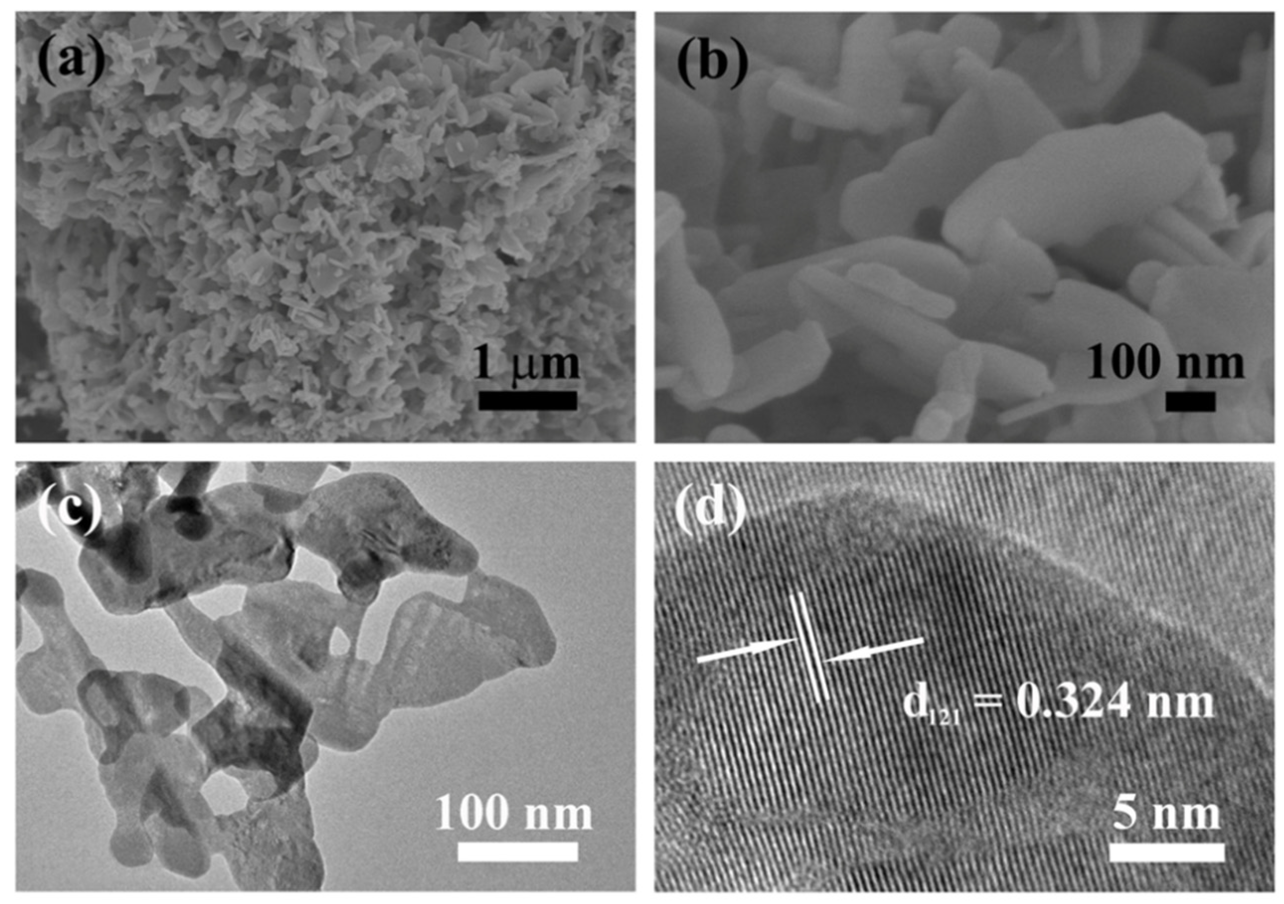
The morphologies of the precursor and BiOIO3 were characterized by FESEM, TEM, and HRTEM. As exhibited in Figure S2, the precursor is of homogeneous granular shapes with diameters of about 20–50 nm. SEM and TEM show that BiOIO3 has a two-dimensional lamellar structure (Figure 2a–c), with these irregular nanosheets stacked together with a size of about tens of nanometers to submicron and thicknesses of about 28 nm. The nanoplate structure may because the (Bi2O2)2+ and (IO3) layers in the crystal structure of BiOIO3 are alternately arranged, thus easily forming a nanosheet morphology (Figure S3). As observed from HRTEM, the spacing between the adjacent lattice stripes is 0.324 nm (Figure 2b), corresponding to the (121) plane of the orthorhombic BiOIO3 crystal, further illustrating the successful synthesis of pure BiOIO3.
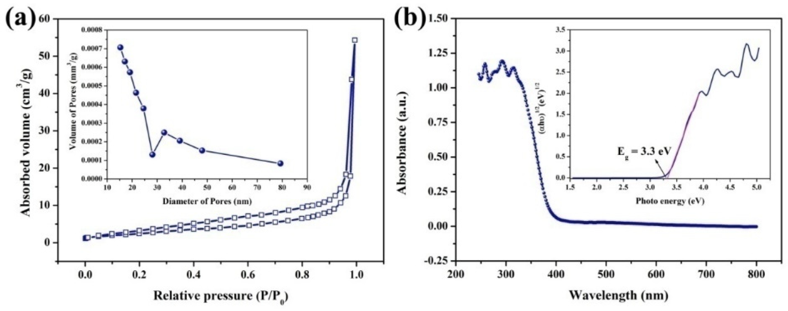
In Figure 3a, the N2 gas adsorption–desorption isotherm of BiOIO3 is measured and type IV with an H3 hysteresis loop was detected, implying the mesopore structure of the photocatalysts. The pore size distribution demonstrated that many mesoporous structures of about 15–25 and 30–45 nm coexist in BiOIO3. The Brunauer-Emmett-Teller (BET) surface area of BiOIO3 calculated from the N2 isotherms is approximately 8.932 m2/g, suggesting the specific surface area is not the main factor affecting performance.
The optical properties of the prepared sample were measured by UV/Vis diffuse reflectance spectroscopy. As can be seen from Figure 3b, BiOIO3 has strong absorption intensities in the region of UV and the absorption edge is about 390 nm. The band gap (Eg) can be fitted by the equation αhν = A(hν − Eg)n/2 [18], where α is the absorption coefficient, hν is photon energy, A is the constant, and the value of n depends on the type of semiconductor. As presented in the insert of Figure 3b, the band gap (Eg) of BiOIO3 is 3.3 eV, further manifesting BiOIO3 as an ultraviolet-light-responsive photocatalyst.
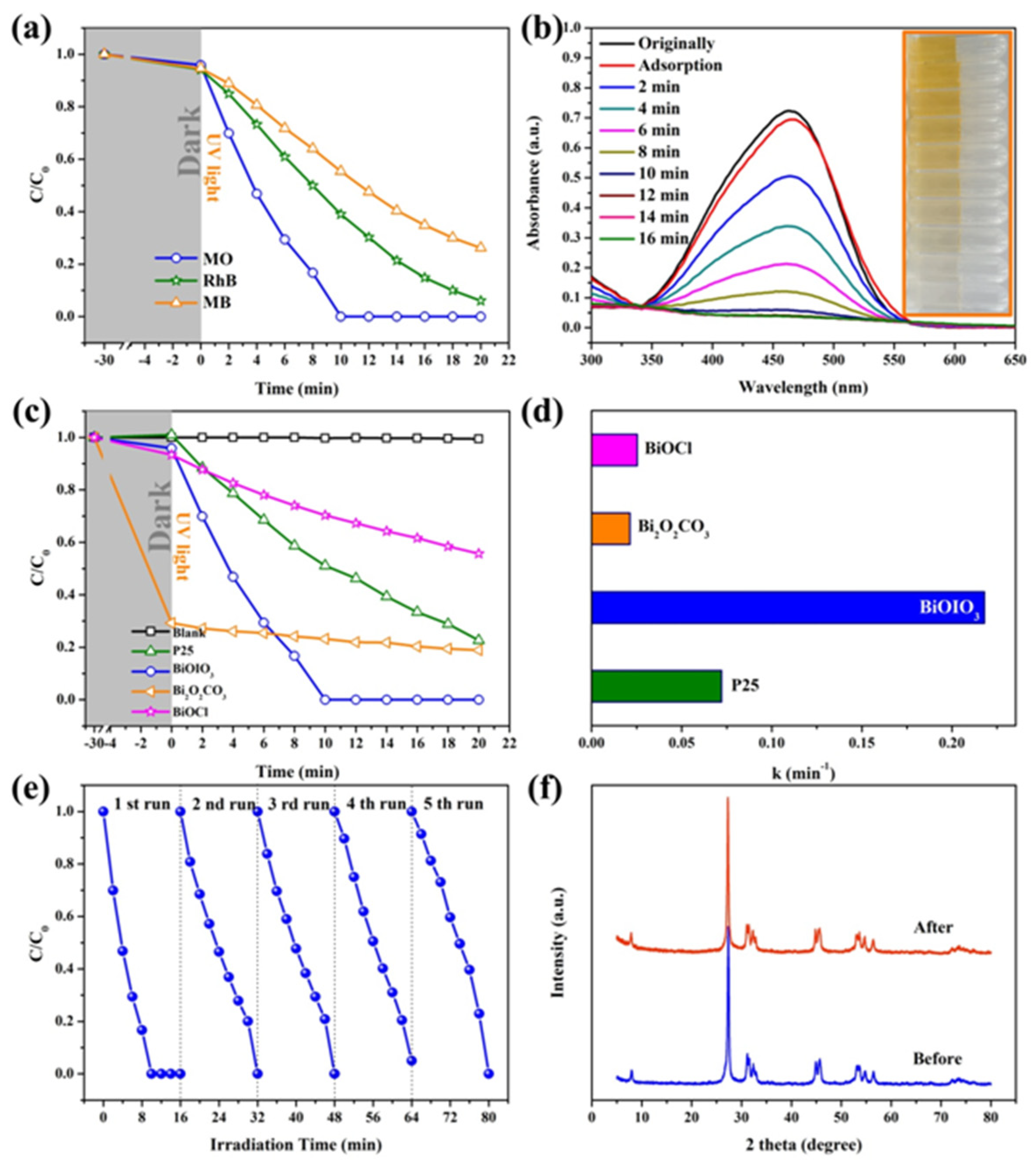
The photocatalytic activity of BiOIO3 nanosheets under UV light was evaluated using anionic dyes (methyl orange) and cationic dyes (rhodamine B and methylene blue) as simulated pollutants, and the concentration of all contaminants is 10 mg/L. Firstly, the effect of calcination atmosphere on the photocatalytic removal performance was investigated; the results are illustrated in Figure S4. Although the performance of the sample obtained under the reducing atmosphere was better than that of the other two samples during dark adsorption, the degradation rate of BiOIO3 calcined in the oxidizing atmosphere (in the air) was the fastest. Therefore, the sample BiOIO3 obtained under an air atmosphere was used for further study.
In addition, the effect of reaction conditions (catalyst concentration and pH) on the photocatalytic performance was also explored. Figure S5 shows the degradation curve of MO by different concentrations of BiOIO3 under UV irradiation. It can be observed that the adsorption capacity is unchanged, while the degradation rate increases with the increase in the catalyst concentration. The degradation efficiency of dye molecules is about 48% and 86% after irradiation for 15 min when the concentration of the photocatalyst in the system is 0.2 g/L and 0.4 g/L, respectively. When the concentration of the photocatalyst in the system is 0.6 g/L or 0.8 g/L, MO is completely degraded when exposed to light for 12 min. Figure S6 presents the degradation efficiencies of MO over BiOIO3 at different pH. The apparent rate constants in a neutral environment (pH = 7) are higher than in acidic and alkaline environments, illustrating that neutral (pH = 7) environments are more suitable for the degradation of MO over BiOIO3. Therefore, the following catalytic experiment was conducted in a neutral environment (pH = 7) with a catalyst concentration of 0.2 g/L. As presented in Figure 4a, the dark adsorption of BiOIO3 for MO, RhB, and MB is almost the same and negligible. However, the degradation efficiency of BiOIO3 nanosheets for MO, RhB, and MB varies widely after 16 min of UV light irradiation, and the removal rates are 100%, 85.2%, and 65.2%, respectively. Figure 4b demonstrates that the characteristic absorption peak of methyl orange gradually decreases with the increase in light irradiation time, and its color also gradually changes from yellow to colorless. As seen in Figure 4c, the photocatalytic activity of BiOIO3 nanosheets was compared with other photocatalysts of bismuth oxygen compound (BiOCl and Bi2O2CO3) synthesized by room-temperature solid-state chemical reaction, commercial TiO2 (P25), and blank (no photocatalyst), using MO as the dye. After the adsorption and desorption equilibrium, there was basically no adsorption of methyl orange over the other samples, except for Bi2O2CO3. Although the adsorption efficiency of Bi2O2CO3 on MO was as high as 70%, MO is quite recalcitrant to degrade by Bi2O2CO3 under UV light illumination. Therefore, the photocatalytic degradation kinetics of MO was investigated using different photocatalytics, and the apparent rate constants for the degradation of MO by BiOIO3 nanosheets were fitted to 0.2179 min−1 under UV light (Figure 4d), which were about 3.02, 8.60, and 10.26 times higher than those of P25 (0.0721 min−1), BiOCl (0.0253 min−1), and Bi2O2CO3 (0.0212 min−1), respectively. Besides, the degradation efficiency for MO over BiOIO3 is in agreement with other reported photocatalysts. The above results indicate that BiOIO3 has superior photocatalytic activity compared with other bismuth-based materials and P25.
In order to evaluate the repeatability and stability of the photocatalyst, a recycling experiment was conducted on the UV photocatalytic degradation of MO. As depicted in Figure 4e, there is almost no significant decrease in BiOIO3 nanosheets during five cycles of the degradation of MO. Moreover, the XRD pattern and the relative peak intensity of the BiOIO3 photocatalyst after five cycles remain unchanged as compared with the fresh catalyst (Figure 4f), and no BiOI or other impurities appeared, indicating that BiOIO3 nanosheets are stable in the photocatalytic reaction.
To explore the mechanism of photooxidation reactions for the decomposition of MO, capture experiments on the main active free radical were carried out. Benzoquinone (BQ), tertiary butanol (TBA), and ethylenediamine tetraacetic acid disodium salt (EDTA-2Na) were applied to capture ·O2, ·OH, and h+, respectively [7,19]. As displayed in Figure 5a, the photocatalytic degradation efficiency of BiOIO3 was greatly inhibited by BQ, illustrating that ·O2 is the main prominent active free radical for removing MO. The addition of EDTA-2Na has some effect on the decomposition, indicating that h+ plays an auxiliary role in photocatalysis, while the inexistence of ·OH has no influence on the photocatalytic activity of BiOIO3 in eliminating MO. In addition, the trapping agent DMPO was used to detect ·OH in water and ·O2 in methanol on ESR to further confirm the active radical ·OH and ·O2. As shown in Figure 5b, there are no any signals in dark, while obviously characteristic signals of DMPO-·OH and DMPO-·O2 were observed after UV light irradiation and the intensity of the DMPO-·O2 signal increased with the prolonged irradiation time, offering conclusive evidence for the increased generation of ·O2. The above result indicates that ·O2 is the major active radical and the oxidation reaction played a leading role for the degradation of MO.
Moreover, the potentials of the valence band (VB) and conduction band (CB) of BiOIO3 can be calculated with the two formulas proposed by Butler and Ginley [20,21]: EVB-Ee + 0.5 Eg and ECB = EVB − Eg, where EVB and ECB are the VB potential and CB potential of the as-prepared material, respectively; Ee is the energy of free electrons on the hydrogen scale (ca. 4.5 eV vs. NHE), and Eg is the absolute electronegativity of the corresponding semiconductor material. Based on the above formulas and the optical absorption spectrum, EVB and ECB are +3.99 and +0.69 eV, respectively, indicating the strong oxidation ability of the valence band maximum. As displayed in Figure 5c, the reduction potentials of O2/·O2 (−0.046 eV vs. NHE) are more negative than the conduction band minimum of BiOIO3; therefore, ·O2 can be generated only when the light is below 308 nm, further manifesting that the degradation of MO over BiOIO3 is largely dependent on the oxidation of ·O2.
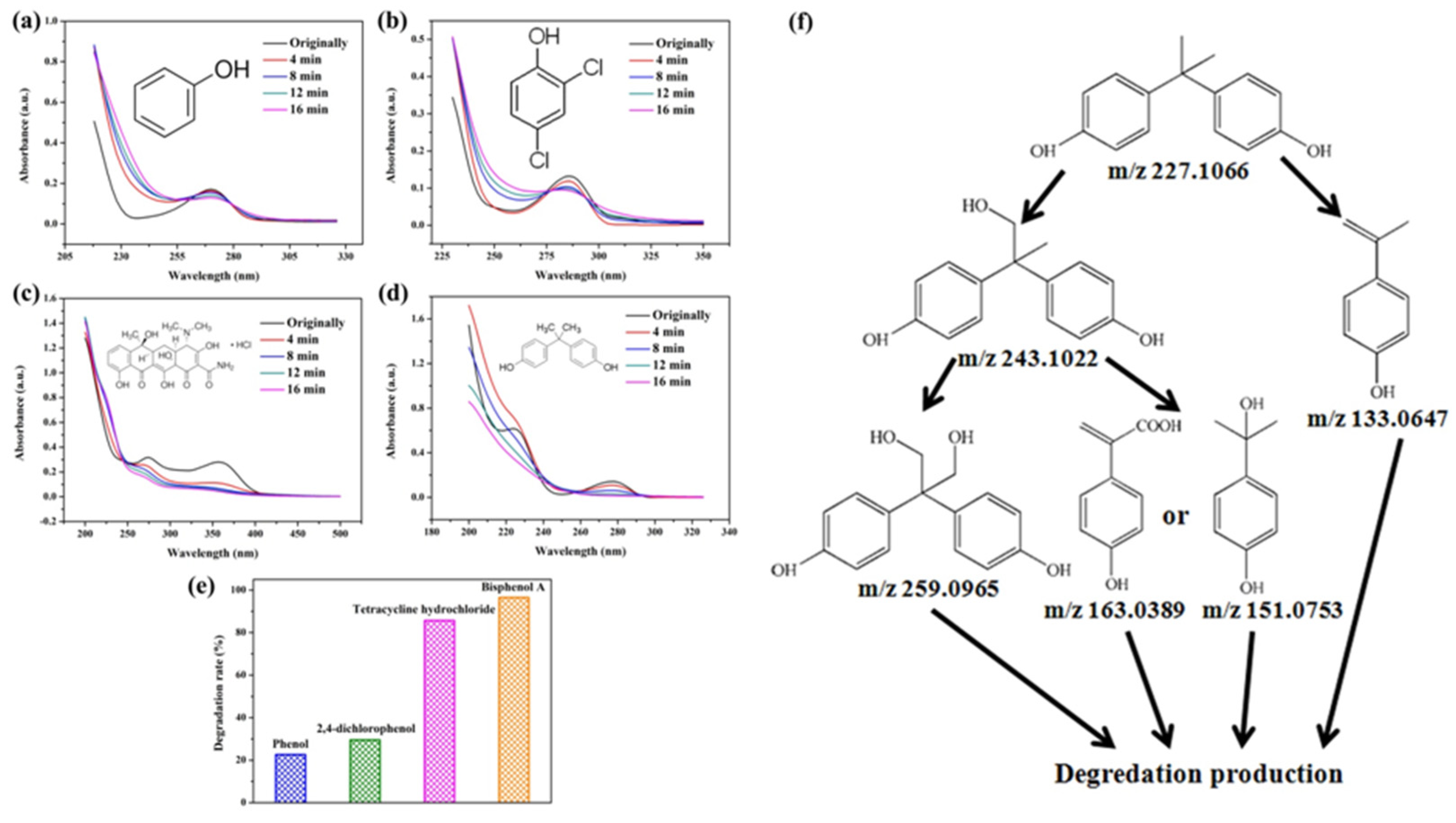
To prove the activity of the BiOIO3 photocatalyst for various organic contaminants, photocatalytic degradation of phenol and phenolic compounds was conducted. Phenol and phenolic compounds are a class of organic pollutants that are highly toxic, carcinogenic, and persistent organic pollutants, which are widely used in plastics and epoxy resin manufacturing. As presented in Figure 6a–d, phenol (ph-OH), 2,4-dichlorophenol (2,4-DCP), tetracycline hydrochloride (TC), and bisphenol A (BPA) were partially or completely degraded by BiOIO3 nanosheets when all of their concentrations were 10 mg/L. In particular, the characteristic absorption peaks of TC and BPA decreased sharply, suggesting the extensive removal ability of BiOIO3. The degradation efficiency of ph-OH, 2,4-DCP, TC, and BPA by BiOIO3 nanosheets was 22.6%, 29.5%, 85.7%, and 96.5%, respectively, after 16 min of light exposure (Figure 6e). To further investigate the intermediates of the photocatalytic process, the residual liquid for the degradation of bisphenol A after 12 min was tested by liquid chromatography coupled with mass spectrometry (LC-MS); the results are shown in Table 1. Five mass spectrometric peaks with different retention times were present in the detection results; the mass-to-charge ratios (m/z) of these intermediates were m/z = 163.0389 ([M-H]), m/z = 259.0965 ([M-H]), m/z = 133.0647 ([M-H]), m/z = 151.0753 ([M-H]), and m/z = 243.1022 ([M-H]), which were analyzed as C9H8O3, C15H16O4, C9H10O, C9H12O2, and C14H14O4, respectively [22,23]. Based on the above analysis and the reported literature, the degradation of BPA is mainly involved the reaction of hydroxylation; the possible degradation pathway is speculatively shown in Figure 6f [24,25].

3. Materials and Methods

3.1. Preparation of BiOIO3

In a typical solid-state chemical reaction, 5 mmol (2.425 g) of bismuth(III) nitrate pentahydrate (Bi(NO3)3·5H2O) and 10 mmol (2.14 g) of potassium iodate (KIO₃) were separately weighed and ground in an agate mortar for about 10 min. The two reactants remained as powders after contact and grinding, and no obvious reaction was observed. After continuous grinding for 30 min, the acquired mixture was packaged in a 50 mL conical flask and placed into a 60 °C water bath for 2 h to ensure the chemical reaction was complete. Subsequently, a white precursor was obtained by washing and filtering with deionized water, and then dried in air at room temperature. The precursor was held in a muffle furnace at 300 °C for 2 h with a heating rate of 3 °C/min to obtain a final white sample, which was denoted as “BiOIO3”. The synthetic procedure of the other samples was the same as above.
The reaction equation is deduced [26] and shown as follows:
Bi(NO3)3·5H2O + KIO3→BiOIO3 + KNO3 + 4H2O + 2HNO3

3.2. Characterization

X-ray diffraction (XRD) was used to analyze the crystalline phase and chemical constitution of the sample by a Bruker D8 using filtered Cu Kα radiation (1.54056 Å) with an operating voltage of 40 kV and a beam current of 40 mA. X-ray photoelectron spectra (XPS) were recorded using the ESCALAB 250 Xi system with monochromatic Al Kα radiation (1486.6 eV); the binding energy was calibrated by a C 1s line at 248.11 eV, 284.25 eV, and 284.75 eV. The oxygen defects were recorded by an in situ electron paramagnetic resonance (EPR, JES FA300) spectrometer at 77 K in liquid nitrogen. Field emission scanning electronic microscopy (FESEM, SU8010, 5 kV) and transmission electron microscopy (TEM, JEOL JEM-2100F) were performed to obtain the morphology and size of the samples. The elementary composition was performed on an energy-dispersive X-ray spectrometer (EDS, Oxfold 2000) connected to an SEM. The specific surface area of the sample was obtained using the Brunauer-Emmett-Teller (BET) method (Quantachrome, AutosorbiQ2). The UV/Vis diffuse reflectance spectroscopy was conducted on a Hitachi U-3010 spectrophotometer using BaSO4 as the reference. The UV/Vis absorption spectra of as-generated adsorbents were obtained using a Hitachi U-3900H spectrophotometer during 200~800 nm. The intermediate products were monitored by liquid chromatography mass spectrometry (LC-MS, Thermo Fisher Scientific Ultimate 3000/Q-Exactive, Waltham, MA, USA). The electron spin resonance (ESR) signals were measured on a JES FA300 spectrometer in 5,5’-dimethyl-1-pirroline-N-oxide (DMPO) solutions (methanol dispersion for DMPO-O2 and distilled water dispersion for DMPO-OH).

3.3. The Degradation Performance Test

Photocatalytic performance test: the XPA-1 photochemical reactor (Xujiang Electromechanical Plant, Nanjing, China) with a 300 W mercury lamp as the UV light source was employed for the photocatalytic degradation experiments. The light source radiates the reaction liquid through a glass cold trap with condensed water. The main contaminants selected for simulation are methyl orange (MO), methylene blue (MB), and rhodamine B (RhB). The procedure was as follows: 25 mg of photocatalyst was ultrasonically dispersed in 50 mL of organic dye and subjected to a dark reaction for 30 min to achieve physical adsorption–desorption equilibrium. Then, light irradiation was performed and 4 mL of sample was taken at 5 min intervals during the reaction; subsequently, the supernate was separated by centrifugation and the absorbance of the clear solution was measured using a UV/Vis spectrophotometer.
Cyclic test: After testing the photocatalytic performance of the samples using the above method, the solid photocatalytic material was separated from the mixture by centrifuging, washing, and drying. Afterwards, the recovered photocatalyst was used again to test the photocatalytic performance with three repetitions.

4. Conclusions

In this paper, BiOIO3 nanosheets were firstly prepared using an easy-to-handle and low-cost two-step solid-state chemical reaction. Thanks to the unique layered crystal structure of BiOIO3, it demonstrates superior photocatalytic degradation activity as well as better repeatability and cycling stability for cationic dye MO than other layered bismuth-based materials (BiOCl and Bi2O2CO3) and P25 under ultraviolet light, which can completely degrade MO within 10 min. Besides, BiOIO3 nanosheets can also be employed to degrade anionic dyes RhB and MB and neutral pollutants ph-OH, 2,4-DCP, BPA, and TC. Unexpectedly, the removal of MO over BiOIO3 is largely dependent on the oxidation of ·O2, while the hydroxylation reaction is the main reason for the degradation of BPA. The study may not only introduce a new simple approach to synthesize BiOIO3 nanomaterials, but also provide a candidate with high photocatalytic activity for environmental purification.

Supplementary Materials

The following supporting information can be downloaded at https://www.mdpi.com/article/10.3390/molecules28093681/s1. Figure S1: XRD patterns of BiOIO3 synthesis with different calcination atmospheres; Figure S2: FESEM image of the precursor; Figure S3: The crystal structure of BiOIO3. Figure S4: Degradation efficiencies of MO over BiOIO3 synthesis with different calcination atmospheres under UV light irradiation. Figure S5: Degradation efficiencies of MO by different concentrations of BiOIO3. Figure S6: Degradation efficiencies of MO over BiOIO3 in different pH. Table S1: The comparison of the degradation for MO in this study with other investigations [11,27,28,29,30,31,32].

Author Contributions

Methodology, J.L. and E.L.; writing—original draft preparation, X.Z.; writing—review and editing, J.X.; supervision, Y.C. All authors have read and agreed to the published version of the manuscript.

Funding

This research was funded by the Xinjiang University Undergraduate Innovation Training Program, grant number XJU-SRT-21089; the Natural Science Foundation of Xinjiang Autonomous Region, grant number 2021D01C093; and the National Natural Science Foundation of China, grant number 52162023.

Institutional Review Board Statement

Not applicable.

Informed Consent Statement

Not applicable.

Data Availability Statement

Not applicable.

Acknowledgments

We also thank the researchers who facilitated the completion of this study.

Conflicts of Interest

The authors declare no conflict of interest.

Sample Availability

Samples of the compounds are available from the authors.

References

  1. Zhang, Z.Q.; Liang, J.L.; Zhang, W.; Zhou, M.; Zhu, X.L.; Liu, Z.Y.; Li, Y.; Guan, Z.Q.; Lee, C.S.; Wong, P.K.; et al. Modified-pollen confined hybrid system: A promising union for visible-light-driven photocatalytic antibiotic degradation. Appl. Catal. B Environ. 2023, 330, 122621. [Google Scholar] [CrossRef]
  2. Chong, M.N.; Jin, B.; Christopher, W.K.; Chow, C.; Chris, S.C. Recent developments in photocatalytic water treatment technology: A review. Water Res. 2010, 44, 2997–3027. [Google Scholar] [CrossRef]
  3. Lu, Q.Y.; Yang, L.L.; Pamela, C.A.; Li, Y.N.; Zhang, X.H.; El-Din, M.G. Enhanced photocatalytic degradation of organic contaminants in water by highly tunable surface microlenses. Chem. Eng. J. 2023, 463, 142345. [Google Scholar] [CrossRef]
  4. Swaraj, R.P.; Marta, P.G.; Dariusz, Ł.; Dmytro, L.; Juan, C.C. Bimetallic TiO2 nanoparticles for lignin-based model compounds valorization by integrating an optocatalytic flow-microreactor. Molecules 2022, 27, 8731. [Google Scholar]
  5. Karolina, S.; Monika, S.M.; Kinga, P.; Magdalena, G.; Grzegorz, D.S. Photoelectrochemical properties of annealed anodic TiO2 layers covered with CuOx. Molecules 2022, 27, 4789. [Google Scholar]
  6. Gao, P.; Yang, Y.N.; Yin, Z.; Kang, F.X.; Fan, W.E.; Sheng, J.Y.; Feng, L.; Liu, Y.Z.; Du, Z.W.; Zhang, L.Q. A critical review on bismuth oxyhalide based photocatalysis for pharmaceutical active compounds degradation: Modifications, reactive sites, and challenges. J. Hazard. Mater. 2021, 412, 125186–125216. [Google Scholar] [CrossRef] [PubMed]
  7. Xu, M.L.; Jiang, X.J.; Li, J.R.; Wang, F.J.; Li, K.; Cheng, X. Self-assembly of a 3D hollow BiOBr@Bi-MOF heterostructure with enhanced photocatalytic degradation of dyes. ACS Appl. Mater. Interfaces 2021, 13, 56171–56180. [Google Scholar] [CrossRef] [PubMed]
  8. Han, X.; Zhang, T.D.; Cui, Y.; Wang, Z.Y.; Dong, R.Y.; Wu, Y.H.; Du, C.W.; Chen, R.Y.; Yu, C.F.; Feng, J.L.; et al. A bifunctional BiOBr/ZIF-8/ZnO photocatalyst with rich oxygen vacancy for enhanced wastewater treatment and H2O2 generation. Molecules 2023, 28, 2422. [Google Scholar] [CrossRef]
  9. Jiang, D.Q.; Song, H.M.; Wen, T.; Jiang, Z.M.; Li, C.; Liu, K.; Yang, W.G.; Huang, H.W.; Wang, Y.G. Pressure-driven two-step second-harmonic-generation switching in BiOIO3. Angew. Chem. Int. Edit. 2022, 61, 16656–16663. [Google Scholar]
  10. Lai, J.H.; Jiang, X.Y.; Zhao, M.; Cui, S.H.; Yang, J.; Li, Y.F. Thickness-dependent layered BiOIO3 modified with carbon quantum dots for photodegradation of bisphenol A: Mechanism, pathways and DFT calculation. Appl. Catal. B Environ. 2021, 298, 120622–120635. [Google Scholar] [CrossRef]
  11. Wang, W.J.; Huang, B.B.; Ma, X.C.; Wang, Z.Y.; Qin, X.Y.; Zhang, X.Y.; Dai, Y.; Whangbo, M.H. Efficient separation of photogenerated electron-hole pairs by the combination of a heterolayered structure and internal polar field in pyroelectric BiOIO3 nanoplates. Chem. Eur. J. 2013, 19, 14777–14780. [Google Scholar] [CrossRef]
  12. Chen, F.; Huang, H.W.; Zeng, C.; Du, X.; Zhang, Y.H. Achieving enhanced UV and visible light photocatalytic activity for ternary Ag/AgBr/BiOIO3: Decomposition for diverse industrial contaminants with distinct mechanisms and complete mineralization ability. ACS Sustain. Chem. Eng. 2017, 5, 7777–7791. [Google Scholar] [CrossRef]
  13. Zhu, Z.J.; Zhang, Q.; Xiao, X.; Sun, F.Q.; Zuo, X.X.; Nan, J.M. Dual-iodine-doped BiOIO3: Bulk and surface co-modification for enhanced visible-light photocatalytic removal of bisphenol AF. Chem. Eng. J. 2021, 404, 126543–126556. [Google Scholar] [CrossRef]
  14. Liu, H.L.; Wu, B.; Wu, S.K.; Liu, Y.S.; Yoriya, S.; Hu, C.; Zhang, H.; Wu, J.; Ling, Y.; He, P.; et al. Ion doping engineering of nickel-doped BiOIO3 nanosheets for gaining oxygen vacancy to enhance light absorption. Fuel Process. Technol. 2023, 241, 170626. [Google Scholar] [CrossRef]
  15. Huang, H.; Ou, H.; Feng, J.; Du, X.; Zhang, Y. Achieving highly promoted visible-light sensitive photocatalyticactivity on BiOIO3 via facile iodine doping. Colloids Surf. A Physicochem. Eng. Asp. 2017, 518, 158–165. [Google Scholar] [CrossRef]
  16. Xiong, T.; Dong, F.; Zhou, Y.; Fu, M.; Ho, W.K. New insights into how RGO influences the photocatalytic performance of BiOIO3/RGO nanocomposites under visible and UV irradiation. J. Colloid Interface Sci. 2015, 447, 16–24. [Google Scholar] [CrossRef] [PubMed]
  17. Wang, W.J.; Cheng, H.F.; Huang, B.B.; Liu, X.L.; Qin, X.Y.; Zhang, X.Y.; Dai, Y. Hydrothermal synthesis of C3N4/BiOIO3 heterostructures with enhanced photocatalytic properties. J. Colloid Interface Sci. 2015, 442, 97–102. [Google Scholar] [CrossRef] [PubMed]
  18. Sarkar, A.; Ghosh, A.B.; Saha, N.; Srivastava, D.N.; Paul, P.; Adhikary, B. Enhanced photocatalytic performance of morphologically tuned Bi2S3 NPs in the degradation of organic pollutants under visible light irradiation. J. Colloid Interface Sci. 2016, 483, 49–59. [Google Scholar] [CrossRef]
  19. Tang, Q.; Wu, J.; Kim, D.H.; Franco, C.; Terzopoulou, A.; Veciana, A.; Josep, P.L.; Chen, X.Z.; Nelson, B.J.; Pané, S. Enhanced piezocatalytic performance of BaTiO3 nanosheets with highly exposed {001} facets. Adv. Funct. Mater. 2022, 32, 2202180. [Google Scholar] [CrossRef]
  20. Xu, Y.; Schoonen, M.A.A. The absolute energy positions of conduction and valence bands of selected semiconducting minerals. Am. Miner. 2000, 85, 543–556. [Google Scholar] [CrossRef]
  21. Wen, X.J.; Niu, C.G.; Zhang, L.; Liang, C.; Zeng, G.M. An in depth mechanism insight of the degradation of multiple refractory pollutants via a novel SrTiO3/BiOI heterojunction photocatalysts. J. Catal. 2017, 356, 283–299. [Google Scholar] [CrossRef]
  22. Chen, D.; Kannan, K.; Tan, H.; Zheng, Z.G.; Feng, Y.L.; Wu, Y.; Widelka, M. Bisphenol analogues other than BPA: Environmental occurrence, human exposure, and toxicity-a review. Environ. Sci. Technol. 2016, 50, 5438–5453. [Google Scholar] [CrossRef]
  23. Ohko, Y.; Ando, I.; Niwa, C.; Tatsuma, T.; Yamamura, T.; Nakashima, T.; Kubota, Y.; Fujishima, A. Degradation of bisphenol A in water by TiO2 photocatalyst. Environ. Sci. Technol. 2001, 35, 2365–2368. [Google Scholar] [CrossRef]
  24. Pan, M.L.; Zhang, H.J.; Gao, G.D.; Liu, L.; Chen, W. Facet-dependent catalytic activity of nanosheet-assembled bismuth oxyiodide microspheres in degradation of bisphenol A. Environ. Sci. Technol. 2015, 49, 6240–6248. [Google Scholar] [CrossRef]
  25. Tan, X.Q.; Wan, Y.F.; Huang, Y.J.; He, C.; Zhang, Z.L.; He, Z.Y.; Hu, L.L.; Zeng, J.W.; Shu, D. Three-dimensional MnO2 porous hollow microspheres for enhanced activity as ozonation catalysts in degradation of bisphenol A. J. Hazard. Mater. 2017, 321, 162–172. [Google Scholar] [CrossRef]
  26. Qi, X.M.; Gu, M.L.; Zhu, X.Y.; Wu, J.; Long, H.M.; He, K.; Wu, Q. Fabrication of BiOIO3 nanosheets with remarkable photocatalytic oxidation removal for gaseous elemental mercury. Chem. Eng. J. 2016, 285, 11–19. [Google Scholar] [CrossRef]
  27. Adeel, M.; Saeed, M.; Khan, I.; Muneer, M.; Akram, N. Synthesis and characterization of Co-ZnO and evaluation of its photocatalytic activity for photodegradation of methyl orange. ACS Omega 2021, 6, 1426–1435. [Google Scholar] [CrossRef]
  28. Wang, R.H.; Wang, Y.N.; Zhang, N.N.; Lin, S.; He, Y.J.; Yan, Y.J.; Hu, K.; Sun, H.J.; Liu, X.F. Synergetic piezo-photocatalytic effect in SbSI for highly efficient degradation of methyl orange. Ceram. Int. 2022, 48, 31818–31826. [Google Scholar] [CrossRef]
  29. Yang, J.; Du, J.; Li, X.; Liu, Y.; Jiang, C.; Qi, W.; Zhang, K.; Gong, C.; Li, R.; Luo, M.; et al. Highly hydrophilic TiO2 nanotubes network by alkaline hydrothermal method for photocatalysis degradation of methyl orange. Nanomaterials 2019, 9, 526. [Google Scholar] [CrossRef]
  30. Yao, W.; Chen, Y.F.; Li, J.L.; Yang, J.; Ren, S.; Liu, W.C.; Liu, Q.C. Photocatalytic degradation of methyl orange by Ca doped β-In2S3 with varying Ca concentration. Res. Chem. Intermediatr. 2022, 48, 1813–1829. [Google Scholar] [CrossRef]
  31. Sun, H.; Lee, S.Y.; Park, S.J. Bimetallic CuPd alloy nanoparticles decorated ZnO nanosheets with enhanced photocatalytic degradation of methyl orange dye. J. Colloid Interface Sci. 2022, 629, 87–96. [Google Scholar] [CrossRef]
  32. Wang, L.; Li, Z.; Chen, J.; Huang, Y.; Zhang, H.; Qiu, H. Enhanced photocatalytic degradation of methyl orange by porous graphene/ZnO nanocomposite. Environ. Pollut. 2019, 249, 801–811. [Google Scholar] [CrossRef] [PubMed]
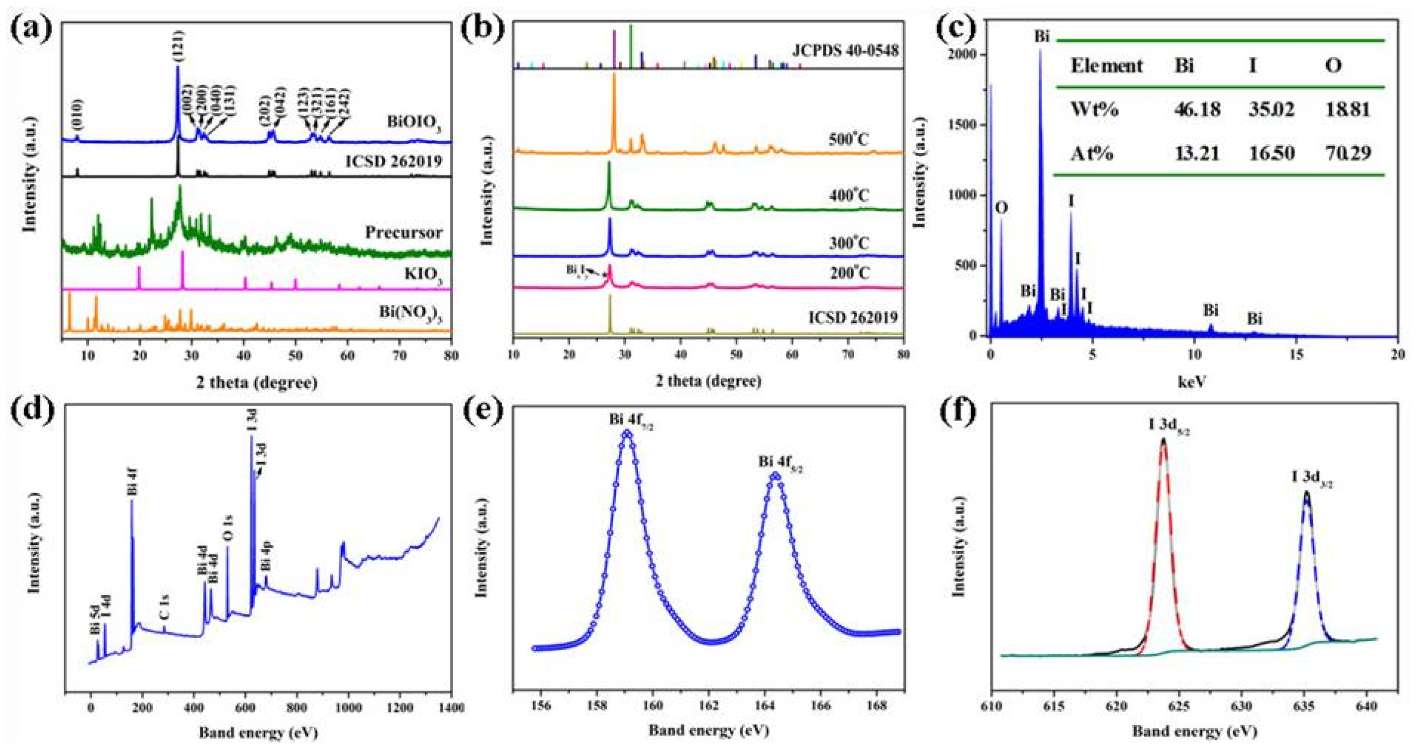
Figure 1. XRD patterns of (a) the precursor and BiOIO3 and (b) the samples obtained by the calcining precursor at different temperatures; (c) the EDS result of the BiOIO3. XPS spectra of BiOIO3: (d) survey scan, (e) Bi 4f, and (f) I 3d.
Molecules 28 03681 g001
Figure 2. (a,b) FESEM images, (c) TEM, and (d) HRTEM of BiOIO3.
Molecules 28 03681 g002
Figure 3. (a) The nitrogen gas absorption–desorption curve and pore size distribution and (b) UV/Vis diffuse reflectance spectra and band gap of BiOIO3.
Molecules 28 03681 g003
Figure 4. (a) Degradation efficiencies of MO, RhB, and MB over BiOIO3 nanoplates under UV light irradiation; (b) UV/Vis spectral change of MO with irradiation time over BiOIO3 nanoplates; (c) degradation efficiencies of MO over BiOIO3, BiOCl, Bi2O2CO3, and P25 under UV light irradiation; (d) apparent first-order rate constants for BiOIO3, BiOCl, Bi2O2CO3, and P25 photocatalysts; (e) recycling experiments conducted on the UV photocatalytic degradation of MO by BiOIO3 nanoplates; and (f) XRD pattern of before and after photocatalysis for BiOIO3 nanoplates.
Molecules 28 03681 g004
Figure 5. (a) Photocatalytic degradation of MO over BiOIO3 nanoplates together with different radical scavengers. (b) ESR spectra of BiOIO3 nanoplates for the detection of ·OH and ·O2. (c) The photocatalytic degradation mechanism of BiOIO3 nanoplates.
Molecules 28 03681 g005
Figure 6. Temporal UV/Vis spectral change of (a) ph-OH, (b) 2,4-DCP, (c) TC, and (d) BPA with irradiation time over BiOIO3 nanoplates; (e) the degradation rate of different pollutants after 16 min of UV light over BiOIO3 nanoplates; and (f) the possible degradation pathway for BPA.
Molecules 28 03681 g006
Table 1. The ions m/z and molecular structures of intermediate products at different retention times.
Peak NO.Retention Time (min)Detected Ions m/zMolecular Structure
11.28163.0389Molecules 28 03681 i001
21.92259.0965Molecules 28 03681 i002
31.99133.0647Molecules 28 03681 i003
41.99151.0753Molecules 28 03681 i004
52.90243.1022Molecules 28 03681 i005
64.00227.1066Molecules 28 03681 i006
Disclaimer/Publisher’s Note: The statements, opinions and data contained in all publications are solely those of the individual author(s) and contributor(s) and not of MDPI and/or the editor(s). MDPI and/or the editor(s) disclaim responsibility for any injury to people or property resulting from any ideas, methods, instructions or products referred to in the content.