Article

Studies Relevant to the Functional Model of Mo-Cu CODH: In Situ Reactions of Cu(I)-L Complexes with Mo(VI) and Synthesis of Stable Structurally Characterized Heterotetranuclear MoVI2CuI2 Complex

by
Umesh I. Kaluarachchige Don
1,
Ahmad S. Almaat
1,
Cassandra L. Ward
2 and
Stanislav Groysman
1,*
1
Department of Chemistry, Wayne State University, 5101 Cass Avenue, Detroit, MI 48202, USA
2
Lumigen Instrument Center, Wayne State University, 5101 Cass Avenue, Detroit, MI 48202, USA
*
Author to whom correspondence should be addressed.
Molecules 2023, 28(8), 3644; https://doi.org/10.3390/molecules28083644
Submission received: 31 March 2023 / Revised: 17 April 2023 / Accepted: 19 April 2023 / Published: 21 April 2023
(This article belongs to the Special Issue Molybdenum and Tungsten Enzymes—State of the Art in Research)

Abstract

:
In this study, we report the synthesis, characterization, and reactions of Cu(I) complexes of the general form Cu(L)(LigH2) (LigH2 = xanthene-based heterodinucleating ligand (E)-3-(((5-(bis(pyridin-2-ylmethyl)amino)-2,7-di-tert-butyl-9,9-dimethyl-9H-xanthen-4-yl)imino)methyl)benzene-1,2-diol); L = PMe3, PPh3, CN(2,6-Me2C6H3)). New complexes [Cu(PMe3)(LigH2)] and [CuCN(2,6-Me2C6H3)(LigH2)] were synthesized by treating [Cu(LigH2)](PF6) with trimethylphosphine and 2,6-dimethylphenyl isocyanide, respectively. These complexes were characterized by multinuclear NMR spectroscopy, IR spectroscopy, high-resolution mass spectrometry (HRMS), and X-ray crystallography. In contrast, attempted reactions of [Cu(LigH2)](PF6) with cyanide or styrene failed to produce isolable crystalline products. Next, the reactivity of these and previously synthesized Cu(I) phosphine and isocyanide complexes with molybdate was interrogated. IR (for isocyanide) and 31P NMR (for PPh3/PMe3) spectroscopy demonstrates the lack of oxidation reactivity. We also describe herein the first example of a structurally characterized multinuclear complex combining both Mo(VI) and Cu(I) metal ions within the same system. The heterobimetallic tetranuclear complex [Cu2Mo2O42-O)(Lig)2]·HOSiPh3 was obtained by the reaction of the silylated Mo(VI) precursor (Et4N)(MoO3(OSiPh3)) with LigH2, followed by the addition of [Cu(NCMe)4](PF6). This complex was characterized by NMR spectroscopy, high-resolution mass spectrometry, and X-ray crystallography.

1. Introduction

Molybdenum-copper carbon monoxide dehydrogenase (Mo-Cu CODH) is an air-stable enzyme that catalyzes the oxidation of CO to CO2. Mo-Cu CODH is a key enzyme in the metabolism of aerobic carboxidotrophic bacteria (such as Oligotropha carboxidovorans), allowing it to use CO as a sole source of energy and carbon [1]. The oxidation of CO is very efficient, and the enzyme detoxifies a significant fraction of total CO from the atmosphere [2]. Thus, there is a longstanding interest in the design of structural and/or functional models which could (a) provide insight into the reaction mechanism, and (b) lead to efficient CO oxidation and potentially CO2 reduction catalysts.
The oxidized active site of Mo-Cu CODH contains square-pyramidal Mo(VI) coordinated to pyranopterindithiolene, [3,4] apical and basal oxo groups, and a basal sulfido bridging to a low-coordinate Cu(I) atom, whose coordination is accomplished by cysteinate and weakly bound water at the resting state (Scheme 1) [5,6]. The reduced active site likely contains Mo(IV)-basal hydroxide in an otherwise similar disposition [7]. Spectroscopic studies suggest that the coordination of CO takes place at the Cu(I) center, replacing weakly bound water [8,9]. Computational studies generally agree with the initial coordination of CO to the coordinatively unsaturated Cu(I) site. However, the subsequent routes differ between different mechanisms, suggesting bridging thiocarbonate or carbon dioxide intermediates [10,11,12,13,14,15,16,17]. It is noted that the bridging thiocarbonate hypothesis relies to a large extent on a high-resolution structure of the product of the reaction of Mo-Cu CODH with alkyl isocyanide (CO analog/inhibitor), which undergoes oxidation and insertion into the Cu-S bond to form bridging thiocarbamate [5]. Since the report of the high-resolution structure of the Mo-Cu CODH enzyme, several structural models of Mo-Cu CODH have been reported; in all these models the metals were linked only by bridging sulfido (or oxo and sulfido) ligands [18,19,20,21,22,23,24]. Although selected closely related structural models were able to rationalize structural, electronic, and spectroscopic features of the active site, only a few have explored the reactivity with CO or CO-like substrates [24]. In our previous work, we explored a different approach toward a more robust functional model of Mo-Cu CODH, which brings two metals together using xanthene-based heterodinucleating ligands [25]. Recently, we have shown that the heterodinucleating ligand LigH2, which combines catecholate chelate for Mo(VI) and dipicolylamine for Cu(I), allows for the formation of mononuclear Cu(I) and Mo(VI) complexes at the designated sites [26]. The combination of LigH2 with both metals forms metastable Mo(VI)-Cu(I) complex (Et4N)[CuMoO3(Lig)] directly observable in solution by 1H NMR and high-resolution mass spectrometry (HRMS). However, our attempts to isolate this complex in a solid state and/or study its reactivity were not successful, as it underwent facile decomposition. In the present work, we explore a different approach toward a functional model, which involves the initial formation of well-defined Cu(I) complexes containing an oxidizable CO-like (or related) ligand (CNR, PR3, CN, olefin), followed by treatment with (Et4N)2[MoO4]. We have also explored a different approach towards stable Mo(VI)-Cu(I) complex that involves the use of a different Mo(VI) precursor (Et4N)[MoO3(OSiPh3)]. Herein we report the first structurally characterized Mo(VI)-Cu(I) complex in our system.

2. Results and Discussion

2.1. Synthesis and Characterization of Cu(I)-LigH2 Complexes Featuring an Additional L Ligand

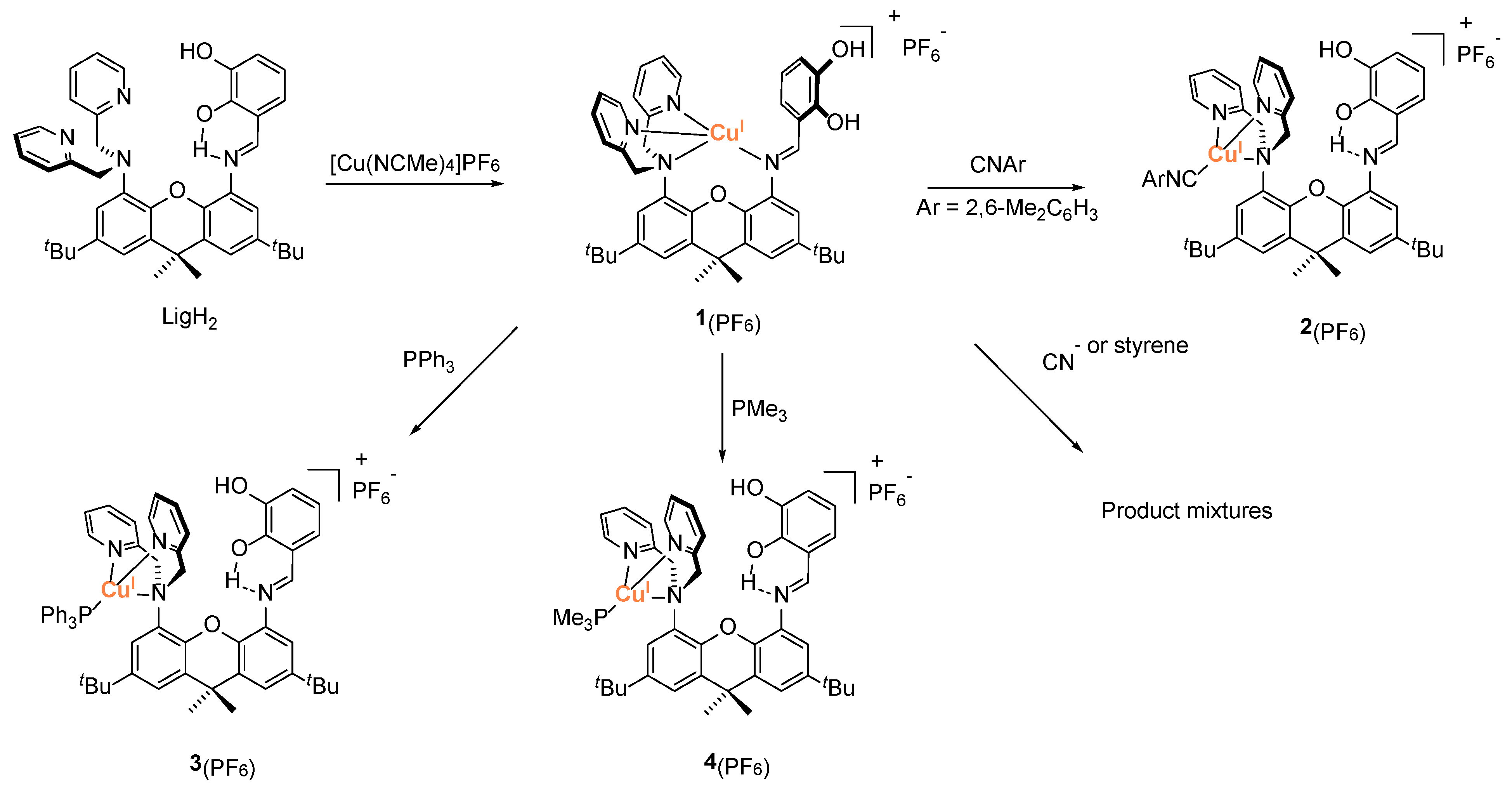
As described above, we have targeted complexes of the general form [Cu(L)(LigH2)](PF6), in which L is either directly related to the reactivity of Mo-Cu CODH (L = CNR), or can undergo similar oxidation reactivity (CN, PR3, olefin). To access these complexes, we have used previously reported Cu(I) complex [Cu(L)(LigH2)](PF6) (1(PF6)) (Scheme 2) as a precursor [24]. Complex 1(PF6) demonstrates strong coordination of Cu(I) by two pyridine and the imine donors; relatively weak interaction with the central amine was observed. A reaction of complex 1(PF6) with a monodentate ligand L is likely to release Cu(I) from the coordination to the imine, while restoring the H-bonding between the catechol proton and the imine [26,27].
Treatment of complex 1(PF6) with 2,6-dimethylphenyl isocyanide CN(2,6-Me2C6H3) produced [Cu(CN(2,6-Me2C6H3))(LigH2)](PF6) (2(PF6)) in 90% yield as yellow-orange crystals. 2(PF6) was characterized by 1H and 13C NMR spectroscopy, FT-IR spectroscopy, HRMS, and X-ray crystallography. The coordination of CN(2,6-Me2C6H3) to the metal is supported by the presence of the isocyanide methyl groups at 2.32 ppm in 1H NMR spectrum. Notably, methylene protons (NCH2py) appear as a sharp singlet, suggesting weak coordination of the central amine donor. The IR spectroscopy demonstrates the C≡NAr stretch at 2137 cm−1. This value suggests the coordination of an isocyanide to Cu(I) through mostly σ-donation [25,28,29]. HRMS demonstrates a peak at 848.3577 in the positive ion mode (expected m/z for 2+ is 848.3595), which agrees well with the expected isotopic distribution (see Supplementary Materials). The X-ray structure of 2(PF6) (Figure 1) confirms the overall composition and is consistent with the spectroscopic features. Coordination of the isocyanide to Cu(I) leads to the release of the metal and the rotation of the Cu(I)-dipicolylamine fragment away from the imine. Copper coordination is replaced by a strong hydrogen bond with one of the catechol-OH groups (OH-N(imine) 1.88 Å). The bond distance between Cu(I) and the central amine is relatively long (Cu1 N1 2.281(1) Å), while the bonds with the pyridine nitrogen are relatively short (2.0289(14) and 2.0274(14) Å). A short bond (1.844(2) Å) is observed between Cu(I) and isocyanide as well.
We have also targeted copper-phosphine complexes (PR3 = PPh3 and PMe3). The synthesis and structure of [Cu(PPh3)(LigH2)](PF6) (3(PF6)) habe been reported previously (Scheme 2) [26]. [Cu(PMe3)(LigH2)](PF6) (4(PF6)), featuring more reactive trimethylphosphine, was obtained by the treatment of 1(PF6) with one equivalent of PMe3. The complex was characterized by 1H, 13C, and 31P NMR and HRMS. Our attempts to obtain its solid-state structure were not successful; however, the spectroscopic data for 4(PF6) is consistent with the spectroscopic data for 3(PF6), suggesting a similar structure. Both compounds give rise to a catechol proton at a highly downfield chemical shift, 14.07 ppm for 3(PF6) and 13.93 ppm for 4(PF6), suggesting strong (catechol)OH-N(imine) hydrogen bonding. In both cases, methylene arms protons NCH2py appear as broad peaks, suggesting weak but not negligible coordination of the central amine. 31P NMR spectroscopy demonstrates 31PMe3 signal at −49.0 ppm in 4(PF6). HRMS contains a molecular ion peak for 4+ at m/z 793.3295 (expected m/z is 793.3302).
In addition to the synthesis of PR3 and isocyanide complexes, we have also attempted the synthesis of the mononuclear cyanide complex [Cu(CN)(LigH2)] and a mononuclear olefin (styrene CH2CHPh) complex. It was previously reported that the reaction of [CuCN]n with LigH2 formed a [Cu2(CN)2(LigH2)] complex featuring two coppers and two cyanides: one terminal and one bridging {Cu(μ2-CN)CuCN} [26]. To prevent the formation of the [CuCN]n chains (likely resulting from the [CuCN]n precursor), complex 1(PF6) was treated with the combination of NaCN and crown ether (18-crown-6). However, the reaction led to a relatively uninformative NMR spectrum containing broad peaks, which suggested product mixture or dynamic processes which could involve species of varying nuclearities. Our attempts to obtain a pure crystalline product from this reaction were not successful. Similarly, the reaction of 1(PF6) with styrene produced a broad NMR spectrum containing excess styrene. Our attempts to purify this compound by crystallization were not successful as well.

2.2. Reactions of Cu-L Complexes (L = CNR, PR3, Styrene) with [MoO4]2−

The reactions of complexes 2(PF6), 3(PF6), and 4(PF6) with (Et4N)2[MoO4] were investigated by adding the equimolar amounts of the acetonitrile solution of molybdate into the stirring acetonitrile solution of the corresponding complex. These reactions were performed similarly to the previously described synthesis of metastable (Et4N)[CuMoO3(Lig)], Ref. [26] suggesting similar incorporation of {MoVIO3} at the initial step of the reaction. Following the addition, the reaction was analyzed by NMR, IR, and UV-vis spectroscopy. Treatment of 2(PF6) with one equivalent of (Et4N)2[MoO4] produced yellow-brown solution. The 1H NMR spectrum indicated the formation of several new products. The UV-vis spectrum of the crude product does not appear to contain any d-d transitions anticipated for Cu(II), suggesting that no Cu(I) oxidation takes place. Although we were not able to separate the products, IR spectroscopy provides a convenient tool to specifically interrogate the oxidation of isocyanide to isocyanate. An isocyanate is expected to exhibit an intense absorption between 2200 cm−1 and 2300 cm−1 (RN=C=O stretch). However, the IR spectrum of the crude product demonstrates a single sharp resonance around 2129 cm−1, consistent with the isocyanide stretch in aryl isocyanides (ArN≡C) [30]. As noted above, a stretching frequency of ~2130 cm−1 suggests either a free isocyanide, or an isocyanide coordinated mostly via σ-donation [28,29,30]. Such coordination would be expected for both Mo(VI) and Cu(I). No significant peaks above 2200 cm−1 were observed.
The reactions of the phosphine complexes 3(PF6) and 4(PF6) with molybdate were investigated similarly. Treatment of the cold yellow solution of 3(PF6) and 4(PF6) with molybdate resulted in a slight color change to brown. In both cases, 1H NMR spectra of the products featured broad resonances. However, 31P NMR was more informative in both cases, allowing us to focus specifically on the phosphine transformation. Thus, the 31P NMR spectrum of the product of the reaction of 3(PF6) with molybdate contains a single peak around −3 ppm (free PPh3 is ~−6 ppm, PPh3 in 3(PF6) ~3 ppm, P(=O)Ph3 ~28 ppm) whereas the product of the reaction of 4(PF6) with molybdate contains a peak around −46 ppm (free PMe3 is ~−62 ppm, PMe3 in 4(PF6) ~−49 ppm, P(=O)Me3 36 ppm). The findings above are consistent with coordinated phosphines and suggest that no oxidation of phosphines to the corresponding phosphine oxides takes place.
The experiments above indicate the lack of in situ reactivity between Cu(I)-bound L (CNR, PR3) and [MoVIO3]2−, and that is despite previously observed in situ nucleophilic attack of [MoVIO3]2− on the Cu(I)-bound imine [25]. We postulate that the lack of reactivity in the present system is due to the structural difference between the two heterodinucleating ligand systems (Figure 2). The iminopyridine system led to a relatively accessible three-coordinate Cu(I)-iminopyridine which, due to the coordination to the (xanthene-) amine, was positioned in the vicinity of Mo(VI)-oxo (Figure 2, left) [25]. In contrast, the coordination of the substrate to the dipicolylamine-coordinate Cu(I) leaves the metal four-coordinate, making it less accessible for attack. Furthermore, the coordination of the substrate L causes the displacement of the imine coordination which, combined with the more significant steric profile of dipicolylamine (vs. iminopyridine), leads to the anti-conformation of the Cu(I) fragment relative to the xanthene bridge (Figure 2, right). This factor is also likely to have a negative effect on reactivity.

2.3. Synthesis and Characterization of Mo2Cu2 Heterodinuclear Complex

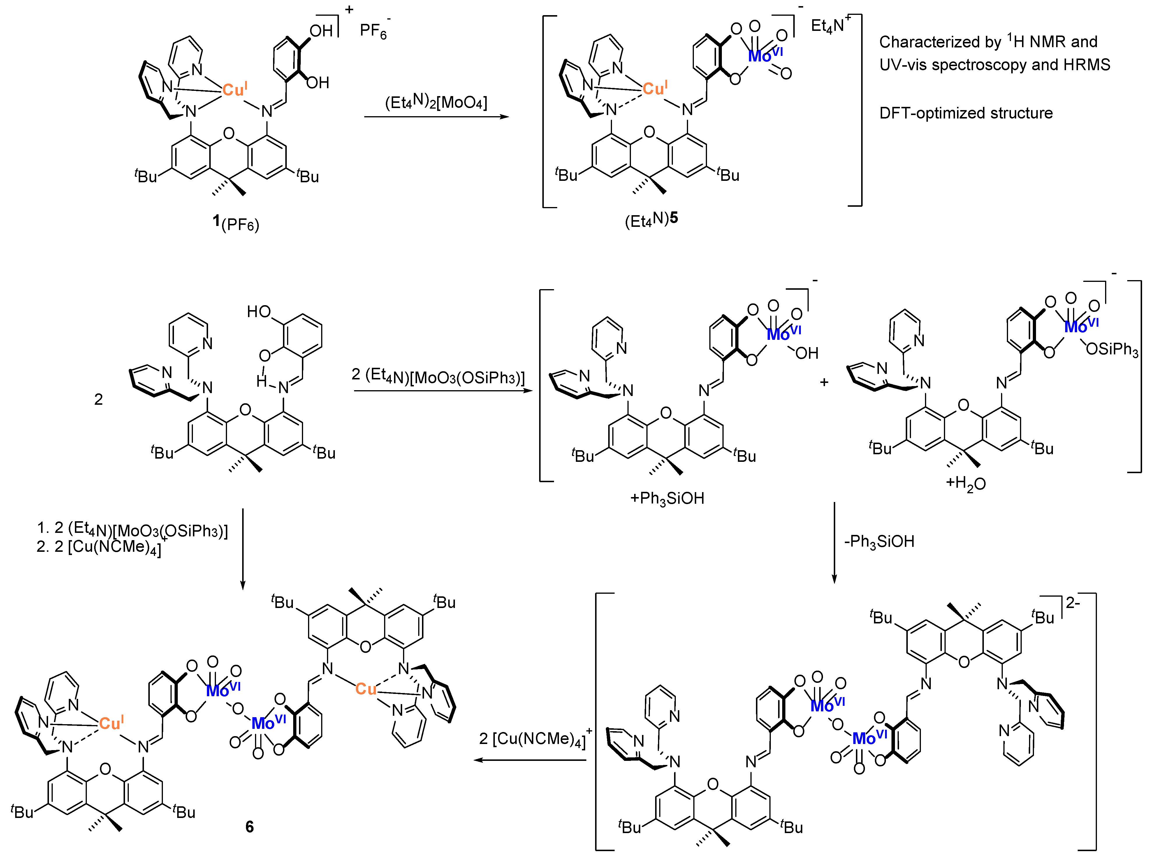
We have previously reported that the reaction of 1(PF6) with molybdate formed red–brown (Et4N)[CuMoO3(Lig)] ((Et4N)5), which had limited stability in solution and therefore was characterized by 1H NMR, UV-vis spectroscopy, and HRMS; its DFT-optimized structure was in an agreement with spectroscopic data (Scheme 3). Our multiple attempts to crystallize (Et4N)5 resulted in its decomposition. We hypothesized that the instability of (Et4N)5 could be due to a highly reactive nature of catecholate-bound [MoVIO3]2−, and thus decided to replace one of the oxo groups with siloxide. Treatment of (Et4N)[MoO3(OSiPh3)] with LigH2, followed by the addition of the resulting solution to [Cu(NCMe)4](PF6) resulted in an orange-brown solution. Recrystallization of the crude product from acetonitrile formed dark orange crystals of [Cu2Mo2O42-O)(Lig)2] (6). X-ray structure determination revealed 6 to be a tetranuclear complex containing two Mo(VI) and two Cu(I) centers (Figure 3 left). The complex is approximately C2-symmetric, albeit the symmetry is not crystallographic due, in part, to the presence of only one molecule of HOSiPh3 that is H-bonded to one of the equatorial oxos (Figure 3 right, see below for the discussion). The coordination geometry at each of the Cu(I) sites is similar to the geometry in 1(PF6), with three relatively short copper-nitrogen (pyridine/imine) bond distances (ranging between 2.000(4) and 2.059(4) Å) and one very long copper-nitrogen (central amine) distance (2.432(4)/2.437(4) Å). The geometry at the molybdenum sites is approximately square-pyramidal, with the chelating catecholate occupying two of the four basal positions. The remaining basal positions and the apical positions are occupied by the oxo ligands, with bridging oxo located in the basal position. Related square-pyramidal [MO3(bdt)]2−/[MO3−xSx(bdt)]2−/[MO2(OSiR3)(bdt)]2− (M = Mo, W; bdt = benzenedithiolate) were previously evaluated by Holm and coworkers as models for Xanthine Oxidoreductase [31,32,33]. The distance between catecholate oxygen and the second molybdenum (Mo1-O8 2.445(3)/Mo2-O7 2.452(3) Å) is likely too long for a bond. Bridging oxo demonstrates a relatively long molybdenum-oxo bond distance of 1.918(4)/1.919(4) Å, compared with 1.696(3)–1.721(4) Å for the terminal oxos. It is noted the relative metals disposition in each of the [Mo-Cu] “halves” is highly reminiscent of the calculated structure of [CuMoO3(Lig)]+ (5+) [26].
As mentioned above, the complex is co-crystallized with one molecule of Ph3SiOH that is hydrogen-bonded to one of the equatorial oxos (Figure 3, right). The presence of one equivalent of triphenylsilanol per two molybdenum ions suggests the dimerization mechanism presented in Scheme 3. Unlike [MoO4]2−, [MoO3(OSiPh3)] contains two different protonation sites. Thus, it is feasible that the acid-base reaction of catechol with [MoO3(OSiPh3)] leads to the formation of siloxo-terminated and hydroxo-terminated products. Subsequent condensation of these complexes results in Mo-(μ2-O)-Mo complex which produces the final product 6 upon the incorporation of two Cu(I) ions.
Complex 6 was also characterized in solution, by 1H NMR spectroscopy and mass spectrometry. Although the spectrum in CD3CN demonstrated only broad peaks, the spectrum in DMF-d7 featured relatively sharp resonances consistent with the overall structure of 6. Additional support for the tetranuclear structure of 6 comes from the observation of the peak corresponding to the molecular ion (6+) at m/z = 1706.3350 (expected m/z = 1706.3273), albeit at low intensity.

3. Summary and Conclusions

Towards the development of the functional model of Mo-Cu CODH, we have previously reported the synthesis of Cu(I)-Mo(VI) complex with a xanthene-bridged heterodinucleating ligand LigH2 featuring dipycolylamine chelate for Cu(I) and catecholate for Mo(VI). However, the complex had only limited stability in solution, and could not be obtained in a solid state; therefore, its subsequent reactivity was not investigated. In the present work, we targeted: (1) the in situ reactivity studies combining Lig, Cu(I), Mo(VI), and a potentially oxidizable ligand L (isocyanide, phosphine, cyanide), and (2) a more stable heterodinuclear MoVICuI complex which could be isolated and structurally characterized. Towards the first goal, we have prepared a series of Lig-supported Cu(I)-L complexes, treated them with [MoO4]2−, and explored the nature of the product (L vs. L=O) using the corresponding spectroscopy. For both phosphine and isocyanide substrates, no oxidation was observed. We propose that the lack of oxidation reactivity can be attributed (at least in part) to the difficulty in accessing a Cu(I)-bound substrate, which is removed from the Mo site and is obscured by the steric congestion at the Cu site. Toward our second goal, we explored the reactivity of the [MoO3(OSiPh3)] precursor, which is expected to produce more stable Mo(VI) complexes. The incorporation of both Mo(VI) and Cu(I) metals within the ligand framework produced a tetranuclear MoVI2CuI2 product [Cu2Mo2O42-O)(Lig)2], in which two heterodinuclear {MoVICuI(Lig1)} fragments were further linked by a μ2-oxo ligand at Mo. The formation of the tetranuclear product was accompanied by the release of silanol directly observed in the crystal. The presence of one equivalent of Ph3SiOH per two molybdenum centers suggests a dimerization mechanism, which involves the reaction of Mo-OSiPh3 with Mo-OH intermediate to form Mo-O-Mo and Ph3SiOH. As in the previously reported DFT-optimized structure of the heterodinuclear [MoVICuI(Lig1)] complex, the metals were relatively far from each other (~7 Å away), which is consistent with the lack of heterobimetallic reactivity. Our future studies in this project will focus on heterodinucleating ligands in which the Cu(I) site is anchored to the Mo site by the nearby imino(phenolate) or amino(phenolate) donor.

4. Materials and Methods

4.1. General

All reactions involving air-sensitive materials were carried out in a nitrogen-filled glovebox. 2,7-Di-tert-butyl-9,9-dimethyl-9H-xanthene-4,5-diamine, (E)-3-(((5-(bis(pyridin-2-ylmethyl) amino)-2,7-di-tert-butyl-9,9-dimethyl-9H-xanthen-4-yl)imino)methyl)benzene-1,2-diol, tetraethylammonium molybdate, 1(PF6), and (Et4N)3 were synthesized using previously published procedures [26,34,35,36]. 2,6-Dimethylphenyl isocyanide, triphenylphosphine, and trimethylphosphine, were purchased from Sigma and used as received. Tetrakis(acetonitrile)copper(I) hexafluorophosphate was purchased from Strem and used as received. All non-deuterated solvents were purchased from Aldrich and were of HPLC grade. The non-deuterated solvents were purified using an MBraun solvent purification system. Dichloromethane-d2 and acetonitrile-d3 were purchased from Cambridge Isotope Laboratories. All solvents were stored over 3 Å molecular sieves. Compounds were generally characterized by 1H and 13C NMR spectroscopy (including 2D techniques such as 1H-1H COSY and HMBC) and high-resolution mass spectrometry. Selected compounds were characterized by X-ray crystallography. Compounds characterization was carried out at the Lumigen Instrument Center at Wayne State University. NMR spectra of metal complexes were recorded on an Agilent DD2-600 MHz spectrometer, and an Agilent 400 MHz spectrometer in CD3CN and DMF at room temperature. Chemical shifts and coupling constants (J) were reported in parts per million (δ) and Hertz, respectively. Detailed assignments of the signals in 1H NMR are given in the ESI. High-resolution mass spectra of the metal complexes (unless otherwise stated) were collected on a Thermofisher Scientific LTQ Orbitrap XL mass spectrometer. The MS survey scan was set from 200 to 2000 m/z. The resolution was set to 60,000. In all cases, only one microscan was used in the analysis.

4.2. Synthetic Procedures and Compounds Characterization

Preparation of [Cu(CN(2,6-Me2C6H3))(LigH2)](PF6) (2(PF6). A 3 mL solution of 2,6-Dimethylphenyl isocyanide (3.80 mg, 0.029 mmol, 1.0 equiv.) in CH3CN was added dropwise to a stirring 5 mL solution of complex [Cu(LH2)](PF6)] (25 mg, 0.029 mmol, 1.0 equiv) in CH3CN. The reaction mixture was stirred for 1 h, upon which the volatiles were removed in vacuo. This solid was purified by recrystallization from CH2Cl2/diethyl ether, which yielded orange-yellow crystals of [Cu(CN(2,6-Me2C6H3))(LigH2)](PF6) (26 mg, 0.028 mmol, 90%). 1H NMR (CD3CN, 400 MHz) δ 8.79 (s, 1H, imine-H), 8.72 (d, 3JHH = 5.1 Hz, 2H, α-H on pyridine), 7.70 (s, 1H, ortho-H on pyridinyl xanthene side), 7.66 (t, 3JHH = 7.8 Hz, 2H, γ-H on pyridine), 7.50 (s, 1H, para-H on pyridinyl xanthene side), 7.43 (s, 1H, para-H on catechol xanthene side), 7.37 (t, 3JHH = 4.8 Hz, 2H, β-H on pyridine),7.31 (t, 3JHH = 7.1 Hz, 1H, para-H on isocyanide), 7.20 (m, 2H, meta-H on isocyanide), 7.18 (s, 1H, ortho-H on catechol xanthene side), 7.12 (d, 3JHH = 7.6 Hz, 1H, 4-H on catechol), 7.07 (d, 3JHH = 7.8 Hz, 2H, β’-H on pyridine), 6.93 (d, 3JHH = 7.9 Hz, 1H, 6-H on catechol), 6.88 (t, 3JHH = 7.7 Hz, 1H, 5-H on catechol), 4.67 (s, 4H, methylene-H), 2.32 (s, 6H, methyl-H on isocyanide), 1.69 (s, 6H, methyl-H), 1.37 (s, 9H, tert-butyl-H on catechol xanthene side), 1.14 (s, 9H, tert-butyl-H on pyridinyl xanthene side) ppm. 13C{1H} NMR (CD2Cl2,150 MHz) δ 165.47, 157.26, 149.37, 149.30, 146.97, 145.57, 145.42, 143.94, 140.00, 138.59, 136.60, 135.28, 134.42, 130.98, 129.77, 129.75, 128.11, 124.26, 124.07, 123.93, 123.27, 122.25, 121.75, 119.14, 118.97, 118.32, 116.05, 58.76, 34.59, 34.30, 32.42, 30.62, 30.47, 17.96. HRMS (ESI+) m/z 848.3577 (calculated m/z for [Cu(CN(2,6-Me2C6H3))(LigH2)]+ 848.3595). IR (ATR) 2137 (C≡NAr) cm−1.
Reaction of [Cu(CN(2,6-Me2C6H3))(LigH2)](PF6) (2(PF6)) with (NEt4)2[MoO4]. A 3 mL solution of tetraethylammonium molybdate (NEt4)2[MoO4] (8.72 mg, 0.020 mmol, 1.0 equiv) in CH3CN and a 3 mL solution of [Cu(LH2)(ArNC)](PF6) (20 mg, 0.020 mmol, 1.0 equiv.) in CH3CN were prepared and cooled to −33 °C. The solution of cold [Cu(LH2)(ArNC)](PF6) was then added dropwise to a stirring solution of cold (NEt4)2[MoO4]. The reaction mixture was stirred for 1hr, after which the volatiles were removed in vacuo to obtain an orang-yellow solid. The crude product was characterized by 1H NMR and IR.
Preparation of [Cu(PMe3)(LigH2)](PF6) (4(PF6)). A 6 µL (0.057 mmol, 1.0 equiv) solution of trimethylphosphine, a 5 mL solution of complex [Cu(LH2)](PF6)] (50 mg, 0.057 mmol, 1.0 equiv) in DCM were prepared and cooled to −33 °C. The solution of cold trimethylphosphine was then added dropwise to a stirring solution of cold[Cu(LH2)](PF6)]. The reaction mixture was stirred for 1 h, upon which the volatiles were removed in vacuo. The product [Cu(LH2)(ArNPMe3)](PF6) was obtained as an orange solid. (51.50 mg, 0.055 mmol, 95%).1H NMR (CD3CN, 400 MHz) δ 13.93 (br, s, 1H, catechol-OH), 8.91 (s, 1H, imine-H), 8.66 (d, 3JHH = 5.0 Hz, 2H, α-H on pyridine), 7.57 (t, 3JHH = 7.7 Hz, 2H, γ-H on pyridine), 7.54 (s, 1H, ortho-H on pyridinyl xanthene side), 7.46 (s, 2H, para-H on pyridinyl xanthene side, para-H on catechol xanthene side), 7.36 (t, 3JHH = 6.4 Hz, 2H, β-H on pyridine), 7.32 (s, 1H, ortho-H on catechol xanthene side), 7.19 (d, 3JHH = 7.4 Hz, 1H, 4-H on catechol), 6.92 (m, 3H, β’-H on pyridine, 6-H on catechol), 6.90 (m, 1H, 5-H on catechol), 6.15 (br, 1H, catechol-OH) 4.66 (s, 4H, methylene-H), 1.72 (s, 6H, methyl-H), 1.41 (s, 9H, tert-butyl-H on catechol xanthene side), 1.29 (s, 6H, methyl-H on isocyanide) 1.15 (s, 9H, tert-butyl-H on pyridinyl xanthene side) ppm. 13C{1H} NMR (CD2Cl2,150 MHz) δ 165.44, 157.57, 149.51, 149.29, 146.99, 145.44, 145.39, 145.02, 140.23, 138.09, 136.34, 134.88, 131.12, 129.82, 125.66, 123.97, 123.73, 123.27, 122.92, 121.90, 119.17, 119.06, 118.26, 115.75, 67.30, 65.27, 58.71, 54.31, 34.65, 34.59, 34.18, 32.45, 30.65, 30.62, 25.26, 14.63. HRMS (ESI+) m/z 793.3295 (calculated m/z for [Cu(LH2)(ArNPMe3)+] 793.3302). 31P NMR (600 MHz) δ −49.0 ppm.
Reaction of [Cu(PMe3)(LigH2)](PF6) (4(PF6)) with (NEt4)2[MoO4]. A 3 mL solution of tetraethylammonium molybdate (NEt4)2[MoO4] (8.95 mg, 0.021 mmol, 1.0 equiv) in CH3CN and a 3 mL solution of [Cu(LH2)(ArNPMe3)](PF6) (20 mg, 0.021 mmol, 1.0 equiv.) in CH3CN were prepared and cooled to −33 °C. The solution of cold [Cu(LH2)(ArNPMe3)](PF6) was then added dropwise to a stirring solution of cold (NEt4)2[MoO4]. The reaction mixture was stirred for 1hr, after which the volatiles were removed in vacuo to obtain an orange solid.
Reaction of [Cu(PPh3)(LigH2)](PF6) (3(PF6)) with (NEt4)2[MoO4]. A 3 mL solution of tetraethylammonium molybdate (NEt4)2[MoO4] (7.38 mg, 0.017 mmol, 1.0 equiv) in CH3CN and a 3 mL solution of [Cu(LH2)(PPh3)](PF6). (20 mg, 0.017 mmol, 1.0 equiv.) in CH3CN were prepared and cooled to −33 °C. The solution of cold [Cu(LH2)(PPh3)](PF6) (2(PF6)). was then added dropwise to a stirring solution of cold (NEt4)2[MoO4]. The reaction mixture was stirred for 1hr, after which the volatiles were removed in vacuo to obtain an orange solid.
Preparation of [Cu2Mo2O42-O)(Lig)2] (6). A 3 mL solution of LH2 (15 mg, 0.022 mmol, 1.0 equiv) in CH3CN and a 3 mL solution of (Et4N)[MoO3(OSiPh3)] (12.59 mg, 0.022 mmol, 1.0 equiv.) in CH3CN were prepared and cooled to −33 °C. The solution of cold LH2 was then added dropwise to a stirring solution of cold (Et4N)[MoO3(OSiPh3)]. The reaction mixture was stirred for 15 min and 3 mL solution of tetrakis(acetonitrile)copper(I) hexafluorophosphate [Cu(NCMe)4](PF6) (8.53 mg, 0.022 mmol, 1.0 equiv.) was added. The reaction mixture was stirred for 30 min, after which the volatiles were removed in vacuo to produce a dark orange solid and crude product was recrystallized using CH3CN to give orange crystals of 6 (13.6 mg, 0.008 mmol, 70%, co-crystallized with HOSiPh3). To remove HOSiPh3, 6 was briefly washed with CH3CN and dissolved in DMF. 1H NMR (DMF, 400 MHz) δ 10.44 (s, 1H, imine-H), 8.88 (s, 1H, 4-H on catechol), 8.15 (s, 1H, ortho-H on pyridinyl xanthene side), 7.89 (m, 3H, para-H on pyridinyl xanthene side, β-H on pyridine), 7.67 (m, 1H, para-H on catechol xanthene side), 7.58 (m, 2H, α-H on pyridine), 7.45 (m, 2H, β-H on pyridine), 7.39 (s, 1H, ortho-H on catechol xanthene side), 7.30 (m, 2H, γ-H on pyridine), 6.55 (s, 1H, 6-H on catechol, 6.39 (m, 1H, 5-H on catechol), 4.88 (br s, 2H, methylene-H) 4.30 (br s, 2H, methylene-H), 1.56 (s, 6H, Me), 1.37 (s, 18H, t-Bu) ppm. HRMS (ESI+) m/z 1706.3350 (calculated m/z for [Cu2Mo2O42-O)(Lig)2]+ 1706.3267).

4.3. X-ray Crystallographic Details

All crystal structures were collected on a Bruker D8 Venture diffractometer with kappa geometry, an Incoatec IµS micro-focus X-ray source (Mo Kα radiation), and a multilayer mirror for monochromatization. The diffraction intensities were measured using a Bruker Photon III CPAD area detector. Data were acquired at 100 K with an Oxford 800 Cryostream low-temperature apparatus. The data were processed using APEX4 software (v.2022.10-0, Bruker, Madison, USA). The structures were solved by Intrinsic Phasing using ShelXT [37] and refined with ShelXL [38] using Olex2 [39]. Hydrogen atoms were placed in calculated positions using a standard riding model and refined isotropically; all other atoms were refined anisotropically. The structure of 2(PF6) contained one co-crystallized molecule of diethyl ether. The structure of 6 contained one co-crystallized molecule of HOSiPh3 and six co-crystallized molecules of acetonitrile. In addition, one of the tert-butyl groups of 6 was found to be disordered over two positions. The main refinement parameters are listed in Table 1. The structures were uploaded in the CCDC under the following numbers: 2252288–2252289.

Supplementary Materials

The following supporting information can be downloaded at: https://www.mdpi.com/article/10.3390/molecules28083644/s1, General experimental details, synthetic procedures, crystallographic, NMR, IR, and mass spectra. Figure S1. 1H NMR spectrum of [Cu(CN(2,6-Me2C6H3))(LigH2)](PF6) (2(PF6)) (CD3CN, 400 MHz). Figure S2. 1H-1H COSY NMR spectrum of [Cu(CN(2,6-Me2C6H3))(LigH2)](PF6) (2(PF6)) (CD3CN, 400 MHz). Figure S3. 13C NMR spectrum of [Cu(CN(2,6-Me2C6H3))(LigH2)](PF6) (2(PF6)) (CD3CN, 150 MHz). Figure S4. 1H NMR spectrum of the reaction between [Cu(CN(2,6-Me2C6H3))(LigH2)](PF6) (2(PF6)) and (NEt4)2[MoO4] (CD3CN, 400 MHz). Figure S5. 1H NMR spectrum of [Cu(PMe3)(LigH2)](PF6) (4(PF6)) (CD3CN, 400 MHz). Figure S6. 1H-1H COSY NMR spectrum of [Cu(PMe3)(LigH2)](PF6) (4(PF6)) (CD3CN 400 MHz). Figure S7. 13C NMR spectrum of [Cu(PMe3)(LigH2)](PF6) (4(PF6)) (CD3CN 150 MHz). Figure S8. 31P NMR spectrum of [Cu(PMe3)(LigH2)](PF6) (4(PF6)) (CD3CN 400 MHz). Figure S9. 1H NMR spectrum of the reaction between [Cu(PMe3)(LigH2)](PF6) (4(PF6)) with (NEt4)2[MoO4] (CD3CN, 400 MHz). Figure S10. 31P NMR spectrum of the reaction between [Cu(PMe3)(LigH2)](PF6) (4(PF6)) with (NEt4)2[MoO4] (CD3CN, 400 MHz). Figure S11. 1H NMR spectrum of the reaction between [Cu(PPh3)(LigH2)](PF6) (3(PF6) and (NEt4)2[MoO4] (CD3CN, 400 MHz). Figure S12. 31P NMR spectrum of the between [Cu(PPh3)(LigH2)](PF6) (3(PF6) and (NEt4)2[MoO4] (CD3CN, 400 MHz). Figure S13. 1H NMR spectrum of the reaction between 1(PF6) and NaCN (CD3CN, 400 MHz). Figure S14. 1H NMR spectrum of the reaction between 1(PF6) and styrene (CD2Cl2, 400 MHz). Figure S15. 1H NMR spectrum of [Cu2Mo2O42-O)(Lig)2] (6) (CD3CN, 400 MHz). Figure S16. 1H NMR spectrum of [Cu2Mo2O42-O)(Lig)2] (6) (DMF-d7, 400 MHz). Figure S17. 1H-1H COSY NMR spectrum of [Cu2Mo2O42-O)(Lig)2] (6) (DMF-d7, 400 MHz). Figure S18. High-resolution mass spectrum of [Cu(CN(2,6-Me2C6H3))(LigH2)]+ (2+). Figure S19. High-resolution mass spectrum of [Cu(PMe3)(LigH2)]+ (3+). Figure S20. High-resolution mass spectrum of [Cu2Mo2O42-O)(Lig)2]+ (6+). Figure S21. IR spectrum of [Cu(CN(2,6-Me2C6H3))(LigH2)](PF6) (2(PF6) (isocyanide CN stretch region). Figure S22. IR spectrum of the reaction product between [Cu(CN(2,6-Me2C6H3))(LigH2)](PF6) (2(PF6) and (NEt4)2[MoO4] (isocyanide CN stretch region).

Author Contributions

S.G. and U.I.K.D. designed this study and wrote the manuscript, U.I.K.D. and A.S.A. carried out the synthetic part of this work, C.L.W. assisted with the crystallographic characterization. All authors have read and agreed to the published version of the manuscript.

Funding

This research was funded by the ACS PRF (grant number 58937-ND3).

Institutional Review Board Statement

Not applicable.

Informed Consent Statement

Not applicable.

Data Availability Statement

Information about Supplementary Materials is provided above in the “Supplementary Materials” statement.

Acknowledgments

Experimental characterization was carried out at the Lumigen Instrument Center of Wayne State University. This work made use of the single-crystal XRD that was partially funded by the National Institutes of Health supplement grant #3R01EB027103-02S1. Umesh I. Kaluarachchige Don is a Rumble fellow.

Conflicts of Interest

The authors declare no competing financial interests.

References

  1. Hille, R.; Dingwall, S.; Wilcoxen, J. The aerobic CO dehydrogenase from Oligotropha carboxidovorans. J. Biol. Inorg. Chem. 2015, 20, 243–251. [Google Scholar] [CrossRef] [PubMed]
  2. Mörsdorf, G.; Frunzke, K.; Gadkari, D.; Meyer, O. Microbial growth on carbon monoxide. Biodegradation 1992, 3, 61–82. [Google Scholar] [CrossRef]
  3. Burgmayer, S.J.N.; Kirk, M.L. The Role of the Pyranopterin Dithiolene Component of Moco in Molybdoenzyme Catalysis. In Structure and Bonding: Metallocofactors that Activate Small Molecules; Ribbe, M., Ed.; Springer Nature: Cham, Switzerland, 2019; Volume 179, pp. 101–151. [Google Scholar]
  4. Basu, P.; Burgmayer, S.J.N. Recent Developments in the Study of Molybdoenzyme Models. J. Biol. Inorg. Chem. 2015, 20, 373–383. [Google Scholar] [CrossRef] [PubMed]
  5. Dobbek, H.; Gremer, L.; Kiefersauer, R.; Huber, R.; Meyer, O. Catalysis at a dinuclear [CuSMo(AO)OH] cluster in a CO dehydrogenase resolved at 1.1-Å resolution. Proc. Natl. Acad. Sci USA 2002, 99, 15971–15976. [Google Scholar] [CrossRef]
  6. Wilcoxen, J.; Hille, R. The hydrogenase activity of the molybdenum/copper-containing carbon monoxide dehydrogenase of Oligotropha carboxidovorans. J. Biol. Chem. 2013, 288, 36052–36060. [Google Scholar] [CrossRef]
  7. Gnida, M.; Ferner, R.; Gremer, L.; Meyer, O.; Meyer-Klaucke, W. A novel binuclear [CuSMo] cluster at the active site of carbon monoxide dehydrogenase: Characterization by X-ray absorption spectroscopy. Biochemistry 2003, 42, 222–230. [Google Scholar] [CrossRef]
  8. Zhang, B.; Hemann, C.F.; Hille, R. Kinetic and spectroscopic studies of the molybdenum-copper CO dehydrogenase from Oligotropha carboxidovorans. J. Biol. Chem. 2010, 285, 12571–12578. [Google Scholar] [CrossRef]
  9. Shanmugam, M.; Wilcoxen, J.; Habel-Rodriguez, D.; Cutsail, G.E.; Kirk, M.L.; Hoffman, B.M.; Hille, R. 13C and 63,65Cu ENDOR studies of CO Dehydrogenase from Oligotropha carboxidovorans. Experimental Evidence in Support of a Copper–Carbonyl Intermediate. J. Am. Chem. Soc. 2013, 135, 17775–17782. [Google Scholar] [CrossRef]
  10. Siegbahn, P.E.M.; Shestakov, A.F. Quantum chemical modeling of CO oxidation by the active site of molybdenum CO dehydrogenase. J. Comput. Chem. 2005, 26, 888–898. [Google Scholar] [CrossRef]
  11. Hofmann, M.; Kassube, J.K.; Graf, T. The mechanism of Mo-/Cu-dependent CO dehydrogenase. J. Biol. Inorg. Chem. 2005, 10, 490–495. [Google Scholar] [CrossRef]
  12. Stein, B.W.; Kirk, M.L. Orbital contributions to CO oxidation in Mo–Cu carbon monoxide dehydrogenase. Chem. Commun. 2014, 50, 1104–1106. [Google Scholar] [CrossRef]
  13. Rokhsana, D.; Large, T.A.; Dienst, M.C.; Retegan, M.; Neese, F. A realistic in silico model for structure/function studies of molybdenum–copper CO dehydrogenase. J. Biol. Inorg. Chem. 2016, 21, 491–499. [Google Scholar] [CrossRef]
  14. Xu, K.; Hirao, H. Revisiting the catalytic mechanism of Mo–Cu carbon monoxide dehydrogenase using QM/MM and DFT calculations. Phys. Chem. Chem. Phys. 2018, 20, 18938–18948. [Google Scholar] [CrossRef]
  15. Rovaletti, A.; Bruschi, M.; Moro, G.; Cosentino, U.; Greco, C. The challenging in silico description of carbon monoxide oxidation as catalyzed by molybdenum-copper CO dehydrogenase. Front. Chem. 2019, 6, 630. [Google Scholar] [CrossRef]
  16. Rovaletti, A.; Bruschi, M.; Moro, G.; Cosentino, U.; Ryde, U.; Greco, C. A thiocarbonate sink on the enzymatic energy landscape of aerobic CO oxidation? Answers from DFT and QM/MM models of MoCu CO-dehydrogenases. J. Catal. 2019, 372, 201–205. [Google Scholar] [CrossRef]
  17. Ritacca, A.G.; Rovaletti, A.; Moro, G.; Cosentino, U.; Ryde, U.; Sicilia, E.; Greco, C. Unraveling the reaction mechanism of Mo/Cu CO dehydrogenase using QM/MM calculations. ACS Catal. 2022, 12, 7336–7343. [Google Scholar] [CrossRef]
  18. Majumdar, A. Bioinorganic Modeling Chemistry of Carbon Monoxide Dehydrogenases: Description of Model Complexes, Current Status and Possible Future Scopes. Dalton Trans. 2014, 43, 12135–12145. [Google Scholar] [CrossRef]
  19. Gourlay, C.; Nielsen, D.J.; White, J.M.; Knottenbelt, S.Z.; Kirk, M.L.; Young, C.G. Paramagnetic active site models for the molybdenum–copper carbon monoxide dehydrogenase. J. Am. Chem. Soc. 2006, 128, 2164–2165. [Google Scholar] [CrossRef] [PubMed]
  20. Gourlay, C.; Nielsen, D.J.; Evans, D.J.; White, J.M.; Young, C.G. Models for Aerobic Carbon Monoxide Dehydrogenase. Chem. Sci. 2018, 9, 876–888. [Google Scholar] [CrossRef] [PubMed]
  21. Takuma, M.; Ohki, Y.; Tatsumi, K. Sulfido-Bridged Dinuclear Molybdenum–Copper Complexes Related to the Active Site of CO Dehydrogenase: [(dithiolate)Mo(O)S2Cu(SAr)]2− (dithiolate = 1,2-S2C6H4, 1,2-S2C6H2-3,6-Cl2, 1,2-S2C2H4). Inorg. Chem. 2005, 44, 6034–6043. [Google Scholar] [CrossRef]
  22. Groysman, S.; Majumdar, A.; Zheng, S.-L.; Holm, R.H. Reactions of monodithiolene tungsten(VI) sulfido complexes with copper(I) in relation to the structure of the active site of carbon monoxide dehydrogenase. Inorg. Chem. 2010, 49, 1082–1089. [Google Scholar] [CrossRef]
  23. Mouchfiq, A.; Todorova, T.K.; Dey, S.; Fontecave, M.; Mougel, V. A Bioinspired Molybdenum–Copper Molecular Catalyst for CO2 Electroreduction. Chem. Sci. 2020, 11, 5503–5510. [Google Scholar] [CrossRef]
  24. Ghosh, D.; Sinhababu, S.; Santarsiero, B.D.; Mankad, N.P. A W/Cu Synthetic Model for the Mo/Cu Cofactor of Aerobic CODH indicates that Biochemical CO Oxidation Requires a Frustrated Lewis Acid/Base Pair. J. Am. Chem. Soc. 2020, 142, 12635–12642. [Google Scholar] [CrossRef]
  25. Hollingsworth, T.S.; Hollingsworth, R.L.; Lord, R.L.; Groysman, S. Cooperative Bimetallic Reactivity of a Heterodinuclear Molybdenum-Copper Model of Mo-Cu CODH. Dalton Trans. 2018, 47, 10017–10024. [Google Scholar] [CrossRef]
  26. Kaluarachchige Don, U.I.; Kurup, S.S.; Hollingsworth, T.S.; Ward, C.L.; Lord, R.L.; Groysman, S. Synthesis and Cu(I)/Mo(VI) reactivity of a bifunctional heterodinucleating ligand on a xanthene platform. Inorg. Chem. 2021, 60, 14655–14666. [Google Scholar] [CrossRef]
  27. Do, T.H.; Brown, S.N. Mono- and Bimetallic Pentacoordinate Silicon Complexes of a Chelating Bis(catecholimine) Ligand. Dalton Trans. 2019, 48, 11565–11574. [Google Scholar] [CrossRef]
  28. Barybin, M.V.; Young, V.G.; Ellis, J.E. Syntheses and Structural Characterizations of the First 16-, 17-, and 18-Electron Homoleptic Isocyanide Complexes of Vanadium:  Hexakis(2,6-dimethyl- phenyl isocyanide)vanadium(I, 0, –I)1. J. Am. Chem. Soc. 1998, 120, 429–430. [Google Scholar] [CrossRef]
  29. Carpenter, A.E.; Rheingold, A.L.; Figueroa, J.S. A Well-Defined Isocyano Analogue of HCo(CO)4. 1: Synthesis, Decomposition, and Catalytic 1,1-Hydrogenation of Isocyanides. Organometallics 2016, 35, 2309–2318. [Google Scholar] [CrossRef]
  30. Stephany, R.W.; de Bie, M.J.A.; Drenth, W. A C-NMR and IR study of isocyanides and some of their complexes. Org. Magn. Reason. 1974, 6, 45–47. [Google Scholar] [CrossRef]
  31. Wang, J.-J.; Holm, R.H. Silylation, Sulfidation, and Benzene-1,2-dithiolate Complexation Reactions of Oxo- and Oxosulfidomolybdates(VI) and -Tungstates(VI). Inorg. Chem. 2007, 46, 11156–11164. [Google Scholar] [CrossRef]
  32. Wang, J.-J.; Groysman, S.; Lee, S.C.; Holm, R.H. Synthesis of Structural Analogues of the Oxidized Sites in the Xanthine Oxidoreductase Enzyme Family. J. Am. Chem. Soc. 2007, 129, 7512–7513. [Google Scholar] [CrossRef] [PubMed]
  33. Groysman, S.; Wang, J.-J.; Tagore, R.; Lee, S.C.; Holm, R.H. A Biomimetic Approach to Oxidized Sites in the Xanthine Oxidoreductase Family: Synthesis and Stereochemistry of Tungsten(VI) Analogue Complexes. J. Am. Chem. Soc. 2008, 130, 12794–12807. [Google Scholar] [CrossRef] [PubMed]
  34. Partyka, D.V.; Holm, R.H. Oxygen/Sulfur Substitution Reactions of Tetraoxometalates Effected by Electrophilic Carbon and Silicon Reagents. Inorg. Chem. 2004, 43, 8609–8616. [Google Scholar] [CrossRef] [PubMed]
  35. Bheemaraju, A.; Beattie, J.W.; Danylyuk, Y.; Rochford, J.; Groysman, S. Synthesis, Structures, and Reactivity of Copper(I) Complexes Supported by a Rigid Dinucleating Ligand. Eur. J. Inorg. Chem. 2014, 34, 5865–5873. [Google Scholar] [CrossRef]
  36. Hollingsworth, R.L.; Bheemaraju, A.; Lenca, N.; Lord, R.L.; Groysman, S. Divergent Reactivity of a New Dinuclear Xanthene-Bridged Bis(iminopyridine) di-Nickel Complex with Alkynes. Dalton Trans. 2017, 46, 5605–5616. [Google Scholar] [CrossRef]
  37. Sheldrick, G.M. SHELXT—Integrated Space-group and Crystal Structure Determination. Acta Crystallogr. Sect. A Found. Adv. 2015, 71, 3–8. [Google Scholar] [CrossRef]
  38. Sheldrick, G.M. Crystal Structure Refinement with SHELXL. Acta Crystallogr. Sect. C Struct. Chem. 2015, 71, 3–8. [Google Scholar] [CrossRef]
  39. Dolomanov, O.V.; Bourhis, L.J.; Gildea, R.J.; Howard, J.A.K.; Puschmann, H. OLEX2: A complete structure solution refinement and analysis program. J. Appl. Cryst. 2009, 42, 339–341. [Google Scholar] [CrossRef]
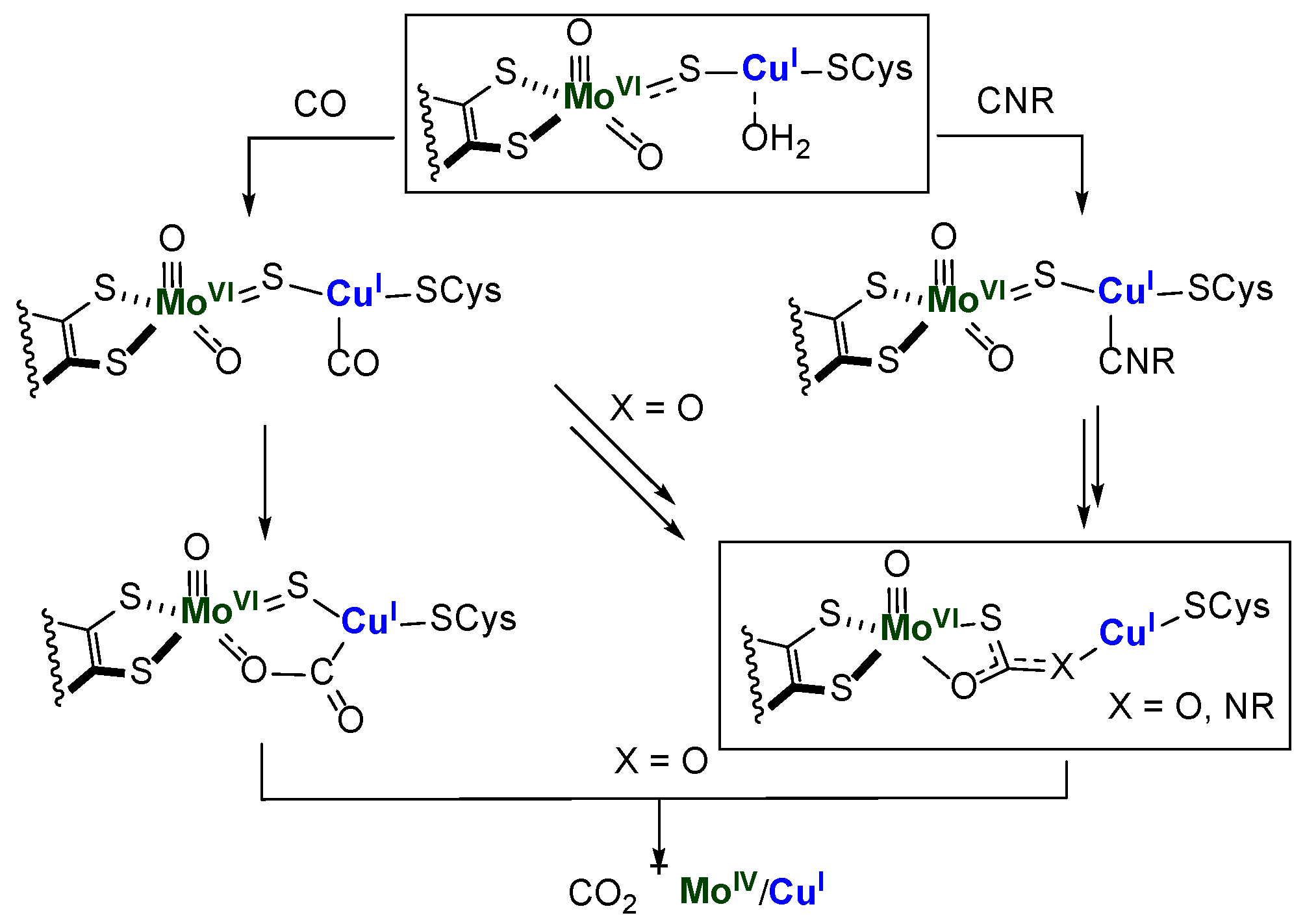
Scheme 1. Resting site (oxidized) structure of Mo-Cu CODH, and its proposed reactivity with CO and CNR. (R = tert-butyl, [SC(R) = C(R)S] ligand = pyranopterindithiolene). Structures inside frames have been characterized by X-ray crystallography (X = NR).
Molecules 28 03644 sch001
Figure 1. The structure of 2(PF6) with 50% probability ellipsoids. PF6 counterion, hydrogen atoms, and co-crystallized solvent (ether) were omitted for clarity.
Molecules 28 03644 g001
Scheme 2. Synthesis of Cu(I) precursors described in this paper.
Molecules 28 03644 sch002
Figure 2. Comparison of the accessibility of Cu(I)-bound substrate to the nucleophilic attack by Mo-oxo in two different xanthene-based heterodinucleating systems [25,26].
Molecules 28 03644 g002
Scheme 3. Top: Previously described formation of complex (Et4N)5. Bottom: Formation of complex 6 and possible reaction mechanism.
Molecules 28 03644 sch003
Figure 3. X-ray structure of 6, 40% probability ellipsoids. Left: top view, without co-crystallized HOSiPh3 molecule. Right: side view, showing H-bonding between one of the basal oxos (O1) and the co-crystallized HOSiPh3 molecule. H atoms, co-crystallized acetonitrile molecules, and an alternative conformation for one of the tert-butyl groups were omitted for clarity. Selected bond distances: Mo1 O1 1.721(3), Mo1 O2 1.696(3), Mo1 O3 1.919(3), Mo2 O4 1.711(3), Mo2 O5 1.706(3), O1–O10 2.769(4) Å.
Molecules 28 03644 g003
Table 1. Crystal and structure refinement data.
2(PF6)6
FormulaC51H55CuN5O3•PF6•C4H10OC84H88Cu2Mo2N8O11•C18H16OSi•6(C2H3N)
Fw, g/mol1068.6702227.30
temperature100 K100 K
X-ray sourceMoKαMoKα
cryst systtriclinicmonoclinic
space groupP-1P21/n
coloryelloworange
Z24
a, Å13.6310(5)20.546(5)
b, Å14.4630(6)21.591(7)
c, Å15.8288(6)25.679(7)
α, deg112.847(1)90.00
β, deg92.606(1)109.253(6)
γ, deg109.383(1)90.00
V, A32657.65(18)10754(5)
dcalcd, g/cm31.3351.376
μ, mm−10.5120.696
2θ, deg55.0055.96
R1 a (all data)0.04280.0996
wR2 b (all data)0.10110.1977
R1 a [(I > 2σ)]0.03720.0682
wR2 b [(I > 2σ)]0.09690.1760
GOF c1.0401.022
a R1 = Σ||Fo − |Fc||/|Fo|. b wR2 = ((w(Fo2 − Fc2)2)/(w(Fo2)2))1/2. c GOF = (w(Fo2 − Fc2)2/(n − p))1/2 where n is the number of data and p is the number of parameters refined.
Disclaimer/Publisher’s Note: The statements, opinions and data contained in all publications are solely those of the individual author(s) and contributor(s) and not of MDPI and/or the editor(s). MDPI and/or the editor(s) disclaim responsibility for any injury to people or property resulting from any ideas, methods, instructions or products referred to in the content.