Article

Five New Cantharidin Derivatives from the Insect Mylabris cichorii L. and Their Potential against Kidney Fibrosis In Vitro

1
School of Pharmacy, Chengdu University of Traditional Chinese Medicine, Chengdu 611137, China
2
Institute for Inheritance-Based Innovation of Chinese Medicine, Medical School of Pharmaceutical Sciences, Shenzhen University, Shenzhen 518060, China
*
Authors to whom correspondence should be addressed.
Lead contact.
Molecules 2023, 28(6), 2822; https://doi.org/10.3390/molecules28062822
Submission received: 22 February 2023 / Revised: 15 March 2023 / Accepted: 18 March 2023 / Published: 21 March 2023

Abstract

:
Five new monoterpenoids including three 1-hydroxymethyl-2-methyl cantharimide-type derivatives (1, 2, and 5) and two 1,2-dimethyl cantharimide-type derivatives (3 and 4), together with three known compounds (68) were isolated from the insect Mylabris cichorii Linnaeus. The structures of these new compounds, including their absolute configurations, were characterized by detailed analysis of NMR, chemical derivatization, and quantum chemical ECD calculations. All of the compounds were tested for their biological activity against kidney fibrosis. The results revealed that compounds 2, 4, and 7 could inhibit kidney fibrosis in vitro at 40 μM by inhibiting the expression of fibronectin and collagen I in TGF-β1-induced NRK-52e cells.

1. Introduction

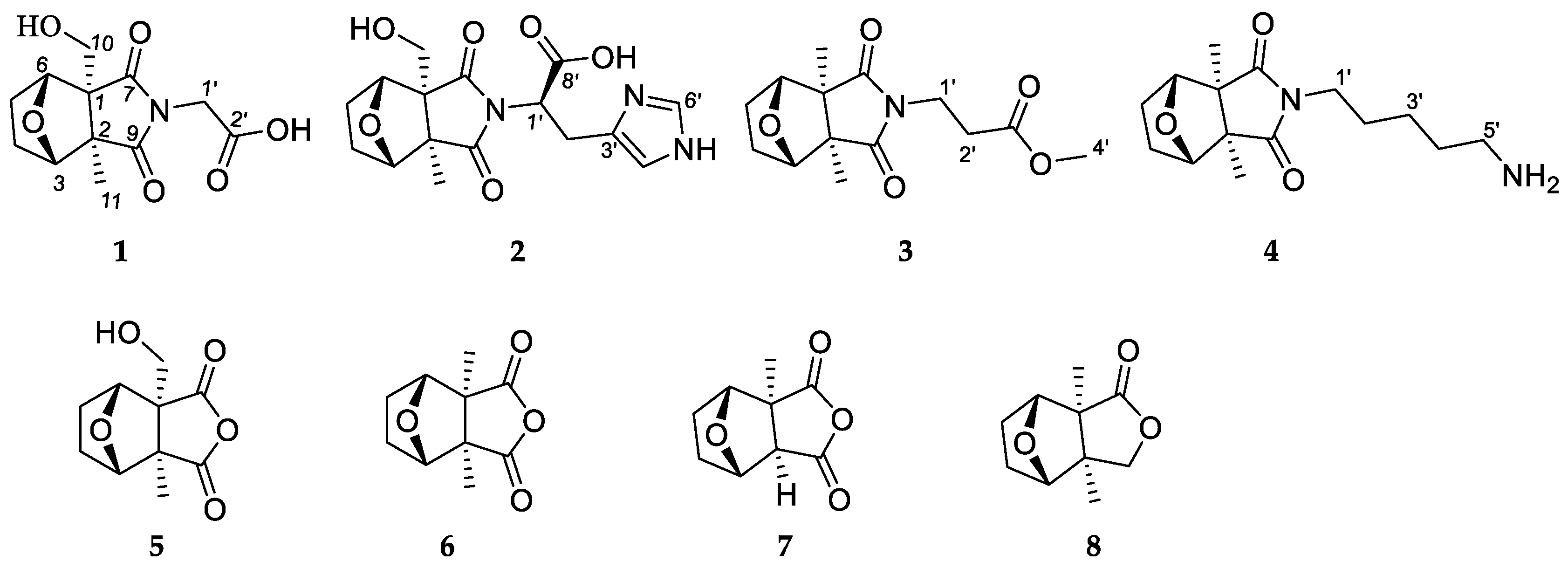
In recent years, small molecules found in insects have been found to have good biological activities such as antiangiogenic activity [1], renal fibrosis inhibition [2], and COX-2 inhibitory activity [3]. The earliest recording of the blister beetle Mylabris cichorii Linnaeus as a medicinal insect is found in Sheng Nong’s Herbal Classic. The dried bodies of M. cichorii have been used as a traditional Chinese medicine for 2000 years, significantly used for the treatment of tumors [4,5,6,7]. Previous studies have revealed cantharidin and its derivatives as a class of small molecules in the genus Mylabris [8,9,10,11]. As a part of our search for characterizing the potent bioactive compounds from insects, research on M. cichorii was carried out. As a result, three new 1-hydroxymethyl-2-methyl cantharimide-type cantharidins (1, 2, and 5), two new 1,2-dimethyl cantharimide-type derivatives (3 and 4), and three known compounds (Figure 1) were isolated from M. cichorii. Recent studies of monoterpene alkaloids have revealed their powerful anti-fibrotic effects [12,13,14]. Because cantharidin derivatives also resemble such a class of monoterpene alkaloids, we were motivated to conduct a study on kidney fibrosis. Biological evaluation revealed the inhibitory properties of kidney fibrosis thereof. Herein, we report the isolation, structure characterization, and biological evaluation of the isolates.

2. Results and Discussion

2.1. Structure Elucidation of the Compounds

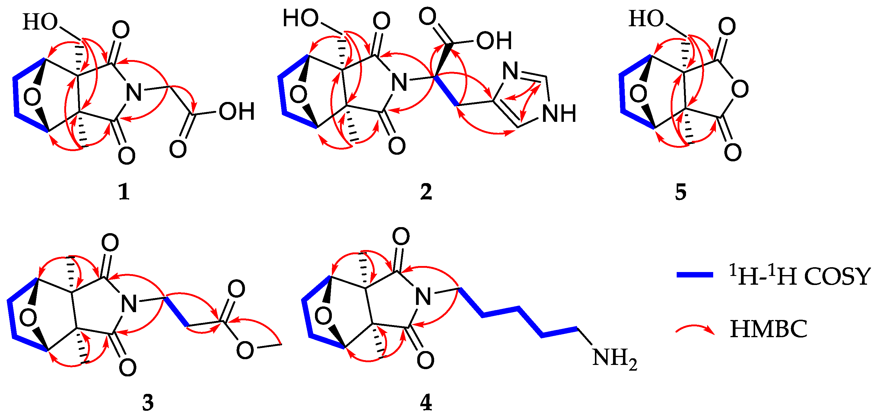
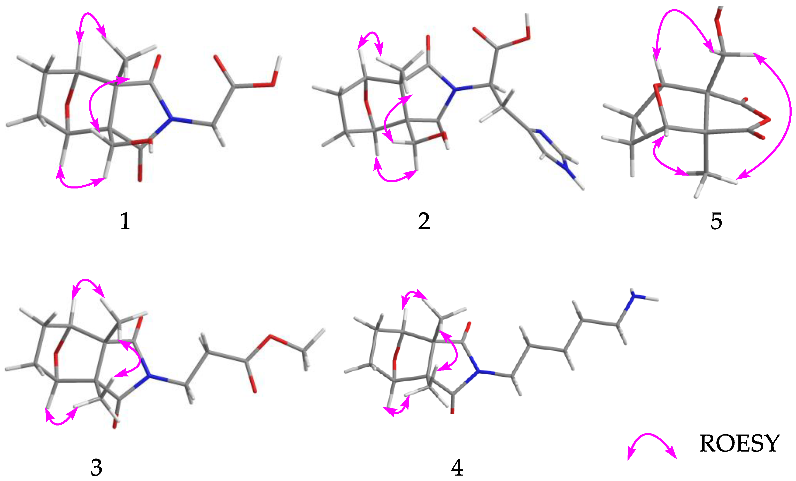
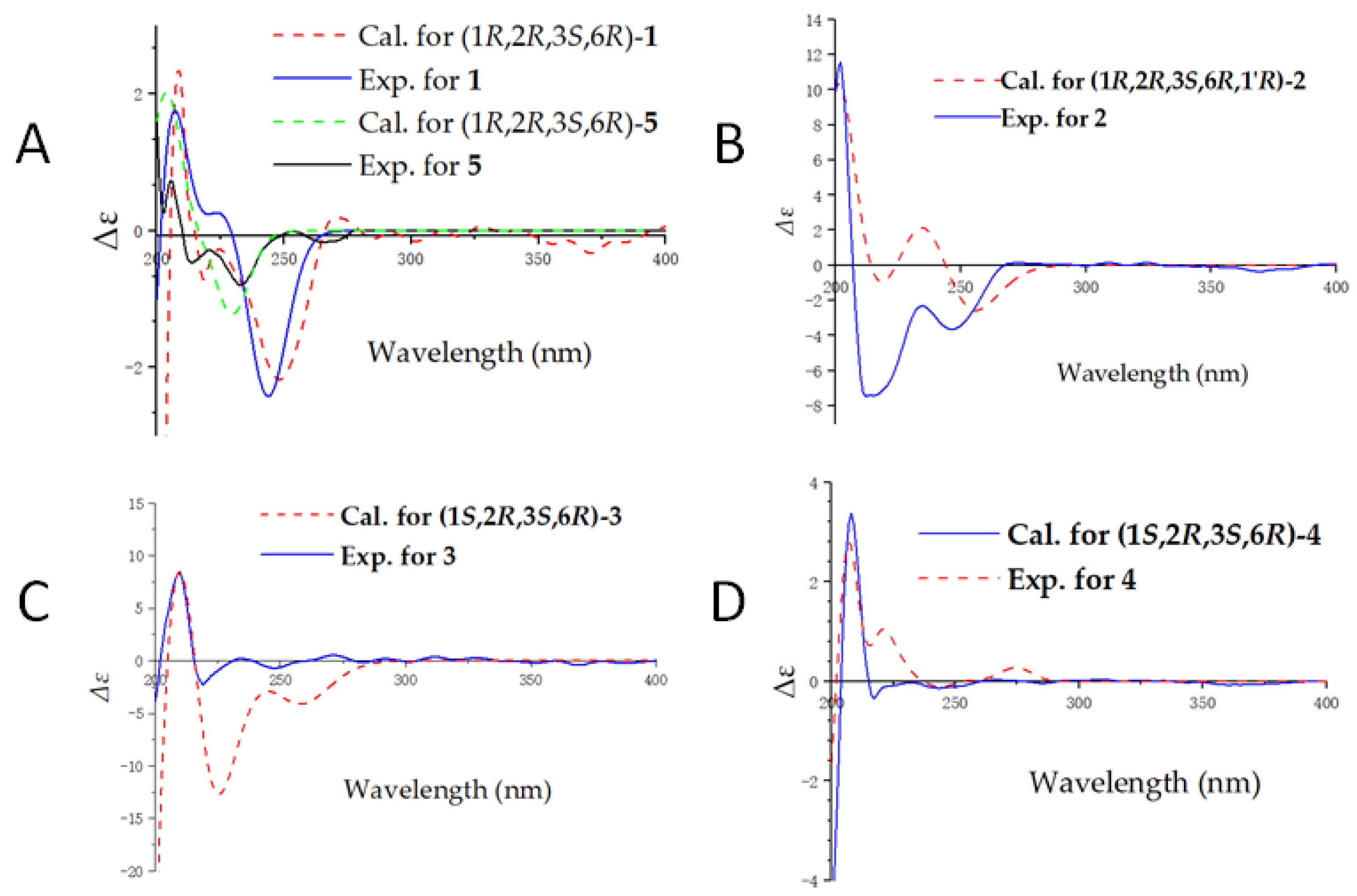
Compound 1, isolated as colorless gums, has a molecular formula C12H15O6N (six degrees of unsaturation) based on analysis of the HRESIMS in the positive ion mode, m/z 270.0959 [M + H]+ (calcd for C12H16O6N, 270.0972) (Figure S7). The UV spectrum of 1 shows an absorption maximum of 203 nm. The 1H NMR spectrum of 1 (Table 1, Figure S1) exhibits characteristic signals of cantharidin derivatives, including one methyl group [δH 1.33 (3H, s, H-11)], four methylenes [δH 1.95 (1H, m, Ha-4), 1.67 (1H, m, Hb-4); 1.75 (1H, m, Ha-5), 1.67 (1H, m, Hb-5); 3.97, 3.73 (each 1H, d, J = 11.3 Hz); 4.21 (2H, d, J = 17.2 Hz)], and two methines [δH 4.52 (1H, br d, J = 4.5 Hz), 4.56 (1H, br d, J = 4.7 Hz)]. The 13C NMR and DEPT spectra (Table 2, Figure S2) exhibit 12 carbon signals including an imide group (δC 182.5, 180.9), a carbonyl group (δC 169.7), two oxygenated methines (δC 85.3, 83.2), and two quaternary carbons (δC 61.9, 55.3). The 1H and 13C NMR data of 1 are similar to cantharimide J [8], except for the signal of one less methylene group at C-2′. The methylene is oxidized as a carbonyl at the C-2′, which could be confirmed by the lower field chemical shift of C-1′ and the 13C NMR data of C-2′ at δC 169.9. An HMBC correlation (Figure 2, Figure S4) of H2-1′/C-2′ (δC 169.7) further confirmed our conclusion. The relative configuration of 1 was assigned by ROESY correlations (Figure 3, Figure S6), which show H-3 (δH 4.52), H2-10 (δH 3.73)/H3-11 (δH 1.33), and H2-10 (δH 3.97)/H-6 (δH 4.56), showing identical relative configuration at each chiral center with cantharidin. The absolute configuration of 1 was verified by the quantum chemical electronic circular dichroism (ECD) calculations. It was found that the experimental ECD spectrum of 1 is similar to the calculated one of (1R,2R,3S,6R)-1 (Figure 4, Figure S8). Thus, the structure of 1 was finally deduced and named cichormide A.
Compound 2 was determined to have a molecular formula of C16H19O6N3 from its HRESIMS (m/z 350.1347 [M + H]+, C16H20O6N3, 350.1360) (Figure S15), indicating the presence of nine degrees of unsaturation. The 1H NMR data (Table 1, Figure S9) of 2 show one methyl [δH 1.24 (3H, s, H-11)], four methylenes [δH 3.94 (1H, d, J = 11.2 Hz, Ha-10), 3.69 (1H, d, J = 11.2 Hz, Hb-10), 1.62-1.91 (4H, m, H2-4, H2-5), δH 3.49 (2H, br d, J = 8.0 Hz, H-2′)], and five methines [δH 8.71 (1H, s, H-6′), 7.26 (1H, s, H-4′), 4.97 (1H, br t, J = 7.4 Hz, H-1′), δH 4.46 (1H, br d, J = 4.6 Hz, H-3), 4.36 (1H, br d, J = 4.8 Hz, H-6)]. The 13C NMR and DEPT spectra (Table 2, Figure S10) show 16 carbon signals, which were assigned as one methyl, four methylenes, five methines, and six non-protonated carbons (including 2 carbonyls and 1 carboxyl). These featured NMR signals are similar to those of canthaminomide F [11], suggesting that 2 is a 1-hydroxymethyl-2-methyl cantharimide-type derivative. The key difference is that the substitution of a methyl at C-1 is replaced by a hydroxymethyl, which is supported by the HMBC correlations of H2-10/C-2, C-6, and C-7 (Figure 2, Figure S12). Thus, the planar structure of 2 was assigned. The ROESY correlations between H-3 (δH 4.46), H2-10 (δH 3.69)/H3-11 (δH 1.24), and between H2-10 (δH 3.94)/H-6 (δH 4.36) suggested that 2 possesses the same relative configuration as compound 1 in the counterpart (Figure 3, Figure S14). The resolution of the configuration at C-1′ has been reported in previous literature [11], and the same method was used to determine the configuration of compound 2. The D or L-histidine was, respectively, submitted to a reaction containing compound 5 under 95% EtOH of solvent at 78 °C for 48 h, which led to the generation of 2a (1R,2R,3S,6R,1′R)/2b (1R,2R,3S,6R,1′S) (Supplementary Materials, Scheme S1). A careful comparison of the NMR data and the retention time in the HPLC chromatogram of 2a and 2b with those of compound 2 revealed that 2a is actually the same as 2 (Supplementary Materials, Figures S41–S45). Thus, the absolute configuration of 2 at C-1′ was assigned. With this information in hand, the absolute configurations at the rest chiral carbons of 2 were solved by comparison of the theoretical and experimental ECD spectra (Figure 4, Figure S16). The results showed that the calculated ECD spectrum of (1R,2R,3S,6R,1′R)-2 is in good accordance with that of 2. Thus, the structure of 2 was determined to be [(1R,2R,3S,6R,1′R)- 1-hydroxymethyl-2-methyl-3,6-epoxycyclohexane-1,2-dicarboximide]-(1′R)-histidine and named cichormide B.
Compound 3 has a molecular formula of C14H19O5N by the HRESIMS at m/z 282.1338 [M + H]+ in a combination of the NMR data, indicating six degrees of unsaturation (Figure S23). The UV spectrum of 3 shows an absorption maximum of 204 nm. The 1H NMR data of 3 (Table 1, Figure S17) show signals for two methyls [δH 1.15 × 2 (6H, s)], four methylenes [δH 3.74 (2H, t, J = 7.0 Hz), 2.58 (2H, t, J = 7.0 Hz), 1.89 (2H, overlapped), 1.64 (2H, overlapped)], two methines [δH 4.47 × 2 (1H, overlapped)], and one methoxy group [δH 3.64 (3H, s)]. The 13C NMR and DEPT spectra (Table 2, Figure S18) exhibit 14 signals including an imide group [δC 183.1 × 2], two oxygenated methines [δC 85.1 × 2], a carbonyl group (δC 172.9), and two quaternary carbons (δC 55.3 × 2). The 13C NMR spectrum of 3 is similar to that of cantharimide E [8], except for one additional methoxy group at δC 52.4. The amine group in cantharimide E at C-3′ is absent in 3 and replaced by a methoxy group, which could be further confirmed by the HMBC (Figure 2, Figure S20) correlation between H3-4′ and the carbonyl group δC 172.9 (C-3′). In the same manner, the relative configuration of 3 was determined by ROESY correlations (Figure 3, Figure S22) of H3-10 or H3-11/H-3 (δH 4.47), H-6 (δH 4.48), suggesting that 3 possesses the same relative configuration as that of cantharidin. Likewise, the absolute configuration of 3 was determined as (1S,2R,3S,4R)-3 by comparing the calculated ECD spectrum with the experimental one (Figure 4, Figure S24). Taken together, the structure of 3 was finally identified and named cichormide C.
Compound 4 was obtained as yellow gums. The molecular formula of 4 was deduced as C15H24O3N2 by its HRESIMS at m/z 281.1851 [M + H]+ (calcd for C15H25O3N2, 281.1860) (Figure S31), indicating five degrees of unsaturation. The UV spectrum of 4 shows an absorption maximum of 204 nm. In the 1H NMR spectrum of 4 (Table 1, Figure S25), two methyls [δH 1.15 × 2 (6H, s)], seven methylenes [δH 3.51 (2H, t, J = 7.0 Hz), 2.89 (2H, t, J = 7.7 Hz), 1.91 (2H, overlapped), 1.66 (4H, overlapped), 1.61 (2H, m), 1.36 (2H, m)], and two methines [δH 4.47 × 2 (1H, overlapped)] were observed. The 13C NMR and DEPT spectra (Table 2, Figure S26) exhibit 15 signals including an imide group [δC 183.6 × 2], two oxygenated methines [δC 85.2 × 2], and two quaternary carbons (δC 55.2 × 2). The 13C NMR data of 4 are similar to those of (2S)-6-amino-2-[(3aR*,4S*,7R*,7aS*)-3a,7a-dimethyl-1,3-dioxo-4,7-epoxyoctahydroisoindol-2-yl]-hexanoic acid [9], except for one disappeared carbonyl group at C-1′, which could be confirmed by the 1H-1H COSY correlations of H2-1′/H2-2′/H2-3′/H2-4′/H2-5′ and HMBC correlation of H2-1′/C-7, C-9 (Figure 2, Figures S28 and 29). The ROESY correlations (Figure 3) of H3-10 or H3-11 (δH 1.15)/H-3 (δH 4.47), and H-6 (δH 4.48), suggested that 4 possesses the same relative configuration as 3 (Figure S30). The absolute configuration of 4 was assigned as (1S,2R,3S,4R)-4 by ECD calculations (Figure 4, Figure S32). Thus, the structure of compound 4 was finally identified and named cichormide D.
Compound 5 was isolated as white powders. Its molecular formula was deduced as C10H12O5 by analysis of its positive HRESIMS, 13C NMR, and DEPT spectra, indicating five degrees of unsaturation (Figure S39). The 1H NMR spectrum of 5 (Table 1, Figure S33) gives a methyl [δH 1.41 (3H, s, H-11)], three methylenes [δH 3.98 (1H, d, J = 11.1 Hz, Ha-10), 3.72 (1H, d, J = 11.1 Hz, Hb-10), 1.68-1.96 (4H, m, H2-4, H2-5)], and two methines [δH 4.67 (1H, br d, H-3), δH 4.66 (1H, br d, H-6)]. The 13C NMR and DEPT spectra (Table 2, Figure S34) display 10 signals including an anhydride group [δC 177.9, 176.0], two oxygenated methines [δC 86.7, 84.2], and two quaternary carbons [δC 64.3, 55.9]. Compound 5 bears the same carbon skeleton as (1R,2R,3S,6R)-1-hydroxymethyl-2-methyl-3,6-epoxycyclohexane-1,2-dicarboximide [15] by inspection of their NMR spectra. The only difference between them is that position 8 is an oxygen atom instead of a nitrogen atom, which is supported by the chemical shifts of C-7 (δC 176.0) and C-9 (δC 177.9) shifting to the upfield 7 ppm. In addition, the ROESY correlations (Figure 3, Figure S38) between H2-10 (δH 3.72) and H-3 (δH 1.67)/H3-11 (δH 1.41), H2-10 (δH 3.98)/H-6 (δH 4.66) suggested that 5 possesses the same relative configurations as cantharidin. In the end, the absolute configuration of 5 was determined as (1S,2R,3S,4R)-5 by comparing the calculated ECD spectrum with those of compounds 1 and 2 (Figure 4, Figure S40). Thus, the structure of 5 was finally identified and named 10-hydroxy-cantharidin.
As mentioned above, cantharidin derivatives have been characterized by the genus Mylabris. It is clear that the side chain attaching to the nitrogen atom could be different amino acid residues. In this study, the side chains belonging to glycine, histidine, alanine, and lysine were observed adding the diversity for the side chain of suan a class of compounds.
The known compounds were identified as cantharidin (6) [16], palasonin (7) [17], and 2.6-dimethy1-4,10-dioxa-3-oxo-tricyclo [5.2.1.02,6]decane (8) [18], respectively, by comparing their spectroscopic data with those reported in the literature.

2.2. Biological Evaluation

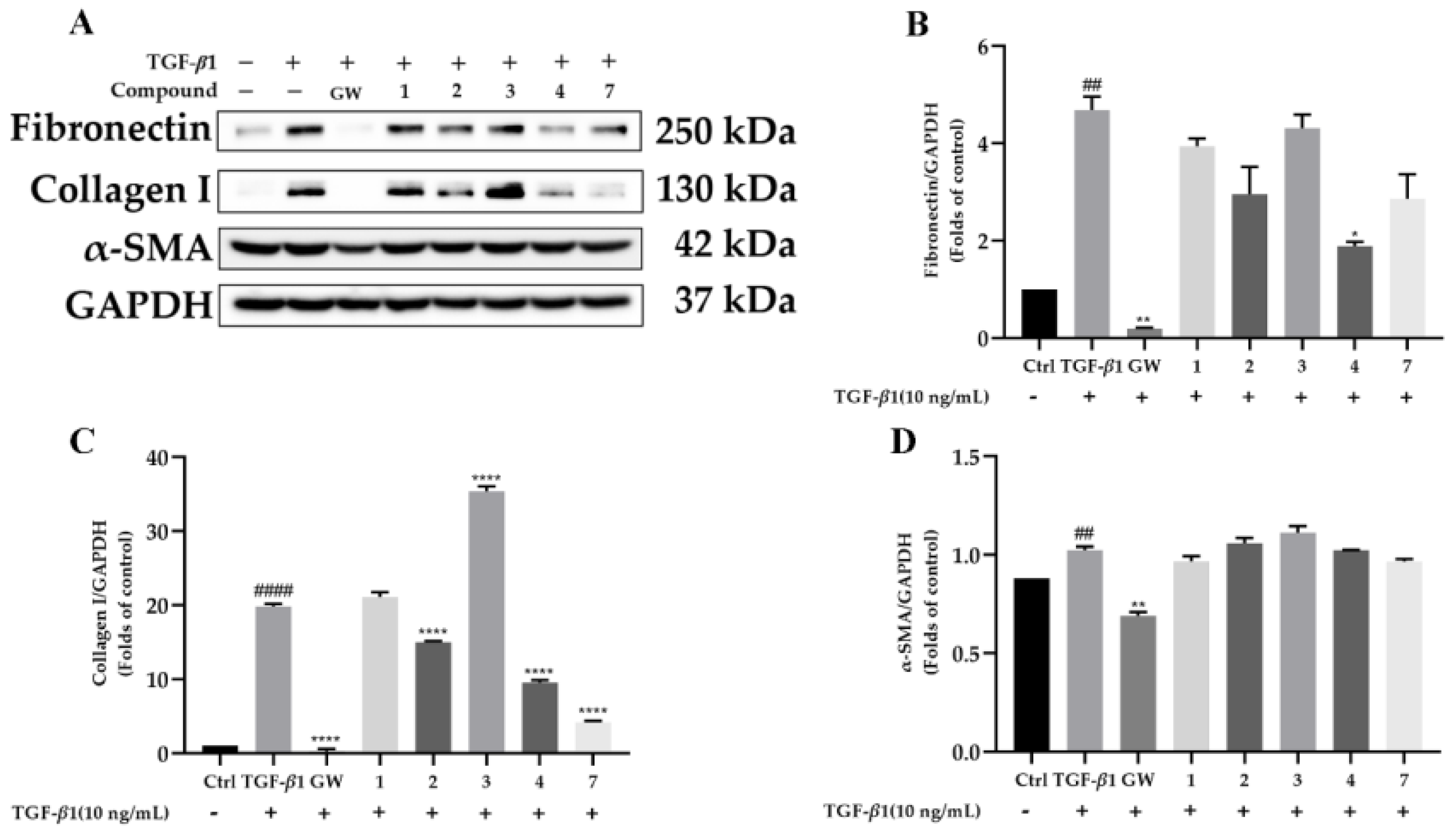
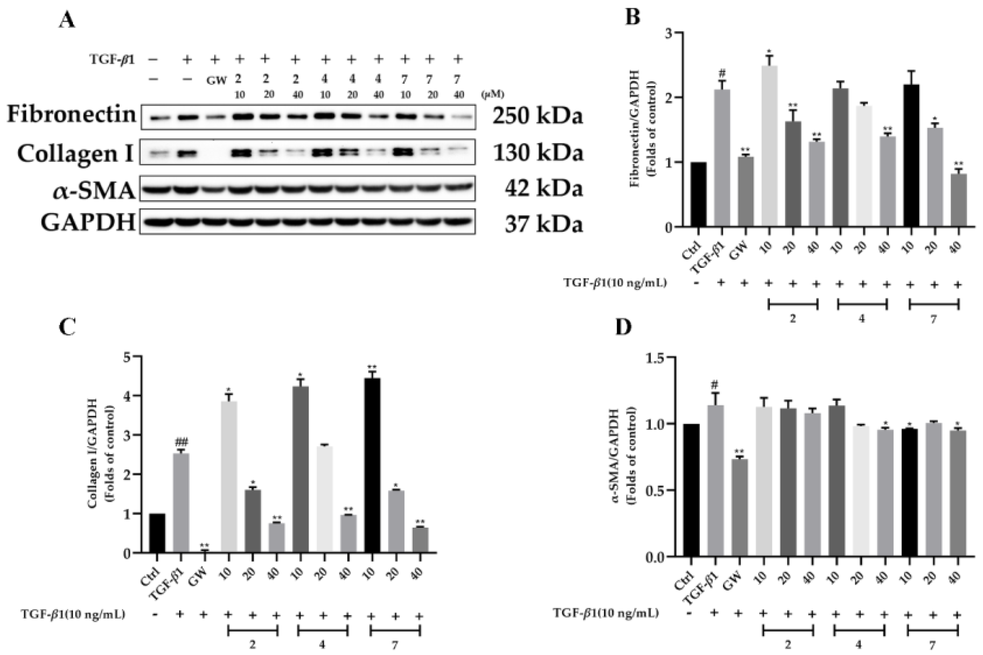
The renal protection of all the isolates was carried out in TGF-β1-induced rat renal proximal tubular cells. To exclude the possibility that the biological effects of the compounds are caused by cytotoxicity, a CCK-8 assay was first carried out (Figure 5). The results showed that the other compounds had slight toxicity toward rat renal proximal tubular cells (NRK-52e) except for 5, 6, and 8. Hence, the renal protection property of compounds 1–4 and 7 were evaluated (Figure 6). As presented in Figure 7, compounds 2, 4, and 7 were found to reduce the expression of fibronectin and collagen I in a dose-dependent manner in TGF-β1-induced NRK-52e cells. Since fibronectin and collagen I are components of the extracellular matrix and overexpression of the extracellular matrix is considered to be the hallmark of renal fibrosis, our current finding disclosed that cantharidin derivatives might be potent agents in renal protection. To our knowledge, this is the first time that cantharidin derivatives have been found to possess biological activity in renal fibrosis.

3. Experimental Section

3.1. General Procedures

Optical rotations were recorded on an Anton Paar MCP-100 digital polarimeter. UV and CD spectra were measured on a Chirascan instrument (Agilent Technologies, Santa Clara, CA, USA). NMR spectra were collected by a Bruker Avance III 600 MHz or a 500 MHz spectrometer, and the internal standard was TMS. HRESIMS were recorded on a Shimazu LC-20AD AB Sciex triple X500R MS spectrometer (Shimadzu Corporation, Tokyo, Japan). Macroporous adsorbents (Rohmhass AMBERLITETM XAD 16N, America), MCI gel CHP 20P (75–150 μm, Mitsubishi Chemical Industries, Tokyo, Japan), chromatography. YMC gel ODS-A-HG (40–60 μm; YMC Co., Tokyo, Japan), Sephadex LH-20 (Amersham Pharmacia, Uppsala, Sweden), and Silica gel (200–300 mesh, Qingdao Marine Chemical Inc., Qingdao, China) were used for column chromatography. Preparative HPLC was carried out using a Chuangxin-Tongheng chromatograph equipped with a Thermo Hypersil GOLD-C18 column (250 mm × 21.2 mm, i.d., 5 μm). Semi-preparative HPLC was taken on a SEP-LC52 chromatograph with a YMC-Pack ODS-A column (250 mm × 10 mm, i.d., 5 μm). Racemic compounds were purified by chiral HPLC on a Daicel Chiralpak column (IC, 250 mm × 4.6 mm, i.d., 5 μm) or a Phenomenex column (OOG-4762-E0 LUX® i-Amylose-1, 250 mm × 4.6 mm, i.d., 5 μm) at a flow rate of 1.0 mL/min.

3.2. Insect Material

M. cichorii were collected from Henan province, China, in July 2020, and identified by Prof. Dang-Rong Yang from the Kunming Institute of Zoology, Chinese Academy of Sciences, Kunming, China. A voucher specimen (CHYX-0643) was deposited at the School of Pharmaceutical Sciences, Shenzhen University Health Science Center, China.

3.3. Extraction and Isolation

The air-dried powdered M. cichorii (9 kg) were extracted with 50% aqueous EtOH (4 × 45 L, 24 h each) at room temperature. The combined extracts were concentrated to obtain a crude extract (1.5 kg), which was then divided into six parts (Fr.A–Fr.F) by using a macroporous adsorbents Rohmhass AMBERLITETM XAD 16N column eluted with gradient aqueous EtOH (0:100–100:0). Fr.B (20.0 g) was isolated by an MCI gel CHP 20P column eluted with gradient aqueous MeOH (3–100%) to afford five fractions (Fr.B1–Fr.B5). Fr.B1 (3.2 g) was subjected to Sephadex LH-20 (MeOH) to afford eight fractions (Fr.B1.1–Fr. B1.8). Fr.B1.3 (310.0 mg) was separated by preparative HPLC (MeCN/H2O with 0.05% TFA, 1–50%, flow rate: 8 mL/min) to yield thirteen portions (Fr.B1.3.1–Fr.B1.3.13). Fr.B1.3.4 (71.0 mg) was purified by semi-preparative HPLC (MeCN/H2O with 0.05% TFA, 7%, flow rate: 3 mL/min) to obtain compound 2 (tR = 10.8 min, 53.1 mg). Fr.B2 (2.8 g) was subjected to Sephadex LH-20 (MeOH) to afford six fractions (Fr.B2.1–Fr.B2.6). Fr.B2.2 (295.0 mg) was separated by preparative HPLC (MeCN/H2O with 0.05% TFA, 1–50%, flow rate: 8 mL/min) to yield six portions (Fr.B2.2.1–Fr.B2.2.6). Fr.B2.2.6 (63.1 mg) was purified by semi-preparative HPLC (MeCN/H2O with 0.05% TFA, 12%, flow rate: 3 mL/min) to obtain compound 1 (tR = 15.5 min, 16.1 mg). Fr.B4 (1.0 g) was subjected to Sephadex LH-20 (aqueous MeOH, 70%) to afford ten fractions (Fr.B4.1–Fr.B4.10). Fr.B4.8 (61.4 mg) was purified by semi-preparative HPLC (MeCN/H2O with 0.05% TFA, 11%, flow rate: 3 mL/min) to obtain compound 7 (tR = 23.0 min, 4.1 mg). Fr.C (180.0 g) was separated into seven fractions (Fr.C1–Fr.C7) by MCI gel CHP 20P column chromatography (aqueous MeOH, 10–100%). Fr.C2 (64.0 g) was subjected to an ODS column on an MPLC system eluted with gradient aqueous MeOH (5–100%, flow rate: 20 mL/min) to afford five fractions (Fr.C2.1–Fr.C2.5). Fr.C2.2 (13.0 g) was subjected to Sephadex LH-20 (MeOH) to afford compound 5 (2.3 g) and eight fractions (Fr.C2.2.1–Fr.C2.2.8). Fr.C2.2.1 (210.1 mg) was separated by preparative HPLC (MeCN/H2O with 0.05% TFA, 5–100%, flow rate: 8 mL/min) to yield four portions (Fr.C2.2.1.1–Fr.C2.2.1.4). Fr.C2.2.1.2 (47.0 mg) was purified by semi-preparative HPLC (MeCN/H2O with 0.05% TFA, 17%, flow rate: 3 mL/min) to obtain compound 4 (tR = 20.3 min, 4.7 mg). Fr.C2.2.4 (1.3 g) was separated into six fractions (Fr.C2.2.4.1–Fr.C2.2.4.6) by Sephadex LH-20 (aqueous MeOH, 70%). Fr.C2.2.4.1 (180.3 mg) was separated by preparative HPLC (MeOH/H2O with 0.05% TFA, 10–100%, flow rate: 8 mL/min) to yield four portions (Fr.C2.2.4.1.1–Fr.C2.2.4.1.4). Fr.C2.2.4.1.3 (72.5 mg) was purified by semi-preparative HPLC (MeOH/H2O with 0.05% TFA, 26%, flow rate: 3 mL/min) to obtain compound 3 (tR = 30.5 min, 2.3 mg). Fr.C2.3 (6.2 g) was separated into six fractions (Fr.C2.3.1–Fr.C2.3.6) by Sephadex LH-20 (MeOH). Fr.C2.3.4 (479.4 mg) was separated by preparative HPLC (MeOH/H2O with 0.05% TFA, 10–100%, flow rate: 8 mL/min) to yield nine portions (Fr.C2.3.4.1–Fr.C2.3.4.9). Fr.C2.3.4.6 (77.2 mg) was purified by semi-preparative HPLC (MeCN/H2O with 0.05% TFA, 20%, flow rate: 3 mL/min) to obtain compound 8 (tR = 22.7 min, 3.1 mg). Fr.F (90.0 g) was separated into ten fractions (Fr. F1–Fr.F10) by MCI gel CHP 20P column chromatography (aqueous MeOH, 60–100%). Fr.F4 (15.0 g) was subjected to Sephadex LH-20 (MeOH) to afford six fractions (Fr.F4.1–Fr.F4.6). Fr.F4.4 (2.3 g) was fractionated into eight parts (Fr.F4.4.1–Fr.F4.4.8) by a silica gel column eluted by increasing ethyl acetate in petroleum ether (10:1–3:1). Fr.F4.4.4 (1.8 g) was gel filtrated over Sephadex LH-20 (MeOH) to get compound 6 (1.5 g).

3.4. Compound Characterization Data

Cichormide A (1): Colorless gum, [α ] D 25 –0.6 (c 0.34, MeOH); CD (MeOH) Δε48 –0.36, Δε205 +0.63, Δε200 –2.26; UV (MeOH) λmax (logε) 203 (2.67) nm; HRESIMS m/z 270.0959 [M + H]+, (calcd for C12H16O6N, 270.0972). 1H and 13C NMR data, see Table 1 and Table 2.
Cichormide B (2): Yellow gum, [α ] D 25 –40.3 (c 0.39, MeOH); CD (MeOH) Δε268 +0.01, Δε246 –0.66, Δε235 –0.42, Δε212 –1.36, Δε202 +2.10; UV (MeOH) λmax (logε) 202 (2.49) nm; HRESIMS m/z 350.1347 [M + H]+, (calcd for C16H20O6N3, 350.1329). 1H and 13C NMR data, see Table 1 and Table 2.
Cichormide C (3): Colorless gum, [α ] D 25 –0.6 (c 0.35, MeOH); CD (MeOH) Δε219 –0.19, Δε209 +0.17, Δε200 –0.33; UV (MeOH) λmax (logε) 204 (2.71) nm; HRESIMS m/z 282.1338 [M + H]+, (calcd for C14H20O5N, 282.1336). 1H and 13C NMR data, see Table 1 and Table 2.
Cichormide D (4): Yellow gum, [α ] D 25 +0.3 (c 0.37, MeOH); CD (MeOH) Δε217 –0.18, Δε208 +1.79, Δε200 –2.57; UV (MeOH) λmax (logε) 204 (2.68) nm; HRESIMS m/z 281.1851 [M + H]+, (calcd for C15H25O3N2, 281.1860). 1H and 13C NMR data, see Table 1 and Table 2.
10-hydroxy-cantharidin (5): white powder, [α ] D 25 0 (c 0.30, MeOH); CD (MeOH) Δε200 +0.15; UV (MeOH) λmax (logε) 200 (0.43) nm; HRESIMS m/z 212.0759 [M + H]+, (calcd for C10H13O5, 211.0757). 1H and 13C NMR data, see Table 1 and Table 2.

3.5. Computational Methods

Molecular Merck force field (MMFF) and DFT/TDDFT were calculated with the Spartan’14 software package and Gaussian 09 program package. Electronic circular dichroism (ECD) calculations were conducted at the B3LYP/6-31g(d,p) level and the CD spectra were produced by the program SpecDis 1.62 (Figures S46–S50 and Tables S1–S5) [19].

3.6. Kidney Fibrosis Activity

3.6.1. Cell Culture

NRK-52e, rat renal proximal tubular cells (Cell Bank of China Science Academy, Shanghai, China) were cultured in high-glucose DMEM (C11995500BT, Gibco, Waltham, MA, USA) supplemented with 10% fetal bovine serum (FBS) (2094468CP, Gibco, Waltham, MA, USA), 100 U/mL penicillin, and 100 μg/mL streptomycin at 37 °C in a humidified environment containing 5% CO2.

3.6.2. Cell Viability Assay

NRK-52e (1× 10⁴ cells/mL) cells were seeded into a 96-well plate with completed DMEM. After overnight culture, cells were treated with various concentrations of compounds or DMSO for 48 h. Then, Cell Count Kit-8 (CCK-8, Beyotime, Shanghai, China) was added into each well for 1 h at 37 °C. The absorbance of each well was recorded at 450 nm using a microplate reader (BioTek, Winooski, VT, USA).

3.6.3. Western Blot

NRK-52e cells were treated with TGF-β1 (10 ng/mL) for 48 h in the absence or presence of 40 μM compounds. Cell lysates were prepared with RIPA buffer (Beyotime, Shanghai, China) containing 1 × protease inhibitor cocktail (Roche, Mannheim, Germany), 1 × phosphatase inhibitor cocktails, 0.1 mM PMSF, and quantified protein samples using the BCA assay (Thermo Scientific, Waltham, MA, USA). Equal amounts of protein extracts were separated by 8% SDS-PAGE and transferred to PVDF membranes (Millipore, Darmstadt, Germany). The membranes were blocked with 5% BSA, then with the indicated antibodies overnight at 4 °C, and were then followed by incubation with horseradish peroxidase (HRP)-conjugated secondary antibody at room temperature. The bands were visualized and measured via the ECL kit (Pierce, Hercules, CA, USA) and analysis system (Bio-Rad, Hercules, CA, USA). The primary antibodies are as follows: Anti-fibronectin antibody [IST-9] (#ab6328; Abcam, Cambridge, UK), Col1A1 antibody (#84336; Cell Signaling Technology, Boston, MA, USA), α-SMA (D4K9N) XP® Rabbit mAb (#19245, Cell Signaling Technology, Boston, MA, USA), and GAPDH (D16H11) XP® Rabbit mAb (#5174, Cell Signaling Technology, Boston, MA, USA).

4. Conclusions

In conclusion, our study resulted in the characterization of cantharidin derivatives from the title material, adding new facets for cantharidin structure diversity. Furthermore, biological comparison found that cantharidin derivatives with an oxygen atom instead of a nitrogen atom at position 8 and an alkyl group at C-1 and C-2 are toxic. To date, a limited number of cantharidin derivatives (<50) have been investigated from the Mylabris species and their potential for kidney fibrosis has not been described. Finally, the anti-fibrotic activity of cantharimide-type derivatives might provide new insight into the biological profiling of chemicals from M. cichorii, an excellent alternative resource for new pharmaceuticals.

Supplementary Materials

The following supporting information can be downloaded at: https://www.mdpi.com/article/10.3390/molecules28062822/s1, Figures S1–S6: NMR spectra of 1. Figure S7: HRESIMS of 1. Figure S8: CD spectrum of 1. Figures S9–S14: NMR spectra of 2. Figure S15: HRESIMS of 2. Figure S16: CD spectrum of 2. Figures S17–S22: NMR spectra of 3. Figure S23: HRESIMS of 3. Figure S24: CD spectrum of 3. Figures S25–S30: NMR spectra of 4. Figure S31: HRESIMS of 4. Figure S32: CD spectrum of 4. Figures S33–S38: NMR spectra of 5. Figure S39: HRESIMS of 5. Figure S40: CD spectrum of 5. Scheme S1: Semi-synthesis of compounds 2a and 2b. Figures S41–S42: NMR spectra of 2a. Figures S43–S44: NMR spectra of 2b. Figure S45: HPLC analyses of compounds 2, 2a, and 2b. Figures S46–S50: Optimized geometries of predominant conformers for 1–5. Tables S1–S5: The cartesian coordinates of the lowest energy conformers for 1–5.

Author Contributions

Y.-X.C. conceived and designed the experiments; J.-J.L. carried out the biological experiments; L.W. analyzed the data; and K.-M.L. contributed to the isolation of partial compounds and wrote the paper. All authors have read and agreed to the published version of the manuscript.

Funding

This study was supported financially by the Shenzhen Science and Technology Program (No. KQTD20210811090219022), the Shenzhen Fundamental Research Program (No. JCYJ20200109114225087), and the National Science Fund for Distinguished Young Scholars (No. 81525026).

Institutional Review Board Statement

The study did not require ethical approval.

Informed Consent Statement

Not applicable.

Data Availability Statement

All the data in this research are presented in manuscript and supplementary material.

Acknowledgments

We are grateful to Yong-Ming Yan at the Institute for Inheritance-Based Innovation of Chinese Medicine, Medical School of Pharmaceutical Sciences, Health Science Center, Shenzhen University, Shenzhen 518060, China for assistance in structure elucidation.

Conflicts of Interest

The authors declare no conflict of interest.

Sample Availability

Samples of compounds 1–8 are available from the authors.

References

  1. Yan, Y.M.; Meng, X.H.; Bai, H.F.; Cheng, Y.X. Nonpeptide small molecules with a ten-membered macrolactam or a morpholine motif from the insect American cockroach and their antiangiogenic activity. Org. Chem. Front. 2021, 8, 1401–1408. [Google Scholar] [CrossRef]
  2. Ding, W.Y.; Yan, Y.M.; Meng, X.H.; Nafie, L.A.; Xu, T.; Dukor, R.K.; Qin, H.B.; Cheng, Y.X. Isolation, total synthesis, and absolute configuration determination of renoprotective dimeric N-acetyldopamine–adenine hybrids from the insect Aspongopus chinensis. Org. Lett. 2020, 22, 5726–5730. [Google Scholar] [CrossRef] [PubMed]
  3. Li, J.; Li, Y.P.; Qin, F.Y.; Yan, Y.M.; Zhang, H.X.; Cheng, Y.X. Racemic xanthine and dihydroxydopamine conjugates from Cyclopelta parva and their COX-2 inhibitory activity. Fitoterapia 2020, 142, 104534. [Google Scholar] [CrossRef] [PubMed]
  4. Verma, A.K.; Prasad, S.B. Bioactive component, cantharidin from Mylabris cichorii and its antitumor activity against ehrlich ascites carcinoma. Cell Biol. Toxicol. 2012, 28, 133–147. [Google Scholar] [CrossRef] [PubMed]
  5. Kadioglu, O.; Kermani, N.S.; Kelter, G.; Schumacher, U.; Fiebig, H.H.; Greten, H.J.; Efferth, T. Pharmacogenomics of cantharidin in tumor cells. Biochem. Pharmacol. 2014, 87, 399–409. [Google Scholar] [CrossRef] [PubMed]
  6. Li, W.; Chen, Z.; Zong, Y.; Gong, F.R.; Zhu, Y.; Zhu, Y.X.; Lv, J.H.; Zhang, J.J.; Xie, L.; Sun, Y.J.; et al. PP2A inhibitors induce apoptosis in pancreatic cancer cell line PANC-1 through persistent phosphorylation of IKKα and sustained activation of the NF-κB pathway. Cancer Lett. 2011, 304, 117–127. [Google Scholar] [CrossRef] [PubMed]
  7. Wang, G.S. Medical uses of mylabris in ancient China and recent studies. J. Ethnopharmacol. 1989, 26, 147–162. [Google Scholar] [CrossRef] [PubMed]
  8. Zeng, Y.B.; Liu, X.L.; Zhang, Y.; Li, C.J.; Zhang, D.M.; Peng, Y.Z.; Zhou, X.; Du, H.F.; Tan, C.B.; Zhang, Y.Y.; et al. Cantharimide and its derivatives from the blister beetle Mylabris phalerata Palla. J. Nat. Prod. 2016, 79, 2032–2038. [Google Scholar] [CrossRef] [PubMed]
  9. Nakatani, T.; Konishi, T.; Miyahara, K.; Noda, N. Three novel cantharidin-related compounds from the Chinese blister beetle, Mylabris phalerata PALL. Chem. Pharm. Bull. 2004, 52, 807–809. [Google Scholar] [CrossRef] [PubMed] [Green Version]
  10. Zeng, Y.B.; Guo, Y.L.; Zhang, Y.; Wang, X.Y.; Jiang, Y.; Yang, D.J. Rapid profiling of cantharidin analogues in Mylabris phalerata Pallas by ultra-performance liquid chromatography-quadrupole time-of-flight-tandem mass spectrometry. Biomed. Chromatogr. 2020, 34, e4801. [Google Scholar] [CrossRef] [PubMed]
  11. Deng, Y.Y.; Zhang, W.; Li, N.P.; Lei, X.P.; Gong, X.Y.; Zhang, D.M.; Wang, L.; Ye, W.C. Cantharidin derivatives from the medicinal insect Mylabris phalerata. Tetrahedron 2017, 73, 5932–5939. [Google Scholar] [CrossRef]
  12. Zhang, X.J.; Cao, Y.; Pan, D.B.; Yao, X.J.; Wang, F.; Zhang, G.L.; Luo, Y.G. Antifibrotic pyridine-containing monoterpene alkaloids from Caryopteris glutinosa. Phytochemistry 2022, 203, 113378. [Google Scholar] [CrossRef] [PubMed]
  13. Anandakumar, P.; Kamaraj, S.; Vanitha, M.K. D-limonene: A multifunctional compound with potent therapeutic effects. J. Food Biochem. 2021, 45, e13566. [Google Scholar] [CrossRef] [PubMed]
  14. Yu, M.; Peng, L.Y.; Liu, P.; Yang, M.X.; Zhou, H.; Ding, Y.R.; Wang, J.J.; Huang, W.; Tan, Q.; Wang, Y.L.; et al. Paeoniflorin ameliorates chronic hypoxia/SU5416-induced pulmonary arterial hypertension by inhibiting endothelial-to-mesenchymal transition. Drug Des. Dev. Ther. 2020, 14, 1191–1202. [Google Scholar] [CrossRef] [PubMed] [Green Version]
  15. Nakatani, T.; Jinpo, K.; Noda, N. Cantharimide dimers from the Chinese blister beetle, Mylabris phalerate Pallas. Chem. Pharm. Bull. 2007, 55, 92–94. [Google Scholar] [CrossRef] [PubMed] [Green Version]
  16. Zeng, Y.B.; Liu, X.L.; Zhou, X.; Zhang, X.M.; Yang, Y.; Du, H.F.; Tan, C.B.; Zhang, Y.Y.; Zhang, Y.; Yang, D.J. Chemical constituents from Mylabris phalerata and their cytotoxic activity in vitro. China J. Chin. Mater. Med. 2016, 41, 859–863. [Google Scholar] [CrossRef]
  17. Dauben, W.G.; Lam, J.Y.L.; Guo, Z.R. Total synthesis of (−)-palasonin and (+)-palasonin and related chemistry. J. Org. Chem. 1996, 61, 4816–4819. [Google Scholar] [CrossRef] [PubMed]
  18. König, W.A. Collection of enantiomeric separation factors obtained by capillary gas chromatography on chiral stationary phases. J. High Res. Chrom. 1993, 16, 569–586. [Google Scholar] [CrossRef]
  19. Frisch, M.J.; Trucks, G.W.; Schlegel, H.B.; Scuseria, G.E.; Robb, M.A.; Cheeseman, J.R.; Scalmani, G.; Barone, V.; Mennucci, B.; Petersson, G.A.; et al. Gaussian 09, Revision B.01; Gaussian, Inc.: Wallingford, CT, USA, 2009. [Google Scholar]
Disclaimer/Publisher’s Note: The statements, opinions, and data contained in all publications are solely those of the individual author(s) and contributor(s) and not of MDPI and/or the editor(s). MDPI and/or the editor(s) disclaim responsibility for any injury to people or property resulting from any ideas, methods, instructions, or products referred to in the content.
Figure 1. The structures of compounds 18 from M. cichorii.
Molecules 28 02822 g001
Figure 2. Key 1H-1H COSY and HMBC correlations of 15.
Molecules 28 02822 g002
Figure 3. Key ROESY correlations of 15.
Molecules 28 02822 g003
Figure 4. Comparison of the calculated ECD and experimental CD spectra in MeOH. (A): The calculated ECD spectrum of (1R,2R,3S,6R)-1 at B3LYP/6-31 (d,p) level, σ = 0.20 eV; shift = −9 nm; The calculated ECD spectrum of (1R,2R,3S,6R)-5 at B3LYP/6-31 (d,p) level, σ = 0.20 eV; shift = −9 nm. (B): The calculated ECD spectrum of (1R,2R,3S,6R,1′R)-2 at B3LYP/6-31 (d,p) level, σ = 0.30 eV; shift = 0 nm. (C): The calculated ECD spectrum of (1S,2R,3S,6R)-3 at B3LYP/6-31 (d,p) level, σ = 0.30 eV; shift = +6 nm. (D): The calculated ECD spectrum of (1S,2R,3S,6R)-4 at B3LYP/6-31 (d,p) level, σ = 0.20 eV; shift = +30 nm.
Molecules 28 02822 g004
Figure 5. NRK-52e cell proliferation in response to compounds at 40 μM by CCK-8 assay. * p < 0.05, ** p < 0.01 compared with Control alone.
Molecules 28 02822 g005
Figure 6. Compounds attenuate renal fibrosis in TGF-β1-induced NRK-52e cells. Cells were incubated in different concentrations of the compound and then exposed to 10 ng/mL TGF-β1 for 48 h. (AD): The protein level of Fibronectin, Collagen I, and α-SMA in NRK-52e were determined by Western blotting, and GAPDH was used as a control. Data represent mean ± SEM values of three experiments. * p < 0.05, ** p < 0.01 and **** p < 0.0001 compared with TGF-β1 alone. ## p < 0.01 and #### p < 0.0001 compared with Control alone. GW788388 (GW) was used as a positive control.
Molecules 28 02822 g006
Figure 7. Compounds attenuate renal fibrosis in TGF-β1-induced NRK-52e cells. NRK-52e cells were incubated with TGF-β1 (10 ng/mL) for 48 h in the absence or presence of different concentrations (10 μM, 20 μM, and 40 μM) of compounds 2, 4, and 7. (AD): The protein level of Fibronectin, Collagen I, and α-SMA in NRK-52e were determined by Western blotting, and GAPDH was used as a control. Data represent mean ± SEM values of three experiments. * p < 0.05, ** p < 0.01 compared with TGF-β1 alone. # p < 0.05 and ## p < 0.01 compared with Control alone. GW788388 (GW) was used as a positive control.
Molecules 28 02822 g007
Table 1. The 1H NMR (600 MHz) data of 1–5 in CD3OD (δ in ppm, J in Hz).
Position12345
34.52 (br d, 4.5)4.46 (br d, 4.6)4.47 (br d, 2.6)4.47 (br d, 2.2)4.67 (br d, 2.7)
4Ha: 1.95 (m)Ha: 1.91 (m)Ha: 1.89 (overlap) aHa: 1.91 (overlap) aHa: 1.97 (m)
Hb: 1.67 (overlap) aHb: 1.61 (overlap) aHb: 1.64 (overlap) aHb: 1.65 (overlap) aHb: 1.68 (overlap) a
5Ha: 1.75 (m)Ha: 1.67 (m)Ha: 1.89 (overlap) aHa: 1.91 (overlap) aHa: 1.68 (overlap) a
Hb: 1.67 (overlap) aHb: 1.61 (overlap) aHb: 1.64 (overlap) aHb: 1.65 (overlap) aHb: 1.68 (overlap) a
64.56 (br d, 4.7)4.36 (br d, 4.8)4.48 (br d, 2.6)4.48 (br d, 2.2)4.66 (br d, 2.7)
10Ha: 3.97 (d, 11.3)Ha: 3.94 (d, 11.2)1.15 (overlap)a1.15 (overlap)aHa: 3.98 (d, 11.1)
Hb: 3.73 (d, 11.3)Hb: 3.69 (d, 11.2) Hb: 3.72 (d, 11.1)
111.33 (s)1.24 (s)1.15 (overlap)a1.15 (overlap)a1.41 (s)
1′Ha: 4.23 (d, 17.2)4.97 (br t, 7.4)3.74 (t, 7.0)3.51 (t, 7.0)
Hb: 4.18 (d, 17.2)
2′ 3.49 (br d, 8.0)2.58 (t, 7.1)1.61 (m)
3′ 1.36 (m)
4′ 7.26 (s)3.64 (s)1.66 (m)
5′ 2.89 (t, 7.7)
7′ 8.71 (s)
a Signals might be interchangeable.
Table 2. The 13C NMR data (150 MHz) of 1–5 in CD3OD (δ in ppm).
Position12345
161.9, C61.6, C55.3, C55.2, C64.3, C
255.3, C55.0, C55.3, C55.2, C55.9, C
385.3, CH85.3, CH85.1, CH85.2, CH86.7, CH
424.6 CH224.5, CH224.6, CH224.6, CH224.7, CH2
525.1, CH224.9, CH224.6, CH224.6, CH224.6, CH2
683.2, CH83.2, CH85.1, CH85.2, CH84.2, CH
7180.9, C180.7, C183.1, C183.6, C176.0, C
9182.5, C182.7, C183.1, C183.6, C177.9, C
1059.5, CH259.4, CH212.6, CH312.5, CH359.1, CH2
1112.2, CH312.2, CH312.6, CH312.5, CH312.7, CH3
1′40.5, CH253.2, CH35.8, CH239.4, CH2
2′169.7, C25.1, CH232.7, CH227.89, CH2
3′ 130.8, C172.9, C24.3, CH2
4′ 119.1, CH52.4, CH327.9, CH2
5′ 40.5, CH2
6′ 134.7, CH
8′ 171.1, C
Disclaimer/Publisher’s Note: The statements, opinions and data contained in all publications are solely those of the individual author(s) and contributor(s) and not of MDPI and/or the editor(s). MDPI and/or the editor(s) disclaim responsibility for any injury to people or property resulting from any ideas, methods, instructions or products referred to in the content.