
Composition and Antioxidative and Antibacterial Activities of the Essential Oil from Farfugium japonicum


Abstract
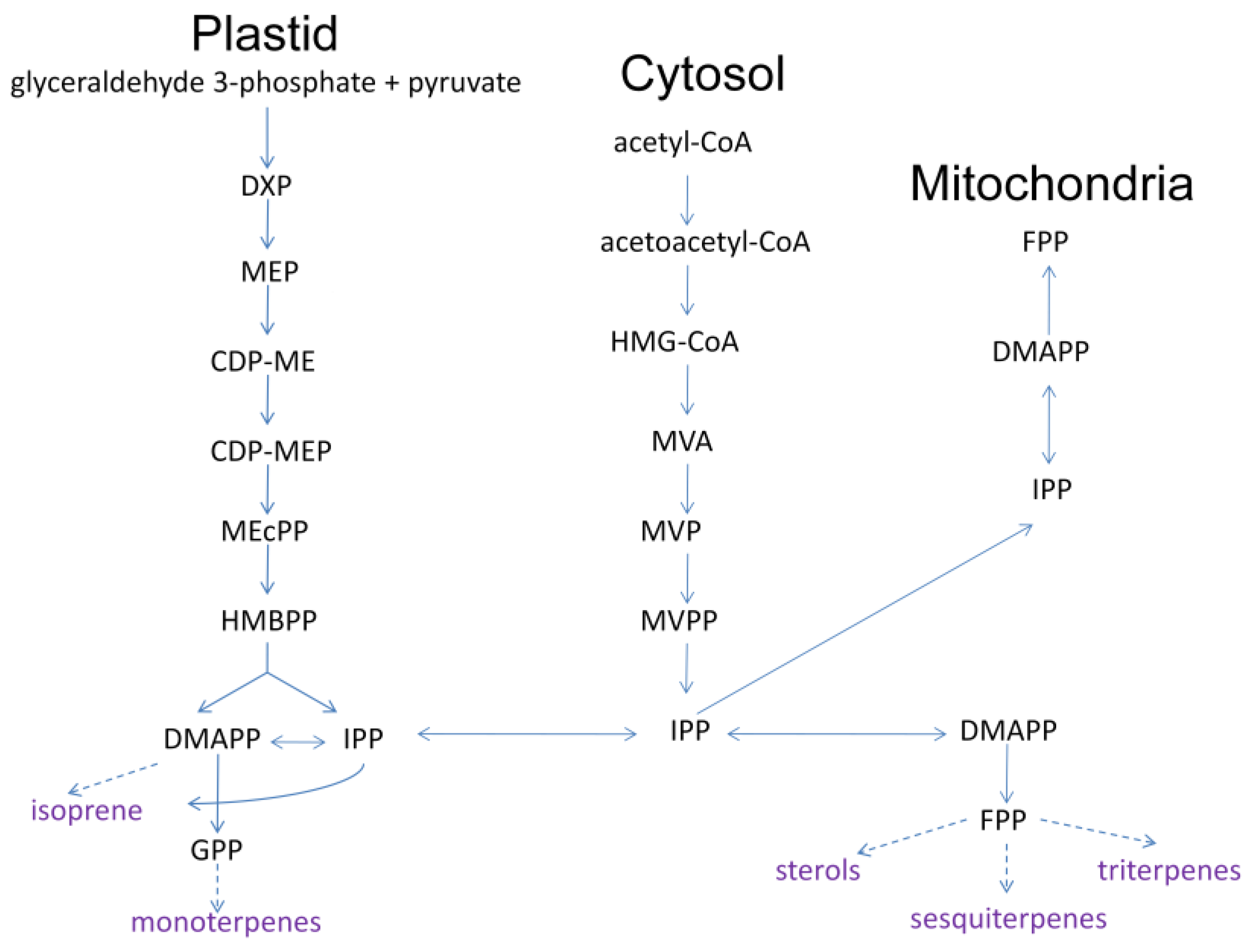
1. Introduction
2. Results
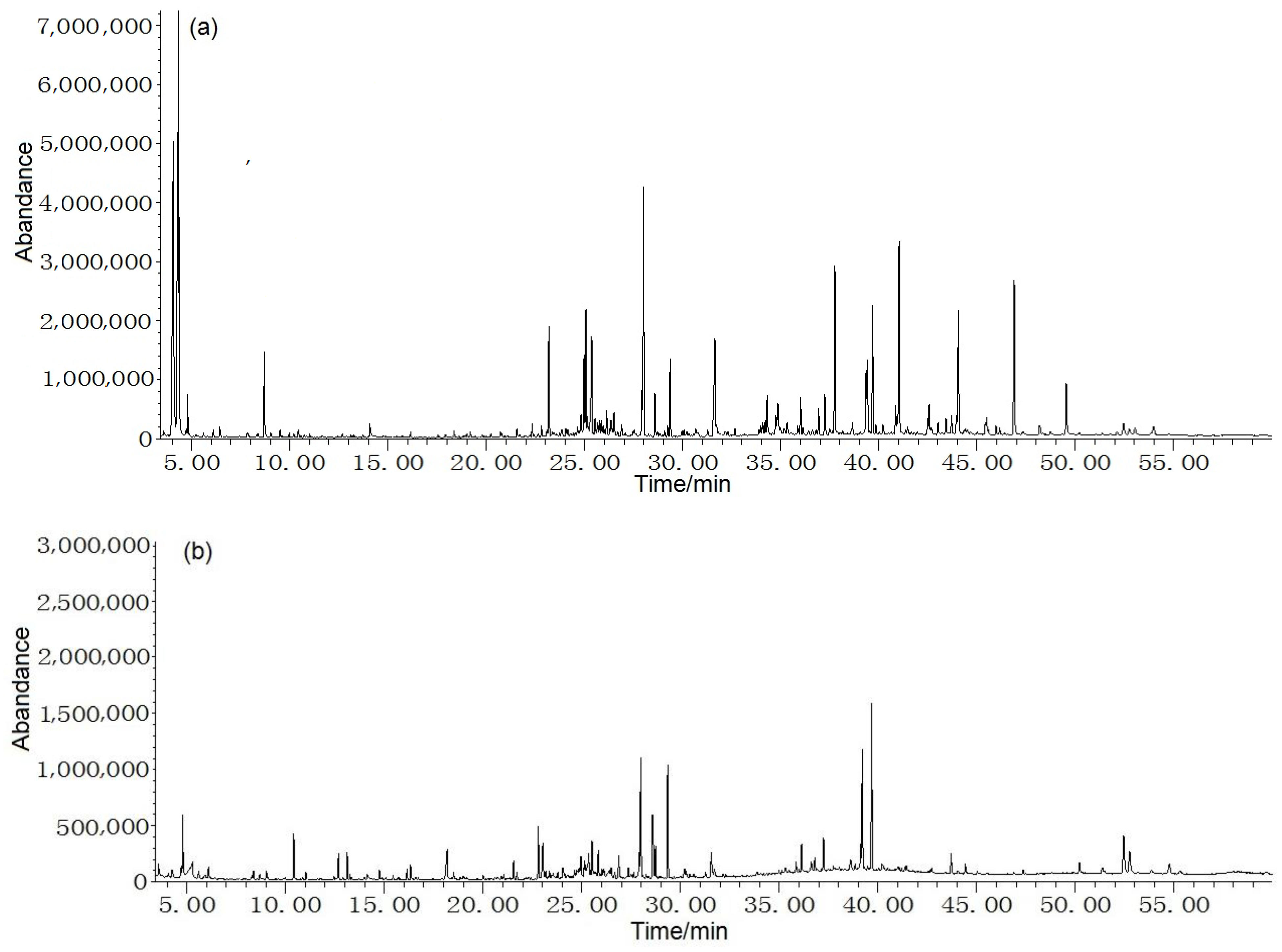
2.1. Analysis of Essential Oil Compositions
2.2. Antioxidative Capacity
2.3. Antibcterial Capacity
3. Materials and Methods
3.1. Plant Material and Reagents
3.2. Essential Oils Extraction
3.3. GC/MS Analysis
3.4. Antioxidative Activity Assay
3.4.1. DPPH Inhibition Test
3.4.2. Hydroxyl Radical (OH) Inhibition Test
3.4.3. Antimicrobial Capacities Assay
4. Conclusions
Author Contributions
Funding
Institutional Review Board Statement
Informed Consent Statement
Data Availability Statement
Conflicts of Interest
Sample Availability
References
- Kim, J.Y.; Oh, T.H.; Kim, B.J.; Kim, S.S. Chemical composition and anti-inflammatory effects of essential oil from Farfugium japonicum flower. J. Oleo Sci. 2008, 57, 623–628. [Google Scholar] [CrossRef] [PubMed]
- Nomura, N.; Setoguchi, H.; Yasuda, K.; Takaso, T. Genetic structure of rheophytic and nonrheophytic populations of Farfugium japonicum on Yaeyama Islands, Japan. Can. J. Bot. 2007, 85, 637–643. [Google Scholar] [CrossRef]
- Wang, X.L. Study of the Neuroprotective Effects and Mechanisms of Farfugium japonicumi Extracts. Master’s Thesis, Shandong University, Weihai, China, 2017. [Google Scholar]
- Chen, J.Z.; Ge, S.L.; Zan, L.F.; Xing, H.C.; Fu, J.; Wang, G.X. Optimization of aqueous two-phase extraction of total flavonoids from Farfugium japonicum (L.f.) Kitam by response surface methodology and antibacterial activity. Food Sci. 2015, 36, 57–62. [Google Scholar]
- Nagano, H.; Moriyama, Y.; Tanahashi, Y.; Takahashi, T. New benzofuranosesquiterpenes from Farfugium japonicum. Farfugin A and Farfugin B. Bull. Chem. Soc. Jpn. 1974, 47, 1994–1998. [Google Scholar] [CrossRef]
- Nagano, H.; Takahashi, T. 3β-Angeloyloxy-10β-hydroxy-9β- senecioyloxyfuranoeremophilane and 3β-Angeloyloxy-10β- hydroxyfuranoeremophilane. new furanoeremophilane derivatives from Farfugium japonicum Kitamura. Bull. Chem. Soc. Jpn. 1978, 51, 3335–3340. [Google Scholar] [CrossRef]
- Niwa, H.; Ishiwata, H.; Kuroda, A.; Yamada, K. Farfugine, a new pyrrolizidine alkaloid isolated from Farfugium japonicum Kitam. Chem. Lett. 1983, 19, 789–790. [Google Scholar] [CrossRef]
- Dima, C.; Dima, S. Essential oils in foods: Extraction, stabilization, and toxicity. Curr. Opin. Food Sci. 2015, 5, 29–35. [Google Scholar] [CrossRef]
- Zehravi, M.; Karthika, C.; Azad, A.K.; Ahmad, Z.; Khan, F.S.; Rahman, M.; Akter, R. A background search on the potential role of scutellaria and its essential oils. BioMed Res. Int. 2022, 2022, 7265445. [Google Scholar] [CrossRef]
- Kultys, E.; Kurek, M.A. Green extraction of carotenoids from fruit and vegetable byproducts: A review. Molecules 2022, 27, 518. [Google Scholar] [CrossRef]
- Shinde, V.; Mahadik, K. Supercritical fluid extraction: A new technology to herbals. Int. J. Herb. Med. 2019, 7, 27–34. [Google Scholar]
- Bhavaniramya, S.; Vishnupriya, S.; Al-Aboody, M.S.; Vijayakumar, R.; Baskaran, D. Role of essential oils in food safety: Antimicrobial and antioxidant applications. Grain Oil Sci. Technol. 2019, 2, 49–55. [Google Scholar] [CrossRef]
- Wei, Q.; Li, Q.Z.; Wang, R.L. Flavonoid components, distribution, and biological activities in Taxus: A review. Molecules 2023, 28, 1713. [Google Scholar] [CrossRef]
- Öz, M.; Deniz, I.; Okan, O.T.; Baltaci, C.; Karatas, S.M. Determination of the chemical composition, antioxidant and antimicrobial activities of different parts of Rosa canina L. and Rosa pimpinellifolia L. essential oils. J Essent. Oil Bear. Pl. 2021, 24, 519–537. [Google Scholar] [CrossRef]
- Bassolé, I.H.; Juliani, H.R. Essential oils in combination and their antimicrobial properties. Molecules 2012, 17, 3989–4006. [Google Scholar] [CrossRef]
- Dutta, A.; Batra, J.; Pandey-Rai, S.; Singh, D.; Kumar, S.; Sen, J. Expression of terpenoid indole alkaloid biosynthetic pathway genes corresponds to accumulation of related alkaloids in Catharanthus roseus (L.) G. Don. Planta 2005, 220, 376–383. [Google Scholar] [CrossRef]
- Sacchettini, J.C.; Poulter, C.D. Creating isoprenoid diversity. Science 1997, 277, 1788–1789. [Google Scholar] [CrossRef]
- Zhou, F.; Pichersky, E. More is better: The diversity of terpene metabolism in plants. Curr. Opin. Plant Biol. 2020, 55, 1–10. [Google Scholar] [CrossRef]
- Muchlinski, A.; Ibdah, M.; Ellison, S.; Yahyaa, M.; Nawade, B.; Laliberte, S.; Senalik, D.; Simon, P.; Whitehead, S.R.; Tholl, D. Diversity and function of terpene synthases in the production of carrot aroma and flavor compounds. Sci. Rep. 2020, 10, 9989. [Google Scholar] [CrossRef]
- Karunanithi, P.S.; Zerbe, P. Terpene synthases as metabolic gatekeepers in the evolution of plant terpenoid chemical diversity. Front. Plant Sci. 2019, 10, 1166. [Google Scholar] [CrossRef]
- Du, Y.; Zhou, H.; Yang, L.; Jiang, L.; Chen, D.; Qiu, D.; Yang, Y. Advances in biosynthesis and pharmacological effects of Cinnamomum camphora (L.) Presl essential oil. Forests 2022, 13, 1020. [Google Scholar] [CrossRef]
- Wang, Q.; Quan, S.; Xiao, H. Towards efficient terpenoid biosynthesis: Manipulating IPP and DMAPP supply. Bioresour. Bioprocess. 2019, 6, 6. [Google Scholar] [CrossRef]
- Zheng, H.; Yu, M.Y.; Pu, C.J.; Chen, M.L.; Li, F.Q.; Shen, Y.; Huang, L.Q. Cloning and expression analysis of 5-phosphomevalonate kinase gene (CcPMK) in Cinnamomum camphora. China J. Chin. Mater. Med. 2020, 45, 78–84. [Google Scholar]
- Lange, B.M.; Croteau, R. Isopentenyl diphosphate biosynthesis via a mevalonate-independent pathway: Isopentenyl monophosphate kinase catalyzes the terminal enzymatic step. Proc. Natl. Acad. Sci. USA 1999, 96, 13714–13719. [Google Scholar] [CrossRef] [PubMed]
- Rehman, R.; Hanif, M.A.; Mushtaq, Z.; Al-Sadi, A.M. Biosynthesis of essential oils in aromatic plants: A review. Food Rev. Int. 2016, 32, 117–160. [Google Scholar] [CrossRef]
- Ichikawa, K.; Sasada, R.; Chiba, K.; Gotoh, H. Effect of side chain functional groups on the DPPH radical scavenging activity of bisabolane-type phenols. Antioxidants 2019, 8, 65. [Google Scholar] [CrossRef]
- Grassmann, J. Grassmann. Terpenoids as plant antioxidants. Vitam. Horm. 2005, 72, 505–535. [Google Scholar] [CrossRef]
- Afshari, M.; Rahimmalek, M. Variation in essential oil composition, anatomical, and antioxidant characteristics of Achillea filipendulina Lam. as affected by different phenological stages. J. Essent. Oil Res. 2021, 33, 283–298. [Google Scholar] [CrossRef]
- Borah, A.; Paw, M.; Gogoi, R.; Loying, R.; Sarma, N.; Munda, S.; Pandey, S.K.; Lal, M. Chemical composition, antioxidant, anti-inflammatory, anti-microbial and in-vitro cytotoxic efficacy of essential oil of Curcuma caesia Roxb. leaves: An endangered medicinal plant of North East India. Ind. Crop. Prod. 2019, 129, 448–454. [Google Scholar] [CrossRef]
- Yu, C.; Zhang, J.; Wang, T. Star anise essential oil: Chemical compounds, antifungal and antioxidant activities: A review. J. Essent. Oil Res. 2021, 33, 1–22. [Google Scholar] [CrossRef]
- Yousefi, M.; Rahimi-Nasrabadi, M.; Pourmortazavi, S.M.; Wysokowski, M.; Jesionowski, T.; Ehrlich, H.; Mirsadeghi, S. Supercritical fluid extraction of essential oils. TrAC-Trends Anal. Chem. 2019, 118, 182–193. [Google Scholar] [CrossRef]
- Valero, M.; Giner, M.J. Effects of antimicrobial components of essential oils on growth of Bacillus cereus INRA L2104 in and the sensory qualities of carrot broth. Int. J. Food Microbiol. 2006, 106, 90–94. [Google Scholar] [CrossRef]
- Vasconcelos, N.G.; Croda, J.; Simionatto, S. Antibacterial mechanisms of cinnamon and its constituents: A review. Microb. Pathogenesis 2018, 120, 198–203. [Google Scholar] [CrossRef]
- Dorman, H.R.D.; Deans, S.G. Antimicrobial agents from plants: Antibacterial activity of plant volatile oils. J. Appl. Microbiol. 2000, 88, 308–316. [Google Scholar] [CrossRef]
- Mann, C.M.; Cox, S.D.; Markham, J.L. The outer membrane of Pseudomonas aeruginosa NCTC 6749 contributes to its tolerance to the essential oil of Melaleuca alternifolia (tea tree oil). Lett. App. Microbiol. 2000, 30, 294–297. [Google Scholar] [CrossRef]
- Soleimani, M.; Arzani, A.; Arzani, V.; Roberts, T.H. Phenolic compounds and antimicrobial properties of mint and thyme. J. Herb. Med. 2022, 36, 100604. [Google Scholar] [CrossRef]
- Liu, J.B.; Gu, J.J.; Wang, K. The expected values for the Gutman index, Schultz index, and some Sombor indices of a random cyclooctane chain. Int. J. Quantum Chem. 2023, 123, e27022. [Google Scholar] [CrossRef]
- Liu, J.B.; Xie, Q.; Gu, J.J. Statistical analyses of a class of random pentagonal xhain networks with respect to several topological properties. J. Funct. Space. 2023, 2023, 6675966. [Google Scholar] [CrossRef]
- Liu, J.B.; Ali, H.; Ain, Q.U.; Ali, P.; Kirmani, S.A.K. On topological properties for benzenoid planar octahedron networks. Molecules 2022, 27, 6366. [Google Scholar] [CrossRef]
- Hsouna, A.B.; Hamdi, N.; Halima, N.B.; Abdelkafi, S. Characterization of essential oil from Citrus aurantium L. flowers: Antimicrobial and antioxidant activities. J. Oleo Sci. 2013, 62, 763–772. [Google Scholar] [CrossRef]
- Wang, X.S.; Wu, Y.F.; Wang, F.F.; Yi, J.P.; Zhang, Y.P.; Liu, P.; Niu, Q.S. Optimization of polysaccharide process from Fructus corni with box-behnken design and antioxidant capacity. Pak. J. Pharm. Sci. 2019, 32, 1537–1544. [Google Scholar]
- Ouadja, B.; Katawa, G.; Toudji, G.A.; Layland, L.; Gbekley, E.H.; Ritter, M.; Anani, K.; Ameyapoh, Y.; Karou, S.D. Anti-inflammatory, antibacterial and antioxidant activities of Chenopodium ambrosioides L. (Chenopodiaceae) extracts. J. Appl. Biosci. 2021, 162, 16764–16794. [Google Scholar] [CrossRef]


| No. | Compounds | LRI | KRI | RA/% | |
|---|---|---|---|---|---|
| Leaf | Stem | ||||
| 1 | Furfural (Ad) | 812 | 810 | - | 1.2 |
| 2 | cis-1,2-Dimethyl-cyclohexane (H) | 822 | 820 | - | 0.6 |
| 3 | Ethyl-cyclohexane (H) | 827 | 829 | - | 2.3 |
| 4 | (E)-3-Hexen-1-ol (Ac) | 849 | 847 | 13.7 | - |
| 5 | (Z)-3-Hexen-1-ol (Ac) | 851 | 847 | 14.0 | - |
| 6 | 1,3-Dimethyl-benzene (B) | 852 | 853 | - | 4.4 |
| 7 | Benzaldehyde (Ad) | 964 | 964 | 0.2 | - |
| 8 | Hydroperoxide, hexyl (Ac) | 977 | 978 | 3.5 | - |
| 9 | 1,2,4-Trimethyl-benzene (B) | 986 | 989 | - | 0.1 |
| 10 | Trimethylenenorbornane (H) | 990 | 990 | - | 0.1 |
| 11 | Decane (H) | 1002 | 1000 | - | 0.1 |
| 12 | 1,2,3-Trimethyl-benzene (B) | 1005 | 1005 | - | 0.4 |
| 13 | β-Linalool (T) | 1098 | 1098 | - | 2.9 |
| 14 | 1,2,4,5-Tetramethyl-benzene (B) | 1108 | 1109 | 0.5 | 0.9 |
| 15 | 2-Ethyl-1-hexanol (Ac) | 1010 | 1010 | 1.7 | 3.4 |
| 16 | 1,4-Diethyl-benzene (B) | 1045 | 1046 | 1.2 | 3.6 |
| 17 | Benzeneacetaldehyde (Ad) | 1047 | 1048 | 1.9 | - |
| 18 | 1-Ethyl-2,3-dimethyl-benzene (B) | 1093 | 1096 | - | 0.9 |
| 19 | 4-Ethenyl-1,2-dimethyl-benzene (B) | 1098 | 1100 | - | 0.1 |
| 20 | Pentyl-cyclohexane (H) | 1123 | 1121 | - | 0.1 |
| 21 | 4-Terpeneol (T) | 1160 | 1160 | - | 1.7 |
| 22 | α-Terpineol (T) | 1170 | 1172 | - | 1.7 |
| 23 | 2,3-Dihydro-benzofuran (B) | 1185 | 1188 | 0.5 | - |
| 24 | Naphthalene (B) | 1191 | 1190 | 1.1 | - |
| 25 | Dodecane (H) | 1201 | 1200 | - | 0.6 |
| 26 | 2-Methoxy-4-vinylphenol (P) | 1272 | 1272 | - | 1.0 |
| 27 | Azulene (T) | 1296 | 1296 | - | 2.6 |
| 28 | 1,1’-Bicyclohexyl (H) | 1302 | 1304 | 1.9 | 4.0 |
| 29 | 5,5,8α-Trimethyl-3,5,6,7,8,8α-hexahydro-2H-chromene (T) | 1308 | 1309 | 0.7 | - |
| 30 | n-Decanoic acid (F) | 1360 | 1360 | - | 3.6 |
| 31 | 2,6,8-Trimethylbicyclo [4.2.0]oct-2-ene-1,8-diol (T) | 1371 | 1370 | 0.4 | - |
| 32 | Tetradecane (H) | 1402 | 1400 | - | 0.1 |
| 33 | Curzerene (T) | 1480 | 1480 | 1.2 | - |
| 34 | 5-Isopropylidene-6-methyldeca-3,6,9-trien-2-one (K) | 1492 | 1494 | - | 2.2 |
| 35 | 2-(1-Cyclopent-1-enyl-1-methylethyl)cyclopentanone (K) | 1495 | 1497 | 3.5 | 11.7 |
| 36 | cis-Calamenene (T) | 1510 | 1511 | 0.7 | - |
| 37 | Shyobunone (T) | 1518 | 1518 | 2.3 | - |
| 38 | 2,4-bis(1,1-Dimethylethyl)-phenol (P) | 1539 | 1540 | 0.4 | 1.2 |
| 39 | trans-Nerolidol (T) | 1547 | 1548 | 0.2 | 3.2 |
| 40 | 2,6,10-Trimethyl-tetradecane (H) | 1555 | 1557 | 0.6 | 0.1 |
| 41 | n-Dodecanoic acid (F) | 1560 | 1561 | - | 3.2 |
| 42 | 2,6-Dimethyl-10-methylene-12-oxatricyclo [7.3.1.0(1,6)] tridec-2-ene (T) | 1577 | 1576 | 2.3 | - |
| 43 | Caryophyllene oxide (T) | 1580 | 1581 | 1.9 | - |
| 44 | Boronia butenal (T) | 1582 | 1584 | - | 2.2 |
| 45 | Calarene epoxide (T) | 1592 | 1592 | 0.2 | - |
| 46 | Diethyl phthalate (E) | 1594 | 1594 | 1.4 | 2.7 |
| 47 | Ledene oxide-(Ⅱ) (T) | 1631 | 1631 | 0.7 | - |
| 48 | α-Cyperone (T) | 1672 | 1673 | 0.4 | - |
| 49 | 6-Isopropenyl-4,8α-dimethyl-1,2,3,5,6,7,8,8α-octahydro-naphthalen-2-ol (T) | 1714 | 1714 | 0.7 | - |
| 50 | 3,5,6,7,8,8α-Hexahydro-4,8α-dimethyl-6-(1-methylethenyl)-2(1H)nahpthalenone (T) | 1790 | 1790 | 0.6 | 2.7 |
| 51 | 5-Heptylresorcinol (P) | 1830 | 1831 | 1.2 | 6.5 |
| 52 | trans-9-Hexadecen-1-ol (Ac) | 1866 | 1868 | - | 0.1 |
| 53 | 1,1,4,6-Tetramethyl-perhydrocyclopropa [e] azulene-4,5,6-triol (T) | 1867 | 1869 | 0.5 | - |
| 54 | 1,2,3,4,5,6,7,8-Octahydro-9,10-dimethyl-anthracene (B) | 1878 | 1879 | 4.2 | 8.4 |
| 55 | 2-Methyl-9-(prop-1-en-3-ol-2-yl)-bicyclo [4.4.0] dec-2-ene-4-ol (T) | 1902 | 1904 | 1.7 | - |
| 56 | bi-1-Cycloocten-1-yl (H) | 1941 | 1942 | 3.1 | - |
| 57 | n-Hexadecanoic acid (F) | 1963 | 1963 | 3.5 | 2.0 |
| 58 | Eicosane (H) | 2000 | 2000 | 3.5 | - |
| 59 | 1-Hexadecanol, acetate (E) | 2008 | 2009 | 0.5 | - |
| 60 | Oralic acid, cyclohexyl octyl ester (E) | 2010 | 2010 | - | 1.0 |
| 61 | Heneicosane (H) | 2100 | 2100 | 4.4 | - |
| 62 | α-Linolenic acid (F) | 2100 | 2102 | 1.4 | - |
| 63 | Phytol (T) | 2104 | 2104 | 1.1 | - |
| 64 | Octadecyl acetate (E) | 2160 | 2161 | - | 2.0 |
| 65 | Isoangenomalin (C) | 2182 | 2186 | 0.5 | - |
| 66 | (Z)-2-(9-Octadecenyloxy)-ethanol (Ac) | 2336 | 2336 | 0.4 | - |
| 67 | Behenic alcohol (Ac) | 2470 | 2470 | 0.8 | - |
| 68 | Heptacosane (H) | 2700 | 2700 | 2.3 | - |
| 69 | Octacosane (H) | 2800 | 2800 | 3.6 | - |
| 70 | 3α-24-Propylidene-cholest-5-en-3-ol (S) | 2881 | 2880 | - | 3.4 |
| 71 | α-Sitosterol (S) | 3065 | 3066 | 0.8 | 5.2 |
| 72 | α-amyrin (T) | 3322 | 3320 | 0.6 | - |
| 73 | Tetratetracontane (H) | 4392 | 4395 | 4.7 | - |
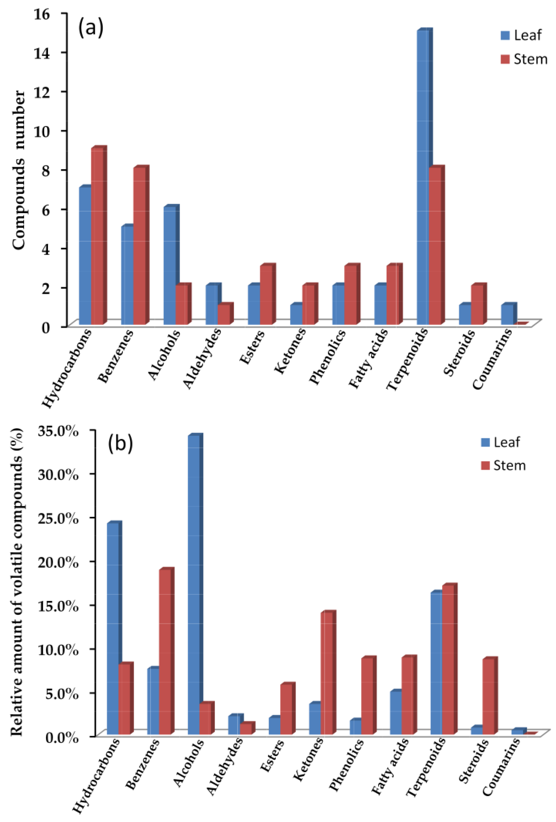
| Hydrocarbons (Sr. No. 2, 3, 10, 11, 20, 25, 28, 32, 40, 56, 58, 61, 68, 69, 73) | 24.1 | 8.0 | |||
| Benzenes (Sr. No. 6, 9, 12, 14, 16, 18, 19, 23, 24, 54) | 7.5 | 18.8 | |||
| Alcohols (Sr. No. 4, 5, 8, 15, 52, 66, 67) | 34.1 | 3.5 | |||
| Aldehydes (Sr. No. 1, 7, 17) | 2.1 | 1.2 | |||
| Esters (Sr. No. 46, 59, 60, 64) | 1.9 | 5.7 | |||
| Ketones (Sr. No. 34, 35) | 3.5 | 13.9 | |||
| Phenolics (Sr. No. 26, 38, 51) | 1.6 | 8.7 | |||
| Fatty acids (Sr. No. 30, 41, 57, 62) | 4.9 | 8.8 | |||
| Terpenoids (Sr. No. 13, 21, 22, 27, 29, 31, 33, 36, 37, 39, 42–45, 47–50, 53, 55, 63, 72) | 16.2 | 17.0 | |||
| Steroids (Sr. No. 70, 71) | 0.8 | 8.6 | |||
| Coumarins (Sr. No. 65) | 0.5 | - | |||
| Total | 97.2 | 94.2 | |||
| Sample | IC50 (mg/mL, n = 3) | |
|---|---|---|
| DPPH | ·OH | |
| Leaf oil | 20.56 ± 0.20 | 1.52 ± 0.03 |
| Stem oil | 9.22 ± 0.11 | 0.90 ± 0.02 |
| Vitamin C a | 4.10 ± 0.18 | 0.65 ± 0.01 |
| Strains of Bacteria | MIC (mg/mL) | |
|---|---|---|
| Leaf Oil | Stem Oil | |
| Methicillin-sensitive Staphylococcus aureus | 0.62 | 0.16 |
| Methicillin-resistant Staphylococcus aureus | 0.31 | 0.16 |
| Pseudomonas aeruginosa | 0.62 | 0.31 |
| Escherichia coli | 1.25 | 0.62 |
| Proteus spp. | 0.62 | 0.31 |
| Klebsiella pneumoniae | 1.25 | 0.62 |
Disclaimer/Publisher’s Note: The statements, opinions and data contained in all publications are solely those of the individual author(s) and contributor(s) and not of MDPI and/or the editor(s). MDPI and/or the editor(s) disclaim responsibility for any injury to people or property resulting from any ideas, methods, instructions or products referred to in the content. |
© 2023 by the authors. Licensee MDPI, Basel, Switzerland. This article is an open access article distributed under the terms and conditions of the Creative Commons Attribution (CC BY) license (https://creativecommons.org/licenses/by/4.0/).
Share and Cite
Wei, Q.; Zhang, Y.-H. Composition and Antioxidative and Antibacterial Activities of the Essential Oil from Farfugium japonicum. Molecules 2023, 28, 2774. https://doi.org/10.3390/molecules28062774
Wei Q, Zhang Y-H. Composition and Antioxidative and Antibacterial Activities of the Essential Oil from Farfugium japonicum. Molecules. 2023; 28(6):2774. https://doi.org/10.3390/molecules28062774
Chicago/Turabian StyleWei, Qiang, and Yi-Han Zhang. 2023. "Composition and Antioxidative and Antibacterial Activities of the Essential Oil from Farfugium japonicum" Molecules 28, no. 6: 2774. https://doi.org/10.3390/molecules28062774
APA StyleWei, Q., & Zhang, Y.-H. (2023). Composition and Antioxidative and Antibacterial Activities of the Essential Oil from Farfugium japonicum. Molecules, 28(6), 2774. https://doi.org/10.3390/molecules28062774