Article

Manipulation in Culture Conditions of Nanofrustulum shiloi for Enhanced Fucoxanthin Production and Isolation by Preparative Chromatography

1
Application and Research Centre for Testing and Analysis (EGE MATAL), Ege University, Bornova, 35100 İzmir, Turkey
2
Department of Bioengineering, Faculty of Engineering, Ege University, Bornova, 35100 İzmir, Turkey
3
Department of Chemistry, Faculty of Science, Ege University, Bornova, 35100 İzmir, Turkey
*
Author to whom correspondence should be addressed.
Molecules 2023, 28(4), 1988; https://doi.org/10.3390/molecules28041988
Submission received: 7 January 2023 / Revised: 13 February 2023 / Accepted: 16 February 2023 / Published: 20 February 2023
(This article belongs to the Section Analytical Chemistry)

Abstract

:
Microalgae produce a variety of high-value chemicals including carotenoids. Fucoxanthin is also a carotenoid that has many physiological functions and biological properties. For this reason, the cost-effective production of fucoxanthin at an industrial scale has gained significant attention. In the proposed study, fucoxanthin production was aimed to be increased by altering the culture conditions of N. shiloi. The effect of light intensity aeration rate, different nitrogen sources, and oxidative stress on the biomass and fucoxanthin productivity have been discussed. Based on these results, the fucoxanthin increased to 97.45 ± 2.64 mg/g by adjusting the light intensity to 50 µmol/m2s, and aeration rate at 5 L/min using oxidative stress through the addition of 0.1 mM H2O2 and 0.1 mM NaOCl to the culture medium. Fucoxanthin was then purified with preparative HPLC using C30 carotenoid column (10 mm × 250 mm, 5 μm). After the purification procedure, Liquid chromatography tandem mass spectrometry (LC–MS/MS) and UV-vis spectroscopy were employed for the confirmation of fucoxanthin. This study presented a protocol for obtaining and purifying considerable amounts of biomass and fucoxanthin from diatom by manipulating culture conditions. With the developed methodology, N. shiloi could be evaluated as a promising source of fucoxanthin at the industrial scale for food, feed, cosmetic, and pharmaceutical industries.

1. Introduction

There is growing interest in microalgae as a source of high-value compounds in the food and medicinal industries. The natural metabolites obtained from microalgae are the most important field of study in microalgal biotechnology. The pigments, vitamins, proteins, and fatty acids they contain are widely used in agriculture, medicine, pharmacy, and the cosmetics sector [1,2]. Particularly, the reasons why microalgae are preferred in this regard can be summarized as being able to increase their daily weight rapidly, easily undergo biotechnological processes, have low production costs, and be resistant to environmental effects [3]. Most of the products beneficial to humans emerge through biotechnological methods [4,5]. For this reason, researchers mainly focus on biotechnologically obtained products rather than synthetic materials for many reasons. Pigments are also important metabolites that have gained importance in recent years. Microalgae are seen as potential organisms in the production of pigments [6]. The formation of free radicals that cause cancer, and a number of chronic diseases that cause various damages to the organism as a result of oxidation during the functioning of the metabolism has increased interest in antioxidant compounds [7]. Carotenoids, which are among the photosynthetic pigments of microalgae, also have a strong antioxidant effect. Carotenoids are a class of oil pigments that can only be produced by phytoplankton algae, plants, and a limited number of fungi and bacteria [8]. They are fat-soluble pigments that give colour to various vegetables, fruits, beverages, flowers, mushrooms, algae, and even birds. Colours range from light yellow to bright orange and red depending on their chemical structure. In general, carotenoids in food are a category of tetraterpenoid (C40); it consists of eight isoprenoid units [9]. From the middle of the molecule, this structure has been reversed, and thus the molecule has gained a symmetrical structure [10]. Structures consisting of structural polyene chains can sometimes end in rings. Hydrocarbon-based carotenoids such as alpha-carotene, beta-carotene, and lycopene are called carotenes. On the other hand, lutein, zeaxanthin, and fucoxanthin belong to the xanthophylls group of carotenoids, as they also contain oxygen [11]. Fucoxanthin is a basic carotenoid found in brown algae, diatoms, and golden algae [12]. Besides, several researchers reported that fucoxanthin has no toxic effect and is a safe pharmaceutical active substance [13,14,15]. Due to its unique molecular structure, fucoxanthin has very important properties. It includes a polyene chain structure as well as functional groups such as epoxy, hydroxyl, carbonyl, and carboxyl. Many benefits of fucoxanthin have been reported for human health. The most important of these are their anti-cancer, anti-hypertensive, anti-inflammatory, and anti-obesity therapeutic properties [16,17,18]. For the industrial production of fucoxanthin, brown macroalgae are used in most studies [19,20,21]. Many studies have indicated that the content of fucoxanthin in microalgae is higher than in macroalgae. In chromist marine microalgae, the amount of fucoxanthin accounts for about 1–2.5% of the dry cell weight, while in brown macroalgae, the content of fucoxanthin is nearly 0.1–1 mg/g (dry cell weight). Brown macroalgae are presently sold as a commercial source of healthy pigment. Notwithstanding, macroalgal production is not an economically viable source due to the low extraction efficiency and slow growth, which is seasonally dependent [22,23]. Therefore, it does not seem appropriate for them to be commercially produced. The production of microalgae is easier and faster. This is because they can double their weight in a very short time. In fact, growth rates increase logarithmically. Today, microalgal biotechnology is progressing rapidly, but there is still a great demand for the commercial production of fucoxanthin from microalgae. Fucoxanthin production is dependent on microalgae type, growth, fucoxanthin concentration, and extraction efficiency. The microalgae’s fucoxanthin synthesis varied depending on the growing conditions, light intensity, temperature, nitrogen source and/or concentration iron and silicate concentration, carbon dioxide, salinity etc. [24] Some microalgal species have a rather high fucoxanthin content (without ideal growth conditions), ranging from 0.22–1.82% of the total dry weight (DW). Recently, discussion of topics such as culture conditions, heterotrophic and mixotrophic cultivation, the use of low-cost by-products as nutrient media, and pilot-scale studies in the cultivation of fucoxanthin accumulating algae has become very popular. In addition to nitrogen, iron, and silicate concentrations, light intensity, and wavelength have a very important effect on the biosynthesis of fucoxanthin, and the production of biomass in microalgae. With their quick growth rate and high fucoxanthin content, microalgae such as Isochrysis spp. (7.5–23.3 mg/g), Nitzschia spp. (12–32.8 mg/g), Phaeodactylum tricornutum (10.9–59.2 mg/g), Tisochrysis lutea (2.1–79.4 mg/g), and others are a promising source for fucoxanthin production [25,26,27,28,29,30,31,32]. With the current findings and recommendations, the economic viability of fucoxanthin production might be improved, possibly leading to a feasible and sustainable production method [33].
When literature studies are examined, most of them focus on the bioactivities of fucoxanthin [12,17,33,34,35,36,37,38]. Studies on microalgae production (for example, optimization of culture conditions, and attempts for pilot scale work) and the use of advanced extraction technologies to obtain fucoxanthin have rarely been discussed [24]. As this information is valuable for future commercial scale production of fucoxanthin, this study demonstrates that fucoxanthin-producing N. shiloi can provide significant increases in both biomass and fucoxanthin content when grown under different culture conditions. Studies on the use of low-cost by-products as a nutrient medium help to fill in gaps in the research in the production of fucoxanthin. In addition, discussions were made on the mechanisms underlying the production of fucoxanthin using different culture conditions in order to guide future studies. This study aimed to present the enhanced productivity of fucoxanthin from N. shiloi by altering the light intensity, aeration rate, nitrogen source, and applying oxidative stress to culture conditions. After that, fucoxanthin was purified by preparative chromatography. The purity of fucoxanthin was proven by UV-vis spectroscopic and mass spectrometric data. Here, it was shown that, by altering culture conditions, it was possible to increase the fucoxanthin content. N. shiloi was shown to be a novel diatom and could serve as a potential source for fucoxanthin production at an industrial level.

2. Results

2.1. Enhanced Productivity of Fucoxanthin

The growth profile and fucoxanthin concentration of N. shiloi were investigated to cultivate under different physical environmental conditions. It is well-known that light intensity, aeration rate, nitrogen sources, and oxidative stressors are factors that affect growth and pigment biosynthesis in microalgae.
Figure 1 shows that biomass productivity and doubling rates are greater at 300 µmol/m2 of light intensity compared to 50 and 150 µmol/m2. According to Table 1, the increase in biomass was the highest at 300 µmol/m2 light intensity. By increasing the aeration rate and light intensity, the mixing speed in the bioreactors was accelerated, and it was established that the suspension duration of the cell biomass increased the amount of biomass. The results emphasize that raising aeration rate and reducing the light intensity significantly increases the amount of fucoxanthin in the living cells.
The results showed that the three different light intensity and aeration rate factors had a significant effect on the fucoxanthin production of the diatom N. shiloi. The amount of fucoxanthin concentration obtained from N. shiloi enhanced with increasing light intensity at different aeration rates.
Table 1 illustrates that the highest amount of fucoxanthin (51.05 ± 1.02 mg/gDW) was obtained at a light intensity of 50 µmol/m2s and an aeration rate of 5 L/min in seawater BG11 medium (used NaNO3). For simplicity, aeration rates (1-3-5 L/min) and light intensities (50-150-300 µmol/m2s) were abbreviated, as given in Table 1. According to these results, low light intensity increases fucoxanthin concentration in N. shiloi.
On the other hand, fucoxanthin production significantly decreased when different nitrogen sources (NaNO2, NH4Cl and CH4N2O instead of NaNO3) were used in BG11 culture medium, separately. The growth curves of NaNO3, NaNO2, NH4Cl, and CH4N2O were monitored to determine the specific growth rates. Adding NH4Cl to a culture medium prevented N. shiloi from reproducing and growing. In contrast to the control (NaNO3), the culture to which CH4N2O was introduced was not a brown color, and phase-contrast microscopy revealed the presence of empty silica walls. Particularly, the use of urea caused the cell death of N. shiloi. It took the cells a long time to adjust to the NH4Cl medium, and they were unable to finish their growth within 16 days. In addition, Figure 2 depicts the inability of cells in NaNO2 media to complete the logarithmic phase.
The biomass productivity results and fucoxanthin yield are summarized in Table 2, where different nitrogen sources were used for the cultivation of N. shiloi under 50 μE/m2s light intensity and at a 5 L/min aeration rate. Based on these results, fucoxanthin could not accumulate excessively in the cells even though the NaNO2 medium was used.
The pH of the culture medium without N. shiloi (Day 1) and culture medium before harvesting the cells (Day 16) are measured and summarized in Table 3. It was seen that the pH dropped dramatically when NH4Cl and CH4N2O were used as nitrogen sources.
The following step was the application of oxidative stress using various sources. Growth of N. shiloi was consistent for each case (Figure 3). Since the N. shiloi cultures were not axenic, the addition of Fe+NaOCl may have destroyed the bacteria in the environment. In addition, the addition of H2O2+NaOCl did not inhibit cell proliferation in comparison to the control group.
Finally, the effect of oxidative stress was investigated using H2O2, NaClO, and Fe2+. It was observed that fucoxanthin productivity reached the maximum (97.45 ± 2.64 mg/gDW) if 0.1 mM H2O2 was used with 0.1 mM NaOCl, while biomass productivity did not change significantly, as presented in Table 4. It was also apparent that each oxidative stress condition leads to the increase in the amount of fucoxanthin.

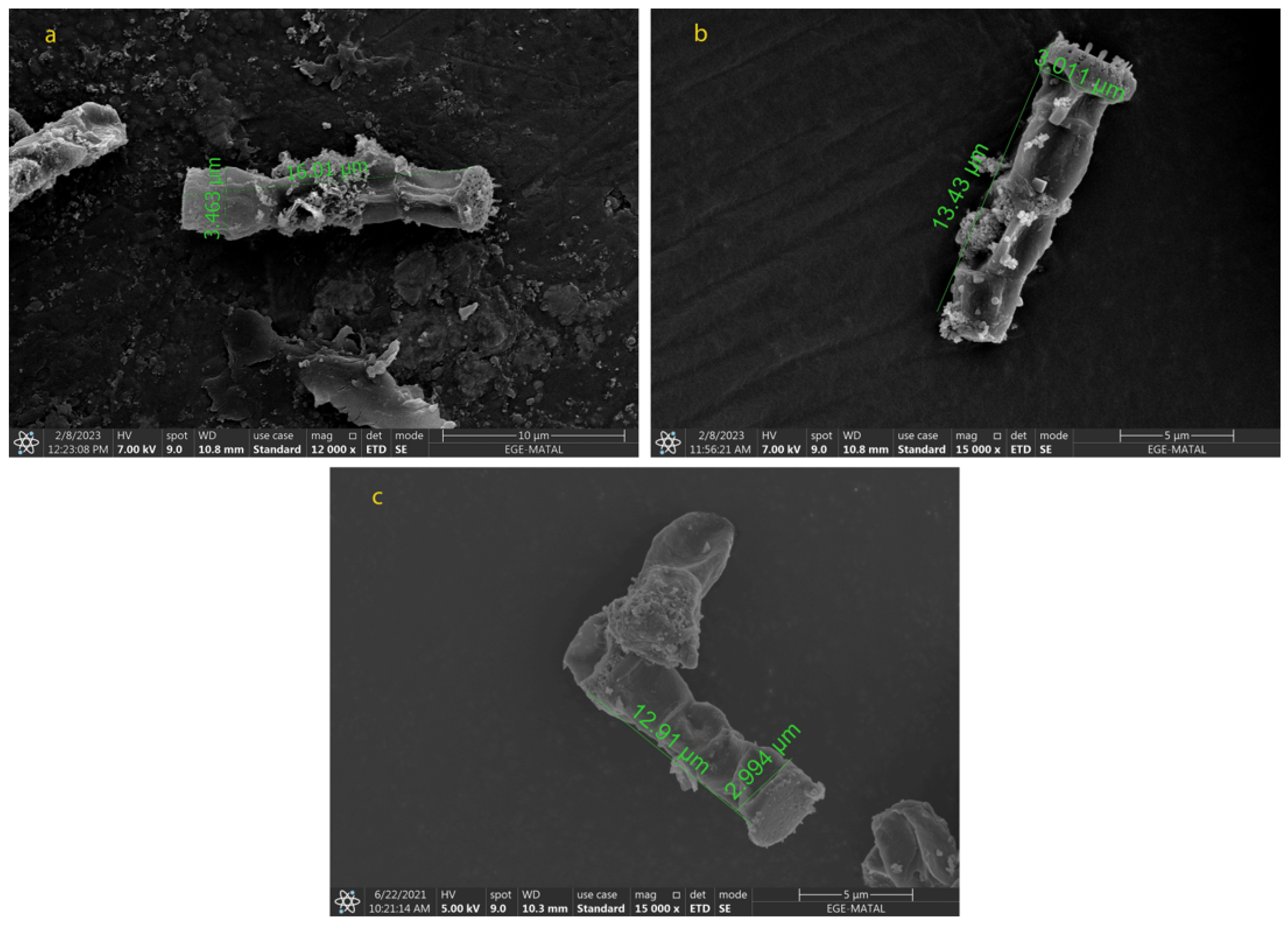
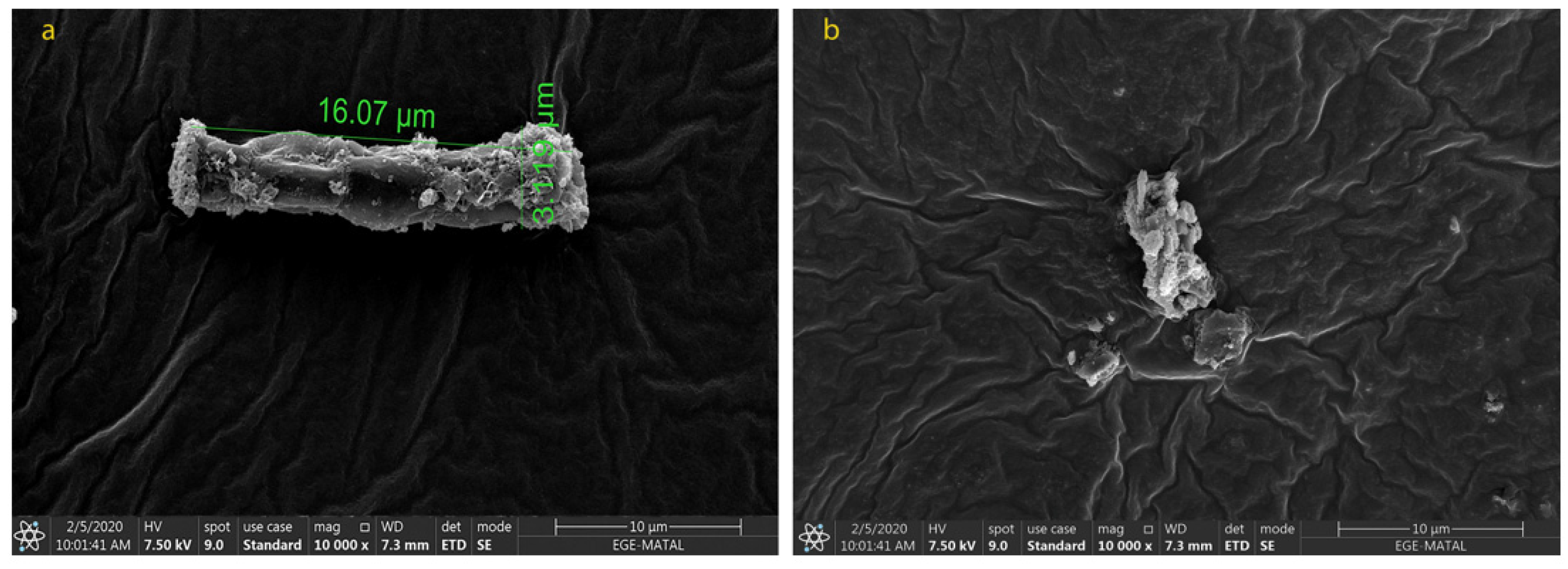
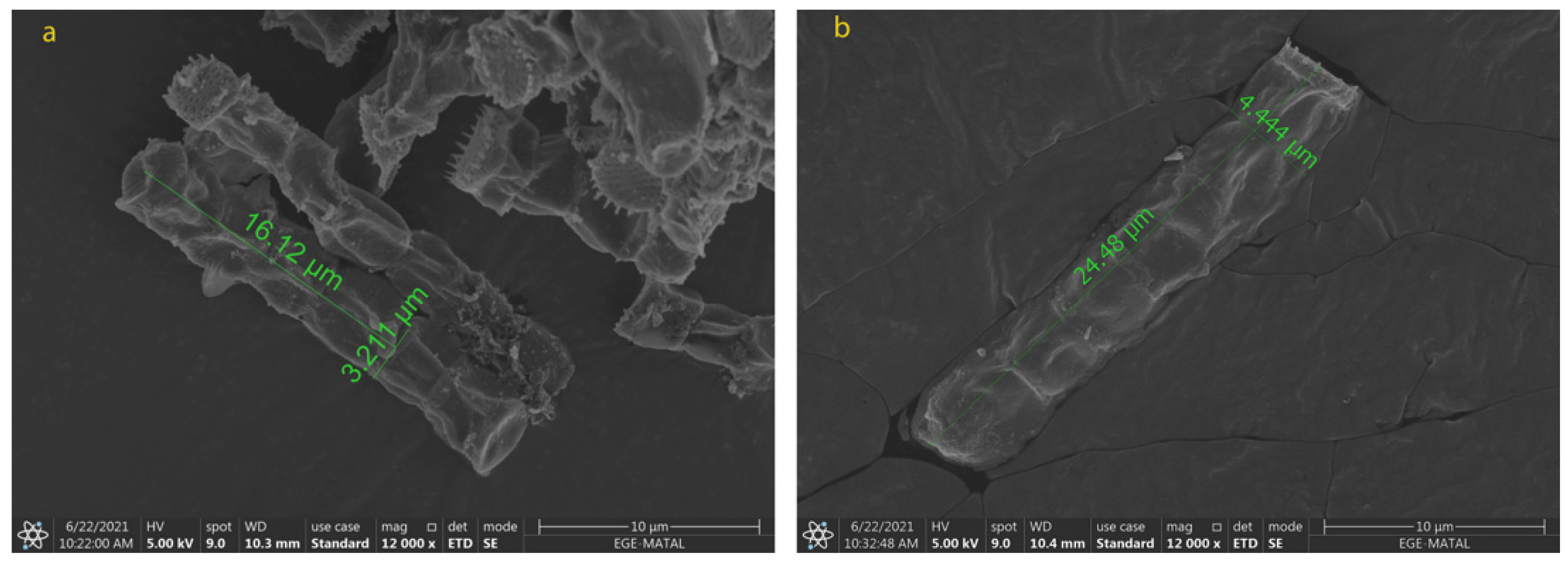
2.2. Morphological Changes in N. shiloi

The cell size and shape directly affect the extraction of biomolecules from each species. To obtain the carotenoids efficiently after the extraction processes, the cell wall of the microalga should be destroyed and dissolved in the appropriate solvent. It is particularly important in repetitive extractions, as the number of extractions increases the cost as well. Due to the structure of N. shiloi, fucoxanthin could be easily extracted by the UAE method. It was reported that Navicula sp. cell size showed a change under indoor and outdoor cultivation conditions [39]. The fucoxanthin of Navicula sp. reduced between 5.40 ± 0.05 (indoor) and 2.61 ± 0.06 mg/gDW (outdoor) of biomass. The cultivation of Amphora sp. was incubated in the optimum conditions at 30 °C and 80 μmol/m2s [40]. In the present study, it was observed from the SEM results that the cell size of N. shiloi also changed with different culture conditions. The variation in light intensity and aeration rate triggered the morphological changes in N. shiloi. The maximum cell size was observed for N5-50 (16.17 ± 0.25 µm × 3.476 ± 0.028 µm). The size of the cells decreased when higher light intensities were applied (Figure 4). On the other hand, the use of different nitrogen sources negatively affected the productivity and morphology of N. shiloi. The cells cultured in urea were dead, and no uniform cells were observed (Figure 5). Particularly, the diatom filament wall size was measured as 12.75 ± 0.29 µm × 2.955 ± 0.057 µm without any oxidative stress, and 24.86 ± 0.69 µm × 4.273 ± 0.303 µm in the presence of 0.1 mM H2O2, and 0.1 mM NaOCl in culture medium, as presented in Figure 6. This corresponds to approximately a 2-fold increase in both the width and height of N. shiloi cells.

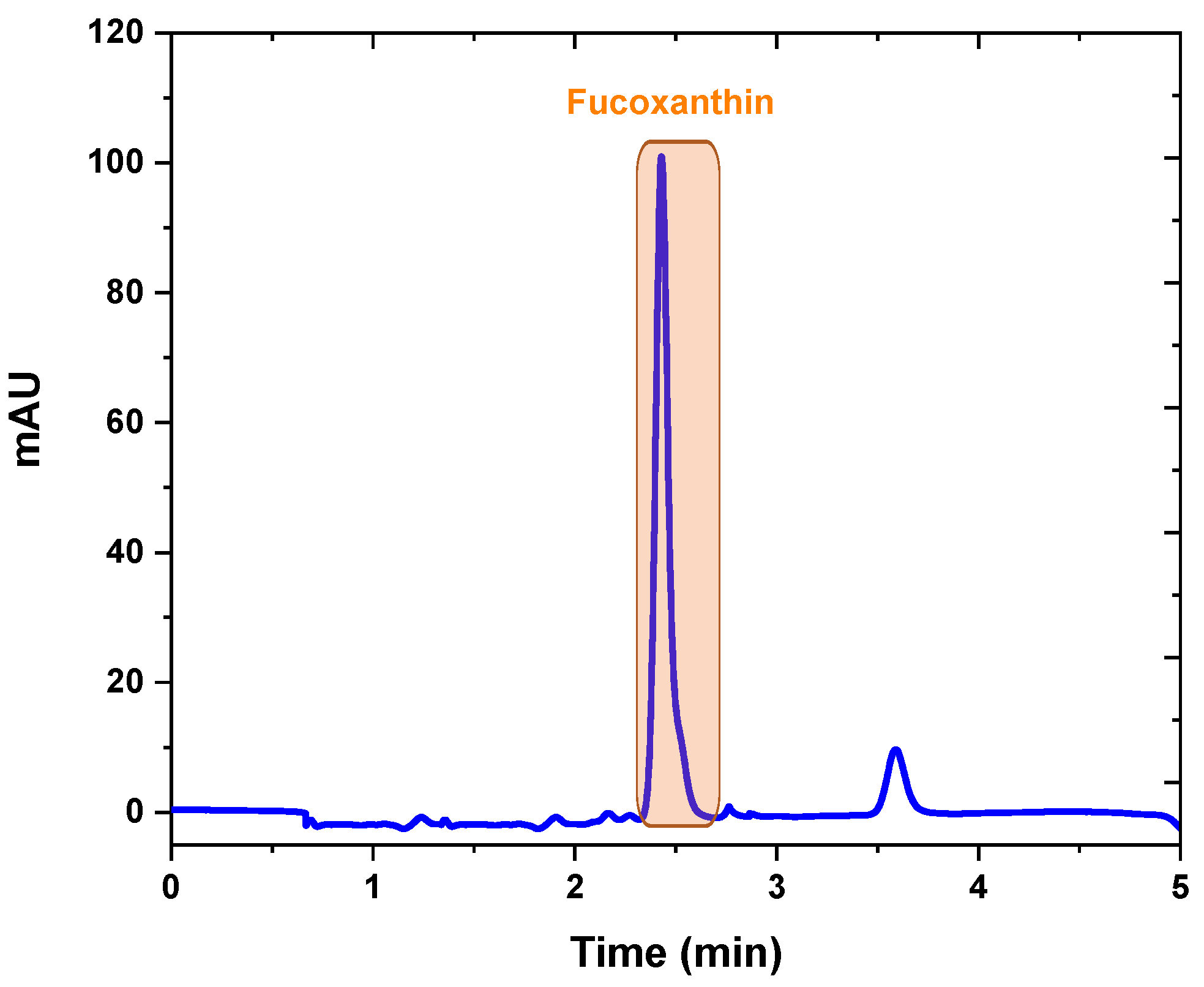
2.3. Fucoxanthin Analysis and Isolation by Preparative Chromatography

Quantification of fucoxanthin was realized by HPLC–DAD with a simple and fast isocratic elution with a modified protocol proposed by Erdoğan, et al. [41] using Methanol: Acetonitrile (85:15) with a flow rate of 1.2 mL/min. Fucoxanthin was isolated by preparative chromatography rapidly with a higher flow rate (4 mL/min) so that fucoxanthin can be collected with sequential injections in a shorter time. No serious problems were encountered in the separation of fucoxanthin by prep-HPLC. The fucoxanthin peak was collected between 2.22 and 2.73 min, as presented in Figure 7. Each fraction was placed in an amber bottle and flushed with nitrogen gas to avoid degradation of fucoxanthin due to light and oxygen.

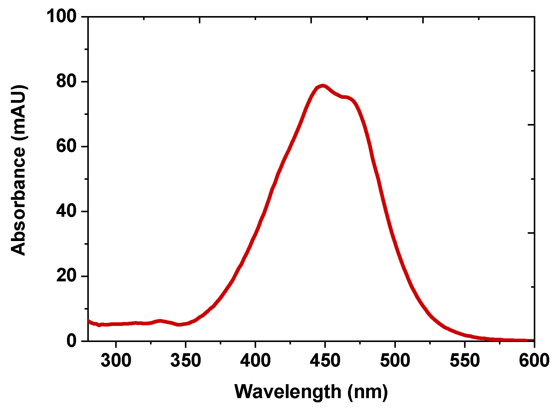
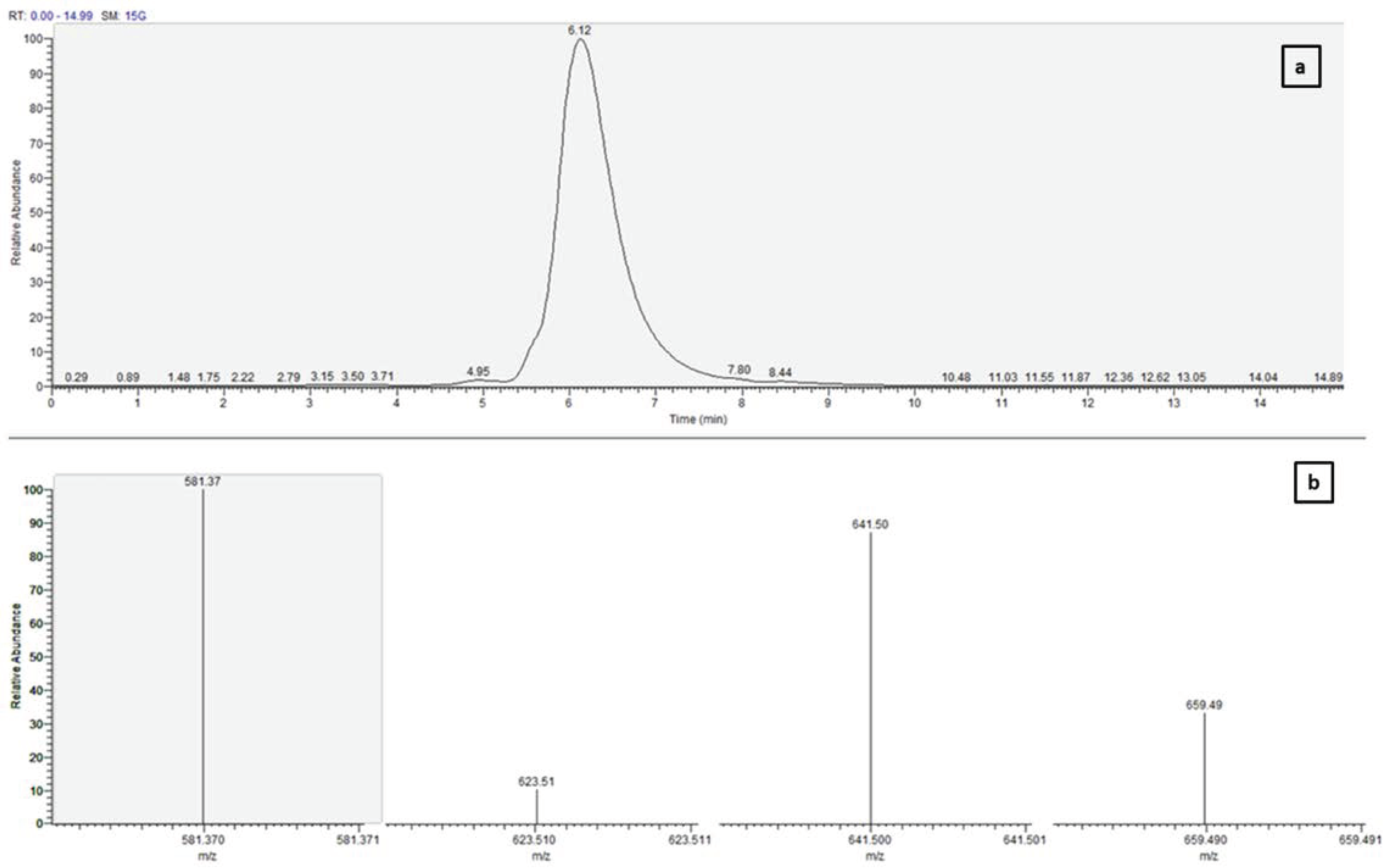
2.4. Confirmation of Purified Fucoxanthin with UV-vis Spectroscopy and Mass Spectrometry

Purified fucoxanthin was dissolved in mobile phase and injected to HPLC-DAD. The absorbance values between 300 and 600 nm were collected during HPLC-DAD analysis. According to the data obtained, fucoxanthin fine structure and absorbance values are in agreement with the literature values, as depicted in Figure 8 [42].
The collected fraction after preparative chromatography was dissolved in 10.0 mL of methanol and subjected to LC–MS/MS analysis for molecular weight. Fucoxanthin was also identified and confirmed by retention time (RT) and Selected Ion Monitoring (SIM) mode. Analysis was performed in positive ion mode and optimized using commercial fucoxanthin standard. Figure 9 demonstrates the LC-MS/MS data for purified fucoxanthin from N. shiloi. Monitoring the ions m/z 659.49 [M+H]+, 641.50 [M+H-18]+, 622.51 [M+H-18-18]+, and 581.37 [M+H-18-60]+ confirms that fucoxanthin was separated, which is consistent with the literature values [42].

3. Discussion

3.1. Variation of Fucoxanthin Content and Biomass Productivity under Different Light Intensities and Aeration Dates

The growth rate of N. shiloi in F/2 medium, and the fucoxanthin content varied significantly under different light intensities and aeration rates. Three types of light responses as low light (50 μmol/m2s), medium light (150 μmol/m2s), and high light (300 μmol/m2s) with varying aeration rates (1-3-5 L/min.) were investigated since the light is one of the most important parameters that influence the amount of carotenoids produced in microalgae. In N. shiloi, low intensities of light caused an increase in the amount of fucoxanthin. Although the adaptability of microalgae to environmental stress, particularly light intensity, is species-specific, there is a similar trend among fucoxanthin-producing microalgae, which states that low light intensity induces a higher accumulation of fucoxanthin [32]. This increase may be due to the Fucoxanthin–chlorophyll proteins (FCP) which are associated with photosystems (PS) I and II in the thylakoid membranes (TM) in algae. The FCP complex is responsible for light-harvesting and electron transfer reactions during photosynthesis [43,44,45]. Apart from light harvesting capacity, FCPs have been also investigated for the photoprotective capacity [46,47]. Under low light (LL) conditions, diatoms require more FCP complexes to compensate for the reduction in photon flux [45]. Thus, photosynthetic pigments including FX and FCP antennae should be provided to form more FCP complexes. In this case, more FCP complexes or larger complexes may be required to overcome the lack of photon flux under LL. In contrast to LL condition, excess photon flux must switch on the photoprotective mechanism to prevent photoinhibition and photodamage, which can threaten microalgal viability [48]. High light (HL) stress may cause alterations in the photosynthetic apparatus, such as chloroplast fragmentation and TM degradation, leading to an increase in secondary carotenoids (DD and DT) rather than in the primary carotenoid FX [49].
When different aeration rates and light intensities are applied, the best result is found in N5-50. Although the amount of biomass is not as high compared to other conditions, the content of fucoxanthin is the highest as 51.05 ± 1.02 mg/gDW. Additionally, it is very important to reduce the steps during purification since the purification step will be required to commercially obtain fucoxanthin. For example, the amount of fucoxanthin obtained in N5-50 is 34% higher than in N3-50, and the growth rate is almost the same (Table 1).
As previously reported in the literature, algal fucoxanthin production was found to be higher under low light intensities [50,51,52,53]. It has been hypothesized that the fucoxanthin variation caused by light may be related to the modulation of Diadinoxanthin Cycle [53,54,55]. When the studies in the literature are examined, it has been reported that the light intensity required for the production of high amounts of fucoxanthin varies between 10 and 100 μmol/m2s. Similarly, N. shiloi produced the maximum amount of fucoxanthin at 50 μmol/m2s light intensity in this study. Therefore, the obtained result is as expected [32].
In order to cope with the abiotic stress caused by the high light intensities, diatom cells tend to convert diadinoxanthin to diatoxanthin at the expense of fucoxanthin, leading to reduced fucoxanthin biosynthesis. However, it should be noted that this is dependent on the species and/or strains. Each microalga has its own capability to overcome the stress conditions or adaptation to different culture conditions [54].

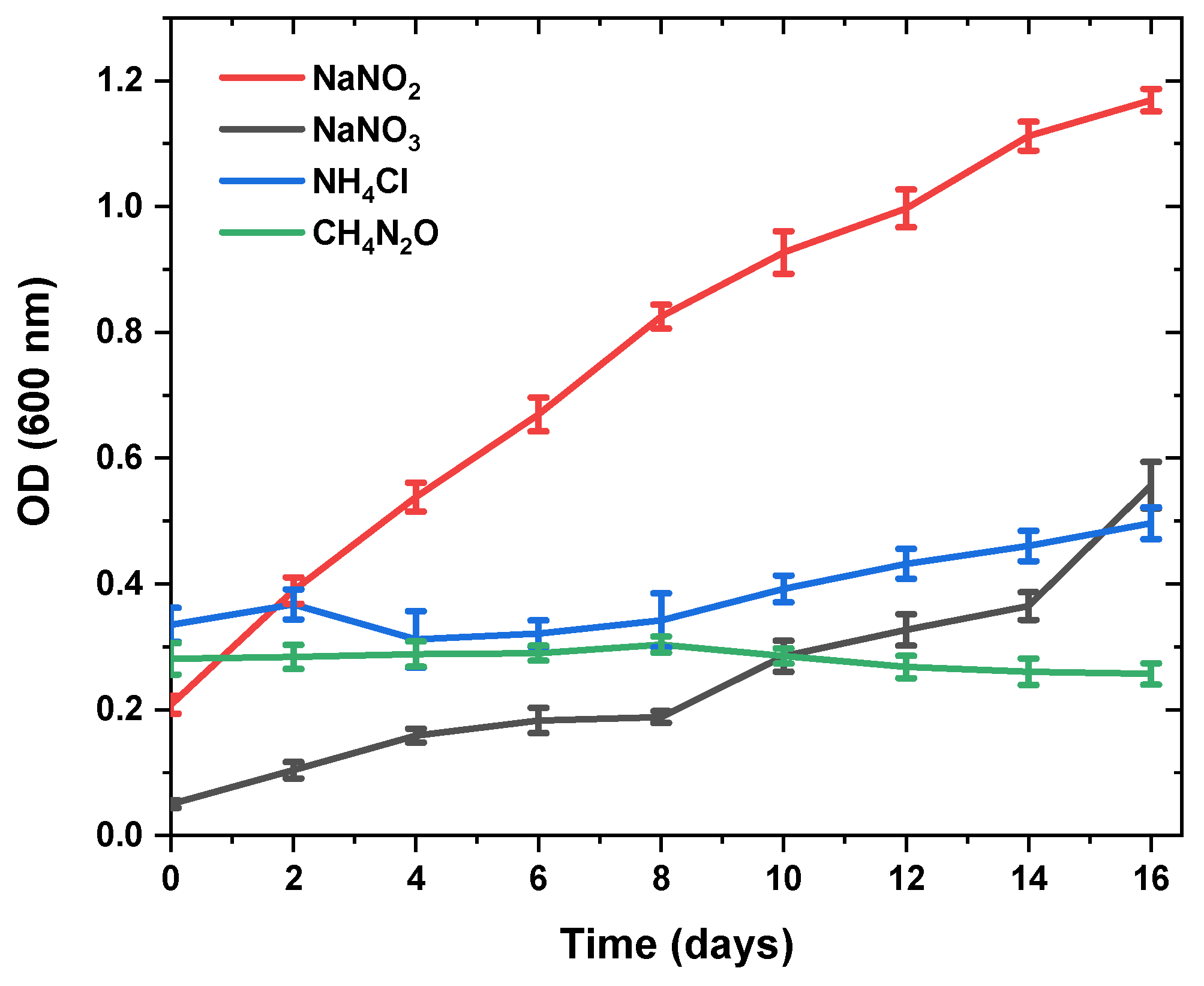
3.2. Changes in Fucoxanthin Content and Biomass Productivity Using Different Nitrogen Sources

The effects of various nitrogen sources on the development and generation of fucoxanthin in microalgae were also studied [56,57,58]. For this reason, N. shiloi cells were cultivated in seawater BG11 medium in which the nitrogen sources were varied. Apart from nitrate, sodium nitrite, ammonium chloride, and urea were used at the same concentrations. The maximum specific growth rate was highest and the same when nitrate and nitrite were used as nitrogen sources. However, cell death was observed when ammonium and urea were used instead. These results have indicated that N. shiloi could not compensate the pH changes. Nitrogen is an important element for growth, and while microalgae can utilize a range of nitrogenous compounds, it has been shown that ammonium ions, if present above a threshold level, are assimilated in preference to other nitrogen sources [59,60,61,62]. The assimilation of various nitrogen sources can have a pronounced effect on the pH of the medium in non-pH-stat cultures.
The extent and direction of the pH change is likely to be due to the rate and form of the assimilated nitrogen, and the buffering capacity of the medium. Apart from the direct energy requirements for assimilating the different nitrogen sources, the observed pH changes may have, in turn, altered the maximum specific growth rate and fucoxanthin.
It is unknown if the higher concentrations of ammonium chloride and urea inhibited the cell division or if they were toxic to the cells. Toxic effects of ammonium ions and urea are often attributed to ammonia (NH3), which may accumulate from the decomposition of urea or from the dissociation of ammonium ions. The potential toxicity could be clarified if, in the future, the concentration of ammonia in the cultivation media is monitored and cell viability assays are undertaken. The increase in the maximum specific growth rate of C. cryptica when cultivated with low concentrations of ammonium chloride or urea could be a direct result of the assimilation efficiency of ammonium ions or urea (as less energy is required for the assimilation of ammonium and urea), or an indirect result based on the pH of the cultivation media. While the preferential assimilation of ammonium ions or urea does not necessarily imply faster growth (growth rate being restricted by the rate-limiting step within the system), the source of nitrogen may alter the way metabolic energy is spent. However, the addition of different nitrogen sources should be more explored and optimized due to the potential toxic effects of ammonium and ammonia concentrations derived from urea conversion [63]. Our experiments show that the addition of different nitrogen sources decreased the fucoxanthin productivity and cell density as compared with cultivation with nitrate.
Possibly, high urea concentrations might lead to growth reduction through toxicity effects. Previous researchers demonstrated that microalgae could assimilate urea as a source of nitrogen by converting it to NH4+ and CO2 through urease activity, or ammonium and bicarbonate via ATP-urea amidolyase [64,65]. This might lead to high ammonium or ammonia concentrations, which tend to be toxic for diatoms such as P. tricornutum, as well as other algae such as chlorophytes [66,67]. Consequently, commercial nitrates such as NaNO3 are still recommended for microalgal culture, despite the considerable need for a substitute nitrogen source that is less expensive.

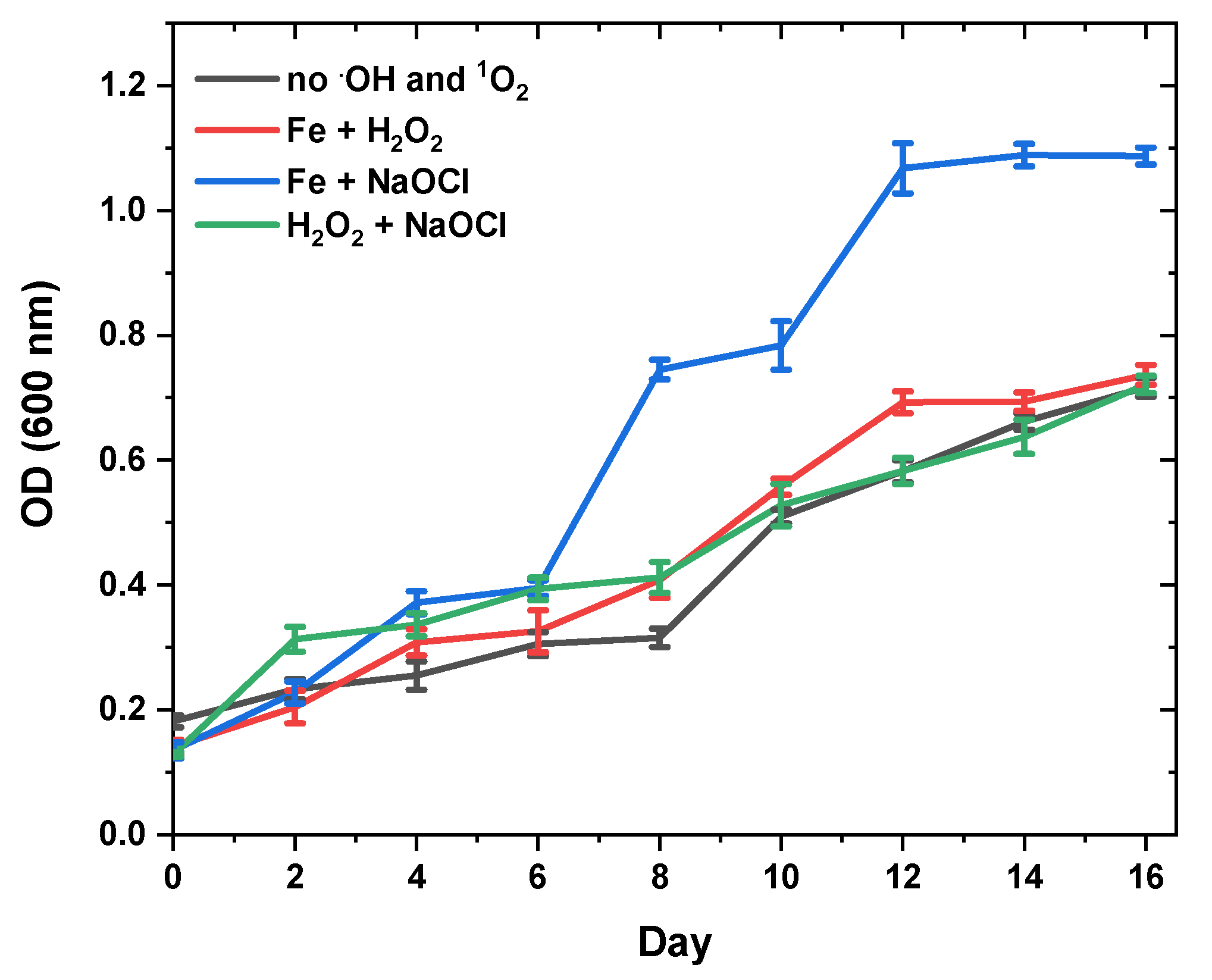
3.3. Effect of Oxidative Stress on Fucoxanthin Content and Biomass Productivity

Reactive oxygen species (ROS) are continuously produced in microalgae, chloroplasts, mitochondria, and peroxisomes. ROS production and cleaning must be balanced so that it does not damage cell components. Therefore, antioxidant protective mechanisms are generally engaged [68]. Hydrogen peroxide, a type of ROS, is also a microalgae product released through oxidative metabolism. Almost all living things decompose hydrogen peroxide into water and oxygen at low concentrations. H2O2 can damage cells at high concentrations, but moderate levels of cells can adapt to this condition. Hydrogen peroxide decomposes, especially in the presence of iron, and forms the highly reactive hydroxyl radical through the Fenton reaction. Uncontrolled production of ROS can also destroy proteins, lipids, and carotenoids. Therefore, as in many other organisms, microalgae develop defence mechanisms to deal with this situation when faced with high ROS levels [69]. One of them is that, in the presence of ROS, it increases the production of carotenoids with high antioxidant activity in order to protect the cells against oxidative damage.
Unfortunately, there is not enough information in the literature about the stimulating mechanism of fucoxanthin formation by ROS. In some of the studies, the production of fucoxanthin was triggered in C. closterium and A. capitellata again by using the same types of ROS [41,70].
In this study, an increase in fucoxanthin accumulation was also observed by using ROS concentrations at the same concentrations. In Table 2, it can be said that the accumulation of fucoxanthin in N. shiloi is remarkable. In the presence of 0.1 mM H2O2 + 0.1 mM Fe2+, the amount of fucoxanthin increased slightly (58.20 ± 1.16 mg/gDW) compared to the control group (50.17 ± 1.02 mg/gDW). This value increased to 65.14 ± 1.95 mg/gDW in the presence of 0.1 mM NaClO + 0.1 mM Fe2+. When 0.1 mM H2O2 + 0.1 mM NaOCl was used, the fucoxanthin value almost doubled (97.45 ± 2.64 mg/gDW). When the data are examined, it is thought that H2O2 and NaClO are very effective on fucoxanthin accumulation and the presence of Fe2+ does not contribute much. When H2O2 was used in combination with NaClO, the remarkable increase in the amount of fucoxanthin supports this. Probably N. shiloi increased the accumulation of fucoxanthin to cope with this stress at the given concentrations.
When the stress sources were examined, it was seen that there was only a partial decrease in the specific growth rate of N. shiloi in only the first two stress sources. In other words, it can be said that these two sources for the concentrations used create stress for N. shiloi. However, the most notable of the results here is that, when H2O2 and NaClO are used together, there is no reduction in growth rate and even remain the same. This shows that microalgae do not accept H2O2 and NaClO as stress factors at given concentrations, and at least produces high amounts of fucoxanthin and easily copes with this stress.
It seems that this species has easily adapted itself to oxidative stress conditions by activating defence mechanisms [71]. As with all living things, microalgae have several defence systems, both enzymatic and non-enzymatic, for detoxification of ROS. In the literature, it has been reported that three reaction mechanisms describe the reaction of free radicals with carotenoids, namely, electron transfer, hydrogen atom transfer, and radical addition to carotenoids [72,73]. To remove free radicals, carotenoids can donate or accept unpaired electrons. Generally, antioxidant molecules are oxidized by giving electrons to free radicals. However, carotenoids can absorb free radicals by accepting an unpaired electron, making it harmless by translocation on the conjugated side chain. For these reasons, it may be possible to induce the synthesis of fucoxanthin during quenching with ROS.

4. Materials and Methods

4.1. Reagents and Chemicals

All-trans-fucoxanthin, triethylamine, pyrogallol, silica gel, and calcium carbonate were provided by Sigma-Aldrich, and all the solvents used in this study were LC-grade purchased from Merck.

4.2. Molecular Identification of N. shiloi

The benthic diatom N. shiloi was previously identified, and the details are given by Demirel, et al. [74]. In that study, benthic diatoms were separated from the Aegean Sea in Türkiye and identified according to their morphological characteristics on the basis of observations in bright field and scanning electron microscopy. For DNA extraction, the cultured strain was harvested by centrifugation, and the cell pellet was used in the DNA Kits (Zymo Research) and stored at −20 °C. PCR primers targeted to link 18S rDNA (F, 5′-YACCTGGTTGATCCTGCCAGTAG-3′ and R, 5′-GCTTGATCCTTCTGCAGGTTCACC-3′). PCR protocol was applied, and products were viewed in an agarose gel stained by Jel Safe Dye and checked the gel under a UV light. In the DNA sequencing step, dye-terminator sequencing was performed using the primers, and the nucleotide chromatograms were determined by DNA sequences. The sequences obtained in the mentioned study were deposited in the NCBI GenBank, and the accession numbers of KR149459. Nanofrustulum shiloi (EGEMACC 47) was deposited in Ege University, Microalgae Culture Collection, Izmir, Turkey (EGEMACC—https://ege-macc.ege.edu.tr/ (accessed on 15 February 2023)).

4.3. Cultivation and Harvesting of N. shiloi

N. shiloi was cultivated in BG11 medium based on artificial seawater [73] at 20 ± 2 °C under continued light intensity. The diatom was added in a bubble-column photobioreactor (BCP, 2 L) and cultivated under the light intensity of 50, 150, and 300 µmol/m2s (Lutron LX-1108, Taiwan), at 21 ± 2 °C, and with the aeration rate of 1, 3, and 5 L/min. The growth of microalgae was pursued by counting microalgae cells optical density at 2-day intervals over the period of 16 days. The specific growth rate for N. shiloi was calculated according to Becker [75] (Equation (1)) using the data obtained by the absorbance values taken at 600 nm.
μ   = lnx 2 ln x 1 Δ t
where μ = specific growth rate, x2 = cell concentration at time, t2, x1 = cell concentration at time t1, and Δt = t2 − t1. Doubling time was determined as 0.693/µ.
The recipe of seawater BG11 was prepared in 1.5 g/L sodium nitrate (NaNO3-17.65 mM), sodium nitrite (NaNO2), ammonium chloride (NH4Cl), and urea (CH4N2O), and were calculated at the same concentrations, separately.
In the control group, the culture medium was prepared using 1.5 g/L sodium nitrate (NaNO3-17.65 mM), as recommended in the Seawater BG11 recipe (American Type Culture Collection, https://www.atcc.org/search#q=bg%2011&sort=relevancy&numberOfResults=24&f:Documenttype=[Media%20formulation] (accessed on 15 February 2023). In order to examine the effect of different nitrogen sources, it was prepared separately with sodium nitrite (NaNO2), ammonium chloride (NH4Cl), and urea (CH4N2O) instead of sodium nitrate, with a constant nitrogen concentration of 17.65 mM.
N. shiloi was grown in a BCP, prepared with different nitrogen sources medium, and then cultivated under the light intensity of 50 µmol/m2s, at 21 ± 2 °C, and with the aeration rate of 5 L/min.
Oxidative stress was created by reactive oxygen species via a series of chemical reactions, as Strati and Oreopoulou 2011 and de Almeida Torres et al. 2018 proposed. The different chemicals (H2O2, NaOCl and FeSO4) of oxidative stress were added to the culture media at a concentration of 0.1 mM. H2O2, NaOCl, and FeSO4, and were added to the culture media at constant concentrations of 0.1 mM in order to enable the release of reactive oxygen species through the 3 reaction templates given in R1, R2 and R3 [76,77].
R1:
Fe2+ + H2O2 → Fe3+ + OH + ∙OH
R2:
NaClO + H2O → HOCl + Na+ + OH and HOCl + Fe2+ → Fe3+ + Cl + ∙OH
R3:
H2O2 + NaClO → H2O + NaCl + 1O2
The control group was cultured in seawater BG11 medium and then cultivated under the light intensity of 50 µmol/m2s, at 21 ± 2 °C, with the aeration rate of 5 L/min, and in BCP. Microalgal cells reached the stationary phase harvested by centrifugation (Pro Research, By Centurion Scientific Ltd., Chichester, UK). After, the pellet was washed with deionized water to remove the growing medium. Then, the paste biomass was lyophilized (Christ Alpha 1-2 LD plus, Osterode am Harz, Germany) and grounded using a mortar for the extraction process. They were stored at −20 °C under dark conditions. The changeable morphology of N. shiloi was examined by scanning electron microscopy (SEM) where characterizations were carried out by using Thermo Scientific Apreo S (Thermo Fisher Scientific, Waltham, MA, USA).

4.4. Preparation of Stock and Standard Fucoxanthin Solutions

For the preparation of 100.0 mg/L stock fucoxanthin solution, 1.0 mg of fucoxanthin was weighed and dissolved in 10.0 mL chloroform (stabilized with 1% ethanol). Calibration standards were prepared by using this stock solution for the construction of a calibration curve. All standard solutions were kept in amber-colored volumetric flasks. Different concentrations (0.05–10.0 mg/L) of fucoxanthin standards were injected into the HPLC-DAD.

4.5. Ultrasound-Assisted Extraction (UAE) of Fucoxanthin from N. shiloi

The extraction of fucoxanthin was performed by Ultrasound-Assisted Extraction (UAE) method by using an ultrasonic bath (Elmasonic, S80H). Ethanol was preferred as an extraction solvent, as it is environmentally preferable and referred to as “green”. Saponification procedure was eliminated, as the saponification causes fucoxanthin to turn into fucoxanthinol under basic conditions [44].
For the extraction process, the biomass (0.20 g) was added with CaCO3 (0.20 g) and 10.0 mL of ethanol containing 0.01% (w/v) pyrogallol. Then, the extraction was conducted by an ultrasonic bath for 15 min at 40 °C. After the UAE process, the solution was centrifuged at 5000 rpm for 2 min and the supernatant was filtered by 47 mm of 0.20 µm nylon filter paper (Sartorius, Goettingen, Germany). Then, the solution was diluted with the mobile phase (%70 Methanol and 30% Acetonitrile containing 0.01% v/v triethylamine) prior to HPLC-DAD analysis [43].

4.6. HPLC-DAD and LC-APCI-MS/MS Analyses of Fucoxanthin

Fucoxanthin was quantified by HPLC 1260 Series (Agilent, Santa Clara, CA, USA) equipped with diode array detector at 450 nm with a flow rate of 1.2 mL/min using Waters YMC C30 Carotenoid column (4.6 mm × 250 mm, 5 µm). The mobile phase consisted of 70% methanol and 30% acetonitrile each including 0.01% triethyl amine (TEA) as a modifier. Fucoxanthin was purified by preparative HPLC (Thermo Scientific/Dionex Ultimate 3000). The preparative purification was performed with a YMC-C30 semi-prep carotenoid column (10 mm × 250 mm, 5 μm) using the same mobile phase composition (85:15, methanol: acetonitrile) at a flow rate of 4.0 mL/min at 450 nm with a column temperature of 25 °C. The injection volume was 1.0 mL.
In order to obtain the absorption profile of fucoxanthin, spectroscopic data were recorded between 300 and 600 nm. After the purification procedure, the absorbance spectrum of fucoxanthin standard and purified fucoxanthin were compared (Data not shown). In the present study, fucoxanthin obtained from N. shiloi extract and the purified fucoxanthin were identified and confirmed by liquid chromatography-tandem mass spectrometry (LC-MS/MS) equipped with an Atmospheric Pressure Chemical Ionization probe (APCI). The mass spectrometer was operated in full scan mode from m/z 50-900 at 350 °C for vaporization temperature.

4.7. Statistical Analysis

Each experiment was performed in triplicate. Tukey’s test at a reliability level (of p < 0.05) was utilized to find differences between treatment levels. Minitab software was utilized to conduct statistical analyses (V18, Minitab Inc., State College, PA, USA).

5. Conclusions

Microalgae have significant potential to produce value-added products such as lipids, proteins, carbohydrates, and carotenoids that play a vital role in human health. The use of carotenoids in the pharmaceutical, nutraceutical, and food industries makes them valuable components of microalgae. As microalgae exhibit adaptation to different environmental factors such as temperature, light, salinity, etc., the production of carotenoids could be increased by changing the cultivation conditions. The results of this study showed that fucoxanthin content could be enhanced by altering the cultivation conditions. Moreover, it was proven that particularly the oxidative stress triggers the production of fucoxanthin in N. shiloi as in other diatoms.
This article has discussed how to obtain high yields of fucoxanthin from N. shiloi as well as methodologies that can be used for new species. Combined with the new and innovative “green” solvent, ultrasound-assisted liquid extraction serves as an economically desirable and environmentally friendly extraction strategy. In addition, it is a pioneering work on obtaining fucoxanthin in a short time using the prep-column obtained with the latest technology. In the future, more intensive research, including pilot studies, should focus on photobioreactor-based bioengineering techniques, the integration of synergistic extraction strategies, and economic and environmental considerations, which are also suitable for industrial production for various purposes.

Author Contributions

Conceptualization, resources, methodology, data curation, writing—original draft preparation, writing—review and editing, supervision, funding acquisition, A.E.; methodology, data curation, writing—review and editing, A.B.K.; data curation, D.D., Ö.Ç. and M.A.; conceptualization methodology, data curation, writing—original draft preparation, Z.D.; conceptualization, methodology, supervision, writing—original draft preparation, M.C.-D. All authors have read and agreed to the published version of the manuscript.

Funding

This research was funded by the Scientific and Technological Research Council of Türkiye (TÜBİTAK) through the COST Project 216Z167.

Institutional Review Board Statement

Not applicable.

Informed Consent Statement

Not applicable.

Data Availability Statement

All data generated or analyzed during this study are included in the article.

Acknowledgments

This publication is based upon work from COST Action CA15136 (EUROCAROTEN) supported by COST (European Cooperation in Science and Technology) program. Moreover, the authors would like to acknowledge Ege University Pharmaceutical Sciences Research Centre (FABAL) for prep-HPLC analyses as well as HÜBTUAM for the mass analyses (LC-APCI-MS/MS). Finally, the authors are grateful to Çiğdem Yengin for her help and patience in purification studies.

Conflicts of Interest

The authors declare no conflict of interest.

Sample Availability

Samples of the brown microalge are available from the authors.

References

  1. Cohen, Z. Chemicals from Microalgae; CRC Press: Boca Raton, FL, USA, 2014. [Google Scholar]
  2. Pulz, O.; Gross, W. Valuable products from biotechnology of microalgae. Appl. Microbiol. Biotechnol. 2004, 65, 635–648. [Google Scholar] [CrossRef]
  3. Saini, D.K.; Chakdar, H.; Pabbi, S.; Shukla, P. Enhancing production of microalgal biopigments through metabolic and genetic engineering. Crit. Rev. Food Sci. Nutr. 2019, 60, 391–405. [Google Scholar] [CrossRef] [PubMed]
  4. de Jesus Raposo, M.F.; de Morais, R.M.; de Morais, A.M. Health applications of bioactive compounds from marine microalgae. Life Sci. 2013, 93, 479–486. [Google Scholar] [CrossRef]
  5. Fung, A.; Hamid, N.; Lu, J. Fucoxanthin content and antioxidant properties of Undaria pinnatifida. Food Chem. 2013, 136, 1055–1062. [Google Scholar] [CrossRef]
  6. Del Campo, J.A.; Garcia-Gonzalez, M.; Guerrero, M.G. Outdoor cultivation of microalgae for carotenoid production: Current state and perspectives. Appl. Microbiol. Biotechnol. 2007, 74, 1163–1174. [Google Scholar] [CrossRef]
  7. Rao, A.V.; Rao, L.G. Carotenoids and human health. Pharmacol. Res. 2007, 55, 207–216. [Google Scholar] [CrossRef]
  8. Wijffels, R.H. Potential of sponges and microalgae for marine biotechnology. Trends Biotechnol. 2008, 26, 26–31. [Google Scholar] [CrossRef] [PubMed]
  9. Delgado-Vargas, F.; Jimenez, A.R.; Paredes-Lopez, O. Natural pigments: Carotenoids, anthocyanins, and betalains--characteristics, biosynthesis, processing, and stability. Crit. Rev. Food Sci. Nutr. 2000, 40, 173–289. [Google Scholar] [CrossRef] [PubMed]
  10. Landrum, J.T.; Chatfield, D.C.; Mebel, A.M.; Alvarez-Calderon, F.; Fernandez, M.V. The conformation of end-groups is one determinant of carotenoid topology suitable for high fidelity molecular recognition: A study of β-and ε-end-groups. Arch. Biochem. Biophys. 2010, 493, 169–174. [Google Scholar] [CrossRef]
  11. Rajesh, K.; Rohit, M.; Mohan, S.V. Microalgae-Based Carotenoids Production. In Algal Green Chemistry; Elsevier: Amsterdam, The Netherlands, 2017; pp. 139–147. [Google Scholar]
  12. Kumar, S.R.; Hosokawa, M.; Miyashita, K. Fucoxanthin: A marine carotenoid exerting anti-cancer effects by affecting multiple mechanisms. Mar. Drugs 2013, 11, 5130–5147. [Google Scholar] [CrossRef] [Green Version]
  13. Beppu, F.; Niwano, Y.; Sato, E.; Kohno, M.; Tsukui, T.; Hosokawa, M.; Miyashita, K. In vitro and in vivo evaluation of mutagenicity of fucoxanthin (FX) and its metabolite fucoxanthinol (FXOH). J. Toxicol. Sci. 2009, 34, 693–698. [Google Scholar] [CrossRef] [PubMed] [Green Version]
  14. Beppu, F.; Niwano, Y.; Tsukui, T.; Hosokawa, M.; Miyashita, K. Single and repeated oral dose toxicity study of fucoxanthin (FX), a marine carotenoid, in mice. J. Toxicol. Sci. 2009, 34, 501–510. [Google Scholar] [CrossRef] [PubMed] [Green Version]
  15. Zaragoza, M.C.; Lopez, D.; Sáiz, M.P.; Poquet, M.; Perez, J.; Puig-Parellada, P.; Marmol, F.; Simonetti, P.; Gardana, C.; Lerat, Y.; et al. Toxicity and antioxidant activity in vitro and in vivo of two Fucus vesiculosus extracts. J. Agric. Food Chem. 2008, 56, 7773–7780. [Google Scholar] [CrossRef]
  16. Dominguez, H. Functional Ingredients from Algae for Foods and Nutraceuticals; Elsevier: Amsterdam, The Netherlands, 2013. [Google Scholar]
  17. Peng, J.; Yuan, J.P.; Wu, C.F.; Wang, J.H. Fucoxanthin, a marine carotenoid present in brown seaweeds and diatoms: Metabolism and bioactivities relevant to human health. Mar. Drugs 2011, 9, 1806–1828. [Google Scholar] [CrossRef]
  18. Xiao, X.; Si, X.; Yuan, Z.; Xu, X.; Li, G. Isolation of fucoxanthin from edible brown algae by microwave-assisted extraction coupled with high-speed countercurrent chromatography. J. Sep. Sci. 2012, 35, 2313–2317. [Google Scholar] [CrossRef] [PubMed]
  19. Koduvayur Habeebullah, S.F.; Surendraraj, A.; Jacobsen, C. Isolation of Fucoxanthin from Brown Algae and Its Antioxidant Activity: In Vitro and 5% Fish Oil-In-Water Emulsion. J. Am. Oil Chem. Soc. 2018, 95, 835–843. [Google Scholar] [CrossRef]
  20. Sun, P.; Wong, C.C.; Li, Y.; He, Y.; Mao, X.; Wu, T.; Ren, Y.; Chen, F. A novel strategy for isolation and purification of fucoxanthinol and fucoxanthin from the diatom Nitzschia laevis. Food Chem. 2019, 277, 566–572. [Google Scholar] [CrossRef]
  21. Zailanie, K.; Purnomo, H. Fucoxanthin content of five species brown seaweed from Talango District, Madura Island. J. Agric. Sci. Technol. A 2011, 1, 1103–1105. [Google Scholar]
  22. Ishika, T.; Laird, D.W.; Bahri, P.A.; Moheimani, N.R. Co-cultivation and stepwise cultivation of Chaetoceros muelleri and Amphora sp. for fucoxanthin production under gradual salinity increase. J. Appl. Phycol. 2019, 31, 1535–1544. [Google Scholar] [CrossRef]
  23. Wang, J.; Ma, Y.; Yang, J.; Jin, L.; Gao, Z.; Xue, L.; Hou, L.; Sui, L.; Liu, J.; Zou, X. Fucoxanthin inhibits tumour-related lymphangiogenesis and growth of breast cancer. J. Cell. Mol. Med. 2019, 23, 2219–2229. [Google Scholar] [CrossRef]
  24. Leong, Y.K.; Chen, C.-Y.; Varjani, S.; Chang, J.-S. Producing fucoxanthin from algae–Recent advances in cultivation strategies and downstream processing. Bioresour. Technol. 2022, 344, 126170. [Google Scholar] [CrossRef] [PubMed]
  25. Mohamadnia, S.; Tavakoli, O.; Faramarzi, M.A. Enhancing production of fucoxanthin by the optimization of culture media of the microalga Tisochrysis lutea. Aquaculture 2021, 533, 736074. [Google Scholar] [CrossRef]
  26. Lu, X.; Sun, H.; Zhao, W.; Cheng, K.-W.; Chen, F.; Liu, B. A hetero-photoautotrophic two-stage cultivation process for production of fucoxanthin by the marine diatom Nitzschia laevis. Mar. Drugs 2018, 16, 219. [Google Scholar] [CrossRef] [Green Version]
  27. Mao, X.; Chen, S.H.Y.; Lu, X.; Yu, J.; Liu, B. High silicate concentration facilitates fucoxanthin and eicosapentaenoic acid (EPA) production under heterotrophic condition in the marine diatom Nitzschia laevis. Algal Res. 2020, 52, 102086. [Google Scholar] [CrossRef]
  28. Sun, Z.; Wang, X.; Liu, J. Screening of Isochrysis strains for simultaneous production of docosahexaenoic acid and fucoxanthin. Algal Res. 2019, 41, 101545. [Google Scholar] [CrossRef]
  29. Mousavi Nadushan, R.; Hosseinzade, I. Optimization of production and antioxidant activity of fucoxanthin from marine haptophyte algae, Isochrysis galbana. Iran. J. Fish. Sci. 2020, 19, 2901–2908. [Google Scholar]
  30. Gao, F.; Teles, I.; Wijffels, R.H.; Barbosa, M.J. Process optimization of fucoxanthin production with Tisochrysis lutea. Bioresour. Technol. 2020, 315, 123894. [Google Scholar] [CrossRef]
  31. Li, Y.; Sun, H.; Wu, T.; Fu, Y.; He, Y.; Mao, X.; Chen, F. Storage carbon metabolism of Isochrysis zhangjiangensis under different light intensities and its application for co-production of fucoxanthin and stearidonic acid. Bioresour. Technol. 2019, 282, 94–102. [Google Scholar] [CrossRef]
  32. Khaw, Y.S.; Yusoff, F.M.; Tan, H.T.; Noor Mazli, N.A.I.; Nazarudin, M.F.; Shaharuddin, N.A.; Omar, A.R.; Takahashi, K. Fucoxanthin Production of Microalgae under Different Culture Factors: A Systematic Review. Mar. Drugs 2022, 20, 592. [Google Scholar] [CrossRef]
  33. Bae, M.; Kim, M.-B.; Park, Y.-K.; Lee, J.-Y. Health benefits of fucoxanthin in the prevention of chronic diseases. Biochim. Et Biophys. Acta (BBA)-Mol. Cell Biol. Lipids 2020, 1865, 158618. [Google Scholar] [CrossRef]
  34. Miyashita, K.; Beppu, F.; Hosokawa, M.; Liu, X.; Wang, S. Nutraceutical characteristics of the brown seaweed carotenoid fucoxanthin. Arch. Biochem. Biophys. 2020, 686, 108364. [Google Scholar] [CrossRef] [PubMed]
  35. Neumann, U.; Derwenskus, F.; Flaiz Flister, V.; Schmid-Staiger, U.; Hirth, T.; Bischoff, S.C. Fucoxanthin, a carotenoid derived from Phaeodactylum tricornutum exerts antiproliferative and antioxidant activities in vitro. Antioxidants 2019, 8, 183. [Google Scholar] [CrossRef] [PubMed] [Green Version]
  36. Yang, H.; Xing, R.; Liu, S.; Li, P. Effect of Fucoxanthin administration on thyroid gland injury induced by cadmium in mice. Biol. Trace Elem. Res. 2021, 199, 1877–1884. [Google Scholar] [CrossRef]
  37. Zarekarizi, A.; Hoffmann, L.; Burritt, D. Approaches for the sustainable production of fucoxanthin, a xanthophyll with potential health benefits. J. Appl. Phycol. 2019, 31, 281–299. [Google Scholar] [CrossRef]
  38. Zheng, J.; Tian, X.; Zhang, W.; Zheng, P.; Huang, F.; Ding, G.; Yang, Z. Protective effects of fucoxanthin against alcoholic liver injury by activation of Nrf2-mediated antioxidant defense and inhibition of TLR4-mediated inflammation. Mar. Drugs 2019, 17, 552. [Google Scholar] [CrossRef] [PubMed] [Green Version]
  39. Telussa, I.; Nurachman, Z. Dynamics of β-carotene and fucoxanthin of tropical marine Navicula sp. as a response to light stress conditions. Algal Res. 2019, 41, 101530. [Google Scholar] [CrossRef]
  40. Csavina, J.L.; Stuart, B.J.; Riefler, R.G.; Vis, M.L. Growth optimization of algae for biodiesel production. J Appl Microbiol 2011, 111, 312–318. [Google Scholar] [CrossRef]
  41. Erdoğan, A.; Karataş, A.B.; Demirel, Z.; Dalay, M.C. Purification of fucoxanthin from the diatom Amphora capitellata by preparative chromatography after its enhanced productivity via oxidative stress. J. Appl. Phycol. 2022, 34, 301–309. [Google Scholar] [CrossRef]
  42. Britton, G.; Liaaen-Jensen, S.; Pfander, H.; Mercadante, A.; Egeland, E. Carotenoids: Handbook; Springer Science & Business Media: Berlin/Heidelberg, Germany, 2004. [Google Scholar]
  43. Wang, W.; Yu, L.-J.; Xu, C.; Tomizaki, T.; Zhao, S.; Umena, Y.; Chen, X.; Qin, X.; Xin, Y.; Suga, M. Structural basis for blue-green light harvesting and energy dissipation in diatoms. Science 2019, 363, eaav0365. [Google Scholar] [CrossRef]
  44. Gardian, Z.; Litvín, R.; Bína, D.; Vácha, F. Supramolecular organization of fucoxanthin–chlorophyll proteins in centric and pennate diatoms. Photosynth. Res. 2014, 121, 79–86. [Google Scholar] [CrossRef]
  45. Truong, T.Q.; Park, Y.J.; Koo, S.Y.; Choi, J.-H.; Enkhbayar, A.; Song, D.-G.; Kim, S.M. Interdependence of fucoxanthin biosynthesis and fucoxanthin-chlorophyll a/c binding proteins in Phaeodactylum tricornutum under different light intensities. J. Appl. Phycol. 2023, 35, 25–42. [Google Scholar] [CrossRef]
  46. Nagao, R.; Yokono, M.; Ueno, Y.; Suzuki, T.; Kumazawa, M.; Kato, K.-H.; Tsuboshita, N.; Dohmae, N.; Ifuku, K.; Shen, J.-R. Enhancement of excitation-energy quenching in fucoxanthin chlorophyll a/c-binding proteins isolated from a diatom Phaeodactylum tricornutum upon excess-light illumination. Biochim. Biophys. Acta (BBA)-Bioenerg. 2021, 1862, 148350. [Google Scholar] [CrossRef] [PubMed]
  47. Giovagnetti, V.; Ruban, A.V. Detachment of the fucoxanthin chlorophyll a/c binding protein (FCP) antenna is not involved in the acclimative regulation of photoprotection in the pennate diatom Phaeodactylum tricornutum. Biochim. Biophys. Acta (BBA)-Bioenerg. 2017, 1858, 218–230. [Google Scholar] [CrossRef] [PubMed]
  48. Domingues, N.; Matos, A.R.; Marques da Silva, J.; Cartaxana, P. Response of the diatom Phaeodactylum tricornutum to photooxidative stress resulting from high light exposure. PLoS ONE 2012, 7, e38162. [Google Scholar] [CrossRef] [PubMed] [Green Version]
  49. Xia, S.; Wang, K.; Wan, L.; Li, A.; Hu, Q.; Zhang, C. Production, characterization, and antioxidant activity of fucoxanthin from the marine diatom Odontella aurita. Mar. Drugs 2013, 11, 2667–2681. [Google Scholar] [CrossRef]
  50. Van De Poll, W.H.; Van Leeuwe, M.A.; Roggeveld, J.; Buma, A.G. Nutrient Limitation And High Irradiance Acclimation Reduce Par And Uv-Induced Viability Loss In The Antarctic Diatom Chaetoceros Brevis (Bacillariophyceae) 1. J. Phycol. 2005, 41, 840–850. [Google Scholar] [CrossRef] [Green Version]
  51. Willemoës, M.; Monas, E. Relationship between growth irradiance and the xanthophyll cycle pool in the diatom Nitzschia palea. Physiol. Plant. 1991, 83, 449–456. [Google Scholar] [CrossRef]
  52. Kolber, Z.; Zehr, J.; Falkowski, P. Effects of Growth Irradiance and Nitrogen Limitation on Photosynthetic Energy Conversion in Photosystem II. Plant Physiol. 1988, 88, 923–929. [Google Scholar] [CrossRef] [Green Version]
  53. Coesel, S.; Obornik, M.; Varela, J.; Falciatore, A.; Bowler, C. Evolutionary origins and functions of the carotenoid biosynthetic pathway in marine diatoms. PLoS ONE 2008, 3, e2896. [Google Scholar] [CrossRef] [Green Version]
  54. Guo, B.B.; Liu, B.; Yang, B.; Sun, P.P.; Lu, X.; Liu, J.; Chen, F. Screening of Diatom Strains and Characterization of Cyclotella cryptica as A Potential Fucoxanthin Producer. Mar. Drugs 2016, 14, 125. [Google Scholar] [CrossRef] [Green Version]
  55. Pahl, S.L.; Lewis, D.M.; Chen, F.; King, K.D. Heterotrophic growth and nutritional aspects of the diatom Cyclotella cryptica (Bacillariophyceae): Effect of some environmental factors. J. Biosci. Bioeng. 2010, 109, 235–239. [Google Scholar] [CrossRef]
  56. Hao, T.B.; Yang, Y.F.; Balamurugan, S.; Li, D.W.; Yang, W.D.; Li, H.Y. Enrichment of f/2 medium hyperaccumulates biomass and bioactive compounds in the diatom Phaeodactylum tricornutum. Algal Res.-Biomass Biofuels Bioprod. 2020, 47, 101872. [Google Scholar] [CrossRef]
  57. Kanamoto, A.; Kato, Y.; Yoshida, E.; Hasunuma, T.; Kondo, A. Development of a Method for Fucoxanthin Production Using the Haptophyte Marine Microalga Pavlova sp. OPMS 30543. Mar. Biotechnol. 2021, 23, 331–341. [Google Scholar] [CrossRef]
  58. Tokushima, H.; Inoue-Kashino, N.; Nakazato, Y.; Masuda, A.; Ifuku, K.; Kashino, Y. Advantageous characteristics of the diatom Chaetoceros gracilis as a sustainable biofuel producer. Biotechnol. Biofuels 2016, 9, 235. [Google Scholar] [CrossRef] [PubMed] [Green Version]
  59. Flores, E.; Guerrero, M.G.; Losada, M. Short-Term Ammonium Inhibition of Nitrate Utilization by Anacystis-Nidulans and Other Cyanobacteria. Arch. Microbiol. 1980, 128, 137–144. [Google Scholar] [CrossRef]
  60. Hildebrand, M. Cloning and functional characterization of ammonium transporters from the marine diatom Cylindrotheca fusiformis (Bacillariophyceae) 1. J. Phycol. 2005, 41, 105–113. [Google Scholar] [CrossRef]
  61. Maestrini, S.Y.; Robert, J.M.; Leftley, J.W.; Collos, Y. Ammonium Thresholds for Simultaneous Uptake of Ammonium and Nitrate by Oyster-Pond Algae. J. Exp. Mar. Biol. Ecol. 1986, 102, 75–98. [Google Scholar] [CrossRef]
  62. Syrett, P.J. Nitrogen-Metabolism of Microalgae. Can. Bull. Fish. Aquat. Sci. 1981, 210, 182–210. [Google Scholar]
  63. Nur, M.M.A.; Muizelaar, W.; Boelen, P.; Buma, A.G.J. Environmental and nutrient conditions influence fucoxanthin productivity of the marine diatom Phaeodactylum tricornutum grown on palm oil mill effluent. J. Appl. Phycol. 2019, 31, 111–122. [Google Scholar] [CrossRef] [Green Version]
  64. Bekheet, I.t.; Syrett, P. Urea-degrading enzymes in algae. Br. Phycol. J. 1977, 12, 137–143. [Google Scholar] [CrossRef]
  65. Solomon, C.M.; Glibert, P.M. Urease activity in five phytoplankton species. Aquat. Microb. Ecol. 2008, 52, 149–157. [Google Scholar] [CrossRef]
  66. Collos, Y.; Harrison, P.J. Acclimation and toxicity of high ammonium concentrations to unicellular algae. Mar. Pollut. Bull. 2014, 80, 8–23. [Google Scholar] [CrossRef] [PubMed]
  67. Fidalgo Paredes, P.; Cid, Á.; Abalde, J.; Herrero, C. Culture of the marine diatom Phaeodactylum tricornutum with different nitrogen sources: Growth, nutrient conversion and biochemical composition. Cah. Biol. Mar. 1995, 36, 165–173. [Google Scholar]
  68. Goiris, K.; Van Colen, W.; Wilches, I.; Leon-Tamariz, F.; De Cooman, L.; Muylaert, K. Impact of nutrient stress on antioxidant production in three species of microalgae. Algal Res.-Biomass Biofuels Bioprod. 2015, 7, 51–57. [Google Scholar] [CrossRef]
  69. Cirulis, J.T.; Scott, J.A.; Ross, G.M. Management of oxidative stress by microalgae. Can. J. Physiol. Pharmacol. 2013, 91, 15–21. [Google Scholar] [CrossRef]
  70. Erdogan, A.; Demirel, Z.; Dalay, M.C.; Eroglu, A.E. Fucoxanthin Content of Cylindrotheca Closterium and Its Oxidative Stress Mediated Enhancement. Turk. J. Fish. Aquat. Sci. 2016, 16, 491–498. [Google Scholar] [CrossRef]
  71. Noctor, G.; Lelarge-Trouverie, C.; Mhamdi, A. The metabolomics of oxidative stress. Phytochemistry 2015, 112, 33–53. [Google Scholar] [CrossRef]
  72. Martinez, A.; Barbosa, A. Antiradical power of carotenoids and vitamin E: Testing the hydrogen atom transfer mechanism. J. Phys. Chem. B 2008, 112, 16945–16951. [Google Scholar] [CrossRef]
  73. Martίnez, A.; Vargas, R.; Galano, A. What is important to prevent oxidative stress? A theoretical study on electron-transfer reactions between carotenoids and free radicals. J. Phys. Chem. B 2009, 113, 12113–12120. [Google Scholar] [CrossRef] [PubMed]
  74. Demirel, Z.; Imamoglu, E.; Dalay, M.C. Growth kinetics of Nanofrustulum shiloi under different mixing conditions in flat-plate photobioreactor. Braz. Arch. Biol. Technol. 2020, 63, e20190201. [Google Scholar] [CrossRef]
  75. Becker, E.W. Microalgae: Biotechnology and Microbiology; Cambridge University Press: Cambridge, UK, 1994; Volume 10, p. 293. [Google Scholar]
  76. Almeida, A.C.; Gomes, T.; Langford, K.; Thomas, K.V.; Tollefsen, K.E. Oxidative stress in the algae Chlamydomonas reinhardtii exposed to biocides. Aquat. Toxicol. 2017, 189, 50–59. [Google Scholar] [CrossRef] [PubMed]
  77. Strati, I.F.; Oreopoulou, V. Effect of extraction parameters on the carotenoid recovery from tomato waste. Int. J. Food Sci. Technol. 2011, 46, 23–29. [Google Scholar] [CrossRef]
Figure 1. Plot showing the effect of light intensities and aeration rates on the optical density of N. shiloi. Reported results are the average of three replicates (i.e., n = 3), with error bars denoting one standard deviation about the mean.
Molecules 28 01988 g001
Figure 2. Plot showing the effect of different nitrogen sources on the optical density of N. shiloi. Reported results are the average of three replicates (i.e., n = 3), with error bars denoting one standard deviation about the mean.
Molecules 28 01988 g002
Figure 3. Plot showing the effect of various oxidative stress conditions on the optical density of N. shiloi. Reported results are the average of three replicates (i.e., n = 3), with error bars denoting one standard deviation about the mean.
Molecules 28 01988 g003
Figure 4. Morphological changes of N. shiloi in response to different light intensities (a) N5-50 (b) N5-300 and (c) N5-150.
Molecules 28 01988 g004
Figure 5. Morphological changes of N. shiloi in response to different nitrogen sources (a) NaNO3 and (b) CH4N2O.
Molecules 28 01988 g005
Figure 6. Morphological changes of N. shiloi in response to oxidative stress (a) no .OH and 1O2 (b) 0.1 mM H2O2 + 0.1 mM NaOCl.
Molecules 28 01988 g006
Figure 7. Prep-HPLC chromatogram for N. shiloi extract obtained at 450 nm.
Molecules 28 01988 g007
Figure 8. UV-vis spectra of purified fucoxanthin form N. shiloi.
Molecules 28 01988 g008
Figure 9. Liquid chromatography mass spectrometric data for fucoxanthin (a) LC chromatogram and (b) LC–MS/MS spectra for fucoxanthin purified from N. shiloi after preparative chromatography.
Molecules 28 01988 g009
Table 1. Data for the production of N. shiloi and fucoxanthin amount under different light intensities and aeration rates.
* Aeration Rate and
Light Intensity
Fucoxanthin
(mg/gDW)
Specific
Growth Rate (day−1)
Dry Weight
(g/L)
N1-5033.52 ± 0.67 b0.175 ± 0.03 a,b0.44 ± 0.02 e
N1-15010.86 ± 0.21 d,e0.203 ± 0.02 a,b0.24 ± 0.08 f
N1-30021.85 ± 0.43 e,f0.269 ± 0.02 a,b1.36 ± 0.08 a
N3-5038.06 ± 0.76 b0.160 ± 0.03 b0.52 ± 0.03 e
N3-15019.75 ± 0.39 f0.224 ± 0.03 a,b0.85 ± 0.07 d
N3-30026.79 ± 0.53 c,d0.188 ± 0.02 a,b1.05 ± 0.06 b,c
N5-5051.05 ± 1.02 a0.166 ± 0.01 b0.43 ± 0.09 e
N5-15023.47 ± 0.46 d,e0.330 ± 0.04 a0.95 ± 0.03 c,d
N5-30028.04 ± 0.56 c0.221 ± 0.02 b1.22 ± 0.08 a,b
* Aeration rates (1-3-5 L/min) and Light intensities (50-150-300 µmol/m2s), abcdef Values for each stage within the same column bearing different superscripts are significantly different (p < 0.05).
Table 2. Data for the production of N. shiloi and variation in the fucoxanthin amount using different N-sources.
Nitrogen
Sources
Fucoxanthin
(mg/gDW)
Specific
Growth Rate (day−1)
Dry Weight
(g/L)
NaNO351.05 ± 1.58 a0.166 ± 0.04 a0.43 ± 0.09 b
NaNO219.18± 0.39 b0.162 ± 0.03 a0.69 ± 0.07 a
NH4 Cl2.56 ± 0.04 c0.040 ± 0.00 b0.64 ± 0.08 a
CH4N2O---
abc Values for each stage within the same column bearing different superscripts are significantly different (p < 0.05).
Table 3. Variation of pH in culture medium.
During Growth PhasepH of Medium with
NaNO3NaNO2NH4ClCH4N2O
Day 0 (culture medium without cells)8.50 ± 0.23 a8.69 ± 0.19 a7.87 ± 0.21 a8.67 ± 0.30 a
Day 16 (culture medium before harvesting)9.02 ± 0.25 a8.95 ± 0.21 a4.2 ± 0.14 b3.7 ± 0.13 b
ab Values for each stage within the same column bearing different superscripts are significantly different (p < 0.05).
Table 4. Changes in biomass productivity of N. shiloi and variation in the fucoxanthin content under oxidative stress conditions.
Oxidative Stress
Sources in Seawaterbg-11
Fucoxanthin
(mg/gDW)
Specific Growth
Rate (day−1)
Dry Weight
(g/L)
no.OH and 1O250.17 ± 1.02 d0.166 ± 0.03 a0.66 ± 0.01 a
0.1 mM H2O2 + 0.1 mM Fe2+58.20 ± 1.16 c0.142 ± 0.01 c0.15 ± 0.03 d
0.1 mM NaClO + 0.1 mM Fe2+65.14 ± 1.95 b0.152 ± 0.00 b0.28 ± 0.01 c
0.1 mM H2O2 + 0.1 mM NaOCl97.45 ± 2.64 a0.163 ± 0.04 a0.48 ± 0.02 b
abcd Values for each stage within the same column bearing different superscripts are significantly different (p < 0.05).
Disclaimer/Publisher’s Note: The statements, opinions and data contained in all publications are solely those of the individual author(s) and contributor(s) and not of MDPI and/or the editor(s). MDPI and/or the editor(s) disclaim responsibility for any injury to people or property resulting from any ideas, methods, instructions or products referred to in the content.

