Article

Creation of a Composite Bioactive Coating with Antibacterial Effect Promising for Bone Implantation

by
Elena G. Zemtsova
1,*,
Lada A. Kozlova
1,
Natalia M. Yudintceva
2,
Daria N. Sokolova
1,
Andrey Yu. Arbenin
1,
Alexandra N. Ponomareva
1,
Petr M. Korusenko
1,
Ludmila A. Kraeva
3,
Elizaveta V. Rogacheva
3 and
Vladimir M. Smirnov
1
1
Institute of Chemistry, St. Petersburg State University, 7/9 Universitetskaya nab, 199034 Saint Petersburg, Russia
2
Institute of Cytology, Russian Academy of Sciences (RAS), Tikhoretsky pr 4, 194064 Saint Petersburg, Russia
3
Pasteur Institute of Epidemiology and Microbiology, 14 Mira Street, 197101 Saint Petersburg, Russia
*
Author to whom correspondence should be addressed.
Molecules 2023, 28(3), 1416; https://doi.org/10.3390/molecules28031416
Submission received: 18 November 2022 / Revised: 16 January 2023 / Accepted: 30 January 2023 / Published: 2 February 2023

Abstract

:
When creating titanium-containing bone implants, the bioactive coatings that promote their rapid engraftment are important. The engraftment rate of titanium implants with bone tissue depends significantly on the modification of the implant surface. It is achieved by changing either the relief or the chemical composition of the surface layer, as well as a combination of these two factors. In this work, we studied the creation of composite coatings with a two-level (the micro- and nanolevel) hierarchy of the surface relief, which have bioactive and bactericidal properties, which are promising for bone implantation. Using the developed non-lithographic template electrochemical synthesis, a composite coating on titanium with a controlled surface structure was created based on an island-type TiO2 film, silver and hydroxyapatite (HAp). This TiO2/Ag/HAp composite coating has a developed surface relief at the micro- and nanolevels and has a significant cytological response and the ability to accelerate osteosynthesis, and also has an antibacterial effect. Thus, the developed biomaterial is suitable for production of dental and orthopedic implants with improved biomedical properties.

1. Introduction

The duration of the recovery of a damaged bone after implant placement affects the quality of the patient’s life. Therefore, the implant engraftment rate is one of the key factors in implantation. This could be improved by the high biological activity of its coating, determined mainly by the chemical composition and topography of its surface.
Usually, implants are metal-based, e.g., alloys of titanium, cobalt, etc., which is due to their high mechanical properties and economic profitability. However, some implants may be rejected due to susceptibility to corrosion, susceptibility to infection, low biological activity and, consequently, inability to effectively integrate into the hostile environment of the body. This means that new bioactive systems for implants are needed [1,2]. Bioactive coatings for metal implants are relevant, since local therapeutic interventions are usually more desirable than systemic therapy because of their better bioavailability, which promotes rapid bone healing [3].
At the same time, it is well known from the literature data and clinical practice that there is a relationship between the morphology of the implant surface, biocompatibility and osseointegration [4,5,6]. It should be noted that the bioactive characteristics of the implant are influenced not only by the chemical composition of its coating, but also by the relief of the surface.
The presence of a micro-rough surface on the implant and nanoscale irregularities that mimic natural bone structure provide improved implant osseointegration [7,8], and, consequently, accelerate the bone’s healing.
The two-level hierarchy of the coating relief (roughness both in the micron and nanometer range) triggers the process of osteoblast differentiation and improves adhesion, respectively, increasing its material bioactivity [5,6,9,10].
It is a non-trivial task to obtain coatings with a two-level hierarchy. A promising strategy for the production of implants with biologically active hierarchical coatings, consisting of inorganic bioactive materials, includes a combination of several technological methods (sol-gel technology, etching, laser technologies) [11].
The implant engraftment is slowed by unwanted infection of the implant area [12]. Antibiotics could lead to bacterial resistance to drugs; therefore, various bactericidal (organic or inorganic) coatings are suitable. For implantation, such systems must be not only antibacterial, but also biocompatible in order to maintain the reproduction and functionality of surrounding tissues [13].
The usual inorganic antibacterial substances are metals and metal oxides, for example Ag (silver), Cu (copper), Au (gold) and Zn (zinc), in their micro- or nanoforms [13,14].
Ag is the most beneficial antibacterial substance due to its broad spectrum of bactericidal properties, as well as a high level of biocompatibility and stability [15,16]. Its high antifungal and antiseptic abilities are known. Despite the excellent antibacterial properties of silver, there are serious problems and controversies regarding the use of silver for medical implants. On the one hand, a solid silver coating has a negative effect on the viability, proliferation and early adhesion of osteoblast cells. The paper [17] describes the negative effect of a solid silver layer (1.29 wt.% or more) on the implant surface for early adhesion of osteoblast cells, mainly due to the toxicity of Ag ions. On the other hand, the osseointegration rate of the implant increases significantly due to the antibacterial effect of silver [18,19,20,21,22].
To increase the speed and improve the quality of implant engraftment, the implant surface is often modified with hydroxyapatite Ca10(PO4)6(OH)2 (HAp) or amorphous calcium phosphate, because HAp is one of the main components of the mineral part of the bones [23,24,25]. Composite coatings based on silver and HAp have good biocompatibility and antibacterial properties [26,27,28]. However, the published methods do not allow for the obtaining of the desired surface with a complex micro- and multi-level structure [29].
In our previous work [30], we showed the possibility of depositing island-type hydroxyapatite on the surface of a TiO2/Ag sample using pulsed electrochemical deposition, and we carried out preliminary comparative cytological studies. As a result, it has been shown that electrochemical deposition is not cytotoxic for osteoblast-like cells.
In this work, we continued the study begun in [30] and created an optimal TiO2/Ag/HAp composite coating with controlled composition and structure at the micro- and nanolevels using a combination of sol-gel synthesis and electrochemical deposition from a solution.
This bioactive island-type coating at the micron level improves the adhesion of osteoblast cells on the surface of the Ti implant and eliminates the negative effect of Ag in the TiO2 coating on the early adhesion of osteoblast cells.
The prospects of using this coating, which has antibacterial and high bioactive properties, for bone implantation have also been investigated.

2. Results and Discussions

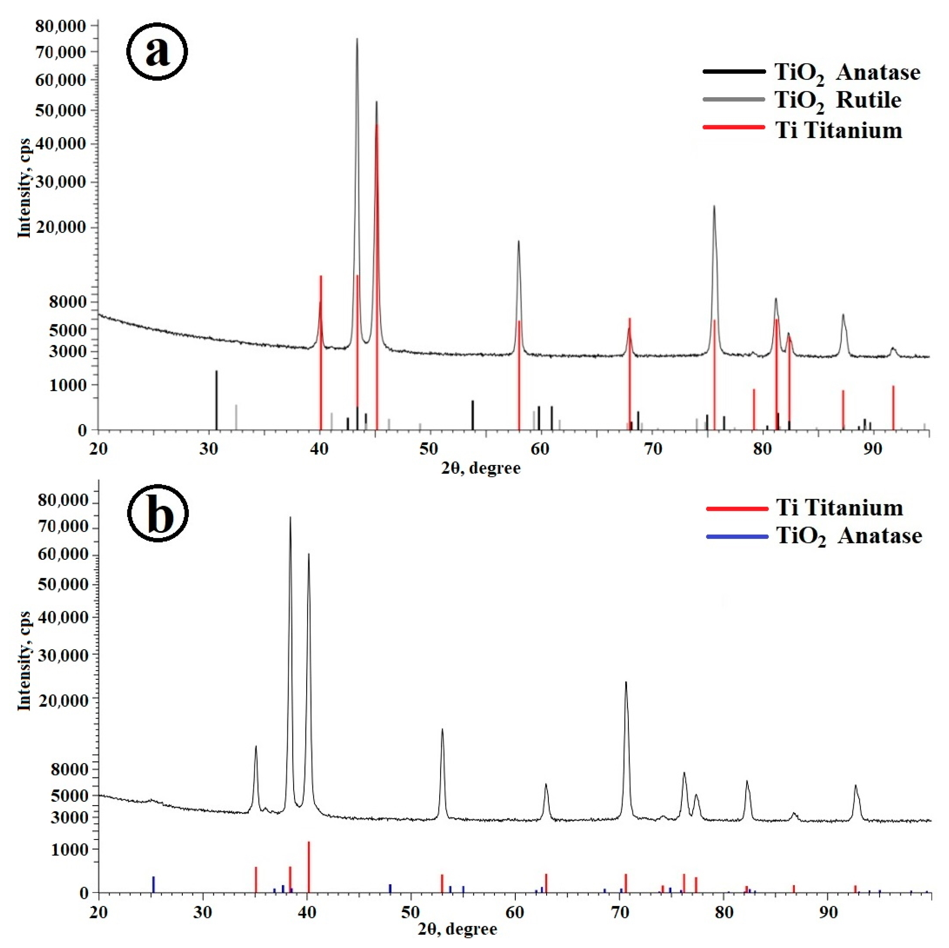
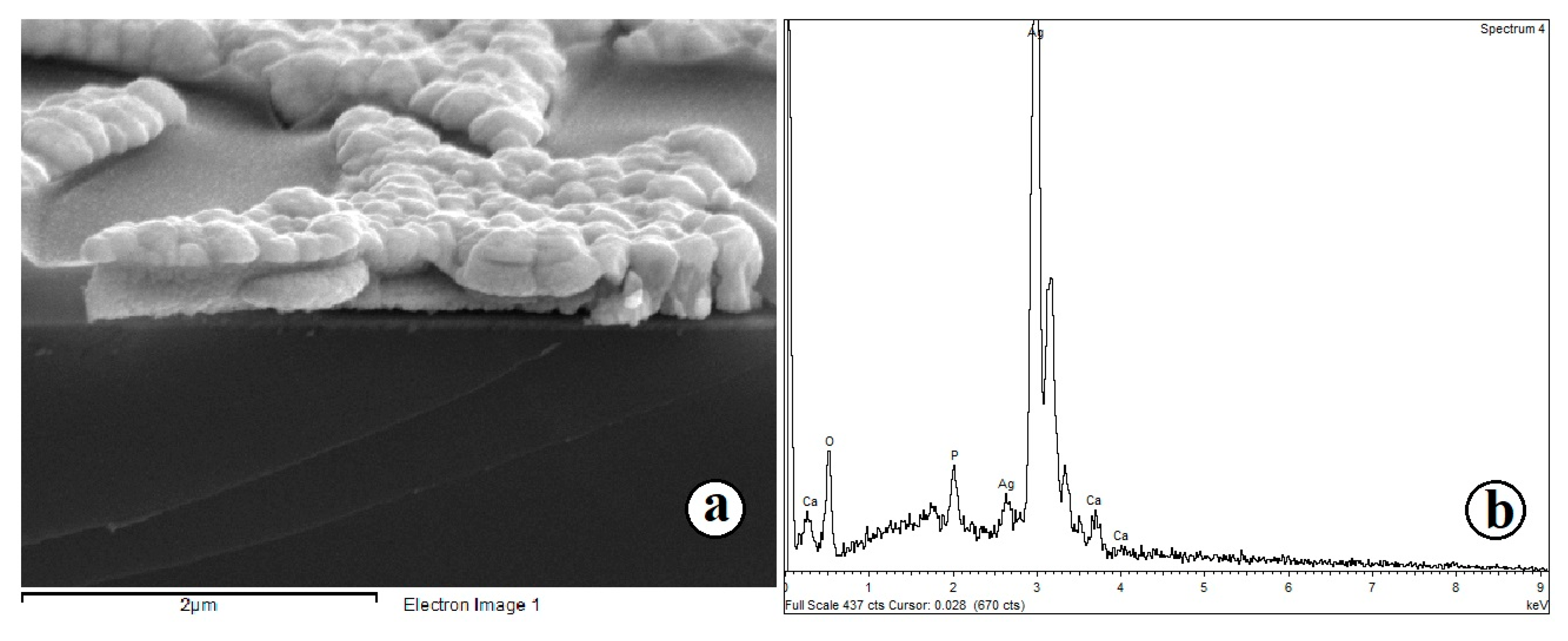
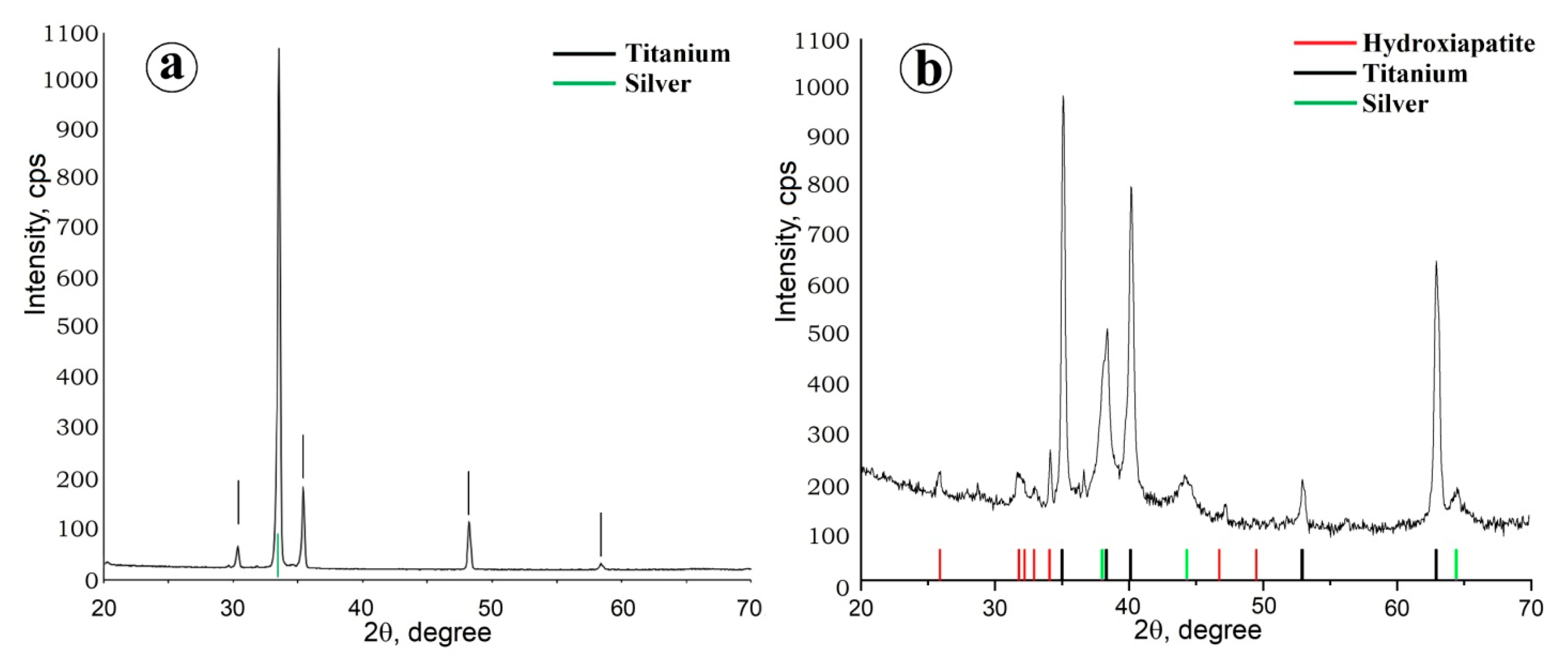
2.1. Preparation of Composite Coating TiO2/Ag/HAp on Titanium

During the deposition of the TiO2 template, post-synthetic heat treatment temperatures between 150 °C and 500 °C in 50 °C increments were tested. The optimal temperature for creating a perforated (insular) oxide coating is 400 °C. At this temperature, the perforation areas are commensurate with the osteoblast cells, which positively affects the rate of the osteoblast cell monolayer formation on the implant surface.
The structures were analyzed using SEM. Figure 1 shows a micrograph of a synthesized TiO2 film with the presence of micro- and nano-roughnesses. The two-level hierarchy, as noted above, significantly increases the bioactivity of the implant surface.
There are no anatase and rutile reflexes on the diffractogram of the resulting TiO2 coating (Figure 2a), which indicates the X-ray amorphousness of the deposited film. However, after 1 h calcination of a TiO2-coated sample at 400 °C, the film becomes polycrystalline. Anatase reflexes (Figure 2b) confirm the presence of the TiO2 phase in the sample.
At the second stage, a TiO2/Ag composite coating was obtained by electrochemical template synthesis using a TiO2 coating with insular perforations as a template.
Ag coatings can be synthesized on the implant surface by various methods, such as electroforming, vapor deposition, layer-by-layer deposition, vacuum plasma spraying, anodizing, electrochemical deposition [18,19,20,21,22,31], etc. In our work, the method of pulsed electrochemical deposition was used for Ag deposition. This method makes it possible to synthesize coatings with a uniform Ag particle distribution, high adhesion to the Ti surface and the ability to create an island-type coating.
When using pulsed current in template electrochemical synthesis, Ag is deposited on a Ti substrate, repeating the geometry of island perforations formed by the TiO2 film. Figure 3 shows the SEM micrograph.
The Ag deposit clearly reproduces the structure of the TiO2 coating. Cracks emanating from micron pores are also filled with Ag particles. Also, the TiO2/Ag composite was studied by X-ray spectral microanalysis (EDX) with an electron probe using an X-ray microanalysis attachment on a scanning electron microscope. It can be seen from the Ti and Ag element maps (Figure 3b) that Ag is mainly deposited in the perforation of the TiO2 film, repeating its geometry. This indicates the successful implementation of the template electrochemical synthesis of the Ag coating.
The presence of silver is also confirmed by X-ray phase analysis data (Figure 4).
At the third stage, an island-type TiO2/Ag/HAp composite coating was obtained by template electrochemical deposition of HAp on the composite TiO2/Ag template coating obtained at the second stage. Pulsed deposition provided the structure of an intermittent HAp layer on the sample surface and made it possible to obtain an island-type coating convenient for the adhesion of osteoblast bone cells.
Micrographs of the samples were obtained using SEM, and their element maps were compiled by X-ray spectral microanalysis with an electron probe (Figure 5).
Micrographs and element maps show that a high level of localization of calcium and phosphorus has been achieved, and the deposited HAp repeats the geometry of the TiO2/Ag template. Microscopy of the sample section showed that the resulting coating had a thickness of several hundred nanometers, and the HAp particles themselves rise above the surface of titanium, creating a micron roughness of the coating (Figure 6). This coating is also chemically close to the mineral composition of bone, which is important for bone implantation.
XRD confirms the crystal structure of the synthesized composite coating. The diffractogram (Figure 7) shows peaks of Ti substrate, silver (Ag microparticles forming the template structure) and HAp.

2.2. Cytological Studies of Samples

Proliferative activity (cytotoxicity) and adhesive properties of MG-63 osteoblast cells cultured on the surface of samples (Table 1) responsible for the formation of a cell monolayer on experimental samples was evaluated at the Institute of Cytology of the Russian Academy of Sciences (St. Petersburg).

2.2.1. MTT Test (Cytotoxicity Assessment)

The results of the evaluation of the proliferative activity of MG-63 cells cultured on the surface of the samples are shown in Figure 8. Based on these data, it is shown that when cells were grown on the surface of all the samples, their viability did not differ from the control during the entire period of joint incubation (48 h). The conclusion is made about the absence of cytotoxicity in the obtained composite coatings.
Based on the obtained microphotographs of samples with osteoblast cells on the surface (Figure 9), it is concluded that a sample with a composite coating is characterized not only by high adhesive properties of osteoblasts, but also by the ability to develop a continuous cell layer, which indicates the possibility of accelerated osteosynthesis on such a surface. This is due to the presence of a two-level composite coating structure formed by micron islands of hydroxyapatite and nanometer pores of TiO2 xerogel for better adhesion of osteoblast cells. In addition, based on micrographs, it can be concluded that the resulting composite coating has no cytotoxicity and has the capacity for of more than 85% in 24 h for MG-63 osteoblast cells to adhere to it.
Cell differentiation in the osteogenic direction was studied using markers of early (alkaline phosphatase—ALP) and late (osteopontin—OPN) differentiation [24,32].

2.2.2. Estimation of Alkaline Phosphatase (ALP) Content

During three weeks of differentiation of MG-63 cells cultured on the surface of samples (no. 1–4), the level of ALP activity in the conditioned medium was higher compared to the control values (cells culture under standard conditions) (Figure 10).
At the same time, a gradual increase in the level of ALP activity was observed during cell differentiation on the surface of two samples (no. 3 and 4), where ALP activity increased by 24 and 105%, respectively. The level of ALP activity in the medium obtained from control cells (culturing on plastic) was taken as 100%. When cells were cultured on the surface of samples no. 1 and 2, the level of ALP activity gradually decreased. For example, after the first week of cell differentiation on the surface of the sample no. 1, the values of the ALP activity level were 34% higher compared to the control, and then decreased and no longer had significant differences with the control (Table 1).
Thus, during the differentiation of MG-63 cells on the surface of the samples, there was an increase in the level of ALP activity compared to the control (cell culture on plastic), which indicated the influence of the nature of the surface treatment of the material on the ability of MG-63 cells to differentiate in the osteogenic direction. At the same time, the treatment (TiO2/Ag, TiO2/Ag/HAp coatings) of the surface of samples no. 3 and 4 had the most significant effect on the differentiation of the cell line.

2.2.3. Estimation of Osteopontin (OPN) Content

During three weeks of differentiation of MG-63 cells cultured on the surface of samples (no. 1–4), a gradual increase in OPN content was observed (Figure 11). The OPN concentration in the medium increased by 29 and 36% compared to the control after cell culture on the surface of samples no. 3 and 4, respectively. The OPN content in the medium obtained from control cells (culturing on plastic) was taken as 100%. When culturing cells on the surface of samples no. 1 and 2, no significant differences in the OPN content compared to the control were revealed.
In a series of in vitro experiments, we have shown that coatings with a complex hierarchical structure at the micro- and nanoscale and the composition of TiO2/Ag/HAp significantly enhanced the differentiation of osteoblasts in the osteogenic direction (Figure 10 and Figure 11) and contribute to improved implant engraftment to the bone.

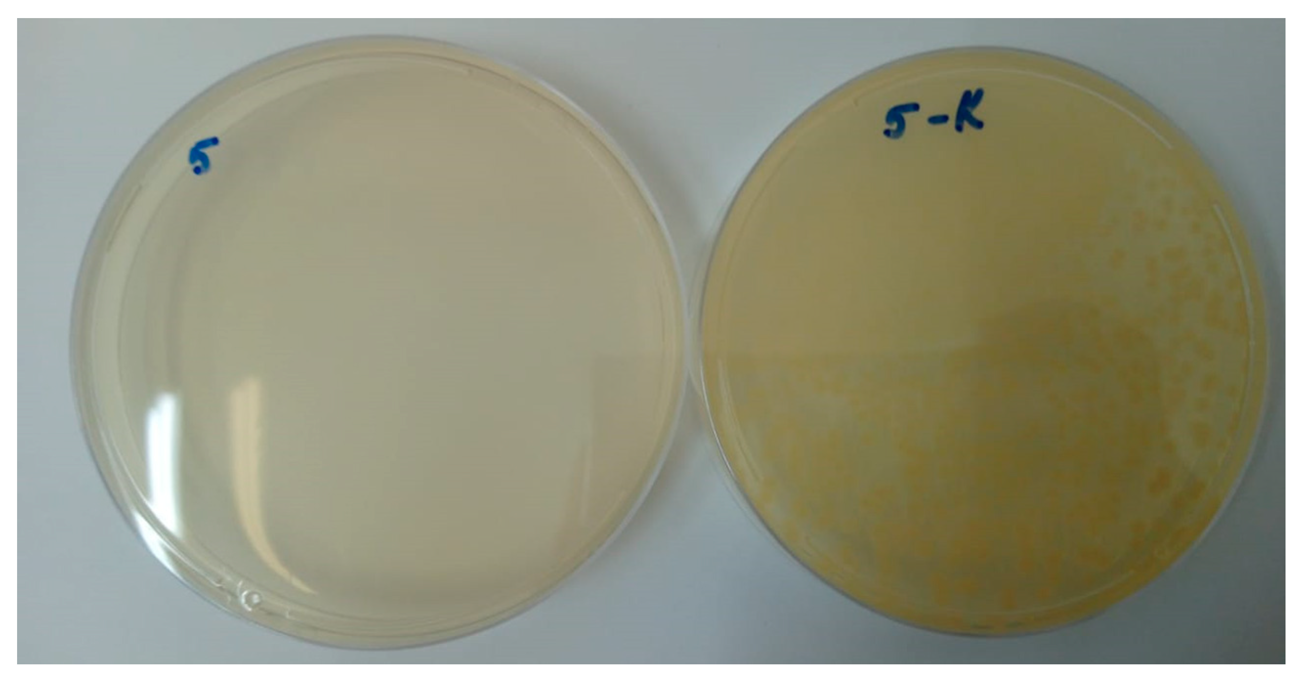
2.3. The Antibacterial Activity of Samples with a Composite Coating TiO2/Ag/HAp

The study was carried out according to ISO Standard 22196 “Measurement of antibacterial activity on the surface of plastics and other non-porous materials.”
Microorganism suspensions were prepared at 107 CFU/mL and 106 CFU/mL according to turbidity standard. Control solutions were sown in the amount of 1 mL on nutrient media, and excess liquid was taken out with a dispenser. Samples were placed in Petri dishes. The samples were incubated in a thermostat at 37 °C for 24 h, then stored until the results were obtained in a refrigerator at 2–4 °C. On the composite coating TiO2/Ag/HAp, microorganism suspensions were planted in the amount of 200 mL. A similar procedure was done with control plates. The samples were covered with a sterile film Parafilm M 40 × 40 mm using tweezers and incubated in a thermostat with a closed lid at 37 °C for 24 h.
Next, the films were transferred from the test samples to separate sterile test tubes containing 9 mL of 0.9% NaCl, with an exposure time of 1 h. After exposure, 1 mL of physiological solution was sown on nutrient media, and the excess liquid was taken out with a dispenser. The samples were incubated in a thermostat at 37 °C for 24 h. Colonies were counted and the results of the experiment were taken into account.
As a result of the study of samples for antibacterial activity, the presence of antibacterial properties of samples containing silver structures in the form of islands was confirmed, compared with samples containing only a layer of titanium oxide (Table 2).
It can be seen from the table that the growth of microorganisms was observed in the seeded control samples and in control samples without silver containing only the titanium oxide coating. In the composite coating of the island-type TiO2/Ag/HAp, there is a 100% absence of culture growth. In particular, Figure 12 shows a photograph illustrating a comparison of culture growth of the reference strain Pseudomonas aeruginosa ATCC 27,853 (no. 7) on the TiO2/Ag/HAp composite sample and the control sample. It can be seen that in the presence of Ag in the composition of the sample, there is a 100% absence of culture growth.
As a result of the study of samples for antibacterial activity, the presence of antibacterial properties of the samples was confirmed. The results of this study allow us to conclude that it is advisable to apply an insular composite silver coating on titanium implants.
The mechanisms of the antibacterial action of silver can be described in several ways [33]. One of them is based on the penetration of Ag+ ions into the peptidoglycan layer or bacterial wall, which leads to damage to DNA and bacterial proteins [34]. The formation of free radicals is also a possible pathway for the action of silver, since this destroys the cell membrane, changing its permeability, which ultimately leads to the death of the bacterium [15]. In addition, bacteria in their composition have sulfur and phosphorus, which are able to react with silver. Silver also disrupts the life cycle of bacteria [35].
Based on a large number of in vitro and in vivo studies and our experimental data, we came to the unequivocal opinion that for the fastest and most successful osseointegration of the implant, a developed relief is necessary both on the micro- and nanoscale scale [36,37,38]. Micron relief, and especially the presence of micropores, significantly improves the adhesion of bone tissue cells [38,39]. The presence of a specific nanorelief plays a significant role in improving circulation and accelerating the adsorption of biomolecules (proteins, nutrients) [38], and can also lead to the occurrence of an antibacterial effect [40,41]. To determine the success of the medical implant material, the composition of the surface is no less important a characteristic than the relief. Studies have shown that the composite coating of the TiO2/Ag/HAp composition with a developed surface relief at the micro- and nanoscale has a significant cytological response and the ability to accelerate osteosynthesis.

3. Materials and Methods

3.1. Preparation of TiO2/Ag/HAp Coating on Ti Surface

Firstly, TiO2 was coated on Ti as the template for further operations [32]. A technical Ti (VT1-6) plate (10 × 38 mm) was used with achieved roughness of <0.01 microns. A solution of absolute isopropanol (iPA), titanium tetraisopropoxide (TTIP), diethanolamine (DEA) and polyethylene glycol (PEG, Mw = 20,000 D, Merck, Rahway, NJ, USA) in the 773/227/105/50 ratio was used for the synthesis of the film. The coating was obtained by the sol-gel method using dip-coating technology using the equipment KSV Nima Dip Coater, Singlevessel. The extraction rate was 100 mm/min. Then the deposited film was heat treated at 400 °C.
Secondly, Ag was coated over the TiO2 film.
Silver was precipitated from an aqueous electrolyte containing AgNO3, sulfosalicylic acid, 25% aqueous ammonia to reach pH 9. Template deposition was carried out in a polypropylene cell with stirring, using a potentiostat-galvanostat Elins P45X. A Ti substrate coated with TiO2 was used as a working electrode, and silver was used as counter electrode.
The silver was deposited from the solution at pulsed current, since constant current gave insufficient repeatability of the template pattern without repeating the geometry of the perforations. Preliminarily, the optimal number of deposition cycles was determined. Each of 300 cycles included the impulse sequences 1 V—5 ms, 0.3 V—3 ms, 2 V—10 ms, 0 V—65 ms.
At the third stage, HAp structures were obtained on the surface of the TiO2/Ag composite coating as the template. As above, potentiostat-galvanostat Ellins P45X was used for electrochemical matrix synthesis (TiO2/Ag composite serves as cathode and matrix and graphite serves as anode). Based on HAp stoichiometry (Ca/P = 1.67), we used 1.18 g of Ca(NO3)2, (Vekton, Krasnodar, Russia) and 0.411 g of KH2PO4 (Vekton, Russia) per 100 mL of deionized water. HAp was deposited at pulsed current [30].

3.2. Evaluation of the Formation of a Cellular Monolayer of MC3T3-E1 Osteoblasts on Titanium Samples with Composite Coating TiO2/Ag/HAp

All samples were placed in Petri dishes and sterilized by ozonation. The suspension of the MS3T3-E1 cell line was applied in a small volume of nutrient medium (100 µL) in such a way that a “drop” formed on the surface of the sample. The concentration of cells during sowing was 1 × 10⁵/cm2 of the sample. Samples with a suspension of cells deposited on their surface in this way were placed in a CO2 incubator at a temperature of +37 °C for 3 h. After this time, during which cell adhesion on the surface of the samples is supposed to occur, a nutrient medium was added to the Petri dishes. As a control, the application of a suspension of cells on the surface of cultural dishes was used.
The assessment of the state of cells (the nature of adhesion and spreading of cells on the surface of the samples) was performed by the SEM method using a JSM-35.7 microscope (JEOL, Tokyo, Japan).

3.3. MTT Test (Cytotoxicity Assessment)

Titanium samples were sterilized under ozonation conditions, then incubated in 1 mL of nutrient medium for 24 h at 37 °C. Two samples were used for each variation. A suspension of MG-63 cells with the same concentration (5000 cells/well) was sifted into the wells of a 96-well board (Nunc, Waltham, MA, USA). The number of repetitions n = 5. After cell adhesion, the medium was changed to the medium obtained after the samples’ incubation. The cytotoxicity of the material on the cells was evaluated after 24 h using a Vibrant MTT Cell Proliferation Assay Kit (Life Technologies, Carlsbad, CA, USA) according to the manufacturer’s protocol.

3.4. Evaluation of Cell Differentiation in the Osteogenic Direction

Ti samples were sterilized using ozonation. The samples were placed in a Petri dish (Nunc, USA), and a suspension of MG-63 cells with a concentration of 1 × 10⁵ in 20 µL of culture medium DMEM/F12 containing 10% fetal cow serum and gentamicin (Gibco, Waltham, MA, USA) was applied to their surface (on the central part). After 2 h of incubation, the cells were adhered to the surface of the samples, then 180 µL of nutrient medium was added to each sample so that the cells on the surface of the samples were completely covered with the medium.
Cells were cultured on the surface of the samples in a CO2 incubator for 3 weeks with a change of medium once a week to a medium that induces differentiation (StemPro Osteogenesis Differentiation Kit, Thermofisher Scientific, Waltham, MA, USA). Standard conditions of cell culture on the surface of culture plastic were used as a control. At each change of medium, a cell-conditioned medium was sampled, which was placed at −80 °C for subsequent assessment of the presence of alkaline phosphatase (ALP) and the content of osteopontin in it using a Human ALP and SSP1 (Osteopontin) ELISA Kit in accordance with the manufacturer’s protocol. The measurements were carried out on a multimodal Thermo Scientific Varioskan LUX reader with a wavelength of 450 nm.

3.5. Studies of the Antibacterial Activity of the Surface of the TiO2/Ag/HAp Composite Coating

During the experiment, titanium plates covered with silver structures as well as pristine titanium samples were examined. The samples were not subjected to additional processing, including sterilization.
For the study, daily cultures of reference strains were used, in the amount of 106 and 107 CFU/mL: Enterococcus faecalis ATCC 29812, Staphylococcus aureus ATCC 25912, Klebsiella pneumoniae ATCC 19882, Acinetobacter baumannii 987, Pseudomonas aeruginosa ATCC 27853.
The controls were taken: (1) seeding of a suspension of microorganisms in the amount of 106 CFU/mL on a Muller-Hinton agar for the reliability of the contamination of the suspension; and (2) Ti plates without Ag coating to confirm the negative antibacterial effect of samples on microorganisms in the absence of Ag.
To assess the presence/absence of microbial growth, the quantitative growth of bacteria on a dense nutrient medium was studied after sowing suspensions on agar from samples without silver after 24 h of incubation. The experiment was repeated twice, and the results are reproducible without errors.
This study was conducted according to ISO Standard 22196 “Measurement of antibacterial activity on the surface of plastics and other non-porous materials”.

3.6. Characterization of the Composite Coating TiO2/Ag/HAp

The structure and morphology of the coatings were studied using a Zeiss Merlin scanning electron microscope (Carl Zeiss Microscopy GmbH, Jena, Germany). For X-ray spectral microanalysis and the construction of element maps, we used the EDX console (Oxford Instruments INCAx-act) for a Zeiss Merlin scanning microscope. The MiniFlex II X-ray diffractometer (Rigaku Corporation, Japan) was used for X-ray phase analysis (λ = 1.5406 Å).

4. Conclusions

In the course of this work, a technique was developed for electrochemical template synthesis of a TiO2/Ag composite coating obtained by sequential application of the sol-gel method using dip-coating technology and electrochemical deposition. The resulting coating has a two-level orderliness of the relief, consisting of micro- and nano-irregularities, and due to this increase, the bioactivity of the implant, since it improves adhesion and promotes an accelerated process of osteoblast differentiation. It was also shown that the combined approach used to obtain coatings based on the sol-gel technology and electrochemical deposition method is not cytotoxic for osteoblast-like cells. The study of coatings containing a deposited layer of silver in the form of islands has shown that the preservation of the antibacterial effect of such coatings also leads to a reduction in the period of implant engraftment. Cytological studies of the obtained TiO2/Ag/HAp coatings have shown that coatings of this composition lead to accelerated osteosynthesis and are promising in the development of implantation products in dentistry and orthopedics.

Author Contributions

Conceptualization, E.G.Z. and V.M.S.; investigation, E.G.Z., L.A.K. (Lada A. Kozlova), N.M.Y., A.Y.A., D.N.S., A.N.P., L.A.K. (Ludmila A. Kraeva) and E.V.R.; writing—original draft preparation, E.G.Z. and V.M.S.; writing—review and editing, E.G.Z. and P.M.K.; project administration, E.G.Z.; funding acquisition, E.G.Z. All authors have read and agreed to the published version of the manuscript.

Funding

The research was conducted under the financial support the Russian Science Foundation grant (project No. 22-21-00573).

Institutional Review Board Statement

Not applicable.

Informed Consent Statement

Not applicable.

Data Availability Statement

Not applicable.

Acknowledgments

The research was carried out using the equipment of the resource centers of the St. Petersburg State University Scientific Park “X-ray Diffraction research methods” and “Nanotechnology”.

Conflicts of Interest

The authors declare no conflict of interest.

Sample Availability

Samples of the compounds are not available from the authors.

References

  1. Murr, L.E. Strategies for creating living, additively manufactured, open-cellular metal and alloy implants by promoting osseointegration, osteoinduction and vascularization: An overview. J. Mater. Sci. Technol. 2019, 35, 231–241. [Google Scholar] [CrossRef]
  2. Oliver, J.N.; Su, Y.; Lu, X.; Kuo, P.H.; Du, J.; Zhu, D. Bioactive glass coatings on metallic implants for biomedical applications. Bioact. Mater. 2019, 4, 261–270. [Google Scholar] [CrossRef]
  3. Gulati, K.; Maher, S.; Findlay, D.M.; Losic, D. Titania nanotubes for orchestrating osteogenesis at the bone-implant interface. Nanomedicine 2016, 11, 1847–1864. [Google Scholar] [CrossRef]
  4. Mendonca, G.; Mendonca, D.B.; Aragao, F.J.; Cooper, L.F. Advancing dental implant surface technology--from micron- to nanotopography. Biomaterials 2008, 29, 3822–3835. [Google Scholar] [CrossRef] [PubMed]
  5. Zemtsova, E.G.; Yudintceva, N.M.; Morozov, P.E.; Valiev, R.Z.; Smirnov, V.M.; Shevtsov, M.A. Improved osseointegration properties of hierarchical microtopographic/nanotopographic coatings fabricated on titanium implants. Int. J. Nanomed. 2018, 13, 2175–2188. [Google Scholar] [CrossRef] [PubMed]
  6. Coelho, P.G.; Jimbo, R.; Tovar, N.; Bonfante, E.A. Osseointegration: Hierarchical designing encompassing the macrometer, micrometer, and nanometer length scales. Dent. Mater. 2015, 31, 37–52. [Google Scholar] [CrossRef] [PubMed]
  7. Wennerberg, A.; Albrektsson, T. On implant surfaces: A review of current knowledge and opinions. Int. J. Oral. Maxillofac. Implant. 2010, 25, 63–74. [Google Scholar]
  8. Arbenin, A.Y.; Zemtsova, E.G.; Orekhov, E.V.; Smirnov, V.M. Features of the sol-gel synthesis of TiO2 nanolayers with calcium phosphate structures on titanium surface. Russ. J. Gen. Chem. 2017, 87, 340–341. [Google Scholar] [CrossRef]
  9. Nazarov, D.V.; Smirnov, V.M.; Zemtsova, E.G.; Yudintceva, N.M.; Shevtsov, M.A.; Valiev, R.Z. Enhanced Osseointegrative Properties of Ultra-Fine-Grained Titanium Implants Modified by Chemical Etching and Atomic Layer Deposition. ACS Biomater. Sci. Eng. 2018, 4, 3268–3281. [Google Scholar] [CrossRef]
  10. Gittens, R.A.; McLachlan, T.; Olivares-Navarrete, R.; Cai, Y.; Berner, S.; Tannenbaum, R.; Schwartz, Z.; Sandhage, K.H.; Boyan, B.D. The effects of combined micron-/submicron-scale surface roughness and nanoscale features on cell proliferation and differentiation. Biomaterials 2011, 32, 3395–3403. [Google Scholar] [CrossRef]
  11. Meirelles, L.; Uzumaki, E.T.; Lima, J.H.; Muller, C.A.; Albrektsson, T.; Wennerberg, A.; Lambert, C.S. A novel technique for tailored surface modification of dental implants—A step wise approach based on plasma immersion ion implantation. Clin. Oral. Implant. Res. 2013, 24, 461–467. [Google Scholar] [CrossRef] [PubMed]
  12. Koo, H.; Allan, R.N.; Howlin, R.P.; Stoodley, P.; Hall-Stoodley, L. Targeting microbial biofilms: Current and prospective therapeutic strategies. Nat. Rev. Microbiol. 2017, 15, 740–755. [Google Scholar] [CrossRef] [PubMed]
  13. Singh, A.; Dubey, A.K. Various Biomaterials and Techniques for Improving Antibacterial Response. ACS Appl. Bio Mater. 2018, 1, 3–20. [Google Scholar] [CrossRef]
  14. Saidin, S.; Jumat, M.A.; Mohd Amin, N.A.A.; Saleh Al-Hammadi, A.S. Organic and inorganic antibacterial approaches in combating bacterial infection for biomedical application. Mater. Sci. Eng. C-Mater. Biol. Appl. 2021, 118, 111382. [Google Scholar] [CrossRef] [PubMed]
  15. Campoccia, D.; Montanaro, L.; Arciola, C.R. A review of the biomaterials technologies for infection-resistant surfaces. Biomaterials 2013, 34, 8533–8554. [Google Scholar] [CrossRef]
  16. Tian, B.; Chen, W.; Yu, D.; Lei, Y.; Ke, Q.; Guo, Y.; Zhu, Z. Fabrication of silver nanoparticle-doped hydroxyapatite coatings with oriented block arrays for enhancing bactericidal effect and osteoinductivity. J. Mech. Behav. Biomed. Mater. 2016, 61, 345–359. [Google Scholar] [CrossRef]
  17. Zhang, Y.Y.; Zhu, Y.; Lu, D.Z.; Dong, W.; Bi, W.J.; Feng, X.J.; Wen, L.M.; Sun, H.; Qi, M.C. Evaluation of osteogenic and antibacterial properties of strontium/silver-containing porous TiO2 coatings prepared by micro-arc oxidation. J. Biomed. Mater. Res. B Appl. Biomater. 2021, 109, 505–516. [Google Scholar] [CrossRef]
  18. Lu, X.; Zhang, B.; Wang, Y.; Zhou, X.; Weng, J.; Qu, S.; Feng, B.; Watari, F.; Ding, Y.; Leng, Y. Nano-Ag-loaded hydroxyapatite coatings on titanium surfaces by electrochemical deposition. J. R. Soc. Interface 2011, 8, 529–539. [Google Scholar] [CrossRef]
  19. Morris, G.V.; Kozdryk, J.; Gregory, J.; Jeys, L. The use of silver-coated orthopaedic implants: Are all silvers the same? Curr. Orthop. Pract. 2017, 28, 532–536. [Google Scholar] [CrossRef]
  20. Liang, R.; Xu, Y.; Zhao, M.; Han, G.; Li, J.; Wu, W.; Dong, M.; Yang, J.; Liu, Y. Properties of silver contained coatings on CoCr alloys prepared by vacuum plasma spraying. Mater. Sci. Eng. C—Mater. Biol. Appl. 2020, 106, 110156. [Google Scholar] [CrossRef]
  21. He, Y.; Kang, Z.; Chen, D. Integrating nanodiamonds to improve anticorrosion property and conductivity simultaneously of Ag composite films via layer by layer spin-spray deposition. Appl. Surf. Sci. 2020, 505, 144643. [Google Scholar] [CrossRef]
  22. Varghese, S.; Elfakhri, S.; Sheel, D.W.; Sheel, P.; Bolton, F.J.; Foster, H.A. Novel antibacterial silver-silica surface coatings prepared by chemical vapour deposition for infection control. J. Appl. Microbiol. 2013, 115, 1107–1116. [Google Scholar] [CrossRef]
  23. Fu, C. Synthesis and Characterization of Hydroxyapatite Coatings for Medical Applications. Ph.D. Thesis, University of Rochester, Rochester, NY, USA, 2016. [Google Scholar]
  24. Chatakun, P.; Nunez-Toldra, R.; Diaz Lopez, E.J.; Gil-Recio, C.; Martinez-Sarra, E.; Hernandez-Alfaro, F.; Ferres-Padro, E.; Giner-Tarrida, L.; Atari, M. The effect of five proteins on stem cells used for osteoblast differentiation and proliferation: A current review of the literature. Cell Mol. Life Sci. 2014, 71, 113–142. [Google Scholar] [CrossRef]
  25. Shi, Z.; Huang, X.; Cai, Y.; Tang, R.; Yang, D. Size effect of hydroxyapatite nanoparticles on proliferation and apoptosis of osteoblast-like cells. Acta Biomater. 2009, 5, 338–345. [Google Scholar] [CrossRef] [PubMed]
  26. Yan, Y.; Zhang, X.; Huang, Y.; Ding, Q.; Pang, X. Antibacterial and bioactivity of silver substituted hydroxyapatite/TiO 2 nanotube composite coatings on titanium. Appl. Surf. Sci. 2014, 314, 348–357. [Google Scholar] [CrossRef]
  27. Pratap Reddy, M.; Venugopal, A.; Subrahmanyam, M. Hydroxyapatite-supported Ag-TiO2 as Escherichia coli disinfection photocatalyst. Water Res. 2007, 41, 379–386. [Google Scholar] [CrossRef] [PubMed]
  28. Tian, B.; Chen, W.; Dong, Y.; Marymont, J.V.; Lei, Y.; Ke, Q.; Guo, Y.; Zhu, Z. Silver nanoparticle-loaded hydroxyapatite coating: Structure, antibacterial properties, and capacity for osteogenic induction in vitro. RSC Adv. 2016, 6, 8549–8562. [Google Scholar] [CrossRef]
  29. Arbenin, A.Y.; Zemtsova, E.G.; Ermakov, S.S.; Gaskov, A.M.; Baburova, P.I.; Sokolova, D.N.; Yaroshenko, S.V.; Smirnov, V.M. Three-component working electrode micron-sized Ag particles/TiO2 layer/Ti: Template electrochemical synthesis and potential use as electrochemical sensor for glutathione detection. Mater. Res. Express 2020, 7, 035401. [Google Scholar] [CrossRef]
  30. Orekhov, E.V.; Arbenin, A.Y.; Zemtsova, E.G.; Sokolova, D.N.; Ponomareva, A.N.; Shevtsov, M.A.; Yudintceva, N.M.; Smirnov, V.M. Template Electrochemical Synthesis of Hydroxyapatite on a Titania–Silver Composite Surface for Potential Use in Implantology. Coatings 2022, 12, 266. [Google Scholar] [CrossRef]
  31. Soo, J.Z.; Chai, L.C.; Ang, B.C.; Ong, B.H. Enhancing the Antibacterial Performance of Titanium Dioxide Nanofibers by Coating with Silver Nanoparticles. ACS Appl. Nano Mater. 2020, 3, 5743–5751. [Google Scholar] [CrossRef]
  32. Wennerberg, A.; Albrektsson, T.; Jimbo, R. Implant Surfaces and Their Biological and Clinical Impact, 1st ed.; Springer: Berlin/Heidelberg, Germany, 2015; p. 182. [Google Scholar]
  33. Prabhu, S.; Poulose, E.K. Silver nanoparticles: Mechanism of antimicrobial action, synthesis, medical applications and toxicity effects. Int. Nano Lett. 2012, 2, 32. [Google Scholar] [CrossRef] [Green Version]
  34. Chaloupka, K.; Malam, Y.; Seifalian, A.M. Nanosilver as a new generation of nanoproduct in biomedical applications. Trends Biotechnol. 2010, 28, 580–588. [Google Scholar] [CrossRef] [PubMed]
  35. Morones, J.R.; Elechiguerra, J.L.; Camacho, A.; Holt, K.; Kouri, J.B.; Ramírez, J.T.; Yacaman, M.J. The bactericidal effect of silver nanoparticles. Nanotechnology 2005, 16, 2346–2353. [Google Scholar] [CrossRef]
  36. Jones, F. Teeth and bones: Applications of surface science to dental materials and related biomaterials. Surf. Sci. Rep. 2001, 42, 75–205. [Google Scholar] [CrossRef]
  37. Li, B.E.; Li, Y.; Min, Y.; Hao, J.Z.; Liang, C.Y.; Li, H.P.; Wang, G.C.; Liu, S.M.; Wang, H.S. Synergistic effects of hierarchical hybrid micro/nanostructures on the biological properties of titanium orthopaedic implants. RSC Adv. 2015, 5, 49552–49558. [Google Scholar] [CrossRef]
  38. Jiang, N.; Zhu, S.; Li, J.; Zhang, L.; Liao, Y.; Hu, J. Development of a novel biomimetic micro/nano-hierarchical interface for enhancement of osseointegration. RSC Adv. 2016, 6, 49954–49965. [Google Scholar] [CrossRef]
  39. Seddiki, O.; Harnagea, C.; Levesque, L.; Mantovani, D.; Rosei, F. Evidence of antibacterial activity on titanium surfaces through nanotextures. Appl. Surf. Sci. 2014, 308, 275–284. [Google Scholar] [CrossRef]
  40. Zemtsova, E.G.; Orehhov, J.V.; Arbenin, A.Y.; Valiev, R.Z.; Smirnov, V.M. The creation of nanocoatings of various morphology on the basis of titanium dioxide on a titanium matrix for bone implant. Mater. Phys. Mech. 2016, 29, 138–144. [Google Scholar]
  41. Variola, F.; Zalzal, S.F.; Leduc, A.; Barbeau, J.; Nanci, A. Oxidative nanopatterning of titanium generates mesoporous surfaces with antimicrobial properties. Int. J. Nanomed. 2014, 9, 2319–2325. [Google Scholar] [CrossRef] [Green Version]
Figure 1. SEM images of TiO2 film: (a) with micro-roughness; and (b) with nanopores.
Molecules 28 01416 g001
Figure 2. XRD patterns of the sample with a TiO2 coating: (a) before calcination; and (b) after calcination at 400 °C.
Molecules 28 01416 g002
Figure 3. SEM image of (a) the TiO2/Ag composite; and (b) the element maps of the Ti and Ag in composite.
Molecules 28 01416 g003
Figure 4. XRD patterns of the sample coated with TiO2/Ag.
Molecules 28 01416 g004
Figure 5. SEM image and element maps of Ti, Ag, Ca, P for TiO2/Ag/HAp composite.
Molecules 28 01416 g005
Figure 6. (a) SEM image of the chip of the sample with a HAp microparticle array obtained with template electrochemical synthesis; and (b) the EDX spectrum recorded at the corresponding points sample with a HAp.
Molecules 28 01416 g006
Figure 7. XRD patterns of the composite coating TiO2/Ag/HAp.
Molecules 28 01416 g007
Figure 8. Proliferation activity of MG-63 cells on the samples.
Molecules 28 01416 g008
Figure 9. SEM images for the samples after adhesion and spreading of the human MG-63 cell line: (a) on polished titanium; and (b) on the sample with the TiO2/Ag/HAp coating.
Molecules 28 01416 g009
Figure 10. Changes in the level of alkaline phosphatase activity during differentiation of the MG-63 line in the osteogenic direction. (Note: Control in vitro is the cultivation of cells on the surface of plastic. The level of ALP activity in the medium from control cells was taken as 100% [cultivation on plastic].)
Molecules 28 01416 g010
Figure 11. The content of osteopontin in the medium conditioned by MG-63 cells during differentiation in the osteogenic direction. (Note: Control is the cultivation of cells on the surface of plastic. The level of osteopontin in the medium from control cells was taken as 100% [cultivation on plastic].)
Molecules 28 01416 g011
Figure 12. Culture of reference strain no. 7: Pseudomonas aeruginosa ATCC 27,853 on the sample with TiO2/Ag/HAp composite coating (left) and on a control sample (right).
Molecules 28 01416 g012
Table 1. Samples for cytological studies.
Sample 1Sample 2Sample 3Sample 4
Polished
VT 6 titanium
TiO2 on
VT 6 titanium
TiO2/Ag on
VT 6 titanium
TiO2/Ag/HAp on
VT 6 titanium
Table 2. Studies of the antibacterial activity of TiO2/Ag/HAp composite coating.
Sample Composition
Cultures of StrainsControl Muller—Hinton AgarControl TiO2TiO2/Ag
Coating
TiO2/Ag/HAp Composite Coating
1. Enterococcus faecalis 812
(107 CFU/mL)
Microorganisms growthMicroorganisms growth100%
no growth
100%
no growth
2. Staphylococcus aureus
ATCC 25,912 (107 CFU/mL)
Microorganisms growthMicroorganisms growth100%
no growth
100%
no growth
3. Klebsiella pneumoniae
ATCC 19,882 (107 CFU/mL)
Microorganisms growthMicroorganisms growth100%
no growth
100%
no growth
4. Acinetobacter baumannii
987 (106 CFU/mL)
Microorganisms growthMicroorganisms growth100%
no growth
100%
no growth
5. Acinetobacter baumannii
987 (107 CFU/mL)
Microorganisms growthMicroorganisms growth100%
no growth
100%
no growth
6. Pseudomonas aeruginosa
ATCC 27,853 (106 CFU/mL)
Microorganisms growthMicroorganisms growth100%
no growth
100%
no growth
7. Pseudomonas aeruginosa
ATCC 27,853 (107 CFU/mL)
Microorganisms growthMicroorganisms growth100%
no growth
100%
no growth
Disclaimer/Publisher’s Note: The statements, opinions and data contained in all publications are solely those of the individual author(s) and contributor(s) and not of MDPI and/or the editor(s). MDPI and/or the editor(s) disclaim responsibility for any injury to people or property resulting from any ideas, methods, instructions or products referred to in the content.

