Article

In Situ Synthesis of Bi2S3/BiFeO3 Nanoflower Hybrid Photocatalyst for Enhanced Photocatalytic Degradation of Organic Pollutants

1
College of Environment Science and Engineering, Guilin University of Technology, Guilin 541004, China
2
Key Laboratory of Jiangxi Province for Persistent Pollutants Control and Resources Recycle, Nanchang Hangkong University, Nanchang 330063, China
3
College of Environmental Sciences, Sichuan Agricultural University, Chengdu 611130, China
*
Authors to whom correspondence should be addressed.
Molecules 2023, 28(24), 8007; https://doi.org/10.3390/molecules28248007
Submission received: 16 October 2023 / Revised: 29 November 2023 / Accepted: 30 November 2023 / Published: 8 December 2023
(This article belongs to the Topic Advanced Oxidation Processes for Wastewater Purification)

Abstract

:
Photocatalytic degradation of Malachite Green oxalate (MG) in a water body is of significant importance to our health protection, as it could cause various serious diseases. However the photocatalytic activity of most catalysts is still unsatisfactory, due to the poor reactive oxygen species production as a result of sluggish charge separation. Here, innovative nanoflower-shaped Bi2S3/BiFeO3 heterojunctions are prepared via a facile sol–gel method, exhibiting an enhanced reactive oxygen species generation, which leads to the excellent photocatalytic performance toward MG degradation. We verify that interfacing BiFeO3 with Bi2S3 could form a fine junction and offers a built-in field to speed up charge separation at the junction area; as a result, this shows much higher charge separation efficiency. By virtue of the aforementioned advantages, the as-prepared Bi2S3/BiFeO3 heterojunctions exhibit excellent photocatalytic performance toward MG degradation, where more than 99% of MG is removed within 2 h of photocatalysis. The innovative design of nanoflower-like Bi2S3/BiFeO3 heterojunctions may offer new viewpoints in designing highly efficient photocatalysts for environmentally related applications.

1. Introduction

In recent years, the world has been grappling with escalating energy crises and environmental pollution issues, particularly the pervasive presence of organic pollutants that significantly affect people’s daily lives. As the organic pollutants in water bodies are mostly highly toxic and may cause various serious diseases, effectively eliminating harmful organic pollutants from the environment has become a paramount concern for the scientific community. As a safe and sustainable way for organic pollutant removal, photocatalytic degradation of organics following a radical oxidative manner in recent years has been intensively studied. The key to achieving highly efficient organic pollutant removal from a water body lies in the development of a cost-effective photocatalyst with high reactive radical generation efficiency [1,2]. Unfortunately, most widely used photocatalysts primarily respond to UV–visible irradiation, limiting their practicality under visible light. Consequently, the quest for highly efficient and stable visible-light-driven photocatalysts has emerged as a hot topic in the field of photocatalysis.
Recently, BiFeO3 photocatalysts have garnered attention, due to their lower energy bandgap, excellent chemical stability at room temperature, and superior carrier transport properties. BiFeO3 holds promise for catalytic applications under visible light. Research has shown that BiFeO3, with a bandgap of approximately 2.2 eV [3,4], exhibits a favorable response to visible light and has been employed for the optical degradation of organic pollutants in water, including the degradation of organic dyes [5,6,7,8,9,10,11,12,13]. To further enhance the photocatalytic activity of BiFeO3, researchers have explored various modification techniques. Common approaches include element doping [14,15,16], which can boost visible-light-driven photocatalytic activity but often fails to effectively suppress the recombination of photogenerated electron–hole pairs, resulting in limited solar energy utilization. Some scholars have also experimented with noble metal deposition methods [17,18]; however, the high cost of precious metals makes these approaches less practical for real-world applications. Additionally, these modifications require further optimization to achieve better responsiveness to the visible light spectrum. As a result, researchers have turned to the incorporation of various materials into BiFeO3 to form composite heterojunction structures, such as CuO [19], TiO2 [20,21], and graphene oxide (GO) [22,23], among others. These heterojunction structures have been effective in enhancing visible light photocatalytic activity by mitigating electron–hole-pair recombination. However, these heterojunctions still lack optimal reducibility and oxidizability for photogenerated electron–hole pairs.
Hence, the search for a new semiconductor material to create heterojunctions with stronger reducibility and oxidizability is imperative. Additionally, since BiFeO3 nanoparticles possess a limited specific surface area, finding a substance capable of effectively increasing its surface area is crucial for enhancing photocatalytic reactions. Based on these considerations, our research aims to improve the reducibility and oxidizability of BiFeO3 through the formation of heterojunction structures with other materials, while simultaneously increasing its surface area. Achieving this under economical, simple, and mild reaction conditions has been a primary objective in our field. Our investigation revealed that Bi2S3, with its narrow band gap (Eg = 1.38–1.71 eV) [24] and the ability to adjust heterojunction morphology, is an ideal material for combining with BiFeO3. While previous studies have explored the potential of Bi2S3 as a visible-light-driven photocatalyst in conjunction with other photocatalysts, such as BiOCl [25,26], ZnO [27], TiO2 [28], CdS [29], WO3 [30], BiFeO3/α-Fe2O3 [31], LaxBa1−xSryCd1−yO3±δ [32], Bi2WO6 [33], BiVO4 [34], and Bi2O3 [35], no reports exist on the Bi2S3/BiFeO3 heterojunction photocatalysis.
In this study, we employed the in situ growth method to synthesize a novel Bi2S3/BiFeO3 nanoflower structure, demonstrating enhanced photocatalytic performance under visible light irradiation. We employed MG as a model contaminant to evaluate the material’s efficiency. Furthermore, we comprehensively investigated the structure, composition, morphology, and mechanism of this novel photocatalyst.

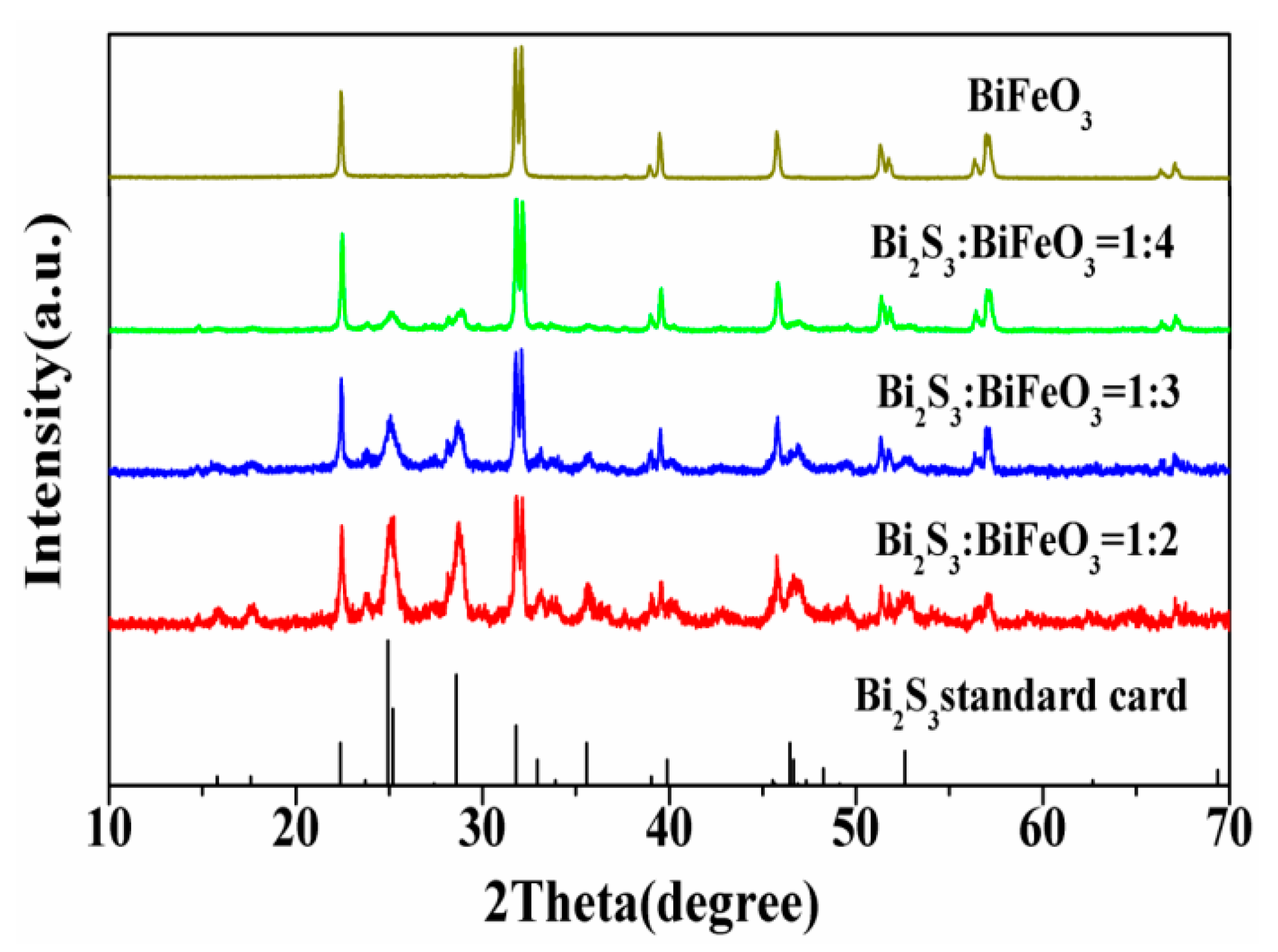
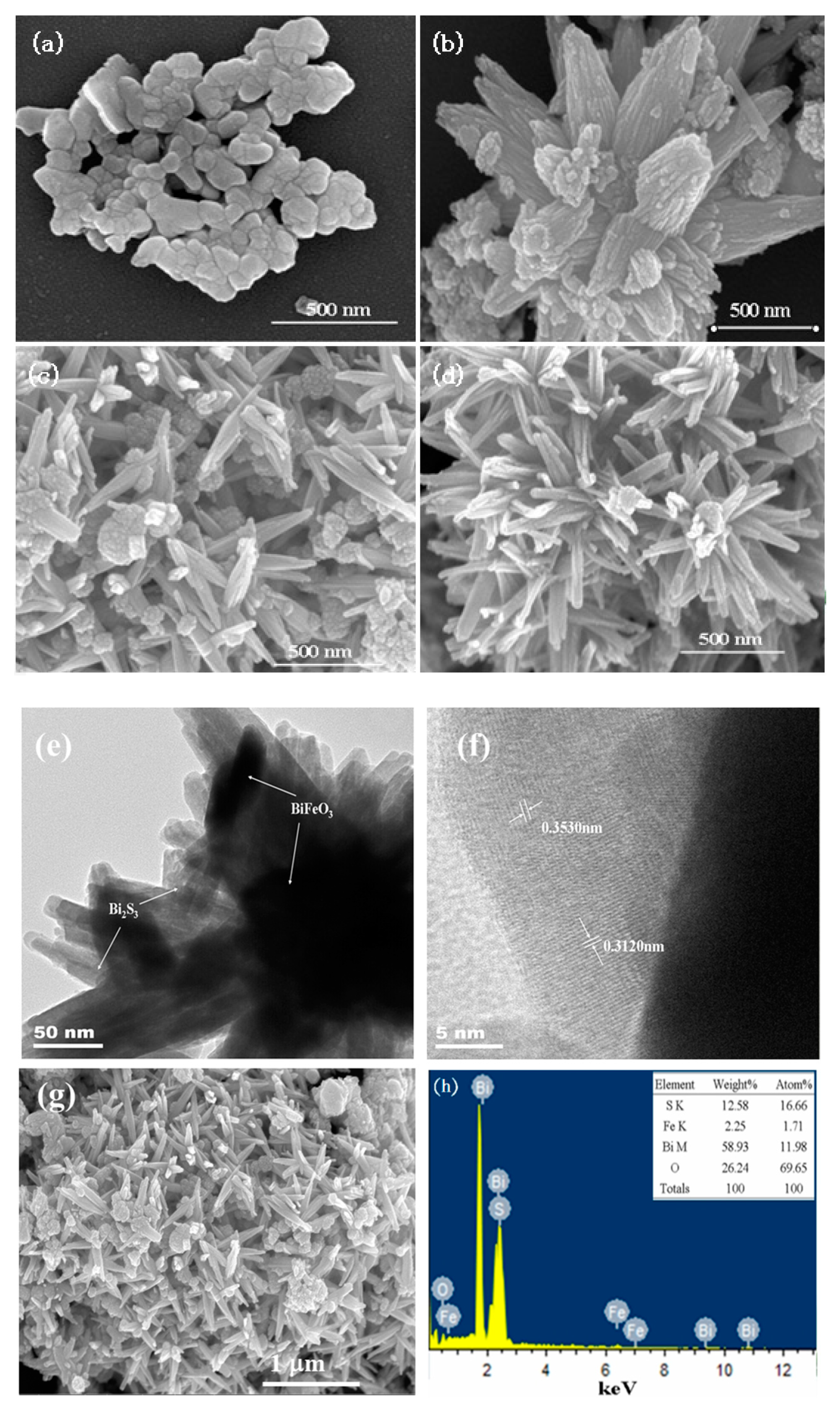
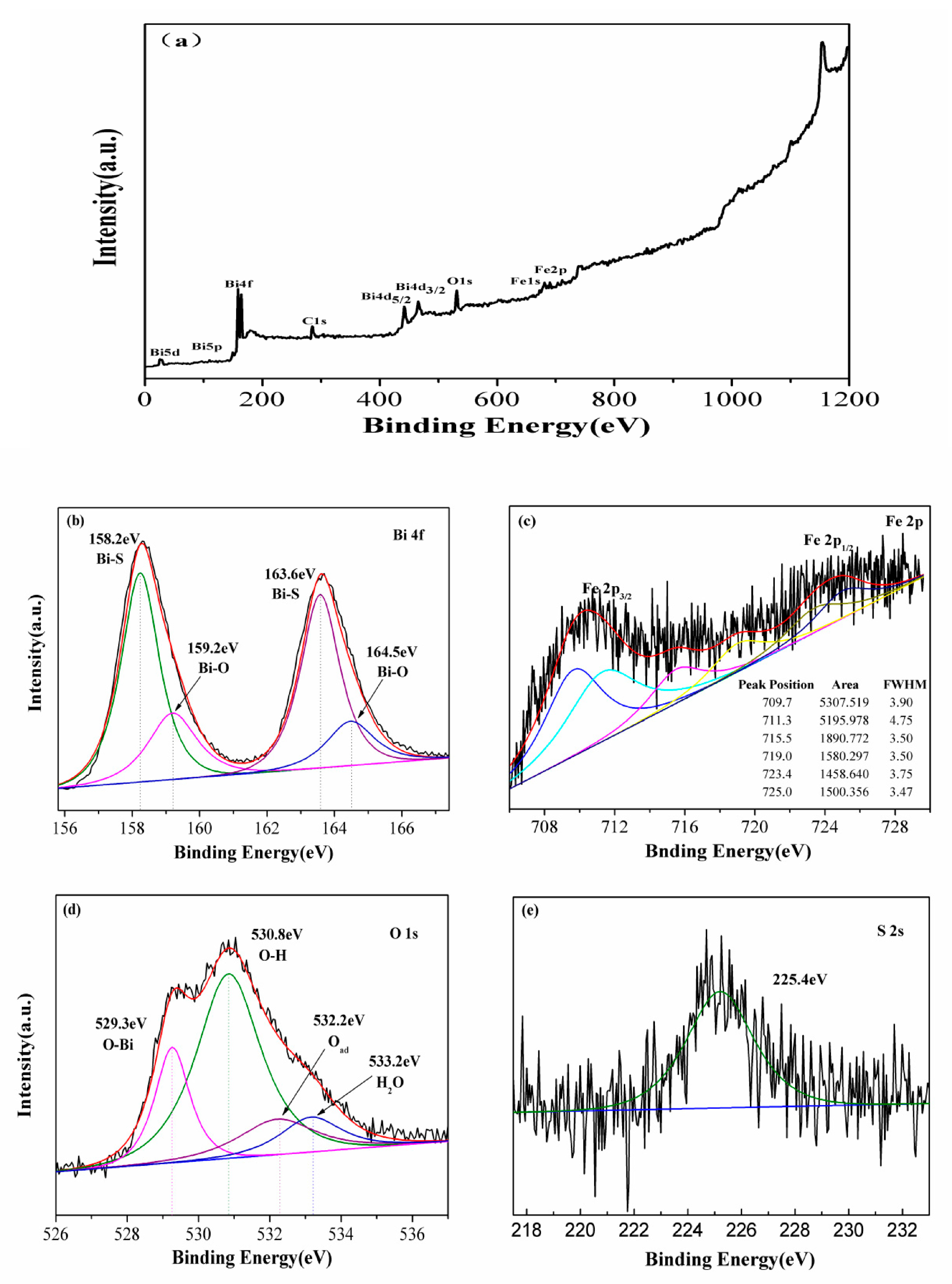
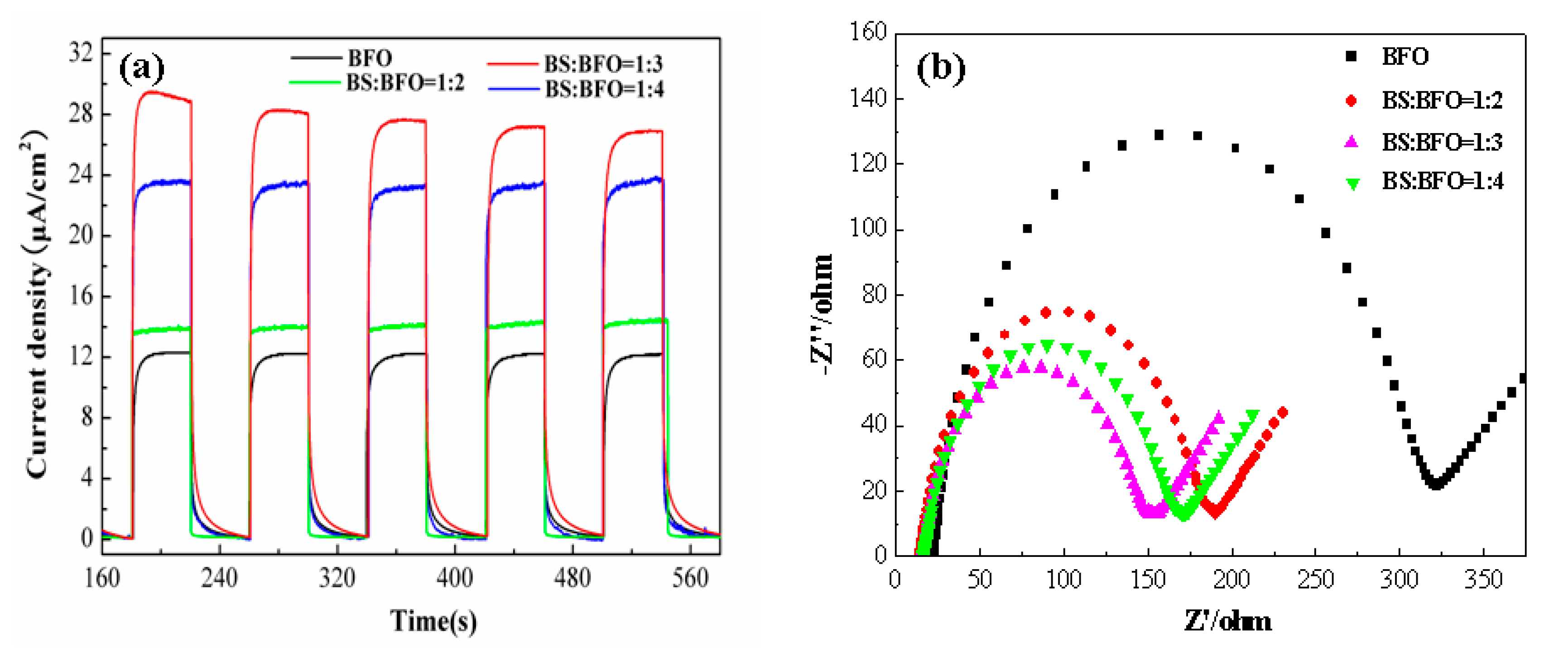
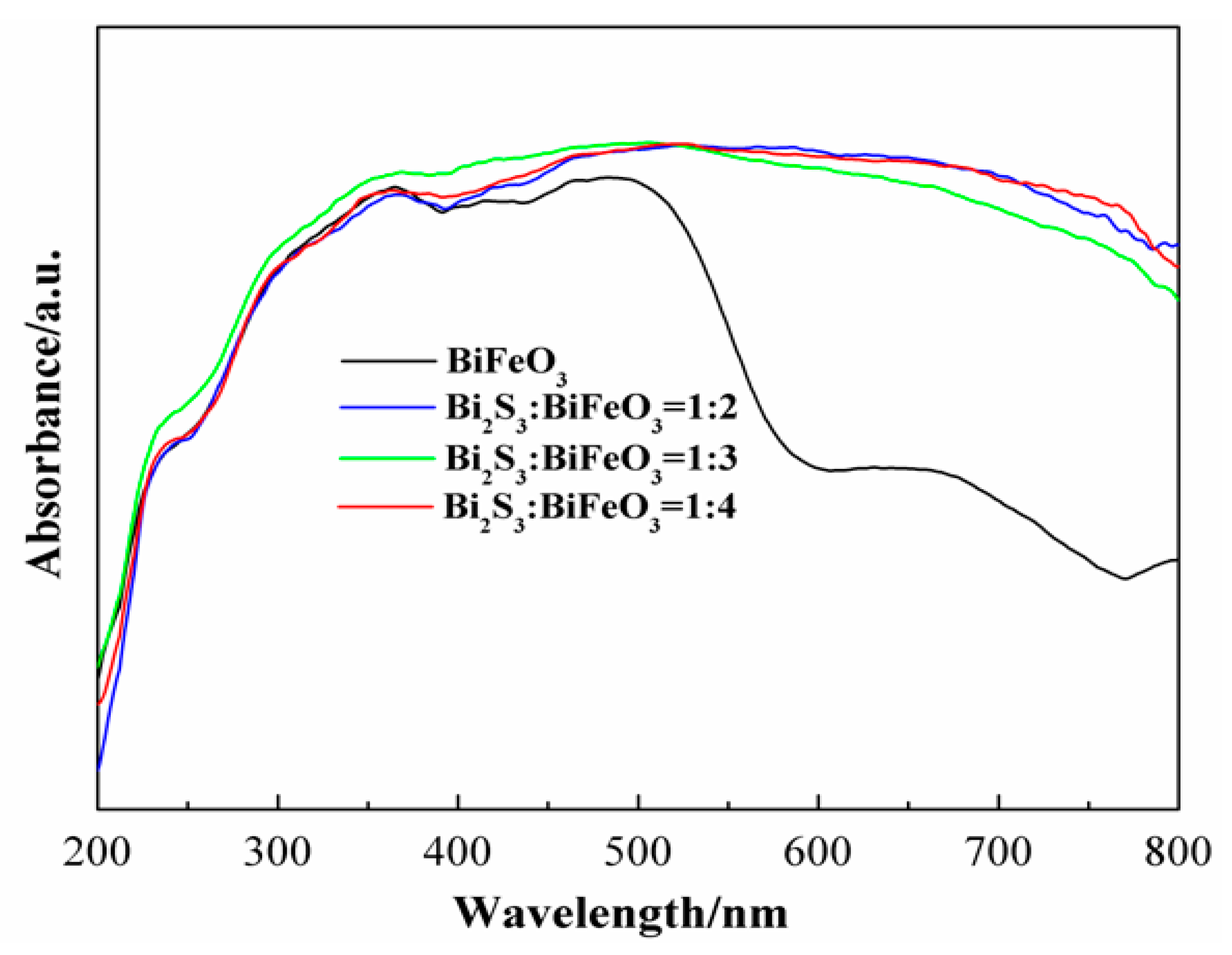
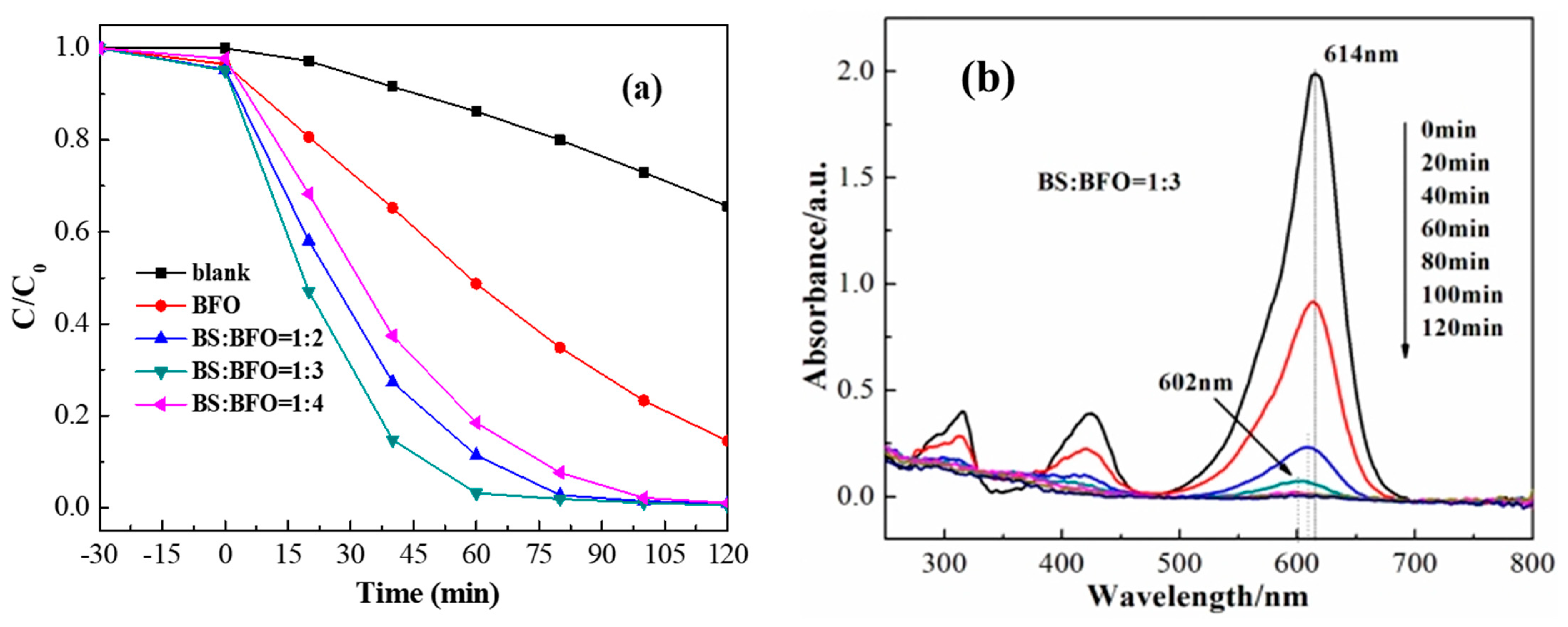
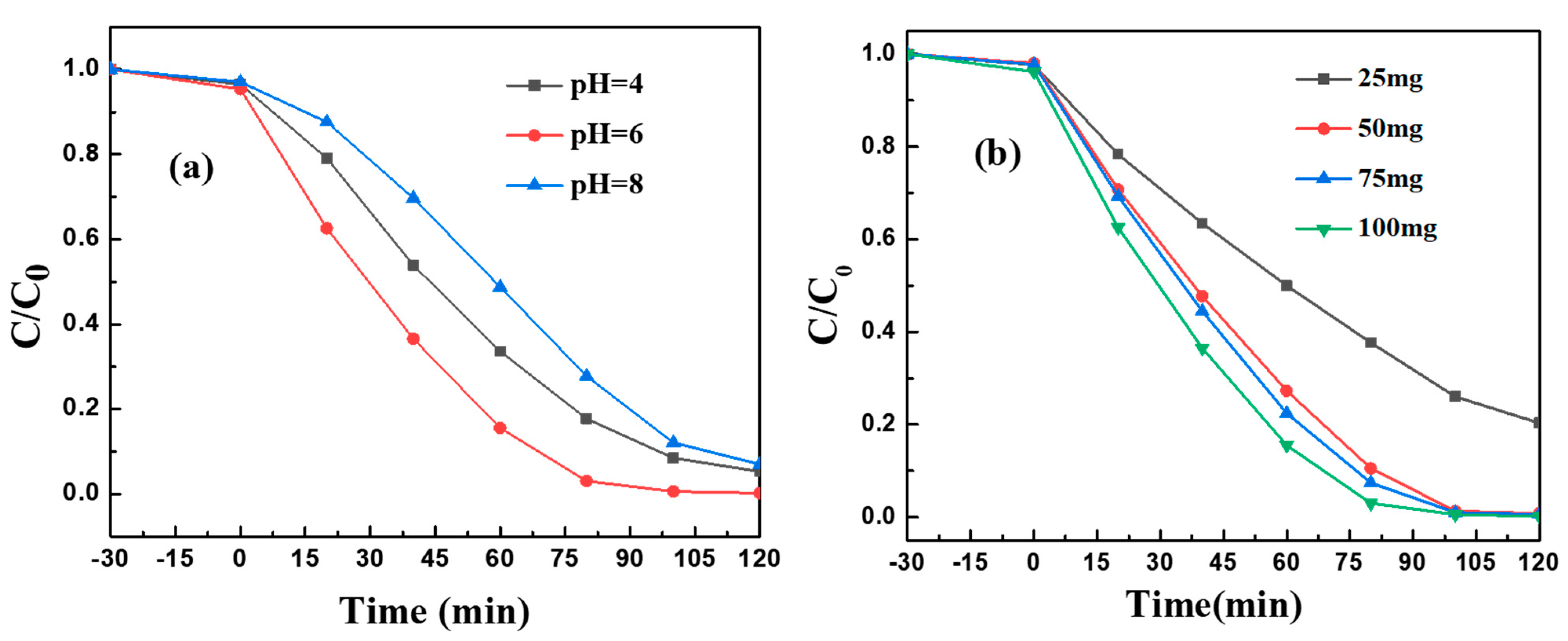
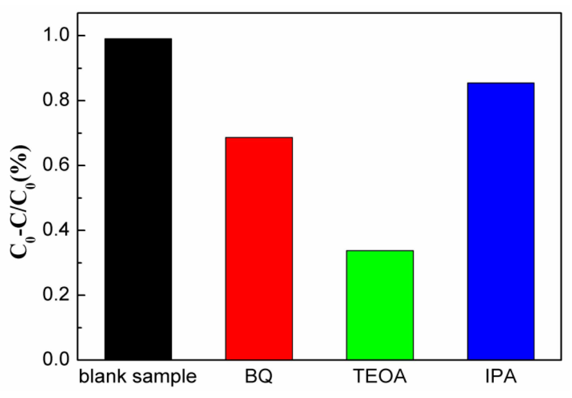
2. Results and Discussion

Figure 1 displays the X-ray diffraction (XRD) patterns of the Bi2S3/BiFeO3 samples with varying molar mass ratios, ranging from 1:2 to 1:4, in addition to pure BiFeO3, for comparison. The XRD patterns of pure BiFeO3 align well with the JCPDS 20-0169 standard [36], indicating the presence of characteristic diffraction peaks associated with this compound. In the composite material, discernible diffraction peaks attributed to Bi2S3 are evident, indicating the successful incorporation of Bi2S3 into the structure. It is worth noting that the diffraction peak of Bi2S3 at 2θ = 25.32 exhibits a slight overlap with BiFeO3 (2θ = 25.40). A noteworthy observation is that the positions of the Bi2S3 diffraction peaks at 25.2 and 28.6 display minor shifts towards larger angles in the composite material. These shifts suggest that Bi2S3 has effectively integrated with BiFeO3, leading to a reduction in the crystalline lattice spacing. Furthermore, it is important to acknowledge that the crystallinity of Bi2S3, synthesized via a hydrothermal method, is comparatively inferior to that of BiFeO3. These XRD findings shed light on the successful synthesis of Bi2S3/BiFeO3 composites and the structural modifications induced by their integration.
The microcosmic morphologies and microstructure of both pure BiFeO3 and Bi2S3/BiFeO3 were meticulously examined using SEM, TEM, and HRTEM, revealing intriguing insights. SEM images of pure BiFeO3, as shown in Figure 2a, depict an assembly of particles characterized by smooth borderlines. However, the introduction of sulfur for the ion exchange process brought about significant changes in the morphology of the composite materials. As evidenced in Figure 2d, there is a noticeable shift towards a nearly spherical flower-like shape. Detailed TEM analysis, displayed in Figure 2e, unequivocally indicates the growth of Bi2S3 on the surface of BiFeO3 through ion exchange. HRTEM provided further elucidation, revealing that Bi2S3 exposes more planes corresponding to crystal planes (310) and (211), which correspond to lattice spacings of 0.3530 nm and 0.3120 nm, respectively. The formation mechanism of these changes can be understood as follows: Initially, the lattice of BiFeO3 is pristine, with filled bonding orbitals and relatively few dangling bonds at the interface, rendering the entire BiFeO3 molecule in a stable state. The introduction of sulfuric acid provides the necessary energy for the surface activation of BiFeO3. This activation leads to damage to the original crystal lattice, exposing elemental bismuth and creating an abundance of dangling bonds on the surface of BiFeO3. Consequently, surface energy increases significantly, enhancing surface activity. When L-cysteine is introduced, a critical chemical transformation occurs. The elemental sulfur in L-cysteine forms stable new chemical bonds with the elemental bismuth on the surface of BiFeO3, resulting in the formation of Bi2S3/BiFeO3 heterojunctions. Moreover, the EDX spectrum analysis corroborates these findings, as it reveals the presence of Bi, O, Fe, and S elements, further confirming the formation mechanism of the Bi2S3/BiFeO3 heterojunction.
To gain further insight into the surface composition of the heterojunction material, X-ray photoelectron spectroscopy (XPS) was employed, providing compelling evidence for the coexistence of Bi2S3 and BiFeO3, as well as the presence of Bi, Fe, O, and S elements (Figure 3). The high-resolution XPS spectra reveal overlapping peaks centered at 158.2 eV and 163.6 eV, as well as 159.2 eV and 164.5 eV. These peaks can be attributed to the binding energy of Bi3+ 4f7/2 and 4f5/2 in Bi2S3 [28] and the Bi-O bond in BiFeO3 [37,38], respectively. The XPS spectrum of Fe(2p) is noteworthy, exhibiting six peaks at 709.9, 711.3, 715.5, 719.0, 723.4, and 725.0 eV. Notably, the Fe(2p) peaks at binding energies of 711.3/725.0 eV and 709.9/723.4 eV, along with a satellite signal peak at 719.0 eV and 715.5 eV, are characteristic of Fe3+ and Fe2+. The Fe(2p) spectra primarily feature two maxima peaks corresponding to the electron levels of Fe 2p3/2 and Fe 2p1/2 [39,40]. It is worth noting that the peak of the high-spin Fe3+ ion is significantly broader, compared to the low-spin Fe2+ ions. This broadening can be attributed to electrostatic interactions and spin–orbit coupling among the 2p empty orbital and the unpaired 3d electrons of iron ions. In the XPS spectrum of O 1s, four distinct peaks were observed at 529.3, 530.8, 532.2, and 533.2 eV, corresponding to oxygen–metal bonds in the lattice O2 of BiFeO3 [41], remaining OH at the material surface [42], adsorbed oxygen (Oads), and surface-adsorbed H2O [43]. The presence of a smaller-intensity peak at a binding energy of 225.4 eV in the S 2s spectrum can be attributed to the S-Bi bond of Bi2S3 [44]. In conclusion, the XPS analysis unequivocally confirms the successful growth of Bi2S3 nanomaterials on the surface of BiFeO3, forming the Bi2S3/BiFeO3 heterojunction.
Photocurrent measurements were conducted for both pure BiFeO3 and various molar ratios of Bi2S3/BiFeO3, to probe electronic interactions, as depicted in Figure 4a. The composite materials exhibited markedly higher photocurrent responses compared to single BiFeO3 during on–off cycles under visible-light irradiation. This enhancement can be directly attributed to the improved separation efficiency of photo-generated electrons within the heterojunction. Notably, when the molar mass ratio of Bi2S3/BiFeO3 is 1:3, the photocurrent response surpassed that of the other ratios, signifying superior performance in charge separation. This enhanced photocurrent in the Bi2S3/BiFeO3 heterojunction indicates heightened efficiency in separating electron–hole pairs, thereby enhancing the photocatalytic properties. To delve further into the charge separation process, electrochemical impedance spectroscopy (EIS) was employed. In EIS, a smaller arc radius signifies more effective separation of photo-generated electron–hole pairs and faster interface charge transfer.
Figure 4b illustrates the fact that the arc radius of the Bi2S3/BiFeO3 heterojunction is notably smaller than that of single BiFeO3. This difference is particularly pronounced when the ratio of Bi2S3/BiFeO3 is 1:3, where the arc radius is minimized. These results underscore the significant role played by the Bi2S3/BiFeO3 heterojunction in improving the separation of photogenerated electron–hole pairs and facilitating efficient charge transfer at the interface. Consequently, the utilization of light energy is greatly enhanced, contributing to the overall photocatalytic efficiency.
The UV-Vis DRS spectra of the Bi2S3/BiFeO3 heterojunction photocatalysts, with varying proportions, along with pure BiFeO3, are presented in Figure 5. These spectra offer valuable insights into the optical properties of the materials. The optical absorption spectra, as revealed by DRS, demonstrate distinct characteristics. The pure BiFeO3 sample exhibits intrinsic semiconductor-like absorption, primarily in the orange region of the visible spectra. In contrast, the Bi2S3/BiFeO3 heterojunction composite materials exhibit absorption across the entire visible region of the spectrum, indicating enhanced light absorption capabilities. Notably, pure BiFeO3 shows an absorption edge at approximately 610 nm, corresponding to a band gap of 2.03 eV for BiFeO3. In the case of the Bi2S3/BiFeO3 heterojunction, the absorption edge shifts towards the visible light region with increased intensity, compared to BiFeO3. This shift signifies the photosensitization effect of Bi2S3 on BiFeO3, where Bi2S3 enhances the absorption of visible light by the composite material. These findings emphasize the significant role of Bi2S3 in expanding the spectral range of light absorption by the BiFeO3 photocatalyst, thereby enhancing its photocatalytic potential.
The photocatalytic activity of the Bi2S3/BiFeO3 heterojunction with varying proportions of Bi2S3 was investigated through the photodegradation of the organic pollutant MG, as illustrated in Figure 6a. In the absence of the catalyst, the rate of photodegradation of MG under visible light (λ > 420 nm) irradiation remained below 40% within 2 h. However, when the Bi2S3/BiFeO3 heterojunction catalysts were introduced, notable improvements were observed. MG was completely photodegraded after 60 min, demonstrating the enhanced photocatalytic efficiency of these materials. The photocatalytic performance followed the following order: Bi2S3/BiFeO3 (1:3) > Bi2S3/BiFeO3 (1:2) > Bi2S3/BiFeO3 (1:4) > BiFeO3. In particular, Bi2S3/BiFeO3 (1:3) exhibited the highest photocatalytic efficiency, with approximately 99% of MG molecules decomposed within 60 min under visible light. This result underscores the significance of the optimal proportion between BiFeO3 and Bi2S3 for enhanced photocatalytic activity. Scanning electron microscopy revealed that Bi2S3/BiFeO3 (1:3) possessed a more uniform morphology, further supporting the superior photocatalytic performance observed. Moreover, full-wavelength scanning in Figure 6b highlights the degradation process: as MG was degraded by the addition of the Bi2S3/BiFeO3 photocatalyst, the UV–visible absorption peak gradually weakened until it disappeared. During this weakening, there was a noticeable blue shift in the UV–visible absorption peak, indicating the generation of intermediate products in the optical degradation process, which, however, did not impede the degradation process. These findings underscore the substantial photocatalytic advantage of the Bi2S3/BiFeO3 heterojunction, particularly when the proportion is optimized, and the importance of uniform particle surfaces in enhancing photocatalytic activity.
As shown in Figure 7a, Bi2S3/BiFeO (1:3) shows pH-dependent photocatalytic activity toward MG degradation, where the heterojunction catalyst exhibits excellent catalytic activity, with a pH around 6. An acidic or alkaline condition brings a negative effect toward MG degradation, which may be attributed to the fact that the heterojunction catalyst is not stable enough against acid or alkaline corrosion. We also investigate the influence of catalyst dosage on the MG photo degradation, and the results are shown in Figure 7b. It is easy to find that, with the increase in the amount of catalyst used, the catalytic activity of heterojunctions toward MG degradation increases, which may be attributed to the increased catalyst amount, which could provide a more active side for oxygen activation to generate more reactive oxygen species. It should be noted here that an excess amount of catalyst would bring a light shading effect, which may bring negative effects toward pollutant removal. As can be seen from Figure 7b, limited activity improvement is found with the amount of catalyst increasing from 75 mg to 100 mg, and we believe that further increasing the amount of catalyst would not bring much improvement in catalytic activity. Thus, we then chose 100 mg of catalyst as the optimized condition to carry out other experiments.
To gain a deeper understanding of the photodegradation mechanism, various sacrificial agents were introduced to elucidate the roles of specific reactive groups in the reaction process. As depicted in Figure 8, when isopropanol (IPA) was introduced as a sacrificial agent to capture hydroxyl (•OH) radicals, the degradation of MG was almost negligible. This observation suggests that •OH radicals play a crucial role in the degradation process, as they are potent oxidants, capable of effectively oxidizing many organic pollutants. Conversely, when triethanolamine (TEOA) was added to capture the photo-generated holes (h+), and 4-benzoquinone (BQ) was used to capture •O2 radicals, the efficiency of MG degradation was significantly reduced. This reduction was particularly pronounced in the presence of TEOA. These results imply that, during the photodegradation reaction, •O2 radicals and photo-generated holes (h+) work in tandem to facilitate the degradation of organic pollutants. The •OH radical, being a robust oxidant capable of oxidizing a wide range of organic pollutants, is likely a key contributor to the degradation process. These findings align with the existing literature, further supporting the significance of •OH radicals in the photodegradation of organic pollutants.
To provide further authentication of the photocatalytic mechanisms at play, several key energy levels and oxidation–reduction potentials were considered (Figure 9). The band gap of BiFeO3 was determined to be 2.05 eV, based on UV-Vis DRS spectra. Additionally, the conduction band of BiFeO3 is at +0.21eV (ECB = +0.21 eV) [45], and the valence band is at +2.26 eV (EVB = +2.26 eV). In contrast, Bi2S3 has a conduction band at 0 V (ECB = 0 V) [46] and a valence band at +1.38 eV (EVB = +1.38 eV) [47]. Notably, the conduction band of Bi2S3 is more negative than that of BiFeO3. Based on the above energy-band structure analysis, the photogenerated electrons are transferred from the conduction band of Bi2S3 to the conduction band of BiFeO3, due to the more negative conduction band of Bi2S3. This facilitates the generation of •O2 on the material surface by oxygen absorption, which can then directly oxidize the target contaminant MG. On the other hand, the oxidation–reduction potential of •OH is 2.38 V [48], significantly more positive than the valence band of BiFeO3 (EVB = +2.26 V) [49] and Bi2S3 (EVB = +1.38 eV) [50]. Consequently, the photoexcited Bi2S3/BiFeO3 generates holes that are unable to oxidize water to form hydroxyl radicals. Furthermore, the valence band energy of Bi2S3 is lower than that of BiFeO3. Thus, the photogenerated holes in the valence band of BiFeO3 are transferred to the valence band of Bi2S3. This energy transfer enables the direct oxidation of MG. Considering the properties of each substance in the reaction system, these insights suggest the procedures and mechanisms of the reaction [50].
B i 2 S 3 + h v h B i 2 S 3 + + e B i 2 S 3
B i F e O 3 + h v h B i F e O 3 + + e B i F e O 3
e B i 2 S 3 + B i F e O 3 e B i F e O 3
e B i F e O 3 + O 2 O 2
O 2 + M G p r o d u c t s
h B i F e O 3 + + B i 2 S 3 h B i 2 S 3 +
h B i 2 S 3 + + M G p r o d u c t s

3. Experimental

3.1. Material Synthesis

BiFeO3 was prepared according to the reported method, with some modification [51]. Typically, 5 mM of bismuth nitrate pentahydrate (Bi(NO3)3•5H2O) and 5 mM of ferric nitrate (Fe(NO3)3•9H2O) were added into 35 mL of ethylene glycol monomethyl ether, to form a fine mixture. The solution was vigorously stirred until it reached a state of homogeneity, resulting in a transparent and stable solution at room temperature. Subsequently, the sample was left to dry for 24 h at 80 °C, yielding a gel-like powder. This powder was then carefully ground and subjected to heating in a muffle furnace at 550 °C for 4 h. After the heating process, the sample was allowed to cool to room temperature, and its final form was obtained through additional grinding.
Following this initial preparation, the next step involved the synthesis of Bi2S3/BiFeO3 through an in situ growth process on the BiFeO3 substrate under hydrothermal conditions, as depicted in Figure 10.
Commencing with three clean beakers, each received 35 mL of deionized water. Sequentially, L-cysteine was introduced at concentrations of 6 mM, 4 mM, and 3 mM into the respective beakers. Complete dissolution of L-cysteine was ensured. Sulfuric acid was employed to carefully adjust the pH level to 2, while maintaining precision in this process. With meticulous care, an equivalent quantity of BiFeO3 (4 mM) was gradually added to each solution. Stirring continued until the BiFeO3 was uniformly dispersed within the mix. This stirring process endured for an additional 30 min. The mixed solutions were then carefully transferred to a 50 mL autoclave. The autoclave was subjected to a hydrothermal reaction at 150 °C for a duration of 12 h. Following completion of the reaction, the contents underwent filtration, and the resulting material was thoroughly washed with deionized water and absolute ethanol. The Bi2S3/BiFeO3 heterojunction materials obtained from the process were dried at 80 °C for 12 h under ambient air conditions. The final samples were categorized as Bi2S3/BiFeO3 (1:2), Bi2S3/BiFeO3 (1:3), and Bi2S3/BiFeO3 (1:4), respectively, based on the ratios used during their preparation.

3.2. Characterization

The crystalline phase of the samples was investigated using X-ray diffraction (XRD, D8 Advance, Bruker, Mannheim, Germany), using graphite monochromatized Cu-Kα (λ = 1.5406 Å) radiation at 40 kv and 40 mA with a scan rate of 2 °/min between 10° and 70°. The UV–vis diffuse reflectance spectra (DRS) of the powders were collected using a U-3900/3900H UV-vis spectrophotometer (UV-vis DRS, U-3900/3900H, Hitachi High-Technologies, Tokyo, Japan) equipped with an integrating sphere attachment. The general morphologies and micro-area chemical analysis of the samples were obtained using scanning electron microscopy images (SEM, Sirion 200, FEI, Eindhoven, The Netherlands) and energy-dispersive X-ray spectroscopy (EDS) with a scanning voltage of 5.00 kV. Further microstructure and crystallinity of the sample was analyzed using transmission electron microscopy (TEM, Tecnai F20, FEI, Hillsboro, OR, USA) and high-resolution transmission electron microscopy (HRTEM) observation. The chemical states of as-prepared photocatalysts were tested using monochromated Al-Kα X-ray photoelectron spectroscopy (XPS, Axis Ultra DLD) at a residual gas pressure of less than 10−8 Pa.
Electrochemical measurements were performed on a CHI 660D electrochemical workstation (Shanghai Chenhua, Shanghai, China) by using a standard three-electrode system with a working electrode. (The working electrode was prepared by dip-coating on a 3 cm × 1 cm fluorine tin oxide (FTO) glass electrode, with the films drying under room condition.) A graphite electrode was used as the electrodes, and a standard calomel electrode in saturated KCl was used as a reference electrode. A total of 0.5 M sodium sulfate was used as the electrolyte solution for the photocurrent test, and 0.5 M sodium sulfate, 2.5 mM of potassium ferricyanide and potassium ferrocyanide were adopted as the supporting electrolyte for Ac impedance.

3.3. Photocatalytic Activity Evaluation

The effectiveness of the Bi2S3/BiFeO3 heterojunction catalysts in photocatalysis was assessed through the degradation of Malachite Green oxalate (MG) under visible light conditions (λ > 420 nm). These photocatalytic reactions were conducted using simulated solar light with an intensity equivalent to 300 W, generated by a Xe-lamp system from Asahi Spectra Co., Ltd. (Torrance, CA, USA) The initial concentration of the Malachite Green solution was set at 20 mg/L. The photocatalytic procedure was performed in a homemade reactor with catalytic conditions quite similar to the reported method; typically, 0.1 g of the catalyst was dispersed into 100 mL of the Malachite Green oxalate (MG) solution [52]. To establish a balance between the adsorption and desorption of Malachite Green on the catalyst surface, the mixture was magnetically stirred for 30 min in the dark.
At specified irradiation time intervals, approximately 5 mL of the suspension was extracted every 10 min. These samples were subsequently centrifuged to obtain clear solutions, which were then analyzed using a UV–vis spectrophotometer (UV-vis DRS, U-3900/3900H, Hitachi High-Technologies, Tokyo, Japan). The maximum absorbance of the solution was determined at 614 nm. To provide a basis for comparison, parallel experiments using pure BiFeO3 were conducted alongside the Bi2S3/BiFeO3 experiments, allowing for an evaluation of the relative performance of Bi2S3/BiFeO3, compared to pure BiFeO3.

4. Conclusions

In summary, we have shown the first instance of fabricating nanoflower-shaped Bi2S3/BiFeO3 heterojunctions via a simple sol–gel process. The formed type II heterojunction is found capable of providing a powerful built-in field to accelerate electron–hole spatial separation, which leads to the enhanced reactive oxygen species generation. As a result, the as-prepared Bi2S3/BiFeO3 heterojunctions exhibit excellent photocatalytic performance toward MG degradation, where more than 99% of MG is removed within 2 h of photocatalysis. The innovative design shown in this work may offer new viewpoints in designing highly efficient photocatalysts for environmental purification. For real water purification, however, we still need to immobilize the catalyst, so as to prevent catalyst loss. The immobilization of the as-prepared Bi2S3/BiFeO3 on a light-weight carrier, for example loofah, to construct floating materials, is another possibility for its practical application.

Author Contributions

Conceptualization, L.Z; Methodology, R.Z. and P.Z.; Validation, L.Z.; Formal analysis, R.Z.; Investigation, R.Z. and P.Z.; Data curation, R.Z. and L.Z.; Writing—original draft, R.Z. and X.T.; Writing—review & editing, X.T. and Z.Z.; Supervision, X.T. and Z.Z.; Project administration, X.T.; Funding acquisition, X.T. All authors have read and agreed to the published version of the manuscript.

Funding

This work was financially supported by the National Natural Science Foundation of China (No. 52060019 and 22276087), the Key Project of Jiangxi Provincial Natural Science Foundation (No. 20202ACBL204015), the Key Research and Development Project of Jiangxi Province (No. 20212BBG73030).

Institutional Review Board Statement

Not applicable.

Informed Consent Statement

Not applicable.

Data Availability Statement

Data are contained within the article.

Conflicts of Interest

The authors declare no conflict of interest.

References

  1. Zeng, Z.; Fan, Y.; Quan, X.; Yu, H.; Chen, S.; Zhang, S. Energy-transfer-mediated oxygen activation in carbonyl functionalized carbon nitride nanosheets for high-efficient photocatalytic water disinfection and organic pollutants degradation. Water Res. 2020, 177, 115798. [Google Scholar] [CrossRef]
  2. Fung, C.-M.; Er, C.-C.; Tan, L.-L.; Mohamed, A.R.; Chai, S.-P. Red phosphorus: An up-and-coming photocatalyst on the horizon for sustainable energy development and environmental remediation. Chem. Rev. 2022, 122, 3879–3965. [Google Scholar] [CrossRef]
  3. Li, S.; Lin, Y.H.; Zhang, B.P.; Wang, Y.; Nan, C.W. Controlled fabrication of BiFeO3 uniform microcrystals and their magnetic and photocatalytic behaviors. J. Phys. Chem. C 2010, 114, 2903–2908. [Google Scholar] [CrossRef]
  4. Gao, F.; Chen, X.Y.; Yin, K.B.; Dong, S.; Ren, Z.F.; Yuan, F.; Yu, T.; Zou, Z.G.; Liu, J.M. Visible-Light Photocatalytic Properties of Weak Magnetic BiFeO3Nanoparticles. Adv. Mater. 2007, 19, 2889–2892. [Google Scholar] [CrossRef]
  5. Yang, X.; Zhang, Y.; Xu, G.; Wei, X.; Ren, Z.; Shen, G.; Han, G. Phase and morphology evolution of bismuth ferrites via hydrothermal reaction route. Mater. Res. Bull. 2013, 48, 1694–1699. [Google Scholar] [CrossRef]
  6. Wang, X.; Lin, Y.; Ding, X.; Jiang, J. Enhanced visible-light-response photocatalytic activity of bismuth ferrite nanoparticles. J. Alloys Compd. 2011, 509, 6585–6588. [Google Scholar] [CrossRef]
  7. Zhang, J.; Gondal, M.A.; Wei, W.; Zhang, T.; Xu, Q.; Shen, K. Preparation of room temperature ferromagnetic BiFeO3 and its application as an highly efficient magnetic separable adsorbent for removal of Rhodamine B from aqueous solution. J. Alloys Compd. 2012, 530, 107–110. [Google Scholar] [CrossRef]
  8. Xian, T.; Yang, H.; Dai, J.F.; Wei, Z.Q.; Ma, J.Y.; Feng, W.J. Photocatalytic properties of BiFeO3 nanoparticles with different sizes. Mater. Lett. 2011, 65, 1573–1575. [Google Scholar] [CrossRef]
  9. He, J.; Guo, R.; Fang, L.; Dong, W.; Zheng, F.; Shen, M. Characterization and visible light photocatalytic mechanism of size-controlled BiFeO3 nanoparticles. Mater. Res. Bull. 2013, 48, 3017–3024. [Google Scholar] [CrossRef]
  10. Liu, Z.; Qi, Y.; Lu, C. High efficient ultraviolet photocatalytic activity of BiFeO3 nanoparticles synthesized by a chemical coprecipitation process. J. Mater.Sci. Mater. Electron. 2010, 21, 380–384. [Google Scholar] [CrossRef]
  11. Soltani, T.; Entezari, M.H. Photolysis and photocatalysis of methylene blue by ferrite bismuth nanoparticles under sunlight irradiation. J. Mol. Catal. A Chem. 2013, 377, 197–203. [Google Scholar] [CrossRef]
  12. Mohan, S.; Subramanian, B. A strategy to fabricate bismuth ferrite (BiFeO3) nanotubes from electro spun nanofibers and their solar light-driven photocatalytic properties. RSC Adv. 2013, 3, 23737–23744. [Google Scholar] [CrossRef]
  13. Zhang, X.; Liu, H.; Zheng, B.; Lin, Y.; Liu, D.; Nan, C.W. Photocatalytic and magnetic behaviors observed in BiFeO3 nanofibers by electrospinning. J. Nanomater. 2013, 2013, 917–948. [Google Scholar] [CrossRef]
  14. Guo, R.; Fang, L.; Dong, W.; Zheng, F.; Shen, M. Enhanced photocatalytic activity and ferromagnetism in Gd Doped BiFeO3 nanoparticles. J. Phys. Chem. C 2010, 114, 21390–21396. [Google Scholar] [CrossRef]
  15. Zhang, Z.; Liu, H.; Lin, Y.; Wei, Y.; Nan, C.W.; Deng, X. Influence of La Doping onMagnetic and Optical Properties of Bismuth Ferrite Nanofibers. J. Nanomater. 2012, 2012, 238605. [Google Scholar] [CrossRef]
  16. Feng, Y.N.; Wang, H.C.; Luo, Y.D.; Shen, Y.; Lin, Y.H. Ferromagnetic and photocatalytic behaviors observed in Ca-doped BiFeO3 nanofibres. J. Appl. Phys. 2013, 113, 146101. [Google Scholar] [CrossRef]
  17. Li, S.; Zhang, J.; Kibria, M.G.; Mi, Z.; Chaker, M.; Ma, D.; Nechache, R.; Rosei, F. Remarkably enhanced photocatalytic activity of laser ablated Au nanoparticle decorated BiFeO3 nanowires under visible-light. Chem. Commun. 2013, 49, 5856–5858. [Google Scholar] [CrossRef]
  18. Schultz, A.M.; Zhang, Y.; Salvador, P.A.; Rohrer, G.S. Effect of Crystal and Domain Orientation on the Visible-Light Photochemical Reduction of Ag on BiFeO3. ACS Appl. Mater. Inter. 2011, 3, 1562–1567. [Google Scholar] [CrossRef]
  19. Niu, F.; Chen, D.; Qin, L.; Zhang, N.; Wang, J.; Chen, Z.; Huang, Y. Facile Synthesis of Highly Efficient p–n Heterojunction CuO/BiFeO3 Composite Photocatalysts with Enhanced Visible-Light Photocatalytic Activity. ChemCatChem 2015, 7, 3279–3289. [Google Scholar] [CrossRef]
  20. Zhang, Y.; Schultz, A.M.; Salvador, P.A.; Rohrer, G.S. Spatially selective visible light photocatalytic activity of TiO2/BiFeO3 heterostructures. J. Mater. Chem. 2011, 21, 4168–4174. [Google Scholar] [CrossRef]
  21. Zhu, A.; Zhao, Q.; Li, X.; Shi, Y. BiFeO3/TiO2 Nanotube Arrays Composite Electrode: Construction, Characterization, and Enhanced Photoelectrochemical Properties. ACS Appl. Mater. Interfaces 2014, 6, 671–679. [Google Scholar] [CrossRef] [PubMed]
  22. Li, T.; Shen, J.; Li, N.; Ye, M. Hydrothermal preparation, characterization and enhanced properties of reduced grapheme-BiFeO3 nanocomposite. Mater. Lett. 2013, 91, 42–44. [Google Scholar] [CrossRef]
  23. Li, Z.; Shen, Y.; Guan, Y.; Hu, Y.; Lin, Y.; Nan, C.W. Bandgap engineering and enhanced interface coupling of graphene–BiFeO3 nanocomposites as efficient photocatalysts under visible light. J. Mater. Chem. A 2014, 2, 1967–1973. [Google Scholar] [CrossRef]
  24. Pineda, E.; Nicho, M.E.; Nair, P.K.; Hu, H. Optoelectronic properties of chemically deposited Bi2S3 thin films and the photovoltaic performance of Bi2S3/P3OT solar cells. Sol. Energy 2012, 86, 1017–1022. [Google Scholar] [CrossRef]
  25. Cao, J.; Xu, B.; Lin, H.; Luo, B.; Chen, S. Novel Bi2S3-sensitized BiOCl with highly visible light photocatalytic activity for the removal of rhodamine B. Catal. Commun. 2012, 26, 204–208. [Google Scholar] [CrossRef]
  26. Cheng, H.; Huang, B.; Qin, X.; Zhang, X.; Dai, Y. A controlled anion exchange strategy to synthesize Bi2S3 nanocrystals/BiOCl hybrid architectures with efficient visible light photoactivity. Chem. Commun. 2012, 48, 97–99. [Google Scholar] [CrossRef] [PubMed]
  27. Chen, P.; Gu, L.; Cao, X. From single ZnO multipods to heterostructured ZnO/ZnS, ZnO/ZnSe, ZnO/Bi2S3 and ZnO/Cu2S multipods: Controlled synthesis and tunable optical and photoelectrochemical properties. CrystEngComm 2010, 12, 3950–3958. [Google Scholar] [CrossRef]
  28. Calva-Yanez, J.C.; Rincon, M.E.; Solís de la Fuente, M.; Alvarado-Tenorio, G. Structural and photoelectrochemical characterization of MWCNT-TiO2 matrices sensitized with Bi2S3. J. Solid State Electrochem. 2013, 17, 2633–2641. [Google Scholar] [CrossRef]
  29. Jana, A.; Bhattacharya, C.; Datta, J. Enhanced photoelectrochemical activity of electro-synthesized CdS-Bi2S3 composite films grown with self-designed cross-linked structure. Electrochim. Acta 2010, 55, 6553–6562. [Google Scholar] [CrossRef]
  30. He, H.; Berglund, S.P.; Xiao, P.; Chemelewski, W.D.; Zhang, Y.; Mullins, C.B. Nanostructured Bi2S3/WO3 heterojunction films exhibiting enhanced photoelectrochemical performance. J. Mater. Chem. A 2013, 1, 12826–12834. [Google Scholar] [CrossRef]
  31. Ata, S.; Shaheen, I.; Aslam, H.; Mohsin, I.U.; Alwadai, N.; Al Huwayz, M.; Iqbal, M.; Younas, U. Barium and strontium doped La-based perovskite synthesis via sol-gel route and photocatalytic activity evaluation for methylene blue. Results Phys. 2023, 45, 106235. [Google Scholar] [CrossRef]
  32. Jiao, S.; Zhao, Y.; Bi, M.; Bi, S.; Li, X.; Wang, B.; Li, C.; Dong, Y. Removal of Methylene Blue from Water by BiFeO3/Carbon Fibre Nanocomposite and Its Photocatalytic Regeneration. Catalysts 2018, 8, 267. [Google Scholar] [CrossRef]
  33. Tseng, W.J.; Lin, R.D. BiFeO3/α-Fe2O3 core/shell composite particles for fast and selective removal of methyl orange dye in water. J. Colloid Interface Sci. 2014, 428, 95–100. [Google Scholar] [CrossRef]
  34. Qin, K.; Zhao, Q.; Yu, H.; Xia, X.; Li, J.; He, S.; Wei, L.; An, T. A review of bismuth-based photocatalysts for antibiotic degradation: Insight into the photocatalytic degradation performance, pathways and relevant mechanisms. Environ. Res. 2021, 199, 111360. [Google Scholar] [CrossRef] [PubMed]
  35. Song, S.; Xing, Z.; Zhao, H.; Li, Z.; Zhou, W. Recent advances in bismuth-based photocatalysts: Environment and energy applications. Green Energy Environ. 2023, 8, 1232–1264. [Google Scholar] [CrossRef]
  36. JCPDS 20-0169; Elements of X-ray Diffraction, B. D. Cullinity, 2nd ed., Addison-Wesley, Reading, MA, 1978. JCPDS (Joint Committee on Powder Diffraction Standards): Newtown Square, PA, USA, 2023.
  37. Dai, Y.; Wang, Y.; Yao, J.; Wang, Q.; Liu, L.; Chu, W.; Wang, G. Phosgene-Free Synthesis of Phenyl Isocyanate by Catalytic Decomposition of Methyl N-Phenyl Carbamate over Bi2O3 Catalyst. Catal. Lett. 2008, 123, 307–316. [Google Scholar] [CrossRef]
  38. Zeng, Z.; Ye, F.; Deng, S.; Fang, D.; Wang, X.; Bai, Y.; Xiao, H. Accelerated organic pollutants mineralization in interlayer confined single Pt atom photocatalyst for hydrogen recovery. Chem. Eng. J. 2022, 444, 136561. [Google Scholar] [CrossRef]
  39. Lu, X.; Pu, F.; Xia, Y.; Huang, W.; Li, Z. Facile fabrication of porous thin films of Bi2O3/Bi2S3 nanocomposite semiconductors at gas/liquid interface and their photoelectrochemical performances. Appl. Surface Sci. 2014, 299, 131–135. [Google Scholar] [CrossRef]
  40. Wang, N.; Zhu, L.; Wang, M.; Wang, D.; Tang, H. Sono-enhanced degradation of dye pollutants with the use of H2O2 activated by Fe3O4 magnetic nanoparticles as peroxidase mimetic. Ultrason. Sonochem. 2010, 17, 526–533. [Google Scholar] [CrossRef]
  41. Jiang, J.; Zou, J.; Anjum, M.N.; Yan, J.; Huang, L.; Zhang, Y.; Chen, J. Synthesis and characterization of wafer-like BiFeO3 with efficient catalytic activity. Solid State Sci. 2011, 13, 1779–1785. [Google Scholar] [CrossRef]
  42. Zhong, Y.; Shi, J.; Li, K.; Guo, H.; Yan, L.; Zeng, Z. Silicotungstic acid as electron transfer mediator in Z-scheme g-C3N4/H4SiW12O40/Ag3PO4: Accelerated charge interface transportation for high-efficient photocatalysis. Process Saf. Environ. 2023, 171, 188–199. [Google Scholar] [CrossRef]
  43. Xiao, H.; Zhang, Q.; Ahmad, M.; Dong, S.; Zhang, Y.; Fang, D.; Wang, X.; Peng, H.; Lei, Y.; Wu, G.; et al. Carbonate mediated hole transfer boosting the photocatalytic degradation of organic pollutants over carbon nitride nanosheets. Sep. Purif. Technol. 2023, 306, 122580. [Google Scholar] [CrossRef]
  44. Marchon, B.; Carrazza, J.; Heinemann, H.; Somorjai, G.A. TPD and XPS studies of O2, CO2, and H2O adsorption on clean polycrystalline graphite. Carbon 1988, 26, 507–514. [Google Scholar] [CrossRef]
  45. Xu, W.; Fang, J.; Chen, Y.; Lu, S.; Zhou, G.; Zhu, X.; Fang, Z. Novel heterostructured Bi2S3/Bi2Sn2O7 with highly visible light photocatalytic activity for the removal of rhodamine B. Mater. Chem. Phys. 2015, 154, 30–37. [Google Scholar] [CrossRef]
  46. Humayun, M.; Zada, A.; Li, Z.; Xie, M.; Zhang, X.; Qu, Y.; Raziq, F.; Jing, L. Enhanced visible-light activities of porous BiFeO3 by coupling with nanocrystalline TiO2 and mechanism. Appl. Catal. B Environ. 2016, 180, 219–226. [Google Scholar] [CrossRef]
  47. Li, X.; Ye, F.; Zhang, H.; Ahmad, M.; Zeng, Z.; Wang, S.; Wang, S.; Gao, D.; Zhang, Q. Ternary rGO decorated W18O49 @g-C3N4 composite as a full-spectrum-responded Z-scheme photocatalyst for efficient photocatalytic H2O2 production and water disinfection. J. Environ. Chem. Eng. 2023, 11, 110329. [Google Scholar] [CrossRef]
  48. Kong, L.; Jiang, Z. Unusual reactivity of visible-light-responsive AgBr-BiOBr heterojunction photocatalysts. J. Catal. 2012, 293, 116–125. [Google Scholar] [CrossRef]
  49. Bard, A.J.; Faulkner, L.R. Electrochemical Methods: Fundamental and Application; John Wiley & Sons: New York, NY, USA, 1980. [Google Scholar]
  50. Li, X.; Zhu, J. Comparative study on the mechanism in photocatalytic degradation of different-type organic dyes on SnS2 and CdS. Appl. Catal. B Environ. 2012, 123–124, 174–181. [Google Scholar] [CrossRef]
  51. Xu, M.L.; Lu, M.; Qin, G.Y.; Wu, X.M.; Yu, T.; Zhang, L.N.; Li, K.; Cheng, X.; Lan, Y.Q. Piezo-Photocatalytic Synergy in BiFeO3@COF Z-Scheme Heterostructures for High-Efficiency Overall Water Splitting. Angew. Chem. Int. Ed. 2022, 61, e202210700. [Google Scholar] [CrossRef]
  52. Tang, J.; Wang, R.; Liu, M.; Zhang, Z.; Song, Y.; Xue, S.; Zhao, Z.; Dionysiou, D.D. Construction of novel Z-scheme Ag/FeTiO3/Ag/BiFeO3 photocatalyst with enhanced visible-light-driven photocatalytic performance for degradation of norfloxacin. Chem. Eng. J. 2018, 351, 1056–1066. [Google Scholar] [CrossRef]
Figure 1. XRD patterns of Bi2S3/BiFeO3 with different proportions.
Molecules 28 08007 g001
Figure 2. SEM images of samples (a) BiFeO3; (b) Bi2S3/BiFeO3(1:4); (c) Bi2S3/BiFeO3(1:3); (d) Bi2S3/BiFeO3(1:2); (e) TEM of Bi2S3/BiFeO3(1:3); (f) HRTEM of Bi2S3/BiFeO3(1:3); (g) the selected area of the EDX; (h) the EDX spectrum of the Bi2S3/BiFeO3(1:3).
Molecules 28 08007 g002
Figure 3. (a) Whole XPS spectra; (b) Bi(4f); (c) Fe(2p); (d) O(1s) and (e) S(2s) lines of the Bi2S3/BiFeO3.
Molecules 28 08007 g003
Figure 4. Photocurrents (a) BiFeO3 and the proportions of Bi2S3 and BiFeO3 ([Na2SO4] = 0.5 M) and (b) electrochemical impedance spectroscopy (EIS) for BiFeO3 and the proportions of Bi2S3 and BiFeO3 ([Na2SO4] = 0.5 M, [K4Fe(CN)6•3H2O] = 2.5 mM, [K3[Fe(CN)6]] = 2.5 mM).
Molecules 28 08007 g004
Figure 5. UV-vis diffuse reflection spectra of pure BiFeO3 and the different proportions of Bi2S3/BiFeO3.
Molecules 28 08007 g005
Figure 6. (a) Photocatalytic degradation of MG under visible-light irradiation using Bi2S3/BiFeO3 heterojunctions as the catalyst; (b) the full-wavelength scanning of Bi2S3/BiFeO3(1:3) heterojunction photocatalyst.
Molecules 28 08007 g006
Figure 7. Photocatalytic performance of Bi2S3/BiFeO (1:3) toward MG degradation at (a) various pH conditions. (b) Effects of catalyst dosage on MG degradation.
Molecules 28 08007 g007
Figure 8. Effects of various scavengers on the photocatalytic efficiencies of Bi2S3/BiFeO3 in MG degradation under visible-light irradiation.
Molecules 28 08007 g008
Figure 9. Schematic illustration of the Bi2S3/BiFeO3 photocatalytic reaction process under visible-light irradiation.
Molecules 28 08007 g009
Figure 10. Schematic illustration of the Bi2S3/BiFeO3 synthetic processes.
Molecules 28 08007 g010
Disclaimer/Publisher’s Note: The statements, opinions and data contained in all publications are solely those of the individual author(s) and contributor(s) and not of MDPI and/or the editor(s). MDPI and/or the editor(s) disclaim responsibility for any injury to people or property resulting from any ideas, methods, instructions or products referred to in the content.

