Article

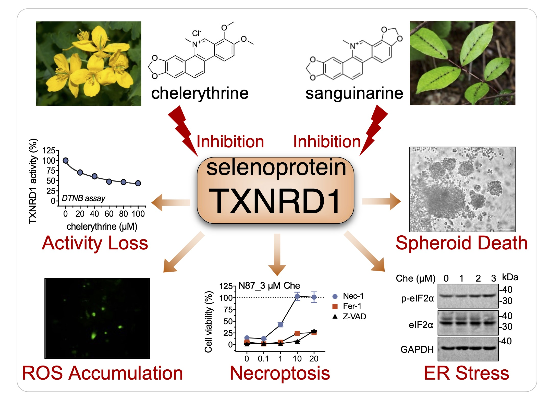
Benzophenanthridine Alkaloid Chelerythrine Elicits Necroptosis of Gastric Cancer Cells via Selective Conjugation at the Redox Hyperreactive C-Terminal Sec498 Residue of Cytosolic Selenoprotein Thioredoxin Reductase

1
School of Life and Pharmaceutical Sciences (LPS), Panjin Institute of Industrial Technology (PIIT), Dalian University of Technology, Panjin 124221, China
2
School of Ocean Science and Technology (OST), Key Laboratory of Industrial Ecology and Environmental Engineering (Ministry of Education), Dalian University of Technology, Panjin 124221, China
3
State Key Laboratory of Fine Chemicals, School of Chemical Engineering (CE), Dalian University of Technology, Dalian 116023, China
*
Author to whom correspondence should be addressed.
These authors contributed equally to this work.
Molecules 2023, 28(19), 6842; https://doi.org/10.3390/molecules28196842
Submission received: 30 August 2023 / Revised: 23 September 2023 / Accepted: 24 September 2023 / Published: 28 September 2023
(This article belongs to the Section Applied Chemistry)

Abstract

:
Targeting thioredoxin reductase (TXNRD) with low-weight molecules is emerging as a high-efficacy anti-cancer strategy in chemotherapy. Sanguinarine has been reported to inhibit the activity of TXNRD1, indicating that benzophenanthridine alkaloid is a fascinating chemical entity in the field of TXNRD1 inhibitors. In this study, the inhibition of three benzophenanthridine alkaloids, including chelerythrine, sanguinarine, and nitidine, on recombinant TXNRD1 was investigated, and their anti-cancer mechanisms were revealed using three gastric cancer cell lines. Chelerythrine and sanguinarine are more potent inhibitors of TXNRD1 than nitidine, and the inhibitory effects take place in a dose- and time-dependent manner. Site-directed mutagenesis of TXNRD1 and in vitro inhibition analysis proved that chelerythrine or sanguinarine is primarily bound to the Sec498 residue of the enzyme, but the neighboring Cys497 and remaining N-terminal redox-active cysteines could also be modified after the conjugation of Sec498. With high similarity to sanguinarine, chelerythrine exhibited cytotoxic effects on multiple gastric cancer cell lines and suppressed the proliferation of tumor spheroids derived from NCI-N87 cells. Chelerythrine elevated cellular levels of reactive oxygen species (ROS) and induced endoplasmic reticulum (ER) stress. Moreover, the ROS induced by chelerythrine could be completely suppressed by the addition of N-acetyl-L-cysteine (NAC), and the same is true for sanguinarine. Notably, Nec-1, an RIPK1 inhibitor, rescued the chelerythrine-induced rapid cell death, indicating that chelerythrine triggers necroptosis in gastric cancer cells. Taken together, this study demonstrates that chelerythrine is a novel inhibitor of TXNRD1 by targeting Sec498 and possessing high anti-tumor properties on multiple gastric cancer cell lines by eliciting necroptosis.

Graphical Abstract

1. Introduction

Cytosolic selenoenzyme thioredoxin reductase 1 (TXNRD1) plays important roles in various human diseases and is a vital target in cancer therapy [1,2,3]. TXNRD1 regulates cellular redox homeostasis and supports cell proliferation and survival when encountering unwanted oxidative damage from cell metabolism and external stress [4,5,6]. In addition to TXNRD, or the entire thioredoxin (TXN) system, glutathione (GSH) is the most abundant antioxidant in cellular components [7,8]. It works cooperatively with the TXN system to establish a cellular antioxidant defense line, along with antioxidant enzymes such as glutathione peroxidases (GPXs), peroxiredoxins (PRXs), and so on [9,10].
Cancer cells up-regulate the expression of TXNRD1 to control excessive reactive oxygen species (ROS) generated from anabolic and catabolic processes [11,12]. This makes TXNRD1 an appealing target for cancer therapy by disturbing cell redox balance [13,14]. Numerous TXNRD1 inhibitors have been discovered in the last decades [15], including three FDA-approved drugs like auranofin [16], ebselen [17], and dimethyl fumarate (DMF) [18]. Additionally, two newly discovered high-efficacy molecules, TRi-1 and TRi-2, have also been identified [19]. Some natural compounds, such as parthenolide [20], piperlongumine [21], and shikonin [22], have also been found to inhibit TXNRD1.
Gastric cancer (GC) is one of the most common tumors and ranks as the third leading cause of new death worldwide [23]. The expression level of TXNRD1 is higher in GC tissues compared to adjacent normal tissues. Additionally, high expression of TXNRD1 is significantly associated with poor outcomes in patients with gastric cancer [24]. Recent studies have revealed that targeting TXNRD1 with small molecules is a viable approach to achieving therapeutic activity and selectivity, indicating the anti-cancer potential of inhibiting TXNRD1 in gastric cancer [25,26].
A recent report has shown that sanguinarine (San) is an inhibitor of TXNRD1, indicating that benzophenanthridine alkaloid is a novel chemical entity among the inhibitors of TXNRD1 [27]. Meanwhile, benzophenanthridine alkaloids have been reported to be potentially valuable due to their anti-tumor activities [28,29,30]. However, it is largely unclear whether the inhibitory activity of TXNRD1 commonly occurs in benzophenanthridine alkaloids that contain the quaternary nitrogen group.
In addition to sanguinarine, chelerythrine (Che) and nitidine (Nit) are two benzophenanthridine alkaloids extracted from the roots of the medicinal plants Chelidonium majus L. (C. majus) [31] and Zanthoxylum nitidum (Roxb.) DC. (ZN) [32], respectively, with a broad spectrum of anti-cancer activity against various types of cancer cells [33]. Differing from sanguinarine, chelerythrine and nitidine contain ortho-methoxy groups at the A ring (Figure 1A), which may alter their pharmacological activity. Till now, the cytotoxicity profiles and anti-tumor mechanisms of benzophenanthridine alkaloids such as sanguinarine, chelerythrine, and nitidine in gastric cancers have been vague. Hence, the interaction between TXNRD1 and Che or Nit is worth disclosing.
In the present study, we focused on the targeting and inhibitory effects of benzophenanthridine alkaloids, commenced on sanguinarine, and further investigated two more compounds, including chelerythrine and nitidine, on their inhibitory effects on TXNRD1 in detail using recombinantly purified wild-type enzymes and their different mutant variants. Our data clearly revealed that chelerythrine and sanguinarine irreversibly inhibit the activity of TXNRD1 while specifically targeting Sec498 of the enzyme, presenting significant anti-cancer activities through necroptotic cell death. So far, our study provides new insights into the discovery of benzophenanthridine alkaloids as novel TXNRD1 inhibitors and discloses their cytotoxicity mechanism.

2. Results

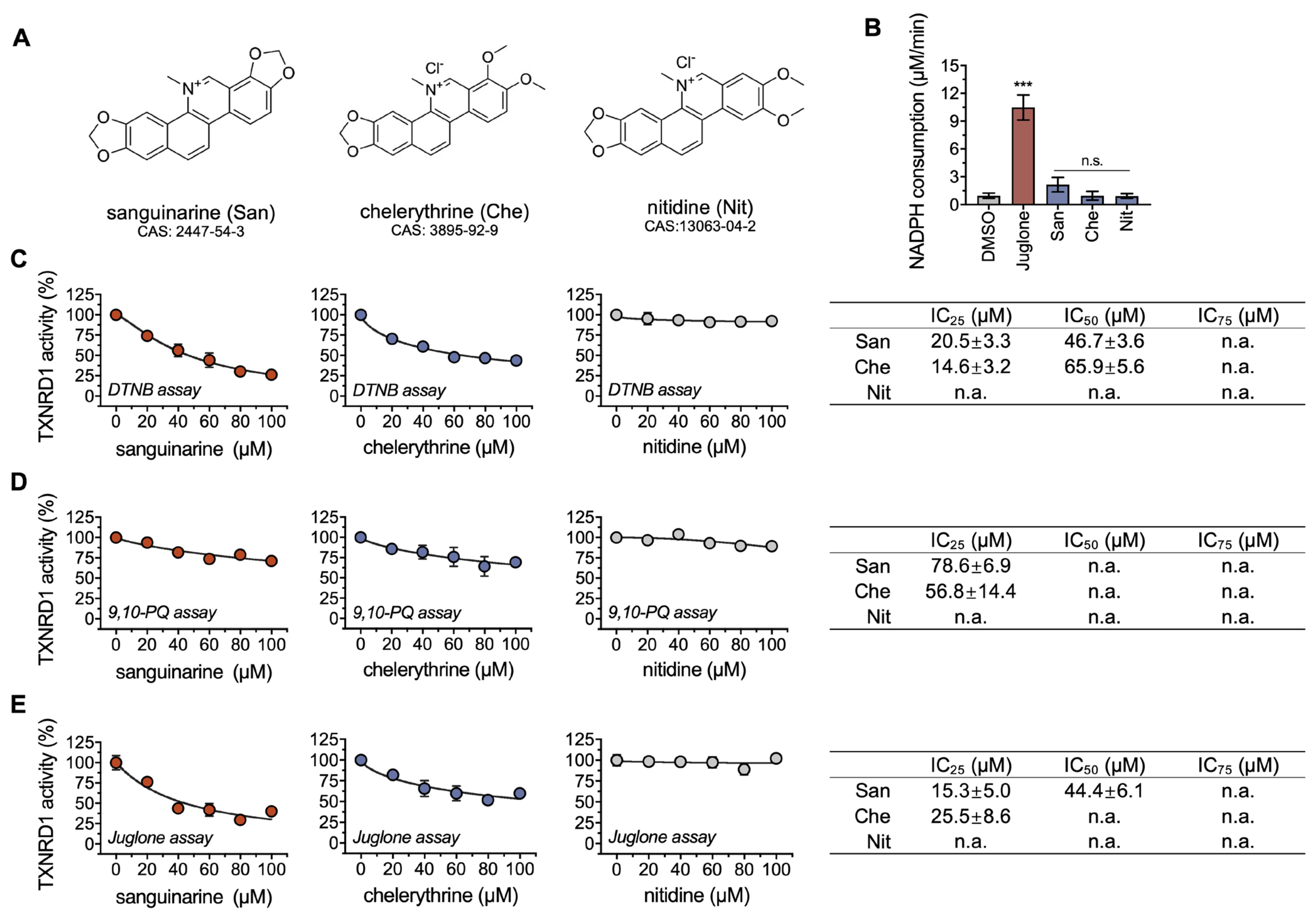
2.1. Chelerythrine and Sanguinarine Are Inhibitors of TXNRD1

TXNRD1 is a redox-active reductase that catalyzes multiple low-weight substrates, such as quinones, disulfide compounds, and selenides [34]. NADPH is the electron donor for TXNRD1 to reduce its substrates. The oxidation activity of NADPH can be monitored to verify whether an electron transfer occurs between TXNRD1 and the aforementioned three benzophenanthridine alkaloids. Compared with DMSO, no significant change was observed in these three compounds. In contrast, juglone, a substrate of TXNRD1, exhibited strong NADPH oxidation (Figure 1A,B). Clearly, these three compounds, in particular chelerythrine and nitidine, are not substrates of TXNRD1. It should be noted that we observed a substantial decrease in absorbance at 340 nm (from ~1.2 to ~1.0) at the first minute of the reaction in the absence of TXNRD1 and NADPH. We suspect that the phenomenon was caused by the compounds themselves and that it did not affect the enzyme-based activity test.
We then assessed the inhibitory effects of three benzophenanthridine alkaloids on wild-type TXNRD1. After a 1 h incubation, chelerythrine and sanguinarine, rather than nitidine, exhibited dose-response inhibitions on TXNRD1 in the DTNB reduction (Figure 1C), 9,10-PQ reduction (Figure 1D), and juglone reduction (Figure 1E). As known, DTNB and 9,10-PQ are Sec-dependent substrates of TXNRD1, while juglone is not. In the DTNB-reduction activity assay, the IC50 value of chelerythrine was 65.9 μM, about 1.5-fold higher than that of sanguinarine (46.7 μM), suggesting that sanguinarine is more potent. Additionally, the inhibitory effects of sanguinarine on TXNRD1’s activity were consistent with a previous study [27]. These results indicated that chelerythrine is a novel inhibitor of TXNRD1 in a dose-dependent manner and led us to further investigate the inhibition details of chelerythrine and sanguinarine on TXNRD1.

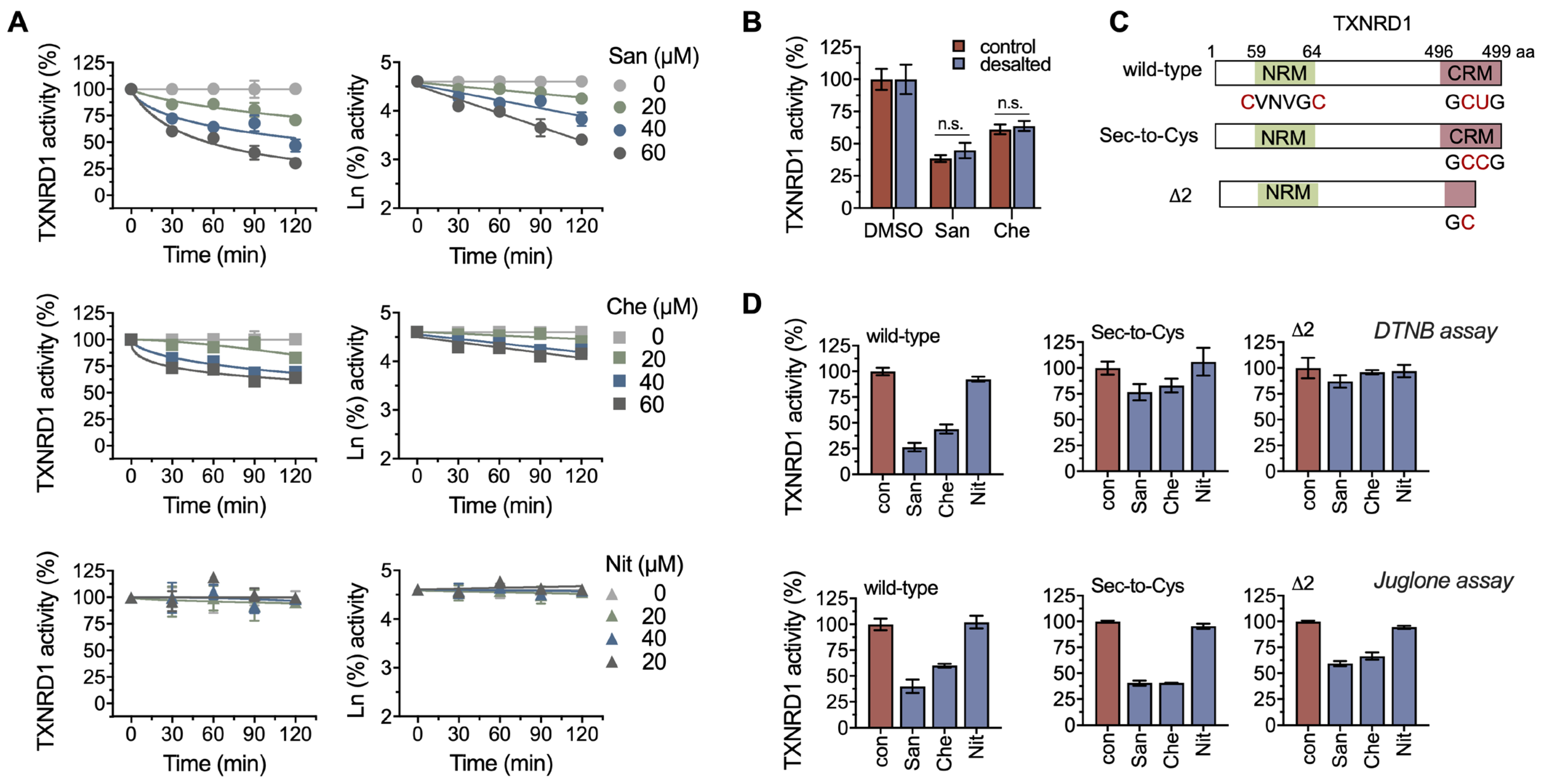
2.2. Chelerythrine and Sanguinarine Primarily Target the Hyperreactive Selenocysteine Residues

Previously, it was reported that benzophenanthridine alkaloids can form adducts with the thiol groups of proteins [35,36]. In this experiment, we tested the possible modification of chelerythrine and sanguinarine on TXNRD1. The results showed that chelerythrine and sanguinarine significantly inhibited TXNRD1 in a time-dependent manner (Figure 2A), with a kinact of 6.29 × 10⁵ μM−1 min−1 for chelerythrine and 1.56 × 10⁻⁴ μM−1 min−1 for sanguinarine. Removing the free compounds using a desalting column could not rescue the enzyme activity, indicating that chelerythrine and sanguinarine irreversibly inhibit TXNRD1 (Figure 2B).
TXNRD1 is a selenoprotein that typically functions as a dimer. The catalytic pocket contains an N-terminal GSR-like “CVNVGC” motif from one subunit and a C-terminal selenocysteine-containing “GCUG” redox motif from the other subunit (Figure 2C) [37,38,39]. Here, we used two TXNRD1 mutants, the Sec-to-Cys and Δ2 mutants of the enzyme, to determine the residue(s) that may be targeted by chelerythrine. The Sec-to-Cys mutant indicated that the selenocysteine at position 498 was replaced with cysteine. The Δ2 mutant indicated that the last two residues (selenocysteine and glycine) of TXNRD1 were deleted. Both chelerythrine and sanguinarine exhibited strong inhibition on wild-type TXNRD1, weaker inhibition on its Sec-to-Cys mutant, and no inhibition on the Δ2 mutant. These above-mentioned results suggest that chelerythrine and sanguinarine may inhibit TXNRD1 by modifying the Sec498 residue of the enzyme.
However, the possibility that chelerythrine and sanguinarine also modify the Cys residues of TXNRD1 (Figure 2D) cannot be ruled out. Juglone has been reported as a Sec-independent substrate of TXNRD1, and the reduction of juglone by TXNRD1 is mainly dependent on the FAD/CVNVGC motif [40,41]. The inhibition of chelerythrine and sanguinarine on juglone reduction suggests that the cysteine residue(s) of TXNRD1 can be targeted by chelerythrine or other benzophenanthridine alkaloids (Figure 2D).

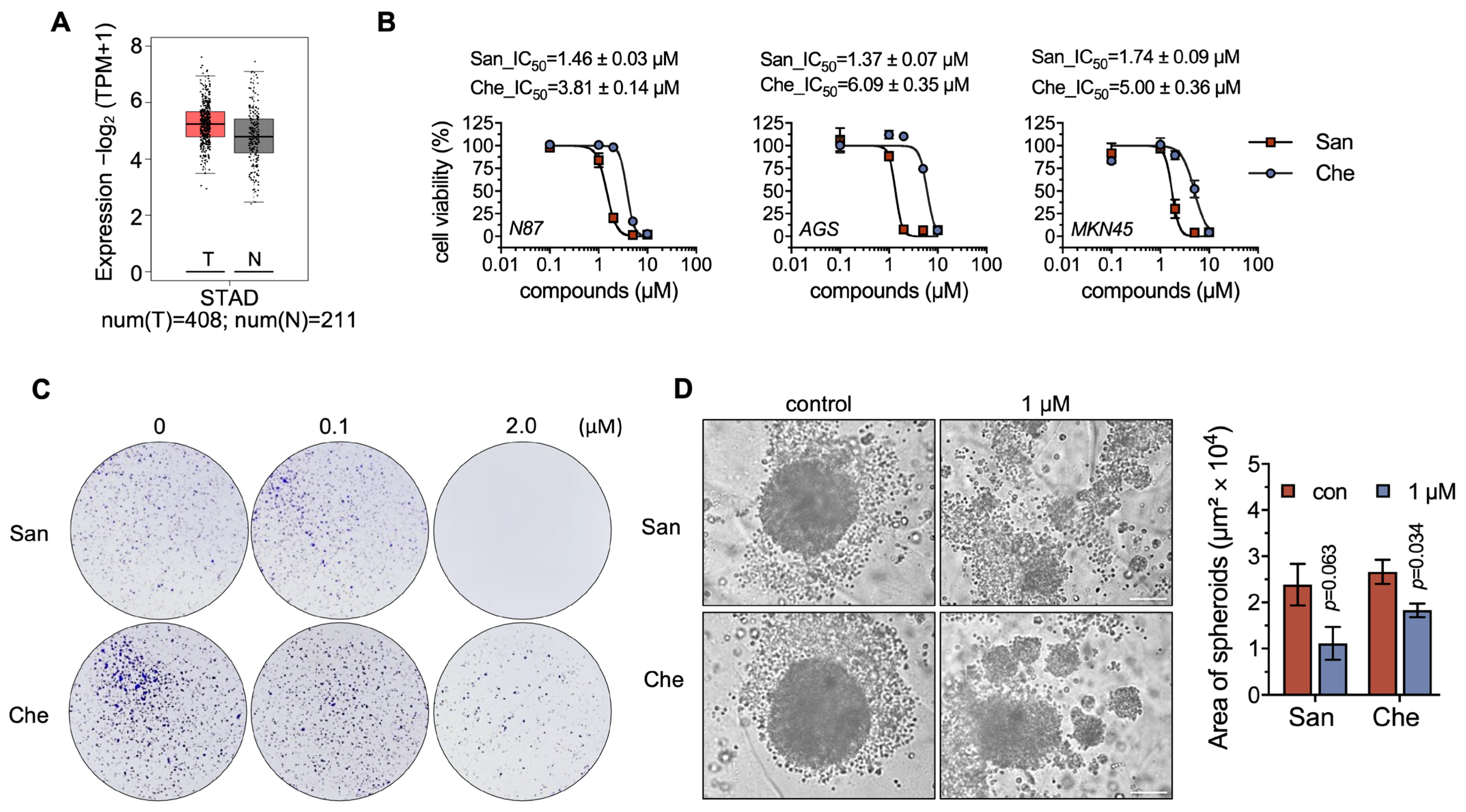
2.3. Chelerythrine and Sanguinarine Suppress the Proliferation of Gastric Cancer Cells

TXNRD1 is commonly overexpressed in cancer cells [42]. We analyzed the expression level of TXNRD1 in gastric cancer using the GEPIA2 software (http://gepia2.cancer-pku.cn (accessed on 1 July 2023)). Compared to normal tissues, TXNRD1 was up-regulated in gastric tumors, suggesting that TXNRD1 could be a potential cellular target for the treatment of gastric cancers (Figure 3A). To elucidate the anti-tumor mechanism of chelerythrine and sanguinarine, we then assessed the cytotoxicity of chelerythrine and sanguinarine in gastric cancer cell lines. After being treated with the indicated compounds for 24 h, the cell viability of NCI-N87 cells decreased (Figure 3B). The IC50 values for chelerythrine and sanguinarine in NCI-N87 cells were 3.81 μM and 1.46 μM, respectively.
In addition to NCI-N87 cells, we tested the cytotoxicity of chelerythrine and sanguinarine on MKN45 and AGS cells (Figure 3B). The cytotoxicity profiles of chelerythrine and sanguinarine were highly similar in these three cancer cells, indicating that benzophenanthridine alkaloids, such as chelerythrine and sanguinarine, may exhibit anti-tumor activity in multiple gastric cancer cell lines. Additionally, the colony formation assay demonstrated that 2 μM chelerythrine or sanguinarine suppressed cell proliferation activity in NCI-N87 cells (Figure 3C).
Tumor spheroids can mimic important aspects of solid tumors, such as hypoxia and metabolic heterogeneity. In this study, we developed tumor spheroids using NCI-N87 cells and subsequently treated the spheroids with chelerythrine and sanguinarine. The size of the spheroids indicated that the growth of tumor spheroids was inhibited by 1 μM chelerythrine to about 70% and by 1 μM sanguinarine to approx. 50% (Figure 3D). A concentration of 5 μM chelerythrine and sanguinarine was also used in this assay, but it resulted in the rapid and forced cell death of the spheroids. These results proved that either chelerythrine or sanguinarine can suppress the proliferation of gastric cancer cells.

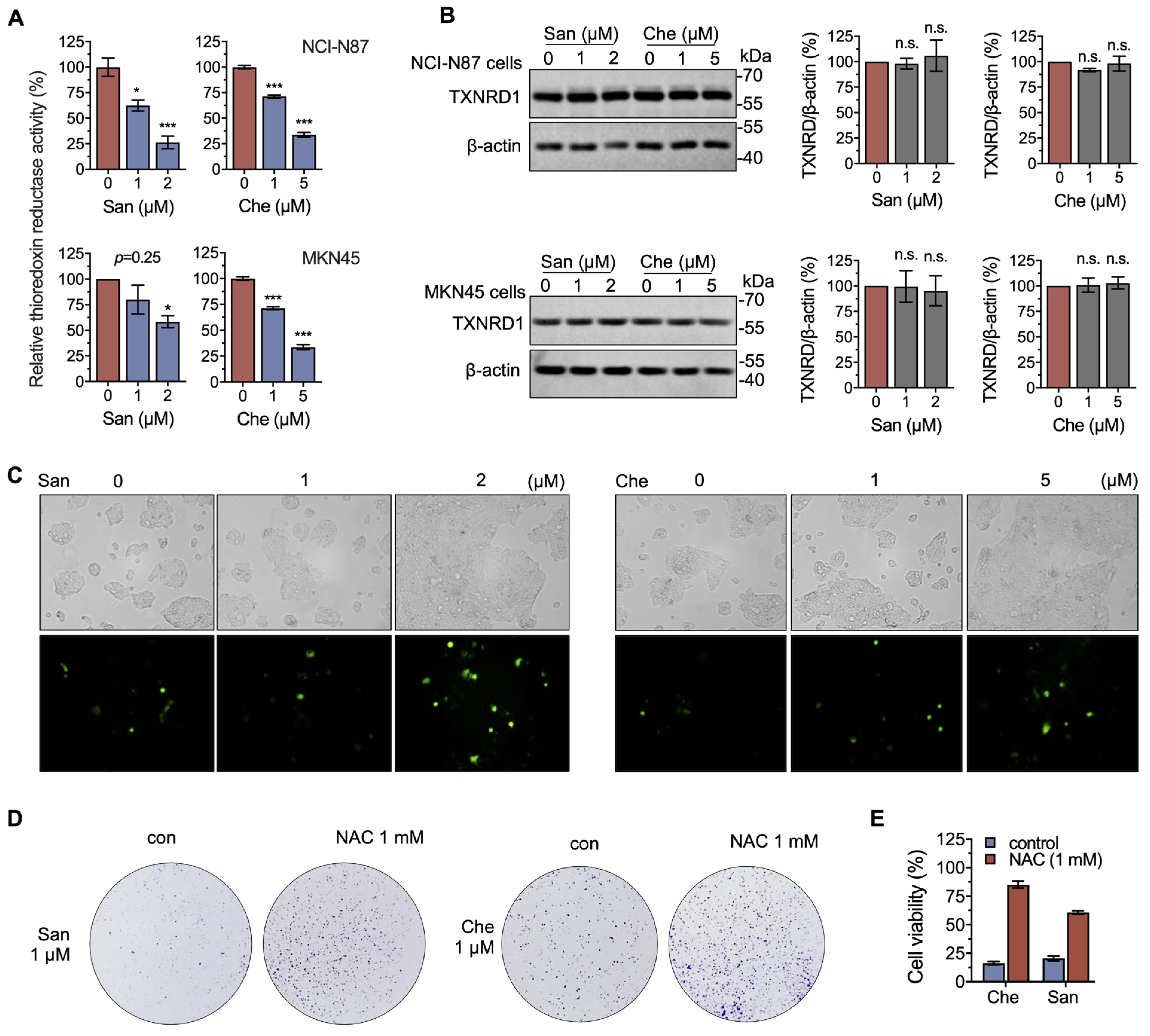
2.4. Chelerythrine and Sanguinarine Induce Oxidative Stress in Gastric Cancer Cells

The cellular redox homeostasis is maintained by the TXN system and the GSH system. These two systems work cooperatively and competitively [5,43]. We next revealed whether the cellular redox balance was perturbed when treating gastric cells with chelerythrine and sanguinarine. We demonstrated that after being treated with either 5 μM chelerythrine or 2 μM sanguinarine for 4 h, cellular TXNRD1 activity in NCI-N87 cells was significantly reduced to approx. 25%, compared to the untreated control (Figure 4A). Around 50% of cellular TXNRD activity in MKN45 cells could be inhibited by 5 μM chelerythrine or 2 μM sanguinarine (Figure 4A). Notably, the expression level of TXNRD1 in these cells was not changed (Figure 4B), indicating that chelerythrine or sanguinarine decreased cellular TXNRD1 activity by directly inhibiting the enzyme.
Furthermore, by using the ROS probe DCFH-DA, we showed a dramatic increase in cellular ROS when treated with 5 μM chelerythrine or 2 μM sanguinarine (Figure 4C), indicating that chelerythrine and sanguinarine induce oxidative stress in NCI-N87 cells. Meanwhile, the anti-proliferation activity of chelerythrine and sanguinarine could be completely rescued by 1 mM NAC (Figure 4D), and the cytotoxicity of these two compounds was counteracted by NAC (Figure 4E). These results suggest that chelerythrine and sanguinarine induce cell death in a ROS-dependent manner.

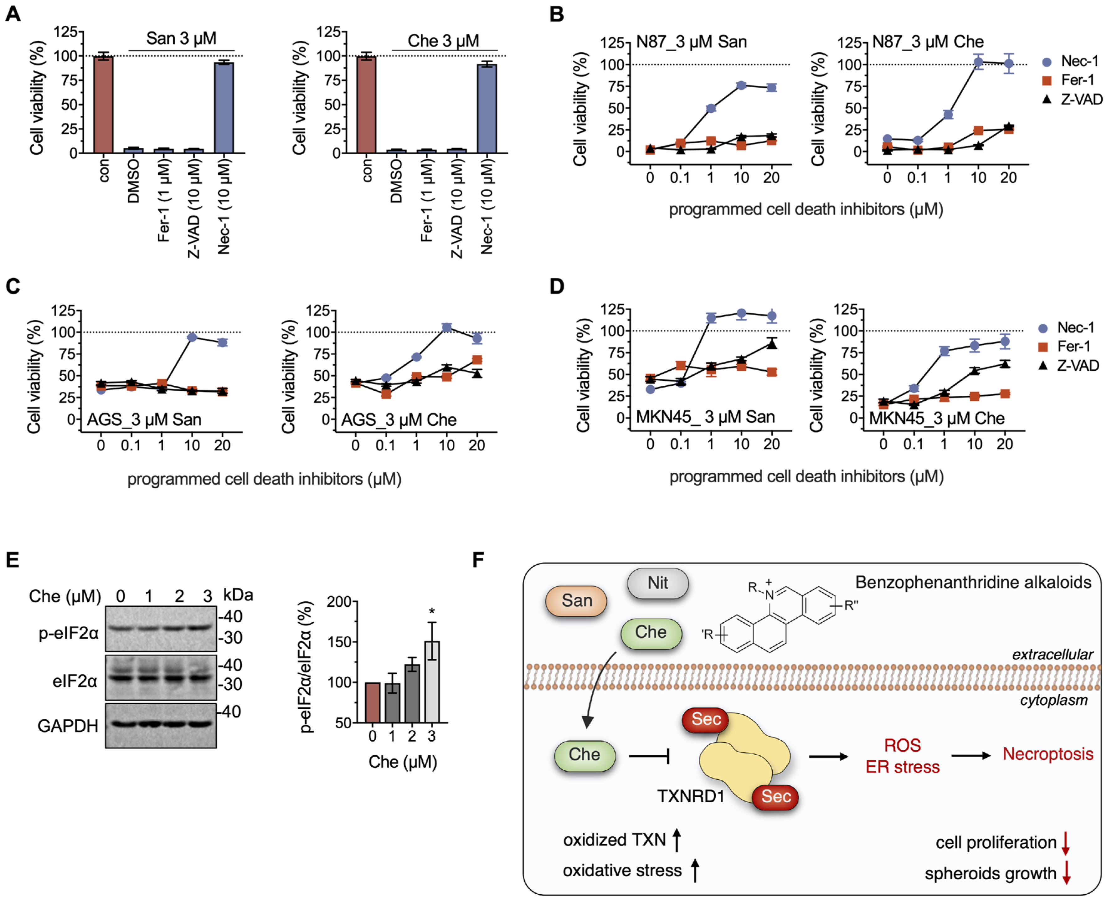
2.5. Chelerythrine and Sanguinarine Promote Necroptosis in Gastric Cancer Cells

To further elucidate the anti-tumor mechanisms of chelerythrine and sanguinarine, especially stress-induced programmed cell death (PCD) in gastric cancer cell lines, we introduced PCD inhibitors to the incubation system. A total of 10 μM RIPK1 inhibitor Nec-1, instead of the ferroptosis inhibitor Fer-1 (1 μM) or the pan-caspase inhibitor Z-VAD-FMK (10 μM), rescued cell death induced by both chelerythrine and sanguinarine in NCI-N87 cells (Figure 5A). The protective effect of Nec-1 on chelerythrine and sanguinarine was observed at concentrations ranging from 0.1 μM to 1 μM. Furthermore, 10 μM Nec-1 completely rescued cell death induced by chelerythrine and sanguinarine (Figure 5B). Meanwhile, Nec-1 showed protective activity in both AGS and MKN45 cells, indicating that chelerythrine and sanguinarine induce necroptosis in multiple gastric cancer cell lines (Figure 5C,D).
Interestingly, Fer-1 and Z-VAD-FMK showed no effect on the rescue of cells against chelerythrine and sanguinarine at low concentrations and only slight amelioration at high concentrations. It can be inferred from the results that 3 μM chelerythrine promoted non-specific lipid peroxidation or DNA damage (Figure 5C,D). However, this circumstance did not ultimately lead to typical ferroptosis and apoptosis in gastric cancer cells [44,45,46].
Endoplasmic reticulum (ER) stress is one of the inducers of necroptosis [47,48,49]. Here, we found that the protein level of p-eIF2α, increased when the NCI-N87 cells were treated with 3 μM chelerythrine for 36 h, suggesting that unfolded protein response (UPR) is initiated by ER stress, which ultimately triggers necroptosis in NCI-N87 cells (Figure 5E,F).

3. Discussion

The high energetic needs for cell growth in cancer cells are naturally occurring but also unavoidably confer redox vulnerabilities in the chemotherapy of various cancers [50,51,52,53]. Antioxidant enzymes, such as TXNRD, glutathione reductase (GSR), and xCT/SLC7A11, are commonly up-regulated in cancer cells to counteract the unwanted oxidation derived from metabolism [51,54]. The perturbations of the cellular redox balance can trigger programmed cell death, including ferroptosis and apoptosis [45,50,55]. Here, we showed that two natural compounds, chelerythrine and sanguinarine, suppress the growth of gastric cancer cells by inhibiting TXNRD1 and inducing oxidative stress in NCI-N87 cells. TXNRD1 is a fundamental enzyme in regulating the reduced status of TXN and a further participant in protein oxidation repair, cystine reduction, and DNA synthesis [4,56,57]. Our data showed that after being exposed to either chelerythrine or sanguinarine for 4 h, cellular TXNRD1 activity rapidly decreased. The loss of activity in TXNRD1 led to ROS-dependent cell death, which supports a redox-targeting strategy in the chemotherapy of gastric cancer.
Apart from the stressed oligomerized forms [58,59], TXNRD1 is generally a homodimeric selenoenzyme with a highly reactive selenocysteine at position 498, which has been considered a potential drug target in cancer therapy [58,59,60]. We showed here that chelerythrine is capable of modifying the Sec498 residue of TXNRD1, which is consistent with most TXNRD1 inhibitors. Meanwhile, our previous results revealed that Cys498 could also be attacked by electrophiles [21,61,62,63,64]. This study also provides evidence that chelerythrine interacts with Cys498. It has been reported that inhibiting TXNRD1 with small molecules may convert the enzyme from antioxidant to pro-oxidant, a phenomenon known as SecTRAPs [42,65]. It can be inferred that the features of TXNRD1 also contribute to the anti-cancer properties of chelerythrine.
Chelerythrine, nitidine, and sanguinarine belong to the group of benzophenanthridine alkaloids. The anti-tumor activity of benzophenanthridine alkaloids has been revealed in recent years. Sanguinarine induces p53-dependent up-regulation of miR-16, leading to cell arrest and apoptosis, as well as inverting EMT through Wnt/β-catenin signaling [28,29,30]. Most studies have revealed that sanguinarine or chelerythrine promotes apoptosis in cancer cells through various mechanisms [33]. Surprisingly, we found that Nec-1, a RIPK1 inhibitor, could rescue the cytotoxicity of both chelerythrine and sanguinarine, indicating that these compounds induce necroptotic cell death in NCI-N87 cells. Meanwhile, it has been reported that chelerythrine promotes necroptosis signaling in gliomas [66]. These results suggest that the cytotoxicity of benzophenanthridine alkaloids in human cancers varies depending on cell type and genetic differences. However, we showed here that chelerythrine and sanguinarine induced necroptosis and ROS-dependent cell death, but the underlying mechanism has not been fully investigated, and this should be clarified in future studies.
Previous studies have shown that 3D cell culture is more representative of the in vivo environment than 2D culture [67]. In this study, we established a cultivation and assay for gastric tumor spheroids to overcome the limitations of 2D cell culture. The tumor spheroids exhibited the physiological characteristics of solid tumors, such as cell–cell contact formation, reduced proliferation, and hypoxia environment, all of which can impact the effectiveness of various anti-tumor drugs. Notably, the results showed that the growth of NCI-N87-derived spheroids was significantly inhibited, indicating the therapeutic potential of chelerythrine and sanguinarine in chemotherapy. Furthermore, the cellular effects of chelerythrine and sanguinarine were recently investigated in vivo [66,68,69]. The results suggest that chelerythrine, as well as sanguinarine, is a potential candidate for cancer chemotherapy and other diseases.

4. Materials and Methods

4.1. Chemicals and Reagents

The chemicals used in this study include sanguinarine (San, HY-N0052, MCE, Shanghai, China), chelerythrine chloride (Che, HY-12048, MCE), nitidine chloride (Nit, HY-N0498), z-VAD-FMK (Z-VAD, S81415, Yuanye, Shanghai, China), necrostatin-1 (Nec-1, HY-15760, MCE), nicotinamide adenine dinucleotide phosphate (NADPH, S10103, Yuanye), ferrostatin-1 (Fer-1, S81461, Yuanye), 5,5′-dithiobis-(2-nitrobenzoic acid) (DTNB, S19139, Yuanye), 5-Hydroxy-1,4-naphthoquinone (juglone, H47003, Sigma-Aldrich, St. Louis, MO, USA), 9,10-phenanthrene quinone (9,10-PQ, P106382, Aladdin, Shanghai, China), and N-acetyl-L-cysteine (NAC, A601127, Sangon, Shanghai, China).
The proteins used in this study include bovine insulin (S12033, Yuanye), recombinant rat TXNRD1 (16.8 U/mg), and recombinant human TXN1. The latter two proteins were recombinantly expressed in E. coli BL21(DE3) strains and chromatographically purified to homogeneity as one band on SDS gels, according to previously reported methods [59,70].
The antibodies used in this study include anti-TXNRD1 (1:5000, 67728, ProteinTech, Wuhan, China), anti-β-actin (1:20,000, 66009, Proteintech), anti-phospho-eIF2A (Ser51) (1:1000, R22946, Zenbio, Chengdu, China), anti-eIF2A (1:1000, 201137, Zenbio), anti-GAPDH (1:10,000, 60004-1-Ig, ProteinTech), goat anti-mouse IgG (SA00001-1, ProteinTech), and goat anti-rabbit IgG (SA00001-2, ProteinTech).
The critical commercial assays used in this study include the BCA protein kit (P0012, Beyotime, Shanghai, China) and Bradford protein kit (C503031, Sangon).

4.2. Cancer Cell Culture

Human gastric cancer cells NCI-N87 (CL-0169, Procell, Wuhan, China), AGS (CL-0022, Procell), and MKN45 (CL-0292, Procell) were cultured in RPMI 1640 medium (PM150110, Procell). All the cultured cells were supplemented with 10% fetal bovine serum (FBS, 164210, Procell), 100 U/mL penicillin, and 100 mg/mL streptomycin (P/S, C0222, Beyotime) in a humidified incubator (HealForce, Shanghai, China) with an atmosphere of 5% CO2 and a temperature of 37 °C.

4.3. Cell Viability

Cell viability was assayed using the MTT assay. Initially, cells were seeded at a density of 5000 cells per well in a 96-well plate and cultured overnight. The next day, cells were exposed to compounds for the indicated time. After incubation, the medium was removed, and a fresh medium containing 0.5 mg/mL of MTT was added and incubated continuously for 2–4 h. Subsequently, the formazan crystals were dissolved in 100 μL of dimethyl sulfoxide (DMSO), and the absorbance at 570 nm was measured with 630 nm as a reference by using a SpectraMax ABS plate reader (Molecular Devices, Sunnyvale, CA, USA).

4.4. Cell Proliferation

Cells were initially seeded at a density of 1000 cells per well in 6-well plates and allowed to adhere overnight. Cells were then exposed to sanguinarine or chelerythrine for 12 h. Subsequently, the cells were cultured for 1–2 weeks, with medium replaced every 3 days. After incubation, the colonies were fixed with a 4% paraformaldehyde solution for 1 h and stained with 0.1% crystal violet stain for 10 min.

4.5. Spheroid Formation

NCI-N87 cells (5000 cells per well) were seeded into ultra-low attachment 96-well plates with 4% Matrix gel (356234, Corning, Corning, NY, USA). The formed spheroids were then treated with compounds and grown for one week. Finally, experimental images of the spheroids were taken with a microscope (DSY-LV2000, Crisoptical, Beijing, China).

4.6. Western Blotting

Cancer cells (200,000 cells per well) were seeded into 6-well plates and treated with compounds for the indicated time. After incubation, cells were lysed using RIPA buffer containing protease inhibitors and phosphatase inhibitors. The protein concentrations were determined by BCA assay, and BSA was used as protein standard. In parallel, the cell lysate samples with equal amounts of proteins were separated using SDS-PAGE, and all protein bands were transferred into polyvinylidene difluoride (PVDF) membranes (0.45 μm, Millipore, Burlington, MA, USA). The membranes were blocked with 5% (m/v) non-fat milk for 60 min at room temperature and then blotted with primary antibodies overnight at 4 °C. After washing with TBST three times, the membranes were incubated with secondary antibodies at room temperature for 1 h and developed in ECL solution (AP34L024, Life-iLab, Shanghai, China).

4.7. Cellular TXNRD Activity

Cellular TXNRD activity was assayed by a TXN1-coupled end-point insulin assay [71]. In brief, an appropriate amount of cell lysates was added into a master mixture containing 80 mM Hepes buffer (pH 7.5), 15 μM TXN1, 300 μM insulin, 660 μM NADPH, and 3 mM EDTA. A reaction mixture without TXN1 was used as a background control. Samples were incubated at 37 °C for 30 min. Subsequently, 6.0 M guanidine hydrochloride (GuHCl), containing 1 mM DTNB and 20 mM EDTA (pH 7.5), was added to each well, and the whole microtitrate plate was shaken for 10 s. Subsequently, an end-point absorbance in each well at 412 nm was measured simultaneously, and the wells without enzymes were used as blanks. The TXNRD activities of cell lysates were normalized to protein concentration for accuracy comparison.

4.8. Inhibition of Compounds on Recombinant TXNRD1

Regarding the incubation system with compounds, TXNRD1 was first pre-reduced by 100 μM NADPH for 10 min and then treated with compounds at different concentrations as indicated in the experiments. After incubation, the TXNRD1 activity was determined by DTNB-reducing activity assay, unless otherwise noted. In the DTNB-reducing assay, the final reaction mixture contained 2.5 mM DTNB, 300 μM NADPH, and 10–20 nM wild-type TXNRD1 or its various mutants (100 nM) in 50 mM TE buffer (pH 7.5), and the enzyme activity was calculated by TNB formation at 412 nm (εTNB = 13,600 M−1 cm−1).
In the 9,10-PQ- and juglone-reducing activity assays, the final mixture contained 30 nM wild-type TXNRD1 or its various mutants, 30 μM 9,10-PQ or juglone, and 200 μM NADPH in 50 mM TE buffer (pH 7.5). The enzymatic activity of 9,10-PQ or juglone reduction was calculated by NADPH oxidation at 340 nm (εNADPH = 6200 M−1 cm−1). All reactions were performed three times by using an Infinite 200 PRO plate reader (Tecan, Männedorf, Switzerland) at 25 °C. The same reaction mixture lacking the enzyme was used as a reference.

4.9. ROS Detection

NCI-N87 cells were seeded into 12-well plates at 100,000 cells per well and treated with different doses of sanguinarine and chelerythrine for 4 h. After incubation, the medium was replaced with serum-free culture medium containing 10 μM DCFH-DA and incubated for 30 min at 37 °C. Cells were washed three times with serum-free cell culture medium and then documented by fluorescence microscopy.

4.10. Statistical Analysis

All experiments were performed in triplicate, and the data were presented as the mean ± SEM. The IC50 values were calculated by non-linear curve fitting using Prism 8.0 (Graphpad, version 8.4.0). Statistical differences between the two groups were analyzed using the Student’s t-test. Comparisons among multiple groups were statistically assessed by one-way analysis of variance (ANOVA), followed by a post hoc Scheffe test. The significant differences between groups were defined as * p < 0.05, ** p < 0.01, *** p < 0.001; n.s. means not significant.

5. Conclusions

This study demonstrated that chelerythrine is a novel inhibitor of TXNRD1 and revealed that both chelerythrine and sanguinarine can inhibit TXNRD1’s activity by modifying the selenocysteine residue. The anti-gastric cancer activity of chelerythrine and sanguinarine was assessed to promote ROS-dependent necroptosis in NCI-N87 cells. The results obtained in this study would be useful in finding novel inhibitors of TXNRD1 and provide new insights into the anti-tumor activity of benzophenanthridine alkaloids.

Author Contributions

Conceptualization: S.S. and J.X.; Resources: W.X. and B.S.; Investigation: M.L., S.S., Y.M., L.W., H.L., W.S. and Q.Z.; Visualization: M.L., S.S. and H.L.; Writing—original draft: S.S. and J.X.; Writing—review and editing: S.S. and J.X.; Funding acquisition: W.X., B.S. and J.X.; Supervision: J.X. All authors have read and agreed to the published version of the manuscript.

Funding

This research was funded by the National Natural Science Foundation of China (U22A20455, 31670767) and Fundamental Research Funds for the Central Universities (DUT21LK29, DUT20LK36, DUT17JC36).

Institutional Review Board Statement

Not applicable.

Informed Consent Statement

Not applicable.

Data Availability Statement

Not applicable.

Acknowledgments

The authors want to thank Elias S.J. Arnér (Karolinska Institutet, Stockholm, Sweden) for his material support; Jianjun Zhang, Yan Zhao, and Fan Sun (Department of Gastric Surgery, Cancer Hospital of Dalian University of Technology, Shenyang, China) for their experimental assistance; and Shulan Sun and Xiaoxi Li (Department of Central Laboratory, Cancer Hospital of Dalian University of Technology, Shenyang, China) for their scientific discussion.

Conflicts of Interest

The authors declare that they have no competing interests.

Sample Availability

Not applicable.

References

  1. Bjorklund, G.; Zou, L.; Wang, J.; Chasapis, C.T.; Peana, M. Thioredoxin Reductase as a Pharmacological Target. Pharmacol. Res. 2021, 174, 105854. [Google Scholar] [CrossRef] [PubMed]
  2. Arnér, E.S.J. Focus on mammalian thioredoxin reductases--important selenoproteins with versatile functions. Biochim. Biophys. Acta 2009, 1790, 495–526. [Google Scholar] [CrossRef] [PubMed]
  3. Delgobo, M.; Goncalves, R.M.; Delazeri, M.A.; Falchetti, M.; Zandona, A.; Nascimento das Neves, R.; Almeida, K.; Fagundes, A.C.; Gelain, D.P.; Fracasso, J.I.; et al. Thioredoxin reductase-1 levels are associated with NRF2 pathway activation and tumor recurrence in non-small cell lung cancer. Free Radic. Biol. Med. 2021, 177, 58–71. [Google Scholar] [CrossRef] [PubMed]
  4. Dagnell, M.; Schmidt, E.E.; Arnér, E.S.J. The A to Z of modulated cell patterning by mammalian thioredoxin reductases. Free Radic. Biol. Med. 2018, 115, 484–496. [Google Scholar] [CrossRef]
  5. Prigge, J.R.; Coppo, L.; Martin, S.S.; Ogata, F.; Miller, C.G.; Bruschwein, M.D.; Orlicky, D.J.; Shearn, C.T.; Kundert, J.A.; Lytchier, J.; et al. Hepatocyte Hyperproliferation upon Liver-Specific Co-disruption of Thioredoxin-1, Thioredoxin Reductase-1, and Glutathione Reductase. Cell Rep. 2017, 19, 2771–2781. [Google Scholar] [CrossRef] [PubMed]
  6. McLoughlin, M.R.; Orlicky, D.J.; Prigge, J.R.; Krishna, P.; Talago, E.A.; Cavigli, I.R.; Eriksson, S.; Miller, C.G.; Kundert, J.A.; Sayin, V.I.; et al. TrxR1, Gsr, and oxidative stress determine hepatocellular carcinoma malignancy. Proc. Natl. Acad. Sci. USA 2019, 116, 11408–11417. [Google Scholar] [CrossRef] [PubMed]
  7. Harris, I.S.; Treloar, A.E.; Inoue, S.; Sasaki, M.; Gorrini, C.; Lee, K.C.; Yung, K.Y.; Brenner, D.; Knobbe-Thomsen, C.B.; Cox, M.A.; et al. Glutathione and thioredoxin antioxidant pathways synergize to drive cancer initiation and progression. Cancer Cell 2015, 27, 211–222. [Google Scholar] [CrossRef]
  8. Bansal, A.; Simon, M.C. Glutathione metabolism in cancer progression and treatment resistance. J. Cell Biol. 2018, 217, 2291–2298. [Google Scholar] [CrossRef]
  9. Lu, J.; Holmgren, A. The thioredoxin antioxidant system. Free Radic. Biol. Med. 2014, 66, 75–87. [Google Scholar] [CrossRef]
  10. Lennicke, C.; Cocheme, H.M. Redox metabolism: ROS as specific molecular regulators of cell signaling and function. Mol. Cell 2021, 81, 3691–3707. [Google Scholar] [CrossRef]
  11. Cheung, E.C.; Vousden, K.H. The role of ROS in tumour development and progression. Nat. Rev. Cancer 2022, 22, 280–297. [Google Scholar] [CrossRef]
  12. Torrente, L.; DeNicola, G.M. Targeting NRF2 and Its Downstream Processes: Opportunities and Challenges. Annu. Rev. Pharmacol. Toxicol. 2022, 62, 279–300. [Google Scholar] [CrossRef]
  13. Zhang, J.; Li, X.; Zhao, Z.; Cai, W.; Fang, J. Thioredoxin signaling pathways in cancer. Antioxid. Redox Signal. 2022, 38, 403–424. [Google Scholar] [CrossRef] [PubMed]
  14. Karunanithi, S.; Liu, R.; Hou, Y.; Gonzalez, G.; Oldford, N.; Roe, A.J.; Idipilly, N.; Gupta, K.; Amara, C.S.; Putluri, S.; et al. Thioredoxin reductase is a major regulator of metabolism in leukemia cells. Oncogene 2021, 40, 5236–5246. [Google Scholar] [CrossRef] [PubMed]
  15. Zhang, J.; Duan, D.; Osama, A.; Fang, J. Natural Molecules Targeting Thioredoxin System and Their Therapeutic Potential. Antioxid. Redox Signal. 2021, 34, 1083–1107. [Google Scholar] [CrossRef]
  16. Pickering, I.J.; Cheng, Q.; Rengifo, E.M.; Nehzati, S.; Dolgova, N.V.; Kroll, T.; Sokaras, D.; George, G.N.; Arner, E.S.J. Direct Observation of Methylmercury and Auranofin Binding to Selenocysteine in Thioredoxin Reductase. Inorg. Chem. 2020, 59, 2711–2718. [Google Scholar] [CrossRef] [PubMed]
  17. Fang, J.; Zhong, L.; Zhao, R.; Holmgren, A. Ebselen: A thioredoxin reductase-dependent catalyst for alpha-tocopherol quinone reduction. Toxicol. Appl. Pharmacol. 2005, 207, 103–109. [Google Scholar] [CrossRef]
  18. Yang, R.; Sun, S.; Guo, Y.; Meng, Y.; Liu, H.; Shi, M.; Guan, S.; Xu, J. Anti-Inflammatory Effect of Dimethyl Fumarate Associates with the Inhibition of Thioredoxin Reductase 1 in RAW 264.7 Cells. Molecules 2022, 28, 107. [Google Scholar] [CrossRef]
  19. Stafford, W.C.; Peng, X.; Olofsson, M.H.; Zhang, X.; Luci, D.K.; Lu, L.; Cheng, Q.; Tresaugues, L.; Dexheimer, T.S.; Coussens, N.P.; et al. Irreversible inhibition of cytosolic thioredoxin reductase 1 as a mechanistic basis for anticancer therapy. Sci. Transl. Med. 2018, 10, eaaf7444. [Google Scholar] [CrossRef]
  20. Duan, D.; Zhang, J.; Yao, J.; Liu, Y.; Fang, J. Targeting Thioredoxin Reductase by Parthenolide Contributes to Inducing Apoptosis of HeLa Cells. J. Biol. Chem. 2016, 291, 10021–10031. [Google Scholar] [CrossRef]
  21. Yang, Y.; Sun, S.; Xu, W.; Zhang, Y.; Yang, R.; Ma, K.; Zhang, J.; Xu, J. Piperlongumine Inhibits Thioredoxin Reductase 1 by Targeting Selenocysteine Residues and Sensitizes Cancer Cells to Erastin. Antioxidants 2022, 11, 710. [Google Scholar] [CrossRef]
  22. Zhang, Y.; Sun, S.; Xu, W.; Yang, R.; Yang, Y.; Guo, J.; Ma, K.; Xu, J. Thioredoxin reductase 1 inhibitor shikonin promotes cell necroptosis via SecTRAPs generation and oxygen-coupled redox cycling. Free Radic. Biol. Med. 2022, 180, 52–62. [Google Scholar] [CrossRef] [PubMed]
  23. Sung, H.; Ferlay, J.; Siegel, R.L.; Laversanne, M.; Soerjomataram, I.; Jemal, A.; Bray, F. Global Cancer Statistics 2020: GLOBOCAN Estimates of Incidence and Mortality Worldwide for 36 Cancers in 185 Countries. CA Cancer J Clin 2021, 71, 209–249. [Google Scholar] [CrossRef] [PubMed]
  24. Zhu, Y.; Hu, Y.; Zhu, X.; Zhang, J.; Yuwen, D.; Wei, X.; Tang, C.; Zhang, W. Plasma thioredoxin reductase: A potential diagnostic biomarker for gastric cancer. Carcinogenesis 2022, 43, 736–745. [Google Scholar] [CrossRef] [PubMed]
  25. Wen, C.; Wang, H.; Wu, X.; He, L.; Zhou, Q.; Wang, F.; Chen, S.; Huang, L.; Chen, J.; Wang, H.; et al. ROS-mediated inactivation of the PI3K/AKT pathway is involved in the antigastric cancer effects of thioredoxin reductase-1 inhibitor chaetocin. Cell Death Dis. 2019, 10, 809. [Google Scholar] [CrossRef]
  26. Zou, P.; Xia, Y.; Ji, J.; Chen, W.; Zhang, J.; Chen, X.; Rajamanickam, V.; Chen, G.; Wang, Z.; Chen, L.; et al. Piperlongumine as a direct TrxR1 inhibitor with suppressive activity against gastric cancer. Cancer Lett. 2016, 375, 114–126. [Google Scholar] [CrossRef]
  27. Yao, J.; Duan, D.; Song, Z.L.; Zhang, J.; Fang, J. Sanguinarine as a new chemical entity of thioredoxin reductase inhibitor to elicit oxidative stress and promote tumor cell apoptosis. Free Radic. Biol. Med. 2020, 152, 659–667. [Google Scholar] [CrossRef]
  28. Zhu, M.; Gong, Z.; Wu, Q.; Shi, X.; Su, Q.; Zhang, Y. Sanguinarine suppresses migration and metastasis in colorectal carcinoma associated with the inversion of EMT through the Wnt/beta-catenin signaling. Clin. Transl. Med. 2020, 10, 1–12. [Google Scholar] [CrossRef]
  29. Su, Q.; Fan, M.; Wang, J.; Ullah, A.; Ghauri, M.A.; Dai, B.; Zhan, Y.; Zhang, D.; Zhang, Y. Sanguinarine inhibits epithelial-mesenchymal transition via targeting HIF-1alpha/TGF-beta feed-forward loop in hepatocellular carcinoma. Cell Death Dis. 2019, 10, 939. [Google Scholar] [CrossRef]
  30. Zhang, B.; Wang, X.; Deng, J.; Zheng, H.; Liu, W.; Chen, S.; Tian, J.; Wang, F. p53-dependent upregulation of miR-16-2 by sanguinarine induces cell cycle arrest and apoptosis in hepatocellular carcinoma. Cancer Lett. 2019, 459, 50–58. [Google Scholar] [CrossRef]
  31. Cho, O.; Lee, J.W.; Kim, H.S.; Jeong, Y.J.; Heo, T.H. Chelerythrine, a novel small molecule targeting IL-2, inhibits melanoma progression by blocking the interaction between IL-2 and its receptor. Life Sci. 2023, 320, 121559. [Google Scholar] [CrossRef]
  32. Hong, Y.; Xu, W.Q.; Feng, J.; Lou, H.; Liu, H.; Wang, L.; Cui, H.; Jiang, L.T.; Xu, R.C.; Xu, H.H.; et al. Nitidine chloride induces cardiac hypertrophy in mice by targeting autophagy-related 4B cysteine peptidase. Acta Pharmacol. Sin. 2023, 44, 561–572. [Google Scholar] [CrossRef] [PubMed]
  33. Plazas, E.; Avila, M.M.; Munoz, D.R.; Cuca, S.L. Natural isoquinoline alkaloids: Pharmacological features and multi-target potential for complex diseases. Pharmacol. Res. 2022, 177, 106126. [Google Scholar] [CrossRef]
  34. Sun, S.; Zhang, Y.; Xu, W.; Yang, R.; Yang, Y.; Guo, J.; Ma, Q.; Ma, K.; Zhang, J.; Xu, J. Plumbagin reduction by thioredoxin reductase 1 possesses synergy effects with GLUT1 inhibitor on KEAP1-mutant NSCLC cells. Biomed. Pharmacother. 2022, 146, 112546. [Google Scholar] [CrossRef] [PubMed]
  35. Walterova, D.; Ulrichova; Preininger, V.; Simanek, V.; Lenfeld, J.; Lasovsky, J. Inhibition of liver alanine aminotransferase activity by some benzophenanthridine alkaloids. J. Med. Chem. 1981, 24, 1100–1103. [Google Scholar] [CrossRef] [PubMed]
  36. Belyaeva, T.; Leontieva, E.; Shpakov, A.; Mozhenok, T.; Faddejeva, M. Sensitivity of lysosomal enzymes to the plant alkaloid sanguinarine: Comparison with other SH-specific agents. Cell Biol. Int. 2003, 27, 887–895. [Google Scholar] [CrossRef]
  37. Liu, S.Y.; Stadtman, T.C. Heparin-binding properties of selenium-containing thioredoxin reductase from HeLa cells and human lung adenocarcinoma cells. Proc. Natl. Acad. Sci. USA 1997, 94, 6138–6141. [Google Scholar] [CrossRef]
  38. Lothrop, A.P.; Snider, G.W.; Ruggles, E.L.; Patel, A.S.; Lees, W.J.; Hondal, R.J. Selenium as an electron acceptor during the catalytic mechanism of thioredoxin reductase. Biochemistry 2014, 53, 654–663. [Google Scholar] [CrossRef]
  39. Fritz-Wolf, K.; Kehr, S.; Stumpf, M.; Rahlfs, S.; Becker, K. Crystal structure of the human thioredoxin reductase-thioredoxin complex. Nat. Commun. 2011, 2, 383. [Google Scholar] [CrossRef]
  40. Xu, J.; Cheng, Q.; Arner, E.S. Details in the catalytic mechanism of mammalian thioredoxin reductase 1 revealed using point mutations and juglone-coupled enzyme activities. Free Radic. Biol. Med. 2016, 94, 110–120. [Google Scholar] [CrossRef]
  41. Sun, S.; Xu, W.; Zhang, Y.; Yang, Y.; Ma, Q.; Xu, J. Menadione inhibits thioredoxin reductase 1 via arylation at the Sec498 residue and enhances both NADPH oxidation and superoxide production in Sec498 to Cys498 substitution. Free Radic. Biol. Med. 2021, 172, 482–489. [Google Scholar] [CrossRef] [PubMed]
  42. Gencheva, R.; Arnér, E.S.J. Thioredoxin Reductase Inhibition for Cancer Therapy. Annu. Rev. Pharmacol. Toxicol. 2022, 62, 4.1–4.20. [Google Scholar] [CrossRef] [PubMed]
  43. Du, Y.; Zhang, H.; Lu, J.; Holmgren, A. Glutathione and glutaredoxin act as a backup of human thioredoxin reductase 1 to reduce thioredoxin 1 preventing cell death by aurothioglucose. J. Biol. Chem. 2012, 287, 38210–38219. [Google Scholar] [CrossRef] [PubMed]
  44. Dixon, S.J.; Stockwell, B.R. The Hallmarks of Ferroptosis. Annu. Rev. Cancer Biol. 2019, 3, 35–54. [Google Scholar] [CrossRef]
  45. Stockwell, B.R. Ferroptosis turns 10: Emerging mechanisms, physiological functions, and therapeutic applications. Cell 2022, 185, 2401–2421. [Google Scholar] [CrossRef]
  46. Mishima, E.; Conrad, M. Nutritional and Metabolic Control of Ferroptosis. Annu. Rev. Nutr. 2022, 42, 275–309. [Google Scholar] [CrossRef]
  47. Saveljeva, S.; Mc Laughlin, S.L.; Vandenabeele, P.; Samali, A.; Bertrand, M.J. Endoplasmic reticulum stress induces ligand-independent TNFR1-mediated necroptosis in L929 cells. Cell Death Dis. 2015, 6, e1587. [Google Scholar] [CrossRef]
  48. Zhu, P.; Hu, S.; Jin, Q.; Li, D.; Tian, F.; Toan, S.; Li, Y.; Zhou, H.; Chen, Y. Ripk3 promotes ER stress-induced necroptosis in cardiac IR injury: A mechanism involving calcium overload/XO/ROS/mPTP pathway. Redox Biol. 2018, 16, 157–168. [Google Scholar] [CrossRef]
  49. Rellmann, Y.; Eidhof, E.; Dreier, R. Review: ER stress-induced cell death in osteoarthritic cartilage. Cell Signal 2021, 78, 109880. [Google Scholar] [CrossRef]
  50. Dixon, S.J.; Pratt, D.A. Ferroptosis: A flexible constellation of related biochemical mechanisms. Mol. Cell 2023, 83, 1030–1042. [Google Scholar] [CrossRef]
  51. Falchetti, M.; Delgobo, M.; Zancanaro, H.; Almeida, K.; das Neves, R.N.; dos Santos, B.; Stefanes, N.M.; Bishop, A.; Santos-Silva, M.C.; Zanotto-Filho, A. Omics-based identification of an NRF2-related auranofin resistance signature in cancer: Insights into drug repurposing. Comput. Biol. Med. 2022, 152, 106347. [Google Scholar] [CrossRef]
  52. Singh, A.; Daemen, A.; Nickles, D.; Jeon, S.M.; Foreman, O.; Sudini, K.; Gnad, F.; Lajoie, S.; Gour, N.; Mitzner, W.; et al. NRF2 Activation Promotes Aggressive Lung Cancer and Associates with Poor Clinical Outcomes. Clin. Cancer Res. 2021, 27, 877–888. [Google Scholar] [CrossRef] [PubMed]
  53. Binkley, M.S.; Jeon, Y.J.; Nesselbush, M.; Moding, E.J.; Nabet, B.Y.; Almanza, D.; Kunder, C.; Stehr, H.; Yoo, C.H.; Rhee, S.; et al. KEAP1/NFE2L2 Mutations Predict Lung Cancer Radiation Resistance That Can Be Targeted by Glutaminase Inhibition. Cancer Discov. 2020, 10, 1826–1841. [Google Scholar] [CrossRef] [PubMed]
  54. Hayes, J.D.; Dinkova-Kostova, A.T.; Tew, K.D. Oxidative Stress in Cancer. Cancer Cell 2020, 38, 167–197. [Google Scholar] [CrossRef] [PubMed]
  55. Yang, L.; Wang, H.; Yang, X.; Wu, Q.; An, P.; Jin, X.; Liu, W.; Huang, X.; Li, Y.; Yan, S.; et al. Auranofin mitigates systemic iron overload and induces ferroptosis via distinct mechanisms. Signal Transduct. Target. Ther. 2020, 5, 138. [Google Scholar] [CrossRef]
  56. Kelleher, Z.T.; Sha, Y.; Foster, M.W.; Foster, W.M.; Forrester, M.T.; Marshall, H.E. Thioredoxin-mediated denitrosylation regulates cytokine-induced nuclear factor kappaB (NF-kappaB) activation. J. Biol. Chem. 2014, 289, 3066–3072. [Google Scholar] [CrossRef]
  57. Zahedi Avval, F.; Holmgren, A. Molecular mechanisms of thioredoxin and glutaredoxin as hydrogen donors for Mammalian s phase ribonucleotide reductase. J. Biol. Chem. 2009, 284, 8233–8240. [Google Scholar] [CrossRef]
  58. Arnér, E.S.J. Effects of Mammalian Thioredoxin Reductase Inhibitors. Handb. Exp. Pharmacol. 2021, 264, 289–309. [Google Scholar] [CrossRef]
  59. Xu, J.; Eriksson, S.E.; Cebula, M.; Sandalova, T.; Hedstrom, E.; Pader, I.; Cheng, Q.; Myers, C.R.; Antholine, W.E.; Nagy, P.; et al. The conserved Trp114 residue of thioredoxin reductase 1 has a redox sensor-like function triggering oligomerization and crosslinking upon oxidative stress related to cell death. Cell Death Dis. 2015, 6, e1616. [Google Scholar] [CrossRef]
  60. Zhong, L.; Arner, E.S.; Holmgren, A. Structure and mechanism of mammalian thioredoxin reductase: The active site is a redox-active selenolthiol/selenenylsulfide formed from the conserved cysteine-selenocysteine sequence. Proc. Natl. Acad. Sci. USA 2000, 97, 5854–5859. [Google Scholar] [CrossRef]
  61. Duan, D.; Zhang, B.; Yao, J.; Liu, Y.; Sun, J.; Ge, C.; Peng, S.; Fang, J. Gambogic acid induces apoptosis in hepatocellular carcinoma SMMC-7721 cells by targeting cytosolic thioredoxin reductase. Free Radic. Biol. Med. 2014, 69, 15–25. [Google Scholar] [CrossRef] [PubMed]
  62. Duan, D.; Zhang, B.; Yao, J.; Liu, Y.; Fang, J. Shikonin targets cytosolic thioredoxin reductase to induce ROS-mediated apoptosis in human promyelocytic leukemia HL-60 cells. Free Radic. Biol. Med. 2014, 70, 182–193. [Google Scholar] [CrossRef] [PubMed]
  63. Zhang, J.; Duan, D.; Song, Z.L.; Liu, T.; Hou, Y.; Fang, J. Small molecules regulating reactive oxygen species homeostasis for cancer therapy. Med. Res. Rev. 2021, 41, 342–394. [Google Scholar] [CrossRef] [PubMed]
  64. Zhang, H.; Cao, D.; Cui, W.; Ji, M.; Qian, X.; Zhong, L. Molecular bases of thioredoxin and thioredoxin reductase-mediated prooxidant actions of (-)-epigallocatechin-3-gallate. Free Radic. Biol. Med. 2010, 49, 2010–2018. [Google Scholar] [CrossRef]
  65. Anestal, K.; Prast-Nielsen, S.; Cenas, N.; Arner, E.S. Cell death by SecTRAPs: Thioredoxin reductase as a prooxidant killer of cells. PLoS ONE 2008, 3, e1846. [Google Scholar] [CrossRef]
  66. Wang, P.; Zheng, S.Y.; Jiang, R.L.; Wu, H.D.; Li, Y.A.; Lu, J.L.; Xiong, Y.; Han, B.; Lin, L. Necroptosis signaling and mitochondrial dysfunction cross-talking facilitate cell death mediated by chelerythrine in glioma. Free Radic. Biol. Med. 2023, 202, 76–96. [Google Scholar] [CrossRef]
  67. Fennema, E.; Rivron, N.; Rouwkema, J.; van Blitterswijk, C.; de Boer, J. Spheroid culture as a tool for creating 3D complex tissues. Trends Biotechnol. 2013, 31, 108–115. [Google Scholar] [CrossRef]
  68. Ge, Q.; Shi, Z.; Zou, K.A.; Ying, J.; Chen, J.; Yuan, W.; Wang, W.; Xiao, L.; Lin, X.; Chen, D.; et al. Protein phosphatase PPM1A inhibition attenuates osteoarthritis via regulating TGF-beta/Smad2 signaling in chondrocytes. JCI Insight 2023, 8, e166688. [Google Scholar] [CrossRef]
  69. Wang, Z.; Lu, J.; Yuan, Z.; Pi, W.; Huang, X.; Lin, X.; Zhang, Y.; Lei, H.; Wang, P. Natural Carrier-Free Binary Small Molecule Self-Assembled Hydrogel Synergize Antibacterial Effects and Promote Wound Healing by Inhibiting Virulence Factors and Alleviating the Inflammatory Response. Small 2023, 19, e2205528. [Google Scholar] [CrossRef]
  70. Arnér, E.S.J.; Sarioglu, H.; Lottspeich, F.; Holmgren, A.; Bock, A. High-level expression in Escherichia coli of selenocysteine-containing rat thioredoxin reductase utilizing gene fusions with engineered bacterial-type SECIS elements and co-expression with the selA, selB and selC genes. J. Mol. Biol. 1999, 292, 1003–1016. [Google Scholar] [CrossRef]
  71. Arner, E.S.; Holmgren, A. Measurement of thioredoxin and thioredoxin reductase. Curr. Protoc. Toxicol. 2001, 24, 7.4.1–7.4.14. [Google Scholar] [CrossRef] [PubMed]
Figure 1. Chelerythrine inhibits the recombinantly purified rat TXNRD1. (A) Chemical structures of sanguinarine (San), chelerythrine (Che), and nitidine (Nit). (B) Reduction of indicated compounds by TXNRD1. The reaction mixture contained 30 nM TXNRD1, 200 μM NADPH, and 30 μM compounds. (CE) Dose-response inhibition of TXNRD1 by chelerythrine and sanguinarine. TXNRD1 was incubated with the indicated compounds for 1 h at room temperature. Then, the activity of TXNRD1 was measured by three classical assays. The IC50 values, together with the IC25 and IC75 values, are shown as mean ± SEM in the table panel on the right. The values of IC25/50/75 were estimated from the dose-response curves by nonlinear curve fitting using Prism 8. n.a. means not applicable, and n.s. represents not significant, ***, p < 0.001.
Molecules 28 06842 g001
Figure 2. Chelerythrine and sanguinarine irreversibly inhibit TXNRD1 through selenol/thiol modifications. (A) Time-course inhibition of TXNRD1 by chelerythrine and sanguinarine. TXNRD1 was incubated with three compounds separately, and afterwards, the enzyme activity was assayed at various indicated times. (B) Irreversible inhibition of TXNRD1 by chelerythrine. TXNRD1 was incubated with 100 μM sanguinarine or chelerythrine for 1 h. Then the samples were subjected to NAP-5TM desalting columns to remove the free compounds. The activity of the eluted fractions was determined by using DTNB as the substrate. (C) Schematic diagram of TXNRD1 and its Sec-to-Cys mutant and UGA-truncation delta2 form used in this study. (D) Priority modification of selenocysteine by sanguinarine or chelerythrine. TXNRD1 and its mutants were incubated with 100 μM sanguinarine or chelerythrine for 1 h. The relative enzyme activity was determined using both the DTNB assay and the juglone assay. n.s. means not significant.
Molecules 28 06842 g002
Figure 3. Chelerythrine and sanguinarine can suppress the proliferation of NCI-N87 cells. (A) Expression level of TXNRD1 in gastric cancer. The analysis was performed using GEPIA2 (http://gepia2.cancer-pku.cn (accessed on 1 July 2023)). The red box indicates the tumor group, and the gray box represents the normal group. (B) Cytotoxicity of chelerythrine (Che) and sanguinarine (San) on gastric cancer cells. NCI-N87, AGS, and MKN45 cells were treated separately with chelerythrine and sanguinarine for 24 h. Afterwards, MTT assay was used to measure the cell viability. IC50 values are shown as mean ± SEM. (C) Suppression of NCI-N87 cell proliferation upon treatment with chelerythrine or sanguinarine. NCI-N87 cells were incubated with sanguinarine or chelerythrine for 12 h. After incubation, the cells were cultured for an additional two weeks before microscopic observation. (D) Tumor spheroid formation assay of NCI-N87 cells being treated with either sanguinarine or chelerythrine. Multicellular tumor spheroids were grown from NCI-N87 cells for 1 day and then treated with chelerythrine and sanguinarine for 3 days. Spheroids were documented under a microscope, scale bar, 100 μm.
Molecules 28 06842 g003
Figure 4. Chelerythrine and sanguinarine induce oxidative stress by inhibiting cellular TXNRD activity. (A) Both chelerythrine (Che) and sanguinarine (San) inhibit the thioredoxin reductase activity of cellular TXNRD1. NCI-N87 cells (upper panel) and MKN45 cells (lower panel) were treated with either chelerythrine or sanguinarine for 4 h, and the cellular TXNRD activity in cell lysates was determined by using an end-point insulin-coupled TXN assay. (B) Expression levels of TXNRD1 following treatment with chelerythrine or sanguinarine. TXNRD1 expression levels were determined after chelerythrine or sanguinarine treatment for 4 h (NCI-N87 cells (upper panel) and MKN45 cells (lower panel)). (C) Chelerythrine and sanguinarine increase cellular ROS levels. NCI-N87 cells were treated with chelerythrine or sanguinarine for 4 h. The level of cellular ROS was quantified by H2DCFH-DA and documented by fluorescence microscopy. (D) The antioxidant NAC rescues the proliferation of NCI-N87 cells from chelerythrine and sanguinarine. A total of 1 mM NAC was co-treated with 1 μM chelerythrine or sanguinarine for 12 h to measure the colony formation. (E) NAC counteracts the cytotoxicity of chelerythrine and sanguinarine. Cells were treated with 2 μM sanguinarine and 5 μM chelerythrine, with or without 1 mM NAC, for 24 h. n.s. means not significant, *, p < 0.05, ***, p < 0.001.
Molecules 28 06842 g004
Figure 5. Chelerythrine and sanguinarine elicit necroptosis in NCI-N87 cells by inducing ER stress. (A) RIPK1 inhibitor Nec-1 suppresses cell death induced by chelerythrine (Che) or sanguinarine (San) in NCI-N87 cells. A total of 1 μM Fer-1, 10 μM Z-VAD, and 10 μM Nec-1 were used in this experiment. (BD) Chelerythrine induces necroptosis in gastric cancer cells. Programed cell death inhibitors ranging from 0 to 20 μM were separately added to the cultured cells that were incubated with 3 μM of either chelerythrine or sanguinarine. After cell cultivation for 24 h, cell viability was determined using MTT assay. (E) Chelerythrine-induced ER stress in NCI-N87 cells. NCI-N87 cells were treated with the indicated concentrations of chelerythrine for 36 h, and the samples were analyzed using Western blots. (F) Schematic diagram of benzophenanthridine alkaloids like chelerythrine inducing necroptosis in gastric cancer cells. Herein, cellular consequences of chelerythrine may include up-regulation of oxidized TXN, increased oxidative stress, and suppression of cell proliferation, in particular hindering the spheroids’ growth. *, p < 0.05.
Molecules 28 06842 g005
Disclaimer/Publisher’s Note: The statements, opinions and data contained in all publications are solely those of the individual author(s) and contributor(s) and not of MDPI and/or the editor(s). MDPI and/or the editor(s) disclaim responsibility for any injury to people or property resulting from any ideas, methods, instructions or products referred to in the content.

