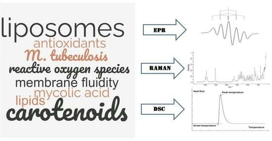
Accessing Properties of Molecular Compounds Involved in Cellular Metabolic Processes with Electron Paramagnetic Resonance, Raman Spectroscopy, and Differential Scanning Calorimetry
Abstract
1. Introduction
2. Electron Paramagnetic Resonance
2.1. EPR Spectroscopy for Studying Antioxidant Capacity
2.2. Biomedical Application of EPR Spectroscopy
3. Some Modern Prospective Applications of Raman Spectroscopy
3.1. Raman Spectroscopy for Mycobacterial Studies
3.2. Raman Spectroscopy for Studying Antioxidants and Their Biochemical Action
4. Differential Scanning Calorimetry
4.1. DSC of Molecules of a Cell Wall and Interaction with Them
4.2. DSC and Studying Antioxidant Action and Activity
5. Combining Different Methods
5.1. Combining EPR and Raman Spectroscopy Methods and Data
5.2. Combining EPR and DSC Methods
6. Conclusions
Author Contributions
Funding
Institutional Review Board Statement
Informed Consent Statement
Data Availability Statement
Conflicts of Interest
References
- Serdyuk, I.N. Physical methods and molecular biology. Biophysics 2009, 54, 238–269. [Google Scholar] [CrossRef]
- Riveline, D.; Kruse, K. Interface between Physics and Biology: Training a New Generation of Creative Bilingual Scientists. Trends Cell Biol. 2017, 27, 541–543. [Google Scholar] [CrossRef] [PubMed]
- Fang, X.; Kruse, K.; Lu, T.; Wang, J. Nonequilibrium physics in biology. Rev. Mod. Phys. 2019, 91, 045004. [Google Scholar] [CrossRef]
- Dobson, C.M. Biophysical Techniques in Structural Biology. Annu. Rev. Biochem. 2019, 88, 25–33. [Google Scholar] [CrossRef]
- Betteridge, D.J. What is oxidative stress? Metabolism 2000, 49, 3–8. [Google Scholar] [CrossRef]
- Yoshikawa, T.; Naito, Y. What is oxidative stress? Jpn. Med. Assoc. J. 2002, 45, 271–276. [Google Scholar]
- Sies, H. Oxidative stress: A concept in redox biology and medicine. Redox Biol. 2015, 4, 180–183. [Google Scholar] [CrossRef]
- Bertram, C.; Hass, R. Cellular responses to reactive oxygen species-induced DNA damage and aging. Biol. Chem. 2008, 389, 211–220. [Google Scholar] [CrossRef] [PubMed]
- Labunskyy, V.M.; Gladyshev, V.N. Role of reactive oxygen species-mediated signaling in aging. Antioxid. Redox Signal. 2013, 19, 1362–1372. [Google Scholar] [CrossRef] [PubMed]
- Chapple, I.L.C. Reactive oxygen species and antioxidants in inflammatory diseases. J. Clin. Periodontol. 1997, 24, 287–296. [Google Scholar] [CrossRef] [PubMed]
- Forrester, S.J.; Kikuchi, D.S.; Hernandes, M.S.; Xu, Q.; Griendling, K.K. Reactive oxygen species in metabolic and inflammatory signaling. Circ. Res. 2018, 122, 877–902. [Google Scholar] [CrossRef] [PubMed]
- Liou, G.Y.; Storz, P. Reactive oxygen species in cancer. Free Radic. Res. 2010, 44, 479–496. [Google Scholar] [CrossRef] [PubMed]
- Saikolappan, S.; Kumar, B.; Shishodia, G.; Koul, S.; Koul, H.K. Reactive oxygen species and cancer: A complex interaction. Cancer Lett. 2019, 452, 132–143. [Google Scholar] [CrossRef]
- Vilchèze, C.; Jacobs, W.R., Jr. The isoniazid paradigm of killing, resistance, and persistence in Mycobacterium tuberculosis. J. Mol. Biol. 2019, 431, 3450–3461. [Google Scholar] [CrossRef] [PubMed]
- Vilchèze, C. Mycobacterial cell wall: A source of successful targets for old and new drugs. Appl. Sci. 2020, 10, 2278. [Google Scholar] [CrossRef]
- Belete, T.M. Recent progress in the development of novel mycobacterium cell wall inhibitor to combat drug-resistant tuberculosis. Microbiol. Insights 2022, 15, 11786361221099878. [Google Scholar] [CrossRef]
- Bayr, H. Reactive oxygen species. Crit. Care Med. 2005, 33, S498–S501. [Google Scholar] [CrossRef]
- Su, L.J.; Zhang, J.H.; Gomez, H.; Murugan, R.; Hong, X.; Xu, D.; Jiang, F.; Peng, Z.Y. Reactive oxygen species-induced lipid peroxidation in apoptosis, autophagy, and ferroptosis. Oxidative Med. Cell. Longev. 2019, 2019, 5080843. [Google Scholar] [CrossRef]
- Cordeiro, R.M. Reactive oxygen species at phospholipid bilayers: Distribution, mobility and permeation. Biochim. Biophys. Acta (BBA)—Biomembr. 2014, 1838, 438–444. [Google Scholar] [CrossRef]
- Yusupov, M.; Wende, K.; Kupsch, S.; Neyts, E.C.; Reuter, S.; Bogaerts, A. Effect of head group and lipid tail oxidation in the cell membrane revealed through integrated simulations and experiments. Sci. Rep. 2017, 7, 5761. [Google Scholar] [CrossRef]
- Tivig, I.; Moisescu, M.G.; Savopol, T. Changes in the packing of bilayer lipids triggered by electroporation: Real-time measurements on cells in suspension. Bioelectrochemistry 2021, 138, 107689. [Google Scholar] [CrossRef] [PubMed]
- Poljsak, B.; Šuput, D.; Milisav, I. Achieving the Balance between ROS and Antioxidants: When to Use the Synthetic Antioxidants. Oxidative Med. Cell. Longev. 2013, 2013, 956792. [Google Scholar] [CrossRef] [PubMed]
- Lesgards, J.F.; Durand, P.; Lassarre, M.; Stocker, P.; Lesgards, G.; Lanteaume, A.; Prost, M.; Lehucher-Michel, M.P. Assessment of lifestyle effects on the overall antioxidant capacity of healthy subjects. Environ. Health Perspect. 2002, 110, 479–486. [Google Scholar] [CrossRef] [PubMed]
- Forman, H.J.; Zhang, H. Targeting oxidative stress in disease: Promise and limitations of antioxidant therapy. Nat. Rev. Drug Discov. 2021, 20, 689–709. [Google Scholar] [CrossRef] [PubMed]
- Andrade, S.; Ramalho, M.J.; Loureiro, J.A.; Pereira, M.C. Liposomes as biomembrane models: Biophysical techniques for drug-membrane interaction studies. J. Mol. Liq. 2021, 334, 116141. [Google Scholar] [CrossRef]
- Sahu, I.D.; Lorigan, G.A. Electron paramagnetic resonance as a tool for studying membrane proteins. Biomolecules 2020, 10, 763. [Google Scholar] [CrossRef]
- Cutshaw, G.; Uthaman, S.; Hassan, N.; Kothadiya, S.; Wen, X.; Bardhan, R. The Emerging Role of Raman Spectroscopy as an Omics Approach for Metabolic Profiling and Biomarker Detection toward Precision Medicine. Chem. Rev. 2023, 123, 8297–8346. [Google Scholar] [CrossRef]
- Bennati, M.; Prisner, T.F. New developments in high field electron paramagnetic resonance with applications in structural biology. Rep. Prog. Phys. 2005, 68. [Google Scholar] [CrossRef]
- Schiemann, O.; Prisner, T.F. Long-range distance determinations in biomacromolecules by EPR spectroscopy. Q. Rev. Biophys. 2007, 40, 1–53. [Google Scholar] [CrossRef]
- Sahu, I.D.; McCarrick, R.M.; Lorigan, G.A. Use of electron paramagnetic resonance to solve biochemical problems. Biochemistry 2013, 52, 5967–5984. [Google Scholar] [CrossRef]
- Umeno, A.; Vasudevanpillai, B.; Yasukazu, Y. In vivo ROS production and use of oxidative stress-derived biomarkers to detect the onset of diseases such as Alzheimer’s disease, Parkinson’s disease, and diabetes. Free Radic. Res. 2017, 51, 413–427. [Google Scholar] [CrossRef]
- Yoshikawa, T.; Naito, Y.; Ueda, S.; Oyamada, H.; Takemura, T.; Yoshida, N.; Sugino, S.; Kondo, M. Role of oxygen-derived free radicals in the pathogenesis of gastric mucosal lesions in rats. J. Clin. Gastroenterol. 1990, 12, 65–71. [Google Scholar] [CrossRef]
- Smith, M.A.; Harris, P.L.; Sayre, L.M.; Perry, G. Iron accumulation in Alzheimer disease is a source of redox-generated free radicals. Proc. Natl. Acad. Sci. USA 1997, 94, 9866–9868. [Google Scholar] [CrossRef]
- Phaniendra, A.; Jestadi, D.B.; Periyasamy, L. Free Radicals: Properties and Sources, Targets and Their Implication in Various Diseases. Indian J. Clin. Biochem. 2015, 30, 11–26. [Google Scholar] [CrossRef] [PubMed]
- Dizdaroglu, M.; Jaruga, P.; Birincioglu, M.; Rodriguez, H. Free radical-induced damage to DNA: Mechanisms and measurement. Free Radic. Biol. Med. 2002, 32, 1102–1115. [Google Scholar] [CrossRef] [PubMed]
- Nibbe, P.; Schleusener, J.; Siebert, S.; Borgart, R.; Brandt, D.; Westphalen, R.; Schűler, N.; Berger, B.; Peters, E.M.J.; Meinke, M.C.; et al. Oxidative stress coping capacity (OSC) value: Development and validation of an in vitro measurement method for blood plasma using electron paramagnetic resonance spectroscopy (EPR) and vitamin C. Free Radic. Biol. Med. 2023, 194, 230–244. [Google Scholar] [CrossRef]
- Morsy, M.A.; Khaled, M.M. Novel EPR characterization of the antioxidant activity of tea leaves. Spectrochim. Acta Part A 2002, 58, 1271–1277. [Google Scholar] [CrossRef]
- Polovka, M.; Brezova, V.; Stasko, A. Antioxidant properties of tea investigated by EPR spectroscopy. Biophys. Chem. 2003, 106, 39–56. [Google Scholar] [CrossRef]
- Unno, T.; Yayabe, F.; Hayakawa, T.; Tsuge, H. Electron spin resonance spectroscopic evaluation of scavenging activity of tea catechins on superoxide radicals generated by a phenazine methosulfate and NADH system. Food Chem. 2002, 76, 259–265. [Google Scholar] [CrossRef]
- Brezova, V.; Slebodova, A.; Stasko, A. Coffee as a source of antioxidants: An EPR study. Food Chem. 2009, 114, 859–868. [Google Scholar] [CrossRef]
- Zalibera, M.; Stasko, A.; Slebodova, A.; Jancovicova, V.; Cermakova, T.; Brezova, V. Antioxidant and radicalscavenging activities of Slovak honeys—An electron paramagnetic resonance study. Food Chem. 2008, 110, 512–521. [Google Scholar] [CrossRef] [PubMed]
- Brezova, V.; Polovka, M.; Stasko, A. The influence of additives on beer stability investigated by EPR spectroscopy. Spectrochim. Acta A 2002, 58, 1279–1291. [Google Scholar] [CrossRef] [PubMed]
- Polak, J.; Bartoszek, M.; Bernat, R. Comprehensive comparison of antioxidant properties of tinctures. Sci. Rep. 2019, 9, 6148. [Google Scholar] [CrossRef] [PubMed]
- Tzika, E.D.; Papadimitriou, V.; Sotiroudis, T.G.; Xenakis, A. Antioxidant properties of fruits and vegetables shots and juices: An Electron Paramagnetic Resonance study. Food Biophys. 2008, 3, 48–53. [Google Scholar] [CrossRef]
- Zang, S.; Tian, S.; Jiang, J.; Han, D.; Yu, X.; Wang, K.; Zhang, Z. Determination of antioxidant capacity of diverse fruits by electron spin resonance (ESR) and UV–vis spectrometries. Food Chem. 2017, 221, 1221–1225. [Google Scholar] [CrossRef]
- Da Porto, C.; Calligaris, S.; Celotti, E.; Nicoli, M.C. Antiradical properties of commercial cognacs assessed by the DPPH test. J. Agric. Food Chem. 2000, 48, 4241–4245. [Google Scholar] [CrossRef]
- Schwarz, M.; Rodríguez, M.C.; Martínez, C.; Bosquet, V.; Guillén, D.; Barroso, C. Antioxidant activity of Brandy de Jerez and other aged distillates and correlation with their polyphenolic content. Food Chem. 2009, 116, 29–33. [Google Scholar] [CrossRef]
- Polak, J.; Bartoszek, M.; Chora̧żewski, M.; Capacity, A. Antioxidant Capacity: Experimental Determination by EPR Spectroscopy and Mathematical Modeling. J. Agric. Food Chem. 2015, 63, 6319–6324. [Google Scholar] [CrossRef]
- Polak, J.; Bartoszek, M.; Lowe, A.R.; Postnikov, E.B.; Chora̧zewski, M. Antioxidant Properties of Various Alcoholic Beverages: Application of a Semiempirical Equation. Anal. Chem. 2020, 92, 2145–2150. [Google Scholar] [CrossRef]
- Bartoszek, M.; Polak, J.; Chora̧żewski, M. Comparison of antioxidant capacities of different types of tea using the spectroscopy methods and semi-empirical mathematical model. Eur. Food Res. Technol. 2018, 244, 595–601. [Google Scholar] [CrossRef]
- Buettner, G.R.; Jurkiewicz, B.A. Catalytic metals, ascorbate and free radicals: Combinations to avoid. Radiat. Res. 1996, 145, 532–541. [Google Scholar] [CrossRef] [PubMed]
- Takeshita, K.; Takajo, T.; Hirata, H.; Ono, M.; Utsumi, H. In vivo oxygen radical generation in the skin of the protoporphyria model mouse with visible light exposure: An L-band ESR study. J. Investig. Dermatol. 2004, 122, 1463–1470. [Google Scholar] [CrossRef] [PubMed]
- Zang, L.Y.; Shi, X.; Misra, H.P. EPR-spin trapping kinetic studies of superoxideradicals produced by photosensitized hypocrellin A. A photodynamic thera-peutic agent. Biochem. Mol. Biol. Int. 1996, 38, 685–691. [Google Scholar]
- Plonka, P.M. Electron paramagnetic resonance as a unique tool for skin and hair research. Exp. Dermatol. 2009, 18, 472–484. [Google Scholar] [CrossRef]
- Yordanov, N.D. Is our knowledge about the chemical and physical properties of DPPH enough to consider it as a primary standard for quantitative EPR spectrometry. Appl. Magn. Reson. 1996, 10, 339–350. [Google Scholar] [CrossRef]
- Mishra, K.; Ojha, H.; Chaudhury, N.K. Estimation of antiradical properties of antioxidants using DPPH·: A critical review and results. Food Chem. 2012, 130, 1036–1043. [Google Scholar] [CrossRef]
- Locatelli, M.; Gindro, R.; Travaglia, F.; Coisson, J.D.; Rinaldi, M.; Arlorio, M. Study of the DPPH· scavenging activity: Development of a free software for the correct interpretation of data. Food Chem. 2009, 114, 889–897. [Google Scholar] [CrossRef]
- Li, H.; Wang, X.; Li, Y.; Li, P.; Wang, H. Polyphenolic compounds and antioxidant properties of selected China wines. Food Chem. 2009, 112, 454–460. [Google Scholar] [CrossRef]
- Stasko, A.; Brezova, V.; Mazur, M.; Certık, M.; Kalinak, M.; Gescheidt, G. A comparative study on the antioxidant properties of Slovakian and Austrian wines. Food Sci. Technol. 2008, 41, 2126–2135. [Google Scholar]
- Stasko, A.; Liptakowa, M.; Malik, F.; Misik, V. Free radical scavening activities of white and red wines: An EPR trapping study. Appl. Magn. Reson. 2002, 22, 101–113. [Google Scholar] [CrossRef]
- Stasko, A.; Polovka, M.; Brezova, V.; Biskupic, S.; Malik, F. Tokay wines as scavengers of free radicals (an EPR study). Food Chem. 2006, 96, 185–196. [Google Scholar] [CrossRef]
- Postnikov, E.B.; Bartoszek, M.; Polak, J.; Chora̧żewski, M. Combination of Machine Learning and Analytical Correlations for Establishing Quantitative Compliance between the Trolox Equivalent Antioxidant Capacity Values Obtained via Electron Paramagnetic Resonance and Ultraviolet–Visible Spectroscopies. Int. J. Mol. Sci. 2022, 23, 11743. [Google Scholar] [CrossRef] [PubMed]
- Polovka, M. EPR spectroscopy: A tool to characterize stability and antioxidant properties of foods. J. Food Nutr. Res. 2006, 45, 1–11. [Google Scholar]
- Yim, M.B.; Chock, P.B.; Stadtman, E.R. Enzyme function of copper, zinc superoxide dismutase as a free radical generator. J. Biol. Chem. 1993, 268, 4099–4105. [Google Scholar] [CrossRef]
- Scott, S.L.; Chen, W.J.; Bakac, A.; Espenson, J.H. Spectroscopic parameters, electrode potentials, acid ionization constants, and electron exchange rates of the 2′-azinobis(3-ethylbenzothiazoline-6-sulfonate) radicals and ions. J. Phys. Chem. 1993, 97, 6710–6714. [Google Scholar] [CrossRef]
- Babić, N.; Peyrot, F. Molecular probes for evaluation of oxidative stress by in vivo EPR spectroscopy and imaging: State-of-the-art and limitations. Magnetochemistry 2019, 5, 13. [Google Scholar] [CrossRef]
- He, G.; Samouilov, A.; Kuppusamy, P.; Zweier, J.L. In vivo imaging of free radicals: Applications from mouse to man. Mol. Cell. Biochem. 2002, 234, 359–367. [Google Scholar] [CrossRef]
- Fuchs, J.; Groth, N.; Herrling, T.; Milbradt, R.; Zimmer, G.; Packer, L. Electron Paramagnetic Resonance (EPR) Imaging in Skin: Biophysical and Biochemcial Microscopy. J. Investig. Dermatol. 1992, 98, 713–719. [Google Scholar] [CrossRef][Green Version]
- Valgimigli, L.; Pedulli, G.F.; Paolini, M. Measurement of oxidative stress by EPR radical-probe technique. Free Radic. Biol. Med. 2001, 31, 708–716. [Google Scholar] [CrossRef]
- Riley, P.A. Free radicals in biology: Oxidative stress and the effects of ionizing radiation. Int. J. Radiat. Biol. 1994, 65, 27–33. [Google Scholar] [CrossRef]
- Droge, W. Free radicals in the physiological control of cell function. J. Physiol. Rev. 2002, 82, 47–95. [Google Scholar] [CrossRef] [PubMed]
- Berlett, D.S.; Stadtman, E. Protein oxidation in aging, disease, and oxidative stress. J. Biol. Chem. 1997, 272, 20313–20316. [Google Scholar] [CrossRef] [PubMed]
- Rikhireva, G.T.; Makletsova, M.G. Application of EPR Spectroscopy in the Study of Iron Metabolism in Parkinson’s Disease. Biophysics 2020, 65, 327–330. [Google Scholar] [CrossRef]
- Gallez, B.; Baudelet, C.; Jordan, B.F. Assessment of tumor oxygenation by electron paramagnetic resonance: Principles and applications. NMR Biomed. 2004, 17, 240–262. [Google Scholar] [CrossRef]
- Mignion, L.; Desmet, C.M.; Harkemanne, E.; Tromme, I.; Joudiou, N.; Wehbi, M.; Baurain, J.F.; Gallez, B. Noninvasive detection of the endogenous free radical melanin in human skin melanomas using electron paramagnetic resonance (EPR). Free Radic. Biol. Med. 2022, 190, 226–233. [Google Scholar] [CrossRef]
- He, G.; Samouilov, A.; Kuppusamy, P.; Zweier, J.L. In vivo EPR imaging of the distribution and metabolism of nitroxide radicals in human skin. J. Magn. Reson. 2001, 148, 155–164. [Google Scholar] [CrossRef]
- Lohan, S.B.; Ahlberg, S.; Mensch, A.; Höppe, D.; Giulbudagian, M.; Calderón, M.; Grether-Beck, S.; Krutmann, J.; Lademann, J.; Meinke, M.C. EPR technology as sensitive method for oxidative stress detection in primary and secondary keratinocytes induced by two selected nanoparticles. Cell Biochem. Biophys. 2017, 75, 359–367. [Google Scholar] [CrossRef]
- Krinsky, N.I. The antioxidant and biological properties of the carotenoids a. Ann. N. Y. Acad. Sci. 1998, 854, 443–447. [Google Scholar] [CrossRef]
- Stahl, W.; Sies, H. Antioxidant activity of carotenoids. Mol. Asp. Med. 2003, 24, 345–351. [Google Scholar] [CrossRef]
- Young, A.J.; Lowe, G.L. Carotenoids—Antioxidant Properties. Antioxidants 2018, 7, 28. [Google Scholar] [CrossRef]
- Johnson, E.J. The role of carotenoids in human health. Nutr. Clin. Care 2002, 5, 56–65. [Google Scholar] [CrossRef] [PubMed]
- Rao, A.V.; Rao, L.G. Carotenoids and human health. Pharmacol. Res. 2007, 55, 207–216. [Google Scholar] [CrossRef] [PubMed]
- Islam, F.; Muni, M.; Mitra, S.; Emran, T.B.; Chandran, D.; Das, R.; Rauf, A.; Safi, S.Z.; Chidambaram, K.; Dhawan, M.; et al. Recent advances in respiratory diseases: Dietary carotenoids as choice of therapeutics. Biomed. Pharmacother. 2022, 155, 113786. [Google Scholar] [CrossRef]
- Martini, D.; Negrini, L.; Marino, M.; Riso, P.; Del Bo, C.; Porrini, M. What is the current direction of the research on carotenoids and human health? An overview of registered clinical trials. Nutrients 2022, 14, 1191. [Google Scholar] [CrossRef] [PubMed]
- Polyakov, N.E.; Leshina, T.V. Certain aspects of the reactivity of carotenoids. Redox processes and complexation. Russ. Chem. Rev. 2006, 75, 1049. [Google Scholar] [CrossRef]
- Focsan, A.L.; Polyakov, N.E.; Kispert, L.D. Carotenoids: Importance in daily life—Insight gained from EPR and ENDOR. Appl. Magn. Reson. 2021, 52, 1093–1112. [Google Scholar] [CrossRef] [PubMed]
- Gruszecki, W.I.; Strzałka, K. Carotenoids as modulators of lipid membrane physical properties. Biochim. Biophys. Acta (BBA)-Mol. Basis Dis. 2005, 1740, 108–115. [Google Scholar] [CrossRef]
- Mostofian, B.; Johnson, Q.R.; Smith, J.C.; Cheng, X. Carotenoids promote lateral packing and condensation of lipid membranes. Phys. Chem. Chem. Phys. 2020, 22, 12281–12293. [Google Scholar] [CrossRef] [PubMed]
- Davies, M.J. Detection and identification of macromolecule-derived radicals by EPR spin trapping. Res. Chem. Intermed. 1993, 19, 669–679. [Google Scholar] [CrossRef]
- Gomez-Mejiba, S.E.; Zhai, Z.; Della-Vedova, M.C.; Mu noz, M.D.; Chatterjee, S.; Towner, R.A.; Hensley, K.; Floyd, R.A.; Mason, R.P.; Ramirez, D.C. Immuno-spin trapping from biochemistry to medicine: Advances, challenges, and pitfalls. Focus on protein-centered radicals. Biochim. Biophys. Acta 2014, 1840, 722–729. [Google Scholar] [CrossRef]
- Abbas, K.; Babić, N.; Peyrot, F. Use of spin traps to detect superoxide production in living cells by electron paramagnetic resonance (EPR) spectroscopy. Methods 2016, 109, 31–43. [Google Scholar] [CrossRef] [PubMed]
- Coghlan, J.G.; Flitter, W.D.; Holley, A.E.; Norell, M.; Mitchell, A.G.; Ilsley, C.D.; Slater, T.F. Detection of free radicals and cholesterol hydroperoxides in blood taken from the coronary sinus of man during percutaneous transluminal coronary angioplasty. Free Radic. Res. Commun. 1991, 14, 409–417. [Google Scholar] [CrossRef] [PubMed]
- Flitter, W.D. Free radicals and myocardial reperfusion injury. Br. Med. Bull. 1993, 49, 545–555. [Google Scholar] [CrossRef] [PubMed]
- Ashton, T.; Young, I.S.; Peters, J.R.; Jones, E.; Jackson, S.K.; Davies, B.; Rowlands, C.C. Electron spin resonance spectroscopy, exercise, and oxidative stress: An ascorbic acid intervention study. J. Appl. Physiol. 1999, 87, 2032–2036. [Google Scholar] [CrossRef] [PubMed]
- Krzyminiewski, R.; Dobosz, B.; Schroeder, G.; Kurczewska, J. ESR as a monitoring method of the interactions between TEMPO-functionalized magnetic nanoparticles and yeast cells. Sci. Rep. 2019, 10, 18733. [Google Scholar] [CrossRef]
- Liu, K.J.; Miyake, M.; Panz, T.; Swartz, H. Evaluation of DEPMPO as a spin trapping agent in biological systems. Free Radic. Biol. Med. 1999, 26, 714–721. [Google Scholar] [CrossRef]
- Timmins, G.S.; Liu, K.J.; Bechara, E.J.H.; Kotake, Y.; Swartz, H.M. Trapping of free radicals with direct in vivo EPR detection: A comparison of 5, 5-dimethyl-1-pyrroline-N-oxide and 5-diethoxyphosphoryl-5-methyl-1-pyrroline-N-oxide as spin traps for HO* and SO4*−. Free Radic. Biol. Med. 1999, 27, 329–333. [Google Scholar] [CrossRef]
- Weaver, J.; Liu, K.J. A Review of Low-Frequency EPR Technology for the Measurement of Brain pO2 and Oxidative Stress. Appl. Magn. Reson. 2021, 52, 1379–1394. [Google Scholar] [CrossRef]
- Li, H.; Pan, Y.; Yang, Z.; Rao, J.; Chen, B. Emerging applications of site-directed spin labeling electron paramagnetic resonance (SDSL-EPR) to study food protein structure, dynamics, and interaction. Trends Food Sci. Technol. 2021, 109, 37–50. [Google Scholar] [CrossRef]
- Smith, E.; Dent, G. Modern Raman Spectroscopy: A practical Approach; John Wiley & Sons: Hoboken, NJ, USA, 2019. [Google Scholar]
- Pezzotti, G. Raman spectroscopy in cell biology and microbiology. J. Raman Spectrosc. 2021, 52, 2348–2443. [Google Scholar] [CrossRef]
- Lee, K.S.; Landry, Z.; Pereira, F.C.; Wagner, M.; Berry, D.; Huang, W.E.; Taylor, G.T.; Kneipp, J.; Popp, J.; Zhang, M.; et al. Raman microspectroscopy for microbiology. Nat. Rev. Methods Primers 2021, 1, 80. [Google Scholar] [CrossRef]
- El-Mashtoly, S.F.; Gerwert, K. Diagnostics and therapy assessment using label-free Raman imaging. Anal. Chem. 2021, 94, 120–142. [Google Scholar] [CrossRef] [PubMed]
- Allakhverdiev, E.S.; Khabatova, V.V.; Kossalbayev, B.D.; Zadneprovskaya, E.V.; Rodnenkov, O.V.; Martynyuk, T.V.; Maksimov, G.V.; Alwasel, S.; Tomo, T.; Allakhverdiev, S.I. Raman spectroscopy and its modifications applied to biological and medical research. Cells 2022, 11, 386. [Google Scholar] [CrossRef]
- Shen, Y.; Wei, L.; Min, W. Raman Imaging Reveals Insights into Membrane Phase Biophysics in Cells. J. Phys. Chem. B 2023. [Google Scholar] [CrossRef] [PubMed]
- Stöckel, S.; Kirchhoff, J.; Neugebauer, U.; Rösch, P.; Popp, J. The application of Raman spectroscopy for the detection and identification of microorganisms. J. Raman Spectrosc. 2016, 47, 89–109. [Google Scholar] [CrossRef]
- Premasiri, W.R.; Lee, J.C.; Sauer-Budge, A.; Théberge, R.; Costello, C.E.; Ziegler, L.D. The biochemical origins of the surface-enhanced Raman spectra of bacteria: A metabolomics profiling by SERS. Anal. Bioanal. Chem. 2016, 408, 4631–4647. [Google Scholar] [CrossRef]
- Allen, D.M.; Einarsson, G.G.; Tunney, M.M.; Bell, S.E.J. Characterization of bacteria using surface-enhanced Raman spectroscopy (SERS): Influence of microbiological factors on the SERS spectra. Anal. Chem. 2022, 94, 9327–9335. [Google Scholar] [CrossRef]
- Mühlig, A.; Bocklitz, T.; Labugger, I.; Dees, S.; Henk, S.; Richter, E.; Andres, S.; Merker, M.; Stöckel, S.; Weber, K.; et al. LOC-SERS: A promising closed system for the identification of mycobacteria. Anal. Chem. 2016, 88. [Google Scholar] [CrossRef]
- Stöckel, S.; Stanca, A.S.; Helbig, J.; Rösch, P.; Popp, J. Raman spectroscopic monitoring of the growth of pigmented and non-pigmented mycobacteria. Anal. Bioanal. Chem. 2015, 407, 8919–8923. [Google Scholar] [CrossRef]
- Rivera-Betancourt, O.E.; Karls, R.; Grosse-Siestrup, B.; Helms, S.; Quinn, F.; Dluhy, R.A. Identification of mycobacteria based on spectroscopic analyses of mycolic acid profiles. Analyst 2013, 138. [Google Scholar] [CrossRef]
- Stöckel, S.; Meisel, S.; Lorenz, B.; Kloß, S.; Henk, S.; Dees, S.; Richter, E.; Andres, S.; Merker, M.; Labugger, I.; et al. Raman spectroscopic identification of Mycobacterium tuberculosis. J. Biophotonics 2017, 10, 727–734. [Google Scholar] [CrossRef]
- Sathyavathi, R.; Dingari, N.C.; Barman, I.; Prasad, P.S.R.; Prabhakar, S.; Rao, D.N.; Dasari, R.R.; Undamatla, J. Raman spectroscopy provides a powerful, rapid diagnostic tool for the detection of tuberculous meningitis in ex vivo cerebrospinal fluid samples. J. Biophotonics 2013, 6. [Google Scholar] [CrossRef] [PubMed]
- Crawford, A.C.; Laurentius, L.B.; Mulvihill, T.S.; Granger, J.H.; Spencer, J.S.; Chatterjee, D.; Hanson, K.E.; Porter, M.D. Detection of the tuberculosis antigenic marker mannose-capped lipoarabinomannan in pretreated serum by surface-enhanced Raman scattering. Analyst 2017, 142. [Google Scholar] [CrossRef] [PubMed]
- Owens, N.A.; Young, C.C.; Laurentius, L.B.; De, P.; Chatterjee, D.; Porter, M.D. Detection of the tuberculosis biomarker mannose-capped lipoarabinomannan in human serum: Impact of sample pretreatment with perchloric acid. Anal. Chim. Acta 2019, 1046, 140–147. [Google Scholar] [CrossRef]
- Perumal, J.; Dinish, U.S.; Bendt, A.K.; Kazakeviciute, A.; Fu, C.Y.; Ong, I.L.H.; Olivo, M. Identification of mycolic acid forms using surface-enhanced Raman scattering as a fast detection method for tuberculosis. Int. J. Nanomed. 2018, 13, 6029–6038. [Google Scholar] [CrossRef]
- Alula, M.T.; Krishnan, S.; Hendricks, N.R.; Karamchand, L.; Blackburn, J.M. Identification and quantitation of pathogenic bacteria via in-situ formation of silver nanoparticles on cell walls, and their detection via SERS. Microchim. Acta 2017, 184, 219–227. [Google Scholar] [CrossRef]
- Rawat, P.; Singh, R.N.; Ranjan, A.; Ahmad, S.; Saxena, R. Antimycobacterial, antimicrobial activity, experimental (FT-IR, FT-Raman, NMR, UV-Vis, DSC) and DFT (transition state, chemical reactivity, NBO, NLO) studies on pyrrole-isonicotinyl hydrazone. Spectrochim. Acta Part A Mol. Biomol. Spectrosc. 2017, 179, 1–10. [Google Scholar] [CrossRef]
- Thomas, R.; Mary, Y.S.; Resmi, K.S.; Narayana, B.; Sarojini, S.B.K.; Armaković, S.; Armaković, S.J.; Vijayakumar, G.; Van Alsenoy, C.; Mohan, B.J. Synthesis and spectroscopic study of two new pyrazole derivatives with detailed computational evaluation of their reactivity and pharmaceutical potential. J. Mol. Struct. 2019, 1181, 599–612. [Google Scholar] [CrossRef]
- Robert, H.M.; Usha, D.; Amalanathan, M.; Geetha, R.R.J.; Mary, M.S.M. Spectroscopic (IR, Raman, UV, NMR) characterization and investigation of reactive properties of pyrazine-2-carboxamide by anti-bacterial, anti-mycobacterial, Fukui function, molecular docking and DFT calculations. Chem. Data Collect. 2020, 30, 100583. [Google Scholar] [CrossRef]
- Gao, M.; Liu, S.; Chen, J.; Gordon, K.C.; Tian, F.; McGoverin, C.M. Potential of Raman spectroscopy in facilitating pharmaceutical formulations development–an AI perspective. Int. J. Pharm. 2021, 597, 120334. [Google Scholar] [CrossRef]
- MacGregor-Fairlie, M.; Wilkinson, S.; Besra, G.S.; Goldberg Oppenheimer, P. Tuberculosis diagnostics: Overcoming ancient challenges with modern solutions. Emerg. Top. Life Sci. 2020, 4, 435–448. [Google Scholar] [CrossRef]
- Wang, K.; Li, S.; Petersen, M.; Wang, S.; Lu, X. Detection and characterization of antibiotic-resistant bacteria using surface-enhanced Raman spectroscopy. Nanomaterials 2018, 8, 762. [Google Scholar] [CrossRef]
- Galvan, D.D.; Yu, Q. Surface-Enhanced Raman Scattering for Rapid Detection and Characterization of Antibiotic-Resistant Bacteria. Adv. Healthc. Mater. 2018, 7, 1701335. [Google Scholar] [CrossRef]
- Hassanain, W.; Johnson, C.; Faulds, K.; Graham, D.; Keegan, N. Recent advances in antibiotic resistance diagnosis using SERS: Focus on the “Big 5” challenges. Analyst 2022, 147, 4674–4700. [Google Scholar] [CrossRef]
- Wang, L.; Zhang, X.D.; Tang, J.W.; Ma, Z.W.; Usman, M.; Liu, Q.H.; Wu, C.Y.; Li, F.; Zhu, Z.B.; Gu, B. Machine learning analysis of SERS fingerprinting for the rapid determination of Mycobacterium tuberculosis infection and drug resistance. Comput. Struct. Biotechnol. J. 2022, 20, 5364–5377. [Google Scholar] [CrossRef] [PubMed]
- Ho, C.S.; Jean, N.; Hogan, C.A.; Blackmon, L.; Jeffrey, S.S.; Holodniy, M.; Banaei, N.; Saleh, A.A.E.; Ermon, S.; Dionne, J. Rapid identification of pathogenic bacteria using Raman spectroscopy and deep learning. Nat. Commun. 2019, 10, 4927. [Google Scholar] [CrossRef] [PubMed]
- Dastgir, G.; Majeed, M.I.; Nawaz, H.; Rashid, N.; Raza, A.; Ali, M.Z.; Shakeel, M.; Javed, M.; Ehsan, U.; Ishtiaq, S.; et al. Surface-enhanced Raman spectroscopy of polymerase chain reaction (PCR) products of Rifampin resistant and susceptible tuberculosis patients. Photodiagnosis Photodyn. Ther. 2022, 38, 102758. [Google Scholar] [CrossRef] [PubMed]
- Paulowski, L.; Yosef, H.; Hillemann, D.; Fischer-Knuppertz, W.; Maurer, F. Single-cell raman spectroscopy: A powerful diagnostic tool for identification and drug susceptibility testing of Mycobacterium tuberculosis. Biophys. J. 2022, 121, 214a. [Google Scholar] [CrossRef]
- Ueno, H.; Kato, Y.; Tabata, K.V.; Noji, H. Revealing the metabolic activity of persisters in mycobacteria by single-cell D2O Raman imaging spectroscopy. Anal. Chem. 2019, 91, 15171–15178. [Google Scholar] [CrossRef]
- Zyubin, A.; Lavrova, A.; Manicheva, O.; Dogonadze, M.; Belik, V.; Samusev, I. Raman spectroscopy reveals M. tuberculosis strains with different antibiotic susceptibility. Laser Phys. Lett. 2019, 16, 085602. [Google Scholar] [CrossRef]
- Zyubin, A.; Lavrova, A.; Manicheva, O.; Dogonadze, M.; Belik, V.; Demin, M.; Samusev, I. The cell-wall structure variation in Mycobacterium tuberculosis with different drug sensitivity using Raman spectroscopy in the high-wavenumber region. Laser Phys. Lett. 2020, 17, 065602. [Google Scholar] [CrossRef]
- Zyubin, A.; Lavrova, A.; Manicheva, O.; Dogonadze, M.; Belik, V.; Samusev, I. Raman spectroscopy for glutathione measurements in Mycobacterium tuberculosis strains with different antibiotic resistance. J. Raman Spectrosc. 2021, 52, 1661–1666. [Google Scholar] [CrossRef]
- Garfinkel, D. Raman Spectra and Ultraviolet Absorption of Glutathione and Possible Thiazoline Derivatives Formed from It. J. Am. Chem. Soc. 1958, 80, 4833–4835. [Google Scholar] [CrossRef]
- Qian, W.; Krimm, S. Vibrational analysis of glutathione. Biopolym. Orig. Res. Biomol. 1994, 34, 1377–1394. [Google Scholar] [CrossRef]
- Raj Rai, S.; Bhattacharyya, C.; Sarkar, A.; Chakraborty, S.; Sircar, E.; Dutta, S.; Sengupta, R. Glutathione: Role in Oxidative/Nitrosative Stress, Antioxidant Defense, and Treatments. ChemistrySelect 2021, 6, 4566–4590. [Google Scholar] [CrossRef]
- Connell, N.D.; Venketaraman, V. Control of Mycobacterium tuberculosis infection by glutathione. Recent Pat. Anti-Infect. Drug Discov. 2009, 4, 214–226. [Google Scholar] [CrossRef] [PubMed]
- Dua, K.; Rapalli, V.K.; Shukla, S.D.; Singhvi, G.; Shastri, M.D.; Chellappan, D.K.; Satija, S.; Mehta, M.; Gulati, M.; Pinto, T.D.J.A.; et al. Multi-drug resistant Mycobacterium tuberculosis & oxidative stress complexity: Emerging need for novel drug delivery approaches. Biomed. Pharmacother. 2018, 107, 1218–1229. [Google Scholar] [CrossRef] [PubMed]
- Baron, V.O.; Chen, M.; Hammarstrom, B.; Hammond, R.J.H.; Glynne-Jones, P.; Gillespie, S.H.; Dholakia, K. Real-time monitoring of live mycobacteria with a microfluidic acoustic-Raman platform. Commun. Biol. 2020, 3, 236. [Google Scholar] [CrossRef]
- Darvin, M.E.; Gersonde, I.; Meinke, M.; Sterry, W.; Lademann, J. Non-invasive in vivo determination of the carotenoids beta-carotene and lycopene concentrations in the human skin using the Raman spectroscopic method. J. Phys. D Appl. Phys. 2005, 38, 2696. [Google Scholar] [CrossRef]
- Ermakov, I.V.; Sharifzadeh, M.; Ermakova, M.; Gellermann, W. Resonance Raman detection of carotenoid antioxidants in living human tissue. J. Biomed. Opt. 2005, 10, 064028. [Google Scholar] [CrossRef]
- Darvin, M.E.; Fluhr, J.W.; Caspers, P.; Van Der Pool, A.; Richter, H.; Patzelt, A.; Sterry, W.; Lademann, J. In vivo distribution of carotenoids in different anatomical locations of human skin: Comparative assessment with two different Raman spectroscopy methods. Exp. Dermatol. 2009, 18, 1060–1063. [Google Scholar] [CrossRef] [PubMed]
- Lademann, J.; Schanzer, S.; Meinke, M.; Sterry, W.; Darvin, M.E. Interaction between carotenoids and free radicals in human skin. Ski. Pharmacol. Physiol. 2011, 24, 238–244. [Google Scholar] [CrossRef] [PubMed]
- Zhang, W.; Rhodes, J.S.; Garg, A.; Takemoto, J.Y.; Qi, X.; Harihar, S.; Chang, C.W.T.; Moon, K.R.; Zhou, A. Label-free discrimination and quantitative analysis of oxidative stress induced cytotoxicity and potential protection of antioxidants using Raman micro-spectroscopy and machine learning. Anal. Chim. Acta 2020, 1128, 221–230. [Google Scholar] [CrossRef] [PubMed]
- Assi, A.; Michael-Jubeli, R.; Jacques-Jamin, C.; Duplan, H.; Baillet-Guffroy, A.; Tfayli, A. Skin surface lipids photo-oxidation: A vibrational spectroscopy study. J. Raman Spectrosc. 2023, 54, 487–500. [Google Scholar] [CrossRef]
- Yakimov, B.P.; Venets, A.V.; Schleusener, J.; Fadeev, V.V.; Lademann, J.; Shirshin, E.A.; Darvin, M.E. Blind source separation of molecular components of the human skin in vivo: Non-negative matrix factorization of Raman microspectroscopy data. Analyst 2021, 146, 3185–3196. [Google Scholar] [CrossRef]
- Lavrova, A.I.; Zyubin, A.; Dogonadze, M.Z.; Borisov, E.V.; Samusev, I.; Postnikov, E.B. Surface-enhanced Raman spectroscopy reveals structure complexity difference in single extrapulmonary Mycobacterium tuberculosis bacteria with different drug resistance. Results Phys. 2023, 44, 106106. [Google Scholar] [CrossRef]
- dos Santos, L.; Tippavajhala, V.K.; Mendes, T.O.; da Silva, M.G.P.; Fávero, P.P.; Soto, C.A.T.; Martin, A.A. Evaluation of penetration process into young and elderly skin using confocal Raman spectroscopy. Vib. Spectrosc. 2019, 100, 123–130. [Google Scholar] [CrossRef]
- Choe, C.; Ri, J.; Schleusener, J.; Lademann, J.; Darvin, M.E. The non-homogenous distribution and aggregation of carotenoids in the stratum corneum correlates with the organization of intercellular lipids in vivo. Exp. Dermatol. 2019, 28, 1237–1243. [Google Scholar] [CrossRef]
- Novikov, V.S.; Kuzmin, V.V.; Darvin, M.E.; Lademann, J.; Sagitova, E.A.; Prokhorov, K.A.; Ustynyuk, L.Y.; Nikolaeva, G.Y. Relations between the Raman spectra and molecular structure of selected carotenoids: DFT study of α-carotene, β-carotene, γ-carotene and lycopene. Spectrochim. Acta Part A Mol. Biomol. Spectrosc. 2022, 270, 120755. [Google Scholar] [CrossRef]
- Sandhiya, L.; Zipse, H. Conformation-dependent antioxidant properties of β-carotene. Org. Biomol. Chem. 2022, 20, 152–162. [Google Scholar] [CrossRef]
- Aguilar-Hernández, I.; Afseth, N.K.; López-Luke, T.; Contreras-Torres, F.F.; Wold, J.P.; Ornelas-Soto, N. Surface enhanced Raman spectroscopy of phenolic antioxidants: A systematic evaluation of ferulic acid, p-coumaric acid, caffeic acid and sinapic acid. Vib. Spectrosc. 2017, 89, 113–122. [Google Scholar] [CrossRef]
- Yao, W.; Sun, Y.; Xie, Y.; Wang, S.; Ji, L.; Wang, H.; Qian, H. Development and evaluation of a surface-enhanced Raman scattering (SERS) method for the detection of the antioxidant butylated hydroxyanisole. Eur. Food Res. Technol. 2011, 233, 835–840. [Google Scholar] [CrossRef]
- Tu, Q.; Lin, Z.; Liu, J.; Dai, H.; Yang, T.; Wang, J.; Decker, E.; McClements, D.J.; He, L. Multi-phase detection of antioxidants using surface-enhanced Raman spectroscopy with a gold nanoparticle-coated fiber. Talanta 2020, 206, 120197. [Google Scholar] [CrossRef]
- Xi, H.; Shi, Z.; Wu, P.; Pan, N.; You, T.; Gao, Y.; Yin, P. A novel SERS sensor array based on AuNRs and AuNSs inverse-etching for the discrimination of five antioxidants. Spectrochim. Acta Part A Mol. Biomol. Spectrosc. 2023, 302, 123082. [Google Scholar] [CrossRef] [PubMed]
- Zhang, Q.; Liu, Z.; Duan, L.; Cao, Z.; Wu, B.; Qu, L.; Han, C. Ultrasensitive determination of lipid soluble antioxidants in food products using silver nano-tripod SERS substrates. Appl. Surf. Sci. 2023, 611, 155577. [Google Scholar] [CrossRef]
- Walsh, C. Molecular mechanisms that confer antibacterial drug resistance. Nature 2000, 406, 775–781. [Google Scholar] [CrossRef]
- Bugg, T.D.H.; Braddick, D.; Dowson, C.G.; Roper, D.I. Bacterial cell wall assembly: Still an attractive antibacterial target. Trends Biotechnol. 2011, 29, 167–173. [Google Scholar] [CrossRef]
- Kurt Yilmaz, N.; Schiffer, C.A. Introduction: Drug resistance. Chem. Rev. 2021, 121, 3235–3237. [Google Scholar] [CrossRef] [PubMed]
- World Health Organization. Global Tuberculosis Report 2021; World Health Organization: Geneva, Switzerland, 2022. [Google Scholar]
- Jarlier, V.; Nikaido, H. Mycobacterial cell wall: Structure and role in natural resistance to antibiotics. FEMS Microbiol. Lett. 1994, 123, 11–18. [Google Scholar] [CrossRef] [PubMed]
- Barry, C.E., III; Mdluli, K. Drug sensitivity and environmental adaptation of myocobacterial cell wall components. Trends Microbiol. 1996, 4, 275–281. [Google Scholar] [CrossRef]
- Liu, J.U.N.; Rosenberg, E.Y.; Nikaido, H. Fluidity of the lipid domain of cell wall from Mycobacterium chelonae. Proc. Natl. Acad. Sci. USA 1995, 92, 11254–11258. [Google Scholar] [CrossRef] [PubMed]
- Liu, J.; Barry, C.E.; Besra, G.S.; Nikaido, H. Mycolic Acid Structure Determines the Fluidity of the Mycobacterial Cell Wall. J. Biol. Chem. 1996, 271, 29545–29551. [Google Scholar] [CrossRef] [PubMed]
- Villeneuve, M.; Kawai, M.; Kanashima, H.; Watanabe, M.; Minnikin, D.E.; Nakahara, H. Temperature dependence of the Langmuir monolayer packing of mycolic acids from Mycobacterium tuberculosis. Biochim. Biophys. Acta (BBA)–Biomembr. 2005, 1715, 71–80. [Google Scholar] [CrossRef]
- Modak, B.; Girkar, S.; Narayan, R.; Kapoor, S. Mycobacterial membranes as actionable targets for lipid-centric therapy in tuberculosis. J. Med. Chem. 2022, 65, 3046–3065. [Google Scholar] [CrossRef]
- Valdemar-Aguilar, C.M.; Manisekaran, R.; Acosta-Torres, L.S.; López-Marín, L.M. Spotlight on mycobacterial lipid exploitation using nanotechnology for diagnosis, vaccines, and treatments. Nanomed. Nanotechnol. Biol. Med. 2023, 48, 102653. [Google Scholar] [CrossRef] [PubMed]
- Sarkar, S.; Mishra, A.; Periasamy, S.; Dyett, B.; Dogra, P.; Ball, A.S.; Yeo, L.Y.; White, J.F.; Wang, Z.; Cristini, V.; et al. Prospective Subunit Nanovaccine against Mycobacterium tuberculosis Infection—Cubosome Lipid Nanocarriers of Cord Factor, Trehalose 6,6′ Dimycolate. ACS Appl. Mater. Interfaces 2023, 15, 27670–27686. [Google Scholar] [CrossRef]
- Ahalwat, S.; Bhatt, D.C.; Rohilla, S. Quality by Design (QbD) based Formulation Optimization of Isoniazid Loaded Novel Nanostructured Lipid Carriers for Controlled Release Effect. J. Pharm. Innov. 2023, 67, 120933. [Google Scholar] [CrossRef]
- Shao, Z.; Chow, M.Y.T.; Chow, S.F.; Lam, J.K.W. Co-delivery of D-LAK antimicrobial peptide and capreomycin as inhaled powder formulation to combat drug-resistant tuberculosis. Pharm. Res. 2023, 40, 1073–1086. [Google Scholar] [CrossRef]
- Pomerantsev, A.L.; Rodionova, O.Y. Hard and soft methods for prediction of antioxidants’ activity based on the DSC measurements. Chemom. Intell. Lab. Syst. 2005, 79, 73–83. [Google Scholar] [CrossRef]
- Sahin, I.; Ceylan, Ç.; Bayraktar, O. Ruscogenin interacts with DPPC and DPPG model membranes and increases the membrane fluidity: FTIR and DSC studies. Arch. Biochem. Biophys. 2023, 733, 109481. [Google Scholar] [CrossRef]
- Bonarska-Kujawa, D.; Pruchnik, H.; Kleszczyńska, H. Interaction of selected anthocyanins with erythrocytes and liposome membranes. Cell. Mol. Biol. Lett. 2012, 17, 289–308. [Google Scholar] [CrossRef] [PubMed]
- Zhao, L.; Feng, S.S.; Kocherginsky, N.; Kostetski, I. DSC and EPR investigations on effects of cholesterol component on molecular interactions between paclitaxel and phospholipid within lipid bilayer membrane. Int. J. Pharm. 2007, 338, 258–266. [Google Scholar] [CrossRef]
- Ma, Y.; Chen, Z.; Chen, R.; Wang, Z.; Zhang, S.; Chen, J. Probing molecular interactions of amylose-morin complex and their effect on antioxidant capacity by 2D solid-state NMR spectroscopy. Food Chem. 2023, 415, 135693. [Google Scholar] [CrossRef] [PubMed]
- Freire, T.B.; Castro Lima, C.R.R.d.; Oliveira Pinto, C.A.S.d.; Borge, L.F.; Baby, A.R.; Velasco, M.V.R. Evaluation of interaction between natural antioxidants and chemical sunscreens aiming the photoprotective efficacy. J. Therm. Anal. Calorim. 2022, 147, 7829–7836. [Google Scholar] [CrossRef]
- Pereira, T.A.; Das, N.P. The effects of flavonoids on the thermal autoxidation of palm oil and other vegetable oils determined by differential scanning calorimetry. Thermochim. Acta 1990, 165, 129–137. [Google Scholar] [CrossRef]
- Kowalski, B. Thermal-oxidative decomposition of edible oils and fats. DSC studies. Thermochim. Acta 1991, 184, 49–57. [Google Scholar] [CrossRef]
- Tan, C.P.; Man, Y.B.C. Differential scanning calorimetric analysis for monitoring the oxidation of heated oils. Food Chem. 1999, 67, 177–184. [Google Scholar] [CrossRef]
- Velasco, J.; Andersen, M.L.; Skibsted, L.H. Evaluation of oxidative stability of vegetable oils by monitoring the tendency to radical formation. A comparison of electron spin resonance spectroscopy with the Rancimat method and differential scanning calorimetry. Food Chem. 2004, 85, 623–632. [Google Scholar] [CrossRef]
- Tan, C.P.; Man, Y.B.C. Recent developments in differential scanning calorimetry for assessing oxidative deterioration of vegetable oils. Trends Food Sci. Technol. 2002, 13, 312–318. [Google Scholar] [CrossRef]
- Kalpoutzakis, E.; Chatzimitakos, T.; Athanasiadis, V.; Mitakou, S.; Aligiannis, N.; Bozinou, E.; Gortzi, O.; Skaltsounis, L.A.; Lalas, S.I. Determination of the Total Phenolics Content and Antioxidant Activity of Extracts from Parts of Plants from the Greek Island of Crete. Plants 2023, 12, 1092. [Google Scholar] [CrossRef]
- Grajzer, M.; Prescha, A.; Korzonek, K.; Wojakowska, A.; Dziadas, M.; Kulma, A.; Grajeta, H. Characteristics of rose hip (Rosa canina L.) cold-pressed oil and its oxidative stability studied by the differential scanning calorimetry method. Food Chem. 2015, 188, 459–466. [Google Scholar] [CrossRef] [PubMed]
- Wei, J.; Liang, Q.; Guo, Y.; Zhang, W.; Wu, L. A Deep Insight in the Antioxidant Property of Carnosic Acid: From Computational Study to Experimental Analysis. Foods 2021, 10, 2279. [Google Scholar] [CrossRef]
- Oellerich, S.; Bill, E.; Hildebrandt, P. Freeze-Quench Resonance Raman and Electron Paramagnetic Resonance Spectroscopy for Studying Enzyme Kinetics: Application to Azide Binding to Myoglobin. Appl. Spectrosc. 2000, 54, 1480–1484. [Google Scholar] [CrossRef]
- Buhrke, D.; Hildebrandt, P. Probing Structure and Reaction Dynamics of Proteins Using Time-Resolved Resonance Raman Spectroscopy. Chem. Rev. 2019, 120, 3577–3630. [Google Scholar] [CrossRef]
- Haag, S.F.; Taskoparan, B.; Darvin, M.E.; Groth, N.; Lademann, J.; Sterry, W.; Meinke, M.C. Determination of the antioxidative capacity of the skin in vivo using resonance Raman and electron paramagnetic resonance spectroscopy. Exp. Dermatol. 2011, 20, 483–487. [Google Scholar] [CrossRef] [PubMed]
- Darvin, M.E.; Gersonde, I.; Ey, S.; Brandt, N.N.; Albrecht, H.; Gonchukov, S.A.; Sterry, W.; Lademann, J. Noninvasive detection of beta-carotene and lycopene in human skin using Raman spectroscopy. Laser Phys. 2004, 14, 231–233. [Google Scholar]
- Lademann, J.; Mansouri, P.; Nahavandi, A.; Ahlers, A.; Zibakalam-Mofrad, F.; Brower, B.; Nahavandi, M.; Feddern, F.; Darvin, M.E.; Schanzer, S.; et al. In vivo Skin Penetration, Radical Protection, and Structural Changes after Topical Application of a Herbal Oil Cream Compared to Topical Calcipotriol in Mild to Moderate Psoriasis. Ski. Pharmacol. Physiol. 2021, 34, 337–350. [Google Scholar] [CrossRef]
- Wesełucha-Birczyńska, A.; Łabanowska, M.; Kurdziel, M.; Filek, M. Resonance Raman and EPR spectroscopy studies of untreated spring wheat leaves. Vib. Spectrosc. 2012, 60, 113–117. [Google Scholar] [CrossRef]
- Kurdziel, M.; Dłubacz, A.; Wesełucha-Birczyńska, A.; Filek, M.; Łabanowska, M. Stable radicals and biochemical compounds in embryos and endosperm of wheat grains differentiating sensitive and tolerant genotypes—EPR and Raman studies. J. Plant Physiol. 2015, 183, 95–107. [Google Scholar] [CrossRef]
- Lohan, S.B.; Vitt, K.; Scholz, P.; Keck, C.M.; Meinke, M.C. ROS production and glutathione response in keratinocytes after application of β-carotene and VIS/NIR irradiation. Chem-Biol. Interact. 2018, 280, 1–7. [Google Scholar] [CrossRef]
- Sun, J.; Wilks, A.; Ortiz de Montellano, P.R.; Loehr, T.M. Resonance Raman and EPR spectroscopic studies on heme-heme oxygenase complexes. Biochemistry 1993, 32, 14151–14157. [Google Scholar] [CrossRef]
- Hofbauer, S.; Howes, B.D.; Flego, N.; Pirker, K.F.; Schaffner, I.; Mlynek, G.; Djinović-Carugo, K.; Furtmüller, P.G.; Smulevich, G.; Obinger, C. From chlorite dismutase towards HemQ–The role of the proximal H-bonding network in haeme binding. Biosci. Rep. 2016, 36, e00312. [Google Scholar] [CrossRef]
- Geeraerts, Z.; Rodgers, K.R.; DuBois, J.L.; Lukat-Rodgers, G.S. Active Sites of O2-Evolving Chlorite Dismutases Probed by Halides and Hydroxides and New Iron–Ligand Vibrational Correlations. Biochemistry 2017, 56, 4509–4524. [Google Scholar] [CrossRef]
- Streit, B.R.; Celis, A.I.; Shisler, K.; Rodgers, K.R.; Lukat-Rodgers, G.S.; DuBois, J.L. Reactions of ferrous coproheme decarboxylase (HemQ) with O2 and H2O2 yield ferric heme b. Biochemistry 2017, 56, 189–201. [Google Scholar] [CrossRef]
- Milazzo, L.; Gabler, T.; Pühringer, D.; Jandova, Z.; Maresch, D.; Michlits, H.; Pfanzagl, V.; Djinović-Carugo, K.; Oostenbrink, C.; Furtmüller, P.G.; et al. Redox cofactor rotates during its stepwise decarboxylation: Molecular mechanism of conversion of coproheme to heme b. ACS Catal. 2019, 9, 6766–6782. [Google Scholar] [CrossRef] [PubMed]
- Takahashi, S.; Nambu, S.; Matsui, T.; Fujii, H.; Ishikawa, H.; Mizutani, Y.; Tsumoto, K.; Ikeda-Saito, M. Unique electronic structures of the highly ruffled hemes in heme-degrading enzymes of Staphylococcus aureus, IsdG and IsdI, by resonance Raman and electron paramagnetic resonance spectroscopies. Biochemistry 2020, 59, 3918–3928. [Google Scholar] [CrossRef] [PubMed]
- Smulevich, G. Raman capability to study heme proteins. Asian J. Phys. 2022, 31, 375–390. [Google Scholar]
- Siebert, E.; Horch, M.; Rippers, Y.; Fritsch, J.; Frielingsdorf, S.; Lenz, O.; Velazquez Escobar, F.; Siebert, F.; Paasche, L.; Kuhlmann, U.; et al. Resonance Raman spectroscopy as a tool to monitor the active site of hydrogenases. Angew. Chem. Int. Ed. 2013, 52, 5162–5165. [Google Scholar] [CrossRef] [PubMed]
- Lorent, C.; Pelmenschikov, V.; Frielingsdorf, S.; Schoknecht, J.; Caserta, G.; Yoda, Y.; Wang, H.; Tamasaku, K.; Lenz, O.; Cramer, S.P.; et al. Exploring structure and function of redox intermediates in [NiFe]-hydrogenases by an advanced experimental approach for solvated, lyophilized and crystallized metalloenzymes. Angew. Chem. Int. Ed. 2021, 60, 15854–15862. [Google Scholar] [CrossRef]
- Singha, A.; Mittra, K.; Dey, A. Synthetic heme dioxygen adducts: Electronic structure and reactivity. Trends Chem. 2022, 4, 15–31. [Google Scholar] [CrossRef]
- Chen, M.; Zhou, X.; Xiong, C.; Yuan, T.; Wang, W.; Zhao, Y.; Xue, Z.; Guo, W.; Wang, Q.; Wang, H.; et al. Facet Engineering of Nanoceria for Enzyme-Mimetic Catalysis. ACS Appl. Mater. Interfaces 2022, 14, 21989–21995. [Google Scholar] [CrossRef] [PubMed]
- Pantic, I.; Paunovic, J.; Pejic, S.; Drakulic, D.; Todorovic, A.; Stankovic, S.; Vucevic, D.; Cumic, J.; Radosavljevic, T. Artificial intelligence approaches to the biochemistry of oxidative stress: Current state of the art. Chem-Biol. Interact. 2022, 358, 109888. [Google Scholar] [CrossRef]
- Zhang, C.; Yu, Y.; Shi, S.; Liang, M.; Yang, D.; Sui, N.; Yu, W.W.; Wang, L.; Zhu, Z. Machine Learning Guided Discovery of Superoxide Dismutase Nanozymes for Androgenetic Alopecia. Nano Lett. 2022, 22, 8592–8600. [Google Scholar] [CrossRef] [PubMed]
- Chia, G.W.N.; Seviour, T.; Kjelleberg, S.; Hinks, J. Carotenoids improve bacterial tolerance towards biobutanol through membrane stabilization. Environ. Sci. Nano 2021, 8, 328–341. [Google Scholar] [CrossRef]
- Bartoszek, M.; Kściuczyk, M. Study of the temperature influence on catalase using spin labelling method. J. Mol. Struct. 2005, 744, 733–736. [Google Scholar] [CrossRef]
- Zhao, J.; Mao, S. Tuning the membrane fluidity of liposomes for desirable in vivo fate with enhanced drug delivery. In Advances in Biomembranes and Lipid Self-Assembly; Iglič, A., Rappolt, M., García-Sáez, A.J., Eds.; Elsevier: Amsterdam, The Netherlands, 2021; Volume 34, pp. 67–106. [Google Scholar] [CrossRef]
- Dos Santos, G.A.; Ferreira-Nunes, R.; Dalmolin, L.F.; dos Santos Ré, A.C.; Anjos, J.L.V.; Mendanha, S.A.; Aires, C.P.; Lopez, R.F.V.; Cunha-Filho, M.; Gelfuso, G.M.; et al. Besifloxacin liposomes with positively charged additives for an improved topical ocular delivery. Sci. Rep. 2020, 10, 19285. [Google Scholar] [CrossRef] [PubMed]
- Pentak, D.; Ploch-Jankowska, A.; Zięba, A.; Kozik, V. The advances and challenges of liposome-assisted drug release in the presence of serum albumin molecules: The influence of surrounding pH. Materials 2022, 15, 1586. [Google Scholar] [CrossRef]


Disclaimer/Publisher’s Note: The statements, opinions and data contained in all publications are solely those of the individual author(s) and contributor(s) and not of MDPI and/or the editor(s). MDPI and/or the editor(s) disclaim responsibility for any injury to people or property resulting from any ideas, methods, instructions or products referred to in the content. |
© 2023 by the authors. Licensee MDPI, Basel, Switzerland. This article is an open access article distributed under the terms and conditions of the Creative Commons Attribution (CC BY) license (https://creativecommons.org/licenses/by/4.0/).
Share and Cite
Postnikov, E.B.; Wasiak, M.; Bartoszek, M.; Polak, J.; Zyubin, A.; Lavrova, A.I.; Chora̧żewski, M. Accessing Properties of Molecular Compounds Involved in Cellular Metabolic Processes with Electron Paramagnetic Resonance, Raman Spectroscopy, and Differential Scanning Calorimetry. Molecules 2023, 28, 6417. https://doi.org/10.3390/molecules28176417
Postnikov EB, Wasiak M, Bartoszek M, Polak J, Zyubin A, Lavrova AI, Chora̧żewski M. Accessing Properties of Molecular Compounds Involved in Cellular Metabolic Processes with Electron Paramagnetic Resonance, Raman Spectroscopy, and Differential Scanning Calorimetry. Molecules. 2023; 28(17):6417. https://doi.org/10.3390/molecules28176417
Chicago/Turabian StylePostnikov, Eugene B., Michał Wasiak, Mariola Bartoszek, Justyna Polak, Andrey Zyubin, Anastasia I. Lavrova, and Mirosław Chora̧żewski. 2023. "Accessing Properties of Molecular Compounds Involved in Cellular Metabolic Processes with Electron Paramagnetic Resonance, Raman Spectroscopy, and Differential Scanning Calorimetry" Molecules 28, no. 17: 6417. https://doi.org/10.3390/molecules28176417
APA StylePostnikov, E. B., Wasiak, M., Bartoszek, M., Polak, J., Zyubin, A., Lavrova, A. I., & Chora̧żewski, M. (2023). Accessing Properties of Molecular Compounds Involved in Cellular Metabolic Processes with Electron Paramagnetic Resonance, Raman Spectroscopy, and Differential Scanning Calorimetry. Molecules, 28(17), 6417. https://doi.org/10.3390/molecules28176417


