Review

Biological Activities of Bismuth Compounds: An Overview of the New Findings and the Old Challenges Not Yet Overcome

by
Jânia dos Santos Rosário
1,
Fábio Henrique Moreira
1,
Lara Hewilin Fernandes Rosa
2,
Wendell Guerra
2,* and
Priscila Pereira Silva-Caldeira
1,*
1
Department of Chemistry, Centro Federal de Educação Tecnológica de Minas Gerais, Belo Horizonte 30421-169, MG, Brazil
2
Institute of Chemistry, Universidade Federal de Uberlândia, Campus Santa Mônica, Uberlândia 38400-142, MG, Brazil
*
Authors to whom correspondence should be addressed.
Molecules 2023, 28(15), 5921; https://doi.org/10.3390/molecules28155921
Submission received: 2 July 2023 / Revised: 4 August 2023 / Accepted: 5 August 2023 / Published: 7 August 2023

Abstract

:
Bismuth-based drugs have been used primarily to treat ulcers caused by Helicobacter pylori and other gastrointestinal ailments. Combined with antibiotics, these drugs also possess synergistic activity, making them ideal for multiple therapy regimens and overcoming bacterial resistance. Compounds based on bismuth have a low cost, are safe for human use, and some of them are also effective against tumoral cells, leishmaniasis, fungi, and viruses. However, these compounds have limited bioavailability in physiological environments. As a result, there is a growing interest in developing new bismuth compounds and approaches to overcome this challenge. Considering the beneficial properties of bismuth and the importance of discovering new drugs, this review focused on the last decade’s updates involving bismuth compounds, especially those with potent activity and low toxicity, desirable characteristics for developing new drugs. In addition, bismuth-based compounds with dual activity were also highlighted, as well as their modes of action and structure–activity relationship, among other relevant discoveries. In this way, we hope this review provides a fertile ground for rationalizing new bismuth-based drugs.

1. Introduction

Historically, the use of bismuth as a medicine is old and bismuth salts have been utilized for treating skin lesions and syphilis for more than 300 years. For instance, bismuth subnitrate was used empirically in the treatment of dyspepsia by Louis Odier in 1786 [1]. Despite a decline in its use due to the emergence of antibiotics, discoveries showcasing its effectiveness against Helicobacter pylori (H. pylori) have led to a resurgence of bismuth in modern medicine [2].
Although a heavy metal, bismuth is non-toxic and has no carcinogenic effect [3]. Thus, its safety and efficacy have attracted the attention of researchers and the pharmaceutical industry, which has led to the development of several drugs, some of which have already been successfully incorporated into clinical practices, such as the examples shown in Figure 1 and Table 1.
In addition to treating gastrointestinal diseases and microbiological infections, such as syphilis, diarrhea, gastritis, and colitis [9,10,11], bismuth compounds exhibit potent antimicrobial activities against Gram-positive and Gram-negative bacterial pathogens [1], Leishmania [12], and fungi [13]. Recently, the pharmacological proprieties of bismuth compounds have been extended, demonstrating their potential for treating multidrug-resistant microbial infections as an antibiotic adjuvant [14,15], cancer, and viral infection, highlighting its promising ability to suppress SARS-CoV-2 [16,17,18]. The advancement of bismuth beyond the fight against H. pylori shows that the development of drugs based on bismuth is an attractive field and that there is still much fertile ground to be explored, considering its safety and pharmacological potential.
Despite the success of bismuth in modern medicine, its low bioavailability in physiological environments represents a challenge that must be faced. Thus, many bismuth compounds with improved physicochemical properties, as well as new administration modalities for bismuth release have been recently prepared and biologically tested. In light of this, the present review explores the recent progress in bismuth-based compounds, particularly in developing more effective drugs with new biological applications, improved bioavailability, potent activity, and low toxicity, as well as those with dual or multiple biological activities, including, when possible, their mechanisms of action and structure–activity relationship.

2. Bioavailability Challenges Associated with Bismuth-Based Compounds

The effectiveness of a drug is determined by its bioavailability, which refers to the amount of the active ingredient that is absorbed and available to produce systemic effects [19]. However, commercially available bismuth compounds such as BSS, CBS, and RBC are not readily soluble in the stomach’s acidic environment (pH 2), leading to poor absorption of Bi3+ ions into the bloodstream. This results in limited bioavailability, with less than 1% of orally ingested bismuth being absorbed and quickly excreted in the urine [20]. Thus, studies have shown that even with multiple doses, the maximum plasma concentration of bismuth remains below the toxic threshold of 50 μg L−1 [21,22,23]. Even during treatment, pathogens are only exposed to small amounts of bismuth for short periods due to its low solubility, low absorption rate, and rapid clearance from the body. Mammalian cells have efficient uptake and detoxification mechanisms of Bi3+ ions, which are mediated by glutathione (GSH) and multidrug-resistant protein (MRP) [24]. The stable conjugate Bi-GSH is transported into perinuclear vesicles by MRP or eliminated through MRP at the cellular level [25,26].
The solubility of a compound in polar solvents is crucial for its biological application. Other factors that affect a compound’s bioavailability, efficacy, and dosage include its molecular structure, intermolecular interactions, and stability as an active pharmaceutical ingredient. Despite being used for a century, determining the molecular structures of commercial bismuth compounds—the specific arrangement of atoms—is challenging. For instance, it was only recently that the structure of BSS was discovered using modern three-dimensional electron diffraction techniques (Figure 1) [5]. In the same direction, the structure and formula of bismuth subgallate were elucidated by continuous rotation electron diffraction only in 2017 (Figure 1) [4]. The structure of CBS, another bismuth-containing pharmaceutical, was identified only after prolonged use [27]. These studies revealed the structures of the bismuth-containing pharmaceuticals as coordination polymers, providing insight into the formula, poor solubility, and acid resistance of these compounds. Interestingly, some reports describe other drugs’ influence on bismuth bioavailability. For example, some medications, such as omeprazole, could increase the bismuth blood level [28].
Considering the above discussions, many researchers have explored several approaches to improve the bioavailability of bismuth in physiological environments. In this regard, this review sheds light on two approaches: developing novel bismuth compounds (Section 3) and incorporating bismuth-based agents into various devices for controlled release (Section 4).

3. Pharmacological Properties of Bismuth-Based Compounds

3.1. Antibacterial Activity

Worldwide, the continuous flow of drug-resistance responses developed by some bacteria and the spread of multidrug-resistant (MDR) pathogens threaten the effective prevention and treatment of common bacterial infections and represent a concern for global public health [29]. Certain agents containing bismuth have displayed significant potential in combatting Gram-positive and Gram-negative bacterial pathogens. In addition, some bismuth agents have also demonstrated synergistic effects when combined with antibiotics, including in cases where antibiotic resistance had previously occurred [14,30].

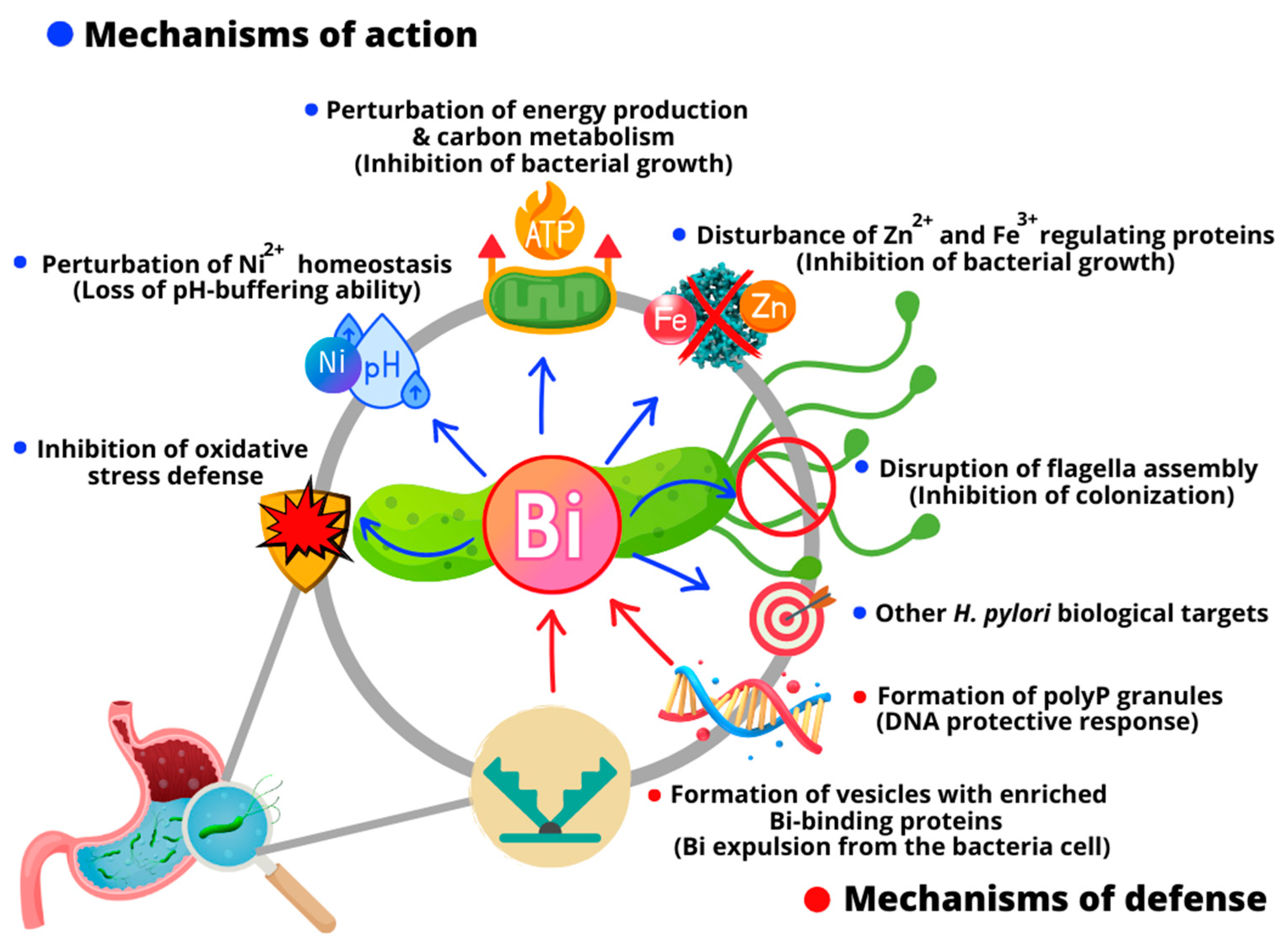
3.1.1. Modes of Action against H. pylori and Bacterial Resistance Mechanisms

H. pylori is a transmissible Gram-negative pathogen that infects the stomach of half of the human population worldwide, causing chronic gastritis and peptic ulcers [31], as Barry Marshall and Robin Warren discovered in 1982 [32]. In addition, this bacterium is also associated with gastric cancer [22]. Due to the effective treatment of H. pylori-associated gastrointestinal ailments, nowadays co-administrated with antibiotics and a proton pump inhibitor, the antiulcer activity of bismuth-based agents has remained in clinical use for decades, although the mechanisms of action were not completely elucidated.
It is known that bismuth-based drugs form a protective coating on the ulcer crater, which prevents the erosion generated by gastric acid and contributes to the healing of the lesion [10,33]. In addition, some studies indicated that the biological targets of the antiulcer activity of bismuth are related to the oxidative stress of H. pylori [34]. Bismuth also can inactivate enzymes involved in respiration, such as F1-ATPase [35], and other enzymes, such as urease [21,36] and alcohol dehydrogenase [37], besides interfering with a range of Zn2+- and Fe3+-regulating proteins [38]. However, recently, through bioinformatics analysis in combination with bioassays, some investigations have provided novel and deeper insights into the complex molecular multi-targeted mode of action by which bismuth eradicates H. pylori [39] (Figure 2). For example, Sun and collaborators demonstrated that bismuth drugs disrupted multiple essential pathways in the pathogen, including damaging the oxidative defense systems and eliminating the bacterial pH-buffering ability by the binding and functional perturbation of several key enzymes [40]. They also discovered a new target (HpDnaK) of bismuth drugs to inhibit bacterium–host cell adhesion [41]. The systemic responses of six H. pylori strains isolated from patients cultured with and without bismuth were recently explored by Yao et al. through integrative proteomic and metabolomic analyses [42]. They found that bismuth inhibited H. pylori growth in vitro via the downregulation of virulence proteins CagA and VacA, the disruption of the flagella assembly responsible for bacterial colonization, and the inhibition of antioxidant enzymes. Furthermore, they observed that diverse metabolic pathways related to growth and RNA translation and biological processes in H. pylori, including antioxidant response and purine, pyrimidine, amino acid, and carbon metabolism, were disrupted by bismuth [42].
While most proteins interact with only a few others, a small number of proteins, termed hubs, are core proteins highly connected with other proteins [43]. Bismuth can bind H. pylori hub proteins [41]; thus, bismuth is expected to be more lethal to this pathogen and less susceptible to developing resistance. In addition, bismuth also has a multifactorial mechanism of action against H. pylori; therefore, the chances of the pathogen developing resistance linked to molecular multi-target agents are lower. However, despite bismuth compounds having potential against H. pylori, their eradication rate is only 10% to 20% when administrated alone [44]. These limited bacterial eradication rates by bismuth agents can be justified by the H. pylori defense mechanisms employed to escape bismuth toxicity (Figure 2). Recently, Kumar et al. reported a study indicating novel defense mechanisms of H. pylori against bismuth [45]. They demonstrated that even low doses of bismuth prompt profound changes in H. pylori physiology, inducing the formation of homogeneously sized membrane vesicles with unique protein cargo content enriched in bismuth-binding proteins. These vesicles are responsible for mediating the bismuth expulsion from the bacteria cell. Additionally, a probable local DNA protective response associated with polyphosphate granule formation induced by bismuth exposure was observed. The poly-P granules cause bacteria nucleoid compaction, protecting it from bismuth toxicity [45].
As to developing new bismuth drugs to combat H. pylori, some new Bi3+ compounds exhibiting significative anti-H. pylori potential were reported in the last decade. For example, a binuclear Bi3+ complex containing pyridine 2,6-dicarboxylate and thiourea was recently prepared and assessed against three strains of H. pylori, displaying excellent activity with MIC (minimum inhibitory concentration) values of 8.84 μM, about two and four times more active than CBS and BSS, respectively [46]. In a similar direction, Kowalik et al. prepared two Bi3+ coordination polymers with the same pyridine-2,3-dicarboxylate ligand. One presented MIC values of 11.4 and 13.4 μM against two H. pylori strains, 26695 and N6, respectively, being about twice more active than BBS in the same bacteria strains [47]. Notably, commercially available medicinal bismuth carboxylates BSS, RBC, and CBS have MIC values against H. pylori ranging from 8 to 12.5 µg mL−1 [48].
In the last decade, the research group of Professor Andrews reported several works containing Bi3+ complexes with various ligands, in which many exhibited significative activity against three H. pylori strains (26695, B128, and 251) [49,50,51,52,53]. Additionally, Andrews and colleagues have conducted various studies on the relationship between the structures of Bi complexes and their antimicrobial activity. One important aspect they have highlighted is the role of the type and coordination mode of the ligand to bismuth, including donor atoms, hapticity, and the influence of substituents. The physicochemical properties of the final complex formed are crucial to its biological action, as evidenced by their research and others [54]. Sun and colleagues also comprehensively examined bismuth-based compounds as antimicrobial agents. Their study focused on identifying the molecular targets of bismuth agents and their mechanisms of action as antimicrobial agents [1].

3.1.2. Antibacterial Activity beyond H. pylori and Reversion of Bacterial Drug Resistance

Bismuth is not only active against H. pylori; for example, Bi compounds with a broad spectrum of activity against Gram-negative and/or Gram-positive bacteria, including antibiotic-resistant bacterial lines, have been continually reported. Andrews et al. reported a series of homoleptic and heteroleptic Bi3+ flavonolate complexes active against some Gram-positive and -negative bacterial lines at the micromolar level, including methicillin-resistant Staphylococcus aureus (MRSA) and vancomycin-resistant Enterococcus (VRE). It was also observed that the heteroleptic complexes exhibited effective antibacterial activity at micromolar levels, although they did show some level of toxicity towards mammalian cells. At the same time, the homoleptic complexes showed lower toxicity to mammalian cells; however, their antibacterial activity was also lower [55]. Subsequently, they evaluated the structure–activity relationships in di-aryl Bi phosphinates on antibacterial activity and demonstrated that the Bi3+-bound aryl group profoundly influences activity. Indeed, some complexes have the most significant activity towards MRSA and VRE in the range of 0.63 to 1.25 μM [56]. In another report, they also observed that a series of phenyl bis-phosphinate Bi3+ complexes exhibited effective antibacterial activity against MRSA and VRE [57]. The most active (1) is shown in Figure 3. Furthermore, they reported a series of novel diphenyl mono-phosphinate Bi complexes that exhibited effective antibacterial activity towards various bacteria, including MRSA and VRE, although these complexes also show some toxicity toward mammalian cells [58]. The above results indicate that exploring bismuth as a source for new antimicrobial agents is challenging, but it could result in the development of novel antibiotics to combat critical pathogens in clinical settings.
Tuberculosis, a poverty-related disease, is also considered neglected in drug research and development. Moreover, there is the emergence of resistant strains to all drugs available for treatment. For example, clinical isolates resistant to isoniazid and rifampicin, plus any fluoroquinolone and at least one of the four injectable second-line drugs (streptomycin, for instance) have already been reported in many countries [59], which makes necessary the search for novel compounds effective against Mycobacterium tuberculosis. In this sense, Machado et al. synthesized an octahedral Bi3+ complex containing the ligand pyridine-2-thiol-1-oxide (2) that showed excellent activity, both on the standard strain H37Rv ATCC 27294 (pan-susceptible) and in five clinical isolates resistant to the standard drugs isoniazid and rifampicin, displaying MIC values in the range of 2.49 to 21.18 µM [60].
Indeed, some studies have proven that bismuth-based combination therapy can effectively treat infections caused by antibiotic-resistant bacteria [1]. For instance, diseases caused by Enterobacteriaceae producing metallo-β-lactamases are hard to treat. Therefore, searching for effective metallo-β-lactamase inhibitors to restore the effectiveness of existing antibiotics is highly desirable. Although these inhibitors are not clinically available to date, a recent study has demonstrated a high potential of bismuth-based drugs to inhibit metallo-β-lactamase enzymes and restore the in vivo efficacy of the antibiotic meropenem [15]. Furthermore, the ability of bismuth to inhibit metallo-β-lactamase activity suggests that bismuth-based drugs could be repurposed alongside clinically used antibiotics as a co-therapy to address the current antibacterial resistance crisis [61]. In other studies, Bisceglie and collaborators tested the synergetic effect of a series of Bi3+ thiosemicarbazone complexes with the antibiotic meropenem in combination therapy in carbapenem-resistant Klebsiella pneumoniae carrying the NDM-1 gene, which is responsible for the production of the correspondent metallo-β-lactamase [62]. One of the complexes (3), with negligible antibacterial activity (MIC >250 μM) when used in monotherapy, displayed synergic activity with meropenem, restoring antibiotic sensitivity in the strain-producing NDM-1 enzyme [62]. Interestingly, the authors reported that complexes bearing a hydroxyl moiety involved in the coordination of the bismuth, O,N,N′,S-chelating mode, did not show any relevant antimicrobial activity and also didn’t displace Bi3+ to Zn2+ in a spectrophotometric titration experiment [62].
Deng and collaborators recently reported another successful example of the capacity of bismuth drugs to reverse resistance and repurpose clinically approved antibiotics [14]. They found that five bismuth drugs, commonly used in the clinical treatment of stomach-associated diseases, effectively boost the antibacterial activity of tigecycline against the gene Tet(X), responsible for conferring tigecycline resistance in Gram-negative bacteria. Furthermore, mechanistic studies showed that bismuth drugs effectively suppress the enzymatic activity of the Tet(X) resistance protein [14].
Periodontitis is a gum disease that could eventually lead to tooth loss and is an increased risk factor for several systemic conditions, such as cardiovascular and pulmonary diseases [63]. Porphyromonas gingivalis is the key pathogen of this oral disease. This bacterium can co-aggregate with other species, forming biofilms in the inflamed gingival tissues [64] and resisting common antimicrobials such as metronidazole [65]. Sun, Jin, et al. conducted a comprehensive investigation indicating that bismuth drugs can suppress this bacterium in its planktonic, biofilm, and internalized states [66]. The study revealed that bismuth upregulated iron metabolism and stress response while downregulating the energy metabolism of P. gingivalis. Furthermore, they demonstrated that bismuth perturbs the expression of a bacterium’s genes, translation, and virulence beyond to reconcile the immuno-inflammatory responses of P. gingivalis-invaded human cells [67].
Other research groups, including ours, have adopted a strategy of coordinating Bi3+ with antibiotics or bioactive molecules because bismuth has been found to exhibit synergistic action with antibiotics, which may help reverse antibiotic resistance or obtain compounds with other pharmacological activities [67,68,69,70,71,72].

3.2. Antifungal Activity

According to Frei et al., there are currently less than ten antifungal drugs in clinical development, a serious concern as new fungal strains resistant to most current antifungals rapidly spread worldwide [73]. Besides the resistance, the usefulness of antifungals is also limited by their narrow spectrum of activity and high toxicity. To make matters worse, developing antifungals has always been challenging since fungi and their human host are eukaryotes, making it difficult to identify unique targets for antifungal drugs [74]. To overcome the resistance phenomenon and prevent a more serious public health situation, new effective antifungals are needed, especially if they present new mechanisms of action.
Since the antibacterial activity of bismuth compounds is well-established, several research groups have naturally prepared bismuth compounds and evaluated their efficacy against fungi [13,75,76,77,78,79,80,81,82]. For instance, three Bi3+ complexes, 46, Figure 4, were tested against Candida albicans: 5 (MIC: 44 µM) showed better activity than fluconazole (MIC: 59 µM), the drug employed as a positive control, while the two other complexes were less active [83]. These results suggest that the para-chloro substituent’s presence on the ligand’s structure favored the antifungal activity. Subsequently, the same research group prepared a Bi3+ complex containing clioquinol (7), an antifungal and antiprotozoal drug. Complex 7 was 70-fold more active than fluconazole and approximately 3-fold more active than the its free ligand against Candida albicans [70].
As to the coordination compounds, a series of eight heteroleptic triorganobismuth(V) carboxylates were prepared and evaluated for their antifungal activity against four fungal strains, namely Aspergillus fumigatus (FCBP-66), A. niger (FCBP-0198), Mucor species (FCBP-0300), and Aspergillus flavus (FCBP-0064). Complexes 8, 9, and 10 exhibited significant antifungal activity against A. flavus with MIC values of 6.25 mg mL−1, comparable to the reference drug(s) [84].
Nanomaterials are an alternative to overcome antimicrobial resistance. An interesting study has revealed the promising properties of bismuth oxide nanoparticles (Bi2O3 NPs) in treating Candida albicans fungal infections and preventing biofilm formation. Notably, Bi2O3 NPs have shown better efficacy than commonly used antifungal agents such as chlorhexidine, nystatin, and terbinafine, known for their oral antiseptic and commercial antifungal properties [85].
In the last decade, several studies have supported the potential antifungal properties of bismuth-based nanomaterials [86,87,88,89,90,91]. For instance, the antimicrobial properties of BiVO4 nanorods were explored toward several fungal strains, and remarkable antifungal activity was observed for Fusarium solani. Additionally, cell culture experiments showed that BiVO4 was not able to induce hemolysis at low concentrations, which denotes its compatibility and low toxicity [92]. In other studies, bismuth nanoparticles (BiNPs), when tested under planktonic conditions, exhibited strong antifungal effects (MIC values ranging from 1 to 4 µg mL−1) against several strains of Candida auris, an emergent multidrug-resistant pathogenic yeast [93]. More recently, it was reported that green and non-toxic bismuth sulphide@graphitic carbon nitride (Bi2S3@g-C3N4) nanosheets (NCs) were synthesized and assessed for their antimicrobial activity. Bi2S3@g-C3N4 NCs exhibited high antifungal activity against C. albicans, with disinfection rates of 99.92% [94].
Despite the outstanding results shown above, it is surprising that bismuth compounds, which have proven effective against several bacterial strains, such as H. pylori, have not been extensively studied for their potential as antifungal agents. This seems to represent a missed opportunity.

3.3. Antiparasitic Activity

Several neglected tropical diseases (NTDs) are caused by parasites, such as leishmaniasis and Chagas disease. According to the World Health Organization (WHO), NTDs are prevalent in tropical areas, primarily affecting the poorest populations, especially women and children. Unfortunately, more than one billion people from developing countries face damaging health, social, and economic issues caused by NTDs [95].
Leishmaniasis is an endemic tropical disease caused by parasites of the Leishmania species, transmitted to humans and other species of mammals primarily via sandfly bites [40,41,42]. Leishmaniasis is mainly treated with antimony-based drugs, such as meglumine antimoniate (MA; Glucantime®), which have severe side effects and may not achieve clinical and parasitological cures [96]. Bismuth and antimony exhibit similar biological chemistry, which suggests that bismuth-based compounds may be a safe alternative to antimonial drugs and, in this regard, should be widely investigated. However, only a few classes of bismuth compounds have been designed and evaluated against leishmaniasis [12] and some of these promising compounds are depicted in Figure 5 and Figure 6.
The first bismuth compounds assessed for anti-leishmanial activity were prepared by Andrews and coworkers using NSAIDs (non-steroidal anti-inflammatory drugs) and substituted benzoic acids as ligands [97]. In this work, bismuth complexes bearing substituted benzoic acids showed significant anti-leishmanial activity against the promastigotes of L. major V121. Although these metal complexes demonstrated generic toxicity, this work encouraged further studies [68,98,99,100,101,102]. Thus, subsequently, Demicheli et al. reported a Bi3+ complex containing dppz (dipyrido [3,2-a:2′,3′-c]phenazine) (11), highly active against the promastigote form of Sb3+-sensitive and -resistant Leishmania infantum chagasi and Leishmania amazonensis strains. Furthermore, it was found that chelation decreased the lipophilicity of dppz, suggesting the role of this factor in improving the anti-leishmanial activity of the complex [99]. The fact that this complex has demonstrated potent activity and selectivity against resistant strains is a significant finding due to the increased clinical resistance involving antimony-based drugs, which can lead to failure in treating leishmaniasis [103].
Over the past decade, some reports have supported the promising anti-leishmanial activity of bismuth compounds [52,104,105] and some outstanding results have been presented. For example, two Bi3+ complexes with glycolic acid (12 and 13) were assessed against promastigote and amastigote forms of Leishmania, as well as human fibroblasts, and both exhibited good activity and a high degree of selectivity (13.2 for 9 and 53.9 for 10) [106].
Two homo- and heteroleptic Bi3+ thiazole–thiolate complexes, 14 and 15, were evaluated against Leishmania major promastigotes. 14 showed the highest activity (IC50 value 0.11 μg mL−1), and it had no toxic effect on human fibroblast cells. 14 was approximately 37 times more effective than 15, demonstrating the phenyl group’s importance in biological activity [107]. It was also reported that seven new Bi3+ complexes containing ligands derived from indole-carboxylic acids had been prepared, and that two of them, 16 and 17, exhibited comparable activity to Amphotericin B against the parasite Leishmania major, without any toxicity towards the mammalian cells at their effective concentration (0.19–0.39 µM) [108]. Furthermore, as reported firstly by A. Luqman et al. [107], the importance of the phenyl group coordinated with bismuth in the biological activity was also demonstrated, since the complexes containing only indole-carboxylates were not active (IC50 > 100 µM) against Leishmania major.
Some Bi5+ compounds, in Figure 6, notably have relevant anti-leishmanial activity. Thus, many tris-tolyl Bi5+ di-carboxylate complexes involving either ortho-, meta-, or para-substituted tolyl ligands were evaluated for their antiparasitic activity against L. major promastigotes and cytotoxicity against human fibroblasts. Two of these complexes showed significant toxicity towards promastigotes at low concentration and high selectivity indices (SI). For example, complexes 18 and 19 showed SI equal to 114 and 838, respectively. It was also demonstrated that the best activity and selectivity are observed with complexes containing o- and m-tolyl ligands, and it appears the primary influence on fibroblast toxicity is the aryl group, while the carboxylate influences promastigote toxicity [109]. Undoubtedly, these findings are noteworthy and motivate further studies.
Furthermore, Andleeb et al. evaluated a series of eight heteroleptic triorganobismuth(V) biscarboxylates (2027) for their anti-leishmanial potential against Leishmania tropica KWH23 strains. The low IC50 values (<1 µM) revealed that the complexes are highly active, except for 25 (IC50 = 20 µM). Additionally, the cytotoxicity profile showed that the tris(tolyl) derivatives presented lower toxicity against isolated lymphocytes with higher anti-leishmanial potential. Finally, stability studies confirmed that Bi5+ complexes are stable in solution and in leishmanial culture M199. This report is fascinating due to the lack of stability involving many Bi5+ compounds reported in the literature [110]. Moreover, these complexes exhibited good activity and need further exploration, even as a starting point for designing new Bi5+ complexes.
Although the mechanism of action of bismuth on the leishmanial parasite is not yet known, Bi5+ compounds presumably act as prodrugs exhibiting their activity through the reductive pathway to generate the more active Bi3+ analog compounds [54]. In addition, the Bi5+ bio-reduction process through redox-active biomolecules, such as GSH and trypanothione, decreases the microbes’ oxidative stress protection, leading to the increased production of reactive oxygen species (ROS) and consequent cell death [54].
As noted above, despite the recent increase in the search for effective bismuth compounds against leishmaniasis, the results have emerged from just a few research groups. However, although there are a small number of manuscripts in the literature, the outcomes reveal that bismuth compounds are promising for treating Leishmania and must be continuously prepared and tested, including in vivo tests.
Chagas disease or American trypanosomiasis is an endemic parasitosis caused by the protozoan Trypanosoma cruzi, affecting millions worldwide. The primary concern is that the two drugs available to treat this illness, nifurtimox and benznidazole, exhibit severe side effects such as neuropathy, anorexia, and weight loss, among others. Add to this the fact that there is no effective treatment for the chronic form of the disease, whose symptoms include mega disease of the esophagus or colon, and cardiomyopathy, the latter being the most common cause of death [111,112]. Furthermore, there is concern that strains resistant to these drugs have already been reported [113]. Given this scenario, searching for new anti-Trypanosoma cruzi drugs is prudent, mainly for the chronic phase.
Beraldo and coworkers prepared two Bi3+ 8-hydroxyquinoline-derived hydrazone complexes, 28 and 29, Figure 7, evaluated against Trypanosoma cruzi. Both complexes were more potent than benznidazole as antiparasitic agents. Complex 29 exhibited the highest activity against the extracellular parasites (EC50 = 0.06 µM) and the highest anti-T. cruzi activity against the amastigote form (EC50 = 2.31 µM). The mode of action of 29 on trypomastigotes was investigated using annexin V-fluorescein isothiocyanate (FITC) and propidium iodide (PI), and the results suggested the occurrence of cell death by necrosis [114]. The pioneering results by Beraldo et al. [114] indicate that Bi3+ complexes with 8-hydroxyquinoline-derived hydrazones are a starting point for developing new bismuth compounds effective against trypanosomes and deserve attention.
The antiparasitic effects of bismuth lipophilic nanoparticles (BisBAL NPs) against Trichomonas vaginalis were recently evaluated. The results showed that after 24 h of exposition to 500 µg mL−1 of BisBAL NPs, the Trichomonas vaginalis growth was inhibited more than 90%, with efficacy similar to metronidazole at 1.3 µg mL−1, the reference drug [115,116]. This finding is noteworthy because Trichomonas vaginalis is the most common vaginal parasitosis in women worldwide, with about 5% of the trichomoniasis clinical cases being caused by parasites resistant to metronidazole. In this sense, it is important to highlight that so far, no resistance to bismuth has been reported [21].

3.4. Antitumoral Activity

Due to the growing interest in medicinal inorganic chemistry since the emergence of cisplatin as a chemotherapeutic, several non-platinum metal complexes with a pivotal role in treating various diseases have been developed or improved/rationalized from old drugs [117,118]. Aiming to overcome some limitations of platinum-based drugs, such as acquired resistance and severe side effects, many bismuth compounds have been increasingly investigated as alternative anticancer agents, since they generally exhibit low toxicity to non-cancerous human cells, although are generally toxic to malignant cells. Still, on bismuth compounds as anticancer drugs, some studies have shown that bismuth drugs minimize the nephrotoxicity induced by cisplatin without affecting its antitumor effects [119,120]. Moreover, it was suggested that bismuth reduces cisplatin-induced nephrotoxicity by enhancing glutathione conjugation and vesicular transport [121]. Some studies suggest that combining bismuth and cisplatin may be a promising strategy for reducing cisplatin toxicity, but further research is necessary.
Several research groups worldwide have dedicated efforts to preparing bismuth compounds with potential anticancer properties. The studies show that low active or inactive free ligands generally enhance their anticancer activity upon bismuth coordination. The ligands can usually act as metal carriers increasing the solubility and bioavailability of Bi3+ [10], the agent responsible for the biological action. In addition, the outcomes have shown that the characteristics of the ligand might influence the anticancer activity of the resulting bismuth complexes, which are partly dependent on the type, the lipophilicity, and the electronic effects of the ligands. It is observed that bismuth complexes containing chelating ligands generally have superior cytotoxic activity against cancer cells compared to compounds bearing monodentate ligands. Thus, the additional thermodynamic stability in the physiological medium provided by the chelating nature of the ligands has a crucial role in the release rate of Bi ions and, consequently, in the biological action [52]. Still, regarding the role of the ligands in the anticancer activity of bismuth complexes, the presence of lipophilic groups on the molecular structure of the ligand may increase the uptake of target bismuth compounds, thereby enhancing the antiproliferative activity [122], as observed for lipophilic chelating thiol compounds [123,124,125,126]. In addition, the electronic effect and the dipole moment of bismuth-based compounds profoundly influence biological activity: polar compounds generally have better activity against cancer cells than non-polar ones [123,127]. Interestingly, the anticancer potential of bismuth compounds extends beyond bismuth complexes; organobismuth compounds have also been reported for their significant anticancer activities [128,129,130,131,132].
Although the mechanisms of antitumor action of bismuth compounds still need to be fully elucidated, some research indicates that cytotoxic pathways could be related to inducing apoptosis in tumor cells. The apoptosis process could be associated with the enhanced generation of intracellular reactive oxygen species (ROS), which are pro-oxidants and are critical in regulating cell death [129,130,131,133,134]. In addition, the cytotoxicity of bismuth could also be related to its capacity to inhibit some enzymes, such as proteases. In this sense, some bismuth compounds activated caspases and cell death by apoptosis [123,128,135,136,137]. A third route to induce apoptosis by bismuth-based compounds is a mitochondrial perturbation, i.e., a reduction of mitochondrial membrane potential [133,134,136]. Furthermore, Bi3+ complexes with aromatic heterocyclic ligands may trigger apoptosis through the DNA fragmentation pathway [83,132]. However, it is essential to mention that apoptosis is not the only regulated type of cell death. In recent decades, different signaling pathways and molecular mechanisms other than apoptosis have been detected, providing potential targets and new approaches for cancer treatment [138]. In this context, Iuchi and collaborators have demonstrated that some heterocyclic organobismuth compounds promote non-apoptotic cell death in vitro in human cancer cells via lipid peroxidation [139].
Considering the above discussions, some bismuth complexes that presented relevant anticancer activity investigated in the last decade are discussed below, and their values of an inhibitory concentration of 50% (IC50) on cancer cell lines are summarized in Table 2. It is relevant to highlight that many of these complexes were also evaluated for their antibacterial potential, and, generally, the complexes presented dual action as antibacterial and anticancer agents [140,141,142]. These findings are fascinating because microbial infection is one of the most common complications during cancer treatment due to surgical complications, chemotherapy- and radiotherapy-related neutropenia, and immunosuppressive drugs during cancer treatment [143]. In this way, developing drugs with dual action is greatly desired due to the increasing number of cancer patients and resistant microbial infections. In this sense, it is interesting to note that several antimicrobial agents, such as anthracyclines, mitomycin, and bleomycin, have been used to treat various cancers [144].

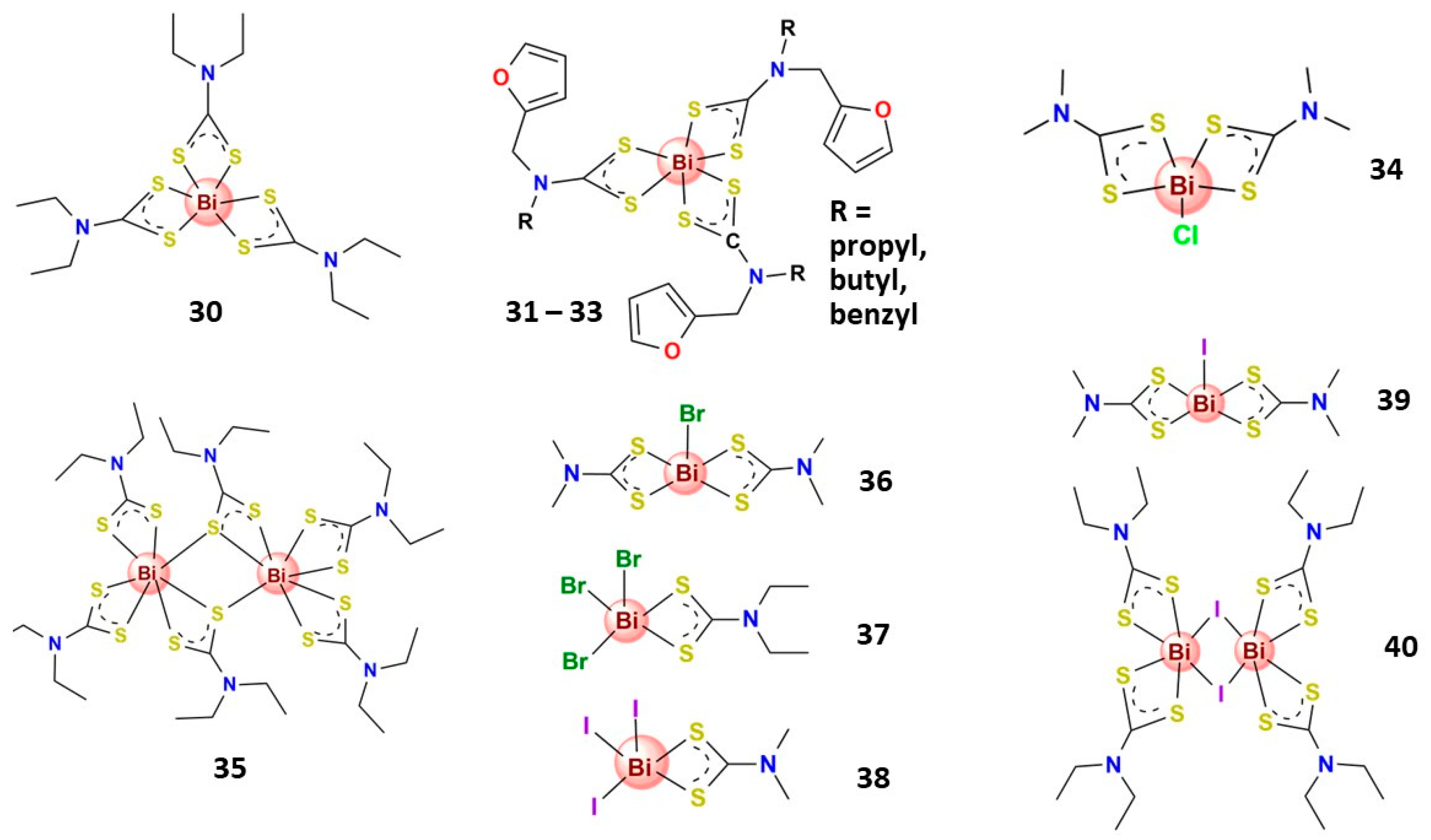
3.4.1. Bismuth(III) Complexes Bearing S-Donor Ligands

Thiol chelation generally improves the solubility and lipophilicity of bismuth-based compounds [122], thus resulting in the potentiation of the antitumoral activity, as observed for antimicrobial activity. The greater thermodynamic stability of Bi–S compounds compared to Bi–O compounds and the relative Bi–S low lability give thiobismuth compounds broad biological applicability [20,32]. Since thiobismuth compounds can promptly exchange with free thiols due to a kinetically labile Bi–S bond, Bi3+ has high mobility inside cells, making it more suitable for reaching the desired biological targets.
Indeed, several Bi3+ complexes with bidentate S,S′-chelating ligands have shown relevant antitumor activity (Figure 8). For instance, Bi3+ dithiocarbamate complexes have demonstrated low toxicity to normal cell lines and remarkable cytotoxicity in various human carcinoma cancer cell lines [123,137,152]. In this context, Ishak and coworkers demonstrated that the bismuth diethyldithiocarbamate compound (30) is highly cytotoxic to hepatocellular carcinoma (HepG2), promotes apoptosis, blocks the cell cycle, and inhibits cell invasion [137]. Complementing these findings, recently, Chan et al. also explored the remarked antiproliferative activity of 30 on MCF-7 breast cancer cells, and further in vitro studies indicated that 30 induced apoptotic cell death through the intrinsic pathway and significantly inhibited MCF-7 cell invasion, which closely associated with angiogenesis and cancer metastasis [123]. Tamilvanan et al. reported three Bi3+ dithiocarbamate complexes containing furfuryl group (3133) that displayed high antiproliferative activity against human cervix carcinoma KB (HeLa derivative) cell lines [124]. In other studies, Ozturk et al. reported mono- and dinuclear Bi3+ complexes with dithiocarbamate ligands (34 and 35) that exhibit expressive antiproliferative activity against MCF-7 and human cervix adenocarcinoma (HeLa) cell lines [125]. Also, the same research group prepared five Bi3+ halide dithiocarbamate complexes (3640) that exhibit expressive activity on MCF-7 and HeLa cell lines with similar or even better activity than tamoxifen (an antiestrogen drug) [126]. Additionally, the most active complexes (39 and 40) could block the estrogen receptors on MCR-7 cells [126]. López-Cardoso and collaborators prepared four Bi3+ complexes of pyrrolidine-dithiocarbamate that presented antiproliferative effects against MCF-7 cells but were not significantly toxic to MDA-MB-231 breast cancer cell lines and showed some toxicity to healthy cells (MCF-10 A) [153].
In contrast, some Bi3+ compounds bearing S-monodentate ligands seem to generally show low cytotoxic values against cancer cells compared to compounds containing chelating ligands. For example, Ozturk et al. reported recently several Bi3+ coordinated in an S-monodentate fashion to thiosemicarbazone and other ligands, finding that most of them give no significative cytotoxicity against MCF-7 cells, IC50 (48 h) > 30 µM, and some of them presented moderate activity, IC50 (48 h) = 5.4–8.0 µM, against this cancer cell line [154,155,156,157]. Very recently, this research group reported five Bi3+ halide complexes with thiosemicarbazone bearing an acetylthiophene moiety that presented moderate cytotoxicity on HeLa cells (IC50 4.5–24.3 µM) [158]. They also reported several Bi3+ complexes coordinated in an S-monodentate mode to thioamide ligands that showed low to moderate activity against human adenocarcinoma cells, HeLa (cervix) and MCF-7 (breast), with most IC50 (48 h) values superior to 30 µM [159,160]. Another Bi3+ compound bearing 6-mercaptopurine, an S-monodentate ligand, presents moderate cytotoxic activity on A549 and H460 human lung cancer cells with IC50 (24 h) ranging from 7 to 11 µM [161]. These outcomes indicated that chelating ligands are crucial for preparing Bi3+ complexes with high antitumoral activity.

3.4.2. Bismuth(III) Complexes Bearing N,S-Donor Ligands

Several thiosemicarbazone Bi3+ complexes (Figure 9) were reported for their anticancer potential. For instance, two Bi3+ complexes containing the pentadentate S,N,N′,N′,S-chelating thiosemicarbazone derivative ligands (41 and 42) were reported [145,146]. Complex 41 exhibited cytotoxic activity against K562 leukemia cells and inhibited H22 xenograft tumor growth in tumor-bearing mice [146]. In the same direction, complex 42 greatly suppressed colony formation and migration and significantly induced the apoptosis of human lung cancer cells A549 and H460 but was not toxic to the non-cancerous human lung fibroblast (HLF) cell line [145]. Moreover, in vivo, 42 inhibited A549 xenograft tumor growth on tumor-bearing mice and did not indicate harmful effects on mouse weight and liver [145]. The same research group reported another Bi3+ thiosemicarbazone derivative complex in which the ligand binds to the metallic center in a bidentate N,S-chelating mode (43). The complex presented moderate cytotoxic activity on A549 and cytotoxic activity on H460 lung cancer cells that was higher than cisplatin. It also induced cell apoptosis and was not toxic to non-cancerous cells (HFL) [147].
Several Bi3+ complexes containing tridentate N,N′,S-chelating thiosemicarbazone derivative ligands with anticancer potential were reported. For example, two Bi3+ thiosemicarbazone complexes prepared by Bisceglie et al. (44 and 45) were evaluated against A549 human lung carcinoma cells and normal HuDe human epithelial tissue {Formatting Citation}. Both complexes were nontoxic to normal cells; 44 was highly selective for cancer cells and approximately 13-fold more active than cisplatin against A549 cancer cells, while 45 was almost inactive in this cell line [62]. A series of five Bi3+ thiosemicarbazone complexes reported by Lessa and coworkers presented moderate activity against glioblastoma multiforme SF-295 and colon adenocarcinoma HCT-116 human tumor cell lines [162]. Li et al. prepared four Bi3+ thiosemicarbazone complexes (4649) that presented significative in vitro antiproliferative activity on HepG2 cells and promoted apoptosis in HepG2 cells associated with an increase in intracellular ROS production and a reduction of mitochondrial membrane potential (47 and 48) and caspase-3 activation (47 and 49) [133,135,148]. The four complexes have similar structures, but the difference in cytotoxic activities and apoptosis mechanisms indicate the relevance of the substituent groups’ ligands in the anticancer activity of the resulting complexes. Interestingly, two other Bi3+ complexes, reported by Li and collaborators, with similar structures to the above-cited complexes, present distinct antitumoral potential. Whereas the complex containing N(4)-pyridylthiosemicarbazone (50) presented high cytotoxic activity on K562 cells, IC50 (24 h) = 1.6 µM [149], the complex containing N(4)-phenylthiosemicarbazone presented just moderate cytotoxic activity under the same conditions (IC50 = 46.2 µM) [163].

3.4.3. Bismuth(III) Complexes Bearing N,O-Donor Ligands

Although bismuth prefers S-donor ligands, it could form relatively stable complexes with N,O-donor chelating ligands, and some of them (Figure 10) presented relevant anticancer activity. For instance, Pereira-Maia et al. recently reported two new Bi3+ complexes with asymmetric N,N′,O-chelating ligands (51 and 52), highly cytotoxic against a chronic myelogenous leukemia cell line (K562), able to induce apoptosis [140]. In another interesting work, Beraldo and coworkers prepared five Bi3+ complexes containing N,N′,O-chelating hydrazones derivatives, which are stable in an aqueous solution. The complexes, 5356, were active on HL-60, Jurkat, and THP-1 leukemia, and MCF-7 and HCT-116 solid tumor cells [141]. Interestingly, these complexes were revealed to be as or more active than the reference drug chloramphenicol in several bacteria lines [141]. The same research group reported three Bi3+ complexes, 36, Figure 4, bearing a planar macrocyclic ligand with expressive cytotoxic activity against several cancer cell lines. The mechanism of action of 36 seems to be related to the apoptotic pathway due to the DNA fragmentation induced by the complexes [83].
Considering the structural aspects in this case, the metal center, some dinuclear Bi3+ complexes with potential anticancer properties deserve attention. For example, Li et al. recently prepared seven dinuclear Bi3+ complexes bearing N,O,N-chelating Schiff-base ligands (5762) that revealed significative cytotoxic activity on human gastric cancer SNU-16 cells [142,150].

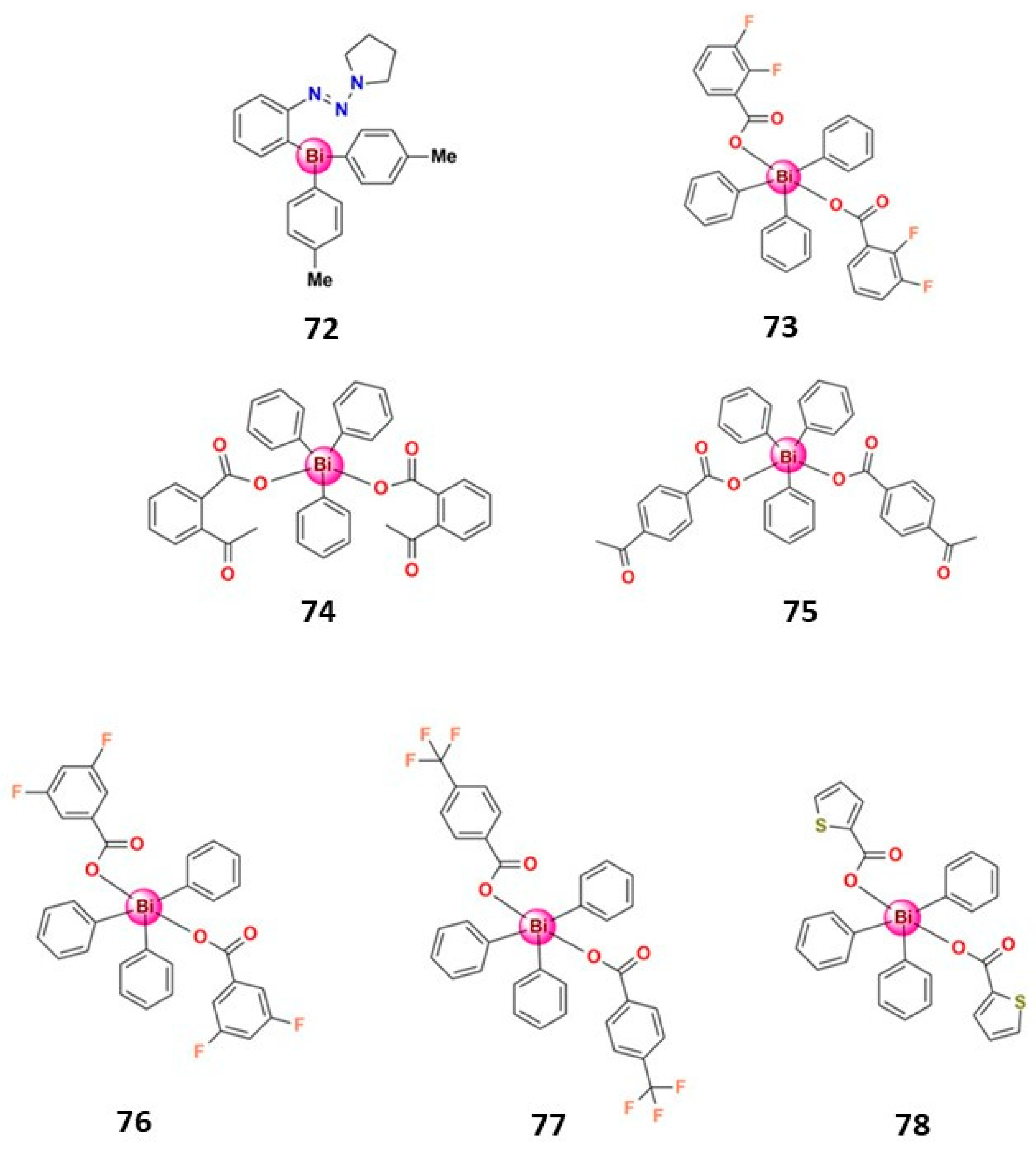
3.4.4. Organobismuth(III) Compounds

Iuchi and coworkers reported three heterocyclic organobismuth(III) compounds, 63–65, Figure 11, with potent antiproliferative activity in vitro in various human cancer cell lines [136,164]. Compounds 63 and 64 exert potent antiproliferative activities in vitro in human cancer cell lines; 63 stimulated cell death in a ROS-dependent manner, inducing both apoptotic and nonapoptotic cell death [164], while 64 induced lipid peroxidation with the concomitant induction of caspase-independent cell death [139]. The eight-membered ring heterocyclic organobismuth(III) compounds (63 and 64) display higher antitumor activity than the six-membered ones (65) [122]. In this context, Liu and collaborators also reported eight-membered heterocyclic hypervalent organobismuth(III) compounds with moderate to high anticancer activity (66–71). The most active compound (67) has selective cytotoxicity against cancer cells, and the toxicological activity is related to the induction of apoptosis by caspase-3 activation [128]. Interestingly, 67 has the nitro substituted in the organogroup, showing the relevance of the ligand’s electronic effect in the metal-based compound’s cytotoxicity [165].
Some organobismuth(V) also presented significant anticancer potential; Figure 12 shows some examples. Onishi and collaborators tested the antitumor activity of tri(p-tolyl)bismuthane and a complex in which one p-tolyl ligand was replaced by phenyldiazenyl-pyrrolidine (72) [129]. While tri(p-tolyl)bismuthane exerted only a weak antiproliferative effect up to 10 μM, 72 exhibited high antiproliferative activity on several human cancer cell lines, indicating that the polar bismuth compound was better than the non-polar derivatives for biological applications [127]. In addition, treating human leukemia NB4 cells with 72 induced apoptosis, a loss of mitochondrial membrane potential, and the generation of ROS [129]. Interestingly, the same chemical structure of 72 with antimony in place of bismuth did not show any cytotoxic activity, confirming the antiproliferative effect of bismuth [129].
Demicheli et al. prepared two organobismuth(V) dicarboxylate complexes (73 and 74) and tested their antiproliferative activity on murine metastatic melanoma (B16F10) and chronic myelogenous leukemia (K562) cell lines as well as in healthy non-cancerous cell lines. Complex 73 was non-toxic and showed cytotoxic activity on K562 cells superior to cisplatin. Moreover, evaluating the pro-apoptotic activity of 73 in B16F10 cells indicates that cell cycle arrest and cell apoptosis contribute to drug cytotoxicity [130]. Another organobismuth(V) carboxylate complex (75) demonstrated significant antiproliferation activity on BT474 breast cancer cells, inducing an increased level of intracellular ROS, which leads to BT474 cell death through the apoptotic pathway [131]. Cui et al. prepared three organobismuth(V) carboxylate complexes, 76–78, that present significative cytotoxic activity on MDA-MB-231 breast cancer cells and bound to CT-DNA via an intercalative mode [132].

3.5. Antiviral Activity

The emergence and reemergence of infectious diseases, especially those of viral origin, have concerned the scientific and medical communities in the last few years and caused global economic welfare challenges. For instance, the recent pandemic caused by coronavirus SARS-CoV-2, which has resulted in several million deaths worldwide, exposed research fragility for developing new antiviral agents. In addition, many neglected viral diseases, such as the Dengue, Zika, and Chikungunya viruses, require the development of effective antivirals. In this scenario, research for bismuth-based antiviral drugs can be a way out in this context of the critical demand for effective drugs to prevent and treat infectious diseases. In this sense, some works have demonstrated the potential of bismuth compounds for treating viral infections, such as rotavirus and HIV [166,167,168]. Moreover, two previous studies conducted by Sun and coworkers have demonstrated that several bismuth compounds could potentially inhibit the helicase enzymatic activities of coronavirus [169,170]. More specifically, it was shown that two of them were highly effective against SARS-CoV, inhibiting both coronavirus’s ATPase and helicase enzymes [170]. However, despite the promising results, only 14 years later, when COVID-19 became a global public health emergency, was interest in the findings of Sun and collaborators resumed.
Thus, in the last few years, the COVID-19 pandemic stimulated the development of new vaccines and drugs, and a few reports confirmed the potential anti-SARS-CoV-2 activity of Bi3+ compounds and provided valuable insight into the anti-SARS-CoV-2 mechanisms of bismuth drugs. The main molecular target of the investigations was the zinc-dependent SARS-CoV-2 non-structural protein 13 (nsp13). This protein, which pertains to the helicase superfamily 1B, in conjunction with the complex nsp7/nsp8/nsp12, plays a pivotal role in viral replication and the infection mechanism [171].
Recently, Sun and collaborators demonstrated that ranitidine bismuth citrate (RBC), a commercially available drug for treating duodenal ulcers, effectively prevents both the in vivo and in vitro replication of SARS-CoV-2 [17]. Furthermore, the affinity of bismuth to infected cells was high, with a selectivity index of 975. The authors also evidenced in in vitro studies that the inhibition of SARS-CoV-2 helicase by bismuth-based compounds can be attributed to the irreversible displacement of zinc(II) ions in SARS-CoV-2 nsp13 by Bi3+ ions [17]. The protein domains of SARS-CoV-2 nsp13 have a cysteine-rich region [172], which is an attractive molecular target for Bi3+ ions since they are known to have a high affinity for thiolate sulfur by forming Bi–S bonds [173]. Sun’s research group also showed that administering bismuth drugs combined with N-acetyl cysteine (NAC) constitutes a broad-spectrum anti-coronavirus cocktail therapy [167]. NAC further stabilized bismuth in stomach-like conditions, owing to the formation of a stable bismuth thiolate complex [Bi(NAC)3], enhancing bismuth drug uptake in tissues. Subsequently, Bi3+ ions suppressed virus replication by inhibiting multiple essential viral enzymes [174].
In 2020, Zhou and coworkers extended the comprehension of the biochemical characteristics of two replicative enzymatic activities associated with SARS-CoV-2 nsp13, nucleoside triphosphate hydrolase (NTPase) and RNA helix unwinding, and demonstrated that bismuth compounds inhibit their activities in a dose-dependent manner [18]. In 2021, Tao and collaborators, in turn, showed that an essential SARS-CoV-2 protease, 3CLpro (3-chymotrypsin-like protease), is one of the targets for colloidal bismuth subcitrate (CBS), a commercially available drug. Furthermore, they demonstrated that Bi3+ first bound to the metal site at the C-terminal domain of 3CLpro and caused dimeric 3CLpro to dissociate into monomers. This alteration resulted in protease dysfunction, suppressing SARS-CoV-2 viral replication in the mammalian cell [16].
More recently, Marrone et al., employing a multilevel computational approach, provide further insight into the antiviral mechanism of bismuth drugs. They demonstrated that the alterations in nsp13 resulting from the process of Zn2+ substitution with Bi3+ generated significative non-local structural impacts [175]. This theoretical study indicated that the Zn2+ to Bi3+ exchange leads to substantial changes in the geometrical and ionization properties of nsp13, which decrease the interaction between nsp13 and nsp12, thus impacting the process of viral replication and the infection mechanism of SARS-CoV-2 [175].
Although metal ions can bind on single and often functionally crucial protein residues, the antiviral activities of metal-based compounds have rarely been explored [176,177]. The efficacy of metallo-antiviral agents is expected to be less liable to new viral variants, thus resulting in an enormous potential for treating viral infections. One attractive strategy employed in some of these recent reports was investigating the possibility of repurposing the old metallodrugs, RBC and CBS (Table 1), as therapies for treating COVID-19. However, it is noteworthy that the required concentrations of drugs for suppressed SARS-CoV-2 viral replication in vivo and cellular-based experiments are much higher than that in a proteolytic assay, probably due to the multiple-targeting property of bismuth drugs [16] and their low bioavailability. This highlights the need for designing more specific bismuth-based compounds for antiviral activity.

4. Strategies to Enhance the Bioavailability of Bismuth beyond the Complex Formation

As bismuth-based compounds easily suffer hydrolysis in physiological conditions, in addition to research for obtaining new bismuth complexes that are more bioavailable, approaches for the targeting and controlled delivery of bismuth-based compounds could be promising strategies for enhancing their bioavailability and, consequently, their efficacy.

4.1. Incorporation of Bismuth in Biopolymeric Systems

Polysaccharides are biopolymers that attract interest as promising drug delivery carriers due to their unique properties, such as biocompatibility, non-toxicity, biodegradability, and high availability [178]. The anion polysaccharides could act as suitable ligands for Bi3+, and the complex formed could reduce bismuth intake and enhance Bi efficacy. In this context, Zhu et al. prepared Bi3+Hericium erinaceus polysaccharide complexes that significantly inhibited H. pylori (MIC = 20 μg mL−1), even with a lower content of Bi3+ than the standard drug CBS [179]. Silva-Caldeira and coworkers also developed a biopolymeric system incorporating Bi3+ ions and the antibiotic furazolidone on an alginate–carboxymethylcellulose blend, obtaining a system able to release these therapeutic agents in a concomitant and controlled way that could be adequate for H. pylori treatment [180]. Jiang et al., on the other hand, prepared pectin–bismuth complexes and investigated their structural changes in an acidic environment. The authors observed that Bi3+ gradually dissociated from carboxyl groups in the acidic environment, which is a beneficial property for treating H. pylori infections [181]. Andrews et al. proceeded with incorporating Bi compounds into microfibrillated cellulose producing composites that display significative antibacterial activity at low complex loadings, with the advantage of controlling the leaching of the Bi complexes from cellulose composites through structural changes in Bi compounds [57,58].
Aiming to obtain a biocompatible antimicrobial hydrogel for active wound dressing applications, Maliha et al. recently prepared a nanocellulose hydrogel incorporated with a phenyl bis-phosphinato Bi3+ complex. The prepared hydrogel showed bactericidal activity against Acinetobacter baumannii and Pseudomonas aeruginosa and a bacteriostatic effect against antibiotic-resistant bacteria MRSA and VRE, while having no toxic impact on mammalian fibroblast cells, thus revealing that bismuth-based compounds could act as a safe antimicrobial additive for preparing materials for biomedical applications [182].
Administering bismuth from polysaccharides is a good strategy for improving Bi bioavailability and diminishing the dose needed to reach the desired therapeutic outcome. In addition, the outcomes evidence the great potential of polysaccharides incorporated with Bi compounds for many applications, including antibacterial surfaces and materials. Even though there are few reports in the literature, the potentialities of biopolymers containing Bi3+ or Bi-based compounds for treating bacterial infections could open a new research trend. In addition, in recent years, nanotechnology-based approaches for targeting and controlling delivery, enhancing bioavailability, and minimizing systemic toxicities of metal-based therapeutic agents have revealed significant potential [183]. In this context, polymer-based nanosized systems designed to load physically or covalently conjugate Bi compounds could also be an interesting strategy for improving their bioavailability and, consequently, their absorption rate. However, it should be noted that, to the best of our knowledge, no research group has yet employed polymeric nanocarriers for bismuth-based agents [183]. In contrast, research on medical applications of metallic bismuth nanoparticles has grown tremendously in recent years [184].

4.2. Bismuth-Based MOFs and Coordination Polymers

Concerning the high porosity and surface area, as well as the infinite building blocks that can be combined to form a variety of materials, metal–organic frameworks (MOFs) are relevant in several fields, including pharmacological purposes [185]. These promising multifunctional platforms are suitable as drug delivery systems because they can accommodate small molecules, act as drug storage, and have low cytotoxicity, high biodegradability, and high biocompatibility [186]. Indeed, some bismuth-based MOFs were recently prepared as drug carriers since bismuth and its compounds are commonly considered biologically safe and nontoxic [187,188]. Interestingly, Huang and coworkers recently prepared two bismuth-based MOFs to act as a reservoir and controlled the delivery of Bi3+ ions to extend the antimicrobial duration, shedding light on a new administration modality of bismuth. In addition, the Bi-MOFs presented low cytotoxicity in human gingival fibroblasts, as expected, and exhibited selective antimicrobial activities against Gram-negative oral pathogens, A. actinomycetemcomitans (MIC = 7.8–31.3 µg mL−1) and P. gingivalis (MIC = 7.8–62.5 µg mL−1), in planktonic and biofilm forms [189].
Burrows et al. prepared some bismuth coordination networks containing deferiprone, an iron chelator drug, aiming to obtain a compound with a potential dual mode of action: simultaneously a combination of a system for deferiprone controlled release and a system with antibacterial activity. The compounds exhibit appreciable H. pylori inhibitory action, comparable to that of BSS, and could release the molecule of interest [190].
Bismuth has immense importance in material chemistry due to its unique properties. Indeed, numerous bismuth-based materials with varied biomedical applications, such as treating cancer and microbial infections, diagnostics imaging, theranostics, and biosensing, have recently been developed [191]. Some of these nanoparticle materials were designed to be administered directly into the bloodstream; therefore, the fates and toxicity of the Bi liberated from the nanoparticles need further research and the additional development of systems to control the release of bismuth.

5. Conclusions and Outlook

The medicinal chemistry of bismuth presents numerous opportunities to address significant health concerns. One of the most pressing global health challenges is antimicrobial resistance, exacerbated by the lack of new and effective antibiotics. Bismuth’s low toxicity in human cells and its ability to combat several microorganisms, including resistant bacteria strains, make it a promising candidate for further exploration. To develop more effective bismuth drugs, it is crucial to find ways to increase the bioavailability of this metal ion in the body’s physiological environment. Preparing and co-administering bioavailable bismuth agents with antibiotics and developing devices to simultaneously release both bismuth and antibiotics in a controlled manner are methods that can repurpose existing antibiotics for clinical use and overcome antimicrobial resistance.
It is also worth noting that various bismuth complexes have demonstrated notable anticancer properties. Interestingly, many of these compounds also possess antimicrobial effects. Developing a drug that can combat both cancer and bacterial infections simultaneously is a forward-thinking step in cancer treatment, as hard-to-treat microbial infections are one of the most common hurdles during treatment.
Another two points to highlight about the medicinal chemistry of bismuth are its antiviral and anti-leishmanial potential. The significant antiviral activities demonstrated by a few bismuth compounds suggest that a large research field can still be explored, which could help develop effective antivirals, especially for neglected diseases caused by viruses. In addition, bismuth’s anti-leishmanial potential makes it a safer alternative to the antimony-based compounds currently used to treat the leishmaniasis, an NTD, indicating reasonable scope for further research on developing bismuth-based anti-leishmanial agents.
Finally, continuous research in the field is necessary because bismuth formulations currently used as pharmaceuticals face insolubility, low absorption, and difficulties with synthetic reproducibility. Developing new bismuth-based agents requires a delicate balance of properties for stability, solubility, targeting, and subsequent activity. In this context, as highlighted in this review, controlled delivery systems for incorporating bismuth agents could be an interesting strategy for enhancing the bioavailability of active bismuth-based compounds.

Author Contributions

Conceptualization, P.P.S.-C. and W.G.; methodology, P.P.S.-C. and W.G.; writing—original draft preparation, J.d.S.R., L.H.F.R., W.G. and P.P.S.-C.; writing—review and editing and supervision, F.H.M., W.G. and P.P.S.-C.; project administration, P.P.S.-C.; funding acquisition, P.P.S.-C. All authors have read and agreed to the published version of the manuscript.

Funding

This research was funded by FAPEMIG, grant number APQ-00995-21 (P.P.S.-C.). CNPq Research Productivity Fellowship 303210/2021-4 (W.G.).

Institutional Review Board Statement

Not applicable.

Informed Consent Statement

Not applicable.

Data Availability Statement

Not applicable.

Acknowledgments

We are grateful to CNPq (Conselho Nacional de Desenvolvimento Científico e Tecnológico, Brazil), CAPES (Coordenação de Aperfeiçoamento de Pessoal de Nível Superior, Brazil), FAPEMIG (Fundação de Amparo à Pesquisa de Minas Gerais, Brazil), and CEFET-MG (Centro Federal de Educação Tecnológica de Minas Gerais, Brazil) for financial support.

Conflicts of Interest

The authors declare no conflict of interest.

References

  1. Wang, R.; Li, H.; Ip, T.K.Y.; Sun, H. Bismuth Drugs as Antimicrobial Agents, 1st ed.; Elsevier Inc.: Amsterdam, The Netherlands, 2020; Volume 75, ISBN 9780128191965. [Google Scholar]
  2. Li, H.; Sun, H. Recent Advances in Bioinorganic Chemistry of Bismuth. Curr. Opin. Chem. Biol. 2012, 16, 74–83. [Google Scholar] [CrossRef] [Green Version]
  3. Yang, N.; Sun, H. Bismuth: Environmental Pollution and Health Effects. In Encyclopedia of Environmental Health; Elsevier: Amsterdam, The Netherlands, 2011; pp. 414–420. [Google Scholar]
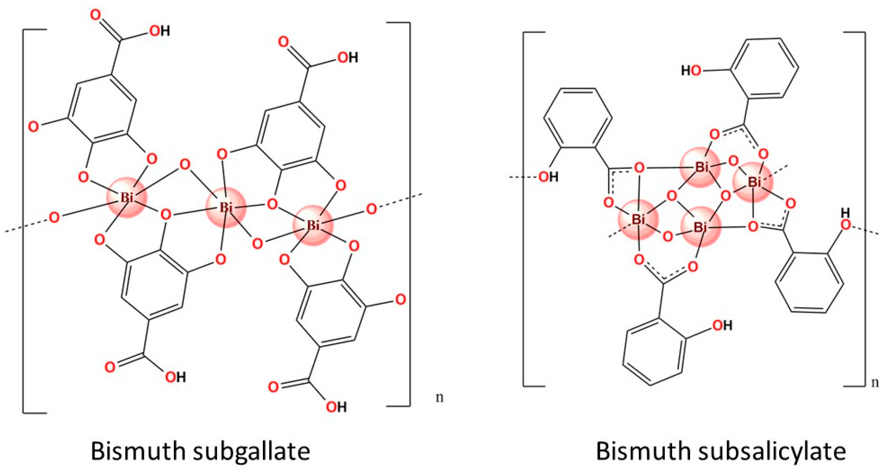
  4. Wang, Y.; Takki, S.; Cheung, O.; Xu, H.; Wan, W.; Öhrström, L.; Inge, A.K. Elucidation of the Elusive Structure and Formula of the Active Pharmaceutical Ingredient Bismuth Subgallate by Continuous Rotation Electron Diffraction. Chem. Commun. 2017, 53, 7018–7021. [Google Scholar] [CrossRef] [PubMed] [Green Version]
  5. Svensson Grape, E.; Rooth, V.; Nero, M.; Willhammar, T.; Inge, A.K. Structure of the Active Pharmaceutical Ingredient Bismuth Subsalicylate. Nat. Commun. 2022, 13, 1984. [Google Scholar] [CrossRef] [PubMed]
  6. Lambert, J.R.; Midolo, P. The Actions of Bismuth in the Treatment of Helicobacter pylori Infection. Aliment. Pharmacol. Ther. 1997, 11, 27–33. [Google Scholar] [CrossRef] [PubMed]
  7. Anthony, E.J.; Bolitho, E.M.; Bridgewater, H.E.; Carter, O.W.L.; Donnelly, J.M.; Imberti, C.; Lant, E.C.; Lermyte, F.; Needham, R.J.; Palau, M.; et al. Metallodrugs Are Unique: Opportunities and Challenges of Discovery and Development. Chem. Sci. 2020, 11, 12888–12917. [Google Scholar] [CrossRef]
  8. Yang, N.; Sun, H. Biocoordination Chemistry of Bismuth: Recent Advances. Coord. Chem. Rev. 2007, 251, 2354–2366. [Google Scholar] [CrossRef]
  9. Griffith, D.M.; Li, H.; Werrett, M.V.; Andrews, P.C.; Sun, H. Medicinal Chemistry and Biomedical Applications of Bismuth-Based Compounds and Nanoparticles. Chem. Soc. Rev. 2021, 50, 12037–12069. [Google Scholar] [CrossRef]
  10. Keogan, D.M.; Griffith, D.M. Current and Potential Applications of Bismuth-Based Drugs. Molecules 2014, 19, 15258–15297. [Google Scholar] [CrossRef] [Green Version]
  11. Shetu, S.A.; Sanchez-Palestino, L.M.; Rivera, G.; Bandyopadhyay, D. Medicinal Bismuth: Bismuth-Organic Frameworks as Pharmaceutically Privileged Compounds. Tetrahedron 2022, 129, 133117. [Google Scholar] [CrossRef]
  12. Ong, Y.C.; Kedzierski, L.; Andrews, P.C. Do Bismuth Complexes Hold Promise as Antileishmanial Drugs? Future Med. Chem. 2018, 10, 1721–1733. [Google Scholar] [CrossRef]
  13. Murafuji, T.; Fujiwara, Y.; Yoshimatsu, D.; Miyakawa, I.; Migita, K.; Mikata, Y. Bismuth Heterocycles Based on a Diphenyl Sulfone Scaffold: Synthesis and Substituent Effect on the Antifungal Activity against Saccharomyces cerevisiae. Eur. J. Med. Chem. 2011, 46, 519–525. [Google Scholar] [CrossRef]
  14. Deng, T.; Jia, Y.; Tong, Z.; Shi, J.; Wang, Z.; Liu, Y. Bismuth Drugs Reverse Tet(X)-Conferred Tigecycline Resistance in Gram-Negative Bacteria. Microbiol. Spectr. 2022, 10, e01578-21. [Google Scholar] [CrossRef] [PubMed]
  15. Wang, R.; Lai, T.P.; Gao, P.; Zhang, H.; Ho, P.L.; Woo, P.C.Y.; Ma, G.; Kao, R.Y.T.; Li, H.; Sun, H. Bismuth Antimicrobial Drugs Serve as Broad-Spectrum Metallo-β-Lactamase Inhibitors. Nat. Commun. 2018, 9, 439. [Google Scholar] [CrossRef] [PubMed] [Green Version]
  16. Tao, X.; Zhang, L.; Du, L.; Liao, R.; Cai, H.; Lu, K.; Zhao, Z.; Xie, Y.; Wang, P.H.; Pan, J.A.; et al. Allosteric Inhibition of SARS-CoV-2 3CL Protease by Colloidal Bismuth Subcitrate. Chem. Sci. 2021, 12, 14098–14102. [Google Scholar] [CrossRef] [PubMed]
  17. Yuan, S.; Wang, R.; Chan, J.F.W.; Zhang, A.J.; Cheng, T.; Chik, K.K.H.; Ye, Z.W.; Wang, S.; Lee, A.C.Y.; Jin, L.; et al. Metallodrug Ranitidine Bismuth Citrate Suppresses SARS-CoV-2 Replication and Relieves Virus-Associated Pneumonia in Syrian Hamsters. Nat. Microbiol. 2020, 5, 1439–1448. [Google Scholar] [CrossRef]
  18. Shu, T.; Huang, M.; Wu, D.; Ren, Y.; Zhang, X.; Han, Y.; Mu, J.; Wang, R.; Qiu, Y.; Zhang, D.Y.; et al. SARS-Coronavirus-2 Nsp13 Possesses NTPase and RNA Helicase Activities That Can Be Inhibited by Bismuth Salts. Virol. Sin. 2020, 35, 321–329. [Google Scholar] [CrossRef]
  19. Toutain, P.L.; Bousquet-Melou, A. Bioavailability and Its Assessment. J. Vet. Pharmacol. Ther. 2004, 27, 455–466. [Google Scholar] [CrossRef]
  20. Himeno, S.; Fujishiro, H.; Sumi, D. Bismuth. In Handbook on the Toxicology of Metals; Elsevier: Amsterdam, The Netherlands, 2022; pp. 121–139. [Google Scholar]
  21. Alkim, H.; Koksal, A.R.; Boga, S.; Sen, I.; Alkim, C. Role of Bismuth in the Eradication of Helicobacter pylori. Am. J. Ther. 2017, 24, e751–e757. [Google Scholar] [CrossRef]
  22. Malfertheiner, P.; Bazzoli, F.; Delchier, J.C.; Celiñski, K.; Giguère, M.; Rivière, M.; Mégraud, F. Helicobacter pylori Eradication with a Capsule Containing Bismuth Subcitrate Potassium, Metronidazole, and Tetracycline given with Omeprazole versus Clarithromycin-Based Triple Therapy: A Randomised, Open-Label, Non-Inferiority, Phase 3 Trial. Lancet 2011, 377, 905–913. [Google Scholar] [CrossRef]
  23. Guiard, E.; Lelievre, B.; Rouyer, M.; Zerbib, F.; Diquet, B.; Mégraud, F.; Tison, F.; Bignon, E.; Lassalle, R.; Perroteau, C.D.; et al. Bismuth Concentrations in Patients Treated in Real-Life Practice with a Bismuth Subcitrate-Metronidazole-Tetracycline Preparation: The SAPHARY Study. Drug Saf. 2019, 42, 993–1003. [Google Scholar] [CrossRef]
  24. Hong, Y.; Lai, Y.T.; Chan, G.C.F.; Sun, H. Glutathione and Multidrug Resistance Protein Transporter Mediate a Self-Propelled Disposal of Bismuth in Human Cells. Proc. Natl. Acad. Sci. USA 2015, 112, 3211–3216. [Google Scholar] [CrossRef]
  25. Larsen, A.; Stoltenberg, M.; Sondergaard, C.; Bruhn, M.; Danscher, G. In Vivo Distribution of Bismuth in the Mouse Brain: Influence of Long-Term Survival and Intracranial Placement on the Uptake and Transport of Bismuth in Neuronal Tissue. Basic Clin. Pharmacol. Toxicol. 2005, 97, 188–196. [Google Scholar] [CrossRef] [PubMed]
  26. Stoltenberg, M.; Hutson, J.C. Bismuth Uptake in Rat Testicular Macrophages: A Follow-up Observation Suggesting That Bismuth Alters Interactions Between Testicular Macrophages and Leydig Cells. J. Histochem. Cytochem. 2004, 52, 1241–1243. [Google Scholar] [CrossRef] [PubMed]
  27. Li, W.; Jin, L.; Zhu, N.; Hou, X.; Deng, F.; Sun, H. Structure of Colloidal Bismuth Subcitrate (CBS) in Dilute HCl: Unique Assembly of Bismuth Citrate Dinuclear Units ([Bi(Cit)2Bi]2−). J. Am. Chem. Soc. 2003, 125, 12408–12409. [Google Scholar] [CrossRef]
  28. Spénard, J.; Aumais, C.; Massicotte, J.; Tremblay, C.; Lefebvre, M. Influence of Omeprazole on Bioavailability of Bismuth Following Administration of a Triple Capsule of Bismuth Biskalcitrate, Metronidazole, and Tetracycline. J. Clin. Pharmacol. 2004, 44, 640–645. [Google Scholar] [CrossRef]
  29. Murray, C.J.; Ikuta, K.S.; Sharara, F.; Swetschinski, L.; Robles Aguilar, G.; Gray, A.; Han, C.; Bisignano, C.; Rao, P.; Wool, E.; et al. Global Burden of Bacterial Antimicrobial Resistance in 2019: A Systematic Analysis. Lancet 2022, 399, 629–655. [Google Scholar] [CrossRef]
  30. Fu, W.; Song, Z.; Zhou, L.; Xue, Y.; Ding, Y.; Suo, B.; Wang, L. Randomized Clinical Trial: Esomeprazole, Bismuth, Levofloxacin, and Amoxicillin or Cefuroxime as First-Line Eradication Regimens for Helicobacter pylori Infection. Dig. Dis. Sci. 2017, 62, 1580–1589. [Google Scholar] [CrossRef]
  31. Robinson, K.; Atherton, J.C. The Spectrum of Helicobacter-Mediated Diseases. Annu. Rev. Pathol. Mech. Dis. 2021, 16, 123–144. [Google Scholar] [CrossRef] [PubMed]
  32. Marshall, B.; Warren, J.R. Unidentified Curved Bacilli in the Stomach of Patients with Gastritis and Peptic Ulceration. Lancet 1984, 323, 1311–1315. [Google Scholar] [CrossRef]
  33. Claudel, M.; Schwarte, J.V.; Fromm, K.M. New Antimicrobial Strategies Based on Metal Complexes. Chemistry 2020, 2, 849–899. [Google Scholar] [CrossRef]
  34. Ge, R.; Chen, Z.; Zhou, Q. The Actions of Bismuth in the Treatment of Helicobacter pylori Infections: An Update. Metallomics 2012, 4, 239–243. [Google Scholar] [CrossRef]
  35. Beil, W.; Birkholz, C.; Wagner, S.; Sewing, K.-F. Bismuth Subcitrate and Omeprazole Inhibit Helicobacter pylori F1-ATPase. Pharmacology 1995, 50, 333–337. [Google Scholar] [CrossRef]
  36. Zhang, L.; Mulrooney, S.B.; Leung, A.F.K.; Zeng, Y.; Ko, B.B.C.; Hausinger, R.P.; Sun, H. Inhibition of Urease by Bismuth(III): Implications for the Mechanism of Action of Bismuth Drugs. BioMetals 2006, 19, 503–511. [Google Scholar] [CrossRef] [PubMed]
  37. Roine, R.P.; Salmela, K.S.; Höök-Nikanne, J.; Kosunen, T.U.; Salaspuro, M. Colloidal Bismuth Subcitrate and Omeprazole Inhibit Alcohol Dehydrogenase Mediated Acetaldehyde Production by Helicobacter pylori. Life Sci. 1992, 51, PL195–PL200. [Google Scholar] [CrossRef] [PubMed]
  38. Ge, R.; Sun, H. Bioinorganic Chemistry of Bismuth and Antimony: Target Sites of Metallodrugs. Acc. Chem. Res. 2007, 40, 267–274. [Google Scholar] [CrossRef]
  39. Liu, J.; Li, X.; Zhu, Y.; Ge, R. Molecular Mechanisms of Bismuth-Containing Drugs Against Helicobacter pylori: A Further Update. Curr. Pharmacol. Rep. 2023, 9, 59–65. [Google Scholar] [CrossRef]
  40. Han, B.; Zhang, Z.; Xie, Y.; Hu, X.; Wang, H.; Xia, W.; Wang, Y.; Li, H.; Wang, Y.; Sun, H. Multi-Omics and Temporal Dynamics Profiling Reveal Disruption of Central Metabolism in Helicobacter pylori on Bismuth Treatment. Chem. Sci. 2018, 9, 7488–7497. [Google Scholar] [CrossRef] [Green Version]
  41. Wang, Y.; Hu, L.; Xu, F.; Quan, Q.; Lai, Y.T.; Xia, W.; Yang, Y.; Chang, Y.Y.; Yang, X.; Chai, Z.; et al. Integrative Approach for the Analysis of the Proteome-Wide Response to Bismuth Drugs in Helicobacter pylori. Chem. Sci. 2017, 8, 4626–4633. [Google Scholar] [CrossRef] [Green Version]
  42. Yao, X.; Xiao, S.; Zhou, L. Integrative Proteomic and Metabolomic Analyses Reveal the Mechanism by Which Bismuth Enables Helicobacter pylori Eradication. Helicobacter 2021, 26, e12846. [Google Scholar] [CrossRef]
  43. Zhu, X.; Gerstein, M.; Snyder, M. Getting Connected: Analysis and Principles of Biological Networks. Genes Dev. 2007, 21, 1010–1024. [Google Scholar] [CrossRef] [Green Version]
  44. Salcedo, J.A.; Al-Kawas, F. Treatment of Helicobacter pylori Infection. Arch. Intern. Med. 1998, 158, 842. [Google Scholar] [CrossRef] [PubMed] [Green Version]
  45. Kumar, S.; Schmitt, C.; Gorgette, O.; Marbouty, M.; Duchateau, M.; Gianetto, Q.G.; Matondo, M.; Guigner, J.M.; De Reuse, H. Bacterial Membrane Vesicles as a Novel Strategy for Extrusion of Antimicrobial Bismuth Drug in Helicobacter pylori. mBio 2022, 13, e01633-22. [Google Scholar] [CrossRef] [PubMed]
  46. Razmara, Z.; Delarami, H.S.; Eigner, V.; Dusek, M. Single Crystal Structure Feature and Quantum Mechanical Studies of a New Binuclear Bi (III) Complex and Its Activity against Helicobacter pylori. Inorg. Chem. Commun. 2022, 146, 110207. [Google Scholar] [CrossRef]
  47. Kowalik, M.; Masternak, J.; Łakomska, I.; Kazimierczuk, K.; Zawilak-Pawlik, A.; Szczepanowski, P.; Khavryuchenko, O.V.; Barszcz, B. Structural Insights into New Bi(III) Coordination Polymers with Pyridine-2,3-Dicarboxylic Acid: Photoluminescence Properties and Anti-Helicobacter pylori Activity. Int. J. Mol. Sci. 2020, 21, 8696. [Google Scholar] [CrossRef] [PubMed]
  48. Andrews, P.C.; Ferrero, R.L.; Junk, P.C.; MacLellan, J.G.; Peiris, R.M. Bismuth(III) Thiobenzoates and Their Activity against Helicobacter pylori. Aust. J. Chem. 2012, 65, 883–891. [Google Scholar] [CrossRef] [Green Version]
  49. Andrews, P.C.; Ferrero, R.L.; Junk, P.C.; Peiris, R.M. A Sweeter Way to Combat Helicobacter pylori? Bismuth(III) Complexes and Oxido-Clusters Derived from Non-Nutritive Sweeteners and Their Activity against H. Pylori. J. Organomet. Chem. 2013, 724, 88–94. [Google Scholar] [CrossRef]
  50. Busse, M.; Trinh, I.; Junk, P.C.; Ferrero, R.L.; Andrews, P.C. Synthesis and Characterisation of Bismuth(III) Aminoarenesulfonate Complexes and Their Powerful Bactericidal Activity against Helicobacter pylori. Chem.-Eur. J. 2013, 19, 5264–5275. [Google Scholar] [CrossRef]
  51. Pathak, A.; Blair, V.L.; Ferrero, R.L.; Junk, P.C.; Tabora, R.F.; Andrews, P.C. Synthesis and Structural Characterisation of Bismuth(III) Hydroxamates and Their Activity against Helicobacter pylori. Dalton Trans. 2015, 44, 16903–16913. [Google Scholar] [CrossRef] [Green Version]
  52. Andrews, P.C.; Blair, V.L.; Ferrero, R.L.; Junk, P.C.; Kedzierski, L.; Peiris, R.M. Bismuth(III) β-Thioxoketonates as Antibiotics against Helicobacter pylori and as Anti-Leishmanial Agents. Dalton Trans. 2014, 43, 1279–1291. [Google Scholar] [CrossRef]
  53. Busse, M.; Border, E.; Junk, P.C.; Ferrero, R.L.; Andrews, P.C. Bismuth(III) Complexes Derived from α-Amino Acids: The Impact of Hydrolysis and Oxido-Cluster Formation on Their Activity against Helicobacter pylori. Dalton Trans. 2014, 43, 17980–17990. [Google Scholar] [CrossRef]
  54. Duffin, R.N.; Werrett, M.V.; Andrews, P.C. Antimony and Bismuth as Antimicrobial Agents, 1st ed.; Elsevier Inc.: Amsterdam, The Netherlands, 2020; Volume 75, ISBN 9780128191965. [Google Scholar]
  55. Burke, K.J.; Stephens, L.J.; Werrett, M.V.; Andrews, P.C. Bismuth(III) Flavonolates: The Impact of Structural Diversity on Antibacterial Activity, Mammalian Cell Viability and Cellular Uptake. Chem.-Eur. J. 2020, 26, 7657–7671. [Google Scholar] [CrossRef]
  56. Herdman, M.E.; Werrett, M.V.; Andrews, P.C. Aryl Bismuth Phosphinates [BiAr2(O(O)PRR′)]: Structure–Activity Relationships for Antibacterial Activity and Cytotoxicity. Dalton Trans. 2022, 51, 9323–9335. [Google Scholar] [CrossRef]
  57. Werrett, M.V.; Herdman, M.E.; Brammananth, R.; Garusinghe, U.; Batchelor, W.; Crellin, P.K.; Coppel, R.L.; Andrews, P.C. Bismuth Phosphinates in Bi-Nanocellulose Composites and Their Efficacy towards Multi-Drug Resistant Bacteria. Chem.-Eur. J. 2018, 24, 12938–12949. [Google Scholar] [CrossRef]
  58. Herdman, M.E.; Werrett, M.V.; Duffin, R.N.; Stephens, L.J.; Brammananth, R.; Coppel, R.L.; Batchelor, W.; Andrews, P.C. Impact of Structural Changes in Heteroleptic Bismuth Phosphinates on Their Antibacterial Activity in Bi-Nanocellulose Composites. Dalton Trans. 2020, 49, 7341–7354. [Google Scholar] [CrossRef] [PubMed]
  59. De Oliveira, L.P.; Carneiro, Z.A.; Ribeiro, C.M.; Lima, M.F.; Paixão, D.A.; Pivatto, M.; de Souza, M.V.N.; Teixeira, L.R.; Lopes, C.D.; de Albuquerque, S.; et al. Three New Platinum Complexes Containing Fluoroquinolones and DMSO: Cytotoxicity and Evaluation against Drug-Resistant Tuberculosis. J. Inorg. Biochem. 2018, 183, 77–83. [Google Scholar] [CrossRef] [PubMed] [Green Version]
  60. MacHado, I.; Marino, L.B.; Demoro, B.; Echeverría, G.A.; Piro, O.E.; Leite, C.Q.F.; Pavan, F.R.; Gambino, D. Bioactivity of Pyridine-2-Thiolato-1-Oxide Metal Complexes: Bi(III), Fe(III) and Ga(III) Complexes as Potent Anti-Mycobacterium tuberculosis Prospective Agents. Eur. J. Med. Chem. 2014, 87, 267–273. [Google Scholar] [CrossRef] [Green Version]
  61. Li, H.; Wang, R.; Sun, H. Systems Approaches for Unveiling the Mechanism of Action of Bismuth Drugs: New Medicinal Applications beyond Helicobacter pylori Infection. Acc. Chem. Res. 2019, 52, 216–227. [Google Scholar] [CrossRef]
  62. Scaccaglia, M.; Rega, M.; Bacci, C.; Giovanardi, D.; Pinelli, S.; Pelosi, G.; Bisceglie, F. Bismuth Complex of Quinoline Thiosemicarbazone Restores Carbapenem Sensitivity in NDM-1-Positive Klebsiella pneumoniae. J. Inorg. Biochem. 2022, 234, 111887. [Google Scholar] [CrossRef]
  63. Falcao, A.; Bullón, P. A Review of the Influence of Periodontal Treatment in Systemic Diseases. Periodontology 2000 2019, 79, 117–128. [Google Scholar] [CrossRef] [PubMed]
  64. Rajakaruna, G.A.; Negi, M.; Uchida, K.; Sekine, M.; Furukawa, A.; Ito, T.; Kobayashi, D.; Suzuki, Y.; Akashi, T.; Umeda, M.; et al. Localization and Density of Porphyromonas gingivalis and Tannerella forsythia in Gingival and Subgingival Granulation Tissues Affected by Chronic or Aggressive Periodontitis. Sci. Rep. 2018, 8, 9507. [Google Scholar] [CrossRef] [Green Version]
  65. Kouidhi, B.; Al Qurashi, Y.M.A.; Chaieb, K. Drug Resistance of Bacterial Dental Biofilm and the Potential Use of Natural Compounds as Alternative for Prevention and Treatment. Microb. Pathog. 2015, 80, 39–49. [Google Scholar] [CrossRef] [PubMed]
  66. Cheng, T.; Lai, Y.T.; Wang, C.; Wang, Y.; Jiang, N.; Li, H.; Sun, H.; Jin, L. Bismuth Drugs Tackle: Porphyromonas gingivalis and Attune Cytokine Response in Human Cells. Metallomics 2019, 11, 1207–1218. [Google Scholar] [CrossRef]
  67. Marzano, I.M.; Franco, M.S.; Silva, P.P.; Augusti, R.; Santos, G.C.; Fernandes, N.G.; Bucciarelli-Rodriguez, M.; Chartone-Souza, E.; Pereira-Maia, E.C. Crystal Structure, Antibacterial and Cytotoxic Activities of a New Complex of Bismuth(III) with Sulfapyridine. Molecules 2013, 18, 1464–1476. [Google Scholar] [CrossRef] [PubMed] [Green Version]
  68. Rocha, M.N.; Nogueira, P.M.; Demicheli, C.; de Oliveira, L.G.; da Silva, M.M.; Frézard, F.; Melo, M.N.; Soares, R.P. Cytotoxicity and In Vitro Antileishmanial Activity of Antimony (V), Bismuth (V), and Tin (IV) Complexes of Lapachol. Bioinorg. Chem. Appl. 2013, 2013, 961783. [Google Scholar] [CrossRef] [PubMed] [Green Version]
  69. Andrews, P.C.; Blair, V.L.; Ferrero, R.L.; Junk, P.C.; Kumar, I. Making Bispirin: Synthesis, Structure and Activity against Helicobacter pylori of Bismuth(III) Acetylsalicylate. Chem. Commun. 2013, 49, 2870. [Google Scholar] [CrossRef]
  70. Ferraz, K.S.O.; Reis, D.C.; Da Silva, J.G.; Souza-Fagundes, E.M.; Baran, E.J.; Beraldo, H. Investigation on the Bioactivities of Clioquinol and Its Bismuth(III) and Platinum(II,IV) Complexes. Polyhedron 2013, 63, 28–35. [Google Scholar] [CrossRef]
  71. Oliver, T.E.; Piantavigna, S.; Andrews, P.C.; Holt, S.A.; Dillon, C.T. Interactions of Non-Steroidal Anti-Inflammatory Drugs and Their Bismuth Analogues (BiNSAIDs) with Biological Membrane Mimics at Physiological PH. Langmuir 2021, 37, 1337–1352. [Google Scholar] [CrossRef]
  72. Oliveira, A.; Ferreira, J.; Farias, L.; Magalhães, P.; Teixeira, L.; Beraldo, H. Antimicrobial Effects of Silver(I) and Bismuth(III) Complexes with Secnidazole-Derived Schiff Base Ligands: The Role of the Nitro Group Reduction. J. Braz. Chem. Soc. 2019, 30, 2299–2307. [Google Scholar] [CrossRef]
  73. Frei, A.; Elliott, A.G.; Kan, A.; Dinh, H.; Bräse, S.; Bruce, A.E.; Bruce, M.R.; Chen, F.; Humaidy, D.; Jung, N.; et al. Metal Complexes as Antifungals? From a Crowd-Sourced Compound Library to the First In Vivo Experiments. JACS Au 2022, 2, 2277–2294. [Google Scholar] [CrossRef]
  74. Bouz, G.; Doležal, M. Advances in Antifungal Drug Development: An Up-To-Date Mini Review. Pharmaceuticals 2021, 14, 1312. [Google Scholar] [CrossRef]
  75. Burrell, R.E.; Corke, C.T.; Goel, R.G. Fungitoxicity of Organoantimony and Organobismuth Compounds. J. Agric. Food Chem. 1983, 31, 85–88. [Google Scholar] [CrossRef]
  76. Murafuji, T.; Miyoshi, Y.; Ishibashi, M.; Mustafizur Rahman, A.F.M.; Sugihara, Y.; Miyakawa, I.; Uno, H. Antifungal Activity of Organobismuth Compounds against the Yeast Saccharomyces cerevisiae: Structure–Activity Relationship. J. Inorg. Biochem. 2004, 98, 547–552. [Google Scholar] [CrossRef] [PubMed]
  77. Chauhan, H.P.S.; Shaik, N.M.; Singh, U.P. Synthetic, Spectroscopic and Antimicrobial Studies of Bis(Dialkyldithiocarbamato)Diorganodithiophosphatobismuth(III) Complexes. Appl. Organomet. Chem. 2005, 19, 1132–1139. [Google Scholar] [CrossRef]
  78. Solanki, J.S.; Tripathi, U.N.; Bhardwaj, A.; Thapak, T.R. Synthesis, Spectral Study and Antimicrobial Activity of Bismuth(III) 3(2′-Hydroxyphenyl)-5-(4-Substituted Phenyl) Pyrazolinates. J. Coord. Chem. 2008, 61, 4025–4032. [Google Scholar] [CrossRef]
  79. Mahajan, K.; Swami, M.; Singh, R.V. Microwave Synthesis, Spectral Studies, Antimicrobial Approach, and Coordination Behavior of Antimony(III) and Bismuth(III) Compounds with Benzothiazoline. Russ. J. Coord. Chem. 2009, 35, 179–185. [Google Scholar] [CrossRef]
  80. Solanki, J.S.; Thapak, T.R.; Bhardwaj, A.; Tripathi, U.N. Synthesis, Structural Characterization, and in Vitro Antimicrobial Properties of Salicylate and Pyrazoline Complexes of Bismuth(III). J. Coord. Chem. 2011, 64, 369–376. [Google Scholar] [CrossRef]
  81. El-Metwaly, N.M.; Refat, M.S. Elaborated 1H NMR Study for the Ligitional Behavior of Two Thiosemicarbazide Derivatives towards Some Heavy Metals (Sn(II), Sb(III), Pb(II) and Bi(III)), Thermal, Antibacterial and Antifungal Studies. Spectrochim. Acta Part A Mol. Biomol. Spectrosc. 2011, 81, 519–528. [Google Scholar] [CrossRef] [PubMed]
  82. Murafuji, T.; Kitagawa, K.; Yoshimatsu, D.; Kondo, K.; Ishiguro, K.; Tsunashima, R.; Miyakawa, I.; Mikata, Y. Heterocyclic Bismuth Carboxylates Based on a Diphenyl Sulfone Scaffold: Synthesis and Antifungal Activity against Saccharomyces cerevisiae. Eur. J. Med. Chem. 2013, 63, 531–535. [Google Scholar] [CrossRef]
  83. Ferraz, K.S.O.; Silva, N.F.; Da Silva, J.G.; De Miranda, L.F.; Romeiro, C.F.D.; Souza-Fagundes, E.M.; Mendes, I.C.; Beraldo, H. Investigation on the Pharmacological Profile of 2,6-Diacetylpyridine Bis(Benzoylhydrazone) Derivatives and Their Antimony(III) and Bismuth(III) Complexes. Eur. J. Med. Chem. 2012, 53, 98–106. [Google Scholar] [CrossRef]
  84. Mehmood, M.; Imtiaz-ud-Din; Abbas, S.; Azam, S.S.; Ihsan-ul-Haq; Tahir, M.N.; Parvaiz, N.; Tameez Ud Din, A. Bioactive Heteroleptic Bismuth(V) Carboxylates: Synthetic Stratagem, Characterization and Binding Pattern Validation. J. Organomet. Chem. 2020, 921, 121357. [Google Scholar] [CrossRef]
  85. Cabral-Romero, C.; Hernandez-Delgadillo, R.; Velasco-Arias, D.; Martinez-Sanmiguel, J.J.; Diaz, D.; Zumeta-Dubé, I.; Arevalo-Niño, K. Bismuth Oxide Aqueous Colloidal Nanoparticles Inhibit Candida albicans Growth and Biofilm Formation. Int. J. Nanomed. 2013, 8, 1645–1652. [Google Scholar] [CrossRef] [PubMed] [Green Version]
  86. Murafuji, T.; Tomura, M.; Ishiguro, K.; Miyakawa, I. Activity of Antifungal Organobismuth(III) Compounds Derived from Alkyl Aryl Ketones against S. Cerevisiae: Comparison with a Heterocyclic Bismuth Scaffold Consisting of a Diphenyl Sulfone. Molecules 2014, 19, 11077–11095. [Google Scholar] [CrossRef] [PubMed] [Green Version]
  87. Raheel, A.; Imtiaz-ud-Din; Andleeb, S.; Ramadan, S.; Tahir, M.N. Synthesis and Structural Characterization of New Bioactive Ligands and Their Bi(III) Derivatives. Appl. Organomet. Chem. 2017, 31, e3632. [Google Scholar] [CrossRef]
  88. Murafuji, T.; Hafizur Rahman, A.F.M.; Yamashita, K.; Narita, M.; Ishiguro, K.; Kamijo, S.; Miyakawa, I.; Mikata, Y. Synthesis and Antifungal Activities of Pyridine Bioisosteres of a Bismuth Heterocycle Derived from Diphenyl Sulfone. Heterocycles 2018, 96, 1037. [Google Scholar] [CrossRef]
  89. Das, P.E.; Majdalawieh, A.F.; Abu-Yousef, I.A.; Narasimhan, S.; Poltronieri, P. Use of A Hydroalcoholic Extract of Moringa oleifera Leaves for the Green Synthesis of Bismuth Nanoparticles and Evaluation of Their Anti-Microbial and Antioxidant Activities. Materials 2020, 13, 876. [Google Scholar] [CrossRef] [PubMed] [Green Version]
  90. Boadi, N.O.; Degbevi, M.; Saah, S.A.; Badu, M.; Borquaye, L.S.; Kortei, N.K. Antimicrobial Properties of Metal Piperidine Dithiocarbamate Complexes against Staphylococcus aureus and Candida albicans. Sci. Afr. 2021, 12, e00846. [Google Scholar] [CrossRef]
  91. Stevanović, N.; Mazzeo, P.P.; Bacchi, A.; Matić, I.Z.; Đorđić Crnogorac, M.; Stanojković, T.; Vujčić, M.; Novaković, I.; Radanović, D.; Šumar-Ristović, M.; et al. Synthesis, Characterization, Antimicrobial and Cytotoxic Activity and DNA-Binding Properties of d-Metal Complexes with Hydrazones of Girard’s T and P Reagents. JBIC J. Biol. Inorg. Chem. 2021, 26, 863–880. [Google Scholar] [CrossRef]
  92. Mohamed, H.E.A.; Afridi, S.; Khalil, A.T.; Zohra, T.; Alam, M.M.; Ikram, A.; Shinwari, Z.K.; Maaza, M. Phytosynthesis of BiVO4 Nanorods Using Hyphaene thebaica for Diverse Biomedical Applications. AMB Express 2019, 9, 200. [Google Scholar] [CrossRef] [Green Version]
  93. Vazquez-Munoz, R.; Lopez, F.D.; Lopez-Ribot, J.L. Bismuth Nanoantibiotics Display Anticandidal Activity and Disrupt the Biofilm and Cell Morphology of the Emergent Pathogenic Yeast Candida auris. Antibiotics 2020, 9, 461. [Google Scholar] [CrossRef]
  94. Abdel-Moniem, S.M.; El-Liethy, M.A.; Ibrahim, H.S.; Ali, M.E.M. Innovative Green/Non-Toxic Bi2S3@g-C3N4 Nanosheets for Dark Antimicrobial Activity and Photocatalytic Depollution: Turnover Assessment. Ecotoxicol. Environ. Saf. 2021, 226, 112808. [Google Scholar] [CrossRef]
  95. World Health Organization. Neglected Tropical Diseases. Available online: https://www.who.int/health-topics/neglected-tropical-diseases#tab=tab_1 (accessed on 1 May 2023).
  96. Carvalho, S.H.; Frézard, F.; Pereira, N.P.; Moura, A.S.; Ramos, L.M.Q.C.; Carvalho, G.B.; Rocha, M.O.C. American Tegumentary Leishmaniasis in Brazil: A Critical Review of the Current Therapeutic Approach with Systemic Meglumine Antimoniate and Short-Term Possibilities for an Alternative Treatment. Trop. Med. Int. Health 2019, 24, 380–391. [Google Scholar] [CrossRef]
  97. Andrews, P.C.; Frank, R.; Junk, P.C.; Kedzierski, L.; Kumar, I.; Maclellan, J.G. Anti-Leishmanial Activity of Homo- and Heteroleptic Bismuth(III) Carboxylates. J. Inorg. Biochem. 2011, 105, 454–461. [Google Scholar] [CrossRef] [PubMed]
  98. Andrews, P.C.; Junk, P.C.; Kedzierski, L.; Peiris, R.M. Anti-Leishmanial Activity of Novel Homo- and Heteroleptic Bismuth(III) Thiocarboxylates. Aust. J. Chem. 2013, 66, 1297. [Google Scholar] [CrossRef]
  99. Lizarazo-Jaimes, E.; Monte-Neto, R.; Reis, P.; Fernandes, N.; Speziali, N.; Melo, M.; Frézard, F.; Demicheli, C. Improved Antileishmanial Activity of Dppz through Complexation with Antimony(III) and Bismuth(III): Investigation of the Role of the Metal. Molecules 2012, 17, 12622–12635. [Google Scholar] [CrossRef] [Green Version]
  100. Duffin, R.N.; Blair, V.L.; Kedzierski, L.; Andrews, P.C. Comparative Stability, Toxicity and Anti-Leishmanial Activity of Triphenyl Antimony(v) and Bismuth(v) α-Hydroxy Carboxylato Complexes. Dalton Trans. 2018, 47, 971–980. [Google Scholar] [CrossRef] [PubMed]
  101. Andleeb, S.; Imtiaz-ud-Din; Rauf, M.K.; Azam, S.S.; Haq, I.; Tahir, M.N.; Ahmad, S. Bioactive Heteroleptic Bismuth(V) Complexes: Synthesis, Structural Analysis and Binding Pattern Validation. Appl. Organomet. Chem. 2019, 33, e5061. [Google Scholar] [CrossRef]
  102. Anamika, A.; Singh, R.; Manar, K.K.; Yadav, C.L.; Kumar, A.; Singh, R.K.; Drew, M.G.B.; Singh, N. Impact of Substituents on the Crystal Structures and Anti-Leishmanial Activity of New Homoleptic Bi(III) Dithiocarbamates. New J. Chem. 2019, 43, 16921–16931. [Google Scholar] [CrossRef]
  103. Ponte-Sucre, A.; Gamarro, F.; Dujardin, J.-C.; Barrett, M.P.; López-Vélez, R.; García-Hernández, R.; Pountain, A.W.; Mwenechanya, R.; Papadopoulou, B. Drug Resistance and Treatment Failure in Leishmaniasis: A 21st Century Challenge. PLoS Negl. Trop. Dis. 2017, 11, e0006052. [Google Scholar] [CrossRef] [Green Version]
  104. Islam, A.; Da Silva, J.; Berbet, F.; da Silva, S.; Rodrigues, B.; Beraldo, H.; Melo, M.; Frézard, F.; Demicheli, C. Novel Triphenylantimony(V) and Triphenylbismuth(V) Complexes with Benzoic Acid Derivatives: Structural Characterization, in Vitro Antileishmanial and Antibacterial Activities and Cytotoxicity against Macrophages. Molecules 2014, 19, 6009–6030. [Google Scholar] [CrossRef] [Green Version]
  105. Ong, Y.C.; Blair, V.L.; Kedzierski, L.; Andrews, P.C. Stability and Toxicity of Heteroleptic Organometallic Bi(V) Complexes towards Leishmania major. Dalton Trans. 2014, 43, 12904–12916. [Google Scholar] [CrossRef]
  106. Loh, A.; Ong, Y.C.; Blair, V.L.; Kedzierski, L.; Andrews, P.C. Bismuth(III) α-Hydroxy Carboxylates: Highly Selective Toxicity of Glycolates towards Leishmania major. JBIC J. Biol. Inorg. Chem. 2015, 20, 1193–1203. [Google Scholar] [CrossRef]
  107. Luqman, A.; Blair, V.L.; Brammananth, R.; Crellin, P.K.; Coppel, R.L.; Kedzierski, L.; Andrews, P.C. Homoleptic and Heteroleptic Bismuth(III) Thiazole–Thiolates and the Influence of Ring Substitution on Their Antibacterial and Antileishmanial Activity. Eur. J. Inorg. Chem. 2015, 2015, 725–733. [Google Scholar] [CrossRef]
  108. Pathak, A.; Blair, V.L.; Ferrero, R.L.; Kedzierski, L.; Andrews, P.C. Structural Influences on the Activity of Bismuth(III) Indole-Carboxylato Complexes towards Helicobacter pylori and Leishmania. J. Inorg. Biochem. 2017, 177, 266–275. [Google Scholar] [CrossRef] [PubMed]
  109. Ong, Y.C.; Blair, V.L.; Kedzierski, L.; Tuck, K.L.; Andrews, P.C. Stability and Toxicity of Tris-Tolyl Bismuth(v) Dicarboxylates and Their Biological Activity towards Leishmania major. Dalton Trans. 2015, 44, 18215–18226. [Google Scholar] [CrossRef] [PubMed]
  110. Andleeb, S.; Imtiaz-ud-Din; Rauf, M.K.; Azam, S.S.; Haq, I.; Tahir, M.N.; Zaman, N. Structural Characterization and Antileishmanial Activity of Newly Synthesized Organo-Bismuth(V) Carboxylates: Experimental and Molecular Docking Studies. JBIC J. Biol. Inorg. Chem. 2022, 27, 175–187. [Google Scholar] [CrossRef] [PubMed]
  111. Paixão, D.A.; Lopes, C.D.; Carneiro, Z.A.; Sousa, L.M.; de Oliveira, L.P.; Lopes, N.P.; Pivatto, M.; Chaves, J.D.S.; de Almeida, M.V.; Ellena, J.; et al. In Vitro Anti-Trypanosoma cruzi Activity of Ternary Copper(II) Complexes and in Vivo Evaluation of the Most Promising Complex. Biomed. Pharmacother. 2019, 109, 157–166. [Google Scholar] [CrossRef]
  112. Paixão, D.A.; de Oliveira, L.P.; Maia, P.I.d.S.; Deflon, V.M.; Carneiro, Z.A.; de Almeida, K.J.; Lopes, N.P.; Pivatto, M.; Chaves, J.D.S.; de Albuquerque, S.; et al. Crystal Structure of Two New Polymeric Copper(II) Complexes Active against Trypanosoma cruzi. J. Saudi Chem. Soc. 2018, 22, 809–815. [Google Scholar] [CrossRef]
  113. Urbina, J.A. Specific Chemotherapy of Chagas Disease: Relevance, Current Limitations and New Approaches. Acta Trop. 2010, 115, 55–68. [Google Scholar] [CrossRef] [PubMed]
  114. Oliveira, A.P.A.; Recio-Despaigne, A.A.; Ferreira, I.P.; Diniz, R.; Sousa, K.A.F.; Bastos, T.M.; Pereira Soares, M.B.; Moreira, D.R.M.; Beraldo, H. Investigation of the Antitrypanosomal Effects of 2-Formyl-8-Hydroxyquinoline-Derived Hydrazones and Their Antimony(III) and Bismuth(III) Complexes. New J. Chem. 2019, 43, 18996–19002. [Google Scholar] [CrossRef]
  115. Neamati, F.; Kodori, M.; Feizabadi, M.M.; Abavisani, M.; Barani, M.; Khaledi, M.; Moghadaszadeh, M.; Azadbakht, M.K.; Zeinali, M.; Fathizadeh, H. Bismuth Nanoparticles against Microbial Infections. Nanomedicine 2023, 17, 2109–2122. [Google Scholar] [CrossRef]
  116. Rodríguez-Luis, O.E.; Hernández-Delgadillo, R.; Pineda-Aguilar, N.; Vargas-Villarreal, J.; González-Salazar, F.; Garza-González, J.N.; Hernández-García, M.E.; Chellam, S.; Cabral-Romero, C. Effect of Bismuth Lipophilic Nanoparticles (BisBAL NPs) on Trichomonas Vaginalis Growth. J. Nanosci. Nanotechnol. 2017, 17, 4618–4622. [Google Scholar] [CrossRef]
  117. Souza, W.A.; Ramos, L.M.S.; de Almeida, A.M.; Tezuka, D.Y.; Lopes, C.D.; Moreira, M.B.; Zanetti, R.D.; Netto, A.V.G.; Ferreira, F.B.; de Oliveira, R.J.; et al. Preparation, Cytotoxic Activity and DNA Interaction Studies of New Platinum(II) Complexes with 1,10-Phenanthroline and 5-Alkyl-1,3,4-Oxadiazol-2(3H)-Thione Derivatives. J. Inorg. Biochem. 2022, 237, 111993. [Google Scholar] [CrossRef] [PubMed]
  118. Sodhi, R.K. Metal Complexes in Medicine: An Overview and Update from Drug Design Perspective. Cancer Ther. Oncol. Int. J. 2019, 14, 555883. [Google Scholar] [CrossRef]
  119. Yun, U.-J.; Lee, J.-H.; Shim, J.; Yoon, K.; Goh, S.-H.; Yi, E.H.; Ye, S.-K.; Lee, J.-S.; Lee, H.; Park, J.; et al. Anti-Cancer Effect of Doxorubicin Is Mediated by Downregulation of HMG-Co A Reductase via Inhibition of EGFR/Src Pathway. Lab. Investig. 2019, 99, 1157–1172. [Google Scholar] [CrossRef]
  120. Wang, R.; Wang, S.; Chan, S.; Wang, Y.; Zhang, Y.; Zuo, Z.; Chan, G.C.-F.; Li, H.; Sun, H. Bismuth Porphyrin Antagonizes Cisplatin-Induced Nephrotoxicity via Unexpected Metallothionein-Independent Mechanisms. iScience 2020, 23, 101054. [Google Scholar] [CrossRef] [PubMed]
  121. Chifiriuc, M.C.; Filip, R.; Constantin, M.; Pircalabioru, G.G.; Bleotu, C.; Burlibasa, L.; Ionica, E.; Corcionivoschi, N.; Mihaescu, G. Common Themes in Antimicrobial and Anticancer Drug Resistance. Front. Microbiol. 2022, 13, 960693. [Google Scholar] [CrossRef]
  122. Iuchi, K.; Yagura, T. Heterocyclic Organobismuth (III) Compounds Containing an Eight-Membered Ring: Inhibitory Effects on Cell Cycle Progression. Toxicol. Vitr. 2018, 50, 172–178. [Google Scholar] [CrossRef]
  123. Chan, P.F.; Ang, K.P.; Hamid, R.A. A Bismuth Diethyldithiocarbamate Compound Induced Apoptosis via Mitochondria-Dependent Pathway and Suppressed Invasion in MCF-7 Breast Cancer Cells. BioMetals 2021, 34, 365–391. [Google Scholar] [CrossRef]
  124. Tamilvanan, S.; Gurumoorthy, G.; Thirumaran, S.; Ciattini, S. Synthesis, Characterization, Cytotoxicity and Antimicrobial Studies on Bi(III) Dithiocarbamate Complexes Containing Furfuryl Group and Their Use for the Preparation of Bi2O3 nanoparticles. Polyhedron 2017, 121, 70–79. [Google Scholar] [CrossRef]
  125. Ozturk, I.I.; Banti, C.N.; Kourkoumelis, N.; Manos, M.J.; Tasiopoulos, A.J.; Owczarzak, A.M.; Kubicki, M.; Hadjikakou, S.K. Synthesis, Characterization and Biological Activity of Antimony(III) or Bismuth(III) Chloride Complexes with Dithiocarbamate Ligands Derived from Thiuram Degradation. Polyhedron 2014, 67, 89–103. [Google Scholar] [CrossRef]
  126. Arda, M.; Ozturk, I.I.; Banti, C.N.; Kourkoumelis, N.; Manoli, M.; Tasiopoulosd, A.J.; Hadjikakou, S.K. Novel Bismuth Compounds: Synthesis, Characterization and Biological Activity against Human Adenocarcinoma Cells. RSC Adv. 2016, 6, 29026–29044. [Google Scholar] [CrossRef]
  127. Das, A.; Das, A.; Banik, B.K. Influence of Dipole Moments on the Medicinal Activities of Diverse Organic Compounds. J. Indian Chem. Soc. 2021, 98, 100005. [Google Scholar] [CrossRef]
  128. Liu, Y.P.; Lei, J.; Tang, L.W.; Peng, Y.; Au, C.T.; Chen, Y.; Yin, S.F. Studies on the Cytotoxicity and Anticancer Performance of Heterocyclic Hypervalent Organobismuth(III) Compounds. Eur. J. Med. Chem. 2017, 139, 826–835. [Google Scholar] [CrossRef] [PubMed]
  129. Onishi, K.; Douke, M.; Nakamura, T.; Ochiai, Y.; Kakusawa, N.; Yasuike, S.; Kurita, J.; Yamamoto, C.; Kawahata, M.; Yamaguchi, K.; et al. A Novel Organobismuth Compound, 1-[(2-Di-p-Tolylbismuthanophenyl)Diazenyl] Pyrrolidine, Induces Apoptosis in the Human Acute Promyelocytic Leukemia Cell Line NB4 via Reactive Oxygen Species. J. Inorg. Biochem. 2012, 117, 77–84. [Google Scholar] [CrossRef] [PubMed]
  130. Islam, A.; Rodrigues, B.L.; Marzano, I.M.; Perreira-Maia, E.C.; Dittz, D.; Lopes, M.T.P.; Ishfaq, M.; Frezard, F.; Demicheli, C. Cytotoxicity and Apoptotic Activity of Novel Organobismuth(V) and Organoantimony(V) Complexes in Different Cancer Cell Lines. Eur. J. Med. Chem. 2016, 109, 254–267. [Google Scholar] [CrossRef]
  131. Yi, N.; Jiang, Z.J.; Wang, L.; Luo, P.; Peng, F. Crystal Structure and Anti-Breast Cancer Activity Evaluation of a Nanosized Bismuth(V)-Containing Coordination Complex Based on the F-Decorated Ligand. Inorg. Nano-Metal Chem. 2020, 50, 562–568. [Google Scholar] [CrossRef]
  132. Cui, L.; Bi, C.; Fan, Y.; Li, X.; Meng, X.; Zhang, N.; Zhang, Z. Synthesis, Crystal Structures, DNA Interaction and Anticancer Activity of Organobismuth(V) Complexes. Inorganica Chim. Acta 2015, 437, 41–46. [Google Scholar] [CrossRef]
  133. Zhang, N.; Tai, Y.; Li, M.; Ma, P.; Zhao, J.; Niu, J. Main Group Bismuth(III), Gallium(III) and Diorganotin(IV) Complexes Derived from Bis(2-Acetylpyrazine)Thiocarbonohydrazone: Synthesis, Crystal Structures and Biological Evaluation. Dalton Trans. 2014, 43, 5182–5189. [Google Scholar] [CrossRef]
  134. Li, Y.K.; Yang, M.; Li, M.X.; Yu, H.; Wu, H.C.; Xie, S.Q. Synthesis, Crystal Structure and Biological Evaluation of a Main Group Seven-Coordinated Bismuth(III) Complex with 2-Acetylpyridine N4-Phenylthiosemicarbazone. Bioorg. Med. Chem. Lett. 2013, 23, 2288–2292. [Google Scholar] [CrossRef]
  135. Fang, Y.; Wang, Y.T.; Zhao, M.; Lu, Y.L.; Li, M.X.; Zhang, Y.H. Bismuth(III) and Diorganotin(IV) Complexes of Bis(2-Acetylpyridine) Thiocarbonohydrazone: Synthesis, Characterization, and Apoptosis Mechanism of Action In Vitro. Polyhedron 2018, 155, 254–260. [Google Scholar] [CrossRef]
  136. Iuchi, K.; Hatano, Y.; Yagura, T. Heterocyclic Organobismuth(III) Induces Apoptosis of Human Promyelocytic Leukemic Cells through Activation of Caspases and Mitochondrial Perturbation. Biochem. Pharmacol. 2008, 76, 974–986. [Google Scholar] [CrossRef]
  137. Ishak, D.H.A.; Ooi, K.K.; Ang, K.P.; Akim, A.M.; Cheah, Y.K.; Nordin, N.; Halim, S.N.B.A.; Seng, H.L.; Tiekink, E.R.T. A Bismuth Diethyldithiocarbamate Compound Promotes Apoptosis in HepG2 Carcinoma, Cell Cycle Arrest and Inhibits Cell Invasion through Modulation of the NF-ΚB Activation Pathway. J. Inorg. Biochem. 2014, 130, 38–51. [Google Scholar] [CrossRef]
  138. Wang, X.; Hua, P.; He, C.; Chen, M. Non-Apoptotic Cell Death-Based Cancer Therapy: Molecular Mechanism, Pharmacological Modulators, and Nanomedicine. Acta Pharm. Sin. B 2022, 12, 3567–3593. [Google Scholar] [CrossRef]
  139. Iuchi, K.; Shirai, S.; Tasaki, Y.; Hisatomi, H. Heterocyclic Organobismuth(III) Compound Induces Nonapoptotic Cell Death via Lipid Peroxidation. Anti-Cancer Drugs 2020, 31, 55–59. [Google Scholar] [CrossRef] [PubMed]
  140. Marzano, I.M.; Tomco, D.; Staples, R.J.; Lizarazo-Jaimes, E.H.; Gomes, D.A.; Bucciarelli-Rodriguez, M.; Guerra, W.; de Souza, Í.P.; Verani, C.N.; Pereira Maia, E.C. Dual Anticancer and Antibacterial Activities of Bismuth Compounds Based on Asymmetric [NN′O] Ligands. J. Inorg. Biochem. 2021, 222, 111522. [Google Scholar] [CrossRef] [PubMed]
  141. Ferreira, I.P.; Piló, E.D.L.; Recio-Despaigne, A.A.; Da Silva, J.G.; Ramos, J.P.; Marques, L.B.; Prazeres, P.H.D.M.; Takahashi, J.A.; Souza-Fagundes, E.M.; Rocha, W.; et al. Bismuth(III) Complexes with 2-Acetylpyridine- and 2-Benzoylpyridine-Derived Hydrazones: Antimicrobial and Cytotoxic Activities and Effects on the Clonogenic Survival of Human Solid Tumor Cells. Bioorg. Med. Chem. 2016, 24, 2988–2998. [Google Scholar] [CrossRef]
  142. Li, C.H.; Jiang, J.H.; Lei, Y.H.; Li, X.; Yao, F.H.; Ji, M.H.; Zhang, K.W.; Tao, L.M.; Ye, L.J.; Li, Q.G. Design, Synthesis, and Biological Evaluation of Dinuclear Bismuth(III) Complexes with Isoniazid-Derived Schiff Bases. J. Inorg. Biochem. 2022, 235, 111931. [Google Scholar] [CrossRef]
  143. Yusuf, K.; Sampath, V.; Umar, S. Bacterial Infections and Cancer: Exploring This Association And Its Implications for Cancer Patients. Int. J. Mol. Sci. 2023, 24, 3110. [Google Scholar] [CrossRef]
  144. Bhattacharya, B.; Mukherjee, S. Cancer Therapy Using Antibiotics. J. Cancer Ther. 2015, 6, 849–858. [Google Scholar] [CrossRef] [Green Version]
  145. Ouyang, R.; Yang, Y.; Feng, K.; Zong, T.; Cao, P.; Xiong, F.; Guo, N.; Li, Y.; Miao, Y.; Tong, X.; et al. Potent Anticancer Activity of a New Bismuth (III) Complex against Human Lung Cancer Cells. J. Inorg. Biochem. 2017, 168, 18–26. [Google Scholar] [CrossRef]
  146. Li, M.X.; Yang, M.; Niu, J.Y.; Zhang, L.Z.; Xie, S.Q. A Nine-Coordinated Bismuth(III) Complex Derived from Pentadentate 2,6-Diacetylpyridine Bis(4N-Methylthiosemicarbazone): Crystal Structure and Both in Vitro and in Vivo Biological Evaluation. Inorg. Chem. 2012, 51, 12521–12526. [Google Scholar] [CrossRef]
  147. Ouyang, R.; Yang, Y.; Tong, X.; Yang, Y.; Tao, H.; Zong, T.; Feng, K.; Jia, P.; Cao, P.; Guo, N.; et al. Potential Anti-Cancer Activity of a Novel Bi(III) Containing Thiosemicarbazone Derivative. Inorg. Chem. Commun. 2016, 73, 138–141. [Google Scholar] [CrossRef]
  148. Li, M.; Lu, Y.; Yang, M.; Li, Y.; Zhang, L.; Xie, S. One Dodecahedral Bismuth(Iii) Complex Derived from 2-Acetylpyridine N(4)-Pyridylthiosemicarbazone: Synthesis, Crystal Structure and Biological Evaluation. Dalton Trans. 2012, 41, 12882–12887. [Google Scholar] [CrossRef] [PubMed]
  149. Zhang, L.Z.; An, G.Y.; Yang, M.; Li, M.X.; Zhu, X.F. Synthesis, Characterization, Crystal Structure and Biological Activities of the Unusual Main Group 8-Coordinate Bismuth (III) Complex Derived from 2-Acetylpyrazine N4- Pyridylthiosemicarbazone. Inorg. Chem. Commun. 2012, 20, 37–40. [Google Scholar] [CrossRef]
  150. Li, C.H.; Ji, M.H.; Zhang, K.W.; Sun, S.Y.; Jiang, J.H. Dinuclear Bismuth(III) Complex Constructed by Isoniazid-Derived Schiff Base: Synthesis, Crystal Structure, and Biological Activity. Appl. Organomet. Chem. 2022, 36, e6876. [Google Scholar] [CrossRef]
  151. De Carvalho, A.B.; de Souza, Í.P.; de Andrade, L.M.; Binatti, I.; Pedroso, E.F.; Krambrock, K.; Oliveira, W.X.C.; Pereira-Maia, E.C.; Silva-Caldeira, P.P. Novel Copper(II) Coordination Polymer Containing the Drugs Nalidixic Acid and 8-Hydroxyquinoline: Evaluation of the Structural, Magnetic, Electronic, and Antitumor Properties. Polyhedron 2018, 156, 312–319. [Google Scholar] [CrossRef]
  152. Li, H.; Lai, C.S.; Wu, J.; Ho, P.C.; de Vos, D.; Tiekink, E.R.T. Cytotoxicity, Qualitative Structure-Activity Relationship (QSAR), and Anti-Tumor Activity of Bismuth Dithiocarbamate Complexes. J. Inorg. Biochem. 2007, 101, 809–816. [Google Scholar] [CrossRef] [PubMed]
  153. López-Cardoso, M.; Tlahuext, H.; Pérez-Salgado, M.; Vargas-Pineda, D.G.; Román-Bravo, P.P.; Cotero-Villegas, A.M.; Acevedo-Quiroz, M.; Razo-Hernández, R.S.; Alvarez-Fitz, P.; Mendoza-Catalán, M.A.; et al. Synthesis, Crystal Structure, Antibacterial, Antiproliferative and QSAR Studies of New Bismuth(III) Complexes of Pyrrolidineditiocarbamate of Dithia-Bismolane and Bismane, Oxodithia- and Trithia-Bismocane. J. Mol. Struct. 2020, 1217, 128456. [Google Scholar] [CrossRef]
  154. Ucar, O.; Grześkiewicz, A.M.; Banti, C.; Hadjikakou, S.K.; Ozturk, I.I. Structural Characterization and Biological Evaluation of Antimony(III) and Bismuth(III) Complexes with Imidazolidine-2-Thione. J. Mol. Struct. 2021, 1235, 130270. [Google Scholar] [CrossRef]
  155. Ozturk, I.I.; Banti, C.N.; Hadjikakou, S.K.; Panagiotou, N.; Tasiopoulos, A.J. Bismuth(III) Halide Complexes of Aromatic Thiosemicarbazones: Synthesis, Structural Characterization and Biological Evaluation. Polyhedron 2021, 208, 115388. [Google Scholar] [CrossRef]
  156. Aygun, O.; Grześkiewicz, A.M.; Banti, C.N.; Hadjikakou, S.K.; Kubicki, M.; Ozturk, I.I. Monomeric Octahedral Bismuth(III) Benzaldehyde-N1-Alkyl Thiosemicarbazones: Synthesis, Characterization and Biological Properties. Polyhedron 2022, 215, 115683. [Google Scholar] [CrossRef]
  157. Turk, K.; Grześkiewicz, A.M.; Banti, C.N.; Hadjikakou, S.K.; Kubicki, M.; Ozturk, I.I. Synthesis, Characterization, and Biological Properties of Mono-, Di- and Poly-Nuclear Bismuth(III) Halide Complexes Containing Thiophene-2-Carbaldehyde Thiosemicarbazones. J. Inorg. Biochem. 2022, 237, 111987. [Google Scholar] [CrossRef]
  158. Ozturk, I.I.; Turk, K.; Grześkiewicz, A.M.; Kubicki, M.; Banti, C.N.; Hadjikakou, S.K. Heteroleptic Six-Coordinate Bismuth(III) Complexes with 2-Acetylthiophene Thiosemicarbazones: Synthesis, Characterization, and Biological Properties. New J. Chem. 2023, 47, 12779–12789. [Google Scholar] [CrossRef]
  159. Ozturk, I.I.; Banti, C.N.; Hadjikakou, S.K.; Panagiotou, N.; Tasiopoulos, A.J. Structural Architectures and Biological Properties of Main Group Bismuth(III) Iodide Complexes with Heterocyclic Thioamides. Inorganica Chim. Acta 2019, 497, 119094. [Google Scholar] [CrossRef]
  160. Cakmak, M.; Ozturk, I.I.; Banti, C.N.; Manoli, M.; Moushi, E.; Tasiopoulos, A.J.; Grzeskiewicz, A.M.; Kubicki, M.; Hadjikakou, S.K. Bismuth(III) Bromide-Thioamide Complexes: Synthesis, Characterization and Cytotoxic Properties. Main Group Met. Chem. 2018, 41, 143–154. [Google Scholar] [CrossRef] [Green Version]
  161. Yang, Y.; Zhou, S.; Ouyang, R.; Yang, Y.; Tao, H.; Feng, K.; Zhang, X.; Xiong, F.; Guo, N.; Zong, T.; et al. Improvement in the Anticancer Activity of 6-Mercaptopurine via Combination with Bismuth(III). Chem. Pharm. Bull. 2016, 64, 1539–1545. [Google Scholar] [CrossRef] [Green Version]
  162. Almeida, J.C.L.; Amim, R.S.; Pessoa, C.; Lourenço, M.C.S.; Mendes, I.C.; Lessa, J.A. Bismuth(III) Complexes with Pyrazineformamide Thiosemicarbazones: Investigation on the Antimicrobial and Cytotoxic Effects. Polyhedron 2020, 189, 114709. [Google Scholar] [CrossRef]
  163. Li, M.X.; Zhang, L.Z.; Yang, M.; Niu, J.Y.; Zhou, J. Synthesis, Crystal Structures, in Vitro Biological Evaluation of Zinc(II) and Bismuth(III) Complexes of 2-Acetylpyrazine N(4)-Phenylthiosemicarbazone. Bioorg. Med. Chem. Lett. 2012, 22, 2418–2423. [Google Scholar] [CrossRef] [PubMed]
  164. Iuchi, K.; Akagi, K.; Yagura, T. Heterocyclic Organobismuth(III) Compound Targets Tubulin to Induce G2/M Arrest in HeLa Cells. J. Pharmacol. Sci. 2009, 109, 573–582. [Google Scholar] [CrossRef] [Green Version]
  165. Shang, X.; Zhao, B.; Xiang, G.; Guedes Da Silva, M.F.C.; Pombeiro, A.J.L. Dimeric Diorganotin(IV) Complexes with Arylhydrazones of β-Diketones: Synthesis, Structures, Cytotoxicity and Apoptosis Properties. RSC Adv. 2015, 5, 45053–45060. [Google Scholar] [CrossRef]
  166. Gorbach, S.L.; Cornick, N.A.; Silva, M. Effect of Bismuth Subsalicylate on Fecal Microflora. Clin. Infect. Dis. 1990, 12, S21–S23. [Google Scholar] [CrossRef]
  167. Brum, J.M.; Gibb, R.D.; Ramsey, D.L.; Balan, G.; Yacyshyn, B.R. Systematic Review and Meta-Analyses Assessment of the Clinical Efficacy of Bismuth Subsalicylate for Prevention and Treatment of Infectious Diarrhea. Dig. Dis. Sci. 2021, 66, 2323–2335. [Google Scholar] [CrossRef]
  168. Ward, R.L.; Sander, D.S.; Knowlton, D.R. In Vitro Activities of Bismuth Salts against Rotaviruses and Other Enteric Viruses. Antimicrob. Agents Chemother. 1985, 27, 306–308. [Google Scholar] [CrossRef] [Green Version]
  169. Yang, N.; Tanner, J.A.; Zheng, B.J.; Watt, R.M.; He, M.L.; Lu, L.Y.; Jiang, J.Q.; Shum, K.T.; Lin, Y.P.; Wong, K.L.; et al. Bismuth Complexes Inhibit the SARS Coronavirus. Angew. Chem. Int. Ed. 2007, 46, 6464–6468. [Google Scholar] [CrossRef]
  170. Yang, N.; Tanner, J.A.; Wang, Z.; Huang, J.D.; Zheng, B.J.; Zhu, N.; Sun, H. Inhibition of SARS Coronavirus Helicase by Bismuth Complexes. Chem. Commun. 2007, 42, 4413–4415. [Google Scholar] [CrossRef] [PubMed]
  171. Newman, J.A.; Douangamath, A.; Yadzani, S.; Yosaatmadja, Y.; Aimon, A.; Brandão-neto, J.; Dunnett, L.; Gorrie-stone, T.; Skyner, R.; Fearon, D.; et al. Structure, Mechanism and Crystallographic Fragment Screening of the SARS-CoV-2 NSP13 Helicase. Nat. Commun. 2021, 12, 4848. [Google Scholar] [CrossRef] [PubMed]
  172. Tanner, J.A.; Watt, R.M.; Chai, Y.-B.; Lu, L.-Y.; Lin, M.C.; Peiris, J.S.M.; Poon, L.L.M.; Kung, H.-F.; Huang, J.-D. The Severe Acute Respiratory Syndrome (SARS) Coronavirus NTPase/Helicase Belongs to a Distinct Class of 5′ to 3′ Viral Helicases. J. Biol. Chem. 2003, 278, 39578–39582. [Google Scholar] [CrossRef] [PubMed] [Green Version]
  173. Sun, H.; Li, H.; Harvey, I.; Sadler, P.J. Interactions of Bismuth Complexes with Metallothionein(II). J. Biol. Chem. 1999, 274, 29094–29101. [Google Scholar] [CrossRef] [PubMed] [Green Version]
  174. Wang, R.; Chan, J.F.W.; Wang, S.; Li, H.; Zhao, J.; Ip, T.K.Y.; Zuo, Z.; Yuen, K.Y.; Yuan, S.; Sun, H. Orally Administered Bismuth Drug Together with N -Acetyl Cysteine as a Broad-Spectrum Anti-Coronavirus Cocktail Therapy. Chem. Sci. 2022, 13, 2238–2248. [Google Scholar] [CrossRef]
  175. Tolbatov, I.; Storchi, L.; Marrone, A. Structural Reshaping of the Zinc-Finger Domain of the SARS-CoV-2 Nsp13 Protein Using Bismuth(III) Ions: A Multilevel Computational Study. Inorg. Chem. 2022, 61, 15664–15677. [Google Scholar] [CrossRef]
  176. De Paiva, R.E.F.; Marçal Neto, A.; Santos, I.A.; Jardim, A.C.G.; Corbi, P.P.; Bergamini, F.R.G. What Is Holding Back the Development of Antiviral Metallodrugs? A Literature Overview and Implications for SARS-CoV-2 Therapeutics and Future Viral Outbreaks. Dalton Trans. 2020, 49, 16004–16033. [Google Scholar] [CrossRef] [PubMed]
  177. Mehrotra, R.; Shukla, S.N.; Gaur, P. Metallo-Antiviral Aspirants: Answer to the Upcoming Virus Outbreak. Eur. J. Med. Chem. Rep. 2023, 8, 100104. [Google Scholar] [CrossRef] [PubMed]
  178. Silva, K.M.M.N.; Costa, B.L.; Nunes Dourado, L.F.; Silva, R.O.; Silva-Cunha, A.; Santos, A.K.; Resende, R.R.; Faria, P.E.; Campos Rubio, J.C.; Goulart, G.A.C.; et al. Four Modified Sodium Alginate/Carboxymethylcellulose Blends for Prednisone Delivery. J. Appl. Polym. Sci. 2021, 138, 50383. [Google Scholar] [CrossRef]
  179. Zhu, Y.; Chen, Y.; Li, Q.; Zhao, T.; Zhang, M.; Feng, W.; Takase, M.; Wu, X.; Zhou, Z.; Yang, L.; et al. Preparation, Characterization, and Anti-Helicobacter pylori Activity of Bi3+-Hericium Erinaceus Polysaccharide Complex. Carbohydr. Polym. 2014, 110, 231–237. [Google Scholar] [CrossRef] [PubMed]
  180. Silva, K.M.M.N.; de Carvalho, D.É.L.; Valente, V.M.M.; Rubio, J.C.C.; Faria, P.E.; Silva-Caldeira, P.P. Concomitant and Controlled Release of Furazolidone and Bismuth(III) Incorporated in a Cross-Linked Sodium Alginate-Carboxymethyl Cellulose Hydrogel. Int. J. Biol. Macromol. 2019, 126, 359–366. [Google Scholar] [CrossRef]
  181. Jiang, W.-X.; Qi, J.-R.; Liao, J.-S.; Wan, Z.-L.; Liang, W.-L.; Huang, J.-Y.; Cao, Y.; Xiao, J.; Yang, X.-Q. Structural Characterization of Pectin-Bismuth Complexes and Their Aggregation in Acidic Conditions. Int. J. Biol. Macromol. 2020, 154, 788–794. [Google Scholar] [CrossRef]
  182. Maliha, M.; Brammananth, R.; Dyson, J.; Coppel, R.L.; Werrett, M.; Andrews, P.C.; Batchelor, W. Biocompatibility and Selective Antibacterial Activity of a Bismuth Phosphinato-Nanocellulose Hydrogel. Cellulose 2021, 28, 4701–4718. [Google Scholar] [CrossRef]
  183. Dantas, K.C.F.; Rosário, J.d.S.; Silva-Caldeira, P.P. Polymeric Nanosystems Applied for Metal-Based Drugs and Photosensitizers Delivery: The State of the Art and Recent Advancements. Pharmaceutics 2022, 14, 1506. [Google Scholar] [CrossRef]
  184. Gomez, C.; Hallot, G.; Laurent, S.; Port, M. Medical Applications of Metallic Bismuth Nanoparticles. Pharmaceutics 2021, 13, 1793. [Google Scholar] [CrossRef]
  185. Gonçalves, I.K.V.; Oliveira, W.X.C.; de Almeida, F.B.; Marinho, M.V.; do Pim, W.D.; Silva-Caldeira, P.P. The Versatile Coordination Chemistry of 1,3-Benzenedicarboxylate in the Last 20 Years: An Investigation from the Coordination Modes to Spectroscopic Insights. Polyhedron 2021, 198, 115068. [Google Scholar] [CrossRef]
  186. Mallakpour, S.; Nikkhoo, E.; Mustansar, C. Application of MOF Materials as Drug Delivery Systems for Cancer Therapy and Dermal Treatment. Coord. Chem. Rev. 2022, 451, 214262. [Google Scholar] [CrossRef]
  187. Orellana-Tavra, C.; Köppen, M.; Li, A.; Stock, N.; Fairen-Jimenez, D. Biocompatible, Crystalline, and Amorphous Bismuth-Based Metal-Organic Frameworks for Drug Delivery. ACS Appl. Mater. Interfaces 2020, 12, 5633–5641. [Google Scholar] [CrossRef] [PubMed]
  188. Zhang, Q.; Liu, Y.; Wang, Z.; Wang, P.; Zheng, Z.; Cheng, H.; Qin, X.; Zhang, X.; Dai, Y.; Huang, B. A Biocompatible Bismuth Based Metal-Organic Framework as Efficient Light-Sensitive Drug Carrier. J. Colloid Interface Sci. 2022, 617, 578–584. [Google Scholar] [CrossRef] [PubMed]
  189. Huang, R.; Zhou, Z.; Lan, X.; Tang, F.K.; Cheng, T.; Sun, H.; Leung, K.C.-F.; Li, X.; Jin, L. Rapid Synthesis of Bismuth-Organic Frameworks as Selective Antimicrobial Materials against Microbial Biofilms. Mater. Today Bio 2023, 18, 100507. [Google Scholar] [CrossRef] [PubMed]
  190. Burrows, A.D.; Jurcic, M.; Mahon, M.F.; Pierrat, S.; Roffe, G.W.; Windle, H.J.; Spencer, J. Bismuth Coordination Networks Containing Deferiprone: Synthesis, Characterisation, Stability and Antibacterial Activity. Dalton Trans. 2015, 44, 13814–13817. [Google Scholar] [CrossRef] [PubMed] [Green Version]
  191. Shahbazi, M.-A.; Faghfouri, L.; Ferreira, M.P.A.; Figueiredo, P.; Maleki, H.; Sefat, F.; Hirvonen, J.; Santos, H.A. The Versatile Biomedical Applications of Bismuth-Based Nanoparticles and Composites: Therapeutic, Diagnostic, Biosensing, and Regenerative Properties. Chem. Soc. Rev. 2020, 49, 1253–1321. [Google Scholar] [CrossRef] [Green Version]
Figure 1. Determined structures of two bismuth-containing pharmaceuticals [4,5].
Molecules 28 05921 g001
Figure 2. Schematic representation of the main action mechanisms of bismuth against H. pylori and probable bacterial resistance mechanisms.
Molecules 28 05921 g002
Figure 3. Structures of some bismuth(III) complexes that present significant antibacterial activity.
Molecules 28 05921 g003
Figure 4. Structures of some bismuth(III/V) complexes that present antifungal activity.
Molecules 28 05921 g004
Figure 5. Structures of some bismuth(III) complexes that present significative anti-leishmaniasis activity.
Molecules 28 05921 g005
Figure 6. Structures of some bismuth(V) complexes that present significative anti-leishmaniasis activity.
Molecules 28 05921 g006
Figure 7. Structures of bismuth(III) complexes with anti-Trypanosoma cruzi activity.
Molecules 28 05921 g007
Figure 8. Structures of some bismuth(III) complexes bearing S-donor ligands that present significative anticancer activity.
Molecules 28 05921 g008
Figure 9. Structures of some bismuth(III) complexes bearing N,S-donor ligands that present significative anticancer activity.
Molecules 28 05921 g009
Figure 10. Structures of some bismuth(III) complexes bearing N,O-donor ligands that present significative anticancer activity.
Molecules 28 05921 g010
Figure 11. Structures of some heterocyclic organobismuth(III) compounds that present significative anticancer activity.
Molecules 28 05921 g011
Figure 12. Structures of some organobismuth(V) compounds that present significative anticancer activity.
Molecules 28 05921 g012
Table 1. Clinical uses of some bismuth compounds.
Bismuth Compound Brand NameClinical Use
Bismuth subgallateImproving stool consistency and odor in colostomy and ileostomy patients
Bismuth oxideWound infection
Bismuth subnitrateIrritable colon, gastric disorders, constipation
Bismuth phosphate, aluminate, and subcarbonateSeveral gastrointestinal disorders
Sodium bismuth tartrateSyphilis and canker sores
Colloidal bismuth subcitrate (CBS) De-Nol®Gastric and duodenal ulcers, non-ulcer dyspepsia, H. pylori
Bismuth subsalicylate (BSS) Pepto-Bismol®Dyspepsia, diarrhea, H. pylori
Ranitidine bismuth citrate (RBC) Tritec®, Pylorid®Gastric and duodenal ulcers, H. pylori
Tribromophenatobismuth(III) Xeroform®Antibiotic in wound dressings
Data from references [3,6,7,8].
Table 2. IC50 values (µM) from several bismuth compounds on cancer cell lines for incubation times of 12 h (), 24 h (*), 48 h (§), or 72 h ().
CompoundsHepG2MCF-7HeLaK562A549H460B16F10BT474MDA-MB-231SNU-16HCT116HL60THP-1NB4Refs.
300.5 * 1.26 *[137]
31–330.40 *–0.95 *[124]
34, 350.023 §; 0.043 §0.33 §; 0.19 §[125]
36–400.070 §–0.100 §0.05 §–0.30 §[126]
413.2 *3.6 *[145]
4226.8 *[146]
4316.4 *20.0 *[147]
44, 455.0 *[62]
46, 471.6 *, 3.4 *2.7 *, 9.0 *1.8 *, 5.2 *1.6 *, 5.57 *[134,148]
48, 492.96 *, 3.42 *[133,135]
501.6 *[149]
51, 520.30 , 0.38 [140]
53–560.23 §–10 §1.42 §–4.47 §0.20 §–1.18 §0.43 §–2.78 §[141]
3–60.27 §–1.07 §2.83 §–10.22 §0.09 §–0.23 §[83]
57–620.3–1.6 [142,150]
63–654.27–4.85 0.69–0.95 1.15–4.29 0.15–1.13 0.05–0.25 [136]
66–712.2 *–26.7 *; 1.8 §–18.6 §; 0.8 –12.6 [128]
725.36 3.33 3.34 1.44 0.88 [129]
73, 743.0 ; 19.6 11.9 ; 11.4 [130]
753.9 *[131]
76–78<20 *[132]
CDDP152.0 *25.9 *; 5.5 §–6.8 § 10.0 §1.10 (23.8–39.8) *; 10.7 §; 9.8 46.2 *>100 §0.05 §>100 §[125,128,137,141,151]
DOX4.6 *0.018 §1.12 §0.23 [125,137,142]
TMX0.046 §[126]
MIT5.3 * 5.98 *8.34 *[148]
Cisplatin (CDDP), Doxorubicin (DOX), Tamoxifen (TMX), and Mitoxantrone (MIT).
Disclaimer/Publisher’s Note: The statements, opinions and data contained in all publications are solely those of the individual author(s) and contributor(s) and not of MDPI and/or the editor(s). MDPI and/or the editor(s) disclaim responsibility for any injury to people or property resulting from any ideas, methods, instructions or products referred to in the content.

