Abstract
Ulcerative colitis (UC) is a chronic, non-specific disease of unknown etiology. The disease develops mainly in the rectum or colon, and the main clinical symptoms include abdominal pain, diarrhea, and purulent bloody stools, with a wide variation in severity. The specific causative factors and pathogenesis of the disease are not yet clear, but most scholars believe that the disease is caused by the interaction of genetic, environmental, infectious, immune, and intestinal flora factors. As for the treatment of UC, medications are commonly used in clinical practice, mainly including aminosalicylates, glucocorticoids, and immunosuppressive drugs. However, due to the many complications associated with conventional drug therapy and the tendency for UC to recur, there is an urgent need to discover new, safer, and more effective drugs. Natural compounds with biodiversity and chemical structure diversity from medicinal plants are the most reliable source for the development of new drug precursors. Evidence suggests that glycosides may reduce the development and progression of UC by modulating anti-inflammatory responses, inhibiting oxidative stress, suppressing abnormal immune responses, and regulating signal transduction. In this manuscript, we provide a review of the epidemiology of UC and the available drugs for disease prevention and treatment. In addition, we demonstrate the protective or therapeutic role of glycosides in UC and describe the possible mechanisms of action to provide a theoretical basis for preclinical studies in drug development.
1. Introduction
Inflammatory bowel disease is a chronic inflammatory disease of unknown etiology, including UC and Crohn’s disease [1]. UC mainly involves the rectum and sigmoid colon, occurring mostly in the mucosal and submucosal layers of the colon and less frequently in the muscular layer, in continuous distribution and with slowly recurrent episodes [2]. The main clinical manifestations are abdominal pain, diarrhea, and purulent stools [3]. Some patients with UC may also have extra-intestinal involvement of joints, spine, bile ducts, skin, eyes, and mouth, resulting in a range of extra-intestinal symptoms. Eventually, complications such as acute peritonitis and intestinal perforation may occur, increasing the risk of hospitalization, surgery, and cancer [4,5].
As a chronic recurrent immune disease, UC is thought to result from the dysregulated expression of molecules involved in pro- and anti-inflammatory processes, often in association with other autoimmune diseases. Although the pathogenesis of epithelial damage due to an abnormal inflammatory response is unknown, it is speculated that its etiology may be related to factors such as diet, genetics, external environment, and intestinal flora [6,7]. UC has been reported to be particularly prevalent in Western developed countries and relatively uncommon in developing countries. However, changes in the standard of living and dietary habits of people in developing countries have led to a global increase in the incidence and prevalence of UC in recent years [8]. Currently, the treatment of UC is mainly divided into surgical and non-surgical therapies. Non-surgical treatments are mostly used in clinical practice and include drugs such as aminosalicylates, immunosuppressants, glucocorticoids, and biologics. However, many of these drugs are limited in clinical application, and their clinical efficacy is unstable and may even bring about a series of problems such as hepatic and renal toxicity, drug dependence, and recurrence of the disease after withdrawal of the drugs [9,10,11]. When drug therapy is ineffective, 15% of patients still need to undergo colorectal resection when it is ineffective at the later stage. Up to now, no effective treatment options for UC have been found to compensate for the shortcomings of conventional treatment [12]. Thus, we need to look for safer and more effective drugs. The bioactive compounds derived from natural products, especially medicinal plants, are emerging as new therapies for a variety of diseases, including those affecting the gastrointestinal tract; these products have produced encouraging results and reduced adverse reactions. The multiple biological activities of plants are due to the diversity of secondary metabolites that bind to macromolecules in the organism. These are grouped according to their synthesis pathway into classes such as alkaloids, flavonoids, and terpenes, among others [13]. As for glycosides, they are ubiquitous natural products, and due to their structural diversity, they therefore possess many pharmacological activities such as anti-inflammatory, antioxidant, and analgesic effects [14,15,16]. In this manuscript, we select 21 glycosides with relatively clear mechanisms and summarize their roles in UC to provide a reference for the drug development of glycoside molecules.
2. Epidemiology of UC
Epidemiological data show that there are significant geographical and ethnic differences in the incidence of UC, that it occurs in people aged 20–40 years, that it causes disability, and that it has a high rate of colon cancer [1,17,18]. The incidence of UC has reached 8.8–23.1/100,000 per year in North America; 0.6–24.3/100,000 per year in Europe; 7.3–17.4/100,000 per year in Oceania; and 2.03–17.8/100,000 per year in Asia [17]. Although the incidence of UC is much lower in developing countries such as Asia than in Western countries, the rate of increase in the incidence of UC is much higher than in Western countries [19], which is a relative reflection of the fact that the development of the disease is closely related to the social transformation of the country, the standard of living of the people, their ethnicity, and their dietary habits.
Surveys estimate that the direct and indirect costs associated with UC range from EUR 12.5 billion to EUR 29.1 billion in Europe and from USD 8.1 billion to USD 14.9 billion in the U.S. each year [20,21]. The root cause is also the fact that the exact pathogenesis of UC is not fully understood, and most of the available therapeutic drugs can only be used to alleviate the symptoms and are hardly curative. This not only adds to the national health care burden but also has a serious impact on the quality of life of patients and their families. In the 21st century, the prevalence of UC has also increased dramatically worldwide due to the proliferation of an aging population and the availability of early UC diagnosis [20]. Today, UC has been included as a global disease and has become a new world burden of disease.
3. Pharmacological Treatment of UC
The majority of patients are currently treated with medications, the choice of which is guided by the severity and progression of the disease [1]. In the course of UC development, drugs with low toxicity are generally chosen first, and if these do not provide the desired relief, other drugs with better results are chosen. The drugs shown in Table 1 have shown good results in clinical use for the treatment of UC, but these conventional drugs are mainly for single-target therapy, have limitations, and are associated with serious side effects.

Table 1.
Drugs commonly used in the treatment of UC.
Aminosalicylates are used to alleviate the disease by inhibiting the production of pro-inflammatory cytokines and oxygen free radicals, blocking neutrophil chemotaxis, and mast cell activation. These drugs are well tolerated by patients, but long-term use can lead to dizziness, headaches, and gastrointestinal adverse effects as well as blood disorders and male infertility as the dose increases over time [22,23]. Glucocorticoids are used for acute and severe UC as well as for mild cases that are intolerant to aminosalicylates or are refractory to treatment. Glucocorticoids can exert anti-inflammatory and immunosuppressive effects by interacting with their corresponding receptors or other nuclear transcription factors. Glucocorticoids have powerful anti-inflammatory effects but are associated with serious side effects, including metabolic disorders, osteoporosis, systemic perverse immune responses, and delayed wound healing [24,25]. For the palliative treatment of hormone-dependent UC patients, immunosuppressive drugs are used. These drugs have high hepatotoxicity and nephrotoxicity and are therefore usually used only in an adjuvant manner [26,27]. Microbial agents are used to improve mild to moderate UC symptoms by regulating the intestinal flora [28,29]. In addition, biologics can be used as appropriate in patients with acute severe UC or severe cases when immunosuppressants are ineffective, but their use is more limited due to their high cost and possible side effects such as leukocytopenia, neutropenia, and allergy [30,31,32]. This is why the search for natural products with abundant supplies and few side effects is of great interest to scholars at home and abroad.
4. Synopsis of Glycosides
Biologically active substances of natural origin are an important source of new drug discovery. In recent years and with improved techniques for the isolation of active ingredients from plants, many sugar-containing active ingredients from plants have been isolated and identified [33,34].
Glycosides are compounds formed when the hemiacetal hydroxyl group of a sugar loses a portion of water or other small molecules by coupling with a ligand to condense, and they consist of both a glycosyl group and aglycone [35,36,37,38,39]. There are many different ways of linking glycosides internally. According to the type of aglycone, glycosides can be classified into phenolic glycosides (e.g., salidroside 10), flavonoid glycosides (vitexin 6), terpenoid glycosides (asperuloside 13), etc. [40,41]. In addition, glycosides can be divided into primary glycosides (glycosides originally present in the plant) and secondary glycosides (hydrolysis or structural change of the primary glycosides) according to the form in which they exist in the organism.
Glycosides, the main form of sugar present in nature, have multiple pharmacological activities. For example, paeoniflorin 12, isolated from peony, has anti-inflammatory, antipyretic, anti-spasmodic and neuroprotective, and cerebral effects [42,43,44,45,46]. The polydatin 11, extracted from the rhizomes of Polygonum cuspidatum, has anti-inflammatory, oxidative-stress-reducing, and apoptosis-inhibiting effects in ulcerative colitis [47]. The multiple pharmacological activities of glycosides are related to their structure–activity relationships [48]. The removal of hydroxyl groups from glycoside ligands as well as hydroxymethylation and the elimination of phenylpropenyl or phenylethyl all decrease the pharmacological activity, while the antioxidant activity is related to the number of unsaturated bonds, the number and position of phenolic hydroxyl groups, and the length of carbon chains [49]. In addition, glycosides have the characteristics of having multiple targets and mechanisms. For example, polydatin 11 regulates the HO-1/NQO1 signaling pathway through the AKT and Nrf2 pathways [50]. Paeoniflorin 12 inhibits ulcerative-colitis-related disease by targeting EGFL7 and has a protective effect on the TLR4/NF-κB signaling pathway [51].
Furthermore, the concept of glycoside drugs has been expanded from the general reference to glycoconjugate molecular drugs only to glycoside-based drugs. For example, digoxin is a representative glycosidic drug with cardiotonic effects [52]. In addition, since glycosides act mainly on the cell surface rather than inside the nucleus, they are characterized by relatively low toxic side effects and have a wide range of applications in the pharmaceutical and food fields, which are of greater importance from a medical point of view [53]. Currently, more and more scholars are turning their attention to glycoconjugates in the hope of discovering and developing new drugs with good activity for the benefit of mankind.
5. Anti-Inflammatory Effects of Glycosides in a Model of UC
The establishment of a suitable animal model is important for the in-depth study of the pathomechanism of UC and preclinical drug screening. There are numerous methods to study animal models of UC, mainly including chemical, immunological, transgenic, or knockout genes [54], among which chemical stimulation models are most commonly used due to their simplicity and economic feasibility. Commonly used chemical induction agents include dextran sodium sulfate (DSS), trinitrobenzene sulfonic acid (TNBS), acetic acid, oxazolone (OXZ), dinitrochlorobenzene (DNCB), etc. Different modeling agents can be selected according to the needs of the experiment [55]. Among them, DSS- and TNBS-induced UC models are the more widely used animal models due to their simple modeling method, good reproducibility, and clinical symptoms and pathological changes that are extremely similar to those of human UC [56,57].
Under normal conditions, the intestine has an intact barrier function that prevents pathogenic antigens in the intestinal lumen from invading the organism and maintains its normal functioning. After modeling, the integrity of the colonic epithelial barrier is disrupted, which can increase the permeability of the intestinal mucosa, leading to increased expression of a range of inflammatory factors and triggering an inflammatory response [58].
In this review, we searched the literature on glycosides in the last decade by using PubMed, Google Scholar, and the Chinese National Knowledge Infrastructure database, from which we selected 21 glycosides that were reported for their clear chemical structures, anti-inflammatory effects, or clear pharmacological mechanisms. We also classified these 21 glycosides with significant anti-inflammatory effects and summarized their roles in UC (Table 2 and Table 3, Figure 1).
Table 2.
Classifications and Chemical Characteristics of Glycosides.
Table 3.
A list of glycosides with inhibitory effects on UC.
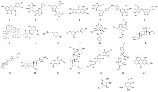
Figure 1.
Structural formulae of several glycosides.
5.1. Quercitrin 1
Quercitrin 1 is a bioflavonoid derived from quercitrin 1 and is widely found in a variety of medicinal or edible plants. It shows a very wide range of biological actions, including anti-tumor, antiviral, anti-thrombotic, anti-inflammatory, anti-allergic, anti-atherosclerotic, and vasodilatory effects as well as stimulating cellular immunity [96,97,98,99,100,101,102].
It has been documented that in DSS-induced colitis in mice, treatment with quercitrin 1 (1 and 5 mg/kg) improves histopathology and reduces biochemical parameters, inflammation, and bacterial translocation [59].
Another article showed that in TNBS-induced experimental colitis, quercitrin 1 (5 mg/kg) prevented early mesenteric vascular hyporesponsiveness by reducing NO overproduction by iNOS. Thus, the mesenteric vascular bed may be a new target for the prevention of UC [60].
In summary, quercitrin 1 has anti-inflammatory and protective effects on the intestinal mucosa in experimental colitis and may be used as an alternative treatment for inflammatory bowel disease. Its role in preventing bacterial translocation and preventing early mesenteric vascular hyporesponsiveness requires further studies to elucidate.
5.2. Baicalin 2
Baicalin 2 is a flavonoid glycoside extracted from Scutellaria baicalensis with a variety of biological activities, including anti-inflammatory, anti-bacterial, and anti-tumor activities [103,104,105,106]. Studies have demonstrated that baicalin 2 (100 mg/kg) can improve the severity of DSS-induced colitis, and the potential mechanism is closely related to the inhibition of the TLR4/NF-κB signaling pathway [61]. Baicalin 2 (100 mg/kg) also alleviated the extent of TNBS-induced colitis in mice by inhibiting the PI3K/AKT signaling pathway [62].
Baicalin 2 (30, 60, and 120 mg/kg) was able to dose-dependently improve the severity of experimental colitis by a mechanism that may be related to the inhibition of oxidative stress and regulation of a range of proteins associated with IEC apoptosis [63]. In addition, baicalein (20, 50, and 100 mg/kg) ameliorated DSS-induced colitis by activating NLRP6 inflammatory vesicles to promote mucus secretion by cupped cells through a mechanism associated with activation of the NLRP6/IL-18 pathway to promote cupped cell production in the mucosa [64].
There is also evidence that baicalin 2 (10 mL/kg) alleviates TNBS-induced colitis in mice concerning the balance between Th17 and Treg cells [65].
In summary, we found that baicalin 2 exerts powerful anti-inflammatory effects in UC treatment by mechanisms related to reducing the release of inflammatory factors, inhibiting oxidative stress and anti-apoptosis, modulating UC-related receptors, modulating immune cells, and regulating different signaling pathways. Thus, baicalin 2 may be a promising drug for UC, and its components may be important lead compounds for the development of novel UC-related chemical drugs.
5.3. Hyperoside 3
Hyperoside 3 is a flavonoid glycoside extracted from Hypericum spp. and Hawthorn spp., with a variety of pharmacological activities including anti-inflammatory, anti-bacterial, anti-tumor, antioxidant, and immunomodulatory functions [107,108].
In DSS-induced colitis in mice, hyperoside 3 (80 and 120 mg/kg) was able to exert a protective effect by inhibiting inflammation and apoptosis, the effect of which may be related to the activation of the Nrf2 signaling pathway [66].
Hyperoside 3 (3, 10, and 30 mg/kg) attenuated DSS-induced colitis in mice in association with modulation of Th17/Treg immune homeostasis and stabilization of PPARγ levels [67].
These findings expand our understanding of the role of hyperoside 3 and may provide potential therapeutic targets for UC.
5.4. Mangiferin 4
Mangiferin 4 is a flavonoid glycoside isolated mainly from the rhizome of mango, with antioxidant, anti-inflammatory, immunomodulatory, anti-bacterial, and analgesic effects [109,110,111,112]. Mangiferin 4 (10, 30, and 100 mg/kg) reduced inflammatory changes in colonic tissue in TNBS-induced colitis in rats, and this protective effect was mainly dependent on its anti-inflammatory and antioxidant properties [68]. There is also evidence that mangiferin 4 (10 and 20 mg/kg) ameliorates inflammatory colitis in TNBS-induced colitis in mice by regulating Th17/Treg cell homeostasis and inhibiting the activation of the NF-κB signaling pathway [69].
Another article clearly showed that mangiferin 4 (50 mg/kg) was able to exert a protective effect on DSS-induced acute colitis by inhibiting the NF-κB and MAPK signaling pathways [70].
The above results provide strong evidence for the use of mangiferin 4 in the treatment of human UC.
5.5. Linarin 5
Linarin 5 is a natural flavonoid compound isolated mainly from plants such as chrysanthemum and peppermint, with anti-inflammatory, antioxidant, analgesic, antipyretic, and anti-tumor as well as sedative, neuroprotective, anti-apoptotic, and anti-osteoporotic effects [113,114,115,116]. It has been shown that linarin 5 (20 and 50 mg/kg) can improve DSS-induced colitis in mice by inhibiting the inflammatory response, maintaining intestinal barrier function, and regulating intestinal flora [71].
5.6. Vitexin 6
Vitexin 6 is mainly a flavonoid glycoside isolated from Hawthorn, with biological activities such as antioxidant, anti-inflammatory, anti-tumor, anti-hypertensive, and anti-convulsant effects [117].
Studies have shown that in DSS-induced colitis mice, vitexin 6 (20 and 80 mg/kg) can combat colitis by inhibiting intestinal mucosal inflammation, maintaining intestinal barrier homeostasis, and remodeling the intestinal flora [72].
It has also been shown that vitexin 6 (40 and 80 mg/kg) not only alleviates DSS-induced colitis in mice but also protects against colitis-induced liver damage from inflammatory responses, which relies heavily on the inhibition of TLR4/NF-κB signaling pathway activation [73].
In summary, vitexin 6 was able to alleviate not only colitis but also colitis-induced liver injury, indicating multiple pharmacological activities of the drug.
5.7. Naringin 7
Naringin 7 is a flavonoid glycoside extracted from grapefruit, lime, and citrus seeds and has a variety of biological activities, including anti-inflammatory and antioxidant effects [118,119].
Naringin 7 (25, 50, and 100 mg/kg) was shown to alleviate DSS-induced colitis, and its anti-UC activity was associated with PPARγ activation. In addition, naringin 7 significantly inhibited DSS-induced NLRP3 inflammasome activation and modulated ZO-1 expression [74].
In addition, naringin 7 (20, 40, and 80 mg/kg) also alleviated TNBS-induced colitis in rats, and its anti-UC activity was associated with antioxidant and anti-inflammatory responses [75].
The above results suggest that naringin 7 may be a potentially effective drug candidate for UC and deserves further development and exploration.
5.8. Punicalagin 8
Punicalagin 8 is a polyphenolic active ingredient extracted from pomegranate, with anti-inflammatory, antioxidant, anti-apoptotic, and anti-proliferative biological activities [120,121,122,123]. It has been shown that in DNBS-induced colitis in rats, administration of punicalagin 8 (4 mg/kg) exhibited significant anti-inflammatory activity and improved inflammatory bowel disease in rats, which may be attributed to direct inhibition of the transcription factor NF-κB [76].
Punicalagin 8 has a significant anti-inflammatory effect on colitis and therefore could be a potential drug for the treatment of colitis, the mechanism of action of which needs to be further explored.
5.9. Curculigoside 9
Curculigoside 9 is a phenolic glycoside component of Curculigo orchioides Gaertn that has various pharmacological activities such as anti-inflammatory, antioxidant, anti-osteoporotic, and neuroprotective effects [124,125,126,127]. It has been documented that curculigoside 9 (500 and 100 mg/kg) inhibits disease activity index, tissue damage, and cell death in DSS-induced colitis mice. It was also able to significantly reverse these alterations in iron-toxicity characteristics such as iron overload, GSH depletion, ROS and MDA production, and reduced expression of SOD and GPX4 [77]. These findings suggest that curculigoside 9 prevents iron sagging in UC by inducing GPX4, suggesting it as a potential therapeutic agent for UC.
In conclusion, in addition to alleviating the symptoms of DSS-induced colitis in mice, curculigoside 9 was also able to reverse the altered iron toxicity profile, which also suggests that the drug’s effects are diverse, while its mechanism requires further study.
5.10. Salidroside 10
Salidroside 10 is a phenolic glycoside extracted from Rhodiola rosea, which has been proven to have a variety of pharmacological effects, including anti-aging, antioxidant, anti-cancer, anti-inflammatory, antioxidant, and neuroprotective activities [128,129,130].
It has been shown that salidroside 10 may exert a protective effect by reducing DSS-induced colonic tissue damage in mice through activation of the SIRT1/FoxOs pathway [78].
Additional data suggest that salidroside 10 protects against experimental colitis by reversing TREM1-associated macrophage pyroptosis and gut microbiota dysregulation-derived Th17/Treg imbalance, suggesting a potential role for UC [79].
In summary, salidroside 10 offers new options as a treatment for UC and merits further drug development.
5.11. Polydatin 11
Polydatin 11 is a phenolic glucoside extracted from the traditional Chinese medicine tiger cane. Many studies have shown that it has a wide range of pharmacological activities, such as anti-fibrotic, anti-tumor, anti-atherosclerotic disease, and anti-hepatitis effects as well as protection against multi-organ ischemia-reperfusion injury and dementia-related diseases [131,132,133,134,135,136,137,138,139].
There is evidence that polydatin 11 (15, 30, and 45 mg/kg) effectively reduce colonic oxidative stress and apoptosis. This effect may be mediated by the up-regulation of the Shh signaling pathway [47,80].
In addition, it has been shown that polydatin 11 inhibits intestinal inflammation and oxidative stress and maintains intestinal epithelial barrier integrity by mechanisms related to NF- κB, MAPK, and AKT/Nrf2/HO-1/NQO1 signaling pathways [50]. Interestingly, however, the authors did not indicate in their article the exact dose of polydatin 11 used in DSS-induced colitis in mice, which needs to be further mapped out.
Polydatin 11 (30 and 60 mg/kg) also alleviates DSS- and TNBS-induced colitis by directly binding to STAT3, specifically inhibiting STAT3 phosphorylation and correcting Th17/Treg homeostasis [81].
In summary, polydatin 11 can exert anti-colitis effects through a variety of mechanisms, so we have reason to believe that polydatin may be a promising candidate for the treatment of UC.
5.12. Paeoniflorin 12
Paeoniflorin 12 is a terpene glycoside isolated from Paeonia lactiflora, with pharmacological effects such as anti-inflammatory, antipyretic, anti-spasmodic, neuroprotective and cerebral, antidepressant, immunomodulatory, and anti-tumor effects as well as scavenging free radicals in the body [42,43,44,45,46,48,49,140].
It has been shown that continuous administration of paeoniflorin 12 (50 mg/kg) for 7 days significantly reduced the severity of DSS-induced colitis and led to a down-regulation of the associated inflammatory parameters, suggesting that its beneficial effects may be related to blocking the activation of NF-κB and MAPK pathways [82]. In addition, we found that paeoniflorin 12 (3 g/kg) had a therapeutic effect on AOM/DSS-induced colitis-associated cancer mice by a mechanism associated with inhibition of TLR4/NF-κB-mediated inflammatory responses and EGFL7 expression [51].
Paeoniflorin 12 (15, 30, and 45 mg/kg) also exerted protective effects against TNBS-induced colitis in mice by inhibiting inflammation and apoptosis through the MAPK/NF-κB pathway [83]. Paeoniflorin 12 (20 mg/kg) also exerted anti-UC activity by suppressing inflammatory responses and eosinophil infiltration [84].
In summary, paeoniflorin 12 can play a role in the treatment of colitis-associated cancers in addition to its protective role in UC, suggesting a diversity of drug actions that merits further in-depth study.
5.13. Asperuloside 13
Asperuloside 13 is a terpenoid extracted from Rubiaceae, Eucommiaceae, and other plants. Recent pharmacological studies have shown that asperuloside 13 has a variety of pharmacological activities that are anti-inflammatory, antioxidant, and immunomodulatory [141]. There is evidence that asperuloside 13 (125 and 0.5 mg/kg) may improve DSS-induced colitis in mice by alleviating inflammation and oxidative stress, activating the Nrf2/HO-1 signaling pathway, and limiting the NF-κB signaling pathway [85].
The above indicates that the drug is characterized by multiple pharmacological mechanisms and predicts a potential application of asperuloside 13 in the treatment of UC.
5.14. Pedunculoside 14
Pedunculoside 14 is a naturally occurring triterpene glycoside derived from the bark of iron holly. Previous studies have shown that pedunculoside 14 has anti-inflammatory, anti-tumor, anti-viral, cholesterol-lowering, and blood-pressure-lowering effects [142,143,144]. There is evidence that pedunculoside 14 (5, 15, and 30 mg/kg) has significant efficacy in DSS-induced UC, suppressing the expression of inflammatory mediators by inhibiting the activation of MAPK and AKT/NF-κB signaling pathways [86].
In summary, pedunculoside 14 has a good therapeutic effect on UC and may be a potential natural product for the treatment of UC.
5.15. Glycyrrhizin 15
Glycyrrhizin 15 is triterpenoid saponin derived from Glycyrrhiza glabra with anti-inflammatory, anti-ulcer, anti-hepatocytotoxic, anti-cancer, and anti-viral biological activities [145,146,147]. In a rat model of acetic-acid-induced UC, glycyrrhizin 15 (40 mg/kg) was able to attenuate the inflammatory response by inhibiting NF-κB, TNF-a, and ICAM-1 in the colonic mucosa [87]. Glycyrrhizin 15 (100 mg/kg) also exerted anti-inflammatory effects through the up-regulation of PPARc [88].
Furthermore, in TNBS-induced experimental colitis, glycyrrhizin 15 (100 mg/kg) was able to modulate the intestinal inflammatory response by regulating the subtle balance of T cells [89].
In conclusion, glycyrrhizin 15 can play a significant anti-inflammatory role in experimental colitis, and its mechanism is related to inhibiting the expression of inflammatory factors, alleviating oxidative stress, up-regulating PPARγ activity, and regulating the expression of immune cells. This provides strong evidence for glycyrrhizin 15 as a new potential therapeutic agent.
5.16. Astragaloside Ⅳ 16
Astragaloside Ⅳ 16 is a triterpenoid saponins isolated from Astragalus membranaceus. Studies have shown that astragaloside IV 16 has immunomodulatory, anti-fibrotic, anti-inflammatory, anti-radiation, anti-viral, antioxidant, anti-tumor, and cardiovascular protective effects [148,149,150]. Studies have demonstrated that astragaloside IV 16 (50 and 100 mg/kg) prevents DSS-induced acute colitis by remodeling Th17/Treg cell homeostasis and anti-oxidative stress, with the potential mechanism closely related to the inhibition of the Notch signaling pathway [90].
5.17. Gentiopicroside 17
Gentiopicroside 17 is a terpenoid glycoside isolated from gentian, with a variety of pharmacological activities, including anti-inflammatory, cholestatic, and anti-hepatotoxic effects [151,152].
Gentiopicroside 17 (50, 100, and 200 mg/kg) may exert anti-inflammatory effects on DSS-induced acute colitis by inhibiting the expression of inflammatory factors, suggesting a possible therapeutic potential in the treatment of colitis, but its exact mechanism of action needs further study [91].
5.18. Ginsenoside Rg1 18
Ginsenoside Rg1 18 is a terpenoid glycoside isolated from Ginseng and Panax notoginseng, which has a variety of pharmacological activities, including anti-inflammatory and neuroprotective effects, effects on obesity, etc. [153,154,155,156,157].
Ginsenoside Rg1 18 (200 mg/kg) significantly improved DSS-induced colonic injury and colonic inflammation in mice, which may be related to the regulation of intestinal flora [92].
5.19. Liriodendrin 19
Liriodendrin 19 is one of the active ingredients extracted from liriodendrin. [158]. Liriodendrin 19 has a variety of biological functions, including anti-inflammatory, antioxidant, anti-tumor, anti-fungal, and anti-platelet coagulation and also has some anti-Alzheimer’s effects [159,160,161,162]. It has been shown that liriodendrin 19 (100 mg/kg) exerts anti-inflammatory activity in DSS-induced colitis in mice by inhibiting oxidative stress and activation of Akt and NF-κB pathways [93].
5.20. Convallatoxin 20
Convallatoxin 20 is a steroidal glycoside isolated from Calendula officinalis with a variety of pharmacological activities, including anti-inflammatory, antioxidant, anti-bacterial, anti-tumor, anti-angiogenic, and cardiotonic [163,164,165,166,167].
The ability of convallatoxin 20 (50 and 150 μg/kg) to ameliorate DSS-induced inflammation in colitis by activating PPARγ and inhibiting NF-κB suggests that it may be a promising compound for the treatment of UC [94].
5.21. Aloin A 21
Aloin A 21 is an anthraquinone glycoside extracted from the secretion of Aloe vera leaves and has anti-inflammatory, anti-bacterial, antioxidant, anti-viral, and anti-cancer pharmacological effects [168]. Aloin A 21 (25 and 50 mg/kg) can prevent DSS-induced colitis by enhancing intestinal barrier function through inhibition of the Notch signaling pathway [95].
6. Anti-Inflammatory Mechanisms of Glycosides in UC
6.1. Suppressing Inflammatory Responses
Excessive production of inflammatory factors and mediators such as TNF-α, IL-1, IL-6, COX-2, and iNOS in the intestine can dominate and perpetuate the inflammatory response [91]. In addition, ICAM-1 acts as a glycoprotein that mediates cell–cell and cell–extracellular matrix adhesion and is proportional to the severity of the inflammatory response when UC occurs [169].
All the glycosides in Table 3 exerted anti-inflammatory effects by inhibiting the expression of inflammatory factors and mediators in the colonic tissues of UC mice to varying degrees. Therefore, the study of the effects of drugs on inflammation-related factors and mediators is important for the treatment of UC.
6.2. Reduction of Oxidative Stress
Oxidative stress is thought to be one of the etiologies involved in inflammatory bowel disease. Excessive oxidative reactions can upset the balance of redox reactions in the colonic mucosa and cause intestinal damage. Whereas antioxidant enzymes are markers for scavenging free radicals generated by oxidative stress, drugs can alleviate the intestinal oxidative stress state in UC by increasing their expression levels [170].
All the glycosides in Table 3 were able to reduce intestinal pathological damage and improve clinical symptoms of UC by modulating the expression of different antioxidant enzymes (Figure 2).
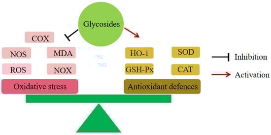
Figure 2.
Glycosides and oxidative stress. All glycosides produced relief of UC by inhibited oxidative stress.
6.3. Anti-Apoptosis
The Bcl-2 protein family consists of Bcl-2, Bcl-xL, and Bax. In animal models, overexpression of Bcl-2 attenuates joint damage in animals, while overproduction of Bax promotes apoptosis.
In addition, Bcl-2 blocks the release of cytochrome c and down-regulates caspase activity. Caspase-3 is a central molecule in apoptosis, and its activation is regulated by a series of signal transduction cascades. Moreover, caspase-9 can be activated through the Bcl-2/Bax-ratio-mediated apoptotic pathway [83].
Studies have shown that salidroside 10 and polydatin 11 inhibit apoptosis in colon cells by down-regulating the expression of Bax, caspase-3, and cleaved-caspase-3 and up-regulating the expression of Bcl-2 [47,78]. Paeoniflorin 12, baicalin 2, and hyperoside 3 can also inhibit colon cell apoptosis by down-regulating Bax expression. Aloin A 21 also inhibited apoptosis in colon cells by down-regulating cleaved-caspase-3 expression [95]. This suggests that apoptosis plays a role in the pathogenesis of UC [63,66,83] (Figure 3).
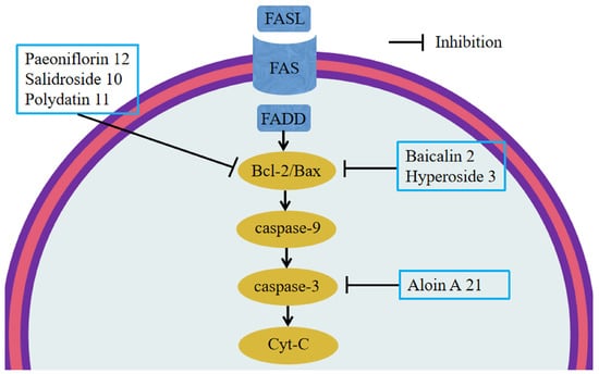
Figure 3.
Glycosides and apoptosis. Salidroside 10 and polydatin 11 down-regulated the expression of Bax, caspase-3, and cleaved caspase-3 and up-regulated the expression of Bcl-2. Paeoniflorin 12, baicalin 2, and hyperoside 3 can down-regulate Bax. Aloin A 21 down-regulated cleaved caspase-3 expression.
6.4. Regulation of Impaired Intestinal Epithelial Barrier Function
The mucus proteins secreted by the cupulae cover the intestinal epithelium to form a dense mucin network that forms the first barrier in the intestinal lumen [171]. In addition, the tight junction (TJ) is the most important component of the intestinal epithelial barrier and is a complex of claudins and occludin proteins, peripheral membrane protein family ZOs, and other proteins. Abnormal expression of TJ can increase the permeability of the intestinal epithelial barrier, leading to the entry of pathogenic antigens such as bacteria into the mucosa and blood circulation, causing inflammation [172].
It was found that MUC2, MUC3A, claudin-1, occludin, and ZO-1 expression levels were significantly increased in colonic tissues of mice with colitis treated with polydatin 11, reducing the intestinal inflammatory response [50]. In addition, baicalin 2, hyperoside 3, aloin A 21, linarin 5, vitexin 6, and naringin 7 can improve the expression of TJ proteins and mucin proteins in intestinal mucosa and inhibit the increase of intestinal mucosal permeability, exerting anti-inflammatory effects [64,67,71,72,74,95]. The above suggests that intestinal epithelial barrier function plays an important role in the pathogenesis of UC (Figure 4).
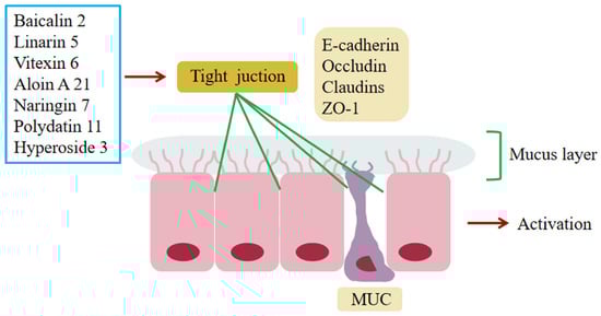
Figure 4.
Glycosides and intestinal epithelial barrier. Polydatin 11, baicalin 2, hyperoside 3, aloin A 21, linarin 5, vitexin 6, and naringin 7 can improve the expression of TJ proteins and mucin proteins, thus inhibiting the increase of the permeability of the intestinal mucosa to achieve regulation of intestinal epithelial barrier function.
6.5. Regulation of Immune Cells
CD4+ T cells can differentiate into Th1, Th2, Th17, and Treg cells in response to different cytokine stimuli [173]. Previous studies have shown that the pro-inflammatory effects of Th17 cells can be antagonized by Treg cells, producing the anti-inflammatory cytokines IL-10 and TGF-β [174]. Conversely, Treg can alleviate colitis by down-regulating Th1 and Th17 through IL-10 and TGF-β [175]. In addition, differentiation of both Treg and Th17 cells requires TGF-β to induce Foxp3 and RORγt. Already-differentiated Treg cells stimulated by IL-6 can inhibit Foxp3 expression and release IL-17, which in turn induces cell differentiation into Th17 cells [176]. Th17/Treg cells remain in balance under normal conditions, and once imbalanced, especially when Th17 cells are overrepresented, they can be reduced to Th17 cells. In particular, an excessive increase in Th17 cells can lead to intestinal mucosal damage and inflammatory bowel disease. Therefore, maintaining the homeostasis of Th17/Treg cells is important to prevent the development of UC.
Studies have shown that polydatin 11 can reduce DSS- and TNBS-induced colitis in mice by directly binding to STAT3 and regulating Th17 cell differentiation and Th17/Treg homeostasis [81]. Meanwhile, the anti-inflammatory effects of salidroside 10, astragaloside Ⅳ 16, hyperoside 3, and mangiferin 4 were also associated with the regulation of Th17/Treg cell homeostasis in experimental UC [65,69,79,90]. In addition, hyperoside 3 was also able to regulate PPARγ levels via MKRN1, thereby restoring Th17/Treg homeostasis to reduce DSS-induced colitis in mice [67] (Figure 5).
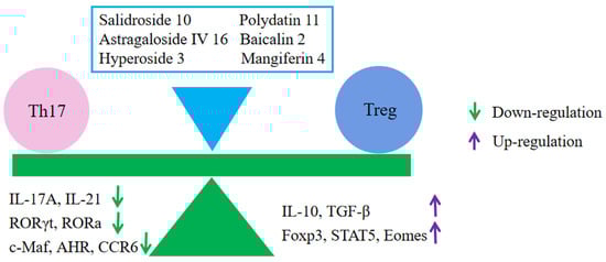
Figure 5.
Glycosides and Th17/Treg balance. Polydatin 11, salidroside 10, astragaloside Ⅳ 16, hyperoside 3, and mangiferin 4 produced relief of UC by regulating the balance of Th17/Treg cells.
6.6. Regulation of UC-Related Receptors
6.6.1. Inhibition of Toll-like Receptors (TLRs)
TLRs are recognition factors that initiate inflammatory responses and immune responses. When activated, they bridle MyD88 protein for signaling and promoting the expression of related inflammatory factors, exacerbating intestinal inflammation. Previous studies have demonstrated that the TLRs/MyD88/NF-κB signaling pathway plays an important role in UC [51,61,73].
Paeoniflorin 12 and vitexin 6 can exert anti-inflammatory effects by reducing TLR4 expression and blocking the TLR4/NF-κB signaling pathway [51,73]. Baicalin 2 can reduce the expression of TLR2, TLR4, and TLR9 and inhibit NF-κB by blocking MyD88 signaling activation, thus inhibiting the production of inflammatory factors and exerting a protective effect [61] (Figure 6).
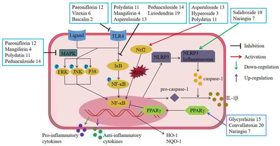
Figure 6.
Glycosides and UC-related receptors AND transcriptional regulation. Paeoniflorin 12, vitexin 6, and baicalin 2 produced relief of UC by reducing TLR4 expression and blocking the TLR4/NF-κB signaling pathway. Glycyrrhizin 15, convallatoxin 20, and naringin 7 produced relief of UC by suppressing the expression of NF-κB via activated PPARγ. Salidroside 10 and naringin 7 produced relief of UC by inhibiting the expression of inflammasome and thereby reducing the release of IL-1β. Punicalagin 8, paeoniflorin 12, pedunculoside 14, liriodendrin 19, baicalin 2, mangiferin 4, polydatin 11, and vitexin 6 produced relief of UC by blocking the NF-κB signaling pathway. Mangiferin 4, polydatin 11, pedunculoside 14, and paeoniflorin 12 produced relief of UC by blocking the MAPK signaling pathway. Asperuloside 13, hyperoside 3, and polydatin 11 produced relief of UC by blocking the Nrf2/HO-1 signaling pathway.
6.6.2. Up-Regulation of Peroxisome Proliferator-Activated Receptor (PPARγ)
PPARr is a member of the nuclear receptor superfamily, most of which are ligand-dependent transcriptional activators. The anti-inflammatory activity of activated PPARγ is mediated through inhibition of NF-κB activity, leading to a reduction in the expression of inflammatory factors and exerting anti-inflammatory effects [88,94].
Glycyrrhizin 15, convallatoxin 20, and naringin 7 were all able to inhibit the expression of NF-κB and other inflammatory factors by increasing the expression of PPARγ, thereby reducing colonic mucosal inflammation and improving experimental colitis [74,88,94] (Figure 6).
6.6.3. Inhibition of Nucleotide-Binding Oligomerization Domain (NOD)-Like Receptors (NLRs)
NOD-like receptor protein 3 (NLRP3) inflammasomes in NLRs are important regulators of intestinal homeostasis. Previous studies have shown that NLRP3 can improve experimental colitis by down-regulating IL-1β levels, and therefore, NLRP3 inflammasome is essential in the pathogenesis of UC.
Studies have shown that naringin 7 can exert anti-UC effects by down-regulating the expression of NLRP3 inflammasome [74]. Furthermore, in LPS-induced macrophages RAW264.7, salidroside 10 was similarly able to exert anti-inflammatory effects by down-regulating NLRP3 inflammasome excitation and apoptosis [79] (Figure 6).
6.7. Regulating Signal Transduction
6.7.1. Inhibition of the NF-κB Pathway
Activation of NF-κB is a key step in the activation and proliferation of the inflammatory response in enteritis [51]. As a heterodimer complex of (p50/p65), NF-κB is regulated by IκB and IKK. Activated in response to external stimuli, IKKs phosphorylates the inhibitory IκB protein. Activated NF-KB is then transferred to the nucleus, where it binds to target DNA elements and encodes multiple inflammatory mediators.
It was shown that paeoniflorin 12 reduced NF-κB expression in DSS/TNBS-induced colonic tissue of UC rats and down-regulated inflammatory mediators in colonic mucosa [51,82,83,84]. In vitro, experiments also revealed that polydatin 11, pedunculoside 14, mangiferin 4, liriodendrin 19, and asperuloside 13 inhibited the nuclear translocation of NF-κB and thus the inflammatory response of LPS-activated macrophages RAW264.7 [50,70,85,86,93]. In addition, the mechanisms by which vitexin 6 and baicalin 2 eliminated experimental colitis were both targeted to inhibit activation of the TLR4/NF-κB pathway [61,73] (Figure 6).
6.7.2. Inhibition of the MAPK Pathway
The MAPK family consists of three main members: JNK, p38MAPK, and ERK [86]. ERK is mainly activated by mitogen. p38MAPK can induce the expression of inflammatory factors such as TNF-α, ILs, and IFN-γ as well as COX-2 by mediating the activation of NF-κB [177]. JNK phosphorylates the transcription factor c-JNK and induces the production of related inflammatory factors that trigger UC. In addition, JNK activates the transcription factor STAT3 and the non-transcription factor Bcl-2, which play an important role in the development of UC [178].
In the DSS-induced UC mouse model, paeoniflorin 12 and mangiferin 4 were able to significantly inhibit the increased phosphorylation levels of ERK1/2, JNK, and p38, exerting anti-inflammatory effects [70,82,83]. In in vitro experiments, polydatin 11 and pedunculoside 14 also significantly inhibited the phosphorylation of ERK1/2, JNK1/2, and p38, thereby reducing the production of IL-1β, IL-6, TNF- α, COX-2, and iNOS and suppressing the inflammatory response [50,86] (Figure 6).
6.7.3. Inhibition of the Nrf2/HO-1 Pathway
The transcription factor Nrf2 is the most important transcription factor against oxidative stress and maintains mucosal homeostasis by inhibiting the production of excess ROS. When activated, Nrf2 is phosphorylated, leading to increased expression of antioxidant genes such as SOD, NQO1, CAT, GSH-Px, and HO-1 [50,85].
Asperuloside 13 can reduce DSS-induced oxidative stress and inflammation in mouse colon tissue by activating the Nrf2/HO-1 signaling pathway; increasing the expression of Nrf2, HO-1, and NQO-1 proteins; and down-regulating p65 levels [85]. Hyperoside 3 was also able to ameliorate the inflammatory response and apoptosis in experimental colitis by activating the Nrf2 signaling pathway [66]. In vitro, experiments have also shown that polydatin 11 can exert anti-inflammatory effects by inhibiting MAPK and NF-κB inflammatory signaling pathways and activating AKT/Nrf2/HO-1/NQO1 signaling pathway [50] (Figure 6).
6.7.4. Inhibition of Other Related Pathways
The pathogenesis of UC also involves the JAK-STAT, PI3K/AKT, Notch, and Shh pathways. Through in vivo and vitro experiments of UC, polydatin 11 was able to specifically block the JAK/STAT3 signaling pathway and inhibit the differentiation of Th17 cells to improve intestinal inflammation [81]. In TNBS-induced colitis, baicalin 2 was able to play a protective role in colitis by inhibiting the PI3K/AKT pathway [62]. Astragaloside Ⅳ 16 and aloin A 21 can effectively inhibit DSS-induced inflammatory damage in colon tissue by inhibiting the Notch signaling pathway and reconstructing the colonic mucosa [90,95]. Oxidative stress and epithelial cell apoptosis in the gastrointestinal tract are associated with the Shh pathway, and polydatin 11 may inhibit experimental colitis by modulating the Shh signaling pathway [47].
7. Conclusions and Outlook
A large number of review papers discuss the possible application of various extracts from natural products as well as pure biologically active substances in medicine [179,180,181,182].
Pure bioactive substances include synthetic drugs and pure natural substances. The decisive factor for the dominance of pure bioactive substances in modern medicine is the ease of drug administration and clinical pharmacological evaluation. Synthetic drugs have been widely used in clinical practice, but there are many side effects as well as resistance and drug-induced diseases; in addition, the development cycle of synthetic drugs is long, the investment is large, and the enterprises often cannot bear it.
With the progress of science, people’s awareness of self-care is enhanced, the understanding of natural medicine is deepened, and the desire to return to nature is rising, so the demand for natural medicine is increasing [183].
Natural products are the largest pool of biologically active substances on Earth [179]. The bioactive compounds derived from natural products, especially medicinal plants, have emerged as new treatments for a variety of diseases. Natural products with biodiversity and chemical diversity, especially higher plants, will always be the most reliable source of leads for the development of new drugs [13]. In addition, the success rate of the development of new drugs by synthetic substances is extremely low, while the success rate of the development of new drugs by natural products from plants is much higher, the development time is greatly shortened, and the financial and human investment is also reduced accordingly.
Despite the great potential of natural products in the treatment of diseases, there are other issues that need to be addressed, such as the extraction, isolation, and standardization of derived compounds as well as their effective treatment modalities. In addition, some natural ingredients have low activity, a narrow antibacterial spectrum, strong drug resistance, poor stability, or serious side effects, and the corresponding technology should be used for structural modification to overcome their defects. Therefore, much research is needed before any natural product can be used as a treatment [13].
The extraction of active ingredients from natural products is an important source of new drug discovery. Understanding the mechanism of action of drugs helps to obtain the best clinical treatment drugs. This manuscript reviews 21 glycosides with relatively clear anti-inflammatory effects and mechanisms of action to provide new insights and ideas for the discovery of new drugs for the treatment of UC.
Glycosides have a wide range of pharmacological effects, including antioxidant, analgesic, antipyretic, anti-hypertensive, anti-tumor, and neuroprotective effects in addition to anti-inflammatory effects [71,113,114,115,116,117]. In this review, we found that glycosides can improve UC symptoms and play a role in the prevention and treatment of UC by inhibiting inflammatory response, reducing oxidative stress and anti-apoptosis, inhibiting abnormal immune response, and regulating signal pathway transduction (Figure 7). The glycosides in this review mainly include flavonoids, phenols, terpenes, etc., which can exert anti-inflammatory effects by inhibiting the expression of inflammatory-related factors and reducing oxidative stress. The main mechanisms include NF-κB, MAPK inflammatory signaling pathways, and the Nrf2/HO-1 signaling pathway. Astragaloside Ⅳ 16 and aloin A 21 can inhibit the colonic inflammatory response by modulating the Notch signaling pathway. Polydatin 11 was also able to reduce the inflammatory response by acting on JAK/STAT3 and Shh signaling pathways. Baicalin 2 can play a protective role in colitis by inhibiting the PI3K/AKT pathway. In addition, the intestinal flora also plays an important role in the pathogenesis of UC, but there are few studies on glycosides in this regard, which need further study. Meanwhile, glycosides are also worthy of further study in the prevention and treatment of UC-related carcinogenesis.
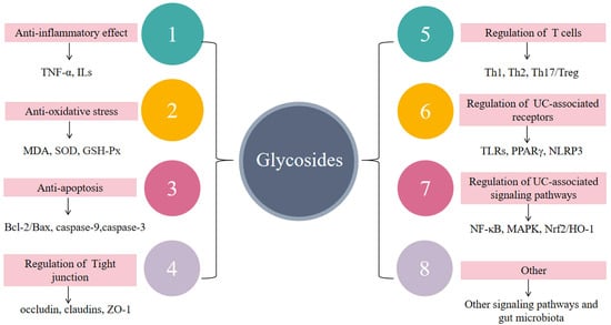
Figure 7.
The mechanism of glycosides in the treatment of UC. Glycosides play a role in the treatment of UC through anti-inflammatory and antioxidant stress mechanisms, regulation of impaired intestinal epithelial barrier function, regulation of immune cells, and regulation of UC-related receptors and signal transduction.
UC is a complex disease, and although we have identified a strong relationship between glycosides and UC, there are still many issues to be resolved. Firstly, oral bioavailability is poor, and glycosides must be hydrolyzed by intestinal enzymes or microflora before they can be absorbed. Secondly, glycosides are unstable and readily hydrolyzed by acids, bases, and enzymes. It has been found that only 10–15% of the glycosides are absorbed in the small intestine, with the remaining glycosides being metabolized by microorganisms in the large intestine to form small molecules, so it is doubtful whether it is the broken-down glycosides and their metabolites or the original glycoside molecules that work in the body [184,185,186]. Thirdly, the side effects of glycosides and the pharmacological control of UC complications are not fully understood.
In summary, these glycosides are valuable candidates for the prevention and treatment of UC even taking into account the problems mentioned above. Therefore, it is urgent to study the pharmacokinetic characteristics of natural-product-isolated glycosides and establish their dose–pharmacology–toxicology relationship. Further clinical studies are essential to prove the effective role of glycosides in the prevention and treatment of UC. Therefore, future research must explore the targets and molecular mechanisms of glycosides, combine laboratory anti-UC studies with clinical practice, test the reliability of glycosides against UC, and promote their use in the practical prevention and treatment of UC.
Author Contributions
Conceptualization, writing—original draft, and writing—review and editing, Y.N.; visualization, J.Z.; project administration, D.S.; data curation, W.Z.; supervision and language embellishment, J.N. All authors have read and agreed to the published version of the manuscript.
Funding
This research received no external funding.
Institutional Review Board Statement
Not applicable.
Informed Consent Statement
Not applicable.
Data Availability Statement
The data that support the findings of this study are available from the corresponding author upon reasonable request.
Conflicts of Interest
The authors declare no conflict of interest.
Sample Availability
Not applicable.
Abbreviations
| IBD | Inflammatory bowel disease |
| TNF-α | Tumor necrosis factor-alpha |
| IL | Interleukin |
| MAPK | Mitogen-activated protein kinase |
| NF-κB | Nuclear factor kappa-B |
| STAT | Signal transducer and activator of transcription |
| Treg | Regulatory T cells |
| IFN-γ | Interferon-γ |
| ERK | Extracellular signal-regulated kinase |
| JNK | C-Jun N-terminal kinase |
| TLR4 | Toll-like receptor4 |
| EGFL7 | Epidermal growth factor-like domain 7 |
| MPO | Myeloperoxidase |
| MDA | Malondialdehyde |
| GSH-Px | Glutathione peroxidase |
| GPX4 | Anti-oxidant enzyme glutathione peroxidase 4 |
| HO-1 | Heme oxygenase-1 |
| NQO1 | Quinone oxidoreductase 1 |
| Nrf2 | Nuclear factor (erythroid-derived 2)-like 2 |
| ROS | Reactive oxygen species |
| SOD | Superoxide dismutase |
| COX-2 | Cyclooxygenase-2 |
| INOs | Inducible nitric oxide synthase |
| ERβ | Estrogen receptor-β |
| ICAM-1 | Intercellular cell adhesion molecule-1 |
| PPARγ | Peroxisome proliferator-activated receptor γ |
| HMGB1 | High-mobility group box 1 |
| DCs | Dendritic cells |
| IFN-γ | Interferon γ |
| NOX1 | Nicotinamide adenine dinucleotide phosphate oxidase 1 |
| Shh | Sonic hedgehog |
| Ptch1 | Patched1 |
| Smo | Smoothened |
| Gli1 | Glioma-associated oncogene homolog 1 |
| ERK | Extracellular signal-regulated kinase |
| JNK | C-Jun N-terminal kinase |
| LPS | Lipopolysaccharide |
| MUC | Mucin |
| Th | T-helper |
| Tregs | Regulatory T cells |
| c-Maf | c-Musculoaponeurotic fibrosarcoma |
| AhR | Aryl hydrocarbon receptor |
| EOMES | Recombinant eomesodermin |
| FOXP3 | Forkhead box P3 |
| CCR6 | Chemokine receptor 6 |
| TGF-β1 | Transforming growth factor-β1 |
| CAT | Catalase |
| DLL3 | Delta-like protein 3 |
| PI3K | Phosphoinositide 3-kinase |
| AKT | Protein kinase B |
| PTEN | Phosphatase and tensin homologue deleted on chromosome ten |
| ROS | Reactive oxygen species |
| RORγt | Retinoic acid-related orphan receptor gamma t |
| ZO | Zonula occludens |
| IEC | Intestinal epithelial cell |
| ASC | Amino acid transporter 1 |
| NLRP6 | Nod-like receptor pyrin domain-containing protein 6 |
| MKRN1 | Makorin ring finger protein 1 |
| OCLN | Occludin |
| ATOH1 | Recombinant human atonal homolog 1 |
| ICAM-1 | Intercellular adhesion molecule-1 |
| IκBα | Inhibitory κB-α |
| ALT | Alanine aminotransferase |
| AST | Aspartate aminotransferase |
| TC | Total cholesterol |
| TG | Triglyceride |
| ASC | Apoptosis-associated particulate protein |
| SGOT | Serum glutamic-oxaloacetic transaminase |
| SGPT | Serum glutamic pyruvic transaminase |
| ALP | Alkaline phosphatase |
| TJ | Tight junction |
| Cyt-C | Cytochrome c |
References
- Ungaro, R.; Mehandru, S.; Allen, P.B.; Peyrin-Biroulet, L.; Colombel, J.-F. Ulcerative colitis. Lancet 2017, 389, 1756–1770. [Google Scholar] [CrossRef]
- Høivik, M.L.; Moum, B.; Solberg, I.C.; Henriksen, M.; Cvancarova, M.; Bernklev, T. IBSEN Group Work disability in inflammatory bowel disease patients 10 years after disease onset: Results from the IBSEN Study. Gut 2013, 62, 368–375. [Google Scholar] [CrossRef]
- Conrad, K.; Roggenbuck, D.; Laass, M.W. Diagnosis and classification of ulcerative colitis. Autoimmun. Rev. 2014, 13, 463–466. [Google Scholar] [CrossRef]
- Anzai, H.; Hata, K.; Kishikawa, J.; Ishii, H.; Nishikawa, T.; Tanaka, T.; Tanaka, J.; Kiyomatsu, T.; Kawai, K.; Nozawa, H.; et al. Clinical pattern and progression of ulcerative proctitis in the Japanese population: A retrospective study of incidence and risk factors influencing progression. Color. Dis. 2016, 18, O97–O102. [Google Scholar] [CrossRef]
- Wu, K.; Zhang, Q.; Sha, S.; Xu, B.; Liang, S. Prevalence of colorectal cancer in patients with ulcerative colitis: A retrospective, monocenter study in China. J. Cancer Res. Ther. 2015, 11, 899–903. [Google Scholar] [CrossRef]
- Rosenzwajg, M.; Lorenzon, R.; Cacoub, P.; Pham, H.P.; Pitoiset, F.; El Soufi, K.; Ribet, C.; Bernard, C.; Aractingi, S.; Banneville, B.; et al. Immunological and clinical effects of low-dose interleukin-2 across 11 autoimmune diseases in a single, open clinical trial. Ann. Rheum. Dis. 2019, 78, 209–217. [Google Scholar] [CrossRef]
- Park, J.H.; Peyrin-Biroulet, L.; Eisenhut, M.; Shin, J.I. IBD immunopathogenesis: A comprehensive review of inflammatory molecules. Autoimmun. Rev. 2017, 16, 416–426. [Google Scholar] [CrossRef]
- Park, S.C.; Jeen, Y.T. Anti-integrin therapy for inflammatory bowel disease. World J. Gastroenterol. 2018, 24, 1868–1880. [Google Scholar] [CrossRef]
- Nielsen, O.H.; Bjerrum, J.T.; Herfarth, H.; Rogler, G. Recent Advances Using Immunomodulators for Inflammatory Bowel Disease. J. Clin. Pharmacol. 2013, 53, 575–588. [Google Scholar] [CrossRef]
- Poitras, P.; Gougeon, A.; Binn, M.; Bouin, M. Extra digestive manifestations of irritable bowel syndrome: Intolerance to drugs? Dig. Dis. Sci. 2008, 53, 2168–2176. [Google Scholar] [CrossRef]
- Triantafillidis, J.K.; Merikas, E.; Georgopoulos, F. Current and emerging drugs for the treatment of inflammatory bowel disease. Drug Des. Dev. Ther. 2011, 5, 185–210. [Google Scholar] [CrossRef]
- Xue, J.-C.; Yuan, S.; Meng, H.; Hou, X.-T.; Li, J.; Zhang, H.-M.; Chen, L.-L.; Zhang, C.-H.; Zhang, Q.-G. The role and mechanism of flavonoid herbal natural products in ulcerative colitis. Biomed. Pharmacother. 2023, 158, 114086. [Google Scholar] [CrossRef]
- Araruna, M.E.; Serafim, C.; Alves Júnior, E.; Hiruma-Lima, C.; Diniz, M.; Batista, L. Intestinal Anti-Inflammatory Activity of Terpenes in Experimental Models (2010–2020): A Review. Molecules 2020, 25, 5430. [Google Scholar] [CrossRef]
- Santos, J.A.M.; Santos, C.L.A.A.; Freitas Filho, J.R.; Menezes, P.H.; Freitas, J.C.R. Polyacetylene Glycosides: Isolation, Biological Activities and Synthesis. Chem. Rec. 2022, 22, e202100176. [Google Scholar] [CrossRef]
- Tian, X.Y.; Li, M.X.; Lin, T.; Qiu, Y.; Zhu, Y.T.; Li, X.L.; Tao, W.D.; Wang, P.; Ren, X.X.; Chen, L.P. A review on the structure and pharmaco-logical activity of phenylethanoid glycosides. Eur. J. Med. Chem. 2021, 209, 112563. [Google Scholar] [CrossRef]
- Khan, H.; Pervaiz, A.; Intagliata, S.; Das, N.; Venkata, K.C.N.; Atanasov, A.G.; Najda, A.; Nabavi, S.M.; Wang, D.; Pittalà, V.; et al. The analgesic potential of glycosides derived from medicinal plants. DARU J. Pharm. Sci. 2020, 28, 387–401. [Google Scholar] [CrossRef]
- Molodecky, N.A.; Soon, I.S.; Rabi, D.M.; Ghali, W.A.; Ferris, M.; Chernoff, G.; Benchimol, E.I.; Panaccione, R.; Ghosh, S.; Barkema, H.W.; et al. Increasing Incidence and Prevalence of the Inflammatory Bowel Diseases With Time, Based on Systematic Review. Gastroenterology 2012, 142, 46–54.e42; quiz e30. [Google Scholar] [CrossRef]
- Torres, J.; Billioud, V.; Sachar, D.B.; Peyrin-Biroulet, L.; Colombel, J.-F. Ulcerative Colitis as A Progressive Disease: The Forgotten Evidence. Inflamm. Bowel Dis. 2012, 18, 1356–1363. [Google Scholar] [CrossRef]
- Kaplan, G.G. The global burden of IBD: From 2015 to 2025. Nat. Rev. Gastroenterol. Hepatol. 2015, 12, 720–727. [Google Scholar] [CrossRef]
- Du, L.; Ha, C. Epidemiology and Pathogenesis of Ulcerative Colitis. Gastroenterol. Clin. N. Am. 2020, 49, 643–654. [Google Scholar] [CrossRef]
- Cohen, R.D.; Yu, A.P.; Wu, E.Q.; Xie, J.; Mulani, P.M.; Chao, J. Systematic review: The costs of ulcerative colitis in Western countries. Aliment. Pharmacol. Ther. 2010, 31, 693–707. [Google Scholar] [CrossRef]
- Neurath, M.F. Current and emerging therapeutic targets for IBD. Nat. Rev. Gastroenterol. Hepatol. 2017, 14, 269–278. [Google Scholar] [CrossRef]
- Schroeder, K.W.; Tremaine, W.J.; Ilstrup, D.M. Coated oral 5-aminosalicylic acid therapy for mildly to moderately active ulcerative colitis. A randomized study. N. Engl. J. Med. 1987, 317, 1625–1629. [Google Scholar] [CrossRef]
- Travis, S.P.L.; Danese, S.; Kupcinskas, L.; Alexeeva, O.; D’Haens, G.; Gibson, P.R.; Moro, L.; Jones, R.; Ballard, E.D.; Masure, J.; et al. Once-daily budesonide MMX in active, mild-to-moderate ulcerative colitis: Results from the randomised CORE II study. Gut 2014, 63, 433–441. [Google Scholar] [CrossRef]
- Damião, A.O.M.C.; De Azevedo, M.F.C.; Carlos, A.D.S.; Wada, M.Y.; Silva, T.V.M.; Feitosa, F.D.C. Conventional therapy for moderate to severe inflammatory bowel disease: A systematic literature review. World J. Gastroenterol. 2019, 25, 1142–1157. [Google Scholar] [CrossRef]
- Sultan, K.S.; Berkowitz, J.C.; Khan, S. Combination therapy for inflammatory bowel disease. World J. Gastrointest. Pharmacol. Ther. 2017, 8, 103–113. [Google Scholar] [CrossRef]
- Broekman, M.M.T.J.; Coenen, M.J.H.; van Marrewijk, C.J.; Wanten, G.J.A.; Wong, D.R.; Verbeek, A.L.M.; TOPIC Recruitment Team. More Dose-dependent Side Effects with Mercaptopurine over Azathioprine in IBD Treatment Due to Relatively Higher Dosing. Inflamm. Bowel Dis. 2017, 23, 1873–1881. [Google Scholar] [CrossRef]
- Moayyedi, P.; Surette, M.G.; Kim, P.T.; Libertucci, J.; Wolfe, M.; Onischi, C.; Armstrong, D.; Marshall, J.K.; Kassam, Z.; Reinisch, W.; et al. Fecal Microbiota Transplantation Induces Remission in Patients With Active Ulcerative Colitis in a Randomized Controlled Trial. Gastroenterology 2015, 149, 102–109.e6. [Google Scholar] [CrossRef]
- Meyers, S.; Shih, J.; Neher, J.O.; Safranek, S. Clinical Inquiries: How effective and safe is fecal microbial transplant in preventing C difficile recurrence? J. Fam. Pract. 2018, 67, 386–388. [Google Scholar]
- Cottone, M.; Kohn, A.; Daperno, M.; Armuzzi, A.; Guidi, L.; D’Inca, R.; Bossa, F.; Angelucci, E.; Biancone, L.; Gionchetti, P.; et al. Advanced Age Is an Independent Risk Factor for Severe Infections and Mortality in Patients Given Anti–Tumor Necrosis Factor Therapy for Inflammatory Bowel Disease. Clin. Gastroenterol. Hepatol. 2011, 9, 30–35. [Google Scholar] [CrossRef]
- Suzuki, Y.; Motoya, S.; Hanai, H.; Hibi, T.; Nakamura, S.; Lazar, A.; Robinson, A.M.; Skup, M.; Mostafa, N.M.; Huang, B.; et al. Four-year maintenance treatment with adalimumab in Japanese patients with moderately to severely active ulcerative colitis. J. Gastroenterol. 2017, 52, 1031–1040. [Google Scholar] [CrossRef]
- Rutgeerts, P.; Feagan, B.G.; Marano, C.W.; Padgett, L.; Strauss, R.; Johanns, J.; PURSUIT-IV Study Group. Randomised clinical trial: A placebo-controlled study of intravenous golimumab induction therapy for ulcerative colitis. Aliment. Pharmacol. Ther. 2015, 42, 504–514. [Google Scholar] [CrossRef]
- Wolfender, J.-L.; Litaudon, M.; Touboul, D.; Queiroz, E.F. Innovative omics-based approaches for prioritisation and targeted isolation of natural products–new strategies for drug discovery. Nat. Prod. Rep. 2019, 36, 855–868. [Google Scholar] [CrossRef]
- Khan, H.; Saeedi, M.; Nabavi, S.M.; Mubarak, M.S.; Bishayee, A. Glycosides from Medicinal Plants as Potential Anticancer Agents: Emerging Trends Towards Future Drugs. Curr. Med. Chem. 2019, 26, 2389–2406. [Google Scholar] [CrossRef]
- Johnson, J.B.; Mani, J.S.; Broszczak, D.; Prasad, S.S.; Ekanayake, C.P.; Strappe, P.; Valeris, P.; Naiker, M. Hitting the sweet spot: A systematic review of the bioactivity and health benefits of phenolic glycosides from medicinally used plants. Phytother. Res. 2021, 35, 3484–3508. [Google Scholar] [CrossRef]
- Grubb, C.D.; Zipp, B.J.; Ludwig-Müller, J.; Masuno, M.N.; Molinski, T.F.; Abel, S. Arabidopsis glucosyltransferase UGT74B1 functions in glucosinolate biosynthesis and auxin homeostasis. Plant J. 2004, 40, 893–908. [Google Scholar] [CrossRef]
- Wang, J.; Ma, X.-M.; Kojima, M.; Sakakibara, H.; Hou, B.-K. N-Glucosyltransferase UGT76C2 is Involved in Cytokinin Homeostasis and Cytokinin Response in Arabidopsis thaliana. Plant Cell Physiol. 2011, 52, 2200–2213. [Google Scholar] [CrossRef]
- Hirade, Y.; Kotoku, N.; Terasaka, K.; Saijo-Hamano, Y.; Fukumoto, A.; Mizukami, H. Identification and functional analysis of 2-hydroxyflavanoneC-glucosyltransferase in soybean (Glycine max). FEBS Lett. 2015, 589, 1778–1786. [Google Scholar] [CrossRef]
- Qian, Z.M.; Wan, J.B.; Zhang, Q.W.; Li, S.P. Simultaneous determination of nucleobases, nucleosides and saponins in Panax noto-ginseng using multiple columns high performance liquid chromatography. J. Pharm. Biomed. Anal. 2008, 48, 1361–1367. [Google Scholar] [CrossRef]
- Zhao, Y.-Z.; Zhang, Y.-Y.; Han, H.; Fan, R.-P.; Hu, Y.; Zhong, L.; Kou, J.-P.; Yu, B.-Y. Advances in the antitumor activities and mechanisms of action of steroidal saponins. Chin. J. Nat. Med. 2018, 16, 732–748. [Google Scholar] [CrossRef]
- Xue, H.; Chen, K.X.; Zhang, L.Q.; Li, Y.M. Review of the Ethnopharmacology, Phytochemistry, and Pharmacology of the Ge-nus Veronica. Am. J. Chin. Med. 2019, 47, 1193–1221. [Google Scholar] [CrossRef]
- Zhang, L.; Wei, W. Anti-inflammatory and immunoregulatory effects of paeoniflorin and total glucosides of paeony. Pharmacol. Ther. 2020, 207, 107452. [Google Scholar] [CrossRef]
- Li, Y.C.; Qiao, J.Y.; Wang, B.Y.; Bai, M.; Shen, J.D.; Cheng, Y.X. Paeoniflorin Ameliorates Fructose-Induced Insulin Resistance and Hepatic Steatosis by Activating LKB1/AMPK and AKT Pathways. Nutrients 2018, 10, 1024. [Google Scholar] [CrossRef]
- Zhang, J.; Wang, F.; Wang, H.; Wang, Y.; Wu, Y.; Xu, H.; Su, C. Paeoniflorin inhibits proliferation of endometrial cancer cells via acti-vating MAPK and NF-κB signaling pathways. Exp. Ther. Med. 2017, 14, 5445–5451. [Google Scholar]
- Zhao, Y.; Zhou, G.; Wang, J.; Jia, L.; Zhang, P.; Li, R.; Shan, L.; Liu, B.; Song, X.; Liu, S.; et al. Paeoniflorin protects against ANIT-induced cholestasis by ameliorating oxidative stress in rats. Food Chem. Toxicol. 2013, 58, 242–248. [Google Scholar] [CrossRef]
- Tu, J.; Guo, Y.; Hong, W.; Fang, Y.; Han, D.; Zhang, P.; Wang, X.; Körner, H.; Wei, W. The Regulatory Effects of Paeoniflorin and Its Derivative Paeoniflorin-6′-O-Benzene Sulfonate CP-25 on Inflammation and Immune Diseases. Front. Pharmacol. 2019, 10, 57. [Google Scholar] [CrossRef]
- Lv, T.; Shen, L.; Yang, L.; Diao, W.; Yang, Z.; Zhang, Y.; Yu, S.; Li, Y. Polydatin ameliorates dextran sulfate sodium-induced colitis by decreasing oxidative stress and apoptosis partially via Sonic hedgehog signaling pathway. Int. Immunopharmacol. 2018, 64, 256–263. [Google Scholar] [CrossRef]
- Li, J.; Huang, S.; Huang, W.; Wang, W.; Wen, G.; Gao, L.; Fu, X.; Wang, M.; Liang, W.; Kwan, H.Y.; et al. Paeoniflorin ameliorates interferon-alpha-induced neuroinflammation and depressive-like behaviors in mice. Oncotarget 2017, 8, 8264–8282. [Google Scholar] [CrossRef]
- Kong, X.; Leng, D.; Liang, G.; Zheng, H.; Wang, Q.; Shen, Y.; Lu, G.; Zhang, H.; Shi, D.; Liu, W. Paeoniflorin augments systemic Candida albicans infection through inhibiting Th1 and Th17 cell expression in a mouse model. Int. Immunopharmacol. 2018, 60, 76–83. [Google Scholar] [CrossRef]
- Chen, G.; Yang, Z.; Wen, D.; Guo, J.; Xiong, Q.; Li, P.; Zhao, L.; Wang, J.; Wu, C.; Dong, L. Polydatin has anti-inflammatory and antioxidant effects in LPS-induced macrophages and improves DSS-induced mice colitis. Immun. Inflamm. Dis. 2021, 9, 959–970. [Google Scholar] [CrossRef]
- Wang, Y.; Zhou, Y.; Lin, H.; Chen, H.; Wang, S. Paeoniflorin Inhibits the Proliferation and Metastasis of Ulcerative Colitis-Associated Colon Cancer by Targeting EGFL7. J. Oncol. 2022, 2022, 7498771. [Google Scholar] [CrossRef] [PubMed]
- Hasegawa, H. Proof of the mysterious efficacy of ginseng: Basic and clinical trials: Metabolic activation of ginsenoside: Degly-cosylation by intestinal bacteria and esterification with fatty acid. J. Pharmacol. Sci. 2004, 95, 153–157. [Google Scholar] [CrossRef]
- Spanogiannopoulos, P.; Bess, E.N.; Carmody, R.N.; Turnbaugh, P.J. The microbial pharmacists within us: A metagenomic view of xenobiotic metabolism. Nat. Rev. Microbiol. 2016, 14, 273–287. [Google Scholar] [CrossRef]
- Katsandegwaza, B.; Horsnell, W.; Smith, K. Inflammatory Bowel Disease: A Review of Pre-Clinical Murine Models of Human Disease. Int. J. Mol. Sci. 2022, 23, 9344. [Google Scholar] [CrossRef]
- Li, Y.-H.; Xiao, H.-T.; Hu, D.-D.; Fatima, S.; Lin, C.-Y.; Mu, H.-X.; Lee, N.P.; Bian, Z.-X. Berberine ameliorates chronic relapsing dextran sulfate sodium-induced colitis in C57BL/6 mice by suppressing Th17 responses. Pharmacol. Res. 2016, 110, 227–239. [Google Scholar] [CrossRef]
- Osman, N.; Adawi, D.; Ahrné, S.; Jeppsson, B.; Molin, G. Probiotics and Blueberry Attenuate the Severity of Dextran Sulfate Sodium (DSS)-Induced Colitis. Dig. Dis. Sci. 2008, 53, 2464–2473. [Google Scholar] [CrossRef]
- Young, Y.; Abreu, M.T. Advances in the pathogenesis of inflammatory bowel disease. Curr. Gastroenterol. Rep. 2006, 8, 470–477. [Google Scholar] [CrossRef]
- Perše, M.; Cerar, A. Dextran Sodium Sulphate Colitis Mouse Model: Traps and Tricks. J. Biomed. Biotechnol. 2012, 2012, 718617. [Google Scholar] [CrossRef]
- Dönder, Y.; Arikan, T.B.; Baykan, M.; Akyüz, M.; Öz, A.B. Effects of quercitrin on bacterial translocation in a rat model of experimental colitis. Asian J. Surg. 2018, 41, 543–550. [Google Scholar] [CrossRef]
- Romero, M.; Vera, B.; Galisteo, M.; Toral, M.; Gálvez, J.; Perez-Vizcaino, F.; Duarte, J. Protective vascular effects of quercitrin in acute TNBS-colitis in rats: The role of nitric oxide. Food Funct. 2017, 8, 2702–2711. [Google Scholar] [CrossRef]
- Feng, J.; Guo, C.; Zhu, Y.; Pang, L.; Yang, Z.; Zou, Y.; Zheng, X. Baicalin down regulates the expression of TLR4 and NFkB-p65 in colon tissue in mice with colitis induced by dextran sulfate sodium. Int. J. Clin. Exp. Med. 2014, 7, 4063–4072. [Google Scholar]
- Zhu, L.; Shen, H.; Gu, P.; Liu, Y.; Zhang, L.; Cheng, J. Baicalin alleviates TNBS-induced colitis by inhibiting PI3K/AKT pathway activation. Exp. Ther. Med. 2020, 20, 581–590. [Google Scholar] [CrossRef] [PubMed]
- Wang, L.-S.; Wang, J.-Y.; Yao, J.; Cao, X.; Zhang, R.; Li, Y.-X.; Xu, Z.-L.; Zhang, D.-G. Protective effect of baicalin against experimental colitis via suppression of oxidant stress and apoptosis. Pharmacogn. Mag. 2016, 12, 225–234. [Google Scholar] [CrossRef] [PubMed]
- Li, Y.; Hu, J.; Cheng, C.; Xu, F.; Au, R.; Zhu, L.; Shen, H. Baicalin Ameliorates DSS-Induced Colitis by Protecting Goblet Cells through Activating NLRP6 Inflammasomes. Evid. Based Complement. Altern. Med. 2022, 2022, 2818136. [Google Scholar] [CrossRef]
- Zou, Y.; Dai, S.X.; Chi, H.G.; Li, T.; He, Z.W.; Wang, J.; Ye, C.G.; Huang, G.L.; Zhao, B.; Li, W.Y.; et al. Baicalin attenuates TNBS-induced colitis in rats by modulating the Th17/Treg paradigm. Arch. Pharmacal Res. 2015, 38, 1873–1887. [Google Scholar] [CrossRef]
- Yang, L.; Shen, L.; Li, Y.; Li, Y.; Yu, S.; Wang, S. Hyperoside attenuates dextran sulfate sodium-induced colitis in mice possibly via activation of the Nrf2 signalling pathway. J. Inflamm. 2017, 14, 25. [Google Scholar] [CrossRef]
- Cheng, C.; Zhang, W.; Zhang, C.; Ji, P.; Wu, X.; Sha, Z.; Chen, X.; Wang, Y.; Chen, Y.; Cheng, H.; et al. Hyperoside Ameliorates DSS-Induced Colitis through MKRN1-Mediated Regulation of PPARγ Signaling and Th17/Treg Balance. J. Agric. Food Chem. 2021, 69, 15240–15251. [Google Scholar] [CrossRef]
- Szandruk, M.; Merwid-Ląd, A.; Szeląg, A. The impact of mangiferin from Belamcanda chinensis on experimental colitis in rats. Inflammopharmacology 2018, 26, 571–581. [Google Scholar] [CrossRef]
- Lim, S.M.; Jeong, J.J.; Choi, H.S.; Chang, H.B.; Kim, D.H. Mangiferin corrects the imbalance of Th17/Treg cells in mice with TNBS-induced colitis. Int. Immunopharmacol. 2016, 34, 220–228. [Google Scholar] [CrossRef]
- Dou, W.; Zhang, J.; Ren, G.; Ding, L.; Sun, A.; Deng, C.; Wu, X.; Wei, X.; Mani, S.; Wang, Z. Mangiferin attenuates the symptoms of dextran sulfate sodium-induced colitis in mice via NF-κB and MAPK signaling inactivation. Int. Immunopharmacol. 2014, 23, 170–178. [Google Scholar] [CrossRef]
- Jin, C.; Liu, J.; Jin, R.; Yao, Y.; He, S.; Lei, M.; Peng, X. Linarin ameliorates dextran sulfate sodium-induced colitis in C57BL/6J mice via the improvement of intestinal barrier, suppression of inflammatory responses and modulation of gut microbiota. Food Funct. 2022, 13, 10574–10586. [Google Scholar] [CrossRef]
- Zhang, J.; Liang, F.; Chen, Z.; Chen, Y.; Yuan, J.; Xiong, Q.; Hou, S.; Huang, S.; Liu, C.; Liang, J. Vitexin Protects against Dextran Sodium Sulfate-Induced Colitis in Mice and Its Potential Mechanisms. J. Agric. Food Chem. 2022, 70, 12041–12054. [Google Scholar] [CrossRef] [PubMed]
- Duan, S.; Du, X.; Chen, S.; Liang, J.; Huang, S.; Hou, S.; Gao, J.; Ding, P. Effect of vitexin on alleviating liver inflammation in a dextran sulfate sodium (DSS)-induced colitis model. Biomed. Pharmacother. 2020, 121, 109683. [Google Scholar] [CrossRef] [PubMed]
- Cao, H.; Liu, J.; Shen, P.; Cai, J.; Han, Y.; Zhu, K.; Fu, Y.; Zhang, N.; Zhang, Z.; Cao, Y. Protective Effect of Naringin on DSS-Induced Ulcerative Colitis in Mice. J. Agric. Food Chem. 2018, 66, 13133–13140. [Google Scholar] [CrossRef]
- Hambardikar, V.R.; Mandlik, D.S. Protective effect of naringin ameliorates TNBS-induced colitis in rats via improving antioxidant status and pro-inflammatory cytokines. Immunopharmacol. Immunotoxicol. 2022, 44, 373–386. [Google Scholar] [CrossRef]
- Shah, T.A.; Parikh, M.; Patel, K.V.; Patel, K.G.; Joshi, C.G.; Gandhi, T.R. Evaluation of the effect of Punica granatum juice and punicalagin on NFκB modulation in inflammatory bowel disease. Mol. Cell. Biochem. 2016, 419, 65–74. [Google Scholar] [CrossRef]
- Wang, S.; Liu, W.; Wang, J.; Bai, X. Curculigoside inhibits ferroptosis in ulcerative colitis through the induction of GPX4. Life Sci. 2020, 259, 118356. [Google Scholar] [CrossRef]
- Li, H.; Shen, L.; Lv, T.; Wang, R.; Zhang, N.; Peng, H.; Diao, W. Salidroside attenuates dextran sulfate sodium-induced colitis in mice via SIRT1/FoxOs signaling pathway. Eur. J. Pharmacol. 2019, 861, 172591. [Google Scholar] [CrossRef]
- Liu, X.; Zhou, M.; Dai, Z.; Luo, S.; Shi, Y.; He, Z.; Chen, Y. Salidroside alleviates ulcerative colitis via inhibiting macrophage pyroptosis and repairing the dysbacteriosis-associated Th17/Treg imbalance. Phytother. Res. 2023, 37, 367–382. [Google Scholar] [CrossRef]
- Ebrahim, H.A.; Elsherbini, D.M.A. Renovation of Intestinal Barrier by Polydatin in Experimentally Induced Ulcerative Colitis: Comparative Ultrastructural Study with L-Carnosine. Cells Tissues Organs 2021, 210, 275–292. [Google Scholar] [CrossRef]
- Liu, Y.J.; Xu, W.H.; Fan, L.M.; Zhang, Y.Q.; Xu, W.; Chen, Y.P.; Chen, L.L.; Chen, L.; Xu, W.; Wang, Y.; et al. Polydatin alleviates DSS- and TNBS-induced colitis by suppressing Th17 cell differentiation via directly inhibiting STAT3. Phytother. Res. 2022, 36, 3662–3671. [Google Scholar] [CrossRef] [PubMed]
- Zhang, J.; Dou, W.; Zhang, E.; Sun, A.; Ding, L.; Wei, X.; Chou, G.; Mani, S.; Wang, Z. Paeoniflorin abrogates DSS-induced colitis via a TLR4-dependent pathway. Am. J. Physiol. Liver Physiol. 2014, 306, G27–G36. [Google Scholar] [CrossRef] [PubMed]
- Gu, P.; Zhu, L.; Liu, Y.; Zhang, L.; Liu, J.; Shen, H. Protective effects of paeoniflorin on TNBS-induced ulcerative colitis through in-hibiting NF-kappaB pathway and apoptosis in mice. Int. Immunopharmacol. 2017, 50, 152–160. [Google Scholar] [CrossRef] [PubMed]
- Li, J.; Ren, S.; Li, M.; Bi, J.; Yang, G.; Li, E. Paeoniflorin protects against dextran sulfate sodium (DSS)-induced colitis in mice through inhibition of inflammation and eosinophil infiltration. Int. Immunopharmacol. 2021, 97, 107667. [Google Scholar] [CrossRef] [PubMed]
- Chen, Y.E.; Xu, S.J.; Lu, Y.Y.; Chen, S.X.; Du, X.H.; Hou, S.Z.; Huang, H.Y.; Liang, J. Asperuloside suppressing oxidative stress and in-flammation in DSS-induced chronic colitis and RAW 264.7 macrophages via Nrf2/HO-1 and NF-κB pathways. Chem. Biol. Interact. 2021, 344, 109512. [Google Scholar] [CrossRef]
- Liu, K.; Li, G.; Guo, W.; Zhang, J. The protective effect and mechanism of pedunculoside on DSS (dextran sulfate sodium) induced ulcerative colitis in mice. Int. Immunopharmacol. 2020, 88, 107017. [Google Scholar] [CrossRef]
- Yuan, H.; Ji, W.-S.; Wu, K.-X.; Jiao, J.-X.; Sun, L.-H.; Feng, Y.-T. Anti-inflammatory effect of Diammonium Glycyrrhizinate in a rat model of ulcerative colitis. World J. Gastroenterol. 2006, 12, 4578–4581. [Google Scholar] [CrossRef]
- Sethuraman, S.N.; Swaminathan, S.; Nelson, S.B.; Palaninathan, P.S.; Gopalan, T.K.; Velayudham, P. Modulation of PPARγ and TNFα by emu oil and glycyrrhizin in ulcerative colitis. Inflammopharmacology 2015, 23, 47–56. [Google Scholar] [CrossRef]
- Chen, X.; Fang, D.; Li, L.; Chen, L.; Li, Q.; Gong, F.; Fang, M. Glycyrrhizin ameliorates experimental colitis through attenuating inter-leukin-17-producing T cell responses via regulating antigen-presenting cells. Immunol. Res. 2017, 65, 666–680. [Google Scholar] [CrossRef]
- Zhong, Y.; Liu, W.; Xiong, Y.; Li, Y.; Wan, Q.; Zhou, W.; Zhao, H.; Xiao, Q.; Liu, D. Astragaloside Ⅳ alleviates ulcerative colitis by regu-lating the balance of Th17/Treg cells. Phytomedicine 2022, 104, 154287. [Google Scholar] [CrossRef]
- Niu, Y.-T.; Zhao, Y.-P.; Jiao, Y.-F.; Zheng, J.; Yang, W.-L.; Zhou, R.; Niu, Y.; Sun, T.; Li, Y.-X.; Yu, J.-Q. Protective effect of gentiopicroside against dextran sodium sulfate induced colitis in mice. Int. Immunopharmacol. 2016, 39, 16–22. [Google Scholar] [CrossRef]
- Cheng, H.; Liu, J.; Zhang, D.; Wang, J.; Tan, Y.; Feng, W.; Peng, C. Ginsenoside Rg1 Alleviates Acute Ulcerative Colitis by Modulating Gut Microbiota and Microbial Tryptophan Metabolism. Front. Immunol. 2022, 13, 817600. [Google Scholar] [CrossRef]
- Zhang, Z.; Yang, L.; Wang, B.; Zhang, L.; Zhang, Q.; Li, D.; Zhang, S.; Gao, H.; Wang, X. Protective role of liriodendrin in mice with dextran sulphate sodium-induced ulcerative colitis. Int. Immunopharmacol. 2017, 52, 203–210. [Google Scholar] [CrossRef]
- Li, M.Y.; Zhang, Z.H.; Wang, Z.; Zuo, H.X.; Wang, J.Y.; Xing, Y.; Jin, C.H.; Xu, G.H.; Piao, L.X.; Ma, J.; et al. Convallatoxin protects against dextran sulfate sodium-induced experimental colitis in mice by inhibiting NF-κB signaling through activation of PPARγ. Pharmacol. Res. 2019, 147, 104355. [Google Scholar] [CrossRef]
- Jiang, H.; Shi, G.-F.; Fang, Y.-X.; Liu, Y.-Q.; Wang, Q.; Zheng, X.; Zhang, D.-J.; Zhang, J.; Yin, Z.-Q. Aloin A prevents ulcerative colitis in mice by enhancing the intestinal barrier function via suppressing the Notch signaling pathway. Phytomedicine 2022, 106, 154403. [Google Scholar] [CrossRef]
- Stavric, B. Role of chemopreventers in human diet. Clin. Biochem. 1994, 27, 319–332. [Google Scholar] [CrossRef] [PubMed]
- Formica, J.; Regelson, W. Review of the biology of quercetin and related bioflavonoids. Food Chem. Toxicol. 1995, 33, 1061–1080. [Google Scholar] [CrossRef]
- Bors, W.; Heller, W.; Michel, C.; Saran, M. [36] Flavonoids as antioxidants: Determination of radical-scavenging efficiencies. Methods Enzymol. 1990, 186, 343–355. [Google Scholar] [CrossRef]
- Skaper, S.D.; Fabris, M.; Ferrari, V.; Carbonare, M.D.; Leon, A. Quercetin Protects Cutaneous Tissue-Associated Cell Types Including Sensory Neurons From Oxidative Stress Induced By Glutathione Depletion: Cooperative Effects of Ascorbic Acid. Free. Radic. Biol. Med. 1997, 22, 669–678. [Google Scholar] [CrossRef]
- Hertog, M.G.; Feskens, E.J.; Hollman, P.C.; Katan, M.B.; Kromhout, D. Dietary antioxidant flavonoids and risk of coronary heart dis-ease: The Zutphen Elderly Study. Lancet 1993, 342, 1007–1711. [Google Scholar] [CrossRef] [PubMed]
- Cao, G.; Sofic, E.; Prior, R.L. Antioxidant and Prooxidant Behavior of Flavonoids: Structure-Activity Relationships. Free Radic. Biol. Med. 1997, 22, 749–760. [Google Scholar] [CrossRef]
- Gryglewski, R.J.; Korbut, R.; Robak, J.; Świȩs, J. On the mechanism of antithrombotic action of flavonoids. Biochem. Pharmacol. 1987, 36, 317–322. [Google Scholar] [CrossRef]
- Dou, W.; Mukherjee, S.; Li, H.; Venkatesh, M.; Wang, H.; Kortagere, S.; Peleg, A.; Chilimuri, S.S.; Wang, Z.T.; Feng, Y.; et al. Alleviation of gut inflammation by Cdx2/Pxr pathway in a mouse model of chemical colitis. PLoS ONE 2012, 7, e36075. [Google Scholar] [CrossRef]
- Hong, T.; Jin, G.-B.; Cho, S.; Cyong, J.-C. Evaluation of the Anti-Inflammatory Effect of Baicalein on Dextran Sulfate Sodium-Induced Colitis in Mice. Planta Medica 2002, 68, 268–271. [Google Scholar] [CrossRef] [PubMed]
- Brackman, G.; Cos, P.; Maes, L.; Nelis, H.J.; Coenye, T. Quorum Sensing Inhibitors Increase the Susceptibility of Bacterial Biofilms to Antibiotics In Vitro and In Vivo. Antimicrob. Agents Chemother. 2011, 55, 2655–2661. [Google Scholar] [CrossRef]
- Yuan, Y.; Shuai, L.; Chen, S.; Huang, L.; Qin, S.; Yang, Z. Flavonoids and antioxidative enzymes in temperature-challenged roots of Scutellaria baicalensis Georgi. Z. Für Nat. C J. Biosci. 2012, 67, 77–85. [Google Scholar]
- Chen, D.; Wu, Y.-X.; Qiu, Y.-B.; Wan, B.-B.; Liu, G.; Chen, J.-L.; Lu, M.-D.; Pang, Q.-F. Hyperoside suppresses hypoxia-induced A549 survival and proliferation through ferrous accumulation via AMPK/HO-1 axis. Phytomedicine 2020, 67, 153138. [Google Scholar] [CrossRef] [PubMed]
- Sun, Y.; Sun, F.; Feng, W.; Qiu, X.; Liu, Y.; Yang, B.; Chen, Y.; Xia, P. Hyperoside inhibits biofilm formation of Pseudomonas aeruginosa. Exp. Ther. Med. 2017, 14, 1647–1652. [Google Scholar] [CrossRef] [PubMed]
- Matkowski, A.; Kuś, P.; Góralska, E.; Woźniak, D. Mangiferin—A bioactive xanthonoid, not only from mango and not just antiox-idant. Mini Rev. Med. Chem. 2013, 13, 439–455. [Google Scholar] [PubMed]
- Jung, K.; Lee, B.; Han, S.J.; Ryu, J.H.; Kim, D.-H. Mangiferin Ameliorates Scopolamine-Induced Learning Deficits in Mice. Biol. Pharm. Bull. 2009, 32, 242–246. [Google Scholar] [CrossRef] [PubMed]
- McKay, D.L.; Blumberg, J.B. A review of the bioactivity of south African herbal teas: Rooibos (Aspalathus linearis) and honeybush (Cyclopia intermedia). Phytother. Res. 2007, 21, 1–16. [Google Scholar] [CrossRef] [PubMed]
- Márquez, L.; García-Bueno, B.; Madrigal, J.L.M.; Leza, J.C. Mangiferin decreases inflammation and oxidative damage in rat brain after stress. Eur. J. Nutr. 2012, 51, 729–739. [Google Scholar] [CrossRef] [PubMed]
- Mottaghipisheh, J.; Taghrir, H.; Dehsheikh, A.B.; Zomorodian, K.; Irajie, C.; Sourestani, M.M.; Iraji, A. Linarin, a Glycosylated Flavonoid, with Potential Therapeutic Attributes: A Comprehensive Review. Pharmaceuticals 2021, 14, 1104. [Google Scholar] [CrossRef]
- Chengyu, Y.; Long, Z.; Bin, Z.; Hong, L.; Xuefei, S.; Congjuan, L.; Caixia, C.; Yan, X. Linarin Protects the Kidney against Ische-mia/Reperfusion Injury via the Inhibition of Bioactive ETS2/IL-12. Biol. Pharm. Bull. 2021, 44, 25–31. [Google Scholar] [CrossRef] [PubMed]
- Qi, W.; Chen, Y.; Sun, S.; Xu, X.; Zhan, J.; Yan, Z.; Shang, P.; Pan, X.; Liu, H. Inhibiting TLR4 signaling by linarin for preventing in-flammatory response in osteoarthritis. Aging 2021, 13, 5369–5382. [Google Scholar] [CrossRef]
- Han, X.; Wu, Y.C.; Meng, M.; Sun, Q.S.; Gao, S.M.; Sun, H. Linarin prevents LPS induced acute lung injury by suppressing oxidative stress and inflammation via inhibition of TXNIP/NLRP3 and NF κB pathways. Int. J. Mol. Med. 2018, 42, 1460–1472. [Google Scholar] [CrossRef]
- Gadioli, I.L.; da Cunha, M.S.B.; de Carvalho, M.V.O.; Costa, A.M.; Pineli, L.L.O. A systematic review on phenolic compounds in Passiflora plants: Exploring biodiversity for food, nutrition, and popular medicine. Crit. Rev. Food Sci. Nutr. 2018, 58, 785–807. [Google Scholar] [CrossRef]
- Rivoira, M.A.; Rodriguez, V.; Talamoni, G.; de Talamoni, N.T. New Perspectives in the Pharmacological Potential of Naringin in Medicine. Curr. Med. Chem. 2021, 28, 1987–2007. [Google Scholar] [CrossRef]
- Li, C.; Zhang, J.; Lv, F.; Ge, X.; Li, G. Naringin protects against bone loss in steroid-treated inflammatory bowel disease in a rat model. Arch. Biochem. Biophys. 2018, 650, 22–29. [Google Scholar] [CrossRef]
- Cao, Y.; Chen, J.; Ren, G.; Zhang, Y.; Tan, X.; Yang, L. Punicalagin Prevents Inflammation in LPS- Induced RAW264.7 Macrophages by Inhibiting FoxO3a/Autophagy Signaling Pathway. Nutrients 2019, 11, 2794. [Google Scholar] [CrossRef]
- Xu, X.; Li, H.; Hou, X.; Li, D.; He, S.; Wan, C.; Yin, P.; Liu, M.; Liu, F.; Xu, J. Punicalagin Induces Nrf2/HO-1 Expression via Upregulation of PI3K/AKT Pathway and Inhibits LPS-Induced Oxidative Stress in RAW264.7 Macrophages. Mediat. Inflamm. 2015, 2015, 380218. [Google Scholar] [CrossRef] [PubMed]
- Adams, L.S.; Seeram, N.P.; Aggarwal, B.B.; Takada, Y.; Sand, D.; Heber, D. Pomegranate juice, total pomegranate ellagitannins, and punicalagin suppress inflammatory cell signaling in colon cancer cells. J. Agric. Food Chem. 2006, 54, 980–985. [Google Scholar] [CrossRef] [PubMed]
- Aqil, F.; Munagala, R.; Vadhanam, M.V.; Kausar, H.; Jeyabalan, J.; Schultz, D.J.; Gupta, R.C. Anti-proliferative activity and protection against oxidative DNA damage by punicalagin isolated from pomegranate husk. Food Res. Int. 2012, 49, 345–353. [Google Scholar] [CrossRef]
- Zhao, Y.; Guo, Y.; Chen, Y.; Liu, S.; Wu, N.; Jia, D. Curculigoside attenuates myocardial ischemia-reperfusion injury by inhibiting the opening of the mitochondrial permeability transition pore. Int. J. Mol. Med. 2020, 45, 1514–1524. [Google Scholar] [CrossRef]
- Tan, S.; Xu, J.; Lai, A.; Cui, R.; Bai, R.; Li, S.; Liang, W.; Zhang, G.; Jiang, S.; Liu, S.; et al. Curculigoside exerts significant anti arthritic effects in vivo and in vitro via regulation of the JAK/STAT/NF κB signaling pathway. Mol. Med. Rep. 2019, 19, 2057–2064. [Google Scholar] [CrossRef] [PubMed]
- Wu, Q.; Fu, D.X.; Hou, A.J.; Lei, G.Q.; Liu, Z.J.; Chen, J.K.; Zhou, T.S. Antioxidative phenols and phenolic glycosides from Curculigo or-chioides. Chem. Pharm. Bull. 2005, 53, 1065–1067. [Google Scholar] [CrossRef] [PubMed]
- Wang, L.; He, Y.-J.; Han, T.; Zhao, L.; Lv, L.; He, Y.-Q.; Zhang, Q.-Y.; Xin, H.-L. Metabolites of curculigoside in rats and their antiosteoporotic activities in osteoblastic MC3T3-E1 cells. Fitoterapia 2017, 117, 109–117. [Google Scholar] [CrossRef] [PubMed]
- Chen, X.; Zhang, Q.; Cheng, Q.; Ding, F. Protective effect of salidroside against H2O2-induced cell apoptosis in primary culture of rat hippocampal neurons. Mol. Cell. Biochem. 2009, 332, 85–93. [Google Scholar] [CrossRef]
- Zhu, Y.; Shi, Y.P.; Wu, D.; Ji, Y.J.; Wang, X.; Chen, H.L.; Wu, S.S.; Huang, D.J.; Jiang, W. Salidroside protects against hydrogen perox-ide-induced injury in cardiac H9c2 cells via PI3K-Akt dependent pathway. DNA Cell Biol. 2011, 30, 809–819. [Google Scholar] [CrossRef]
- Gao, J.; Zhou, R.; You, X.; Luo, F.; He, H.; Chang, X.; Zhu, L.; Ding, X.; Yan, T. Salidroside suppresses inflammation in a D-galactose-induced rat model of Alzheimer’s disease via SIRT1/NF-κB pathway. Metab. Brain Dis. 2016, 31, 771–778. [Google Scholar] [CrossRef]
- Ahmad, P.; Alvi, S.S.; Iqbal, D.; Khan, M.S. Insights into pharmacological mechanisms of polydatin in targeting risk factors-mediated atherosclerosis. Life Sci. 2020, 254, 117756. [Google Scholar] [CrossRef] [PubMed]
- Chen, P.; Wang, L.; Sun, S.; Zhou, Q.; Zeng, Z.; Zehua, Z.; Hussain, M.; Lu, C.; Du, H. High-throughput screening suggests glutathione synthetase as an anti-tumor target of polydatin using human proteome chip. Int. J. Biol. Macromol. 2020, 161, 1230–1239. [Google Scholar] [CrossRef] [PubMed]
- Sun, Z.; Wang, X. Protective effects of polydatin on multiple organ ischemia-reperfusion injury. Bioorg. Chem. 2020, 94, 103485. [Google Scholar] [CrossRef] [PubMed]
- Gao, Y.; Dai, X.; Li, Y.; Li, G.; Lin, X.; Ai, C.; Cao, Y.; Li, T.; Lin, B. Role of Parkin-mediated mitophagy in the protective effect of polydatin in sepsis-induced acute kidney injury. J. Transl. Med. 2020, 18, 114. [Google Scholar] [CrossRef]
- Chen, X.; Chan, H.; Zhang, L.; Liu, X.; Ho, I.H.T.; Zhang, X.; Ho, J.; Hu, W.; Tian, Y.; Kou, S.; et al. The phytochemical polydatin ameliorates non-alcoholic steatohepatitis by restoring lysosomal function and au-tophagic flux. J. Cell. Mol. Med. 2019, 23, 4290–4300. [Google Scholar] [CrossRef]
- Huang, Q.H.; Xu, L.Q.; Liu, Y.H.; Wu, J.Z.; Wu, X.; Lai, X.P.; Li, Y.C.; Su, Z.R.; Chen, J.N.; Xie, Y.L. Polydatin Protects Rat Liver against Etha-nol-Induced Injury: Involvement of CYP2E1/ROS/Nrf2 and TLR4/NF-κB p65 Pathway. Evid. Based Complement. Altern. Med. 2017, 2017, 7953850. [Google Scholar] [CrossRef]
- Huang, B.; Liu, J.; Meng, T.; Li, Y.; He, D.; Ran, X.; Chen, G.; Guo, W.; Kan, X.; Fu, S.; et al. Polydatin Prevents Lipopolysaccharide (LPS)-Induced Parkinson’s Disease via Regulation of the AKT/GSK3β-Nrf2/NF-κB Signaling Axis. Front. Immunol. 2018, 9, 2527. [Google Scholar] [CrossRef]
- Peng, Y.; Xu, J.; Zeng, Y.; Chen, L.; Le Xu, X. Polydatin attenuates atherosclerosis in apolipoprotein E-deficient mice: Role of reverse cholesterol transport. Phytomedicine 2019, 62, 152935. [Google Scholar] [CrossRef]
- Bheereddy, P.; Yerra, V.G.; Kalvala, A.K.; Sherkhane, B.; Kumar, A. SIRT1 Activation by Polydatin Alleviates Oxidative Damage and Elevates Mitochondrial Biogenesis in Experimental Diabetic Neuropathy. Cell. Mol. Neurobiol. 2021, 41, 1563–1577. [Google Scholar] [CrossRef]
- Xiao, H.-B.; Liang, L.; Luo, Z.-F.; Sun, Z.-L. Paeoniflorin regulates GALNT2-ANGPTL3-LPL pathway to attenuate dyslipidemia in mice. Eur. J. Pharmacol. 2018, 836, 122–128. [Google Scholar] [CrossRef]
- Lal, R.; Dhaliwal, J.; Dhaliwal, N.; Dharavath, R.N.; Chopra, K. Activation of the Nrf2/HO-1 signaling pathway by dimethyl fumarate ameliorates complete Freund’s adjuvant-induced arthritis in rats. Eur. J. Pharmacol. 2021, 899, 174044. [Google Scholar] [CrossRef] [PubMed]
- Liu, C.; Zhang, W.; Yang, H.; Sun, W.; Gong, X.; Zhao, J.; Sun, Y.; Diao, G. A Water-Soluble Inclusion Complex of Pedunculoside with the Polymer β-Cyclodextrin: A Novel Anti-Inflammation Agent with Low Toxicity. PLoS ONE 2014, 9, e101761. [Google Scholar] [CrossRef] [PubMed]
- Liu, C.; Shen, Y.-J.; Tu, Q.-B.; Zhao, Y.-R.; Guo, H.; Wang, J.; Zhang, L.; Shi, H.-W.; Sun, Y. Pedunculoside, a novel triterpene saponin extracted from Ilex rotunda, ameliorates high-fat diet induced hyperlipidemia in rats. Biomed. Pharmacother. 2018, 101, 608–616. [Google Scholar] [CrossRef] [PubMed]
- Ma, X.; Chen, G.; Wang, J.; Xu, J.; Zhao, F.; Hu, M.; Xu, Z.; Yang, B.; Guo, J.; Sun, S.; et al. Pedunculoside attenuates pathological phe-notypes of fibroblast-like synoviocytes and protects against collagen-induced arthritis. Scand. J. Rheumatol. 2019, 48, 383–392. [Google Scholar] [CrossRef] [PubMed]
- Gao, L.; Tang, H.; He, H.; Liu, J.; Mao, J.; Ji, H.; Lin, H.; Wu, T. Glycyrrhizic acid alleviates bleomycin-induced pulmonary fibrosis in rats. Front. Pharmacol. 2015, 6, 215. [Google Scholar] [CrossRef]
- Deng, Q.-P.; Wang, M.-J.; Zeng, X.; Chen, G.G.; Huang, R.-Y. Effects of Glycyrrhizin in a Mouse Model of Lung Adenocarcinoma. Cell. Physiol. Biochem. 2017, 41, 1383–1392. [Google Scholar] [CrossRef]
- Yang, Q.; Wang, J.; Liu, R.; Wang, Z.; Li, Y.; Zhang, Y.; Hao, X.; Huang, Y.; Xie, W.; Wei, H. Amelioration of concanavalin A-induced au-toimmune hepatitis by magnesium isoglycyrrhizinate through inhibition of CD4(+)CD25(-)CD69(+) subset proliferation. Drug Des. Dev. Ther. 2016, 10, 443–453. [Google Scholar]
- Sui, Y.-B.; Zhang, K.-K.; Ren, Y.-K.; Liu, L.; Liu, Y. The role of Nrf2 in astragaloside IV-mediated antioxidative protection on heart failure. Pharm. Biol. 2020, 58, 1201–1207. [Google Scholar] [CrossRef]
- Chen, J.K.; Guo, M.K.; Bai, X.H.; Chen, L.Q.; Su, S.M.; Li, L.; Li, J.Q. Astragaloside IV ameliorates intermittent hypoxia-induced inflam-matory dysfunction by suppressing MAPK/NF-κB signalling pathways in Beas-2B cells. Sleep Breath. 2020, 24, 1237–1245. [Google Scholar] [CrossRef]
- Wang, Y.; Jiang, F.; Cheng, H.; Tan, X.; Liu, Y.; Wei, C.; Song, E. Astragaloside IV Protects Against Oxidative Stress in Calf Small Intestine Epithelial Cells via NFE2L2-Antioxidant Response Element Signaling. Int. J. Mol. Sci. 2019, 20, 6131. [Google Scholar] [CrossRef]
- Hase, K.; Li, J.; Basnet, P.; Xiong, Q.; Takamura, S.; Namba, T.; Kadota, S. Hepatoprotective Principles of Swertia japonica Makino on D-Galactosamine/Lipopolysaccharide-Induced Liver Injury in Mice. Chem. Pharm. Bull. 1997, 45, 1823–1827. [Google Scholar] [CrossRef] [PubMed]
- Oztürk, N.; Korkmaz, S.; Oztürk, Y.; Başer, K.H. Effects of gentiopicroside, sweroside and swertiamarine, secoiridoids from gentian (Gentiana lutea ssp. symphyandra), on cultured chicken embryonic fibroblasts. Planta Med. 2006, 72, 289–294. [Google Scholar] [CrossRef] [PubMed]
- Zhao, A.; Liu, N.; Yao, M.; Zhang, Y.; Yao, Z.; Feng, Y.; Liu, J.; Zhou, G. A Review of Neuroprotective Effects and Mechanisms of Gin-senosides From Panax Ginseng in Treating Ischemic Stroke. Front. Pharmacol. 2022, 13, 946752. [Google Scholar] [CrossRef]
- Zhang, L.; Zhu, M.; Li, M.; Du, Y.; Duan, S.; Huang, Y.; Lu, Y.; Zhang, J.; Wang, T.; Fu, F. Ginsenoside Rg1 attenuates adjuvant-induced arthritis in rats via modulation of PPAR-γ/NF-κB signal pathway. Oncotarget 2017, 8, 55384–55393. [Google Scholar] [CrossRef]
- Wang, T.; Gao, Y.; Yue, R.; Wang, X.; Shi, Y.; Xu, J.; Wu, B.; Li, Y. Ginsenoside Rg1 Alleviates Podocyte Injury Induced by Hyperlipidemia via Targeting the mTOR/NF-κB/NLRP3 Axis. Evid. Based Complement. Altern. Med. 2020, 2020, 2735714. [Google Scholar] [CrossRef] [PubMed]
- Liu, H.; Wang, J.; Liu, M.; Zhao, H.; Yaqoob, S.; Zheng, M.; Cai, D.; Liu, J. Antiobesity Effects of Ginsenoside Rg1 on 3T3-L1 Preadipocytes and High Fat Diet-Induced Obese Mice Mediated by AMPK. Nutrients 2018, 10, 830. [Google Scholar] [CrossRef]
- Alolga, R.N.; Nuer-Allornuvor, G.F.; Kuugbee, E.D.; Yin, X.; Ma, G. Ginsenoside Rg1 and the control of inflammation implications for the therapy of type 2 diabetes: A review of scientific findings and call for further research. Pharmacol. Res. 2020, 152, 104630. [Google Scholar] [CrossRef]
- Li, D.H.; Wang, Y.; Lv, Y.S.; Liu, J.H.; Yang, L.; Zhang, S.K.; Zhuo, Y.Z. Preparative Purification of Liriodendrin from Sargentodoxa cu-neata by Macroporous Resin. BioMed Res. Int. 2015, 2015, 861256. [Google Scholar]
- Jin, C.M.; Lee, J.J.; Yang, Y.J.; Kim, Y.M.; Kim, Y.K.; Ryu, S.Y.; Lee, M.K. Liriodenine inhibits dopamine biosynthesis and L-DOPA-induced dopamine content in PC12 cells. Arch. Pharmacal Res. 2007, 30, 984–990. [Google Scholar] [CrossRef]
- Clark, A.M.; Watson, E.S.; Ashfaq, M.K.; Hufford, C.D. In vivo efficacy of antifungal oxoaporphine alkaloids in experimental dis-seminated candidiasis. Pharm. Res. 1987, 4, 495–498. [Google Scholar] [CrossRef]
- Chen, K.-S.; Wu, Y.-C.; Teng, C.-M.; Ko, F.-N.; Wu, T.-S. Bioactive Alkaloids from Illigera luzonensis. J. Nat. Prod. 1997, 60, 645–647. [Google Scholar] [CrossRef] [PubMed]
- Wu, Y.C.; Yamagishi, T.; Lee, K.H. Cytotoxic isoquinoline alkaloids from Xanthorhiza simplicissima. Gaoxiong Yi Xue Ke Xue Za Zhi 1989, 5, 409–411. [Google Scholar] [PubMed]
- Shang, X.; Miao, X.; Yang, F.; Wang, C.; Li, B.; Wang, W.; Pan, H.; Guo, X.; Zhang, Y.; Zhang, J. The Genus Adonis as an Important Cardiac Folk Medicine: A Review of the Ethnobotany, Phytochemistry and Pharmacology. Front. Pharmacol. 2019, 10, 25. [Google Scholar] [CrossRef]
- Zhang, Z.H.; Li, M.Y.; Wang, Z.; Zuo, H.X.; Wang, J.Y.; Xing, Y.; Jin, C.; Xu, G.; Piao, L.; Piao, H.; et al. Convallatoxin promotes apoptosis and inhibits proliferation and angiogenesis through crosstalk between JAK2/STAT3 (T705) and mTOR/STAT3 (S727) signaling pathways in colorectal cancer. Phytomedicine 2020, 68, 153172. [Google Scholar] [CrossRef]
- Anderson, S.E.; Barton, C.E. The cardiac glycoside convallatoxin inhibits the growth of colorectal cancer cells in a p53-independent manner. Mol. Genet. Metab. Rep. 2017, 13, 42–45. [Google Scholar] [CrossRef]
- Lee, J.; Kang, J.S.; Nam, L.B.; Yoo, O.K.; Keum, Y.S. Suppression of NRF2/ARE by convallatoxin sensitises A549 cells to 5-FU-mediated apoptosis. Free Radic. Res. 2018, 52, 1416–1423. [Google Scholar] [CrossRef] [PubMed]
- Jiang, B.-W.; Zhang, W.-J.; Wang, Y.; Tan, L.-P.; Bao, Y.-L.; Song, Z.-B.; Yu, C.-L.; Wang, S.-Y.; Liu, L.; Li, Y.-X. Convallatoxin induces HaCaT cell necroptosis and ameliorates skin lesions in psoriasis-like mouse models. Biomed. Pharmacother. 2020, 121, 109615. [Google Scholar] [CrossRef]
- Sánchez, M.; González-Burgos, E.; Iglesias, I.; Gómez-Serranillos, M.P. Pharmacological Update Properties of Aloe Vera and its Major Active Constituents. Molecules 2020, 25, 1324. [Google Scholar] [CrossRef]
- Carty, E.; Macey, M.; Rampton, D.S. Inhibition of platelet activation by 5-aminosalicylic acid in inflammatory bowel disease. Aliment. Pharmacol. Ther. 2000, 14, 1169–1179. [Google Scholar] [CrossRef]
- Biasi, F.; Leonarduzzi, G.M.; Oteiza, P.I.; Poli, G. Inflammatory Bowel Disease: Mechanisms, Redox Considerations, and Therapeutic Targets. Antioxid. Redox Signal. 2013, 19, 1711–1747. [Google Scholar] [CrossRef]
- Siegmund, B.; Zeitz, M. Innate and adaptive immunity in inflammatory bowel disease. World J. Gastroenterol. 2011, 17, 3178–3183. [Google Scholar] [PubMed]
- Xu, C.-M.; Li, X.-M.; Qin, B.-Z.; Liu, B. Effect of tight junction protein of intestinal epithelium and permeability of colonic mucosa in pathogenesis of injured colonic barrier during chronic recovery stage of rats with inflammatory bowel disease. Asian Pac. J. Trop. Med. 2016, 9, 148–152. [Google Scholar] [CrossRef] [PubMed]
- Van Dieren, J.M.; Kuipers, E.J.; Samsom, J.N.; Nieuwenhuis, E.E.; Van Der Woude, J.C. Revisiting the immunomodulators tacrolimus, methotrexate, and mycophenolate mofetil: Their mechanisms of action and role in the treatment of IBD. Inflamm. Bowel Dis. 2006, 12, 311–327. [Google Scholar] [CrossRef] [PubMed]
- Eastaff-Leung, N.; Mabarrack, N.; Barbour, A.; Cummins, A.; Barry, S. Foxp3+ Regulatory T Cells, Th17 Effector Cells, and Cytokine Environment in Inflammatory Bowel Disease. J. Clin. Immunol. 2010, 30, 80–89. [Google Scholar] [CrossRef] [PubMed]
- Ogino, H.; Nakamura, K.; Ihara, E.; Akiho, H.; Takayanagi, R. CD4+CD25+ regulatory T cells suppress Th17-responses in an ex-perimental colitis model. Dig. Dis. Sci. 2011, 56, 376–386. [Google Scholar] [CrossRef]
- Lee, Y.K.; Mukasa, R.; Hatton, R.D.; Weaver, C.T. Developmental plasticity of Th17 and Treg cells. Curr. Opin. Immunol. 2009, 21, 274–280. [Google Scholar] [CrossRef]
- Hollenbach, E.; Neumann, M.; Vieth, M.; Roessner, A.; Malfertheiner, P.; Naumann, M. Inhibition of p38 MAP kinase- and RICK/NF-kappaB-signaling suppresses inflammatory bowel disease. FASEB J. 2004, 18, 1550–1552. [Google Scholar] [CrossRef]
- Roy, P.K.; Rashid, F.; Bragg, J.; Ibdah, J.A. Role of the JNK signal transduction pathway in inflammatory bowel disease. World J. Gastroenterol. 2008, 14, 200–202. [Google Scholar] [CrossRef]
- Li, Y.; Zhang, L.; Wang, W.; Liu, Y.; Sun, D.; Li, H.; Chen, L. A review on natural products with cage-like structure. Bioorganic Chem. 2022, 128, 106106. [Google Scholar] [CrossRef]
- Low, Z.; Lani, R.; Tiong, V.; Poh, C.; AbuBakar, S.; Hassandarvish, P. COVID-19 Therapeutic Potential of Natural Products. Int. J. Mol. Sci. 2023, 24, 9589. [Google Scholar] [CrossRef]
- Ooi, S.L.; Pak, S.C. Editorial: A Feasible Approach for Natural Products to Treatment of Diseases. Molecules 2023, 28, 3791. [Google Scholar] [CrossRef]
- Lu, Q.; Tan, D.; Luo, J.; Ye, Y.; Zuo, M.; Wang, S.; Li, C. Potential of natural products in the treatment of irritable bowel syndrome. Phytomedicine 2022, 106, 154419. [Google Scholar] [CrossRef]
- Li, J.H. The prospect of development and application of natural drugs. Med. Rev. 2002, 8, 472–475. [Google Scholar]
- Coussens, L.M.; Werb, Z. Inflammation and cancer. Nature 2002, 420, 860–867. [Google Scholar] [CrossRef] [PubMed]
- D’Archivio, M.; Filesi, C.; Di Benedetto, R.; Gargiulo, R.; Giovannini, C.; Masella, R. Polyphenols, dietary sources and bioavailability. Ann. Ist. Super. Sanita 2007, 43, 348–361. [Google Scholar] [PubMed]
- Jacobs, D.; Gaudier, E.; van Duynhoven, J.; Vaughan, E. Non-Digestible Food Ingredients, Colonic Microbiota and the Impact on Gut Health and Immunity: A Role for Metabolomics. Curr. Drug Metab. 2009, 10, 41–54. [Google Scholar] [CrossRef]


Disclaimer/Publisher’s Note: The statements, opinions and data contained in all publications are solely those of the individual author(s) and contributor(s) and not of MDPI and/or the editor(s). MDPI and/or the editor(s) disclaim responsibility for any injury to people or property resulting from any ideas, methods, instructions or products referred to in the content. |
© 2023 by the authors. Licensee MDPI, Basel, Switzerland. This article is an open access article distributed under the terms and conditions of the Creative Commons Attribution (CC BY) license (https://creativecommons.org/licenses/by/4.0/).