Article

Thiocarbonyl Derivatives of Natural Chlorins: Synthesis Using Lawesson’s Reagent and a Study of Their Properties

1
Department of Chemistry and Technology of Biologically Active Compounds, Medicinal and Organic Chemistry, Institute of Fine Chemical Technologies, MIREA-Russian Technological University, 86 Vernadsky Avenue, 119571 Moscow, Russia
2
P. Hertsen Moscow Oncology Research Institute—Branch of the National Medical Research Radiological Centre of the Ministry of Health of the Russian Federation, 2nd Botkinsky pr., 3, 125284 Moscow, Russia
3
Department of Oral Surgery, Borovsky Institute of Dentistry, I.M. Sechenov First Moscow State Medical University (Sechenov University), str Trubetskaya 8\2, 119435 Moscow, Russia
4
Department of Operative Surgery and Topographic Anatomy, I.M. Sechenov First Moscow State Medical University (Sechenov University), str Trubetskaya 8\2, 119435 Moscow, Russia
*
Author to whom correspondence should be addressed.
Molecules 2023, 28(10), 4215; https://doi.org/10.3390/molecules28104215
Submission received: 24 April 2023 / Revised: 17 May 2023 / Accepted: 18 May 2023 / Published: 20 May 2023
(This article belongs to the Special Issue Macrocyclic Compounds: Derivatives and Applications)

Abstract

:
The development of sulfur-containing pharmaceutical compounds is important in the advancement of medicinal chemistry. Photosensitizers (PS) that acquire new properties upon incorporation of sulfur-containing groups or individual sulfur atoms into their structure are not neglected, either. In this work, a synthesis of sulfur-containing derivatives of natural chlorophyll a using Lawesson’s reagent was optimized. Thiocarbonyl chlorins were shown to have a significant bathochromic shift in the absorption and fluorescence bands. The feasibility of functionalizing the thiocarbonyl group at the macrocycle periphery by formation of a Pt(II) metal complex in the chemotherapeutic agent cisplatin was shown. The chemical stability of the resulting conjugate in aqueous solution was studied, and it was found to possess a high cytotoxic activity against sarcoma S37 tumor cells that results from the combined photodynamic and chemotherapeutic effect on these cells.

1. Introduction

Photodynamic therapy (PDT) is a relatively new and promising method for the treatment of malignant tumors. Despite the achievements in cancer treatment by standard methods (surgery, radiation therapy, chemotherapy), certain problems, such as resistance of tumor cells, metastasis, and, as a consequence, recurrence of the disease, have not been solved yet [1,2,3,4]. PDT is advantageous over traditional treatment methods because it is localized (the cytotoxic effect develops in the irradiated area only), making it possible to overcome multiple drug resistance (MDR) of tumor cells, activate the body’s immune response, and prevent surgical risks and severe systemic complications, and the cost of treatment is low [5,6,7]. The method is based on the ability of a PS to be selectively accumulated in the tumor tissue due to specific features of tumor cells and then generate reactive oxygen species (ROS) under laser light with a certain wavelength. These species have a cytotoxic effect on the tumor nodule cells. The membranes, organelles, and various biomolecules in tumor cells are destroyed during a PDT session due to the main cytotoxic agent, i.e., singlet oxygen. The efficiency of the method depends on many factors, including the PS type, tumor histogenesis, parameters and mode of light irradiation, degree of tumor oxygenation, etc. (Figure 1) [8,9,10,11].
The structure of natural chlorins allows their directed functionalization by incorporation of heteroatoms or biomolecules with desired properties on the periphery of the macrocycle [12,13]. Modified amino acids that are widely used in anticancer therapy are examples of such biomolecules [14]. Previously, our research team obtained derivatives of natural chlorins with various sulfur-containing molecules, including cysteine, lipoic acid, glutathione, etc. [15,16,17,18,19].
Trung Thanh Nguyen et al. suggested a method for the thionation of 2-hydroxyacetophenone by treatment of a solution of the latter in ethanol with gaseous H2S and HCl. To prevent the trimerization or oligomerization of the reaction products due to formation of disulfide bonds, the substrate concentration, reaction temperature, intensity, and duration of feeding the reacting gases were controlled. However, the low rate of this reaction limits its use in fine organic synthesis [20].
An alternative thionation method involves the use of phosphorus pentasulfide (P4S10). For example, a thioketone was obtained in a high yield (70%) from a framework ketone, pentacyclo[5.4.0.02,6.03,10.05,8]undecan-8-one, using P4S10 in pyridine. However, the capabilities of this approach are limited by the need to handle phosphorus pentasulfide under anhydrous conditions because of the flammability and hydrolysis of the latter in the presence of moisture [21].
A substituted dithiophosphethane known as Lawesson’s reagent is the best-known thionating agent for ketones, amides, and lactams. It is widely used in the chemistry of tetrapyrrole compounds to convert carbonyl derivatives into thio derivatives [22,23]. Such a modification of porphyrins results in a bathochromic shift in the long-wave band in the electron absorption spectrum in comparison with oxygen-containing analogues [24,25].
For example, replacement of the carbonyl oxygen atom with sulfur in a synthetic bacteriochlorin molecule shifts the absorption maximum to the near-infrared spectrum region, while the stability of the resulting dye decreases [26].
It was shown previously that in a group of natural chlorins, pyropheophorbide a derivative containing the thiocarbonyl functional group had a greater photodynamic activity than the oxo derivative [27].
Moreover, it should be taken into account that thioketones are susceptible to hydrolysis in physiological media [28,29]; therefore, metal complexes with a coordination center on the macrocycle periphery are often obtained to stabilize them. M.A. Carvalho et al. synthesized a porphyrin metal complex containing a thiocarbonyl group chelating the Pt(II) cation. In subsequent works of this research team, they created nanoribbons based on the porphyrin metal complexes obtained that manifested a batochromic shift in all absorption maxima [30,31].
The purpose of this work was to obtain sulfur-containing chlorins and Pt(II) complexes based thereon and to study their spectral and biological properties.

2. Results and Discussion

2.1. Synthesis Thiocarbonyl Derivatives

Natural chlorins with absorption maxima in the 660–760 nm range, low dark cytotoxicity, and functional groups on the periphery of the macrocycle were chosen as the starting compounds.
Previously, our team developed synthetic approaches to cyclic chlorin p6 thioanhydride (2), which was found to be a promising photosensitizer [32,33]. Because the method for the synthesis of thioanhydrides from phthalic and 1,8-naphthalic anhydrides and their derivatives [34,35,36] using thionating Lawesson’s reagent is known in the literature, in this work, we performed a similar reaction with purpurin-18 (1) under basic conditions (Scheme 1).
It was found that the reaction occurred ambiguously due to the presence of three carbonyl groups, and as a result, the target thioanhydride (2) was obtained in a low yield (15%).
To make the thionation reaction with Lawesson’s reagent occur regioselectively, pheophorbide a and pyropheophorbide a obtained by the technique developed previously from Spirulina platensis [37] and containing an isolated carbonyl group in exocycle E were taken as the starting compounds. The starting reagents were converted to thiocarbonyl derivatives using Lawesson’s reagent in toluene, but the yields of the reactions were rather low, i.e., 36 and 39%, respectively (Scheme 2).
To increase the yield, the reaction was performed at various temperatures from 0 °C to 110 °C and with organic bases such as pyridine, DIPEA in toluene, Et3N in toluene, and K2CO3 in acetonitrile (Table 1).
Based on the data in Table 1, the optimum yields of thioketones (5) and (6) of 73% and 82%, respectively, are achieved using toluene and Et3N at 35 °C. Under these conditions, the conversion of the starting pigments occurs at a high rate in 45 min.
The assumed mechanism of this reaction consists of a number of stages (Scheme 3). The first one involves the decomposition of Lawesson’s reagent into two nucleophilic dithiophosphine ylides and addition of one of these at the oxygen atom of the keto group in the cyclopentanone ring to yield thiaoxaphosphetane (4a). At the last stage, the C-O bond in compound (4b) is broken and a thiocarbonyl group C=S is formed in the target product (6).
A possible thionation mechanism was already suggested previously for compounds (5) and (6) [27], and our research team succeeded in an indirect confirmation of the mechanism of this reaction by high-performance chromatography–mass spectroscopy that detected a molecular ion of the intermediate compound (4b) (Figure S1).
The structures of the target chlorins (5) and (6) were confirmed by 1H and 13C NMR spectra. The 13C NMR spectrum manifests a downfield shift in the signal of the 131 carbon atom, indicating that the oxygen atom is replaced by a sulfur atom (Figure S5).
The purity of the compounds obtained was confirmed by high-resolution chromatography–mass spectrometry spectra, where characteristic molecular ions of pheophorbide a 131-thioketone (5) and pyropheophorbide a 131-thioketone (6) were recorded (Figure S1).

2.2. UV-Vis Spectroscopy, Fluorescence, and Stability

Because the pigments noted above are photoactive compounds, it was important to determine the effect of the sulfur atom on their spectral characteristics. The UV-Vis electronic absorption spectra of the thiocarbonyl derivatives manifest a considerable bathochromic shift in the long-wave absorption maximum by 40 nm in comparison with the original compounds, where it is 666 nm and 668 nm for compounds (3) and (4), respectively. The Q2 band of compound (5) in the EAS is at 706 nm, and that of compound (6) is at 708 nm (Figure 2a). The fluorescence spectra were recorded under excitation into the absorption band at 550 nm, with fluorescence maxima at 719 nm and 723 nm for thiocarbonyl derivatives (5) and (6), which are 40 nm higher than those of the parent compounds (3) and (4), having the fluorescence maxima at 680 nm and 684 nm, respectively (Figure 2b).
The thiocarbonyl derivative of pyropheophorbide a (6) has a number of advantages over pheophorbide a thioketone (5), including higher synthetic yields, better stability in storage, etc., so the former was chosen for the further studies.
The synthesis of the water-soluble pigment form (6) revealed its instability in aqueous solutions, so we studied this fact in particular detail. The stability of pyropheophorbide a 131-thioketone (6) in 4% aqueous micellar solution of Kolliphor ELP over time was studied at a neutral pH by monitoring the variation in the Q2 band intensity in the absorption spectra of the sample.
After 3 h, the Q2 band of thioketone (6) decreased by more than 50%, and after 24 h, the thioketone was completely converted to the original pyropheophorbide a (4) (Figure 3).
We suggest that the oxidation of thioketones to ketones is associated with PS self-photosensitization. Photosensitized oxidation of thioketones produces ketones as isolated photoproducts, and the proposed reaction mechanism involves the formation of intramolecular “SO” and the release of atomic oxygen to form [38,39].

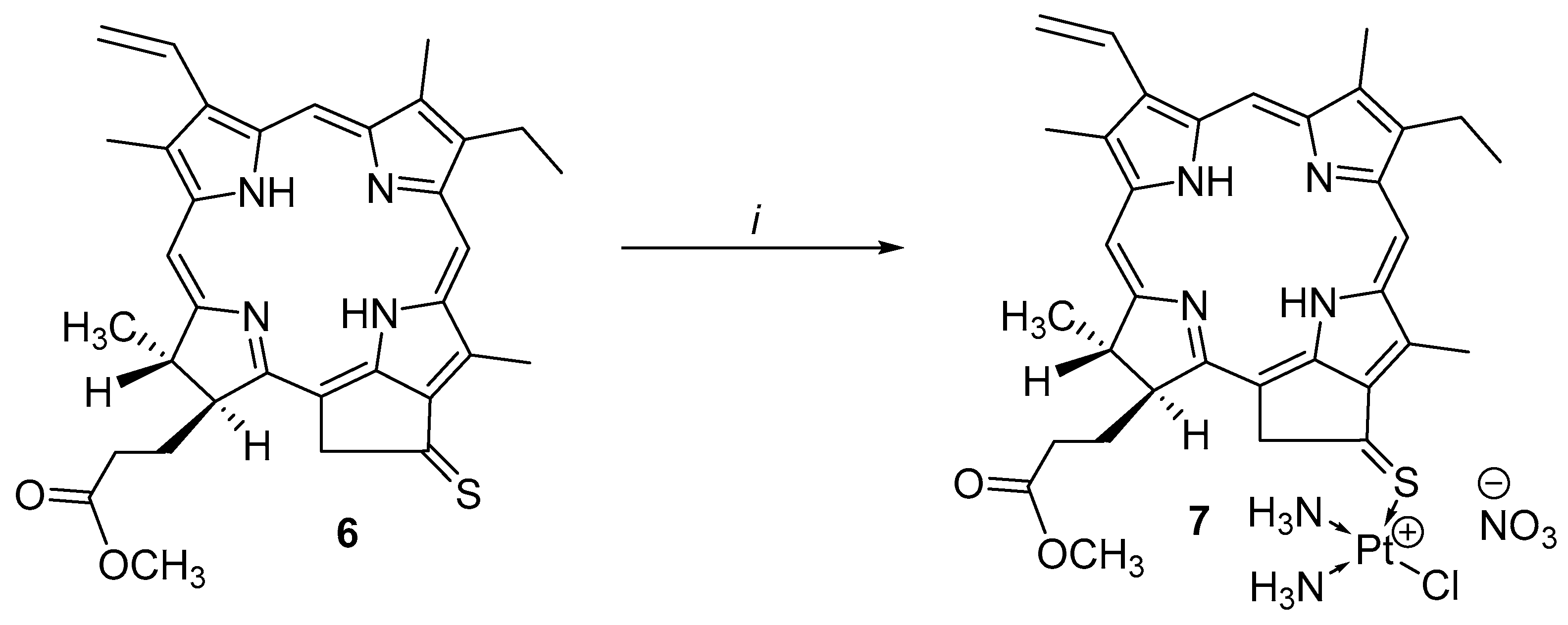
2.3. Synthesis Platinum Complex (II)

To stabilize the thiocarbonyl group in chlorin (6), a Pt(II) metal complex was obtained by attaching a cisplatin residue to the periphery of the macrocycle through the sulfur atom. The choice of the well-known chemotherapeutic agent is not accidental, as studies performed abroad and in our laboratory show that combining moieties implementing photodynamic and chemotherapeutic effects on the tumor in a single molecule increases the PDT efficiency [4,5,6,40].
Previously, metal complexes of chlorins with platinum were obtained by our team by incorporating chelators based on nicotinic acid, isonicotinic acid, terpyridine, etc., into the PS structure [41]. It is known that apart from the nitrogen atom, the sulfur atom also tends to chelate d-metals due to transfer of unshared electron pairs of the chalcogen to the free orbitals of the metal. To perform metalation, the cisplatin derivative was brought into the reaction with pyropheophorbide a 131-thioketone (6) (Scheme 4).
Compound 7 was isolated by preparative TLC and characterized by physicochemical methods, including NMR spectroscopy, mass spectrometry, and elemental analysis.

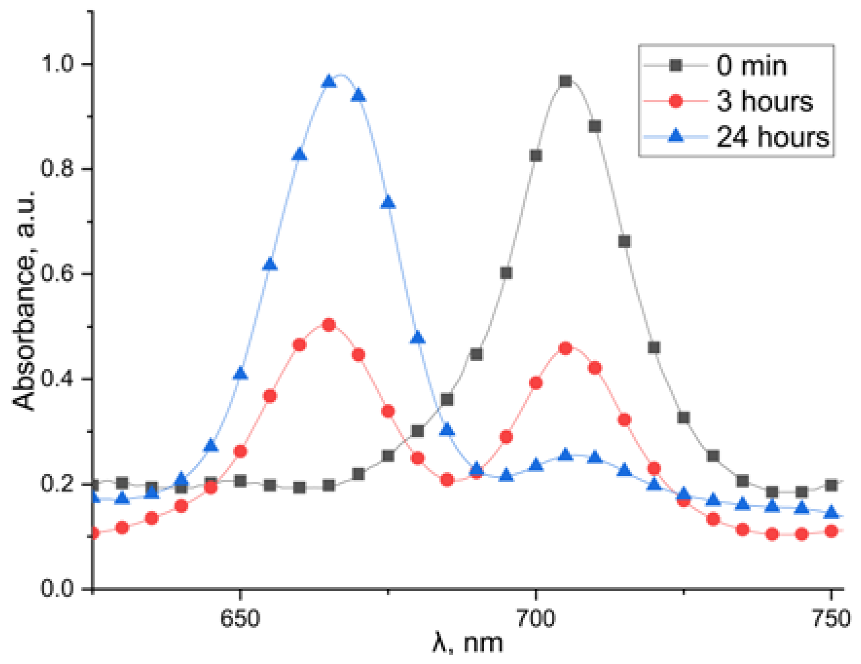
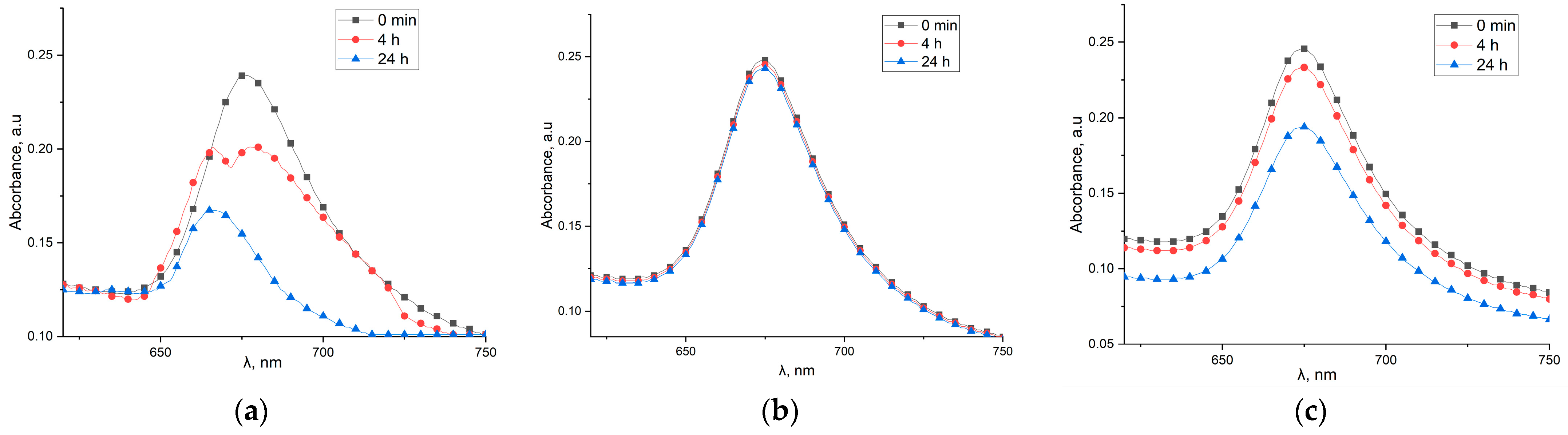
2.4. Stability of Platinum Complexes (II)

At the next step, we studied the chemical stability of complex 7 obtained in Kolliphor ELP aqueous micellar solutions vs. time at various pH values based on the variation in the intensity of the Qy band in the EAS of the samples (Figure 4).
At neutral pH = 7, compound 7 remained stable for 24 h, while in an alkaline solution (pH = 9), a precipitate formed and the intensity of the Qy absorption band decreased. In an acid solution (pH = 4), a hypsochromic shift in the Qy band to the 666 nm region occurred due to elimination of the platinum complex and hydrolysis of the thiocarbonyl derivative to the methyl ester of pyropheophorbide a (4), whose structure was confirmed by 1H NMR and mass spectra.
The chemical stability of compounds 4 and 7 was also studied in the lysate of S37 sarcoma cells (pH = 7.8), as well as in the cell culture medium at different pH values. The stability was assessed by the change in the intensity of the Qy bands as described above. The results of the study are shown in Table 2.
At neutral and basic pH, both compounds remained stable in the lysate of S37 sarcoma cells and in the cell culture medium, while a significant decrease in the intensity of the Qy band was observed in acidic medium.

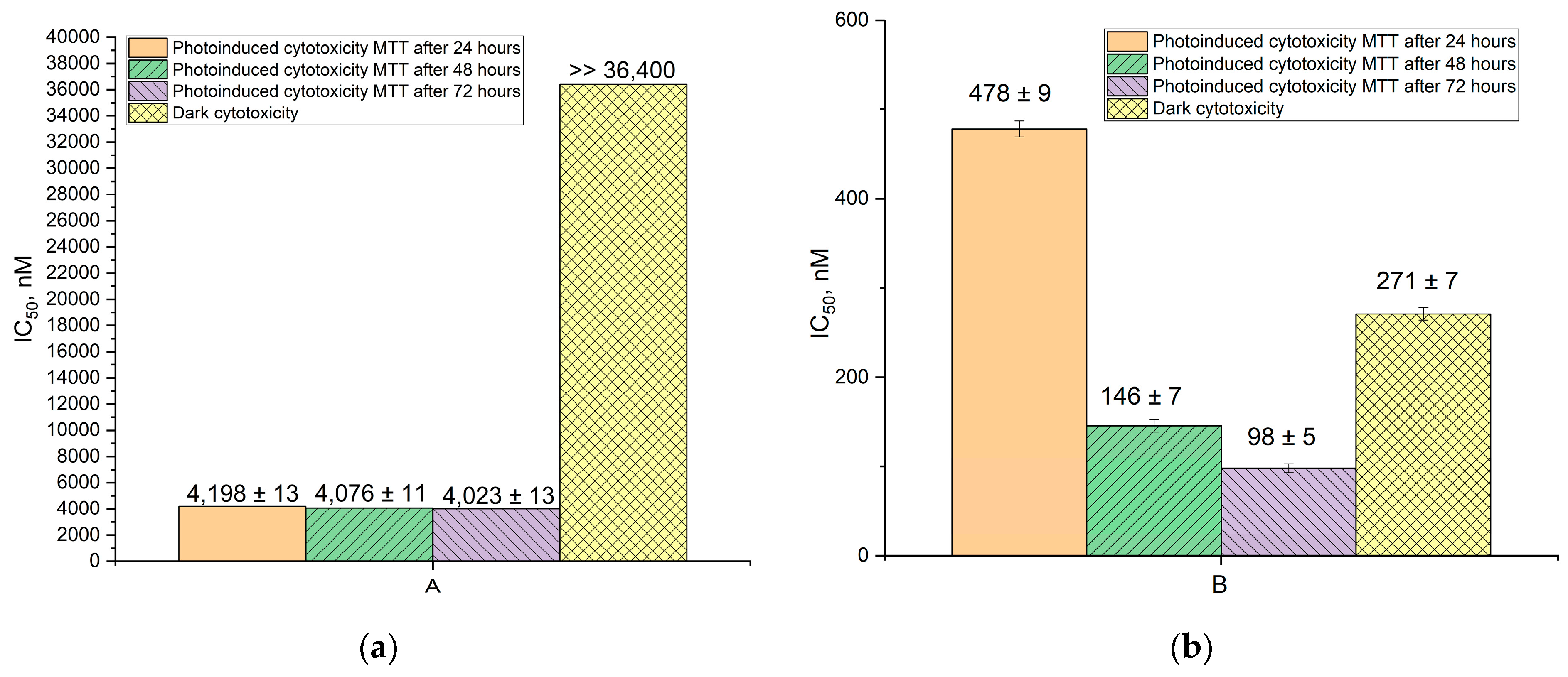
2.5. Biological Studies In Vitro

To estimate the combined effect (photoinduced and chemocytotoxic) of compound (4) and platinum complex (7), a two-step experiment was designed: incubation of cells with PS for 4 h and irradiation and subsequent incubation for 48 or 72 h followed by estimation of cell survival by the MTT assay. The results of these studies are presented in Table 3.
Incubation of these dyes with S37 sarcoma cells for 72 h showed that the compound comprising the Pt(II) complex (7) had a pronounced cytotoxic effect: its IC50 value was 271 ± 7 nM, whereas the parent compound (4) had no cytotoxic effect on cells.
A pronounced additive effect was identified for the PS with a platinum residue (7), whose photocytotoxicity increased 5-fold on increasing the incubation time after irradiation from 24 h (IC50 478 ± 9 nM) to 72 h (IC50 98 ± 5 nM) (Figure 5).
No additive effect was observed for pyropheophorbide a methyl ester (4), and its photoinduced activity was 8.8 times lower than that of the platinum complex.
It was found that the metal-free compound (4) does not have a pronounced cytotoxicity without light irradiation. Moreover, platinum complex 7, even without light irradiation, has a higher cytotoxicity with respect to S-37 cells than compound 4 upon irradiation. From this, it follows that the introduction of a fragment of cisplatin changes the effect of the drug in the dark. We hypothesize that this mechanism involves the binding of the cisplatin fragment to DNA [42]. In the absence of irradiation, the chlorin, which has proven tumor tropism [43], acts as a transporter that delivers the cisplatin fragment to the tumor cell, while when irradiated with light, there is a combined (photocytotoxic and chemotherapeutic) effect, leading to an increase in the cytotoxic effect.
Thus, a high cytotoxic activity against S37 sarcoma tumor cells was detected for the platinum complex (7) due to the combined photodynamic and chemotherapeutic effect on the latter.

3. Materials and Methods

All the solvents used in this work were prepared by conventional techniques as described elsewhere. Column chromatography was performed on silica gel 40/60 (Merck, Darmstadt, Germany). Preparative thin-layer chromatography (TLC) was performed on glass plates with silica gel 60 (Merck, Darmstadt, Germany). Analytical TLC was performed on aluminum plates with Silica gel 60 F254 with a fluorescent probe (Merck, Darmstadt, Germany).
Absorption and fluorescence spectra were recorded on Shimadzu UV1800 UV/VIS (Shimadzu, Duisburg, Germany) and Shimadzu RF-5301 (Shimadzu, Duisburg, Germany) spectrophotometers in quartz cells (0.4 × 1.0 cm) with an optical path length of 1 cm (spectral slit width 1 nm), using dichloromethane, acetone, and water as the solvents. Absorption spectra were recorded in the range of 350–750 nm. Fluorescence spectra were recorded in the range of 600–800 nm (excitation wavelength 550 nm), using dichloromethane, acetone, and water as the solvents. The fluorescence quantum yield was determined by the relative method. A solution of purpurin-18 in toluene was used as the standard [44]. Both 1H and 13C NMR spectra were recorded on a Bruker DPX 300 spectrometer (Bruker, Alzenau, Germany). NMR samples were prepared in d6-acetone and CDCl3. Matrix-assisted laser desorption/ionization (MALDI) mass spectra were recorded on a Bruker Ultraflex TOF/TOF spectrometer (Bruker, Alzenau, Germany), with 2,5-dihydroxybenzoic acid as the matrix. Elemental analysis was performed on a Vario Micro Cube analyzer (Elementar, Berlin, Germany).
Extraction of chlorophyll a from Spirulina platensis and synthesis of purpurin-18 (1), pheophorbide a (3), pyropheophorbide a (4), and cyclic thioanhydride of chlorin p6 (2) were carried out as described previously [32,37,45,46].

3.1. Synthesis

3.1.1. Cyclic Thioanhydride of Chlorin p6 (2)

Purpurine-18 173-methyl ester (1) (173 mmol, 100 mg) was dissolved in 10 mL of toluene and 1 mL of triethylamine. Then, 140 mg (346 mmol) of Lawesson’s reagent was added. The reaction was carried out under reflux for 24 h with stirring in an inert argon atmosphere. After that, the solvent was removed by evaporation on a rotary evaporator. The compound was purified by column chromatography in the system: (CH2Cl2/CH3OH, 60/1, v/v). The yield of the target compound (2) was 15 mg (15%).

3.1.2. Pheophorbide a 131-Thioketone (5)

Pheophorbide a (3) (82.4 mmol, 50 mg) was dissolved in 5 mL of toluene. Then, 67 mg (165 mmol) of Lawesson’s reagent and 100 μL of triethylamine was added. The reaction was carried out at room temperature in an inert argon atmosphere with stirring for 24 h. The solution was concentrated and purified by preparative TLC (CH2Cl2/hexane 1/80, v/v). The yield of the target compound (5) amounted to 37 mg (73%).
UV/VIS (CH2Cl2) λmax, nm (ε, M−1 sm−1): 386 (27,381), 410 (22,072), 706 (20,252).
HRMS m/z [M+H]+ calculated for C36H38N4O4S [M+H]: 623.2667. Found: 623.2666.
1H NMR (300 MHz, CDCl3) δ 9.43 (s, 1H, 10-H), 9.25 (s, 1H, 5-H), 8.46 (s, 1H, 20-H), 7.95 (dd, J = 17.8, 11.5 Hz, 1H, 31-H), 7.73 (dd, 1H, J = 1.4, 18 Hz, 32-Ha), 7.55 (dd, 1H, J = 1.4, 13 Hz, 32-Hb), 6,58 (s, 1H, 132-H), 6.25 (m, 1H, 18-H), 5.39 (m, 1H, 17-H), 3,85 (s, 3H, 134-CH3), 3.75 (s, 3H, 121-CH3), 3,60 (m, 5H, 81-H, 174-CH3), 3.37 (s, 3H, 21-CH3), 3.18 (s, 3H 71-CH3), 2.33 (m, 4H, 171-H, 172-H), 1.82 (d, J = 7.3 Hz, 3H, 181-H), 1.68 (t, J = 7.7 Hz, 3H, 82-H), 0.69 (s, 1H, NH), −1.11 (s, 1H, NH).
13C NMR (75 MHz, CDCl3) δ 219.54, 173.24, 173.10, 169.76, 167.72, 160.20, 156.19, 151.47, 149.47, 146.59, 145.21, 142.63, 138.93, 136.30, 132.38, 125.38, 122.85, 107.82, 104.77, 97.61, 93.21, 74.64, 74.27, 52.85, 51.64, 51.09, 50.01, 38.65, 31.87, 29.65, 28.86, 19.32, 17.28, 12.00, 11.62, 11.11, 0.97.

3.1.3. Pyropheophorbide a 131-Thioketone (6)

Pyropheophorbide a (4) (91 mmol, 50 mg) was dissolved in 5 mL of toluene. Then, 74 mg (182 mmol) of Lawesson’s reagent and 100 μL of triethylamine were added. The reaction was carried out at room temperature in an inert argon atmosphere with stirring for 4 h. The solution was concentrated, and the target compound was isolated by preparative TLC (CH2Cl2/hexane 1/80, v/v). The yield of compound (6) was 41 mg (82%).
UV-Vis λmax nm (relative intensities of peaks): 386 (23,673), 410 (19,175), 708 (17,518) (1:0.81:0.74).
HRMS m/z [M+H]+ calculated for C34H36N4O2S [M+H]: 565.2613. Found: 565.2611.
1H NMR (300 MHz, CDCl3) δ 9.42 (s, 1H, 10-H), 9.27 (s, 1H, 5-H), 8.42 (s, 1H, 20-H), 7.96 (dd, J = 17.8, 11.5 Hz, 1H, 31-H), 6.21 (m, 4H, 32-H, 132-H), 5.62 (q, J = 19.5 Hz, 2H, 82-H), 4.43 (m, 1H, 18-H), 4.25 (m, 2H, 17-H), 3.69 (s, 3H, 12-CH3), 3.66 (m, 2H, 81-H), 3.67 (s, 3H, 12-CH3), 3.62 (s, 3H, 174-CH3), 3.36 (s, 3H, 2-CH3), 3.20 (s, 3H, 7-CH3), 2.33 (m, 4H, 171-H, 172-H), 1.82 (d, J = 18.2 Hz, 3H, 18-CH3), 1.69 (t, J = 7.7 Hz, 3H, 82-CH3), 0.07 (s, 1H, NH), −1.1 (s, 1H, NH).
13C NMR (75 MHz, CDCl3) δ 226.51, 173.39, 172.50, 159.53, 148.65, 145.04, 142.19, 138.78, 136.71, 136.07, 136.01, 129.02, 122.63, 108.81, 104.52, 97.23, 93.16, 77.38, 77.15, 76.95, 76.53, 59.51, 51.66, 49.91, 31.87, 30.94, 29.64, 22.92, 19.36, 17.31, 12.01, 11.59, 11.14, 0.97.

3.1.4. Platinum Complex of Pyropheophorbide a 131-Thioketone (7)

Cisplatin (15 mg, 0.05 mmol, 1 equiv.) and silver nitrate (9 mg, 0.05 mmol, 1 equiv.) were dissolved in DMF (2 mL). The mixture was stirred for 24 h at room temperature in the dark. The resulting solution was centrifugated to remove the silver chloride precipitate. The supernatant was added to pyropheophorbide a 131-thioketone (6) (0.033 mmol), and the mixture was stirred for 48 h at room temperature. The solvent was distilled off under reduced pressure. The residue was purified by preparative TLC (CH2Cl2/CH3OH, 5/1, v/v).
UV-Vis (4% micellar solutions Kolliphor ELP) λmax, nm (relative intensities of peaks): 406 (33,858), 508 (10,500), 675 (12,703).
HRMS m/z [M+H]+ calculated for C34H41ClN6O2PtS [M+H]: 828.2423. Found: 828.2425.
1H NMR (300 MHz, C3D6O) δ 9.40 (s, 1H, 10-H), 9.21 (s, 1H, 5-H), 8.78 (s, 1H, 20-H), 8.00 (m, 1H, 31-H), 6.25 (dd, 1H, J = 17.8, 1.5 Hz, 32-Ha), 6.10 (dd, 1H, J = 11.6, 1.5 Hz, 32-Hb), 4.69 (s, 1H, 151-H), 4.57 (m, 1H, 18-H), 4.32 (m, 1H, 17-H), 3.57 (s, 3H, 173-COOCH3), 3.48 (s, 3H, 12-CH3), 3.38 (s, 3H, 2-CH3), 3.04 (s, 3H, 7-CH3), 2.95 (m, 2H, 81-CH2), 2.64 (m, H, 172-CH2a), 2.40 (m, H, 171-CH2a), 2.25 (m, 172-CH2b), 2.18 (m, H, 171-CH2b), 1.81 (d, 3H, J = 7.3 Hz, 18-CH3), 1.55 (t, 3H, J = 7.6 Hz, 18-CH3), 0.87 (t, 3H, J = 7.8 Hz, 82-CH3), 0.14 (s, 1H, NH), −2.11 (s, 1H, NH).
13C NMR (75 MHz, C3D6O) δ 195.69, 174.11, 172.97, 162.17, 155.56, 151.44, 145.73, 142.11, 138.52, 136.87, 136.42, 132.64, 131.52, 130.56, 130.21, 128.71, 122.96, 107.45, 104.74, 97.59, 94.50, 52.71, 51.83, 50.89, 32.47, 31.66, 23.55, 23.45, 19.80, 17.84, 14.50, 12.33, 11.92, 11.22.
Elemental analysis for CHNS, calculated for C34H42ClN7O5PtS (%): C, 45.82; H, 4.75; N, 11.00; S, 3.60. Found (%): C, 45.83; H, 4.77; N, 11.01; S, 3.59.

3.2. Preparation of Micellar Solutions of Compounds (4) and (7)

A solution of compounds (4) or (7) in dichloromethane (1–2 mL) was added dropwise with continuous argon bubbling to a freshly prepared 4% aqueous solution of Kolliphor ® ELP (5 mL) heated to 45 °C. Bubbling was continued until strong foaming started. A clear dark-red solution with a concentration of 0.25 mg/mL was obtained, which was then successively filtered through a 0.45 µm PTFE filter and a 0.22 µm PTFE filter. The concentrations of the resulting micellar solutions were monitored spectrophotometrically.

3.3. Biological Studies In Vitro

3.3.1. Characterization of Test Systems In Vitro

The S37 murine sarcoma cell line (collection of the N.N. Blokhin Research Institute of Oncology) was used to estimate photoinduced activity and cytotoxicity.
Cells were cultured in DMEM medium (PanEco, Russia) with addition of 2 mM L-glutamine (PanEco, Moscow, Russia) and 10% fetal calf blood serum (FCS) (Corning, New York, NY, USA) in 25 cm2 culture vials (Corning, USA) at 37 °C in a humid atmosphere containing 5% carbon dioxide. Upon passivation, the Versene solution (PanEco, Russia) was used to remove the cells from the substrate. For in vitro studies, cells of the 3rd and 4th passages were used with an optimal seeding concentration of 6 × 10⁴ cells/mL.

3.3.2. Preparation of the Substances to Be Studied

The concentrations of the solutions obtained were 0.6 mg/mL for compound (4) and 0.1 mg/mL for compound (7). All the volumes obtained were filtered through a Millipore membrane filter with a pore size of 0.22 μm. Next, the samples were diluted in 24-well plates (Corning, USA) on the cell-culturing medium. The final concentration of the substances ranged from 32 ng/mL to 20 μg/mL.

3.3.3. Procedure for Studying Photoinduced and Dark Activity In Vitro

To perform the experiments, S37 tumor cells were seeded into 96-well plates in the amount of 6 × 10³ cells per well in a 100 μL volume. Exposure was performed in the exponential phase of cell growth 24 h after seeding. Solutions of compounds in the complete culture medium were added to the wells at final concentrations of 32 ng/mL to 20 μg/mL, in triplicate. The time of cell incubation with compounds before irradiation was 4 h. The culture medium was used as the blank. Light exposure was performed with a halogen lamp through a KS-10 broadband filter (λ > 620 nm). The light dose was 10 J/cm2. After light exposure, the plates with cells were placed in a CO2 incubator for 24, 48, or 72 h.
To estimate cytotoxicity, cells were incubated with the substances for 72 h under dark conditions in a CO2 incubator.
Cell survival was estimated using the colorimetric MTT test (24 and 72 h after exposure), which is based on the ability of dehydrogenases in living cells to convert yellow water-soluble 3-(4-5-dimethyl-tetrazolyl-2)-2,5-diphenyltetrazolium bromide to blue crystals of formazan, whose amount is measured spectrophotometrically.
Before the test, cells were washed from cytotoxic agents. Then, 0.5% solution of 3-(4,5-dimethylthiazolyl-2)-2,5-diphenyltetrazolium bromide was added to the wells and the system was incubated for 3 h under standard conditions. After incubation, the medium with the MTT-reagent was removed and formazan crystals were dissolved for 10 min with dimethyl sulfoxide. Optical density was measured on a Multiscan FC multichannel plate photometer (Thermo scientific) at 550 nm.
The growth inhibition (GI) of cells in a culture was calculated using the following formula:
GI(%) = [ODk − ODo/ODk)] × 100%,
where ODo is the optical density of formazan in the test wells and ODk is the optical density of formazan in the control wells.
Inhibition of cell growth in culture by more than 50% (IC50) was considered a biologically significant effect. Quantitative parameters were calculated using the results of three independent tests.

4. Conclusions

In this work, methyl esters of chlorin p6 thioanhydride (2) and thiocarbonyl derivatives of pheophorbide a (5) and pyropheophorbide a (6) were obtained using Lawesson’s reagent. We indirectly confirmed the mechanism of thionation of compound (4) by identifying an intermediate compound by means of high-performance chromatography–mass spectrometry. The spectral properties of thiocarbonyl derivatives of pheophorbide a (5) and pyropheophorbide a (6) were studied. A considerable bathochromic shift in the long-wave absorption and fluorescence maxima by about 40 nm compared to the carbonyl precursors (3) and (4) was shown.
The chelating capabilities of the thiocarbonyl group at the periphery of the macrocycle were shown by formation of a Pt(II) metal complex in cisplatin, a chemotherapeutic agent. The chemical stability in aqueous solutions of the conjugate of pyropheophorbide with cisplatin was studied, and the high cytotoxic activity against sarcoma S37 tumor cells due to the combined photodynamic and chemotherapeutic effects was shown.

Supplementary Materials

The following supporting information can be downloaded at: https://www.mdpi.com/article/10.3390/molecules28104215/s1, Figure S1: Mass chromatogram of the reaction mass, target compound mass time 11.27 min, m/z [M+H]+ = 751.2538; Figure S2: Mass chromatogram of compounds (5) and (6): (I) chromatogram of 131-thioketone pheophorbide a (5) retention time 11.43 min; (II) HRMS spectrum of 131-thioketone pheophorbide a (5); (III) chromatogram of 131-thioketone pyropheophorbide a (6) retention time 12.39 min; (IV) HRMS spectrum of 131-thioketone pyropheophorbide a (6); Figure S3: 1H NMR spectrum of compound 5; Figure S4: 1H NMR spectrum of compound 6; Figure S5: 13C NMR spectra. (a): pheophorbide a (3)–red, pheophorbide a thioketone (5)–blue; (b): pyropheophorbide a (4)–red, pyropheophorbide a thioketone (6)–green (created with MestRenova); Figure S6: 1H NMR spectrum of compound 7; Figure S7: 13C NMR spectrum of compound 7; Figure S8: HRMS spectrum of compound 7.

Author Contributions

Conceptualization, V.P., M.G. and E.D.; methodology, V.E., O.B., A.P. and M.G.; validation, V.P.,V.E., P.O., E.P. and M.G.; data curation, V.P., V.E., P.O., A.P. and Y.V.; writing—original draft preparation, V.P., V.E., E.P., P.O., M.G., Y.V. and E.D.; writing—review and editing, A.P., O.B. and M.G.; visualization, V.P., V.E., P.O. and E.P.; funding acquisition, M.G. All authors have read and agreed to the published version of the manuscript.

Funding

This work was carried out with the financial support of the Ministry of Science and Higher Education of the Russian Federation (project no. FSFZ-2023-0004).

Institutional Review Board Statement

Not applicable.

Informed Consent Statement

Not applicable.

Data Availability Statement

The data presented in this study are available from the authors.

Acknowledgments

This work was performed using the equipment of the Shared Science and Training Center for Collective Use RTU MIREA and supported by the Ministry of Science and Higher Education of the Russian Federation.

Conflicts of Interest

The authors declare no conflict of interest.

References

  1. Quirk, B.J.; Brandal, G.; Donlon, S.; Vera, J.C.; Mang, T.S.; Foy, A.B.; Lew, S.M.; Girotti, A.W.; Jogal, S.; LaViolette, P.S. Photodynamic therapy (PDT) for malignant brain tumors—Where do we stand? Photodiagn. Photodyn. Ther. 2015, 12, 530–544. [Google Scholar] [CrossRef] [PubMed]
  2. Filonenko, E.V. Clinical implementation and scientific development of photodynamic therapy in Russia in 2010–2020. Biomed. Photonics 2021, 10, 4–22. [Google Scholar] [CrossRef]
  3. Yi, M.; Xiong, B.; Li, Y.; Guo, W.; Huang, Y.; Lu, B. Manipulate tumor hypoxia for improved photodynamic therapy using nanomaterials. Eur. J. Med. Chem. 2022, 247, 115084. [Google Scholar] [CrossRef]
  4. Otvagin, V.F.; Kuzmina, N.S.; Kudriashova, E.S.; Nyuchev, A.V.; Gavryushin, A.E.; Fedorov, A.Y. Conjugates of porphyrinoid-based photosensitizers with cytotoxic drugs: Current progress and future directions toward selective photodynamic therapy. J. Med. Chem. 2022, 65, 1695–1734. [Google Scholar] [CrossRef] [PubMed]
  5. Grin, M.; Suvorov, N.; Ostroverkhov, P.; Pogorilyy, V.; Kirin, N.; Popov, A.; Sazonova, A.; Filonenko, E. Advantages of combined photodynamic therapy in the treatment of oncological diseases. Biophys. Rev. 2022, 14, 941–963. [Google Scholar] [CrossRef]
  6. Krishnaswami, V.; Natarajan, B.; Sethuraman, V.; Natesan, S.; RajSelvaraj, B. Nano based photodynamic therapy to target tumor microenvironment. Nano Trends 2023, 1, 100003. [Google Scholar] [CrossRef]
  7. Li, X.; Chen, L.; Huang, M.; Zeng, S.; Zheng, J.; Peng, S.; Wang, Y.; Cheng, H.; Li, S. Innovative strategies for photodynamic therapy against hypoxic tumor. Asian J. Pharm. Sci. 2023, 18, 100775. [Google Scholar] [CrossRef]
  8. Xu, B.; He, P.; Wang, Y.; Wang, H.; Zhang, J.; Zhu, J.; Pu, W.; Chen, H. PDT for Gastric Cancer—The View from China. Photodiagn. Photodyn. Ther. 2023, 42, 103366. [Google Scholar] [CrossRef]
  9. Allison, R.R.; Sibata, C.H.; Downie, G.H.; Cuenca, R.E. A clinical review of PDT for cutaneous malignancies. Photodiagn. Photodyn. Ther. 2006, 3, 214–226. [Google Scholar] [CrossRef]
  10. He, P.; Zhang, F.; Xu, B.; Wang, Y.; Pu, W.; Wang, H.; Wang, B.; Zhang, J.; Chen, H.; Li, Y. Research progress of potential factors influencing photodynamic therapy for Gastrointestinal Cancer. Photodiagn. Photodyn. Ther. 2023, 41, 103271. [Google Scholar] [CrossRef]
  11. Abrahamse, H.; Hamblin, M.R. New photosensitizers for photodynamic therapy. Biochem. J. 2006, 473, 347–364. [Google Scholar] [CrossRef]
  12. Zhao, X.; Liu, J.; Fan, J.; Chao, H.; Peng, X. Recent progress in photosensitizers for overcoming the challenges of photodynamic therapy: From molecular design to application. Chem. Soc. Rev. 2021, 50, 4185–4219. [Google Scholar] [CrossRef]
  13. Pham, T.C.; Nguyen, V.N.; Choi, Y.; Lee, S.; Yoon, J. Recent strategies to develop innovative photosensitizers for enhanced photodynamic therapy. Chem. Rev. 2021, 121, 13454–13619. [Google Scholar] [CrossRef]
  14. Kuzmin, I.S.; Toporkov, G.A.; Yuriev, D.Y.; Kalistratova, A.V.; Kovalenko, L.V. Synthesis and biological activity of N-phosphonacetyl-L-aspartate’s structural analogs N-(α-dietoxyphosphorylcyclopropylcarbonyl)-amino acids. Fine Chem. Technol. 2020, 15, 26–35. [Google Scholar]
  15. Koifman, O.I.; Ageeva, T.A.; Kuzmina, N.S.; Otvagin, V.F.; Nyuchev, A.V.; Fedorov, A.Y.; Belykh, D.V.; Lebedeva, N.S.; Yurina, E.S.; Syrbu, S.A.; et al. Synthesis Strategy of Tetrapyrrolic Photosensitizers for Their Practical Application in Photodynamic Therapy. Macroheterocycles 2022, 15, 207–302. [Google Scholar] [CrossRef]
  16. Grin, M.A.; Tikhonov, S.I.; Petrova, A.S.; Pogorilyy, V.A.; Noev, A.N.; Tatarskiy, V.V.; Shpakovsky, D.B.; Milaeva, E.V.; Kalinina, E.V.; Chernov, A.A.; et al. New derivatives of bacteriopurpurin with thiolated Au (I) complexes: Dual darkand light activated antitumor potency. Anti-Cancer Agents Med. Chem. 2020, 20, 49–58. [Google Scholar] [CrossRef]
  17. Grin, M.A.; Pogorilyy, V.A.; Noev, A.N.; Tikhonov, S.I.; Majouga, A.G.; Mironov, A.F. Bacteriochlorophyll a derivatives with sulfur-containing amino acids as promising photosensitizers for cancer PDT. Macroheterocycles 2018, 11, 89–94. [Google Scholar] [CrossRef]
  18. Pantiushenko, I.V.; Rudakovskaya, P.G.; Starovoytova, A.V.; Mikhaylovskaya, A.A.; Abakumov, M.A.; Kaplan, M.A.; Tsygankov, A.A.; Majouga, A.G.; Grin, M.A.; Mironov, A.F. Development of bacteriochlorophyll a-based near-infrared photosensitizers conjugated to gold nanoparticles for photodynamic therapy of cancer. Biochemistry 2015, 80, 752–762. [Google Scholar] [CrossRef]
  19. Mironov, A.F.; Grin, M.A.; Pantushenko, I.V.; Ostroverkhov, P.V.; Ivanenkov, Y.A.; Filkov, G.I.; Plotnikova, E.A.; Karmakova, T.A.; Starovoitova, A.V.; Burmistrova, N.A.; et al. Synthesis and Investigation of photophysical and biological properties of novel s-containing bacteriopurpurinimides. J. Med. Chem. 2017, 60, 10220–10230. [Google Scholar] [CrossRef]
  20. Nguyen, T.T.; Le, T.N.; Hansen, P.E.; Duus, F. Preparation and structural characterization of a new class of stable thioketones: Ortho-hydroxythioacetophenones. Tetrahedron Lett. 2006, 47, 8433–8435. [Google Scholar] [CrossRef]
  21. Polshettiwar, V.; Kaushik, M.P. Recent advances in thionating reagents for the synthesis of organosulfur compounds. J. Sulfur Chem. 2006, 27, 353–386. [Google Scholar] [CrossRef]
  22. Khatoon, H.; Abdulmalek, E. A focused review of synthetic applications of Lawesson’s reagent in organic synthesis. Molecules 2021, 26, 6937. [Google Scholar] [CrossRef] [PubMed]
  23. Kaur, N. Lawesson’s Reagent in Heterocycle Synthesis; Springer: Luxembourg, 2022. [Google Scholar] [CrossRef]
  24. Yu, Y.; Czepukojc, B.; Jacob, B.; Jiang, Y.; Zeller, M.; Brueckner, C.; Zhang, J.L. Porphothionolactones: Synthesis, structure, physical, and chemical properties of a chemodosimeter for hypochlorite. Org. Biomol. Chem. 2013, 11, 4613. [Google Scholar] [CrossRef] [PubMed]
  25. Richeter, S.; Jeandon, C.; Gisselbrecht, J.P.; Graff, R.; Ruppert, R.; Callot, H.J. Synthesis of New Porphyrins with Peripheral Conjugated Chelates and Their Use for the Preparation of Porphyrin Dimers Linked by Metal Ions. Inorg. Chem. 2004, 43, 251–263. [Google Scholar] [CrossRef]
  26. Tamiaki, H.; Xu, M.; Kinoshita, Y. Synthesis of oxo-, thioxo- and methylene-substituted bacteriochlorins by modifying chlorophyll-a and their electronic absorption in visible and near-infrared regions. J. Photochem. Photobiol. A Chem. 2013, 252, 60–68. [Google Scholar] [CrossRef]
  27. Pandey, R.K.; Shiau, F.Y.; Sumlin, A.B.; Dougherty, T.J.; Smith, K.M. Structure/activity relationships among photosensitizers related to pheophorbides and bacteriopheophorbides. Bioorg. Med. Chem. Lett. 1992, 2, 491–496. [Google Scholar] [CrossRef]
  28. Bruns, R.F.; Watson, I.A. Rules for Identifying Potentially Reactive or Promiscuous Compounds. J. Med. Chem. 2012, 55, 9763–9772. [Google Scholar] [CrossRef]
  29. Nhat, P.V.; Si, N.T.; Nguyen, T.; Duonh, L.; Nguyen, M.T. Elucidating the binding mechanism of thione-containing mercaptopurine and thioguanine drugs to small gold clusters. J. Comput. Chem. 2020, 41, 1748–1758. [Google Scholar] [CrossRef]
  30. Dekkiche, H.; Carvalho, M.-A.; Jeandon, C.; Karmazin, L.; Boundon, C.; Ruhlmann, L.; Ruppert, R. Synthesis and electrochemistry of nickel(II)porphyrins bearing external palladium(II) or platinum(II) complexes. J. Porphyr. Phthalocyanines 2021, 25(10n12), 1133–1142. [Google Scholar] [CrossRef]
  31. Carvalho, M.-A.; Dekkiche, H.; Nagasaki, M.; Kikkawa, Y.; Ruppert, R. Coordination-Driven Construction of Porphyrin Nano-Ribbons at a HOPG/Liquid Interface. J. Am. Chem. Soc. 2019, 141, 10137–10141. [Google Scholar] [CrossRef]
  32. Pogorilyy, V.; Kirin, N.; Mironov, A.; Grin, M. A new cyclic thioanhydride derived from chlorophyll a and its aurophilic properties. Dyes Pigments 2021, 184, 108858. [Google Scholar] [CrossRef]
  33. Pogorilyy, V.; Plyutinskaya, A.; Suvorov, N.; Diachkova, E.; Vasil’ev, Y.; Pankratov, A.; Mironov, A.; Grin, M. The First Selenoanhydride in the Series of Chlorophyll a Derivatives, Its Stability and Photoinduced Cytotoxicity. Molecules 2021, 26, 7298. [Google Scholar] [CrossRef]
  34. van Asselt, R.; Vanderzande, D.; Gelan, J.; Froehling, P.E.; Aagaard, O. New synthetic routes to poly (isothianaphthene). II. Mechanistic aspects of the reactions of phthalic anhydride and phthalide with phosphorus pentasulfide. J. Polym. Sci. Part A Polym. Chem. 1996, 34, 1553–1560. [Google Scholar] [CrossRef]
  35. Huang, T.B.; Qian, X.; Tao, Z.F.; Wang, K.; Song, G.H.; Liu, L.F. The improved synthesis, Diels-Alder reactions, and desulfuration of trithio-1, 8-naphthalic anhydride. Heteroat. Chem. Int. J. Main Group Elem. 1999, 10, 141–146. [Google Scholar] [CrossRef]
  36. Lakshmikantham, M.V.; Chen, W.; Cava, M.P. Thioanhydrides. 3. Synthesis, properties and Diels-Alder reactions of sulfur analogs of 1, 8-naphthalic anhydride. J. Org. Chem. 1989, 54, 4746–4750. [Google Scholar] [CrossRef]
  37. Kuchin, A.V.; Karmanova, L.P.; Belykh, D.V. Method of Preparing Pheophorbid Methyl Ester. RU Patent No. 2180342C2, 24 March 1999. [Google Scholar]
  38. Rajee, R.; Ramamurthy, V. Oxidation of thiones by singlet and triplet oxygen. Tetrahedron Lett. 1978, 19, 5127–5130. [Google Scholar] [CrossRef]
  39. Sánchez-Arroyo, A.J.; Pardo, Z.; Moreno, F.; Herrera, A.; Martín, N.; García-Fresnadillo, D. Photochemical Oxidation of Thioketones by Singlet Molecular Oxygen Revisited: Insights into Photoproducts, Kinetics, and Reaction Mechanism. J. Org. Chem. 2015, 80, 10575–10584. [Google Scholar] [CrossRef]
  40. Yu, Y.; Wang, N.; Wang, Y.; Shi, Q.; Yu, R.; Gu, B.; Maswikiti, E.P.; Chen, H. Photodynamic therapy combined with systemic chemotherapy for unresectable extrahepatic cholangiocarcinoma: A systematic review and meta-analysis. Photodiagn. Photodyn. Ther. 2023, 41, 103318. [Google Scholar] [CrossRef]
  41. Grin, M.; Ostroverkhov, P.; Suvorov, N.; Tikhonov, S.; Popov, A.; Shelyagina, A.; Kirin, N.; Nichugovskiy, A.; Usachev, M.; Bragina, N. Potential agents for combined photodynamic and chemotherapy in oncology based on Pt (II) complexes and pyridine-containing natural chlorins. J. Porphyr. Phthalocyan. 2023, 27, 728–740. [Google Scholar] [CrossRef]
  42. Aldossary, S.A. Review on pharmacology of cisplatin: Clinical use, toxicity and mechanism of resistance of cisplatin. Biomed Pharm. J. 2019, 12, 7–15. [Google Scholar] [CrossRef]
  43. Plekhova, N.; Shevchenko, O.; Korshunova, O.; Stepanyugina, A.; Tananaev, I.; Apanasevich, V. Development of Novel tetrapyrrole structure Photosensitizers for cancer Photodynamic therapy. Bioengineering 2022, 9, 82. [Google Scholar] [CrossRef] [PubMed]
  44. Zenkevich, E.; Sagun, E.; Knyukshto, V.; Shulga, A.; Mironov, A.; Efremova, O.; Bonnett, R.; Songca, S.P.; Kassem, M. Photophysical and photochemical properties of potential porphyrin and chlorin photosensitizers for PDT. J. Photochem. Photobiol. B Biol. 1996, 33, 171–180. [Google Scholar] [CrossRef]
  45. Brandis, A.; Kozyrev, A.; Kurnygina, V.; Mironov, A.; Nekrasova, V.; Nikitina, T.; Fragina, A.I. The Method of Producing Purpurin-18. RU Patent No. 2054944, 27 February 1996. [Google Scholar]
  46. Pandey, R.K.; Dougherty, T.J.; Pallenberg, A.J. Efficient Synthesis of Pyropheophorbide a and its Derivatives. Org. Process Res. Dev. 2004, 8, 287–290. [Google Scholar]
Figure 1. A chlorin and its application in experimental oncology (created with Biorender).
Molecules 28 04215 g001
Scheme 1. Reagents and conditions. i: Lawesson’s reagent, toluene, Et3N, 110 °C, 24 h, inert argon atmosphere.
Molecules 28 04215 sch001
Scheme 2. Reagents and conditions. i: Lawesson’s reagent; the reaction conditions are listed in Table 1.
Molecules 28 04215 sch002
Scheme 3. Scheme for the interaction of Lawesson’s reagent with ketones of pyropheophorbide a.
Molecules 28 04215 sch003
Figure 2. Absorption (a) and fluorescence (b) electronic spectra: pheophorbide a (3), pyropheophorbide a (4), pheophorbide a 131-thioketone (5), pyropheophorbide a 131-thioketone (6). The absorption and fluorescence spectra were obtained at the same concentration using dichloromethane as the solvent (created with Origin).
Molecules 28 04215 g002
Figure 3. Electronic absorption spectra of compound (6) in 4% micellar solution of Kolliphor ELP recorded versus time (created with Origin).
Molecules 28 04215 g003
Scheme 4. Reagents and conditions. i: cisplatin, AgNO3, DMF.
Molecules 28 04215 sch004
Figure 4. Electronic absorption spectra of pigment (7) in 4% micellar solutions of Kolliphor ELP at various pH values vs. time: (a) pH = 4, (b) pH = 7, (c) pH = 9 (created with Origin).
Molecules 28 04215 g004
Figure 5. Photoinduced and cytotoxic activity. (a) pyropheophorbide a methyl ester (4): orange sans stripes—4 h PS incubation with cells, irradiation at 10 J/cm2, MTT after 24 h; green stripes to the left—4 h PS incubation with cells, irradiation at 10 J/cm2, MTT after 48 h; purple with stripes to the right—4 h PS incubation with cells, irradiation at 10 J/cm2, MTT after 72 h; yellow with stripes to the right and left—72 h incubation of compounds with cells, MTT. (b) chlorin-based Pt(II) metal complex (7): orange sans stripes—4 h PS incubation with cells, irradiation at 10 J/cm2, MTT after 24 h; green stripes to the left—4 h PS incubation with cells, irradiation at 10 J/cm2, MTT after 48 h; purple with stripes to the right—4 h PS incubation with cells, irradiation at 10 J/cm2, MTT after 72 h; yellow with stripes to the right and left—72 h incubation of compounds with cells, MTT (created with Origin).
Molecules 28 04215 g005
Table 1. Thionation conditions.
ThioketoneSolventTemperatureTimeYield
5toluene0 °C48 h24%
toluene20 °C4 h36%
toluene35 °C1 h56%
toluene70 °C30 min43%
toluene110 °C30 min19%
pyridine35 °C30 min61%
toluene, DIPEA35 °C45 min67%
toluene, Et3N35 °C45 min73%
K2CO3 in acetonitrile82 °C60 min32%
6toluene0 °C48 h27%
toluene20 °C4 h39%
toluene35 °C1 h62%
toluene70 °C30 min54%
toluene110 °C30 min21%
pyridine35 °C30 min69%
toluene, DIPEA35 °C45 min77%
toluene, Et3N35 °C45 min82%
K2CO3 in acetonitrile82 °C60 min33%
Table 2. Intensity of the absorption maxima of compounds (4) and (7) under cell culture medium and cell lysate S37.
Compound 7Compound 4
TimeIncubation
Medium
pHIntensity at 675 nmContent, %Intensity at 666 nmContent, %
0 minCell culture
medium
40.912991000.95382100
70.913000.95379
90.912980.95381
Cell lysate S37-0.913020.95380
4 hCell culture
medium
40.22824250.6676770
70.88561970.8870293
90.79429870.8107385
Cell lysate S37-0.81257890.8679591
24 hCell culture
medium
40.15520170.5722960
70.85822940.8298087
90.68473750.7439778
Cell lysate S37-0.76692840.7821282
72 hCell culture
medium
40.10042110.5532158
70.83297910.7534979
90.52770650.7058273
Cell lysate S37-0.72127790.7248976
Table 3. Photoinduced and cytotoxic activity of pyropheophorbide a methyl ester (4) and chlorin-based Pt(II) metal complex (7). S37 cell culture.
CompoundIC50, nM
Photoinduced cytotoxicityCombined cytotoxicityDark cytotoxicity
4 h PS incubation with cells
Irradiation at
10 J/cm2, MTT
after 24 h
4 h PS incubation with cells
Irradiation at
10 J/cm2, MTT after 48 h
4 h PS incubation with cells
Irradiation at
10 J/cm2, MTT after 72 h
72 h incubation of
compounds with cells, MTT
(4)4198 ± 13 nM4076 ± 11 nM4023 ± 13 nM>>36,400 nM
(7)478 ± 9 nM146 ± 7 nM98 ± 5 nM271 ± 7 nM
Cisplatin---17,185 nM ± 15 nM
Disclaimer/Publisher’s Note: The statements, opinions and data contained in all publications are solely those of the individual author(s) and contributor(s) and not of MDPI and/or the editor(s). MDPI and/or the editor(s) disclaim responsibility for any injury to people or property resulting from any ideas, methods, instructions or products referred to in the content.

