Abstract
We present a visual tool and facile method to detect MCF-7 breast cancer cells by using YVO4:Eu3+@silica-NH-GDA-IgG bio-nanocomplexes. To obtain these complexes, YVO4:Eu3+ nanoparticles with a uniform size of 10–25 nm have been prepared firstly by the hydrothermal process, followed by surface functionalization to be bio-compatible and conjugated with cancer cells. The YVO4:Eu3+@silica-NH-GDA-IgG nanoparticles exhibited an enhanced red emission at 618 nm under an excitation wavelength of 355 nm and were strongly coupled with MCF-7 breast cancer cells via biological conjugation. These bio-nanocomplexes showed a superior sensitiveness for MCF-7 cancer cell labelling with a detection percentage as high as 82%, while no HEK-293A healthy cells were probed under the same conditions of in vitro experiments. In addition, the detection percentage of MCF-7 breast cancer cells increased significantly via the functionalization and conjugation of YVO4:Eu3+ nanoparticles. The experimental results demonstrated that the YVO4:Eu3+@silica-NH-GDA-IgG bio-nanocomplexes can be used as a promising labelling agent for biomedical imaging and diagnostics.
1. Introduction
Nanostructured materials containing rare earth elements with numerous advantages such as high stability, strong luminescence, easy surface functionalization, and being friendly to environment and human body have been designed for new applications, especially for biomedical fluorescence labelling [1,2,3,4,5,6,7,8]. Most nanoparticles reported in past immunoassays are smaller than 200 nm in diameter for the biomolecular [9,10]. The size of these particles is proven to provide obvious prolonged equilibration time and enhanced nonspecific adsorption [11,12,13]. Therefore, the kinetic properties of smaller nanoparticles can be improved, and the nonspecific adsorption decreases. On the other hand, nanoparticles should be controlled to be big enough in size to bind with several proteins and cells on the surface, which is expected to increase the immunological affinity [14]. Among nanostructured materials containing rare-earth elements, YVO4:Eu3+ nanomaterials have received a great deal of interest because of biologically appropriate emission in the visible region and biocompatibility. It is surveyed from the literature that yttrium(III) orthovanadate, YVO4, is one of the most commonly used host lattices containing rare earth ions to prepare efficient luminescent materials with different color emittings because of its high luminescence quantum yields of f–f transitions [15]. Accordingly, when being doped with Eu3+ ions, YVO4:Eu3+ nanomaterials with high quantum efficiency have strong fluorescence emission at 618 nm. The luminescent 4f–4f transitions of europium(III) ion and the effective energy transfer from ligands to europium(III) ion lead to a strong emission of red light that makes them widely employed in detection, biomedical imaging, and luminescent labels [16,17].
The functionalization of nanostructured materials is a key step toward biomedical applications. The applications of nanostructured materials require preliminary grafting at the nanophosphor’s surface by organic or bio-organic functional groups. Different approaches are used such as encapsulation with functional polymers or direct grafting of biofunctional ligands. The 3-aminopropyl triethoxysilane (APTES) solution is well-known as a functionalized bio-compatible agent, because its ligands can create ethoxy groups on an inorganic surface. Meanwhile, glutaraldehyde (GDA) is one of the most popular bis-aldehyde homobifunctional crosslinkers that can be incorporated into nanostructured materials containing rare earth elements for many biomedical applications, including labelling, biosensing, drug delivery, and other therapies [18,19,20]. However, the structure of GDA is complicated, and its reaction mechanism is not fully understood. The reactions of GDA on proteins and other amine-containing molecules through the formation of a Schiff base that is lacking and still in progress [21]. In this work, we reported the results of the synthesis and in vitro testing of YVO4:Eu3+@silica-NH-GDA-IgG bio-nanocomplexes for labelling cancer cells.
2. Results and Discussion
2.1. Morphological Characterization
Figure 1a,b show the scanning electron microscopy (SEM) images of the YVO4:Eu3+ and YVO4:Eu3+@silica-NH-GDA-IgG nanoparticles, respectively. It indicated that YVO4:Eu3+ nanomaterials were nearly spherical with diameters ranging from 10 to 20 nm (Figure 1a). After being covered with silica and functionalized with APTES and IgG, the spherical nanoparticles with diameters increasing up to 20–25 nm were observed (Figure 1b).
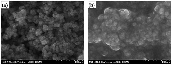
Figure 1.
SEM images of the YVO4:Eu3+ sample (a) and YVO4:Eu3+@silica-NH-GDA-IgG (b) samples.
2.2. Structure Characterization
Figure 2a shows the X-ray diffraction (XRD) pattern of the YVO4:Eu3+ sample. One can realize that all diffraction peaks of the sample coincided well with the standard data of the tetragonal-phase YVO4:Eu3+ structure (JCPDS No. 17-0341). Furthermore, no impurity peaks were detected, indicating that the dopant Eu3+ ions were well inserted into the host lattice of YVO4.
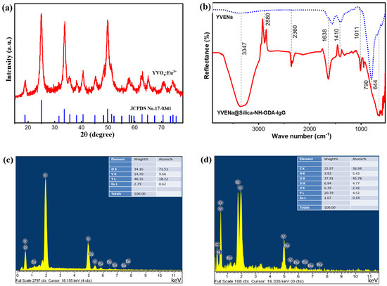
Figure 2.
(a) X-ray diffraction pattern of the YVO4:Eu3+ sample in comparison with the standard data of tetragonal-phase YVO4:Eu3+ nanoparticles (JCPDS No. 17-0341) as a reference. (b) FTIR spectra of the YVO4:Eu3+@silica-NH-GDA-IgG sample and the YVO4:Eu3+ samples. (c) EDX spectrum of the YVO4:Eu3+ sample. (d) EDX spectrum of the YVO4:Eu3+@silica-NH-GDA-IgG sample.
The characteristic chemical bonds of the YVO4:Eu3+ and YVO4:Eu3+@silica-NH-GDA-IgG samples were analyzed via Fourier-transform infrared (FTIR) spectra as shown in Figure 2b. For both cases, the observed absorption peaks at low frequencies of vibration such as at 644 and 790 cm−1 corresponded to the characteristic Y–O and Eu–O bonds, respectively. We also observed oscillations of the O–H bond at around 1638 and 3347 cm−1 [22]. In the case of YVO4:Eu3+@silica-NH-GDA-IgG nanoparticles, the band around 2880 cm−1 was attributed to C–H stretching vibration of alkanes. The N–H stretching vibration and the O–H bond were around 3347 cm−1. The characteristic band of the Si–O∓R bond was observed at around 1011 cm−1. Furthermore, an intense peak at 2360 cm−1 was associated with C=N stretching vibration that was formed by the reaction between the glutaraldehyde with amine–NH2 groups of functionalized YVO4:Eu3+@silica-NH-GDA-IgG antibodies. It indicated that the conjugation between luminescent nanoparticles and IgG was formed in the YVO4:Eu3+@silica-NH-GDA-IgG nanocomplex [20]. Compared to the case of the unconjugated YVO4:Eu3+ sample (Figure 2c) in addition to the strong peaks belonging to Y, V, and O elements, there were additionally weaker characteristic peaks of Eu and other peaks of Si, N, and C in the energy-dispersive X-ray (EDX) spectrum of the YVO4:Eu3+@silica-NH-GDA-IgG sample (Figure 2d). It confirmed that the silica shell and the amine group were successfully coated on the surface of the YVO4:Eu3+ core.
2.3. Luminescence Properties
Figure 3a shows the photoluminescence (PL) spectra of the YVO4:Eu3+, YVO4:Eu3+@silica-NH2, and YVO4:Eu3+@silica-NH-GDA-IgG samples at a 355 nm excitation wavelength. The PL spectra consisted of a narrow band corresponding to the well-known Eu3+ emission from intra 4f transitions (5D0–7F1, 5D0–7F2, 5D0–7F3, and 5D0–7F4). The strongest emission peaks were yielded by the 5D0–7F2 transition at around 618 nm (red light). Specially, the positions of PL peaks of the YVO4:Eu3+@silica-NH-GDA-IgG sample were almost the same as those of the YVO4:Eu3+ sample and the YVO4:Eu3+@silica-NH2 sample. It implies that the PL property of the YVO4:Eu3+@silica-NH-GDA-IgG sample was unchanged after functionalization and biological conjugation. Moreover, the PL intensity of the YVO4:Eu3+@silica-NH-GDA-IgG sample was enhanced. This result can be explained by substitution of the O–H luminescence quenching with O=C and C–H in the case of surface modification. These fluorescence properties of samples containing YVO4:Eu3+ nanoparticles have attracted a great deal of attention in biology and medicine. It can be indicated that the surface modification of the YVO4:Eu3+ nanoparticles not only improved their bio-compatibility, but also increased their PL intensity. Furthermore, the YVO4:Eu3+@silica-NH-GDA-IgG sample remained stable for quite a long time. The PL intensity of the YVO4:Eu3+@silica-NH-GDA-IgG sample had insignificant changes after three and six months of the synthesis as shown in Figure 3b. Thus, these nanoparticles can be used for bio-labelling applications.
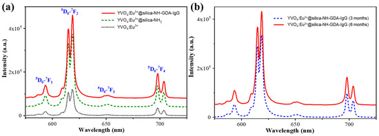
Figure 3.
(a) PL spectra of the YVO4:Eu3+, YVO4:Eu3+@silica-NH2, and YVO4:Eu3+@silica-NH-GDA-IgG nanoparticles under a 355 nm excitation. (b) PL spectra of the YVO4:Eu3+@silica-NH-GDA-IgG nanoparticles measured after three and six months of the synthesis.
2.4. In Vitro Cellular Imaging
We used fluorescence microscopy to evaluate the linking ability between YVO4:Eu3+@silica-NH-GDA-IgG conjugates and MCF-7 breast cancer cells after the incubation process. Figure 4a–c show the fluorescent images for three cases of MCF-7 breast cancer cells (negative control), incubated MCF-7 breast cancer cells with YVO4:Eu3+@silica-NH2 nanoparticles, and incubated MCF-7 breast cancer cells with YVO4:Eu3+@silica-NH-GDA-IgG nanoparticles, respectively.

Figure 4.
Fluorescent microscopy images of MCF-7 breast cancer cells after 3 h of incubation without nanoparticles (negative control) (a) and with YVO4:Eu3+@silica-NH2 nanoparticles at a concentration of 20 µg/mL (b) and YVO4:Eu3+@silica-NH-GDA-IgG conjugates at a concentration of 20 µg/mL (c) under an excitation wavelength in the UV region.
For the first case, we did not observe PL emission in the reference sample (Figure 4a). In the second case, the incubated MCF-7 breast cancer cells with the YVO4:Eu3+@silica-NH2 sample showed a blur and tiny PL intensity (Figure 4b). This can be explained by the existence of the weak bonds between YVO4:Eu3+@silica-NH2 nanoparticles and the cancer cells. As reported, muscarinic acetylcholine receptors (mAChR) belong to the G-protein-coupled receptor family and are extensively expressed in human breast tumor cells. In addition, immunoglobulin G (IgG) has been described that the presence of IgG in tumor cells establishes correlations between high antibody levels and promotion of cancer cell proliferation, invasion, and poor clinical prognosis for tumor patients. Blocking tumor-cell-derived IgG inhibits tumor cells. Tumor-cell-derived IgG might impede antigen-dependent cellular cytotoxicity by binding antigens such as mAChR while, at the same time, lacking the capacity for complement activation [23]. However, we can observe bright red pixels in the case of YVO4:Eu3+@silica-NH-GDA-IgG nanoparticles in Figure 4c. This evidence demonstrated a strong coupling between YVO4:Eu3+@silica-NH-GDA-IgG nanoparticles and MCF-7 breast cancer cells due to biological conjugation.
Furthermore, it can be seen that YVO4:Eu3+@silica-NH-GDA-IgG nanoparticles were localized within the cell cytoplasm. The high GDA concentration allowed the ligand binding between cells and luminescent labelling particles. After that, the YVO4:Eu3+@silica-NH-GDA-IgG nanoparticles were internalized into the cell via the invagination process. Therefore, YVO4:Eu3+@silica-NH-GDA-IgG nanoparticles could be used as a potential bio-label for MCF-7 breast cancer cells. In comparison to the other techniques, the use of YVO4:Eu3+@silica-NH-GDA-IgG nanoparticles is expected to be more advantageous by providing a facile method without requirements of any complex apparatus as well as processing such as spectral equipment and data analysis [24,25]. Additionally, it is a visual tool that is needed for some specific studies in biology.
In addition, the nanocomplex exhibited much fewer probing activities on HEK-293A human embryonic kidney cells, which were non-cancerous cell lines. Figure 5a–c show the fluorescence images of the HEK-293A cells with YVO4:Eu3+@silica-NH-GDA-IgG nanoparticles with different detection modes: (a)—negative control, (b)—Dark field, (c)—Merge, respectively. The experimental conditions were the same as those for the MCF-7 breast cancer cells, and the images were taken in the cases of the bright field, dark field, and merged modes. Moreover, the YVO4:Eu3+@silica-NH-GDA-IgG nanoparticles could not probe healthy cells of HEK-293A.
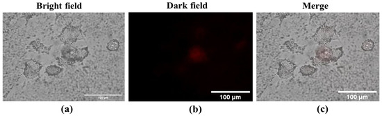
Figure 5.
Fluorescence images of HEK-293A embryonic kidney cells after incubated with YVO4:Eu3+@silica-NH-GDA-IgG bio-nanocomplexes with different detection modes: (a)—negative control, (b)—Dark field, (c)—Merge.
The flowcytometry results also provided the percentage of probed cells using the YVO4:Eu3+@silica-NH-GDA-IgG nanocomplex. As shown in Figure 6, YVO4:Eu3+@silica-NH-GDA-IgG nanocomplexes were found in about 82.11% of MCF-7 cells when stained with YVO4:Eu3+@silica-NH-GDA-IgG nanocomplexes. A similar result (1.55%) was found in MCF-7 cells that were incubated with YVO4:Eu3+@silica-NH2 nanoparticles. The percentage of MCF-7 cells was only 0.57% for MCF-7 cells incubated with unconjugated YVO4:Eu3+ nanoparticles. Thus, the detection percentage of MCF-7 breast cancer cells increased pronouncedly from 0.57% for the case of unconjugated YVO4:Eu3+ nanoparticles to 1.55% for the case of YVO4:Eu3+@silica-NH2 nanoparticles and achieved a highest value of 82.11% with YVO4:Eu3+@silica-NH-GDA-IgG nanoparticles, indicating the crucial role of the functionalization and conjugation of YVO4:Eu3+ nanoparticles.
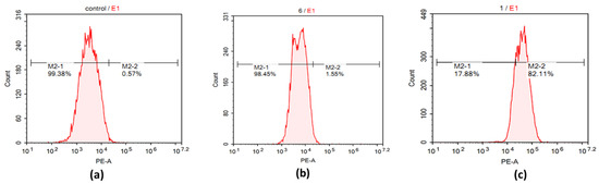
Figure 6.
Flowcytometry analysis of labelled MCF-7 cells incubated with the negative control (a), YVO4:Eu3+@silica-NH2 nanoparticles (b), and YVO4:Eu3+@silica-NH-GDA-IgG nanoparticles (c).
3. Materials and Methods
3.1. Preparation of YVO4:Eu3+@Silica-NH2 Nanoparticles
In a typical synthesis of YVO4:Eu3+ nanoparticles, yttrium nitrate hexahydrate (Y(NO3)3∙6H2O, 99.9%; Sigma-Aldrich), sodium orthovanadate (Na3VO4 90%; Sigma-Aldrich), and europium nitrate pentahydrate (Eu(NO3)3∙5H2O, 99.9%; Sigma-Aldrich) were mixed with a molar ratio of 0.99/1/0.01, and sodium hydroxide (NaOH 99%, Merck) was added to control pH values of 6–8. The solution was stirred for 180 min, and then, it was transferred into an autoclave and heated at 190 °C for 24 h. The YVO4:Eu3+ nanoparticles were separated by using centrifugation (5800 rpm) and washed with deionized water for three times and dispersed in ethanol to achieve a colloidal solution. YVO4:Eu3+ nanoparticles were coated by silica via the Stöber process. In detail, 10 mL of tetraethyl orthosilicate (TEOS), 10 mL of ethanol (NH4OH), 1 mL of acetic acid (CH3COOH), and 2 mL of deionized water (H2O) were mixed and stirred at room temperature for 24 h. Ten millimeters of the colloidal solution of YVO4:Eu3+ nanoparticles was slowly added into the above mixture and continuously stirred for 24 h. The YVO4:Eu3+@silica nanoparticles were separated by using a centrifuge and dispersed in 20 mL of ethanol. Then, they were added into a mixture of 22.5 mL absolute ethanol and 2 mL 3-aminopropyltriethoxysilane (APTES) at 60 °C for 5 h. YVO4:Eu3+@silica-NH2 nanoparticles were obtained by centrifugation [26,27,28].
By using APTES, a reagent contains a short organic 3-amino propyl group, which terminates in a primary amine, and ethoxy groups are not reactive enough to couple spontaneously with OH groups on an inorganic surface without prior hydrolysis to make silanol. This protocol can be used to modify the surface of particles with this reagent as described in Figure 7. Firstly, the reaction involves the hydrolysis of the alkoxysilane group to create highly reactive silanol that undergoes hydrogen bonding with other silanol groups in solutions and on the particle surface, resulting in the associated organosilane derivatives. Then, a condensation reaction takes place to form a polymerized coating of the organosilane on the particle surface [21]. Secondly, APTES is coated on the YVO4:Eu3+@silica surface to create a covalent shell containing a primary amine group. The reaction occurs in a partially aqueous environment, because ethoxy groups are unreactive enough to substrate OH groups without prior hydrolysis. This is typically performed in 5% water in ethanol that is acidified with acetic acid to pH values of 4.5–5.5. The process results in a layer containing about 3–8 organosilanes in thickness and masks the inorganic substrate with aminopropyl groups. The advantage of this process is providing a thin and controllable silane layer that can be created a monolayer of the aminopropyl group on the surface.
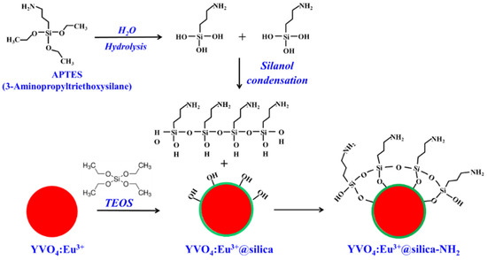
Figure 7.
The coupling reaction of the silanol group to the YVO4:Eu3+ nanoparticles and APTES coating on the YVO4:Eu3+@silica surface to create a covalent shell of the amine group.
3.2. Preparation of YVO4:Eu3+@silica-NH-GDA-IgG Bio-Nanocomplexes
Figure 8 describes the functionalization of the YVO4:Eu3+@silica-NH nanoparticles with glutaraldehyde (GDA) and immunoglobulin G (IgG). The YVO4:Eu3+@silica-NH2 nanoparticles and GDA were dispersed in vanadate-buffered saline (PBS, 0.1 M, pH 5) with a concentration of 5 gL−1. Then, this compound was added to different concentrations of IgG. These reaction mixtures were incubated with glycerol at room temperature for 4 h. The YVO4:Eu3+@silica-NH-GDA-IgG products were collected by centrifugation (5900 rpm) with water for three times and stored at 4 °C in a closing box. The reaction mechanism can be explained via a Schiff base linkage with amine on proteins [21]. Proteins can be coupled to the -NH2 groups via an amine-reactive linker—glutaraldehyde—to form activated derivatives that enable to make crosslink with other proteins. As shown in Figure 8, the amine groups react with the aldehyde groups to form a Schiff base, resulting in a polymeric coating that contains both aldehydes and double bonds for further coupling with amine-containing molecules.
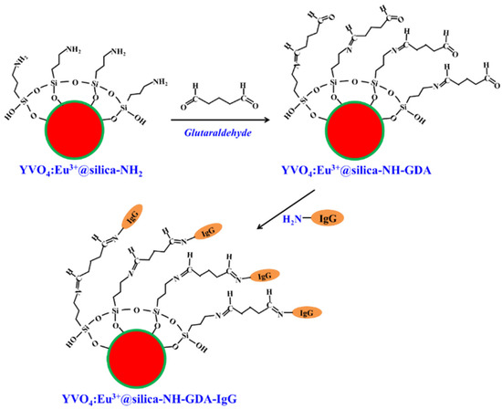
Figure 8.
Functionalization of the YVO4:Eu3+@silica-NH nanoparticles with GDA and IgG. This conjugation strategy has been used to associate biomolecules containing amine groups with aminated YVO4:Eu3+@silica-NH2 nanomaterials, usually utilizing glutaraldehyde, a molecule containing two aldehyde moieties. One aldehyde group forms a Schiff base with the amine groups, while the other binds to the amino groups of the biomolecules. The reactions with glutaraldehyde are favored in alkaline media, being more efficient at high pH values [21].
3.3. MCF-7 Breast Cancer Cell and HEK-293A Cell Culture and Fluorescence Imaging of Cells
In this study, the experiments were implemented on MCF-7 breast cancer cells (MCF-7 was kindly presented by Prof. Chi-Ying Huang, National Yangming University, Taiwan-MCF7 (ATCC # HTB-22). The results were then compared with HEK-293A healthy cells (these cells were kindly provided from Prof. Young-Pil Kim, HanYang University, Korea-HEK-293A (Invitrogen # P/N 51-0036)) that were maintained in a cultured medium—Dulbecco’s Modified Eagle Medium (DMEM) with fetal bovine serum (10%) (Sigma) and gentamicin (50 µg/mL) at 37 °C and 5% CO2 in a humidified atmosphere [29,30]. The cells were seeded at a density of 5.104 cells/mL. To study the uptake capacity of the YVO4:Eu3+@silica-NH-GDA-IgG nanoparticles, the MCF-7 breast cancer cells and HEK-293A cells (106 cells/mL) at the log phase were seeded in 24-well plates and then incubated for 24 h. The YVO4:Eu3+@silica-NH-GDA-IgG nanoparticles (with a concentration of 20 µg/mL) were then added to the cell-seeded wells for 3 h. After the assigned time, the cultured medium was discarded. The cells were then washed three times with phosphate-buffered saline. At the end of the process, phosphate-buffered saline was added to the wells, before and after MCF-7 breast cancer cells and HEK-293A cells were incubated with YVO4:Eu3+@silica-NH-GDA-IgG bio-nanocomplexes. The cell images were obtained using an Olympus ScanR 100X fluorescent microscope.
3.4. Cellular Surface Labelling Analysis Using Flowcytometry
MCF-7 cancer cells, a cultured medium (DMEM), a fetal bovine serum (FBS), trypsin-EDTA, and bovine insulin were obtained from Invitrogen (Carlsbad, CA, USA). MCF-7 cells (5 × 10⁴ cells/mL) were cultured in 6-well plates at 37 °C and 5% CO2 for 24 h. Then, the cells were detached with 0.05% trypsin-EDTA and centrifuged at 1000 rpm for 5 min to obtain the cell pellets. The cells were fixed with 4% formaldehyde for 24 h at 4 °C and washed with cold phosphate-buffered saline (PBS). To access cellular surface labeling, the YVO4:Eu3+-NH2 and YVO4:Eu3+@silica-NH-GDA-IgG nanoparticles were employed and incubated with fixed cells for 2 h. The labelled cells were washed with cold PBS twice before resuspending in PBS for analyzing with a flowcytometry Novocyte system (ACEA Bioscience inc.) and NovoExpress software. The cells were requested for light protection.
3.5. Characterization Techniques
The X-ray diffraction (XRD) analysis of the samples was carried out on a Siemens D5000 diffractometer with λ = 1.5406 Å. The morphologies and the energy-dispersive X-ray spectra were observed and measured by field emission scanning electron microscopy (S-4800; Hitachi) attached with an energy-dispersive X-ray spectrometer. The infrared absorption spectra were performed by employing a Fourier-transform infrared spectrometer (FTIR NEXUS 670). The photoluminescence (PL) spectra of the samples were studied by using an iHR550 photoluminescence system (Horiba). The cells were observed under an Olympus ScanR fluorescence microscope (Olympus Europa SE & Co.KG, Hamburg, DE).
4. Conclusions
YVO4:Eu3+ nanoparticles with a uniform size of 10–25 nm were synthesized via a hydrothermal process, followed by further functionalizations to form the YVO4:Eu3+@silica-NH-GDA-IgG bio-nanocomplexes. The surface modification of the YVO4:Eu3+ nanoparticles not only improved the bio-compatible media, but also increased their PL intensity due to enhancement of chemical stability. The experimental evidence indicated that the YVO4:Eu3+@silica-NH-GDA-IgG nanoparticles could selectively detect MCF-7 breast cancer cells while it could not probe HEK-293A healthy cells for in vitro tests. The YVO4:Eu3+@silica-NH-GDA-IgG bio-nanocomplexes with a significant bio-compatible capability exhibited a strong, enhanced red emission, which provides a visual tool for bio-labelling applications.
Author Contributions
Conceptualization, T.T.H., L.T.V., and H.T.P.; methodology, T.T.H., L.T.V., H.T.P., H.T.K. and D.T.T.; validation, T.T.H. and H.T.P.; formal analysis, T.T.H., L.T.V., H.T.P. and H.T.K.; investigation, T.T.H., L.D.T., D.T.T. and N.D.V.; resources, L.T.V. and H.T.P.; data curation, T.T.H. and H.T.P.; writing—original draft preparation, T.T.H., D.T.T. and L.D.T.; writing—review and editing, T.T.H., L.D.T. and N.D.V.; supervision, T.T.H., L.T.V. and H.T.P.; project administration, T.T.H. All authors have read and agreed to the published version of the manuscript.
Funding
This research was funded by Vietnam National Foundation for Science and Technology Development (NAFOSTED) under grant number 103.03-2017.66.
Institutional Review Board Statement
Not applicable.
Informed Consent Statement
Not applicable.
Data Availability Statement
All data are available in this publication.
Conflicts of Interest
The authors declare no conflict of interest.
Sample Availability
Samples of the compounds are available from the authors.
References
- Tan, M.; Chen, G. Rare Earth-Doped Nanoparticles for Advanced In Vivo Near Infrared Imaging. In Near Infrared-Emitting Nanoparticles for Biomedical Applications; Benayas, A., Hemmer, E., Hong, G., Jaque, D., Eds.; Springer: Cham, Switzerland, 2020; pp. 63–81. [Google Scholar] [CrossRef]
- Shao, J.; Yan, J.; Li, X.; Li, S.; Hu, T. Novel fluorescent label based on YVO4: Bi3+, Eu3+ for latent fingerprint detection. Dyes Pigments 2018, 160, 555–562. [Google Scholar] [CrossRef]
- Giang, L.T.K.; Trejgis, K.; Marciniak, Ł.; Opalińska, A.; Koltsov, I.E.; Łojkowski, W. Synthesis and characterizations of YZ-BDC:Eu3+,Tb3+ nanothermometers for luminescence-based temperature sensing. RSC Adv. 2022, 12, 13065–13073. [Google Scholar] [CrossRef] [PubMed]
- Sevic, D.; Rabasovic, M.S.; Krizan, J.; Savic-Sevic, S.; Nikolic, M.G.; Marinkovic, B.P. YVO4:Eu3+ nanopowders: Multi-mode temperature sensing technique. J. Phys. D Appl. Phys. 2019, 53, 015106. [Google Scholar] [CrossRef]
- Fernández-Osorio, A.; Redón, R.; Medina-Pérez, J.; Pedroza-Montero, M.; Acosta, M. Photoluminescence and Thermoluminescence Properties of Nanophosphors, YVO4:Eu3+ and YVO4:Eu3+:Dy3+. J. Clust. Sci. 2022, 33, 653–664. [Google Scholar] [CrossRef]
- Giaume, D.; Poggi, M.; Casanova, D.; Mialon, G.; Lahlil, K.; Alexandrou, A.; Gacoin, T.; Boilot, J.-P. Organic Functionalization of Luminescent Oxide Nanoparticles toward Their Application As Biological Probes. Langmuir 2008, 24, 11018–11026. [Google Scholar] [CrossRef]
- Senty, T.R.; Yalamanchi, M.; Zhang, Y.; Cushing, S.K.; Seehra, M.S.; Shi, X.; Bristow, A.D. Photoluminescence spectroscopy of YVO4:Eu3+ nanoparticles with aromatic linker molecules: A precursor to biomedical functionalization. J. Appl. Phys. 2014, 115, 163107. [Google Scholar] [CrossRef]
- Ascenzi, P.; Bettinelli, M.; Boffi, A.; Botta, M.; De Simone, G.; Luchinat, C.; Marengo, E.; Mei, H.; Aime, S. Rare earth elements (REE) in biology and medicine. Rendiconti Lincei. Scienze Fisiche Naturali 2020, 31, 821–833. [Google Scholar] [CrossRef]
- Soukka, T.; Paukkunen, J.; Härmä, H.; Lönnberg, S.; Lindroos, H.; Lövgren, T. Supersensitive Time-resolved Immunofluorometric Assay of Free Prostate-specific Antigen with Nanoparticle Label Technology. Clin. Chem. 2001, 47, 1269–1278. [Google Scholar] [CrossRef]
- Fan, Q.; Cui, X.; Guo, H.; Xu, Y.; Zhang, G.; Peng, B. Application of rare earth-doped nanoparticles in biological imaging and tumor treatment. J. Biomater. Appl. 2020, 35, 237–263. [Google Scholar] [CrossRef]
- Kolesnikov, I.E.; Mamonova, D.V.; Kurochkin, M.A.; Kolesnikov, E.Y.; Lähderanta, E. Optical Thermometry by Monitoring Dual Emissions from YVO4 and Eu3+ in YVO4:Eu3+ Nanoparticles. ACS Appl. Nano Mater. 2021, 4, 1959–1966. [Google Scholar] [CrossRef]
- Bouzigues, C.; Gacoin, T.; Alexandrou, A. Biological Applications of Rare-Earth Based Nanoparticles. ACS Nano 2011, 5, 8488–8505. [Google Scholar] [CrossRef] [PubMed]
- Wang, F.; Tan, W.B.; Zhang, Y.; Fan, X.; Wang, M. Luminescent nanomaterials for biological labelling. Nanotechnology 2005, 17, R1–R13. [Google Scholar] [CrossRef]
- Huong, T.T.; Vinh, L.; Anh, T.K.; Khuyen, H.T.; Phuong, H.T.; Minh, L.Q. Fabrication and optical characterization of multimorphological nanostructured materials containing Eu(iii) in phosphate matrices for biomedical applications. New J. Chem. 2014, 38, 2114–2119. [Google Scholar] [CrossRef]
- Lee, S.Y.; Lin, M.; Lee, A.; Park, Y.I. Lanthanide-doped nanoparticles for diagnostic sensing. Nanomaterials 2017, 7, 411. [Google Scholar] [CrossRef]
- Ren, Q.-F.; Zhang, B.; Chen, S.-H.; Wang, S.-L.; Zheng, Q.; Ding, Y.; Qian, H.-S.; Jin, Z. Amine salts assisted controllable synthesis of the YVO4:Eu3+ nanocrystallines and their luminescence properties. Phys. B Condens. Matter 2019, 557, 1–5. [Google Scholar] [CrossRef]
- Reddy, M.L.P.; Divya, V.; Pavithran, R. Visible-light sensitized luminescent europium(iii)-β-diketonate complexes: Bioprobes for cellular imaging. Dalton Trans. 2013, 42, 15249–15262. [Google Scholar] [CrossRef]
- Tamimi, E.; Ardila, D.C.; Haskett, D.G.; Doetschman, T.; Slepian, M.J.; Kellar, R.S.; Geest, J.P.V. Biomechanical Comparison of Glutaraldehyde-Crosslinked Gelatin Fibrinogen Electrospun Scaffolds to Porcine Coronary Arteries. J. Biomech. Eng. 2015, 138, 011001. [Google Scholar] [CrossRef]
- Peng, Y.Y.; Glattauer, V.; Ramshaw, J.A.M. Stabilisation of Collagen Sponges by Glutaraldehyde Vapour Crosslinking. Int. J. Biomater. 2017, 2017, 8947823. [Google Scholar] [CrossRef]
- Niekamp, S.; Stuurman, N.; Vale, R.D. A 6-nm ultra-photostable DNA FluoroCube for fluorescence imaging. Nat. Methods 2020, 17, 437–441. [Google Scholar] [CrossRef]
- Hermanson, G.T. Bioconjugate Techniques; Elsevier: New York, NY, USA, 2008. [Google Scholar]
- Stauffer, T.M. Applications of Molecular Spectroscopy to Current Research in the Chemical and Biological Sciences-Fourier Transform Infrared and Raman Characterization of Silica-Based Materials; Capeletti, L.B., Zimnoch, J.H., Eds.; IntechOpen: London, UK, 2016; Chapter 1; pp. 1–20. [Google Scholar] [CrossRef]
- Negroni, M.P.; Fiszman, G.L.; Azar, M.E.; Morgado, C.C.; Español, A.J.; Pelegrina, L.T.; de la Torre, E.; Sales, M.E. Immunoglobulin G from Breast Cancer Patients in Stage I Stimulates Muscarinic Acetylcholine Receptors in MCF7 Cells and Induces Proliferation. Participation of Nitric Oxide Synthase-Derived Nitric Oxide. J. Clin. Immunol. 2010, 30, 474–484. [Google Scholar] [CrossRef]
- Yang, Y.; Fu, Y.; Su, H.; Mao, L.; Chen, M. Sensitive detection of MCF-7 human breast cancer cells by using a novel DNA-labeled sandwich electrochemical biosensor. Biosens. Bioelectron. 2018, 122, 175–182. [Google Scholar] [CrossRef] [PubMed]
- Moallem, G.; Pore, A.A.; Gangadhar, A.; Sari-Sarraf, H.; Vanapalli, S.A. Detection of live breast cancer cells in bright-field microscopy images containing white blood cells by image analysis and deep learning. J. Biomed. Opt. 2022, 27, 076003. [Google Scholar] [CrossRef] [PubMed]
- Vinh, L.T.; Huong, T.T.; Phuong, H.T.; Khuyen, H.T.; Hung, N.M.; Thao, D.T.; Minh, L.Q. Folic Acid-Conjugated Silica-Modified TbPO4·H2O Nanorods for Biomedical Applications. J. Nanomater. 2021, 2021, 9888856. [Google Scholar] [CrossRef]
- Labrador-Páez, L.; Ximendes, E.C.; Rodríguez-Sevilla, P.; Ortgies, D.H.; Rocha, U.; Jacinto, C.; Rodríguez, E.M.; Haro-González, P.; Jaque, D. Core–shell rare-earth-doped nanostructures in biomedicine. Nanoscale 2018, 10, 12935–12956. [Google Scholar] [CrossRef] [PubMed]
- Huong, T.T.; Vinh, L.T.; Phuong, H.T.; Khuyen, H.T.; Anh, T.K.; Tu, V.D.; Minh, L.Q. Controlled fabrication of the strong emission YVO4:Eu3+ nanoparticles and nanowires by microwave assisted chemical synthesis. J. Lumin. 2016, 173, 89–93. [Google Scholar] [CrossRef]
- Huong, T.T.; Phuong, H.T.; Vinh, L.T.; Khuyen, H.T.; Thao, D.T.; Tuyen, L.D.; Anh, T.K.; Minh, L.Q. Upconversion NaYF4:Yb3+/Er3+@silica-TPGS Bio-Nano Complexes: Synthesis, Characterization, and In Vitro Tests for Labeling Cancer Cells. J. Phys. Chem. B 2021, 125, 9768–9775. [Google Scholar] [CrossRef]
- Jain, A.; Fournier, P.G.J.; Mendoza-Lavaniegos, V.; Sengar, P.; Guerra-Olvera, F.M.; Iñiguez, E.; Kretzschmar, T.G.; Hirata, G.A.; Juárez, P. Functionalized rare earth-doped nanoparticles for breast cancer nanodiagnostic using fluorescence and CT imaging. J. Nanobiotechnol. 2018, 16, 26. [Google Scholar] [CrossRef]


Disclaimer/Publisher’s Note: The statements, opinions and data contained in all publications are solely those of the individual author(s) and contributor(s) and not of MDPI and/or the editor(s). MDPI and/or the editor(s) disclaim responsibility for any injury to people or property resulting from any ideas, methods, instructions or products referred to in the content. |
© 2022 by the authors. Licensee MDPI, Basel, Switzerland. This article is an open access article distributed under the terms and conditions of the Creative Commons Attribution (CC BY) license (https://creativecommons.org/licenses/by/4.0/).