Article

Insight of the Functional and Biological Activities of Coconut (Cocos nucifera L.) Protein by Proteomics Analysis and Protein-Based Bioinformatics

1
College of Food Sciences & Engineering, Hainan University, 58 People Road, Haikou 570228, China
2
Key Laboratory of Aquatic Product Processing, Ministry of Agriculutre and Rural Affairs, National R&D Center for Aquatic Product Processing, South China Sea Fisheries Research Institute, Chinese Academy of Fishery Sciences, Guangzhou 510300, China
3
Collaborative Innovation Center of Provincial and Ministerial Co-Construction for Marine Food Deep Processing, Dalian Polytechnic University, Dalian 116034, China
*
Authors to whom correspondence should be addressed.
These authors contributed equally to this work and should be regarded as co-first authors.
Molecules 2022, 27(9), 2987; https://doi.org/10.3390/molecules27092987
Submission received: 20 April 2022 / Revised: 27 April 2022 / Accepted: 4 May 2022 / Published: 6 May 2022
(This article belongs to the Topic Label-Free Proteome Profiling)

Abstract

:
Coconut (Cocos nucifera L.) is one of the most critical economic crops in the tropics and sub-tropics. Although coconut protein has attracted more and more attention due to its nutritional potential, the lack of proteomic information has limited its practical application. The present study aimed to investigate the coconut meat proteome by shotgun proteomics and protein-based bioinformatic analysis. A grand total of 1686 proteins were identified by searching the National Center for Biotechnology Information (NCBI) protein database and self-constructed C. nucifera transcriptome repository. Among them, 17 and 9 proteins were identified as antioxidant proteins and globulins, respectively. Network analysis of the globulins referred to the sub-works of Cupin and Oleosin, and the antioxidant proteins were related to the sub-networks of glutathione metabolism and peroxisome. The bioactive peptides acquired by in-silico digestion of the targeted proteins have the potential to be applied as antioxidants and emulsifiers for both healthcare and food stabilization.

1. Introduction

A global focus on sustainability has accelerated research into alternative non-animal food protein sources and functional food ingredients [1]. Proteins sourced from plants are considered valuable ingredients by the food industry in preparing functional foods [2]. Coconut (Cocos nucifera L.) belongs to the Palm family (Arecaceae) with a 2n = 32 ploidy and is one of the most critical economic crops in the tropics and sub-tropics [3]. According to the Food and Agriculture Organization, the annual world production of coconut is approximately 60.7 million tons [4]. The protein content of fresh coconut meat is about 2.6–4.4% on a wet basis (w/w), and the large mass of coconut grown globally makes recovery of these proteins desirable [5]. Most of the studies on coconut meat mainly focus on the effects of processing on the nutritional quality of coconut proteins [6] and characterizing the major proteins present in coconut via classical biochemical purification tools [7,8]. Although coconut is a large part of the human diet with potential health benefits [9], information on the components, functional, and biological activities of coconut proteins are scarce. Therefore, evaluating the protein components in coconut and understanding its functional and biological activities are essential, which is good for the processing and application of coconut.
Proteomics is being increasingly used to assess the diversity, quality, and bioactivity of plant proteins, such as almond kernel [10], lupin seed [11], maize seed [12], avocado seed [13], and soybean [14]. It can describe the composition, function, interaction, and modification of direct and coordinated cell activities over a given time or under specific environmental cell conditions of proteins [3]. Liquid chromatography-tandem mass spectrometry (LC-MS/MS) is the method of choice for shotgun proteomics [15]; based on this method, the proteomics analysis could acquire a large-scale protein identification. Proteomics analysis has been proved much better than two-dimensional electrophoresis (2-DE), which has certain disadvantages, such as the limited number of identified proteins, narrow linear detection dynamic range, long analysis time, and low sensitivity and reproducibility [15]. At present, proteomics research in coconut meat is poorly represented. To our knowledge, few studies have been reported. D’Amato, Fasoli, and Righetti [16] analyzed the proteome of coconut milk and identified 307 unique gene products; the most abundant proteins were the glutelin and 7S globulin, homologous to proteins present in oil palm and that have a nutrient reservoir activity. Huang et al. [17] investigated the proteome of coconut meat and found 200 protein spots on 2-DE maps; the identified proteins were limited, which were nonetheless very informative in that some of the proteins in coconut endosperm were found to be “storage proteins”. Recently, coconut storage proteins have been paid growing attention to their nutritional and health benefits [3]. Conclusively, protein-based bioinformatic information on the coconut is relatively scarce, and further studies are needed.
The lack of information regarding the functional and biological properties of individual protein or protein groups of coconut has limited their applications. Given these circumstances, the present study aimed to identify and characterize the coconut meat proteins by a shotgun proteomic approach. Meanwhile, the bioinformatics analysis was performed to illuminate the functional and biological activities of all the identified proteins. Furthermore, different protein-based bioinformatic analyses were combined to acquire the bioactive peptides of globulins and antioxidant proteins and reveal their potential function.

2. Results and Discussion

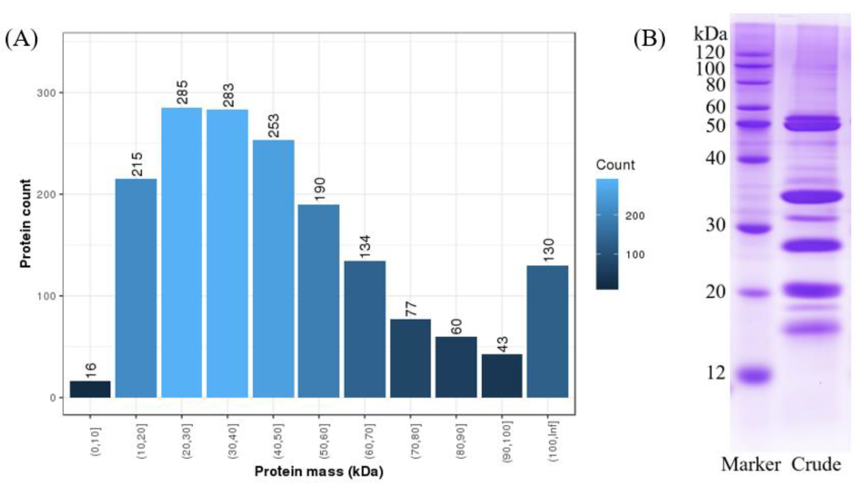
2.1. Coconut Meat Proteome

A shotgun proteomics analysis was carried out to obtain comprehensive proteome information on coconut meat. In the NCBI protein database, a total of 22,572 proteins for C. nucifera were registered. In addition, a transcriptome repository of C. nucifera was constructed by de novo assembly as a supplementary database.
The preliminary analysis of the coconut meat proteome showed that the proteins in coconut are abundant. A total of 279,953 spectra were obtained from the sample, 16,199 spectra were matched, and 1686 proteins were identified by searching in both the NCBI protein database and transcriptome repository, which were much more than the proteins identified by previous studies [16,17]. The molecular weights of the proteins were predominantly distributed among 10 and 60 kDa: a total of 1226 proteins were found in this range (72.72%) (Figure 1A). In addition, the SDS-PAGE analysis corresponds well with the identification results (Figure 1B). Demason and Sekhar [18] reported that reduced coconut proteins fractionated into seven major bands ranging from 17 to 55 kDa. Kwon, Park, and Rhee [19] also found that coconut proteins consisted of about seven major bands with molecular weights of between 14 and 52 kDa in a reduced state. These previous results are consistent with the protein pattern in our study.

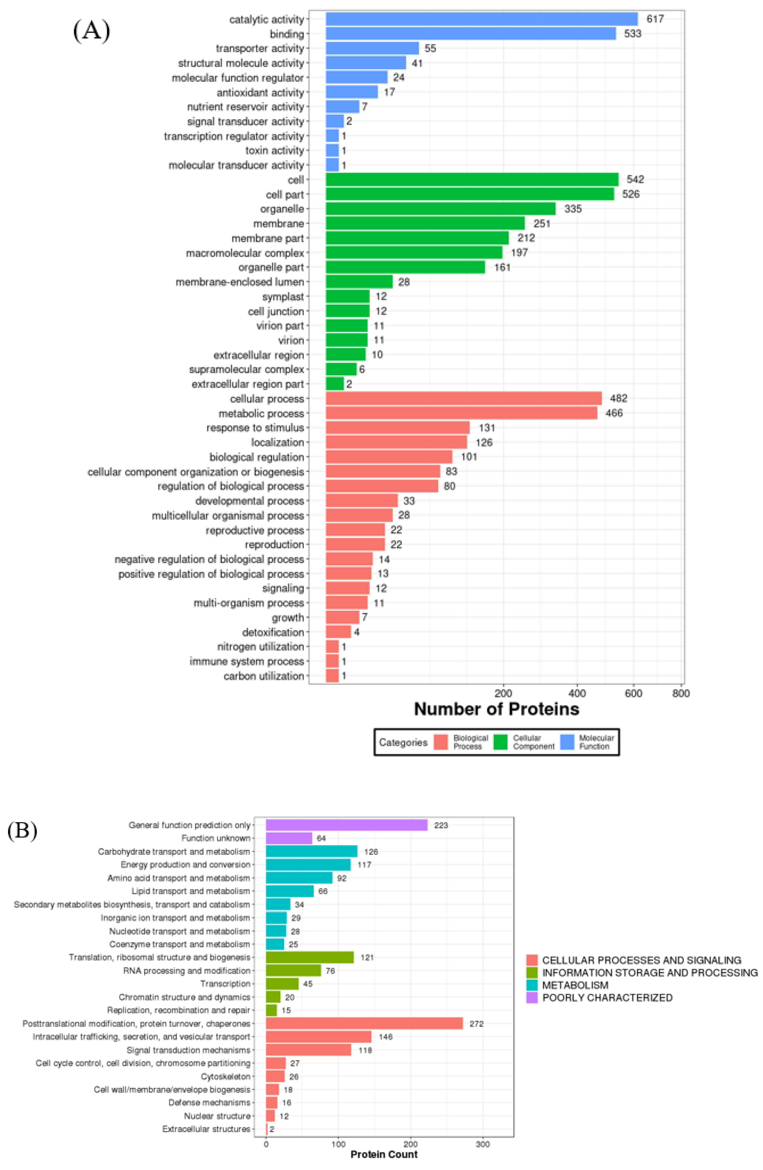
2.2. Functional and Bioinformatic Analysis

As the functional agents which are created by genomic transcripts and are the final products of gene expression, proteins function through an integral and coordinated network to regulate metabolism in cells, tissues, and organisms as a whole [3]. The functional classification and bioinformatic analysis of all the identified proteins were based on the molecular function, biological process, and cellular component (Figure 2).

2.2.1. Molecular Functions

As shown in Figure 2A, the results of molecular functional analyses demonstrated that the proteins with catalytic (36.6%) and binding (31.6%) activities were predominant in these groups. The exuberant metabolism and biosynthesis in the coconut endosperm need various proteins with different catalytic activities. Therefore, proteins represent the catalytic activities in various aspects, such as hydrolase activity, transferase activity, and phosphatase activity. Multiple studies have analyzed the individual enzymatic functions of coconut proteins during seed development, such as the tyrosine kinase activity [20,21], polyphenol oxidase activity [22], and lipase activity [23]. For fruit development, coconut meat is imbued with various nutrients, such as minerals and bioactive molecules, so a mass of proteins with binding abilities is needed.
Seed storage proteins have the pivotal role of providing the seedling with amino acids and nitrogen source for metabolism during germination and growth [24]. In this study, seven proteins were identified with nutrient reservoir activity, and all the proteins are globulins except the glutelin (ID: KAG1339186.1). Globulin is the predominant protein in coconut meat (40%) and the essential storage protein as well [25]. In recent years, there are many studies on its purification and identification [7,8]. 11S globulin (300–400 kDa, also known as cocosin), owning six subunits (50–60 kDa), accounted for 86% of the coconut globulin, whereas 7S globulin (150–200 kDa, also called vicilin-like protein), owning three subunits (40–70 kDa), only take up 14%. In this study, six 7S globulins (ALQ56981.1, CL8941.Contig1, CL3433.Contig1, CL4719.Contig2, CL4719.Contig3.1, and KAG1368674.1) and three 11S globulins (KAG1361520.1, ASQ40963.1, and Unigene39216) were identified (Table 1). This result could guide the extraction and purification of coconut globulin. Globulin has many potential applications in the food industry because of its good functional activity, such as emulsifying properties due to its amphiphilic molecular structure [26]. Currently, the most widely used protein-based emulsifiers are dairy proteins, like casein and whey proteins. However, animal protein presents growing costs and limited supply. It has been highly associated with climate change, freshwater depletion, biodiversity loss, and hazards for human health related to cardiovascular diseases and others [27]. Plant proteins are increasingly being used as a versatile alternative, replacing animal sources and functional ingredients for product formulation. The big part of globulin of coconut may have good application in emulsifiers.
Hitherto, the bioactive properties of coconut proteins are rarely investigated. Li et al. [28] found that the coconut proteins exhibited radical-scavenging activity and ion chelating ability; they can also protect DNA from oxidative damage. In this work, 17 proteins were identified with antioxidant activities (Table 2). Most identified antioxidant proteins were peroxidase, superoxide dismutase, catalase isozyme, and glutathione reductase. The protein mass of the antioxidant proteins is mainly concentrated in low molecular weight (18–35 kDa). Environmental or biotic stress could generate reactive oxygen species (ROS) and break the cellular redox balance. Therefore, a large variety of antioxidant enzymes are needed. Antioxidants play a vital role in both food systems and the human body to reduce oxidative stress. Since synthetic antioxidants have been suspected of threatening human health, antioxidants from natural sources have attracted more attention [28]. The coconut proteins with antioxidant activities identified here could provide a reference for their further applications.

2.2.2. Cellular Components

Regarding cellular components, the coconut proteins were mainly distributed in the cell, cell part, organelle, membrane, membrane part, macromolecular complex, and organelle part (Figure 2A). The proteins are present in coconut meat in various forms and cellular locations, such as as a component of the cell wall. They also bind to other cellular components, such as carbohydrates and membranes. Lipids are the primary source of energy in coconut endosperm, thus a great number of membrane structure is needed for its storage. Generally, membrane proteins possess emulsifying properties due to their amphiphilic structure. Yesiltas et al. [1] found that the proteins which are membrane-associated or form highly complex membrane-like macrostructures are more likely to include highly hydrophilic–hydrophobic regions. Therefore, except for the globulin, the membrane proteins also have potential applications as emulsifiers.

2.2.3. Biological Processes

As shown in Figure 2A, the proteins primarily participate in the cellular process, metabolic process, response to stimulus, localization, and biological regulation when focusing on the biological processes. These results indicated that, as a vegetative organ, coconut meat needs to perform various biological processes, and most coconut proteins are involved in multiple types of metabolism and cellular processes. From the KOG analysis (Figure 2B), we know that the main metabolic processes are carbohydrate transport, energy production, amino acid transport, and lipid transport, which correspond to the properties of seed proteins for embryo development. The ability of coconut to resist highly stressed environments originates from biological regulation and responses to stimulus behavior—most antioxidant proteins respond to oxidative stress and cell redox homeostasis. In addition, the heat shock proteins (HSPs) can assist protein folding and help to refold damaged proteins under stressful conditions [29]. This contributes to a better understanding of the molecular mechanisms of tropical plants responding and adapting to environmental change and facilitates the development of some anti-stress proteins. Additionally, further investigations on the functions of poorly known proteins are needed, considering there are 223 proteins that were poorly characterized and 64 proteins were uncharacterized (Figure 2B).
The globulins and antioxidant proteins from coconut meat may be promising in future applications as emulsifiers and antioxidants, respectively; more knowledge about them is needed. Therefore, in this work, we chose globulins and antioxidant proteins for the further bioinformatic analysis (network analysis and putative bioactive peptides).

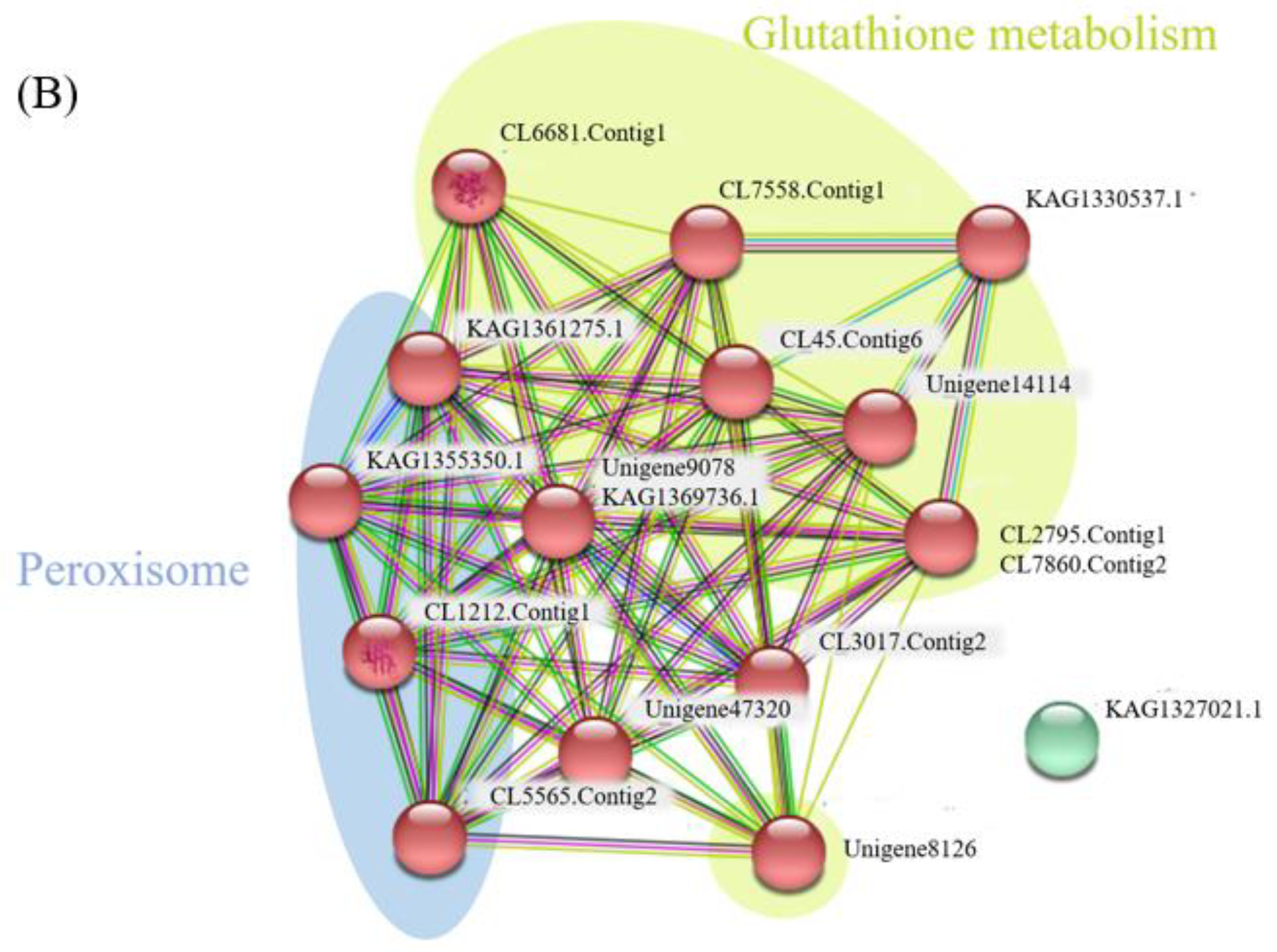
2.3. Network Analysis for Globulins and Antioxidant Proteins

Proteins do not exist independently in vivo, as their functions are associated with, or regulated by, other proteins [30]. Therefore, we performed a network analysis of the target proteins using protein–protein interaction. All the globulins and antioxidant proteins identified from the coconut meat proteome were merged on the STRING software (v.11.0, https://string-db.org/, accessed on 21 September 2021) to obtain cross-correlation information. The oil palm (Elaeis guineensis Jacq), date palm (Phoenix dactylifera L.), carnauba palm (Copernicia prunifera (Mill.) H.E.Moore), and coconut palm (Cocos nucifera L.) all belong to palm family that is restricted to tropical and subtropical climates [31]. The P. dactylifera L. was selected as a reference organism considering the fact that the genome of C. nucifera is not available in the STRING database. As shown in Figure 3, there were 4 nodes (9 proteins) and 2 edges (interactions) for the globulins (Figure 3A), and 15 nodes (17 proteins) and 78 edges (interactions) for the antioxidant proteins (Figure 3B).
The storage proteins are a group of plant structurally-conserved polypeptides lacking catalytic activities. Along with the seed development, they accumulate in the endosperm and catabolize to be used as a nitrogen source to support the growth of the seedling [3]. The topological analysis of the globulins revealed two sub-networks (Figure 3A): Cupins with two vicilin-like proteins (ALQ56981.1 and CL3433.Contig1.) and Oleosin with three 11S globulins (KAG1361520.1, ASQ40963.1, and Unigene39216). Cupins are classified as storage proteins and are involved in such other relevant functions as seed germination and stress defense [24]. The results suggested that the two vicilin-like seed storage proteins (ALQ56981.1 and CL3433.Contig1.) are mainly involved in seed development and external pressure alleviation. The lipids are major nutrient reserves in the Arecaceae; the 11S globulin, as the Oleosin, further proves it is the leading seed storage protein related to the endosperm germination progression, complexed with lipids. Similarly, Nascimento et al. [31] found the 11S globulin seed storage proteins in the endosperm of Euterpe oleracea decreased dramatically with germination progression.
The topological analysis of the antioxidant protein also revealed two sub-networks (Figure 3B): glutathione metabolism with eight proteins (CL6681.Contig1, Unigene8126, CL7558.Contig1, KAG1330537.1, CL45.Contig6, Unigene14114, CL2795.Contig1 and CL7860.Contig2.) and peroxisome with four proteins (KAG1361275.1, KAG1355350.1, CL1212.Contig1 and CL5565.Contig2.) Most antioxidant proteins from the glutathione metabolism sub-networks were mainly involved in cellular processes and signaling, such as posttranslational modification, protein turnover, and chaperones. The proteins in the peroxisome sub-network are designated with metabolism, including inorganic ion transport and metabolism.

2.4. Putative Bioactive Peptides for Globulins and Antioxidant Proteins

Food protein-derived peptides are analyzed using different approaches, including in silico, in vitro, and ex vivo/in vivo studies [32]. The in-silico digestion (known as bioinformatics) is based on the elaboration of databases of peptides by computer technologies; it applies to information technologies for studying the potential of proteins as sources of peptides. Considering the advantages of in-silico digestion, studying bioactive peptides, is less costly and time-consuming. Therefore, the final part of the work is dedicated to investigating the possible physiological roles of peptides deriving from globulins and antioxidant proteins—the major storage proteins and bioactive proteins in coconut meat. They were subjected to in-silico digestion by sequential hydrolysis with pepsin and trypsin.
For the globulins, after digesting by pepsin, a total of 11 peptides were selected as potential bioactive peptides (Table 3) with a PeptideRanker score higher than 0.8 (7–27 amino acid residues), all of which corresponded to seven proteins. After tryptic digestion, 50 tryptic peptides (7–34 amino acid residues) were predicted as bioactive peptides (Table 4). The potential bioactive peptides obtained from all the globulins showed as non-toxic. For the antioxidant proteins, digestion by pepsin released 17 potential bioactive peptides (Table 5), which corresponded to eight proteins. Most of the bioactive pepsin digested peptides showed as non-toxic except for the APPVCCRF obtained from CL2795.Contig1_coconut and the FHPPMVSF which was obtained from KAG1369736. After tryptic digestion, 46 peptides (7–34 amino acid residues) were considered as bioactive peptides (Table 6). Similarly, most of the bioactive trypsin digested peptides showed non-toxic except the GVSFPFPVSSSSAAPPVCCR obtained from protein CL2795.Contig1_coconut. The toxicity of the bioactive peptides would impede their application in the food industry. It was previously indicated that Val, Thr, Arg, Gln, Met, Leu, Lys, Ile, Phe, and Ala are primary components of the non-toxic antioxidant peptides, while the Pro, His, Cys, and Asn amino acid residues are predominant in toxic peptides [33], which agreed well with the present results. In addition, the bioactive peptides obtained after digestion by pepsin were far less than that obtained by trypsin, however, oligopeptides were dominant after pepsin digestion.
Globulin is the main protein in coconut meat, but research on its bioactive peptides is scarce. Li et al. [28] demonstrated that the globulin had relatively high antioxidation properties, and the peptides after tryptic cleavage showed antioxidant activity. Although the coconut globulin lacks catalytic activities, the functional peptides released after digestion by trypsin and pepsin in this work could provide preliminary data for future studies and applications. For example, emulsifying peptides represent a class of promising biomolecules to replace chemical emulsifiers in food emulsions. Amphiphilicity is crucial in peptide self-assembly, and therefore also of tremendous importance in the interfacial properties of peptides [1]. The results suggest that the QSFQQSESEQQGEKGQRRRSRDEHQRI from KAG1361520.1 and most peptides digested by trypsin, determined in this study, with both positively and negatively charged polar amino acids, may be used as emulsifiers.
In recent years, an increasing number of studies have been conducted on the antioxidant capacity of plant protein-derived peptides. Plant proteins have been considered as a green source of antioxidant peptides, which help save energy, strengthen the treatment of oxidation-related diseases, and delay the oxidation of food [34]. Proteins and peptides are good antioxidants due to their ability to inhibit lipid peroxidation in the human body. Their mechanisms of action include inactivating ROS, scavenging free radicals, chelating pro-oxidative transition metals, and reducing hydroperoxide formation [35]. Plants continuously generate ROS under various abiotic and biotic stress conditions, such as heat, drought, high salinity, cold, and pathogen infection. The ascorbate (AsA)-glutathione (GSH) cycle plays an important role in the detoxification of ROS, but only AsA is specific and highly abundant in plants [36]. Thus, the potential digested peptides from the antioxidant protein of the glutathione metabolism sub-networks determined in this study may be utilized as natural antioxidants for healthcare. These potential bioactive peptides need to be synthesized and further verified. Nevertheless, compared with classical approaches, bioinformatic analysis is a faster and cheaper alternative method that reduces the number of potential targets to be researched.

3. Materials and Methods

3.1. Materials

The fresh local Hainan Tall coconut (C. nucifera) specimens were harvested in Wenchang, Hainan, China. Coconut meats were collected and immediately frozen in liquid nitrogen before being transported to the laboratory, where they were stored at −80 °C before use.

3.2. Transcriptome De Novo Assembly

The transcriptome repository was assembled according to the method reported by Pan et al. [36]. The polyA-tailed mRNA was enriched and fragmented before the synthesis of double-strand DNA and adaptor ligation. The PCR products were then heat-separated to a single strand of DNA and circularized with a bridge primer to obtain the DNA library before sequencing. The raw data were filtered to obtain the clean reads performed with Trimmomatic (v0.36). De novo assembly of the clean reads was performed with Trinity (v2.0.6). Quality of de novo assembly was analyzed via BUSCO (v5.0.0) (http://busco.ezlab.org/, accessed on 8 April 2021) [37].

3.3. Protein Extraction

Fresh coconut meat (300 mg) was grounded with homogeneous buffer containing 20 mM Tris-HCl (pH 8), 30% saccharose, 2% β-mercaptoethanol, 1 mM dithiothreitol (DTT), 100 mM EDTA, 1% Triton X-100 (Sigma-Aldrich, St. Louis, MA, USA), and protease inhibitor. This mixture was further blended with 2 volumes of saturation phenol, which was centrifuged at 25,000× g for 15 min at 4 °C after being shaken for 15 min. The supernatant was collected afterward and mixed with 5 volumes of cold methyl alcohol and 10 mM DTT, followed by incubation at −20 °C for 2 h first and centrifugation at 25,000× g for 15 min at 4 °C. The acquired sediments were mixed with cold acetone (1 mL) and incubated at −20 °C for 30 min. The protein precipitate was collected after air-drying the sediments from centrifugation. To reduce the protein, the protein precipitate was re-dissolved by the lysis buffer containing 20 mM Tris-HCl (pH 8), 7 M urea, 2 M thiourea, 4% SDS, 2 mM EDTA, and protease inhibitor. The reduction was carried out by adding DTT to a final concentration of 10 mM in a 56 °C water bath for 1 h. Then, 55 mM iodoacetamide (IAM) was added, and the solution was placed in a dark room. After 45 min, cold acetone (1 mL) was added to the tube, followed by incubation at −20 °C for 2 h, centrifugation, and air-drying as above, to obtain the protein precipitate, which was grounded with lysis buffer (without SDS) and centrifuged again. The protein concentration in the supernatant was determined by the Bradford method [38].

3.4. SDS-Polyacrylamide Gel Electrophoresis

SDS-polyacrylamide gel electrophoresis (SDS-PAGE) was performed with a 12% (w/v) acrylamide resolving gel, as previously described by Laemmli [39]. Proteins (10 μg) were loaded on the gel. Pre-stained Color Protein Marker (12–120 kDa, Beijing Solarbio Science & Technology Co., Ltd., Beijing, China) was used as a molecular weight indicator. Electrophoresis was conducted in a vertical electrophoresis unit (Bio-Rad Mini-PROTEAN®Tetra, Bio-Rad Laboratories, Hercules, CA, USA) at 80 V for 30 min, followed by 120 V for 60 min. The gel was then stained with Coomassie blue R-250 stain solution (45% methanol, 10% acetic acid, and 0.25% R-250), decolorized with water/methanol/acetic acid (5:4:1, v/v), and photographed.

3.5. In-Solution Protein Digestion

A total of 150 μg of coconut protein was denatured with 8 M urea and then reduced with 5 mM Tris (2-carboxyethyl) phosphine (TCEP) at 37 °C for 60 min. After alkylating (40 min, in the dark) with 55 mM IAM in 50 mM ammonium bicarbonate, the urea concentration was decreased by diluting with 50 mM ammonium bicarbonate. Proteins were digested with trypsin (1:20 protease-to-protein ratio) at 37 °C for 4 h.

3.6. Proteomic Identification

The digested peptides were dissolved with mobile phase A (2% acetonitrile, 0.1% formic acid). The supernatant was collected by centrifuging (20,000× g, 10 min) and analyzed by LC-MS/MS. The sample (2 μg) was first enriched in a trap column and desalted, and then entered a self-packed C18 column (75μm internal diameter, 3 μm particles, 25 cm column length) and was separated at a flow rate of 300 nL/min by a 60 min linear gradient from 5% to 80% solution B (98% acetonitrile, 0.1% formic acid). The peptides that were separated by liquid-phase chromatography were ionized by a nanoESI source and then passed through a tandem mass spectrometer Q-Exactive HF X (Thermo Fisher Scientific, San Jose, CA, USA) for DDA (Data Dependent Acquisition) mode detection. All MS/MS spectra were analyzed by the Mascot (v2.3.02) search engine against the National Center for Biotechnology Information (NCBI) protein database and the transcriptome de novo database constructed. The Percolator algorithm was used to filter the results by keeping a false discovery rate (FDR) below 1% [40].

3.7. Bioinformatic Analysis

The non-redundant protein IDs were submitted to the PANTHER program (https://geneontology.org/, accessed on 19 May 2021) for gene ontology (GO) enrichment analysis. The classification is based on protein class and biological processes. The identified proteins were blasted in the eukaryotic orthologous groups (KOG) database for the KOG annotation. Network analysis for globulins and antioxidant proteins was implemented by submitting the selected proteins dataset to the STRING software (v11.0, https://string-db.org/, accessed on 21 September 2021). Cluster networks were created using the MCL inflation algorithm, and a value of 3 was selected for all analyses [41].

3.8. Bioactive Peptides Prediction

Bioactive peptides encrypted in the globulins and antioxidant proteins were predicted by in-silico protein hydrolysates with pepsin and trypsin enzymes. All the proteolytic digestions were performed in-silico using the MS-Digest software in ProteinProspector (v.5.24.0, http://prospector.ucsf.edu/prospector/mshome.htm, accessed on 21 September 2021). To evaluate the results, the PeptideRanker (https://distilldeep.ucd.ie/PeptideRanker/, accessed on 21 September 2021) was used to rank the potential peptides at a threshold of 0. In addition, for ensuring the application of the activated peptides, they were put into ToxinPred (https://webs.iiitd.edu.in/raghava/toxinpred/index.html, accessed on 21 September 2021) for toxicity identification.

4. Conclusions

For such an important agricultural species, coconut has received only limited study. In the present study, the coconut meat proteome was researched based on shotgun proteomics and protein-based bioinformatic analysis. The proteins in coconut are abundant with a grand total of 1686 proteins identified. In addition, a total of 17 proteins were identified as antioxidant proteins and 9 globulins were found through the functional and bioinformatic analysis. The globulins and antioxidant proteins and their corresponding bioactive peptides may be promising in future applications as emulsifiers and antioxidants. This work presents a systematic analysis of coconut meat proteins and could direct the development of coconut processing technology. The bioinformatic analysis sometimes cannot faithfully reflect the functional and biological activities because it misses out on several important aspects, such as actual environment variations and biotic conditions effects. However, compared with classical approaches, bioinformatic analysis is a broader, faster, and deeper alternative method for a system searching for uncharacterized proteins; it is a feasible way to make predictions and will have a greater application in future foodomics.

Author Contributions

J.M.: writing—original draft, investigation; C.P.: methodology, data curation, software; H.C.: writing—review and editing, greatly helped to revise the manuscript, resources; W.C. (Weijun Chen): project administration; W.C. (Wenxue Chen): data curation, resources; M.Z.: supervision; Q.Z.: validation. All authors have read and agreed to the published version of the manuscript.

Funding

This work was funded by Hainan Province Science and Technology Special Fund (ZDYF2022XDNY166), the Research and Development of Key Technology of Coconut Product Processing (zzzz002003240).

Institutional Review Board Statement

Not applicable.

Informed Consent Statement

Not applicable.

Data Availability Statement

The data presented in this study are available on request from the corresponding author.

Acknowledgments

This work was supported by Hainan Province Science and Technology Special Fund (ZDYF2022XDNY166), the Research and Development of Key Technology of Coconut Product Processing (zzzz002003240).

Conflicts of Interest

The authors declare no conflict of interest.

References

  1. Yesiltas, B.; Gregersen, S.; Lægsgaard, L.; Brinch, M.L.; Olsen, T.H.; Marcatili, P.; Overgaard, M.T.; Hansen, E.B.; Jacobsen, C.; García-Moreno, P. Emulsifier peptides derived from seaweed, methanotrophic bacteria, and potato proteins identified by quantitative proteomics and bioinformatics. Food Chem. 2021, 362, 130217. [Google Scholar] [PubMed]
  2. Ghorab, H.; Lammi, C.; Arnoldi, A.; Kabouche, Z.; Aiello, G. Proteomic analysis of sweet algerian apricot kernels (Prunus armeniaca L.) by combinatorial peptide ligand libraries and LC-MS/MS. Food Chem. 2018, 239, 935–945. [Google Scholar] [PubMed]
  3. Ignacio, I.F.; Miguel, T.S. Research opportunities on the coconut (Cocos nucifera L.) using new technologies. S. Afr. J. Bot. 2021, 141, 414–420. [Google Scholar]
  4. Kumar, M.; Saini, S.S.; Agrawal, P.K.; Roy, P.; Sircar, D. Nutritional and metabolomics characterization of the coconut water at different nut developmental stages. J. Food Compos. Anal. 2021, 96, 103738. [Google Scholar] [CrossRef]
  5. Thaiphanit, S.; Anprung, P. Physicochemical and emulsion properties of edible protein concentrate from coconut (Cocos nucifera L.) processing by-products and the influence of heat treatment. Food Hydrocoll. 2016, 52, 756–765. [Google Scholar]
  6. Mepba, H.D.; Achinewhu, S.C. Effects of processing on protein nutritive quality of coconut Cocos nucifera products. Plant Foods Hum. Nutr. 2003, 58, 15–25. [Google Scholar]
  7. Garcia, R.N.; Arocena, R.V.; Laurena, A.; Tecson-Mendoza, E.M. 11S and 7S globulins of coconut (Cocos nucifera L.): Purification and characterization. J. Agric. Food Chem. 2005, 53, 1734–1739. [Google Scholar] [CrossRef]
  8. Balasundaresan, D.; Sugadev, R.; Ponnuswamy, M.N. Purification and crystallization of coconut globulin cocosin from Cocos nucifera. Biochim. Biophys. Acta 2002, 1601, 121–122. [Google Scholar]
  9. Nneli, R.O.; Woyike, O.A. Antiulcerogenic effects of coconut (Cocos nucifera) extract in rats. Phytother. Res. 2008, 22, 970–972. [Google Scholar]
  10. Li, S.; Fang, G.; Ping, W.; Lu, J.; Ma, M. Proteome analysis of the almond kernel (Prunus dulcis). J. Sci. Food Agric. 2016, 96, 3351–3357. [Google Scholar] [CrossRef]
  11. Cabello-Hurtado, F.; Keller, J.; Ley, J.; Sanchez-Lucas, R.; Jorrín-Novo, J.V.; Aïnouche, A. Proteomics for exploiting diversity of lupin seed storage proteins and their use as nutraceuticals for health and welfare. J. Proteom. 2016, 143, 57–68. [Google Scholar] [CrossRef] [PubMed]
  12. Liu, W.; Li, L.; Zhang, Z.; Dong, M.; Jin, W. iTRAQ-based quantitative proteomic analysis of transgenic and non-transgenic maize seeds. J. Food Compos. Anal. 2020, 92, 103564. [Google Scholar] [CrossRef]
  13. Juarez-Escobar, J.; Guerrero-Analco, J.A.; Zamora-Briseo, J.A.; Elizalde-Contreras, J.M.; Ruíz-May, E. Tissue-specific proteome characterization of avocado seed during postharvest shelf life. J. Proteom. 2021, 235, 104112. [Google Scholar] [CrossRef] [PubMed]
  14. Komatsu, S.; Ahsan, N. Soybean proteomics and its application to functional analysis. J. Proteom. 2009, 72, 325–336. [Google Scholar] [CrossRef]
  15. Galindo-Luján, R.; Pont, L.; Minic, Z.; Berezovski, M.V.; Benavente, F. Characterization and differentiation of quinoa seed proteomes by label-free mass spectrometry-based shotgun proteomics. Food Chem. 2021, 5, 130250. [Google Scholar] [CrossRef]
  16. D’Amato, A.; Fasoli, E.; Righetti, P.G. Harry Belafonte and the secret proteome of coconut milk. J. Proteom. 2012, 75, 914–920. [Google Scholar] [CrossRef]
  17. Huang, J.; Liu, X.Q.; Lan, Q.; Lai, X.; Yang, G.W. Proteomic profile of coconuts. Eur. Food Res. Technol. 2016, 242, 449–455. [Google Scholar] [CrossRef]
  18. DeMason, D.A.; Sekhar, K.N.C. Electrophoretic characterization and immunological localization of coconut (Cocos nucifera L.) endosperm storage proteins. Bot. Gaz. 1990, 151, 302–313. [Google Scholar] [CrossRef]
  19. Kwon, K.; Park, K.H.; Rhee, K.C. Fractionation and characterization of proteins from coconut (Cocos nucifera L.). J. Agric. Food Chem. 1996, 44, 1741–1745. [Google Scholar]
  20. Islas-Flores, I.; Oropeza, C.; Hernández-Sotomayor, S.M.T. Protein phosphorylation during coconut zygotic embryo development. Plant Physiol. 1998, 118, 257–263. [Google Scholar] [CrossRef] [Green Version]
  21. Islas-Flores, I.; Chan, J.L. Occurrence of phosphorylated proteins and kinase activity in coconut tissues cultured in vitro in a medium that induces somatic embryogenesis. Plant Physiol. Biochem. 2000, 38, 825–836. [Google Scholar] [CrossRef]
  22. Tan, T.C.; Cheng, L.H.; Bhat, R.; Rusul, G.; Easa, A.M. Composition, physicochemical properties and thermal inactivation kinetics of polyphenol oxidase and peroxidase from coconut (Cocos nucifera) water obtained from immature, mature and overly mature coconut. Food Chem. 2014, 142, 121–128. [Google Scholar] [CrossRef] [PubMed]
  23. Ejedegba, B.O.; Onyeneke, E.C.; Oviasogie, P.O. Characteristics of lipase isolated from coconut (Cocos nucifera Linn) seed under different nutrient treatments. Afr. J. Biotechnol. 2007, 6, 723–727. [Google Scholar]
  24. Ialicicco, M.; Viscosi, V.; Arena, S.; Scaloni, A.; Trupiano, D.; Rocco, M.; Chiatante, D.; Scippa, G.S. Lens culinaris Medik. seed proteome: Analysis to identify landrace markers. Plant Sci. 2012, 197, 1–9. [Google Scholar] [CrossRef] [PubMed]
  25. Kotecka-Majchrzak, K.; Sumara, A.; Fornal, E.; Montowska, M. Oilseed proteins -Properties and application as a food ingredient. Trends Food Sci. Technol. 2020, 106, 160–170. [Google Scholar] [CrossRef]
  26. Tang, C.H. Globular proteins as soft particles for stabilizing emulsions: Concepts and strategies. Food Hydrocoll. 2020, 103, 105664. [Google Scholar] [CrossRef]
  27. Sá, A.G.A.; Moreno, Y.M.F.; Carciofi, B.A.M. Plant proteins as high-quality nutritional source for human diet. Trends Food Sci. Technol. 2020, 97, 170–184. [Google Scholar] [CrossRef]
  28. Li, Y.; Zheng, Y.; Zhang, Y.; Xu, J.; Gao, G. Antioxidant activity of coconut (Cocos nucifera L.) protein fractions. Molecules 2018, 23, 707. [Google Scholar] [CrossRef] [Green Version]
  29. Osama, S.K.; Kerr, E.D.; Yousif, A.M.; Phung, T.K.; Kelly, A.M.; Fox, G.P.; Schulz, B.L. Proteomics reveals commitment to germination in barley seeds is marked by loss of stress response proteins and mobilisation of nutrient reservoirs. J. Proteom. 2021, 242, 104221. [Google Scholar] [CrossRef]
  30. Chang, Y.; Yin, C.; Peng, H.; Shi, Y. Differentially proteomic analysis of the hemocytes against Aeromonas hydrophila infection in oriental river prawn Macrobrachium nipponense by iTRAQ approach. Fish Shellfish Immunol. 2020, 104, 324–336. [Google Scholar] [CrossRef]
  31. Nascimento, J.R.S.; Neto, D.F.; Coutinho, Í.C.; Domont, G.B.; Nogueira, F.C.S.; Camposa, F.A.P. Proteome dynamics of the cotyledonary haustorium and endosperm in the course of germination of Euterpe oleracea seeds. Plant Sci. 2020, 298, 110569. [Google Scholar] [CrossRef] [PubMed]
  32. Iwaniak, A.; Darewicz, M.; Mogut, D.; Minkiewicz, P. Elucidation of the role of in silico methodologies in approaches to studying bioactive peptides derived from foods. J. Funct. Foods 2019, 61, 103486. [Google Scholar] [CrossRef]
  33. Pal, G.K.; Suresh, P.V. Physico-chemical characteristics and fibril-forming capacity of carp swim bladder collagens and exploration of their potential bioactive peptides by in silico approaches. Int. J. Biol. Macromol. 2017, 101, 304–313. [Google Scholar] [CrossRef] [PubMed]
  34. Wen, C.; Zhang, J.; Zhang, H.; Duan, Y.; Ma, H. Plant protein-derived antioxidant peptides: Isolation, identification, mechanism of action and application in food systems: A review. Trends Food Sci. Technol. 2020, 105, 308–322. [Google Scholar] [CrossRef]
  35. Chang, S.K.; Ismail, A.; Yanagita, T.; Esa, N.M.; Baharuldin, M. Antioxidant peptides purified and identified from the oil palm (Elaeis guineensis Jacq.) kernel protein hydrolysate. J. Funct. Foods 2015, 14, 63–75. [Google Scholar] [CrossRef]
  36. Pan, C.; Ma, J.; Tao, F.; Ji, C.; Zhao, Y.; Chen, S.; Yang, X. Novel insight into the antioxidant proteins derived from laver (porphyra haitanensis) by proteomics analysis and protein based bioinformatics. Food Biosci. 2021, 42, 101134. [Google Scholar] [CrossRef]
  37. Zakaria, W.N.A.W.; Loke, K.K.; Goh, H.H.; Noor, N.M. RNA-seq analysis for plant carnivory gene discovery in Nepenthes × ventrata. Genom. Data 2016, 7, 18–19. [Google Scholar] [CrossRef] [Green Version]
  38. Bradford, M.M. A rapid and sensitive method for the quantitation of microgram quantities of protein utilizing the principle of protein-dye binding. Anal. Biochem. 1976, 72, 248–254. [Google Scholar] [CrossRef]
  39. Laemmli, U.K. Cleavage of structural proteins during the assembly of the head of bacteriophage. Nature 1970, 227, 680–685. [Google Scholar] [CrossRef]
  40. Lukas, K.; Jesse, D.C.; Jason, W.; William, S.N.; Michael, J.M. Semi-supervised learning for peptide identification from shotgun proteomics datasets. Nat. Methods 2007, 4, 923–925. [Google Scholar]
  41. Carrera, M.; Ezquerra-Brauer, J.M.; Aubourg, S.P. Characterization of the jumbo squid (Dosidicus gigas) skin by-product by shotgun proteomics and protein-based bioinformatics. Mar. Drugs 2020, 18, 31. [Google Scholar] [CrossRef] [PubMed] [Green Version]
Figure 1. (A) Distribution of protein mass. (B) SDS-PAGE 12% profiles of the extracted proteins of C. nucifera sample.
Molecules 27 02987 g001
Figure 2. (A) Gene ontology (GO) term enrichment for coconut derived proteins. (B) Bar chart of the Eukaryotic orthologous group (KOG) analysis.
Molecules 27 02987 g002
Figure 3. Protein network for the globulin (A) and antioxidant proteins (B) using the STRING (v11.0) software. The solid lines represent the physical direct interactions.
Molecules 27 02987 g003aMolecules 27 02987 g003b
Table 1. C. nucifera proteome (globulin, FDR < 1%).
NProtein_IDProtein Mass (kDa)DescriptionUni. Pep.Cov. (%)
1ALQ56981.154.81vicilin-like protein (Cocos nucifera)23.47
2CL8941.Contig1_coconut90.00vicilin-like seed storage protein At2g18540 (Elaeis guineensis)55.12
3KAG1361520.152.71cocosin 1 (Cocos nucifera)1528.60
4ASQ40963.152.62cocosin (Cocos nucifera) 1250.43
5CL3433.Contig1_coconut52.48vicilin-like seed storage protein At2g28490 (Elaeis guineensis)812.16
6Unigene39216_coconut51.48cocosin 1 (Elaeis guineensis)3255.97
7CL4719.Contig2_coconut64.757S globulin (Elaeis guineensis)1141.83
8CL4719.Contig3_coconut65.987S globulin (Elaeis guineensis)1934.32
9KAG1368674.150.38putative vicilin-like antimicrobial peptides 2-1 (Cocos nucifera)426.37
N: Identification Numbers, FDR: False Discovery Rate, Uni. Pep.: Unique Peptides, Cov.: Protein coverage.
Table 2. C. nucifera proteome (GO class=antioxidant activity, FDR < 1%).
NProtein_IDProtein Mass (kDa)DescriptionUni. Pep.Cov. (%)
1CL7558.Contig1_coconut26.66phospholipid hydroperoxide glutathione peroxidase 1, chloroplastic (Cocos nucifera)26.58
2CL2795.Contig1_coconut28.12probable phospholipid hydroperoxide glutathione peroxidase (Elaeis guineensis)312.55
3Unigene9078_coconut22.16peroxiredoxin-2 (Capsaspora owczarzaki ATCC 30864)15.03
4CL7860.Contig2_coconut25.27probable glutathione peroxidase 5 (Elaeis guineensis) 310.71
5Unigene47320_coconut18.82peroxiredoxin-2C (Elaeis guineensis) 18.05
6CL1212.Contig1_coconut19.83superoxide dismutase [Cu-Zn], chloroplastic (Elaeis guineensis)214.67
7Unigene8126_coconut33.93probable L-ascorbate peroxidase 4, peroxisomal (Elaeis guineensis)37.77
8Unigene14114_coconut22.19probable phospholipid hydroperoxide glutathione peroxidase isoform X1 (Elaeis guineensis)28.67
9KAG1327021.136.53peroxidase 63 (Cocos nucifera) 39.52
10KAG1355350.1106.60putative Catalase isozyme 1 (Cocos nucifera)47.29
11KAG1361275.156.38catalase isozyme 2 (Cocos nucifera)38.81
12CL6681.Contig1_coconut29.17L-ascorbate peroxidase, cytosolic (Elaeis guineensis)13.37
13CL5565.Contig2_coconut32.61superoxide dismutase [Mn], mitochondrial (Elaeis guineensis)311.78
14KAG1330537.116.58microsomal glutathione S-transferase 3 (Cocos nucifera)211.49
15CL45.Contig6_coconut41.20glutathione reductase, cytosolic (Phoenix dactylifera) 27.37
16KAG1369736.129.802-Cys peroxiredoxin BAS1, chloroplastic (Cocos nucifera)29.26
17CL3017.Contig2_coconut24.281-Cys peroxiredoxin (Elaeis guineensis)840.83
N: Identification Numbers, FDR: False Discovery Rate, Uni. Pep.: Unique Peptides, Cov.: Protein coverage.
Table 3. Selected potential bioactive peptides of the globulin predicted by in-silico digestions with pepsin.
NProteinsPeptidesModificationsPeptideRanker ScoreToxin Prediction
3KAG1361520.1QSFQQSESEQQGEKGQRRRSRDEHQRIGln->pyro-Glu (N-term Q)0.870457Non-Toxin
7CL4719.Contig2_coconutSEGWSRPF 0.853573Non-Toxin
9KAG1368674.1QPVATPGMFGln->pyro-Glu (N-term Q) Oxidation (M)0.851509Non-Toxin
8CL4719.Contig3_coconutHPVATPGMFOxidation (M)0.840543Non-Toxin
7CL4719.Contig2_coconutHPVATPGMFOxidation (M)0.840543Non-Toxin
6Unigene39216_coconutMPGCPTTFOxidation (M)0.83791Non-Toxin
3KAG1361520.1QFGRSPWGln->pyro-Glu (N-term Q)0.804836Non-Toxin
N: Identification Numbers were according to Table 1.
Table 4. Selected potential bioactive peptides of the globulin predicted by in-silico digestions with trypsin.
NProteinsPeptidesModificationsPeptideRanker ScoreToxin Prediction
5CL3433.Contig1_coconutEMGAMGDRTAVMLLMLLLSSWCLTAVTGNROxidation (M)0.998699Non-Toxin
6Unigene39216_coconutSSTASLLSFSLCLLLLCHSSLSQRECQLDR 0.9969Non-Toxin
3KAG1361520.1AMATSAATLLPFSLCLLLLCRASLAQFGROxidation (M)0.994361Non-Toxin
3KAG1361520.1MKAMATSAATLLPFSLCLLLLCRMet-loss+Acetyl (Protein N-term) Oxidation (M)0.99011Non-Toxin
7CL4719.Contig2_coconutAFVPFLLLLSILLVSATLTFSVTTEDPKR 0.98242Non-Toxin
8CL4719.Contig3_coconutAFVPFLLLLSILLVSATLTFSVTTEDPKR 0.98242Non-Toxin
2CL8941.Contig1_coconutMGKMASTLLATIYLWSLVAINGKOxidation (M)0.977959Non-Toxin
1ALQ56981.1EMDSDDDDEAEQEEDDVWTWRCLLKOxidation (M)0.941081Non-Toxin
7CL4719.Contig2_coconutMTTKPRAFVPFLLLLSILLVSATLTFSVTTEDPKAcetyl (Protein N-term) Oxidation (M)0.930602Non-Toxin
8CL4719.Contig3_coconutMTTKPRAFVPFLLLLSILLVSATLTFSVTTEDPKAcetyl (Protein N-term) Oxidation (M)0.930602Non-Toxin
7CL4719.Contig2_coconutMTTKPRAFVPFLLLLSILLVSATLTFSVTTEDPKMet-loss+Acetyl (Protein N-term)0.930602Non-Toxin
8CL4719.Contig3_coconutMTTKPRAFVPFLLLLSILLVSATLTFSVTTEDPKMet-loss+Acetyl (Protein N-term)0.930602Non-Toxin
9KAG1368674.1AYIPFLLLLSILFLSATLALSSNEQEDPELKQCK 0.927218Non-Toxin
6Unigene39216_coconutQLMATSRSSTASLLSFSLCLLLLCHSSLSQROxidation (M)0.916637Non-Toxin
6Unigene39216_coconutQLMATSRSSTASLLSFSLCLLLLCHSSLSQRGln->pyro-Glu (N-term Q)0.916637Non-Toxin
8CL4719.Contig3_coconutSWPFGESRRPFNLFHK 0.899487Non-Toxin
1ALQ56981.1TASAILALLLLSSWSLMVVMAYQGRGMEGROxidation (M)0.879123Non-Toxin
2CL8941.Contig1_coconutMASTLLATIYLWSLVAINGKDFPSFGPLVTROxidation (M)0.878424Non-Toxin
7CL4719.Contig2_coconutDGPLELFAF 0.868349Non-Toxin
2CL8941.Contig1_coconutSHPDPMROxidation (M)0.828137Non-Toxin
1ALQ56981.1NRPQFLVGKSSLLHSMROxidation (M)0.823569Non-Toxin
9KAG1368674.1QQPFYDEGMRROxidation (M)0.823569Non-Toxin
9KAG1368674.1QQPFYDEGMRRGln->pyro-Glu (N-term Q)0.815333Non-Toxin
7CL4719.Contig2_coconutEGDPYFFDK 0.815321Non-Toxin
9KAG1368674.1EGDPYFFDK 0.815321Non-Toxin
N: Identification Numbers were according to Table 1.
Table 5. Selected potential bioactive peptides of the antioxidant proteins predicted by in-silico digestions with pepsin.
NProteinsPeptidesModificationsPeptideRanker ScoreToxin Prediction
15CL45.Contig6_coconutSSEVVGGVGGTCVIRGCVPKKI 0.988623Non-Toxin
2CL2795.Contig1_coconutAPPVCCRF 0.969325Toxin
10KAG1355350.1SGGSPFPGL 0.949596Non-Toxin
1CL7558.Contig1_coconutPSGFPKSPF 0.893866Non-Toxin
16KAG1369736.1FHPPMVSFOxidation (M)0.888879Toxin
1CL7558.Contig1_coconutSSKFPSGF 0.872751Non-Toxin
2CL2795.Contig1_coconutRGVSFPF 0.866817Non-Toxin
13CL5565.Contig2_coconutQGSGWVWGln->pyro-Glu (N-term Q)0.84448Non-Toxin
17CL3017.Contig2_coconutGISCDDVKCHMEWOxidation (M)0.820966Non-Toxin
10KAG1355350.1VGNNFPVF 0.818058Non-Toxin
13CL5565.Contig2_coconutWKVMNWOxidation (M)0.812069Non-Toxin
9KAG1327021.1MSDWRTRPFOxidation (M)0.801392Non-Toxin
N: Identification Numbers were according to Table 2.
Table 6. Selected potential bioactive peptides of the antioxidant proteins predicted by in-silico digestions with trypsin.
NProteinsPeptidesModificationsPeptideRanker ScoreToxin Prediction
14KAG1330537.1LKIGGFNFLALFGLIICTASSGIHLLIR 0.968709Non-Toxin
6CL1212.Contig1_coconutFVTFCELLICFQWFHFAIGPTTVKVR 0.960217Non-Toxin
1CL7558.Contig1_coconutFPSGFPKSPFR 0.954895Non-Toxin
9KAG1327021.1HLQHSSALLAAVLAVVVALSFPAPSAAKLTPDYYQR 0.953075Non-Toxin
14KAG1330537.1IGGFNFLALFGLIICTASSGIHLLIREVL 0.930888Non-Toxin
6CL1212.Contig1_coconutGGHELSLTTGNAGGRLACGVVGLTPLE 0.92959Non-Toxin
9KAG1327021.1FDNMYFKOxidation (M)0.920339Non-Toxin
11KAG1361275.1EGNWDLLGNNFPVFFIR 0.902904Non-Toxin
16KAG1369736.1IFHPPMVSFLROxidation (M)0.890783Non-Toxin
13CL5565.Contig2_coconutMPVAAFIFHCROxidation (M)0.8833Non-Toxin
2CL2795.Contig1_coconutGVSFPFPVSSSSAAPPVCCR 0.882809Toxin
12CL6681.Contig1_coconutNCAPLMLROxidation (M)0.877201Non-Toxin
4CL7860.Contig2_coconutDLEILAFPCNQFLR 0.876087Non-Toxin
9KAG1327021.1LFFHDCFVGGCDASILISSSAFNRAER 0.86701Non-Toxin
8Unigene14114_coconutMQPSLSWPVVFLGLALLFFFLRNPTPPDKMet-loss+Acetyl (Protein N-term)0.855954Non-Toxin
8Unigene14114_coconutMQPSLSWPVVFLGLALLFFFLRNPTPPDKAcetyl (Protein N-term) Oxidation (M)0.855954Non-Toxin
1CL7558.Contig1_coconutSSAGGFLGDLIKWNFEK 0.851297Non-Toxin
7Unigene8126_coconutNCAPIMLROxidation (M)0.850796Non-Toxin
13CL5565.Contig2_coconutTLTLGLGFGPRGAVAASCPGLGSGLR 0.849617Non-Toxin
11KAG1361275.1VHYVKFHWKPTCGVSCLLEEEATVVGGK 0.840276Non-Toxin
9KAG1327021.1HLQHSSALLAAVLAVVVALSFPAPSAAK 0.836338Non-Toxin
4CL7860.Contig2_coconutNKDLEILAFPCNQFLR 0.833597Non-Toxin
1CL7558.Contig1_coconutSSLFQKNPSFAAKPLR 0.830042Non-Toxin
2CL2795.Contig1_coconutACSLLPPMLYSSPLALSRAcetyl (Protein N-term) Oxidation (M)0.826857Non-Toxin
10KAG1355350.1EGNFDLVGNNFPVFFIR 0.826634Non-Toxin
7Unigene8126_coconutTSTVLAQSAFGVAVAAGVVILSYLYEVSRR 0.82028Non-Toxin
6CL1212.Contig1_coconutLDKFVTFCELLICFQWFHFAIGPTTVK 0.818727Non-Toxin
16KAG1369736.1SFHGLRR 0.814079Non-Toxin
10KAG1355350.1FHWRPTCGVKCLLEDEAVIVGGNNHSHATK 0.809949Non-Toxin
9KAG1327021.1MARHLQHSSALLAAVLAVVVALSFPAPSAAKMet-loss+Acetyl (Protein N-term)0.80904Non-Toxin
9KAG1327021.1MARHLQHSSALLAAVLAVVVALSFPAPSAAKAcetyl (Protein N-term) Oxidation (M)0.80904Non-Toxin
N: Identification Numbers were according to Table 2.
Publisher’s Note: MDPI stays neutral with regard to jurisdictional claims in published maps and institutional affiliations.

