
Study of Chemical Compositions and Anticancer Effects of Paris polyphylla var. Chinensis Leaves

 ,
,  ,
,
Abstract
:
1. Introduction
2. Results and Discussion
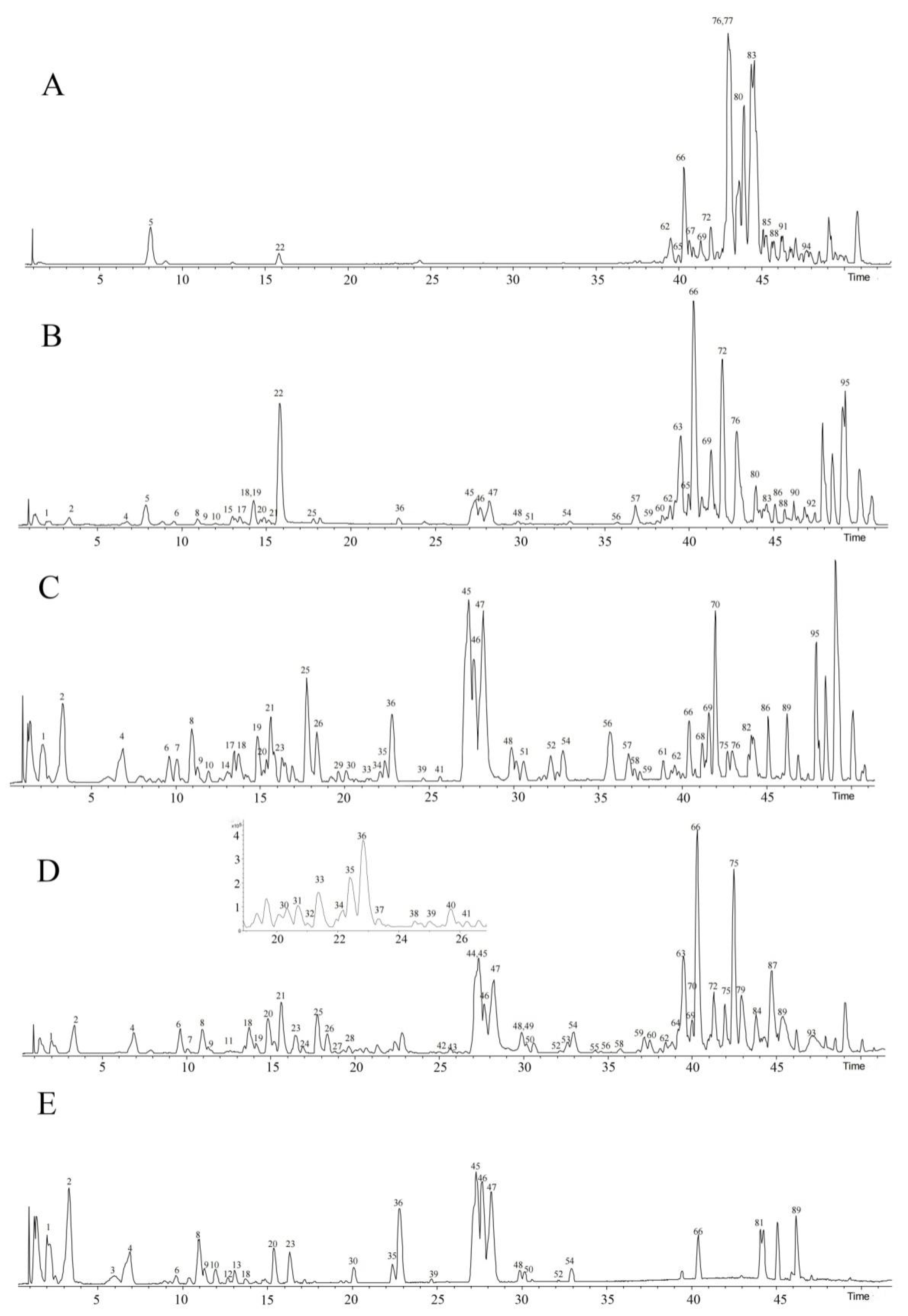
2.1. Identification of the Chemical Composition from Different Fractions
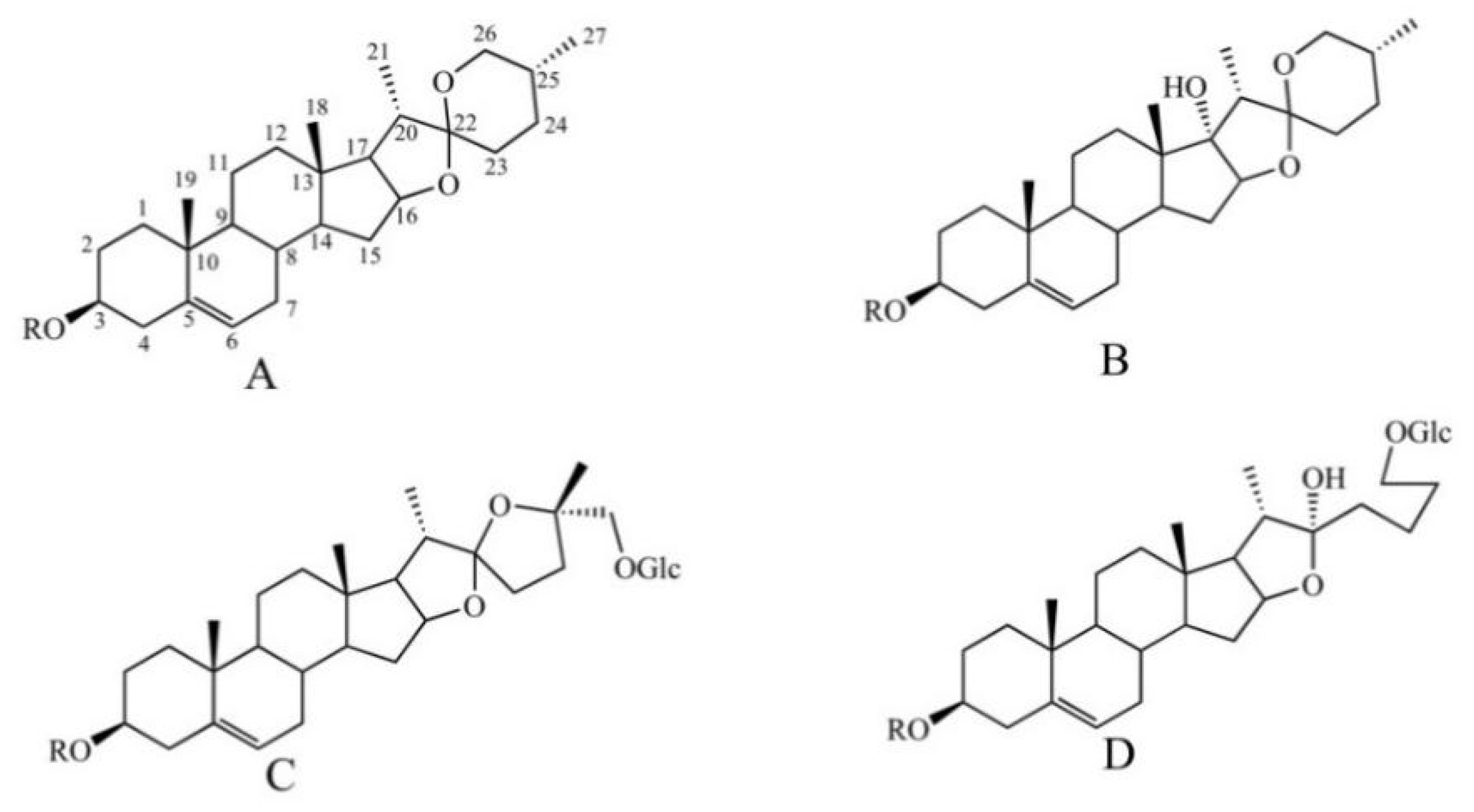
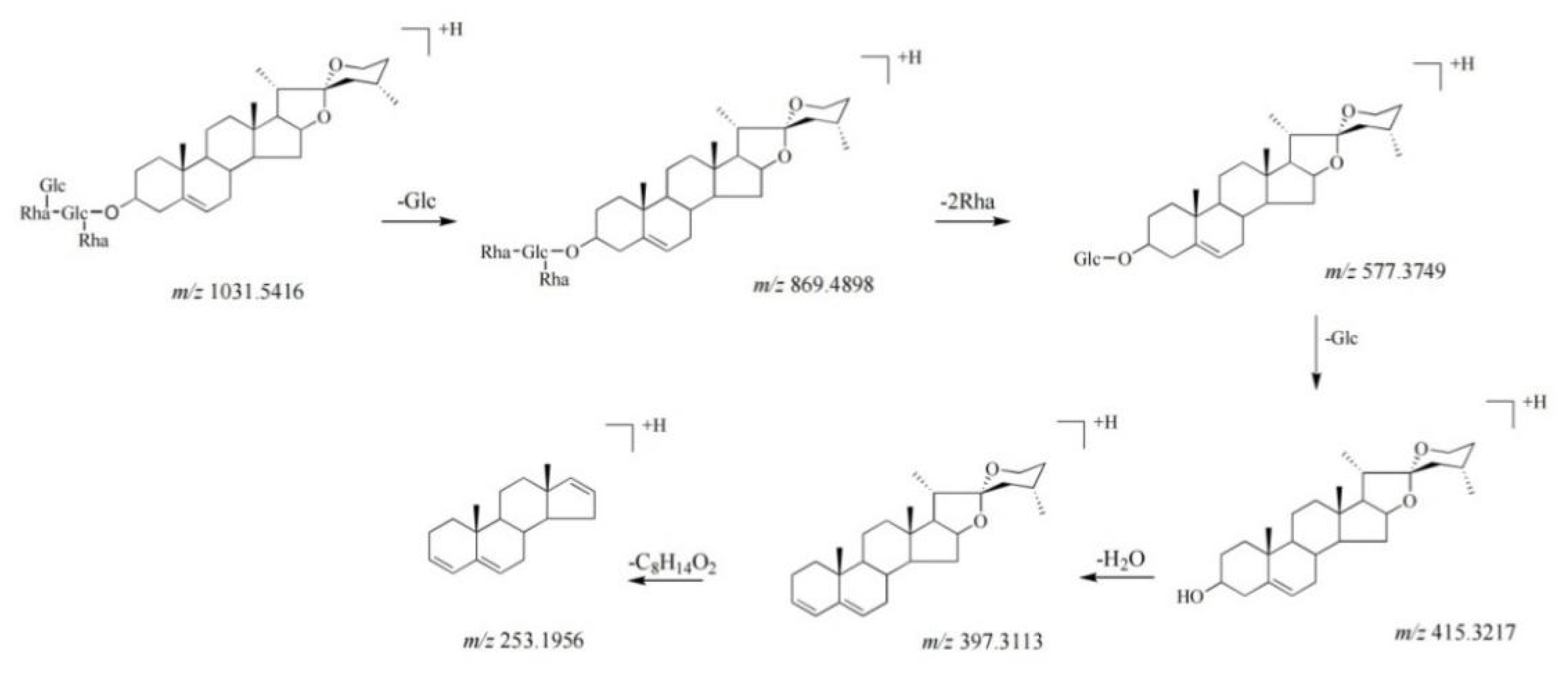
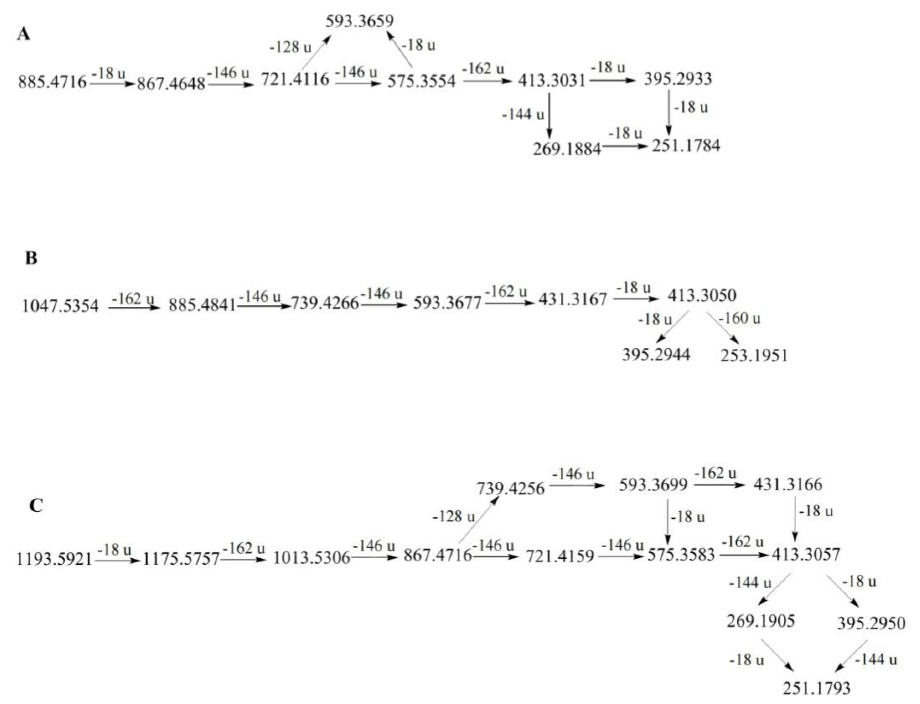
2.1.1. Identification of Steroidal Saponins
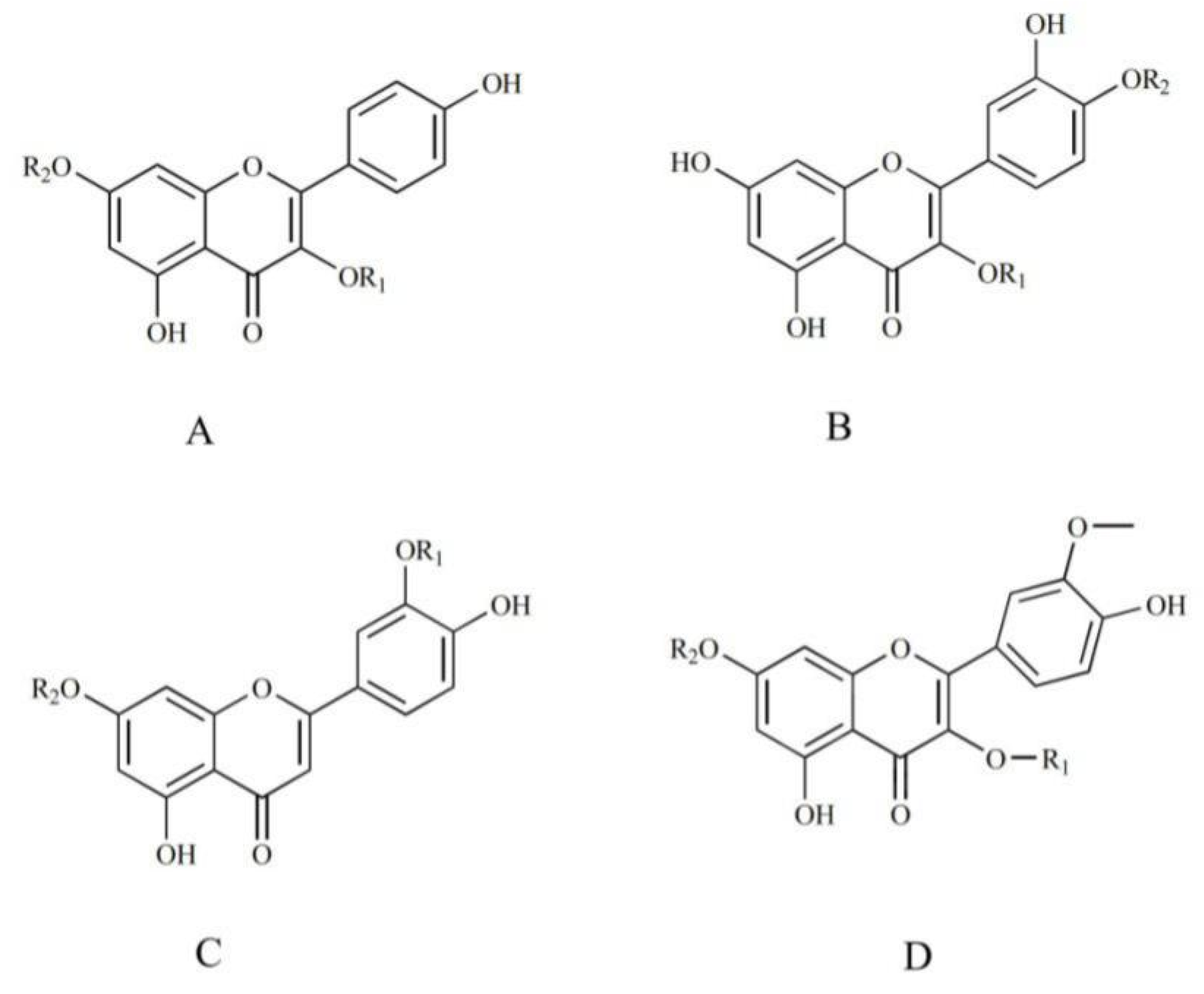
2.1.2. Identification of Flavonoids
2.1.3. Identification of Ceramides
2.1.4. Identification of Organic Acids
2.2. Antitumor Activity of Different Fractions In Vitro
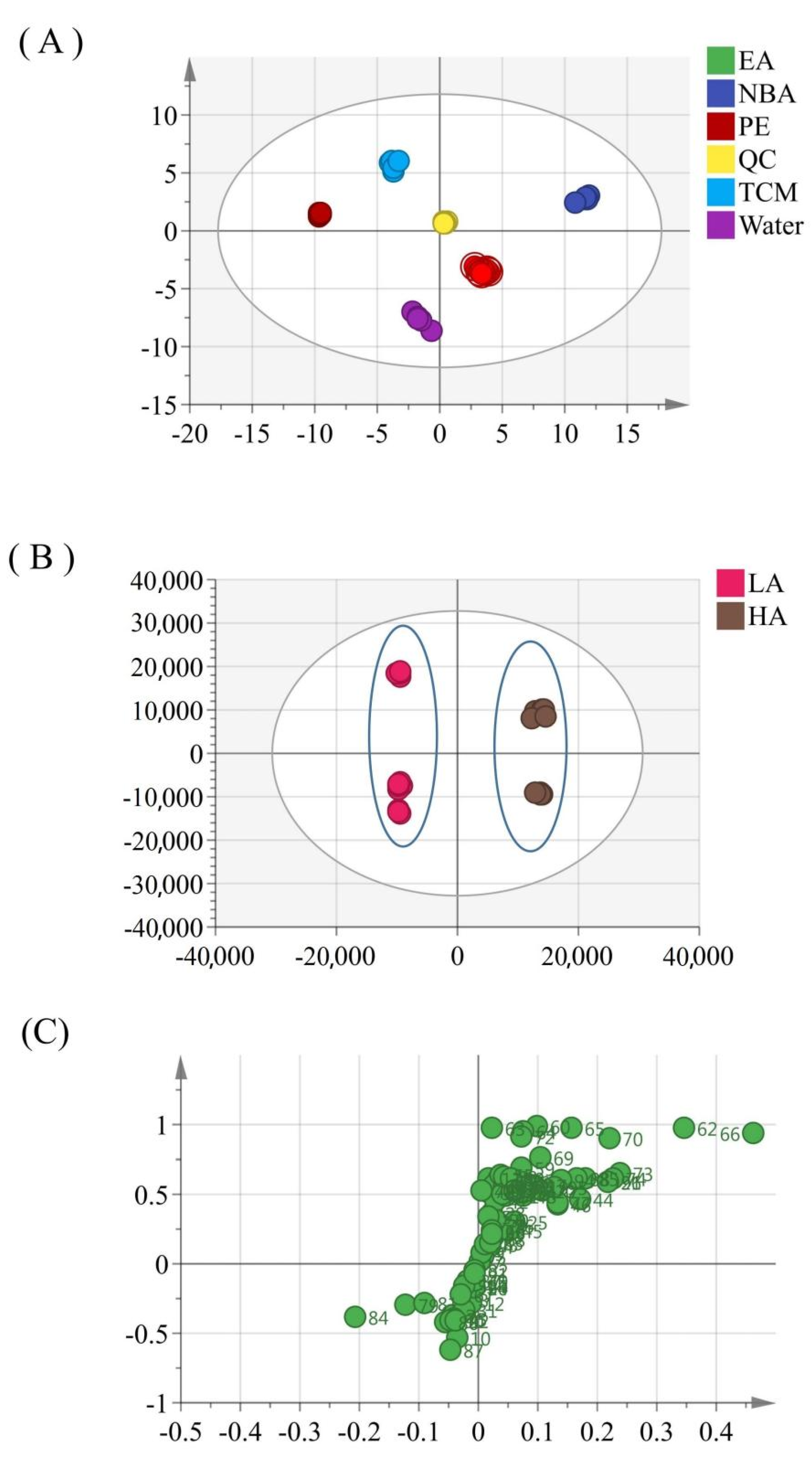
2.3. Multivariate Statistical Analysis
3. Materials and Methods
3.1. Plant Materials and Chemicals
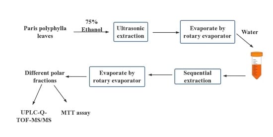
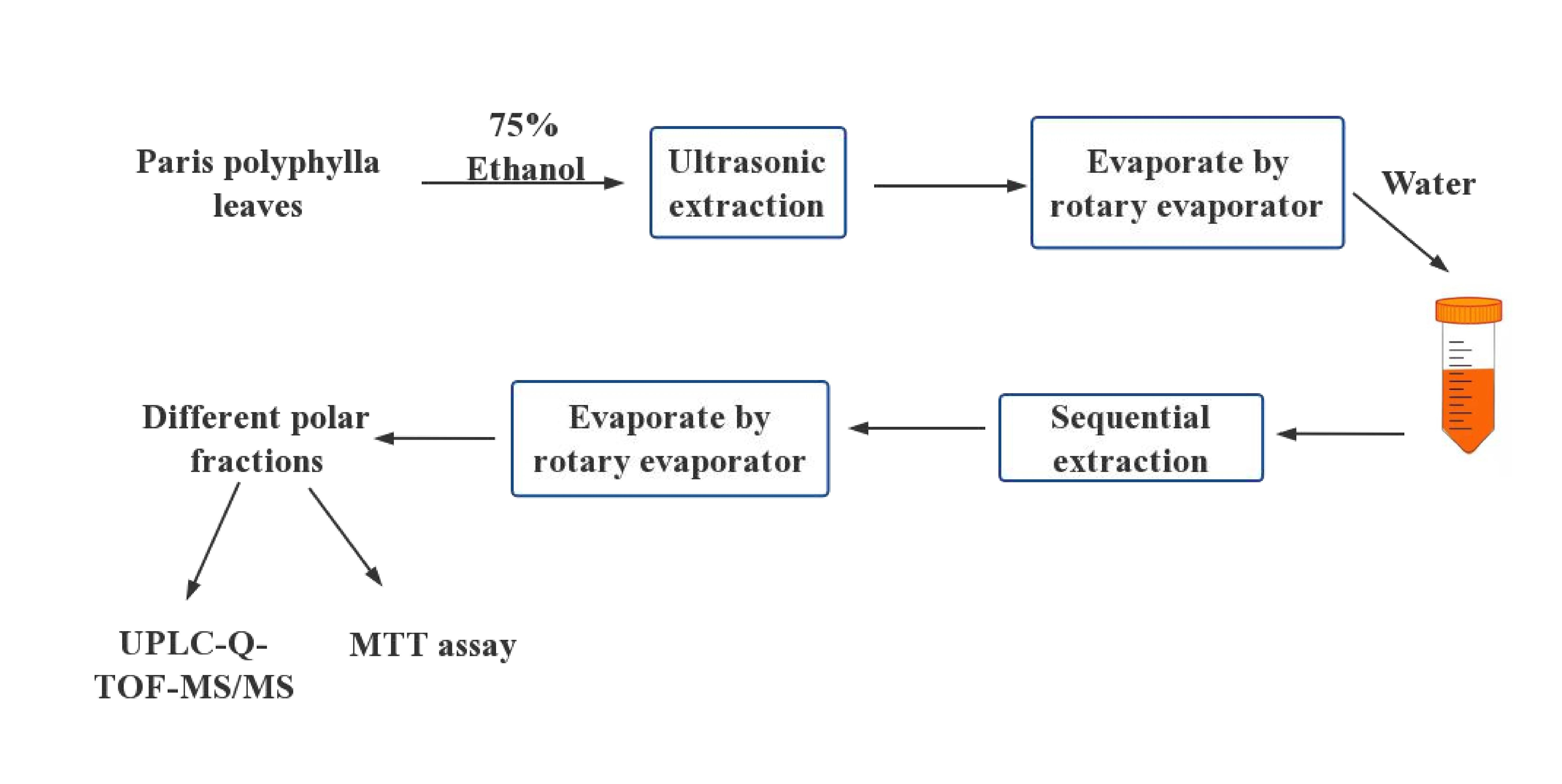
3.2. Preparation of Samples
3.3. Preparation of Standard Solution
3.4. Operating Conditions of UPLC and MS Analysis
3.5. Cytotoxic Assay
3.6. Data Analysis
4. Conclusions
Author Contributions
Funding
Informed Consent Statement
Data Availability Statement
Acknowledgments
Conflicts of Interest
Sample Availability
References
- Liu, F.; Meng, Y.; He, K.; Song, F.; Cheng, J.; Wang, H.; Huang, Z.; Luo, Z.; Yan, X. Comparative analysis of proteomic and metabolomic profiles of different species of Paris. J. Proteom. 2019, 200, 11–27. [Google Scholar] [CrossRef] [PubMed]
- Ding, Y.G.; Zhao, Y.L.; Zhang, J.; Zuo, Z.T.; Zhang, Q.Z.; Wang, Y.Z. The traditional uses, phytochemistry, and pharmacological properties of Paris L. (Liliaceae): A review. J. Ethnopharmacol. 2021, 278, 114293. [Google Scholar] [CrossRef] [PubMed]
- Cunningham, A.; Brinckmann, J.; Bi, Y.F.; Pei, S.J.; Schippmann, U.; Luo, P. Paris in the spring: A review of the trade, conservation and opportunities in the shift from wild harvest to cultivation of Paris polyphylla (Trilliaceae). J. Ethnopharmacol. 2018, 222, 208–216. [Google Scholar] [CrossRef] [PubMed]
- Jing, S.; Wang, Y.; Li, X.; Man, S.; Gao, W. Chemical constituents and antitumor activity from Paris polyphylla Smith var. yunnanensis. Nat. Prod. Res. 2016, 31, 660–666. [Google Scholar] [CrossRef] [PubMed]
- Shian, S.; Zhou, X.; Shiling, F.; Handong, W.; Jing, L.; Lijun, Z.; Yuan, M.; Huang, Y.; Ding, C. Structural elucidation and antiaging activity of polysaccharide from Paris polyphylla leaves. Int. J. Biol. Macromol. 2018, 107, 1613–1619. [Google Scholar] [CrossRef]
- Qin, X.J.; Sun, D.J.; Ni, W.; Chen, C.X.; Hua, Y.; He, L.; Liu, H.-Y. Steroidal saponins with antimicrobial activity from stems and leaves of Paris polyphylla var. yunnanensis. Steroids 2012, 77, 1242–1248. [Google Scholar] [CrossRef]
- Wei, J.C.; Gao, W.Y.; Yan, X.D.; Wang, Y.; Jing, S.S.; Xiao, P.G. ChemInform Abstract: Chemical Constituents of Plants from the Genus Paris. ChemInform 2014, 45, 1277–1297. [Google Scholar] [CrossRef]
- Wang, G.X.; Han, J.; Zhao, L.W.; Jiang, D.X.; Liu, Y.T.; Liu, X.L. Anthelmintic activity of steroidal saponins from Paris polyphylla. Phytomedicine 2010, 17, 1102–1105. [Google Scholar] [CrossRef]
- Yue, J.; Li, Z.; Zuo, Z.; Zhao, Y.; Zhang, J.; Wang, Y. Study on the identification and evaluation of growth years for Paris polyphylla var. yunnanensis using deep learning combined with 2DCOS. Spectrochim. Acta Part A Mol. Biomol. Spectrosc. 2021, 261, 120033. [Google Scholar] [CrossRef]
- Pei, Y.F.; Wu, L.H.; Zhang, Q.Z.; Wang, Y.Z. Geographical traceability of cultivated Paris polyphylla var. yunnanensis using ATR-FTMIR spectroscopy with three mathematical algorithms. Anal. Methods 2018, 11, 113–122. [Google Scholar] [CrossRef]
- Zhao, B.; Wang, Z.; Han, J.; Wei, G.; Yi, B.; Li, Z. Rhizoma Paridis total saponins alleviate H2O2-induced oxidative stress injury by upregulating the Nrf2 pathway. Mol. Med. Rep. 2019, 21, 220–228. [Google Scholar] [CrossRef] [PubMed] [Green Version]
- Liu, F.; Li, L.; Tian, X.; Zhang, D.; Sun, W.; Jiang, S. Chemical Constituents and Pharmacological Activities of Steroid Saponins Isolated from Rhizoma Paridis. J. Chem. 2021, 2021, 1442906. [Google Scholar] [CrossRef]
- Yan, T.; Hu, G.; Wang, A.; Sun, X.; Yu, X.; Jia, J. Paris saponin VII induces cell cycle arrest and apoptosis by regulating Akt/MAPK pathway and inhibition of P-glycoprotein in K562/ADR cells. Phytother. Res. 2018, 32, 898–907. [Google Scholar] [CrossRef] [PubMed]
- Pei, Y.-F.; Zuo, Z.-T.; Zhang, Q.-Z.; Wang, Y.-Z. Multi-source information fusion strategies of aerial parts in FTIR-ATR spectroscopic characterization and classification of Paris polyphylla var. yunnanensis. J. Mol. Struct. 2019, 1196, 478–490. [Google Scholar] [CrossRef]
- Qin, X.J.; Ni, W.; Chen, C.X.; Liu, H.Y. Seeing the light: Shifting from wild rhizomes to extraction of active ingredients from above-ground parts of Paris polyphylla var. yunnanensis. J. Ethnopharmacol. 2018, 224, 134–139. [Google Scholar] [CrossRef]
- Hu, R.; Yu, W.; Zhuo, Y.; Yang, Y.; Hu, X. Paris polyphylla extract inhibits proliferation and promotes apoptosis in A549 lung cancer cells. Trop. J. Pharm. Res. 2017, 16, 2121. [Google Scholar] [CrossRef] [Green Version]
- Pei, Y.F.; Zhang, Q.Z.; Zuo, Z.T.; Wang, Y.Z. Comparison and Identification for Rhizomes and Leaves of Paris yunnanensis Based on Fourier Transform Mid-Infrared Spectroscopy Combined with Chemometrics. Molecules 2018, 23, 3343. [Google Scholar] [CrossRef] [Green Version]
- Qin, X.J.; Zhang, L.J.; Zhang, Y.; Ni, W.; Yang, X.Z.; Yu, Q.; Yan, H.; An, L.K.; Liu, H.Y. Polyphyllosides A–F, six new spirostanol saponins from the stems and leaves of Paris polyphylla var. chinensis. Bioorganic Chem. 2020, 99, 103788. [Google Scholar] [CrossRef]
- Wakeel, A.; Jan, S.A.; Ullah, I.; Shinwari, Z.K.; Xu, M. Solvent polarity mediates phytochemical yield and antioxidant capacity of Isatis tinctoria. PeerJ 2019, 7, e7857. [Google Scholar] [CrossRef] [Green Version]
- Zhang, T.; Liu, H.; Liu, X.-T.; Xu, D.-R.; Chen, X.-Q.; Wang, Q. Qualitative and quantitative analysis of steroidal saponins in crude extracts from Paris polyphylla var. yunnanensis and P. polyphylla var. chinensis by high performance liquid chromatography coupled with mass spectrometry. J. Pharm. Biomed. Anal. 2010, 51, 114–124. [Google Scholar]
- Liang, M.Y.; Wang, Y.Z.; Qiao, X.; Lu, Y.W.; Chen, M.H.; Li, P.; Wen, X.D.; Yang, J. Structural characterisation and discrimination of the aerial parts of Paris polyphylla var. yunnanensis and Paris polyphylla var. chinensis by UHPLC-QTOF-MS coupled with multivariate data analysis. Phytochem. Anal. 2019, 30, 437–446. [Google Scholar] [CrossRef] [PubMed]
- Huang, Y.Y.; Liu, D.H.; Peng, H.S.; Hao, Q.X.; Chen, M.L.; Zhang, A.Z.; Chen, M.; Kang, L.P.; Huang, L.Q. Determination of eight steroidal saponins in 15 kinds of genus Paris. China J. Chin. Mater. Med. 2017, 42, 3443–3451. [Google Scholar]
- Kang, L.P.; Huang, Y.Y.; Zhan, Z.L.; Liu, D.H.; Peng, H.S.; Nan, T.G.; Zhang, Y.; Hao, Q.X.; Tang, J.F.; Zhu, S.D.; et al. Structural characterization and discrimination of the Paris polyphylla var. yunnanensis and Paris vietnamensis based on metabolite profiling analysis. J. Pharm. Biomed. Anal. 2017, 142, 252–261. [Google Scholar] [CrossRef] [PubMed]
- Wang, G.; Hao, R.; Luo, C.; Wang, Y.; Man, S.; Gao, W. Pharmacokinetics profiles of polyphyllin II and polyphyllin VII in rats by liquid chromatography with tandem mass spectrometry. Biomed. Chromatogr. 2021, 35, e5083. [Google Scholar] [CrossRef] [PubMed]
- Geng, P.; Chen, P.; Lin, L.Z.; Sun, J.; Harrington, P.; Harnly, J.M. Classification of structural characteristics facilitate identifying steroidal saponins in Alliums using ultra-high performance liquid chromatography high-resolution mass spectrometry. J. Food Compos. Anal. 2021, 102, 103994. [Google Scholar] [CrossRef]
- Kang, L.P.; Yu, K.; Zhao, Y.; Liu, Y.-X.; Yu, H.-S.; Pang, X.; Xiong, C.Q.; Tan, D.W.; Gao, Y.; Liu, C.; et al. Characterization of steroidal glycosides from the extract of Paris polyphylla var. yunnanensis by UPLC/Q-TOF MSE. J. Pharm. Biomed. Anal. 2012, 62, 235–249. [Google Scholar] [CrossRef]
- Liu, Z.; Wang, J.; Gao, W.; Man, S.; Guo, H.; Zhang, J.; Liu, C. Formulation and in vitro absorption analysis of Rhizoma paridis steroidal saponins. Int. J. Pharm. 2012, 441, 680–686. [Google Scholar] [CrossRef]
- Li, Y.M.; Guan, L.J.; Chen, L.M.; Zhao, M.; Ding, L.S.; Meng, C.X.N.; Gao, H.M.; Wang, Z.M. Qualitative and quantitative analysis of Paris polyphylla var. chinensis by UPLC-Q-TOF-MS/MS and HPLC. Zhongguo Zhong Yao Za Zhi China J. Chin. Mater. Med. 2021, 46, 2900–2911. [Google Scholar]
- Lyu, C.G.; Kang, C.Z.; Kang, L.P.; Yang, J.; Wang, S.; He, Y.L.; Deng, A.P.; Wang, H.Y.; Huang, L.Q.; Guo, L.P. Structural characterization and discrimination of Ophiopogon japonicas (Liliaceae) from different geographical origins based on metabolite profiling analysis. J. Pharm. Biomed. Anal. 2020, 185, 113212. [Google Scholar] [CrossRef]
- Guan, L.; Ju, B.; Zhao, M.; Zhu, H.; Chen, L.; Wang, R.; Gao, H.; Wang, Z. Influence of drying process on furostanoside and spirostanoside profiles of Paridis Rhizoma by combination of HPLC, UPLC and UPLC-QTOF-MS/MS analyses. J. Pharm. Biomed. Anal. 2021, 197, 113932. [Google Scholar] [CrossRef]
- Ling, Y.; He, X.; Jiang, R.; Zhang, Q.; Yuan, S.; Liang, Y.; Li, C.; Zhao, Y.; Zhang, Q.; Liu, K. Rapid Detection and Characterization of Steroidal Saponins in the Root of Asparagus cochinchinensis by High-Performance Liquid Chromatography Coupled to Electrospray Ionization and Quad-rupole Time-of-Flight Mass Spectrometry. J. Chromatogr. Sci. 2020, 58, 454–463. [Google Scholar] [CrossRef] [PubMed]
- Mohd, T.; Belwal, T.; Bhatt, I.D.; Pande, V.; Nandi, S.K. Polyphenolics in leaves of Paris polyphylla: An important high value Him-alayan medicinal herb. Ind. Crops Prod. 2018, 117, 66–74. [Google Scholar] [CrossRef]
- Yang, Y.; Zhao, Y.; Zuo, Z.; Wang, Y. Determination of Total Flavonoids for Paris Polyphylla var. Yunnanensis in Different Geo-graphical Origins Using UV and FT-IR Spectroscopy. J. AOAC Int. 2019, 102, 457–464. [Google Scholar] [CrossRef]
- Beck, S.; Stengel, J. Mass spectrometric imaging of flavonoid glycosides and biflavonoids in Ginkgo biloba L. Phytochemistry 2016, 130, 201–206. [Google Scholar] [CrossRef] [PubMed]
- Xu, X.; Xie, H.; Hao, J.; Jiang, Y.; Wei, X. Flavonoid Glycosides from the Seeds of Litchi chinensis. J. Agric. Food Chem. 2011, 59, 1205–1209. [Google Scholar] [CrossRef] [PubMed]
- Bartke, N.; Fischbeck, A.; Humpf, H.U. Analysis of sphingolipids in potatoes (Solanum tuberosum L.) and sweet potatoes (Ipomoea batatas (L.) Lam.) by reversed phase high-performance liquid chromatography electrospray ionization tandem mass spec-trometry (HPLC-ESI-MS/MS). Mol. Nutr. Food Res. 2006, 50, 1201–1211. [Google Scholar] [CrossRef] [PubMed]
- Cajka, T.; Fiehn, O. Comprehensive analysis of lipids in biological systems by liquid chromatography-mass spectrometry. Trends Anal. Chem. 2014, 61, 192–206. [Google Scholar] [CrossRef] [Green Version]
- Touge, T.; Kuwana, M.; Komatsuki, Y.; Tanaka, S.; Nara, H.; Matsumura, K.; Sayo, N.; Kashibuchi, Y.; Saito, T. Development of Asymmetric Transfer Hydro-genation with a Bifunctional Oxo-Tethered Ruthenium Catalyst in Flow for the Synthesis of a Ceramide (d-erythro-CER[NDS]). Org. Process Res. Dev. 2018, 23, 452–461. [Google Scholar] [CrossRef]
- Lu, H.; Li, Y.; Zhang, H.; Chingin, K.; Wei, Y.; Huang, K.; Feng, S. Direct quantitative profiling of amino acids in tissues for the assessment of lung cancer. Talanta 2021, 233, 122544. [Google Scholar] [CrossRef]
- Lu, H.; Li, Y.; Zhang, H.; Chingin, K.; Wei, Y.; Huang, K.; Feng, S. Determination of Amino Acids Content in Paris polyphylla Smith var. yunnanensis (Franch.) Hand.-Mazz. and Its Nutritional Evaluation. Southwest China J. Agric. Sci. 2017, 30, 1760–1766. [Google Scholar]
- He, H.; Zheng, L.; Sun, Y.P.; Zhang, G.W.; Yue, Z.G. Steroidal saponins from Paris polyphylla suppress adhesion, migration and invasion of human lung cancer A549 cells via down-regulating MMP-2 and MMP-9. Asian Pac. J. Cancer Prev. 2015, 15, 10911–10916. [Google Scholar] [CrossRef] [PubMed]
- Hu, W.Y.; Li, J.; He, Z.Y.; Fu, K.; Zhang, R.P. The in vivo and in vitro inhibitory effect of pariphyllin I on human breast cancer cell MCF-7. Zhongchengyao 2015, 37, 1582–1585. [Google Scholar]
- Lim, W.C.; Kim, H.; Kim, Y.J.; Choi, K.C.; Lee, I.H.; Lee, K.H.; Kim, M.K.; Ko, H. Dioscin suppresses TGF-b1-induced epithelial-mesenchymal transition and suppresses A549 lung cancer migration and invasion. Bioorg. Med. Chem. Lett. 2017, 27, 3342–3348. [Google Scholar] [CrossRef] [PubMed]
- Qin, X.J.; Yu, M.Y.; Ni, W.; Yan, H.; Chen, C.X.; Cheng, Y.C.; He, L.; Liu, H.Y. Steroidal saponins from stems and leaves of Paris polyphylla var. yunnanensis. Phytochemistry 2016, 121, 20–29. [Google Scholar] [CrossRef]
- Miao, C.; Chen, J.; Li, X.; Lin, P.; Hu, Q. Effect of luteolin on TGF-beta1-induced epithelial-mesenchymal transition in lung cancer A549 cells. Chin. J. Pathophysiol. 2019, 35, 1163–1168. [Google Scholar]
- Stith, J.L.; Velazquez, F.N.; Obeid, L.M. Advances in determining signaling mechanisms of ceramide and role in disease. J. Lipid Res. 2019, 60, 913–918. [Google Scholar] [CrossRef] [Green Version]
- Long, Y.; Xu, X.; Luo, C. The relationship of human colon carcinorma HT-29 cells apoptosis induced by C-2-ceramide and the changes of mitochondrial membrane potential. Jiangsu Med. J. 2007, 33, 149–152. [Google Scholar]
- Bi, F.C.; Liu, Z.; Wu, J.X.; Liang, H.; Xi, X.L.; Fang, C.; Sun, T.J.; Yin, J.; Dai, G.Y.; Rong, C.; et al. Loss of Ceramide Kinase in Arabidopsis Impairs Defenses and Promotes Ceramide Accumulation and Mitochondrial H2O2 Bursts. Plant Cell. 2014, 26, 3449–3467. [Google Scholar] [CrossRef] [Green Version]
- Hannun, Y.A.; Obeid, L.M. Sphingolipids and their metabolism in physiology and disease. Nat. Rev. Mol. Cell Biol. 2017, 19, 175–191. [Google Scholar] [CrossRef]


| No | tR/Min | [M + H]+ m/z | Molecular Formula | Error (ppm) | ESI/Q–TOF MS/MS Fragments | Tentative Identification | Existing PART |
|---|---|---|---|---|---|---|---|
| 1 | 2.2 | 294.1547 | C12H23NO7 | 0.3 | 276.1440 (100), 258.1336 (50.4), 230.1386 (39.5), 132.1020 (15.5), 248.1488 (10.9) | N-fructosyl isoleucine | TCM, EA, NBA, Water |
| 2 | 3.4 | 328.1392 | C15H21NO7 | −0.3 | 310.1283 (100), 292.1178 (57.3), 264.1228 (33.2), 166.0861 (28.3) | N-fructosyl phenylalanine | TCM, EA, NBA, Water |
| 3 | 6.0 | 367.1501 | C17H22N2O7 | −0.5 | 229.0966 (100), 349.1386 (81.4), 188.0699 (74.8), 332.1128 (61.5), 276.1229 (38.3), 258.1122 (21.3), 350.1417 (14.9) | N.A. | Water |
| 4 | 6.8 | 205.0969 | C11H12N2O2 | 1.3 | 188.0706 (100), 146.0605 (82.0), 144.0813 (17.1), 159.0918 (11.9) | Tryptophan | TCM, EA, NBA, Water |
| 5 | 8.2 | 192.0923 | C9H9N3O2 | 0.5 | 160.0502 (100), 192.0760 (29.1) | Carbendazim | PE, TCM, EA |
| 6 | 9.6 | 217.0970 | C12H12N2O2 | 0.9 | 144.0811 (100), 145.0842 (10.0) | Lycoperodine I | TCM, EA, NBA, Water |
| 7 | 10.1 | 169.0502 | C8H8O4 | −3.7 | 125.0603 (100), 151.0389 (83.5), 269.0499 (30.0) | 2,4,6-trihydroxyacetophenone | EA, NBA |
| 8 | 11.0 | 611.1634 | C27H30O16 | −4.5 | 287.0550 (100), 449.1079 (26.0), 288.0583 (14.9) | Luteolin-3’,7-di-O-glucoside | TCM, EA, NBA, Water |
| 9 | 11.4 | 641.1738 | C28H32O17 | −4.0 | 317.0664 (100), 479.1193 (25.8), 318.0690 (13.6) | Isorhamnetin-3,7-di-O-glucoside | TCM, EA, NBA, Water |
| 10 | 12.1 | 697.1630 | C30H32O19 | −2.9 | 287.0548 (100), 449.1086 (52.9), 288.0582 (14.4), 127.0401 (11.6), 450.1119 (11.3) | 3-[[6-O-(2-Carboxyacetyl)-4-O-hexopyranosyl-β-d-glucopyranosyl]oxy]-5,7-dihydroxy-2-(4-hydroxyphenyl)-4H-1-benzopyran-4-one | TCM, EA, EA, Water |
| 11 | 12.4 | 627.1555 | C27H30O17 | 0.1 | 303.0497 (100), 304.0528 (13.2) | Quercetin-3,4’-O-di-beta-glucoside | NBA |
| 12 | 12.7 | 551.2689 | C25H42O13 | 1.6 | 209.1533 (100), 149.0959 (68.9), 227.1637 (55.2), 191.1426 (25.6) | Tricalysionoside A | Water |
| 13 | 13.1 | 252.0865 | C12H13NO5 | 0.5 | 206.0809 (100), 120.0806 (93.5), 188.0703 (48.1), 146.0597 (15.9), 207.0842 (10.0) | N.A. | Water |
| 14 | 13.1 | 165.0553 | C9H8O3 | −3.9 | 147.0446 (100), 148.0418 (9.3), 123.0919 (2.4) | O-coumaric acid | EA |
| 15 | 13.2 | 227.1649 | C13H22O3 | −2.7 | 125.0970 (100), 149.0970 (87.7), 123.0818 (63.7), 153.0918 (43.1), 209.1541 (33.7), 191.1437 (23.1) | 4-(1-Hydroxy-4-keto-2,6,6-trimethyl-2-cyclohexen-1-yl)-butan-2-ol | TCM |
| 16 | 13.5 | 611.1605 | C27H30O16 | 0.3 | 303.1493 (100), 304.0525 (13.3) | Rutin | NBA |
| 17 | 13.5 | 225.1499 | C13H20O3 | −5.3 | 161.1335 (100), 179.1440 (26.0), 121.1024 (25.1), 162.1369 (10.3) | 2-(6-methyl-7-oxooctyl)-2H-furan-5-one | TCM, EA |
| 18 | 13.8 | 611.1627 | C27H30O16 | −3.3 | 287.0559 (100), 288.0586 (14.7) | Luteolin-3’-O-Glc-(l→2)-glucoside | TCM, EA, NBA, Water |
| 19 | 14.2 | 641.1742 | C28H30O17 | −4.7 | 317.0672 (100), 318.0700 (15.2) | Isorhamnetin-3-O-glc-(l→2)-gal | TCM, EA, NBA |
| 20 | 14.9 | 595.1685 | C27H30O15 | −4.7 | 287.0558 (100), 288.0595 (14.6) | Kaempferol 7-O-neohesperidoside | TCM, EA, NBA, Water |
| 21 | 15.6 | 481.3170 | C27H44O7 | −2.2 | 445.2956 (100), 371.2228 (39.0), 427, 2857 (33.2), 165.1281 (26.7), 409.2732 (11.6), 428.2894 (10.1) | Crustecdysone | TCM, EA, NBA |
| 22 | 15.9 | 197.1167 | C11H16O3 | 0.5 | 179.1063 (100), 197.1163 (49.7), 135.1171 (34.4), 133.1013 (33.1), 161.0955 (26.0) | Loliolide | PE, TCM |
| 23 | 16.4 | 1225.5500 | C56H88O29 | −1.3 | 621.3268 (100), 553.3008 (70.0), 459.2736 (59.6), 391.2473 (47.8), 373.2366 (33.2), 622.3300 (29.2), 441.2841 (26.3), 767.3855 (24.9), 477.2841 (24.4), 605.3317 (24.3), 699.3578 (22.7), 639.3374 (20.8), 309.1176 (19.6), 783.3797 (16.0), 929.4381 (7.5) | Parisyunnanoside G | EA, NBA, Water |
| 24 | 16.5 | 481.3164 | C27H44O7 | −0.9 | 427.2852 (100), 303.1957 (69.5), 143.1070 (66.3), 125.0964 (48.2), 285.1850 (44.2), 409.2744 (42.0), 428.2883 (25.6), 301.1802 (22.1) | Crustecdysone or its isomer | NBA |
| 25 | 17.8 | 449.1093 | C21H20O11 | −3.2 | 287.0555 (100), 288.0589 (14.9) | Kaempferol 3-Glucoside | TCM, EA, NBA |
| 26 | 18.4 | 479.1189 | C22H22O12 | −1.0 | 317.0655 (100), 318.0688 (16.7) | Isorhamnetin-3-O-glucoside | EA, NBA |
| 27 | 18.9 | 1063.5301 | C51H82O23 | 1.8 | 293.1236 (100), 427.2850 (78.0), 147.0653 (42.6), 445.2956 (40.6), 239.0920 (36.6), 257.1024 (32.3), 409.2743 (29.6), 461.2898 (22.7), 309.1185 (21.8), 589.3380 (21.3), 607.3489 (16.8), 735.3948 (14.3) | N.A. | NBA |
| 28 | 19.7 | 901.4769 | C45H72O18 | 2.5 | 269.1894 (100), 287.1998 (69.7), 147.0647 (67.1), 441.2631 (57.4), 427.2829 (55.7), 595.3133 (46.6), 739.4247 (45.9), 901.4781 (39.0), 721.4162 (33.9), 409.2742 (30.9) | 26-O-glc-furost-5-ene-3β,22α,26-triol-3-O-rha-(1→2)-glc or its isomer | NBA |
| 29 | 19.7 | 1047.5328 | C51H82O22 | 3.2 | 413.3053 (100), 147.0647 (70.1), 395.2945 (65.8), 129.0549 (51.8), 1047.5367 (36.1), 269.1890 (31.8), 431.3149 (30.7), 281.2269 (29.9), 377.2836 (23.4), 885.4798 (18.0), 849.4619 (13.7), 1063.5284 (4.2) | 26-O-Glc-nuatigenin-5-ene-3β,17-diol-3-O-rha (1→2) -[rha (1→4)]-glc | EA, NBA |
| 30 | 20.1 | 1193.5995 | C57H94O27 | −3.8 | 413.3053 (100), 1193.5980 (52.8), 431.3162 (50.4), 293.1230 (49.1), 395.2947 (37.8), 147.0658 (29.4), 414.3086 (22.5), 593.3688 (16.9), 739.4295 (5.9) | Th or its isomer | EA, NBA, Water |
| 31 | 20.7 | 901.4815 | C45H72O18 | −2.6 | 269.1906 (100), 287.2003 (72.0), 147.0661 (27.9), 413.3043 (26.6), 595.3112 (22.1), 901.4785 (21.6), 739.4256 (21.1), 431.3165 (12.0) | (25S)-spirost-5-ene-3β,25-diol-3-O-glc (1→3)-[rha (1→2)]-glc | EA, NBA |
| 32 | 21.0 | 1063.5289 | C51H82O23 | 2.9 | 445.2944 (100), 293.1227 (90.2), 147.0649 (53.6), 239.0912 (41.3), 607.3480 (39.9), 427.2839 (38.8), 753.4068 (34.1), 257.1014 (32.9), 129.0552 (29.5), 275.1111 (25.2), 446.2976 (23.6), 271.2045 (21.6) | 26-O-β-d-Glc-3β,12,22,26-tetrahydroxyfurost-5-ene-22,25-epoxy-3-O-Rha-(1→2)-[Rha-(1→4)-Rha-(1→4)]-β-D-Glc | NBA |
| 33 | 21.4 | 755.4245 | C39H62O14 | −4.3 | 269.1908 (100), 449.2554 (65.4), 593.3715 (38.3), 287.2021 (37.5), 755.4265 (30.0), 270.1949 (18.5), 413.3075 (16.2), 251.1811 (15.4), 594.3748 (15.3), 450.2583 (13.3) | (25S)-spirost-5-ene-3-O-glc-(1→2)-glc | EA, NBA |
| 34 | 22.0 | 933.4671 | C45H72O20 | 2.0 | 445.2948 (100), 147.0652 (91.1), 427.2846 (65.9), 129.0549 (41.5), 309.1183 (36.3), 271.2054 (30.3), 253.1942 (20.3), 163.0605 (12.4), 607.3457 (7.0) | (23S,24S)-spirost-5-ene-1β,3β,23,24-tetrol-1-O-rha(1→2)-glc 24-O-gal | EA, NBA |
| 35 | 22.4 | 1047.5393 | C51H82O22 | −2.2 | 413.3048 (100), 867.4745 (75.1), 147.0655 (48.6), 395.2945 (35.5), 129.0552 (29.0), 868.4683 (28.5), 431.3157 (26.7), 293.1235 (26.3), 414.3080 (24.5), 181.1224 (20.7), 309.1170 (12.8), 239.0913 (10.5) | 27-O-glc-(25R)-spirost-5-ene-3β,27-diol-3-O-rha(1→4)-[rha(1→2)]-glc | EA, NBA, Water |
| 36 | 22.9 | 1193.6061 | C57H94O27 | −5.0 | 413.3065 (100), 293.1242 (88.7), 431.3173 (42.0), 147.0660 (36.5), 257.1027 (25.0), 593.3709 (16.4), 721.4181 (6.2), 1013.5360 (6.3) | Th | TCM, EA, NBA, Water |
| 37 | 23.3 | 917.4738 | C45H72O19 | 0.3 | 269.1890 (100), 737.4085 (71.6), 287.2001 (64.7), 147.0645 (48.2), 755.4207 (48.0), 411.2890 (47.8), 429.2990 (46.3), 595.3108 (21.6), 756.4239 (21.5) | 26-O-β-d-Glc-3β,22,26-trihydroxyfurost-5-ene-3-O-Rha-glc | NBA |
| 38 | 24.5 | 917.4745 | C45H72O19 | −0.5 | 269.1903 (100), 755.4188 (82.8), 287.1999 (70.4), 429.2998 (70.4), 447.3101 (47.0), 411.2904 (38.6), 756.4218 (29.1), 609.3613 (25.5), 595.3117 (18.4), 251.1799 (16.2) | (25R)-spirost-5-ene-3β,17α,27-triol-3-O-glc(1→3)-[rha(1→2)]-glc or its isomer | NBA |
| 39 | 24.7 | 1209.5925 | C57H92O27 | −2.2 | 591.3527 (100), 429.3001 (85.0), 293.1231 (67.0), 737.4091 (54.8), 197.1168 (37.0), 592.3553 (30.1), 147.0651 (26.3), 573.3424 (26.1), 411.2886 (25.0), 447.3101 (21.6), 430.3039 (20.5), 899.4631 (18.4) | Parisverticoside C | EA, NBA, Water |
| 40 | 25.0 | 593.3695 | C33H52O9 | −1.8 | 287.2008 (100), 431.3165 (52.1), 593.3685 (21.8), 269.1901 (20.3), 288.2037 (17.6), 432.3180 (12.7), 167.1070 (10.3) | Chonglouoside SL-1 | NBA |
| 41 | 25.7 | 1047.5370 | C51H82O22 | 0.1 | 431.3143 (100), 885.4838 (70.6), 148.0652 (59.8), 413.3040 (56.2), 129.0547 (36.0), 271.2046 (32.6), 593.3659 (31.1), 253.1947 (25.0), 739.4249 (17.9), 395.2919 (13.1), 447.3102 (11.7) | 26-O-Glc-nuatigenin-3-O-rha (1→2) -[rha (1→4)]-glc | EA, NBA |
| 42 | 26.0 | 901.4779 | C45H72O18 | −3.1 | 129.0547 (100), 147.0652 (97.7), 411.2902 (69.2), 393.2795 (62.5), 281.2270 (42.2), 269.1905 (41.2), 429.3003 (37.2), 251.1793 (36.5), 901.4808 (34.5), 557.3534 (9.8), 739.4267 (9.2) | (25R)-spirost-5-ene-3β,17β,27-triol-3-O-rha(1→4)-[rha(1→2)]-glc or its isomer | NBA |
| 43 | 26.2 | 1047.5347 | C51H82O22 | 2.2 | 447.3119 (100), 147.0654 (69.0), 293.1229 (62.8), 429.3003 (55.2), 411.2911 (55.2), 129.0554 (49.9), 239.0918 (39.0), 593.3685 (37.8), 755.4194 (37.0), 609.3632 (27.8), 275.1123 (24.2), 257.1023 (24.1), 393.2802 (20.5), 885.4821 (7.0) | N.A. | NBA |
| 44 | 27.4 | 739.4271 | C39H62O13 | −1.1 | 577.3748 (100), 433.2591 (73.9), 253.1952 (69.5), 271.2058 (38.7), 739.4270 (31.5), 578.3780 (29.1), 167.1065 (20.5), 397.3122 (9.3) | Polyphyllin VI | NBA, Water |
| 45 | 27.4 | 1031.5461 | C51H82O21 | −3.8 | 415.3228 (100), 869.4946 (99.0), 1031.5479 (92.1), 147.0663 (83.6), 271.2068 (70.0), 253.1964 (61.4), 129.0557 (52.8), 167.1076 (43.5), 870.4980 (41.8), 725.3785 (26.5), 577.3760 (11.2) | Pariphyllin A | TCM, EA, NBA, Water |
| 46 | 27.7 | 1177.6053 | C57H92O25 | −4.5 | 415.3224 (100), 1177.6060 (81.2), 293.1240 (60.1), 577.3763 (40.2), 147.0661 (30.0), 271.2066 (23.8), 416.3260 (23.3), 397.3114 (19.9), 723.4351 (14.3), 869.4926 (5.0) | Pseudoproto-Pb | TCM, EA, NBA, Water |
| 47 | 28.2 | 1047.5436 | C51H82O22 | −4.3 | 415.3230 (100), 147.0665 (81.4), 271.2070 (77.9), 1047.5440 (76.7), 885.4900 (60.1), 253.1964 (55.4), 397.3124 (40.9), 129.0559 (39.9), 577.3772 (17.4), 741.3739 (11.3) | Pseudoproto-gracillin | TCM, EA, NBA, Water |
| 48 | 29.9 | 1047.5408 | C51H82O22 | −3.6 | 431.3168 (100), 885.4877 (60.4), 147.0662 (48.8), 413.3067 (48.1), 593.3703 (35.9), 129.0557 (33.2), 271.2065 (30.4), 739.4277 (25.3), 432.3203 (23.9), 309.1182 (12.7) | 26-O-Glc-nuatigenin-3-O-rha (1→2) -[rha (1→4)]-glc or its isomer | TCM, EA, NBA, Water |
| 49 | 29.9 | 901.4798 | C45H72O18 | −0.8 | 739.4271 (100), 593.3709 (74.4)147.0654 (57.9), 253.1951 (51.3), 271.2062 (43.9), 129.00546 (43.3), 431.3155 (40.8), 413.3056 (37.6), 579.3176 (24.2), 395.2963 (19.7), 575.3583 (16.1) | 27-O-glc-(25R)-spirost-5-ene-3β,27-diol-3-O-rha(1→4)-glc | NBA |
| 50 | 30.2 | 1193.6005 | C57H94O27 | −4.7 | 431.3172 (100), 593.3710 (53.5), 293.1240 (44.3), 413.3068 (33.0), 432.3208 (22.5), 739.4322 (22.5), 129.0556 (13.6), 885.4888 (10.0) | Chonglouoside SL-14 | TCM, EA, NBA, Water |
| 51 | 30.7 | 1063.5366 | C51H84O24 | −4.3 | 431.3172 (100), 413.3061 (51.5), 593.3703 (43.6), 147.0661 (40.0), 432.3206 (24.0), 309.1185 (23.6), 575.3598 (23.1), 129.0557 (19.8), 755.4256 (13.6), 414.3098 (13.2), 271.2067 (11.0), 901.4828 (6.3) | 26-O-glc-furost-5-ene-3β,12α,22α,26-trihydroxy-3-O-glc-(1→3)-[rha-(1→2)]-glc | TCM, EA, NBA, Water |
| 52 | 32.2 | 1209.5821 | C57H92O27 | 4.7 | 431.3149 (100), 593.3679 (32.3), 413.3048 (20.0), 293.1221 (16.7), 739.4238 (15.4), 309.1170 (9.4), 885.4810 (4.0) | N.A. | EA, NBA, Water |
| 53 | 32.6 | 1047.5404 | C51H82O22 | −3.2 | 885.4870 (100), 431.3160 (83.7), 886.4898 (45.7), 867.4745 (43.8), 147.0658 (39.9), 413.3054 (36.9), 253.1946 (24.9), 271.2061 (24.4), 129.0550 (22.6), 868.4754 (16.2), 725.3727 (13.4), 593.3687 (13.4), 739.4339 (11.6), 239.0934 (10.0) | 27-O-glc-(25R)-spirost-5-ene-3β,27-diol-3-O-rha(1→4)-[rha(1→2)]-glc or its isomer | NBA |
| 54 | 33.0 | 1193.5962 | C57H92O26 | −1.1 | 431.3158 (100), 593.3685 (27.2), 413.3051 (24.5), 432.3192 (24.4), 293.1231 (23.7), 739.4278 (21.0), 885.4858 (14.3), 147.0655 (10.3), 886.4903 (6.3) | Chonglouoside SL-14 or its isomer | TCM, EA, NBA, Water |
| 55 | 34.3 | 885.4836 | C45H72O17 | 0.7 | 147.0651 (100), 431.3159 (83.6), 129.0548 (80.2), 413.3053 (67.6), 253.1952 (66.8), 885.4835 (50.4), 271.2062 (48.8), 293.1229 (37.0), 593.3691 (19.9), 739.4281 (13.2) | (25S)-isonuatigenin-3-O-rha-(1→4)-[rha-(1→2)]-glc | NBA |
| 56 | 35.8 | 769.2314 | C45H36O12 | 0.7 | 645.1775 (100), 375.0871 (40.1), 646.1813 (32.7), 389.1025 (30.8), 137.0605 (25.4), 521.1247 (24.7), 513.1553 (22.1), 259.0979 (16.0), 769.2319 (11.2), 377.1014 (11.2) | N.A. | TCM, EA, NBA |
| 57 | 36.9 | 657.4610 | C36H64O10 | −5.0 | 173.1182 (100), 155.1077 (95.0), 275.2016 (44.5), 293.2122 (24.2) | N.A. | TCM, EA |
| 58 | 37.2 | 1031.5364 | C51H82O21 | 5.0 | 885.4802 (100), 593.3672 (49.3), 1031.5364 (31.2), 739.4238 (27.5), 431.3147 (26.1), 293.1225 (9.3), 413.3041 (6.0) | Parisyunnanside C | EA, NBA |
| 59 | 37.5 | 901.4815 | C45H72O18 | −2.6 | 431.3168 (100), 147.0660 (89.0), 413.3064 (76.8), 129.0556 (43.6), 309.1185 (26.7), 432.3209 (24.7), 271.2062 (23.4), 414.3080 (20.9), 253.1957 (18.5), 273.0974 (13.6), 163.0608 (12.4), 145.0500 (11.8) | (25S)-spirost-5-ene-3β,25-diol-3-O-rha (1→2)-[glc (1→3)] -glc | TCM, EA, NBA |
| 60 | 38.4 | 494.3339 | C24H47NO9 | −3.1 | 332.2801 (100), 494.3340 (80.8), 495.3372 (19.8), 333.2835 (17.4) | HexCer t18:0 | TCM, NBA |
| 61 | 38.9 | 683.4726 | C38H66O10 | 0.3 | 353.2304 (100), 354.2336 (18.6) | n.a. | EA |
| 62 | 39.5 | 496.3487 | C24H49NO9 | −0.7 | 334.2950 (100), 496.3483 (92.3), 497.3514 (25.1), 335.2981 (18.6) | Hydroxyl 1-O-(β-d-glc)-(2S, 3S)-2-acetamide-4 (E)-octadecane-1,3,6-triol | PE, TCM, EA, NBA |
| 63 | 39.8 | 1031.5388 | C51H82O21 | 3.2 | 413.3034 (100), 293.1219 (72.9), 721.4122 (52.5), 395.2929 (49.6), 575.3558 (40.0), 147.0644 (29.4), 239.0901 (29.2), 257.1009 (24.5), 431.3138 (13.6), 867.4667 (4.4) | Polyphyllin VII | TCM, EA, NBA |
| 64 | 40.0 | 885.4798 | C45H72O17 | 5.0 | 413.3036 (100), 395.2930 (71.9), 147.0645 (45.5), 129.0542 (29.8), 293.1214 (29.7), 575.3556 (22.8), 239.0897 (19.3), 396.2958 (16.2), 431.3143 (15.2), 275.1113 (12.8), 721.4121 (7.7) | Chonglouside H | NBA |
| 65 | 40.0 | 334.2965 | C18H39NO4 | −3.8 | 316.2849 (100), 334.2963 (53.3), 298.2745 (30.4), 280.2635 (18.7), 317.2887 (17.5), 335.2985 (0.5) | 3,3’-(dodecylimino)bispropane-1,2-diol | PE, TCM, EA |
| 66 | 40.3 | 480.3542 | C24H49NO8 | −1.1 | 318.3004 (100), 480.3538 (56.0), 300.2897 (11.1) | 1-O-(β-d-glc)-(2S,3S)-2-acetamide-4 (E)-octadecane-1,3,6-triol | PE, TCM, EA, NBA, Water |
| 67 | 40.7 | 275.2006 | C18H26O2 | 0.0 | 173.1328 (100), 137.0969 (78.9), 257.1398 (27.0), 275.2002 (14.1), 239.1792 (11.0) | Nandrolone | PE |
| 68 | 41.2 | 392.3012 | C20H41NO6 | −1.3 | 279.2319 (100), 356.2796 (62.4), 374.2898 (35.4), 338.2690 (20.7), 280.2345 (15.9), 144.0663 (15.4), 357.2822 (12.3) | N-tetradecyl-d-gluconamide | EA |
| 69 | 41.3 | 318.3004 | C18H39NO3 | −0.1 | 318.2999 (100), 300.2893 (67.6), 282.2788 (63.7), 301.2924 (12.3) | Phytosphingosine | PE, TCM, EA, NBA |
| 70 | 41.6 | 506.3698 | C26H51NO8 | −2.0 | 344.3158 (100), 506.3693 (68.3), 300.2897 (32.7), 345.3192 (20.6), 507.3729 (18.2), 282.2792 (11.0) | N-[1-[(β-d-Galactopyranosyloxy)methyl]-2-hydroxyheptadecyl]acetamide | EA, NBA |
| 71 | 41.7 | 1015.5420 | C51H82O20 | 4.2 | 293.1222 (100), 415.3197 (91.7), 397.3089 (81.0), 147.0647 (75.6), 239.0904 (57.4), 129.0541 (50.6), 257.1011 (39.6), 723.4291 (25.8), 577.3709 (21.8), 398.3118 (18.8), 309.1166 (16.3), 869.4854 (5.8) | Polyphyllin II | EA, NBA |
| 72 | 42.0 | 376.3059 | C20H41NO5 | −0.2 | 340.2854 (100), 280.2635 (58.1), 358.2951 (34.2), 263.2377 (28.2), 322.2738 (14.7), 262.2528 (13.5) | 1-Deoxy-1-(tetradecylamino)-d-fructose | PE, TCM, EA, NBA |
| 73 | 42.2 | 869.4864 | C45H72O16 | 3.4 | 869.4865 (100), 147.0648 (97.2), 253.1944 (83.4), 129.0545 (75.4), 271.2047 (46.8), 293.1226 (35.2), 397.3090 (34.8), 415.3199 (29.4), 725.3731 (22.5), 239.0905 (19.2), 577.3711 (5.2) | Dioscin | NBA |
| 74 | 42.5 | 534.3999 | C28H55NO8 | 0.2 | 434.3999 (100), 372.3469 (82.0), 300.2892 (49.5), 535.4034 (31.4), 462.3423 (26.5), 373.3504 (19.6), 282.2786 (16.8), 222.0969 (16.8) | N-[1-[(β-d-Galactopyranosyloxy)methyl]-2-hydroxyheptadecyl]butanamide | NBA |
| 75 | 42.7 | 344.3159 | C20H41NO3 | 0.0 | 300.2900 (100), 282.2795 (70.4), 344.3162 (51.4), 301.2930 (17.5), 283.2827 (12.1) | d-erythro-N-Acetylsphinganine | EA, NBA |
| 76 | 43.0 | 346.3312 | C20H43NO3 | 1.2 | 346.3311 (100), 328.3206 (54.1), 310.3101 (43.3), 347.3343 (22.5), 329.3239 (11.3) | 1-[Bis(2-hydroxyethyl)amino]-2-hexadecanol | PE, TCM, EA, NBA |
| 77 | 43.0 | 277.2163 | C18H28O2 | −0.1 | 121.1023 (100), 135.1177 (94.4), 277.2162 (65.6), 149.1330 (34.2), 133.1021 (23.5), 147.1174 (15.0), 278.2196 (10.1) | N.A. | PE |
| 78 | 43.5 | 293.2111 | C18H28O3 | 0.0 | 219.1745 (100), 275.2003 (16.6), 220.1779 (14.4) | N.A. | PE |
| 79 | 43.8 | 372.3458 | C22H45NO3 | 3.9 | 300.2881 (100), 282.2777 (59.6), 372.3455 (57.9), 301.2915 (18.7), 373.3492 (13.0), 283.2811 (10.1) | N-[2-Hydroxy-1-(hydroxymethyl)heptadecyl]butanamide | NBA |
| 80 | 44.0 | 279.2324 | C18H30O2 | −0.6 | 279.2324 (100), 123.1181 (88.3), 137.1335 (77.6), 173.1331 (43.3), 135.1179 (33.3), 209.1542 (26.5) | Linolenic acid | PE, TCM |
| 81 | 44.1 | 300.2894 | C18H37NO2 | 0.3 | 282.2791 (100), 265.2525 (24.9), 247.2418 (23.2), 283.2820 (15.8) | N.A. | Water |
| 82 | 44.2 | 372.3478 | C22H45NO3 | −1.4 | 328.3213 (100), 310.3107 (65.9), 372.3473 (58.3), 329.3243 (18.3), 373.3505 (12.7), 311.3136 (11.1) | N-[2-Hydroxy-1-(hydroxymethyl)eicosyl]acetamide | EA |
| 83 | 44.6 | 295.2264 | C18H30O3 | 1.4 | 151.1114 (100), 277.2162 (82.7), 249.2212 (23.0), 133.1005 (14.5), 161.1321 (14.0) | FA 18:3+1O | PE, TCM |
| 84 | 44.7 | 590.4619 | C32H63NO8 | 2.6 | 590.4617 (100), 300.2888 (75.9), 462.3417 (69.6), 222.0965 (36.7), 428.4089 (31.4), 282.2783 (22.1) | N-1-[(β-d-Glucopyranosyloxy) methyl]-2-hydroxyheptadecyl] octanamide | NBA |
| 85 | 45.1 | 279.1604 | C16H22O4 | −1.3 | 149.0246 (100), 167.0347 (1.2) | Dibutyl phthalate | PE |
| 86 | 45.1 | 743.4219 | C38H62O14 | −0.9 | 383.2051 (82.6), 361.2231 (37.5), 185.0814, 129.0188 | N.A. | TCM, EA |
| 87 | 45.4 | 400.3765 | C24H49NO3 | 5.0 | 328.3193 (100), 400.3767 (63.0), 310.3087 (57.7), 329.3226 (21.8) | N-[2-Hydroxy-1-(hydroxymethyl)eicosyl]butanamide | NBA |
| 88 | 45.6 | 324.2903 | C20H37NO2 | −0.6 | 324.2895 (100), 179.1070 (65.5), 307.2647 (48.0), 263.2367 (25.0) | Linoleoyl ethanolamide | PE, TCM |
| 89 | 46.2 | 403.2320 | C20H34O8 | 1.6 | 185.0804 (100), 157.0127 (38.0), 129.0180 (37.7), 259.1532 (22.6), 139.0023 (14.7), 217.0334 (10.9) | Acetyl tributyl citrate | EA, NBA, Water |
| 90 | 46.2 | 425.2144 | C17H32N2O10 | −3.5 | 425.2142 (100), 426.2172 (20.9), 365.1931 (14.4) | N.A. | TCM |
| 91 | 46.7 | 429.3004 | C27H40O4 | −0.5 | 429.3002 (100), 411.2890 (34.7), 271.2051 (11.6) | 9(11)-dehydrohecogenin | PE |
| 92 | 46.8 | 607.2536 | C34H38O10 | 0.2 | 607.2537 (100), 608.2537 (38.8), 609.2640 (13.6) | N.A. | TCM |
| 93 | 47.2 | 428.4093 | C26H53NO3 | 1.3 | 300.2887 (100), 282.2779 (42.4), 428.4084 (36.3), 301.2922 (19.7) | N-[2-Hydroxy-1-(hydroxymethyl)heptadecyl]octanamide | NBA |
| 94 | 47.7 | 323.2584 | C20H34O3 | −0.3 | 277.2158 (100), 151.1121 (59.7), 179.1430 (43.3), 135.1174 (16.1), 161.1329 (14.1) | N.A. | PE |
| 95 | 47.9 | 609.2691 | C34H40O10 | 0.5 | 609.2687 (100), 610.2719 (39.2) | N.A. | TCM, EA, NBA |
| Extraction of Parts | IC50 (μg/mL) | ||||
|---|---|---|---|---|---|
| A549 | MCF-7 | HepG2 | A431 | HBE | |
| Petroleum ether | 6.12 × 101 | 1.50 × 10² | 3.29 × 101 | 1.32 × 10² | 1.78 × 10² |
| Chloroform | 1.47 × 101 | 1.26 × 101 | 1.36 × 101 | 1.43 × 101 | 1.52 × 10² |
| Ethyl acetate | 4.99 × 10² | 2.87 × 10² | 1.32 × 10² | 1.45 × 10² | 2.82 × 10² |
| n-Butanol | 1.41 × 101 | 1.18 × 101 | 0.910 × 101 | 1.68 × 101 | 1.09 × 10² |
| Water | 8.30 × 10³ | 7.30 × 10³ | 7.60 × 10³ | 6.20 × 10³ | − |
| Cisplatin | 0.512 × 101 | 0.664 × 101 | 0.339 × 101 | 0.361 × 101 | 4.35 × 101 |
| Var ID | RT (min) | Compound | VIP | p-Value | HA | LA |
|---|---|---|---|---|---|---|
| 66 | 40.3 | 1-O-(β-d-glc)-(2S, 3S)-2-acetamide-4 (E)-octadecane-1,3,6-triol | 4.76 | 1.80 × 10⁻¹⁴ | * | |
| 62 | 39.5 | Hydroxyl 1-O-(β-d-glc)-(2S, 3S)-2-acetamide-4 (E)-octadecane-1,3,6-triol | 3.81 | 8.59 × 10⁻¹⁹ | * | |
| 83 | 44.6 | FA 18:3+1O | 3.49 | 4.33 × 10⁻² | * | |
| 74 | 42.5 | N.A. | 3.21 | 3.76 × 10⁻⁴ | * | |
| 72 | 42.0 | 1-Deoxy-1-(tetradecylamino)-d-fructose | 3.19 | 1.12 × 10⁻⁴ | * | |
| 22 | 15.9 | Loliolide | 3.03 | 8.79 × 10⁻⁴ | * | |
| 45 | 27.4 | Pariphyllin A | 2.94 | 1.03 × 10⁻² | * | |
| 69 | 41.3 | Phytosphingosine | 2.81 | 1.16 × 10⁻¹¹ | * | |
| 76 | 43.0 | 1-[Bis(2-Hydroxyethyl)Amino]Hexadecan-2-Ol | 2.77 | 2.01 × 10⁻² | * | |
| 84 | 44.7 | N-[1-[(β-d-Glucopyranosyloxy) methyl]-2-hydroxyheptadecyl] octanamide | 2.75 | 4.58 × 10⁻⁴ | * | |
| 87 | 45.4 | N.A. | 2.60 | 4.16 × 10⁻⁴ | * | |
| 47 | 28.2 | Pseudoproto-gracillin | 2.58 | 1.96 × 10⁻² | * | |
| 80 | 44.0 | Linolenic acid | 2.38 | 3.95 × 10⁻² | * | |
| 75 | 42.8 | d-erythro-N-Acetylsphinganine | 2.37 | 1.61 × 10⁻² | * | |
| 93 | 47.2 | N-[2-Hydroxy-1-(hydroxymethyl)heptadecyl]octanamide | 2.36 | 4.71 × 10⁻⁴ | * | |
| 95 | 47.9 | N.A. | 2.30 | 2.04 × 10⁻³ | * | |
| 64 | 40.0 | Chonglouside H | 2.26 | 9.81 × 10⁻²⁰ | * | |
| 21 | 15.6 | Crustecdysone | 2.12 | 3.49 × 10⁻³ | * | |
| 46 | 27.7 | Pseudoproto-Pb | 1.99 | 1.72 × 10⁻² | * | |
| 20 | 14.9 | Kaempferol 7-O-neohesperidoside | 1.95 | 1.69 × 10⁻³ | * | |
| 18 | 13.8 | Luteolin-3’-O-Glc-(l→2)-glucoside | 1.93 | 1.29 × 10⁻³ | * | |
| 68 | 41.2 | N-tetradecyl-d-gluconamide | 1.91 | 1.70 × 10⁻⁶ | * | |
| 25 | 17.8 | Kaempferol 3-Glucoside | 1.84 | 1.26 × 10⁻² | * | |
| 49 | 29.9 | 27-O-glc-(25R)-spirost-5-ene-3β,27-diol-3-O-rha(1→4)-[rha(1→2)]-glc or its isomer | 1.82 | 7.30 × 10⁻³ | * | |
| 54 | 33.0 | Chonglouoside SL-14 or its isomer | 1.80 | 4.18 × 10⁻³ | * | |
| 60 | 38.4 | HexCer t18:0 | 1.79 | 2.31 × 10⁻²³ | * | |
| 53 | 32.6 | 27-O-glc-(25R)-spirost-5-ene-3β,27-diol-3-O-rha(1→4)-[rha(1→2)]-glc or its isomer | 1.70 | 9.45 × 10⁻⁴ | * | |
| 36 | 22.9 | Th | 1.56 | 2.70 × 10⁻² | * | |
| 8 | 11.0 | Luteolin-3’,7-di-O-glucoside | 1.34 | 4.92 × 10⁻² | * | |
| 59 | 37.5 | (25S)-spirost-5-ene-3β,25-diol-3-O-rha (1→2)-[glc (1→3)] -glc | 1.16 | 4.34 × 10⁻⁵ | * |
Publisher’s Note: MDPI stays neutral with regard to jurisdictional claims in published maps and institutional affiliations. |
© 2022 by the authors. Licensee MDPI, Basel, Switzerland. This article is an open access article distributed under the terms and conditions of the Creative Commons Attribution (CC BY) license (https://creativecommons.org/licenses/by/4.0/).
Share and Cite
Su, F.; Ye, L.; Zhou, Z.; Su, A.; Gu, J.; Guo, Z.; Zhu, P.; Su, W. Study of Chemical Compositions and Anticancer Effects of Paris polyphylla var. Chinensis Leaves. Molecules 2022, 27, 2724. https://doi.org/10.3390/molecules27092724
Su F, Ye L, Zhou Z, Su A, Gu J, Guo Z, Zhu P, Su W. Study of Chemical Compositions and Anticancer Effects of Paris polyphylla var. Chinensis Leaves. Molecules. 2022; 27(9):2724. https://doi.org/10.3390/molecules27092724
Chicago/Turabian StyleSu, Feng, Lv Ye, Zilin Zhou, An Su, Jinping Gu, Zili Guo, Peixi Zhu, and Weike Su. 2022. "Study of Chemical Compositions and Anticancer Effects of Paris polyphylla var. Chinensis Leaves" Molecules 27, no. 9: 2724. https://doi.org/10.3390/molecules27092724
APA StyleSu, F., Ye, L., Zhou, Z., Su, A., Gu, J., Guo, Z., Zhu, P., & Su, W. (2022). Study of Chemical Compositions and Anticancer Effects of Paris polyphylla var. Chinensis Leaves. Molecules, 27(9), 2724. https://doi.org/10.3390/molecules27092724