
Two Antimicrobial Heterodimeric Tetrahydroxanthones with a 7,7′-Linkage from Mangrove Endophytic Fungus Aspergillus flavus QQYZ

 , ,
, ,
Abstract
:
1. Introduction
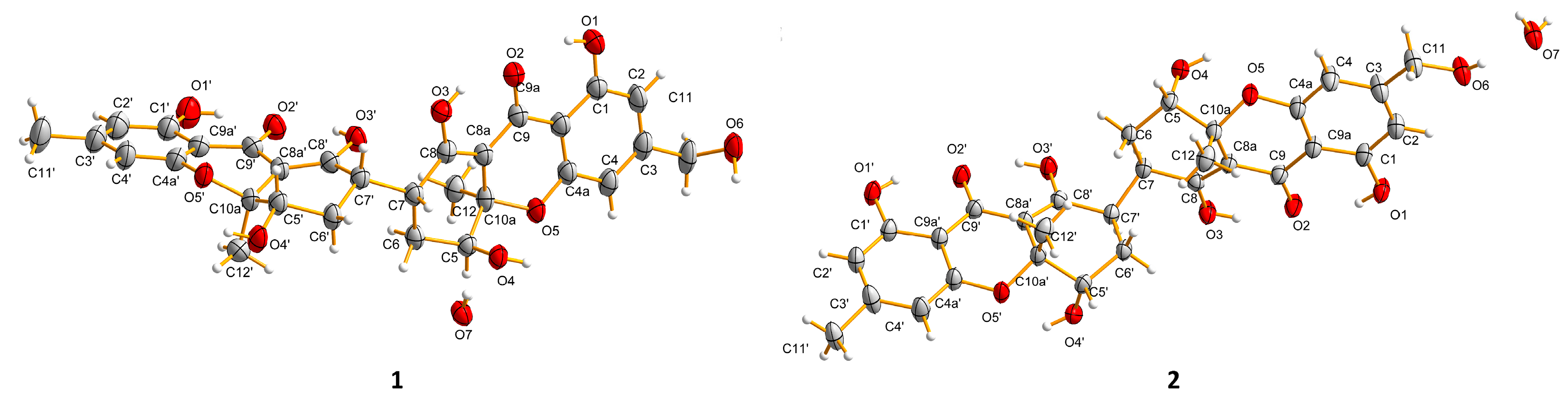
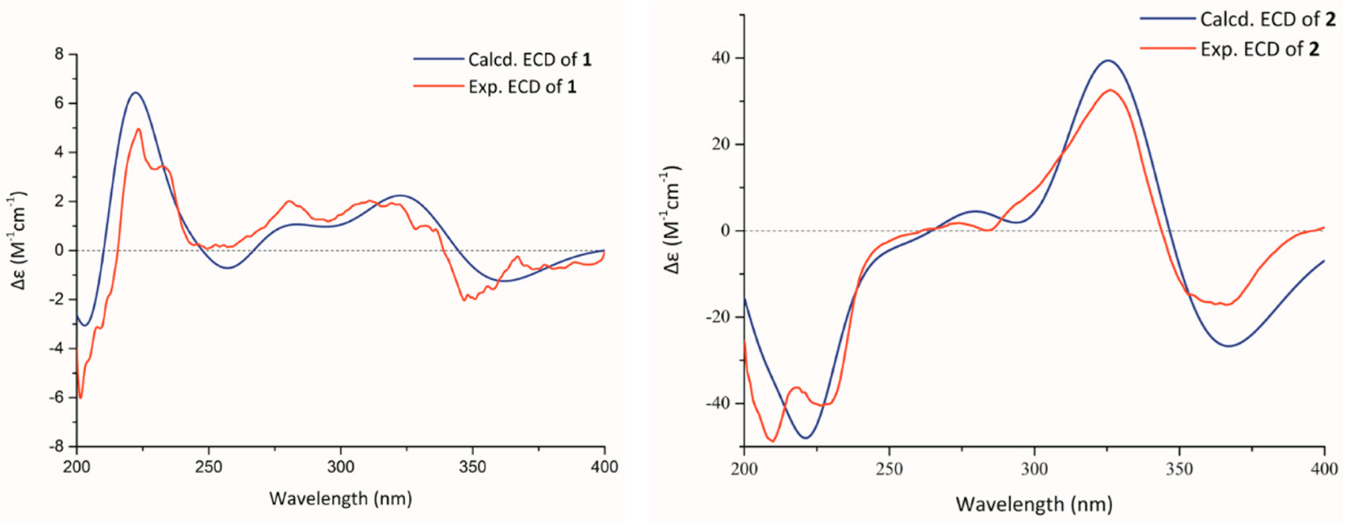
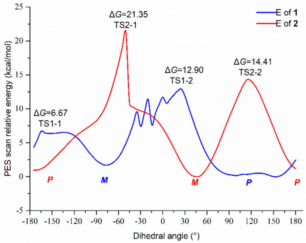
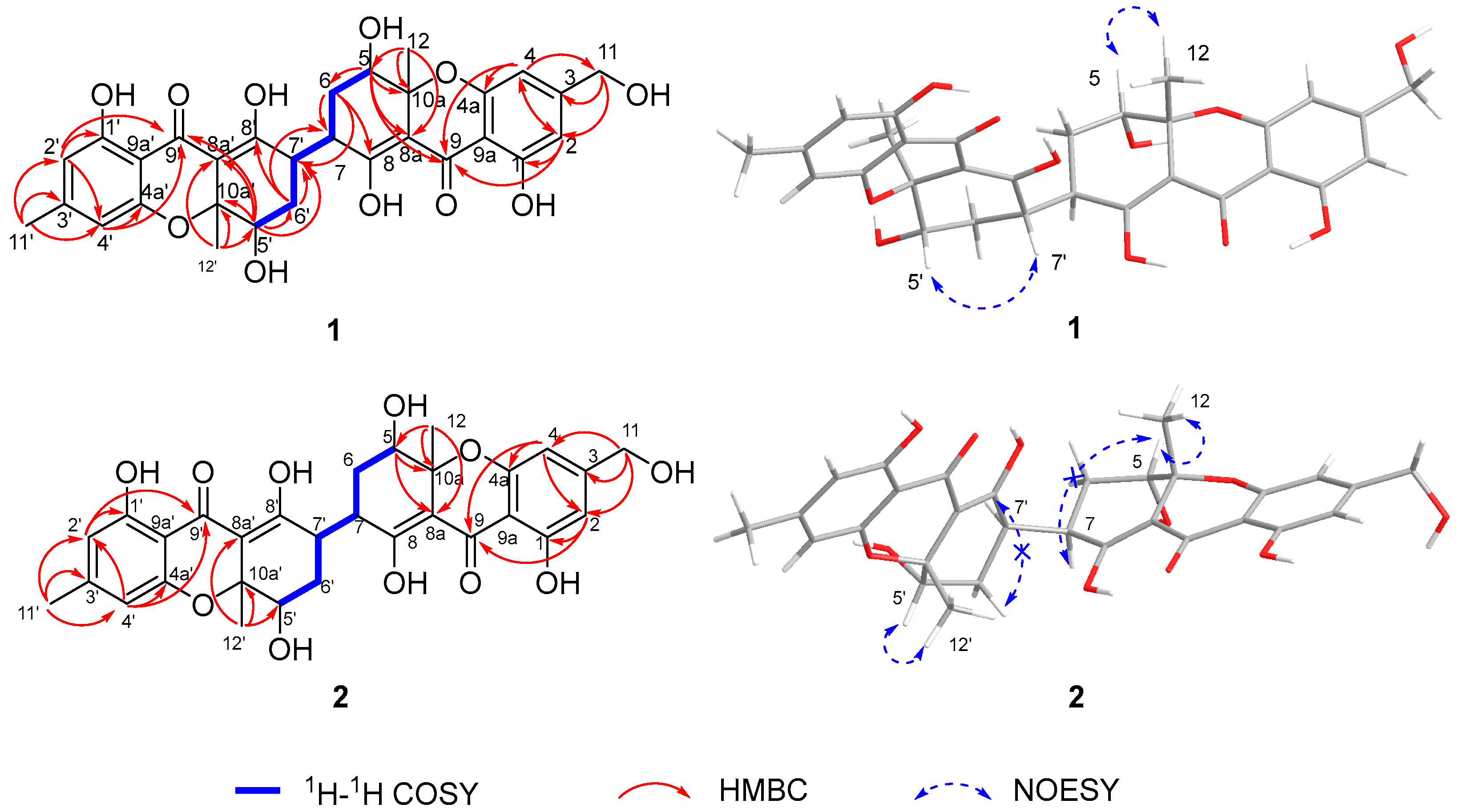
2. Results
3. Materials and Methods
3.1. General Experimental Procedures
3.2. Fungal Material
3.3. Fermentation and Isolation
3.4. X-ray Crystallographic Analysis of Compounds 1 and 2
3.5. Antimicrobial Assays
3.6. Computation Methods
4. Conclusions
Supplementary Materials
Author Contributions
Funding
Institutional Review Board Statement
Informed Consent Statement
Data Availability Statement
Acknowledgments
Conflicts of Interest
Sample Availability
References
- Roberts, J.C. Naturally Occurring Xanthones. Chem. Rev. 1961, 61, 591–605. [Google Scholar] [CrossRef]
- Wezeman, T.; Bräese, S.; Masters, K.S. Xanthone dimers: A compound family which is both common and privileged. Nat. Prod. Rep. 2015, 32, 6–28. [Google Scholar] [CrossRef] [PubMed] [Green Version]
- Masters, K.S.; Bräse, S. Xanthones from fungi, lichens, and bacteria: The natural products and their synthesis. Chem. Rev. 2012, 112, 3717–3776. [Google Scholar] [CrossRef] [PubMed]
- Santos, C.M.M.; Freitas, M.; Fernandes, E. A comprehensive review on xanthone derivatives as alpha-glucosidase inhibitors. Eur. J. Med. Chem. 2018, 157, 1460–1479. [Google Scholar] [CrossRef] [PubMed]
- Liu, Z.; Chen, S.; Qiu, P.; Tan, C.; Long, Y.; Lu, Y.; She, Z. (+)- and (−)-Ascomlactone A: A pair of novel dimeric polyketides from a mangrove endophytic fungus Ascomycota sp. SK2YWS-L. Org. Biomol. Chem. 2017, 15, 10276–10280. [Google Scholar] [CrossRef]
- Cao, S.G.; McMillin, D.W.; Tamayo, G.; Delmore, J.; Mitsiades, C.S.; Clardy, J. Inhibition of tumor cells interacting with stromal cells by xanthones isolated from a Costa Rican Penicillium sp. J. Nat. Prod. 2012, 75, 793–797. [Google Scholar] [CrossRef] [Green Version]
- Ding, B.; Yuan, J.; Huang, X.; Wen, W.; Zhu, X.; Liu, Y.; Li, H.; Lu, Y.; He, L.; Tan, H.; et al. New dimeric members of the phomoxanthone family: Phomolactonexanthones A, B and deacetylphomoxanthone C isolated from the fungus Phomopsis sp. Mar. Drugs 2013, 11, 4961. [Google Scholar] [CrossRef] [Green Version]
- Wu, G.; Yu, G.; Kurtan, T.; Mandi, A.; Peng, J.; Mo, X.; Liu, M.; Li, H.; Sun, X.; Li, J.; et al. Versixanthones A-F, cytotoxic xanthone-chromanone dimers from the marine-derived fungus Aspergillus versicolor HDN1009. J. Nat. Prod. 2015, 78, 2691–2698. [Google Scholar] [CrossRef]
- Liu, F.; Lin, X.; Zhou, X.; Chen, M.; Huang, X.; Yang, B.; Tao, H. Xanthones and quinolones derivatives produced by the deep-sea-derived dungus Penicillium sp. SCSIO Ind16F01. Molecules 2017, 22, 1999. [Google Scholar] [CrossRef] [Green Version]
- Jo, Y.H.; Kim, S.B.; Ahn, J.H.; Turk, A.; Kwon, E.-B.; Kim, M.O.; Hwang, B.Y.; Lee, M.K. Xanthones from the stems of Cudrania tricuspidata and their inhibitory effects on pancreatic lipase and fat accumulation. Bioorg. Chem. 2019, 92, 103234. [Google Scholar] [CrossRef]
- Cao, H.Y.; Yi, C.; Sun, S.F.; Li, Y.; Liu, Y.B. Anti-inflammatory dimeric tetrahydroxanthones from an endophytic Muyocopron laterale. J. Nat. Prod. 2022, 85, 148–161. [Google Scholar] [CrossRef] [PubMed]
- Xue, Q.; Chen, Y.; Yin, H.; Teng, H.; Qin, R.; Liu, H.; Li, Q.; Mei, Z.; Yang, G. Prenylated xanthones and benzophenones from the fruits of Garcinia bracteata and their potential antiproliferative and anti-inflammatory activities. Bioorg. Chem. 2020, 104, 04. [Google Scholar] [CrossRef] [PubMed]
- Yoganathan, K.; Cao, S.; Crasta, S.C.; Aitipamula, S.; Whitton, S.R.; Ng, S.; Buss, A.D.; Butler, M.S. Microsphaerins A-D, four novel benzophenone dimers with activity against MRSA from the fungus Microsphaeropsis sp. Tetrahedron 2008, 64, 10181–10187. [Google Scholar] [CrossRef]
- El-Elimat, T.; Figueroa, M.; Raja, H.A.; Graf, T.N.; Swanson, S.M.; Falkinham, J.O.; Wani, M.C.; Pearce, C.J.; Oberlies, N.H. Biosynthetically distinct cytotoxic polyketides from Setophoma terrestris. Eur. J. Org. Chem. 2015, 2015, 109–121. [Google Scholar] [CrossRef] [Green Version]
- Chutrakul, C.; Boonruangprapa, T.; Suvannakad, R.; Isaka, M.; Sirithunya, P.; Toojinda, T.; Kirtikara, K. Ascherxanthone B from Aschersonia luteola, a new antifungal compound active against rice blast pathogen Magnaporthe grisea. J. Appl. Microbiol. 2009, 107, 1624–1631. [Google Scholar] [CrossRef]
- Peng, X.; Sun, F.; Li, G.; Wang, C.; Zhang, Y.; Wu, C.; Zhang, C.; Sun, Y.; Wu, S.; Zhang, Y.; et al. New xanthones with antiagricultural fungal pathogen activities from the endophytic fungus Diaporthe goulteri L17. J. Agric. Food. Chem. 2021, 69, 11216–11224. [Google Scholar] [CrossRef]
- Araújo, J.; Fernandes, C.; Pinto, M.; Tiritan, M.E. Chiral derivatives of xanthones with antimicrobial activity. Molecules 2019, 24, 314. [Google Scholar] [CrossRef] [Green Version]
- Lv, X.J.; Ding, F.; Wei, Y.J.; Tan, R.X. Antiosteoporotic tetrahydroxanthone dimers from Aspergillus brunneoviolaceus FB-2 residing in human gut. Chin. J. Chem. 2021, 39, 1580–1586. [Google Scholar] [CrossRef]
- Roensberg, D.; Debbab, A.; Mandi, A.; Vasylyeva, V.; Boehler, P.; Stork, B.; Engelke, L.; Hamacher, A.; Sawadogo, R.; Diederich, M.; et al. Pro-apoptotic and immunostimulatory tetrahydroxanthone dimers from the endophytic fungus Phomopsis longicolla. J. Org. Chem. 2013, 78, 12409–12425. [Google Scholar] [CrossRef]
- Li, S.J.; Jiao, F.W.; Li, W.; Zhang, X.; Yan, W.; Jiao, R.H. Cytotoxic xanthone derivatives from the mangrove-derived endophytic fungus Peniophora incarnata Z4. J. Nat. Prod. 2020, 83, 2976–2982. [Google Scholar] [CrossRef]
- Wang, F.; Jiang, J.; Hu, S.; Hao, X.; Cai, Y.; Ye, Y.; Ma, H.; Sun, W.; Cheng, L.; Huang, C.; et al. Nidulaxanthone A, a xanthone dimer with a heptacyclic 6/6/6/6/6/6/6 ring system from Aspergillus sp.-F029. Org. Chem. Front. 2020, 7, 953–959. [Google Scholar] [CrossRef]
- Yamazaki, H.; Ukai, K.; Namikoshi, M. Asperdichrome, an unusual dimer of tetrahydroxanthone through an ether bond, with protein tyrosine phosphatase 1B inhibitory activity, from the Okinawan freshwater Aspergillus sp. TPU1343. Tetrahedron Lett. 2016, 57, 732–735. [Google Scholar] [CrossRef]
- Wang, P.; Luo, Y.F.; Zhang, M.; Dai, J.G.; Wang, W.J.; Wu, J. Three xanthone dimers from the Thai mangrove endophytic fungus Phomopsis sp. xy21. J. Asian Nat. Prod. Res. 2018, 20, 217–226. [Google Scholar] [CrossRef] [PubMed]
- Chen, S.; Cai, R.; Liu, Z.; Cui, H.; She, Z. Secondary metabolites from mangrove-associated fungi: Source, chemistry and bioactivities. Nat. Prod. Rep. 2022, 39, 560–595. [Google Scholar] [CrossRef] [PubMed]
- Chen, Y.; Liu, Z.; Huang, Y.; Liu, L.; He, J.; Wang, L.; Yuan, J.; She, Z. Ascomylactams A-C, cytotoxic 12-or 13-membered-ring macrocyclic alkaloids isolated from the mangrove endophytic fungus Didymella sp. CYSK-4, and structure revisions of phomapyrrolidones A and C. J. Nat. Prod. 2019, 82, 1752–1758. [Google Scholar] [CrossRef] [PubMed]
- Cai, R.; Jiang, H.; Xiao, Z.; Cao, W.; Yang, T.; Liu, Z.; Lin, S.; Long, Y.; She, Z. (-)- and (+)-asperginulin A, a pair of indole diketopiperazine alkaloid dimers with a 6/5/4/5/6 pentacyclic skeleton from the mangrove endophytic fungus Aspergillus sp. SK-28. Org. Lett. 2019, 21, 9633–9636. [Google Scholar] [CrossRef] [PubMed]
- Cai, R.; Jiang, H.; Mo, Y.; Guo, H.; Li, C.; Long, Y.; Zang, Z.; She, Z. Ophiobolin-type sesterterpenoids from the mangrove endophytic fungus Aspergillus sp. ZJ-68. J. Nat. Prod. 2019, 82, 2268–2278. [Google Scholar] [CrossRef]
- Cui, H.; Lin, Y.; Luo, M.; Lu, Y.; Huang, X.; She, Z. Diaporisoindoles A-C: Three isoprenylisoindole alkaloid derivatives from the mangrove endophytic fungus Diaporthe sp. SYSU-HQ3. Org. Lett. 2017, 19, 5621–5624. [Google Scholar] [CrossRef]
- Liu, Z.; Chen, Y.; Chen, S.; Liu, Y.; Lu, Y.; Chen, D.; Lin, Y.; Huang, X.; She, Z. Aspterpenacids A and B, two sesterterpenoids from a mangrove endophytic fungus Aspergillus terreus H010. Org. Lett. 2016, 18, 1406–1409. [Google Scholar] [CrossRef]
- Yu, G.; Wu, G.; Sun, Z.; Zhang, X.; Che, Q.; Gu, Q.; Zhu, T.; Li, D.; Zhang, G. Cytotoxic tetrahydroxanthone dimers from the mangrove-associated fungus Aspergillus versicolor HDN1009. Mar. Drugs 2018, 16, 335. [Google Scholar] [CrossRef] [Green Version]
- Cui, H.; Liu, Y.; Li, J.; Huang, X.; Yan, T.; Cao, W.; Liu, H.; Long, Y.; She, Z. Diaporindenes A-D: Four unusual 2,3-dihydro-1H-indene analogues with anti-inflammatory activities from the mangrove endophytic fungus Diaporthe sp. SYSU-HQ3. J. Org. Chem. 2018, 83, 11804–11813. [Google Scholar] [CrossRef] [PubMed]
- LaPlante, S.R.; Edwards, P.J.; Fader, L.D.; Jakalian, A.; Hucke, O. Revealing atropisomer axial chirality in drug discovery. Chemmedchem 2011, 6, 505–513. [Google Scholar] [CrossRef] [PubMed]
- LaPlante, S.R.; Fader, L.D.; Fandrick, K.R.; Fandrick, D.R.; Hucke, O.; Kemper, R.; Miller, S.P.F.; Edwards, P.J. Assessing atropisomer axial chirality in drug discovery and development. J. Med. Chem. 2011, 54, 7005–7022. [Google Scholar] [CrossRef] [PubMed]
- Zhou, G.; Wu, H.; Wang, T.; Guo, R.; Xu, J.; Zhang, Q.; Tang, L.; Wang, Z. C-glycosylflavone with rotational isomers from Vaccaria hispanica (Miller) Rauschert seeds. Phytochem. Lett. 2017, 19, 241–247. [Google Scholar] [CrossRef]
- Stuart, A.C.; Tumbleston, J.R.; Zhou, H.; Li, W.; Liu, S.; Ade, H.; You, W. Fluorine substituents reduce charge recombination and drive structure and morphology development in polymer solar cells. J. Am. Chem. Soc. 2013, 135, 1806–1815. [Google Scholar] [CrossRef] [PubMed]
- Xing, Q.; Gan, L.-S.; Mou, X.F.; Wang, W.; Wang, C.Y.; Wei, M.Y.; Shao, C.L. Isolation, resolution and biological evaluation of pestalachlorides E and F containing both point and axial chirality. RSC Adv. 2016, 6, 22653–22658. [Google Scholar] [CrossRef]
- Barrett, K.T.; Metrano, A.J.; Rablen, P.R.; Miller, S.J. Spontaneous transfer of chirality in an atropisomerically enriched two-axis system. Nature 2014, 509, 71–75. [Google Scholar] [CrossRef] [Green Version]
- Cheng, G.; Bai, Y.J.; Zhao, Y.Y.; Tao, J.; Lin, Y.; Tu, G.Z.; Ma, L.B.; Liao, N.; Xu, X.J. Flavonoids from Ziziphus jujuba Mill var. spinosa. Tetrahedron 2000, 56, 8915–8920. [Google Scholar] [CrossRef]
- Pierce, C.G.; Uppuluri, P.; Tristan, A.R.; Wormley, F.L.; Mowat, E.; Ramage, G.; Lopez Ribot, J.L. A simple and reproducible 96-well plate-based method for the formation of fungal biofilms and its application to antifungal susceptibility testing. Nat. Protoc. 2008, 3, 1494–1500. [Google Scholar] [CrossRef]
- Chen, Y.; Yang, W.; Zou, G.; Chen, S.; Pang, J.; She, Z. Bioactive polyketides from the mangrove endophytic fungi Phoma sp. SYSU-SK-7. Fitoterapia 2019, 139, 104396. [Google Scholar] [CrossRef]
- Spartan’14; IWavefunction Inc.: Irvine, CA, USA, 2013.
- Frisch, M.J.; Trucks, G.W.; Schlegel, H.B.; Scuseria, G.E.; Robb, M.A.; Cheeseman, J.R.; Scalmani, G.; Barone, V.; Petersson, G.A.; Nakatsuji, H.; et al. Gaussian 09 Rev. A.02; Gaussian, Inc.: Wallingford, CT, USA, 2016. [Google Scholar]
- Bruhn, T.; Hemberger, Y.; Schaumlöffel, A.; Bringmann, G. SpecDis, Version 1.53; University of Würzburg: Würzburg, Germany, 2011. [Google Scholar]
- Dennington, R.; Keith, T.A.; Millam, J.M. GaussView, Version 6; Semichem Inc.: Shawnee Mission, KS, USA, 2016. [Google Scholar]


| 1 | 2 at 298 K | 2 at 243 K | |||||
|---|---|---|---|---|---|---|---|
| Atom No. | δC, Type | δH (Mult, J in Hz) | δC, Type | δH, (Mult, J in Hz) | δC, Type | δH, (Mult, J in Hz) | |
| 1 | 162.2, C | 162.3, C | 162.3, C | 162.2, C | |||
| 2 | 107.6, CH | 6.45, s | 107.6, CH | 6.45, s | 107.2, CH | 107.1, CH | 6.45, s |
| 3 | 153.6, C | 153.8, C | 153.7, C | 153.3, C | |||
| 4 | 106.6, CH | 6.42, s | 106.7, CH | 6.42, s | 106.9, CH | 106.9, CH | 6.42, s |
| 4a | 159.3, C | 159.2, C | 159.3, C | 159.2, C | |||
| 10a | 81.5, C | 81.4, C | 81.5, C | 81.4, C | |||
| 5 | 70.8, CH | 4.20, dd (12.4, 4.2) | 70.9, CH | 4.10–4.05, m | 70.9, CH | 4.08–4.04, m | |
| 6 | 29.9, CH2 | 2.10–2.02, m 1.71–1.62, m | 33.7, CH2 | 2.56–2.48, m 2.23–2.14, m | |||
| 7 | 39.9, CH | 3.44–3.35, m | 38.2, CH | 3.02–2.95, m | |||
| 8 | 175.0, C | 175.4, C | 174.9, C | ||||
| 8a | 105.8, C | 105.8, C | |||||
| 9 | 188.4, C | 188.4, C | 188.3, C | ||||
| 9a | 105.3, C | 105.2, C | 105.8, C | ||||
| 11 | 64.1, CH2 | 4.51, s | 64.1, CH2 | 4.51, s | 64.8, CH2 | 4.53, s | |
| 12 | 26.1, CH3 | 1.48, s | 26.1, CH3 | 1.50, s | 25.5, CH3 | 1.52, s | |
| 1′ | 162.4, C | 162.5, C | 162.1, C | 161.9, C | |||
| 2′ | 110.7, CH | 6.27, s | 110.8, CH | 6.25, s | 110.7, CH | 110.5, CH | 6.25, s |
| 3′ | 150.8, C | 150.6, C | 150.7, C | 150.3, C | |||
| 4′ | 109.8, CH | 6.26, s | 106.7, CH | 6.27, s | 109.9, CH | 109.9, CH | 6.27, s |
| 4a’ | 159.3, C | 159.4, C | 159.0, C | 158.9, C | |||
| 10a’ | 81.8, C | 81.5, C | 81.2, C | 81.0, C | |||
| 5′ | 71.8, CH | 4.12, dd (4.3, 1.9) | 71.9, CH | 4.10–4.05, m | 70.2, CH | 4.13–4.09, m | |
| 6′ | 28.0, CH2 | 2.20–2.14, m 1.87–1.79, m | 25.8, CH2 | 2.14–2.07, m 1.59–1.67, m | |||
| 7′ | 37.1, CH | 3.44–3.35, m | 35.0, CH | 3.80–3.73, m | |||
| 8′ | 176.9, C | 177.4, C | 176.9, C | ||||
| 8a’ | 107.8, C | 107.8, C | |||||
| 9′ | 188.6, C | 188.4, C | 188.3, C | ||||
| 9a’ | 106.1, C | 106.1, C | 106.4, C | ||||
| 11′ | 22.5, CH3 | 2.25, s | 22.5, CH3 | 2.25, s | 22.6, CH3 | 2.25, s | |
| 12′ | 20.1, CH3 | 1.45, s | 20.1, CH3 | 1.50, s | 27.0, CH3 | 1.45, s | |
| MIC of Compounds/μM | ||||
|---|---|---|---|---|
| 1 | 2 | Ketoconazole a | Ampicillin b | |
| F. oxysporum | 12.5 | 12.5 | 6.25 | NT |
| P. italicum | 50 | >100 | 1.56 | NT |
| C. musae | 25 | 12.5 | 1.56 | NT |
| C. gloeosporioides | 3.13 | >100 | 0.1 | NT |
| C. albicans | 12.5 | 25 | 0.1 | NT |
| MRSA | 12.5 | >100 | NT | 0.39 |
| P. aeruginosa | >100 | >100 | NT | 0.19 |
| B. subtilis | 25 | 25 | NT | 0.39 |
Publisher’s Note: MDPI stays neutral with regard to jurisdictional claims in published maps and institutional affiliations. |
© 2022 by the authors. Licensee MDPI, Basel, Switzerland. This article is an open access article distributed under the terms and conditions of the Creative Commons Attribution (CC BY) license (https://creativecommons.org/licenses/by/4.0/).
Share and Cite
Zang, Z.; Yang, W.; Cui, H.; Cai, R.; Li, C.; Zou, G.; Wang, B.; She, Z. Two Antimicrobial Heterodimeric Tetrahydroxanthones with a 7,7′-Linkage from Mangrove Endophytic Fungus Aspergillus flavus QQYZ. Molecules 2022, 27, 2691. https://doi.org/10.3390/molecules27092691
Zang Z, Yang W, Cui H, Cai R, Li C, Zou G, Wang B, She Z. Two Antimicrobial Heterodimeric Tetrahydroxanthones with a 7,7′-Linkage from Mangrove Endophytic Fungus Aspergillus flavus QQYZ. Molecules. 2022; 27(9):2691. https://doi.org/10.3390/molecules27092691
Chicago/Turabian StyleZang, Zhenming, Wencong Yang, Hui Cui, Runlin Cai, Chunyuan Li, Ge Zou, Bo Wang, and Zhigang She. 2022. "Two Antimicrobial Heterodimeric Tetrahydroxanthones with a 7,7′-Linkage from Mangrove Endophytic Fungus Aspergillus flavus QQYZ" Molecules 27, no. 9: 2691. https://doi.org/10.3390/molecules27092691
APA StyleZang, Z., Yang, W., Cui, H., Cai, R., Li, C., Zou, G., Wang, B., & She, Z. (2022). Two Antimicrobial Heterodimeric Tetrahydroxanthones with a 7,7′-Linkage from Mangrove Endophytic Fungus Aspergillus flavus QQYZ. Molecules, 27(9), 2691. https://doi.org/10.3390/molecules27092691