Article

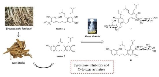
Microbial Transformation and Biological Activities of the Prenylated Aromatic Compounds from Broussonetia kazinoki

College of Pharmacy, Chonnam National University, Gwangju 61186, Korea
*
Author to whom correspondence should be addressed.
Current address: Advanced Radiation Technology Institute, Korea Atomic Energy Research Institute, Jeongeup-si 56212, Korea.
Molecules 2022, 27(6), 1879; https://doi.org/10.3390/molecules27061879
Submission received: 17 February 2022 / Revised: 11 March 2022 / Accepted: 12 March 2022 / Published: 14 March 2022
(This article belongs to the Special Issue Microbial Biotransformation of Natural Products)

Abstract

:
Broussonetia kazinoki has been used as a traditional medicine for the treatment of burns and acne, and its extracts have been found to show tyrosinase inhibitory and anticancer activities. In this study, the tyrosinase inhibitory and cytotoxic activities of B. kazinoki were explored, leading to the isolation of kazinol C (1), kazinol E (2), kazinol F (3), broussonol N (4), and kazinol X (5), of which the compounds 4 and 5 have not been previously reported. Microbial transformation has been recognized as an efficient tool to generate more active metabolites. Microbial transformation of the major compounds 1 and 3 was conducted with Mucor hiemalis, where four glucosylated metabolites (69) were produced from 1, while one hydroxylated (10) and one glucosylated (11) metabolites were obtained from 3. Structures of the isolated metabolites were determined by extensive spectroscopic analyses. All compounds were evaluated for their tyrosinase inhibitory and cytotoxic activities. Compound 3 and its metabolites, kazinol Y (10) and kazinol F-4″-O-β-d-glucopyranoside (11), exhibited the most potent tyrosinase inhibitory activities with the IC50 values ranging from 0.71 to 3.36 µM. Meanwhile, none of the metabolites, except for kazinol C-2′,3″-di-O-β-d-glucopyranoside (7), showed moderate cytotoxic activities (IC50 17.80 to 24.22 µM) against A375P, B16F10 and B16F1 cell lines.

Graphical Abstract

1. Introduction

Broussonetia kazinoki, a deciduous shrub tree belonging to the family Moraceae, is widely distributed throughout Korea, China, and Japan [1,2,3]. Since ancient times, its leaves, branches, roots and fruits have been used for various therapeutic purposes including the amelioration of vision, and suppression of edema [1,2,3,4]. In addition, it has been traditionally used for dermatologic diseases such as burns and acne in Korea according to the Principles and Practice of Eastern Medicine, an encyclopedia of medical knowledge [5,6]. Previous biological investigations have demonstrated that B. kazinoki exhibited a variety of pharmacological effects, such as antioxidant, anti-inflammatory, anticancer, anti-allergic, anti-diabetic, and anti-hyperglycemic activities [2,3,4,5,6,7]. Moreover, the extract of B. kazinoki has been registered as a skin-whitening agent by the Korea Food and Drug Administration (KFDA) due to its potent tyrosinase inhibitory and anti-melanogenic effects [5,8]. Additionally, a series of prenylated polyphenols including kazinol F and broussonin C isolated from B. kazinoki have been reported to exhibit significant tyrosinase inhibitory effects [8].
Tyrosinase (EC 1.14.18.1), also known as catecholase or diphenol oxidase, is a multifunctional copper-containing enzyme and is widely distributed in plants, fungi, bacteria and animals [9,10]. It catalyzes the hydroxylation of L-tyrosine to L-DOPA (3,4-dihydroxy-L-phenylalanine) and the subsequent oxidation of L-DOPA to L-dopaquinone [10]. Then, the dopaquinone forms melanin through polymerization with a series of enzymatic and nonenzymatic reactions [11,12]. Melanin is responsible for skin color and plays an important role in human skin, for example, the prevention of skin injury under normal physiological conditions [12]; however, an overproduction and accumulation of melanin can result in hyperpigmentary disorders of the skin, such as freckles, melasma, age spots, and melanoma [9]. More seriously, tyrosinase catalyzes the formation of neuromelanin, which is associated with neurodegenerative disorders like Parkinson’s disease [10,11,13]. Therefore, inhibiting tyrosinase activity applies to the treatment of pigmentation disorders associated with melanin hyperpigmentation and some related diseases [9,14]. Though a number of well-known tyrosinase inhibitors, like arbutin, kojic acid, and hydroquinone, have been reported from natural or synthetic sources during the past few decades, their applications have been limited due to serious side effects such as dermatitis, cytotoxicity and hepatotoxicity [9,14]. Thus, it is desirable to find new tyrosinase inhibitors with improved safety.
Microbial transformation is known as a useful method to generate more active derivatives with minor structural modifications in bioactive substrates using the metabolic activities of microorganisms [15,16]. The transformation is accomplished by a series of enzymatic reactions including hydroxylation, oxidation, and glycosylation under mild conditions [16,17,18,19]. Furthermore, microbial transformation is considered to be an environmentally friendly tool and has been used successfully to produce pharmaceuticals from natural products [15,16].
In the present study, we isolated and characterized the constituents of the root barks of B. kazinoki with tyrosinase inhibitory properties based on the bioactivity-guided fractionation process (Figure 1). Further transformation of kazinols C (1) and F (3) was performed with Mucor hiemalis, which led to the isolation of five glucosylated and one oxidized metabolite (611). All of the isolated metabolites were evaluated for their tyrosinase inhibitory and cytotoxic activities, and it was revealed that kazinol F-4″-O-β-d-glucopyranoside (11) had the strongest anti-tyrosinase activity with no cytotoxic activity against melanoma cells.

2. Results and Discussion

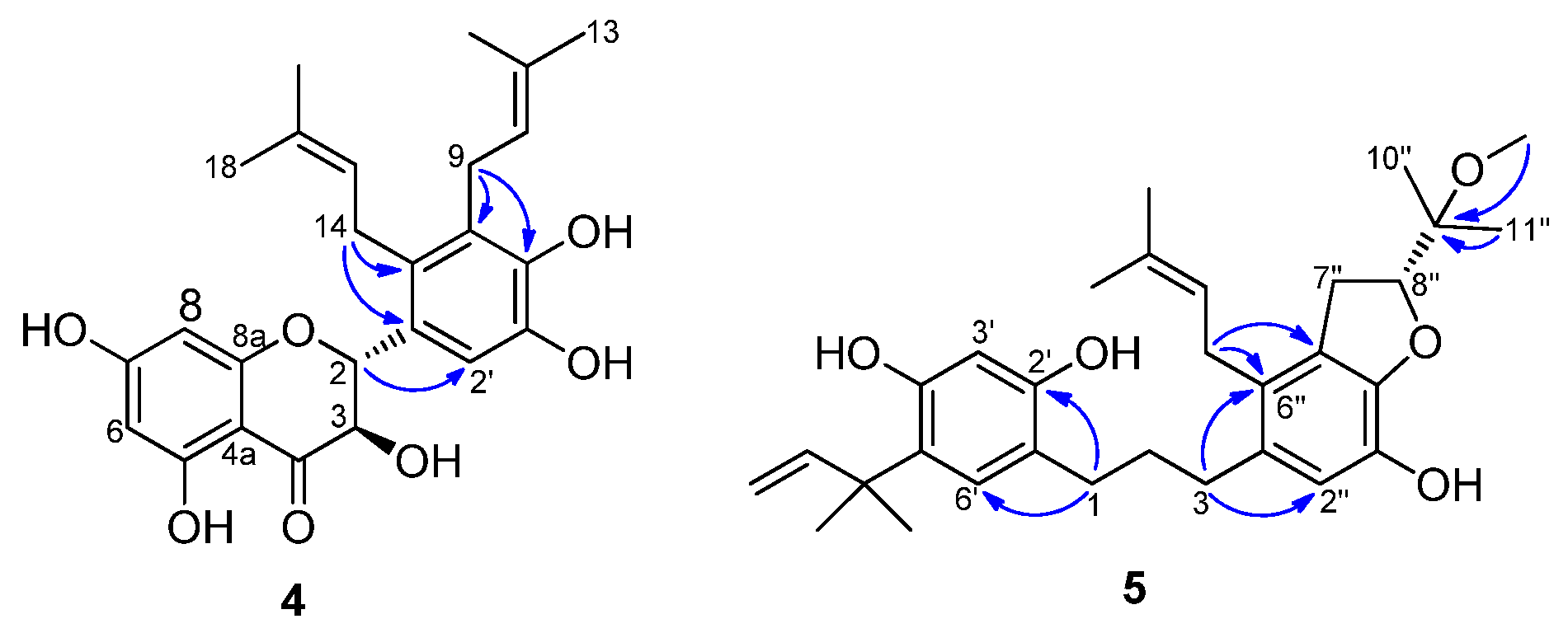
2.1. Structure Elucidation of Compounds from Broussonetia kazinoki

To isolate and identify the secondary metabolites of B. kazinoki for anti-tyrosinase activity, the EtOH extract of its root barks was investigated [8]. The CH2Cl2 fraction of the EtOH extract showed promising tyrosinase inhibitory effects, and the subsequent activity-guided fractionation led to the isolation of five prenylated polyphenols 15 (Figure 1). Structures of the compounds were determined using the 1D- and 2D-NMR (nuclear magnetic resonance), including COSY (correlation spectroscopy), HSQC (heteronuclear single quantum correlation), and HMBC (heteronuclear multiple bond correlation), as well as mass spectroscopic data analyses. The three known compounds were identified and confirmed as kazinol C (1), kazinol E (2), and kazinol F (3) by comparison of their spectroscopic data with those reported in the literatures [8,20].
Compound 4 showed a molecular formula of C25H28O7 by the HRFDMS peak at m/z 440.1841 [M]+ (calcd. 440.1835). The UV spectrum of 4 exhibited two distinct peaks at 210 and 291 nm, characteristic of a flavanonol system. The 1H NMR spectrum of compound 4 displayed: three aromatic proton signals at δH 7.05 (1H, s), 5.95 (1H, s), and 5.89 (1H, s); two sets of prenyl group proton signals at δH 5.12 (1H, t, J = 6.6 Hz), 5.08 (1H, t, J = 6.1 Hz), 3.46 (1H, dd, J = 6.1, 16.4 Hz), 3.40 (2H, d, J = 6.2 Hz), 3.33 (1H, dd, J = 6.1, 16.4 Hz), 1.75 (3H, s), 1.66 (3H, s), 1.62 (3H, s), and 1.61 (3H, s); and a set of AB-type proton signals at δH 5.31 (1H, d, J = 11.5 Hz), and 4.64 (1H, t, J = 11.5 Hz) due to H-2 and H-3. The positions of the two prenyl groups were determined on the basis of its HMBC spectrum (Figure 2). The long-range correlations from H-9 to C-4′/5′, H-14 to C-1′/6′, and H-2 to C-2′ indicated that two prenyl groups were adjacent to each other and attached at the C-5′ and C-6′ positions of ring B. The large coupling constant (11.5 Hz) between H-2 and H-3 indicated that ring C had a 2,3-trans-configuration, not a cis-configuration which gave a small J value (~2 Hz) [3], and the absolute configuration of compound 4 was considered to be 2R,3R based on its positive specific rotation ( [ α ] D 20   +74.6°) by comparison with the reported data of related structures [3,21]. Based on the above analysis, the structure of compound 4 was assigned (2R,3R)-3,5,7,3′,4′-pentahydroxy-5′,6′-diprenylflavanonol, and was named broussonol N.
Compound 5 exhibited a molecular formula of C31H42O5 by its HRFDMS peak at m/z 494.3034 [M]+ (calcd. 494.3032). The UV spectrum of 5 exhibited absorption maxima at 228 and 285 nm, which was similar to the kazinol, indicating that it was also a 1,3-diphenylpropane. The 1H NMR spectrum of compound 5 revealed: three aromatic proton signals at δH 6.93 (1H, s), 6.61 (1H, s), and 6.32 (1H, s); a 1,1-dimethylallyl group proton signal at δH 6.17 (1H, dd, J = 10.6, 17.7 Hz), 5.32 (1H, dd, J = 0.6, 17.7 Hz), 5.26 (1H, dd, J = 0.6, 10.6 Hz), and 1.39 (6H, s); one prenyl group proton signal at δH 5.01 (1H, t, J = 6.6 Hz), 3.16 (2H, d, J = 6.6 Hz), 1.70 (3H, s), and 1.67 (3H, s); and the signals of a 1,3-disubstituted propane moiety at δH 2.59 (2H, m), 2.54 (2H, m), and 1.81 (2H, m). In addition, the proton signals of a 2-(1-methoxy-1-methylethyl)-dihydrofuran moiety at δH 4.73 (1H, t, J = 9.2 Hz), 3.31 (3H, s), 3.09 (2H, dd, J = 9.6, 15.8 Hz), 1.24 (3H, s), and 1.20 (3H, s) were also observed in 5, which was quite similar to that of kazinol T [8], except for the presence of a methoxy group instead of a hydroxyl group at the C-9″ position. This was supported by the long-range correlation from 9″-OCH3 to C-9″ (δC 77.03) (Figure 2). The absolute configuration of compound 5 at C-9″ was considered to be R based on its negative specific rotation ( [ α ] D 20 −6.31°) by comparison with the reported data of related structures [18,22,23]. Based on the above analysis, the structure of compound 5 was assigned to be 5′-(2-methylbut-3-en-2-yl)-6″-(3-methylbut-2-enyl)-4″,5″-[(R)-2-(1-methoxy-1-methylethyl)]-dihydrofuranyl-2′,4′,3″-trihydroxydiphenylpropane, and was named kazinol X.

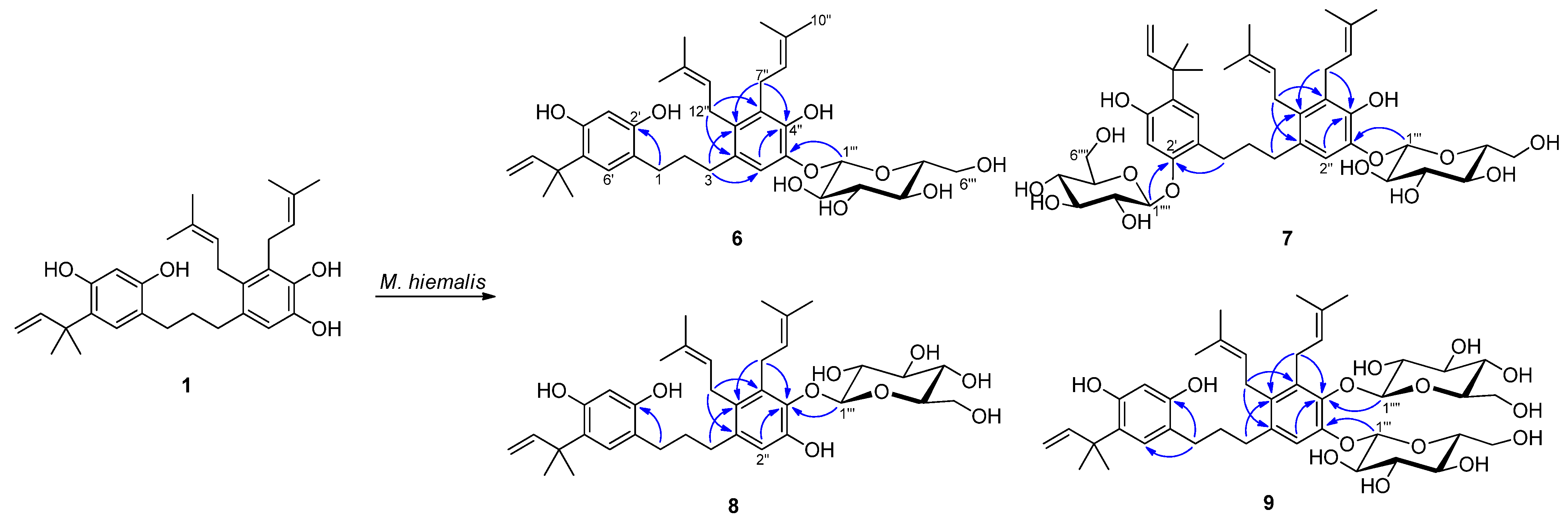
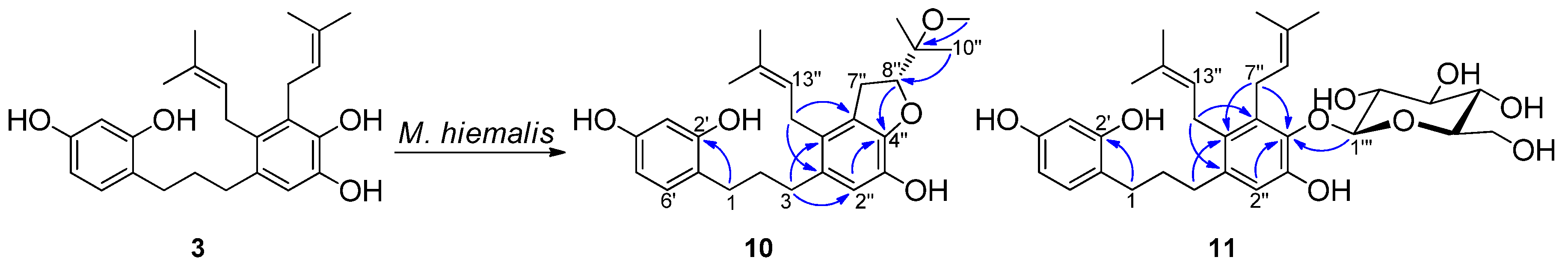
2.2. Microbial Transformation of Kazinols C and F by Mucor hiemalis

A total of 14 microbial cultures were screened to evaluate their ability to metabolize the isolated compounds 1 and 3 under a standard two-stage fermentation procedure [18,19]. Based on the analysis of TLC plates involving the substrates and culture controls, it was observed that Gliocladium deliquescens and Mucor hiemalis showed the ability to metabolize 1, and Alternaria alternata, Absidia coerulea, G. deliquescens and M. hiemalis showed the ability to metabolize 3 (Supplementary Table S1). Among the active strains, the fungus M. hiemalis was selected for preparative-scale fermentation studies since it exhibited the highest transformational capability towards 1 and 3. The subsequent transformation studies led to the production of five glucosylated and one oxidized metabolite (611).
Compound 6 had a molecular formula of C36H50O9, as established by its HRFDMS peak at m/z 626.3439 [M]+ (calcd. 626.3455), which was one glucose unit higher than that of 1, indicating that 6 was a glucosylated derivative of 1. This was supported by the occurrence of six new carbon signals in the 13C NMR spectrum of 6, including five methine carbon resonances at δC 103.4, 77.1, 75.8, 73.4, 69.7, and one methylene carbon at δC 60.7. The corresponding proton signals at δH 4.50 (1H) and 3.69–3.19 (6H) were observed in the 1H NMR spectrum. All of these data were consistent with previous reports on the d-glucose moiety [19,24,25], and the glucose was determined to be in a β-configuration by the large coupling constant (7.1 Hz) of the anomeric proton signal at δH 4.50 (H-1‴). The significantly downfield-shifted aromatic proton signal at δH 6.81 (H-2″) suggested the glucose moiety was attached to C-3″ through an ether linkage. This was confirmed by a long-range correlation between the anomeric proton signal at δH 4.50 (H-1‴) and carbon signal at δC 143.1 (C-3″) in the HMBC spectrum (Figure 3). Thus, compound 6 was assigned to be kazinol C-3″-O-β-d-glucopyranoside.
Compound 7 had a molecular formula of C42H60O14, as established by the peak at m/z 788.3997 [M]+ (calcd. 788.3983) in its HRFDMS spectrum, which was one glucose unit higher than that of 6, indicating that two glucose units had been introduced into the molecule of 1. The additional proton signals at δH 4.64, 4.51 and 3.71–3.18 (12H), as well as the carbon signals at δC 103.5, 101.5, 77.1, 76.9, 76.8, 75.8, 73.5, 73.4, 69.7, 69.6, 60.7, and 60.7 observed in the 1H and 13C NMR spectra of 7, indicated the presence of two D-glucose residues [24,25]. The glucose units were identified as β-glucose by their large coupling constants (6.5 Hz and 6.6 Hz) of the anomeric proton signals δH 4.64 and 4.51, respectively. The significantly downfield-shifted aromatic proton signals at δH 6.60 (H-3′) and 6.82 (H-2″) suggested the two glucose moieties were linked to C-2′ and C-3″, respectively. These connections were confirmed by the long-range correlations between the anomeric proton signal at δH 4.51 (H-1‴) and the carbon signal at δC 142.9 (C-3″), as well as between δH 4.64 (H-1″″) and δC 154.1 (C-2′) in the HMBC spectrum of 7 (Figure 3). Thus, compound 7 was assigned to be kazinol C-2′,3″-di-O-β-d-glucopyranoside.
Compound 8 had a molecular formula of C36H50O9, as deduced from the peak at m/z 626.3469 [M]+ (calcd. 626.3455) in its HRFDMS spectrum, which was one glucose unit higher than that of 1, indicating that 8 was also a monoglucosylated derivative of 1. The additional proton signals at δH 4.42 and 3.64–3.15 (6H), as well as the carbon signals at δC 105.9, 77.3, 76.2, 74.1, 69.5, and 60.8 in the 1H and 13C NMR spectra of 8, indicated the presence of a D-glucose residue. The coupling constant (7.6 Hz) of anomeric proton (δH 4.42) in the 1H NMR spectrum of 8 indicated a β-configuration of this glucose moiety. The significantly downfield-shifted aromatic carbon signals at δC 147.0 (C-3″) and 133.9 (C-5″) suggested this glucose moiety was attached to C-4″, and it was confirmed by the correlation between H-1‴ and C-4″ in the HMBC spectrum of 8 (Figure 3). Thus, compound 8 was assigned to be kazinol C-4″-O-β-d-glucopyranoside.
Compound 9 had a molecular formula of C42H60O14, as established by the peak at m/z 788.4016 [M]+ (calcd. 788.3983) in its HRFDMS spectrum, which was one glucose unit higher than that of 8, indicating that two glucose units had been introduced into the molecule of 1. The additional proton signals at δH 4.85, 4.65 and 3.69–3.02 (12H), as well as the carbon signals at δC 103.5, 102.3, 77.1, 77.1, 76.5, 75.8, 74.3, 73.5, 70.0, 69.8, 61.1, and 60.7 in the 1H and 13C NMR spectra of 9, indicated the presence of two D-glucose residues. The coupling constants (7.4 Hz and 7.6 Hz) of the anomeric protons (δH 4.85 and 4.64) in the 1H NMR spectrum of 9 indicated the β-configuration of these two glucose moieties. The aromatic proton signals of ring A at δH 6.70 (s, H-6′) and 6.29 (s, H-3′) were quite similar to those of compound 1, and together with the significantly downfield-shifted aromatic proton signal at δH 6.93 (H-2″) suggested that these two glucose moieties should be linked to C-3″ and C-4″. These connections were confirmed by the cross-peaks of H-1‴ and C-3″ as well as H-1″″ and C-4″ in the HMBC spectrum of 10 (Figure 3). Based on the above analyses, compound 9 was assigned to be kazinol C-3″,4″-di-O-β-d-glucopyranoside.
Compound 10 showed a [M]+ peak at m/z 426.2414 (calcd. for C26H34O5, 426.2406) in its HRFDMS spectrum, which established a molecular formula of C26H34O5. Significant differences were observed in the 1H and 13C NMR spectra of 10 compared with those of 3. The highly downfield-shifted oxymethine proton signal at δH 4.58 (1H, t, J = 9.1 Hz) together with the corresponding carbon signal at δC 89.3 indicated that a dihydrobenzofuran group was formed from the prenyl group substituted on ring B [26]. The spectroscopic data of 10 were quite similar to those of kazinol V [22], except for the signal of the methoxy group, which was supposed to be located at the C-9″ position. The location of the additional methoxy group was confirmed by the cross-peak of the proton signal at δH 3.16 (3H, s) and carbon signal at δC 77.5 in its HMBC spectrum (Figure 4). The absolute configuration of compound 10 at C-8″ was considered to be R based on its negative specific rotation ( [ α ] D 20 −12.38°) by comparison with the reported data of related compounds which had a dihydrofuran group in their structures [18,22,23]. Based on the above analyses, compound 10 was assigned to be 6″-(3-methylbut-2-enyl)-4″,5″-[(R)-2-(1-methoxy-1-methylethyl)]-dihydrofuranyl-2′,4′,3″-trihydroxydiphenylpropane, and was named kazinol Y.
Compound 11 had a molecular formula of C31H42O9, as deduced from the peak at m/z 558.2839 [M]+ (calcd. 558.2829) in its HRFDMS spectrum, which was one glucose unit higher than that of 3, indicating that 11 was also a monoglucosylated derivative of 3. The additional proton signals at δH 4.43 and 3.65–3.16 (6H), as well as the carbon signals at δC 105.9, 77.3, 76.2, 74.1, 69.6, and 60.8 in the 1H and 13C NMR spectra of 11, indicated the presence of a D-glucose residue. The coupling constant (7.7 Hz) of the anomeric proton (δH 4.43) in the 1H NMR spectrum of 11 indicated a β-configuration of this glucose moiety. The significantly downfield-shifted aromatic carbon signals at δC 147.1 (C-3″) and 133.9 (C-5″) suggested this glucose moiety was attached to C-4″, and it was confirmed by the correlation between H-1‴ and C-4″ in the HMBC spectrum of 11 (Figure 3). Thus, compound 11 was assigned to be kazinol C-4″-O-β-d-glucopyranoside.

2.3. Tyrosinase Inhibitory Activity

All the compounds were investigated for their tyrosinase inhibitory effects using L-tyrosine as the substrate. Kojic acid, a well-known tyrosinase inhibitor currently used in cosmetics as a skin-whitening agent, was used as a positive control. Metabolite 11 exhibited the most potent inhibitory effect against tyrosinase (IC50, 0.71 μM), followed by its parent compound 3 and metabolite 10, with IC50 values of 2.12 and 3.36 μM, respectively (Table 1). Compound 4, which also exhibited a stronger inhibitory effect than the kojic acid, showed moderate activity with an IC50 value of 24.11 μM. All of the other compounds, which have a 1,1-dimethylallyl group in their A-ring, were considered to be inactive, as their IC50 values were over 80 μM. Moreover, it has been reported that kazinol F and broussonin C have exhibited much stronger tyrosinase inhibition than the ring A-prenylated 1,3-diphenylpropanes [8], and similar results were observed in studies reporting the flavonoids as tyrosinase inhibitors from Broussonetia papyrifera [27]. The above analyses indicated that prenylation in the ring A of the compounds with a 1,3-diphenylpropane skeleton might weaken their tyrosinase inhibition activities.

2.4. Cytotoxic Activity

To evaluate the anticancer potential of compounds 111, an MTT assay was used to determine the cell viability of human melanoma (A375P) and murine melanoma (B16F10 and B16F1) cell lines following a 24 h treatment with compounds 111. From the results shown in Table 1, it was observed that compounds 14 and metabolite 7 displayed potent cytotoxic effects against all cancer cell lines tested. Compounds 5 and 10 exhibited quite weak cytotoxicity compared to compounds 1 and 3, suggesting that cyclization between the prenyl moiety and the adjacent phenolic hydroxyl group might decrease the cytotoxic activity. This was similar to the result that kazinol R, possessing a pyran ring in its structure, exhibited weaker cytotoxic effects than kazinol Q [28]. In addition, metabolite 7, which had a glucose moiety in the ring A of its structure, showed much stronger activities than the other glucosylated metabolites, suggesting that O-glycosylation of 1,3-diphenylpropanes in the ring A might enhance their cytotoxic activities.

3. Materials and Methods

3.1. General Experimental Procedures

The NMR spectra were recorded in CDCl3, acetone-d6 or DMSO-d6 on Varian Unity Inova 500 and 600 spectrometers (Varian, Palo Alto, CA, USA) and a Bruker Avance Ⅲ HD 400 spectrometer (Bruker, Billerica, MA, USA), using TMS as the internal standard. The chemical shift values (δ) are reported in ppm units, and the coupling constants (J) are in Hertz (Hz). Optical rotations and IR spectra were measured with a Perkin Elmer 343 Plus polarimeter and a Perkin Elmer Spectrum 400 FT-IR/FT-NIR spectrometer (Waltham, MA, USA), respectively. HRFDMS was performed on a JEOL GC-MS: JMS-T200GC AccuTOF GCx-plus High Performance Gas Chromatograph—Time-of-Flight Mass Spectrometer (Seoul, Korea). TLC analyses were carried out on precoated silica gel 60 F254 glass plates (Merck, Darmstadt, Germany). Visualization of the TLC plates was performed under UV light (254 and 365 nm) and using an anisaldehyde-H2SO4 spray reagent followed by heating (120 °C, 1 min). The adsorbents used for the open column chromatography were Intertec silica gel 70–230 mesh (Intertechnologies Co., Ltd., Seoul, Korea) and Sephadex LH-20 (Amersham Pharmacia Biotech AB, Uppsala, Sweden). The HPLC was performed on a Waters 515 HPLC pump connected to a Waters 996 Photodiode Array detector (Waters Corp., Milford, MA, USA) using a Phenomenex Luna C18 column (25 cm × 10 mm) with HPLC grade methanol and water.

3.2. Materials and Microorganisms

The root barks of Broussonetia kazinoki were collected and identified by Eden farm in Jeonju, Korea, in August 2020, and a voucher specimen has been deposited at the College of Pharmacy, Chonnam National University. The microorganisms were obtained from the Korean Culture Center of Microorganisms (KCCM) and Korean Collection for Type Cultures (KCTC). Fourteen cultures were used for the preliminary screening procedure and are listed below: Aspergillus oryzae KCCM 60345, Absidia coerulea KCTC 6936, Alternaria alternata 6005, Aspergillus fumigatus 6145, Cunninghamella elegans var. elegans 6992, Gliocladium deliquescens 6173, Glomerella cingulata 6075, Hormoconis resinae 6966, Monascus rubber 6122, Mortierella ramanniana var. angulispora 6137, Mucor hiemalis 26779, Fusarium merismoides 6153, Penicillium chrysogenum 6933, and Trichoderma koningii 6042. The cultures of the microorganisms were stored at −60 °C with 20% glycerol.
Two types of media were used in the screening experiments and are listed below: A. coerulea, A. alternata, A. fumigatus, M. hiemalis, P. chrysogenum, and T. koningii were incubated on a malt medium (malt extract 20 g/L, dextrose 20 g/L, and peptone 1 g/L). Other microbes were cultured on a potato dextrose medium (24 g/L).

3.3. Isolation of Active Compounds from Broussonetia kazinoki

The root barks (650 g) of B. kazinoki were extracted with 94% ethanol (3 × 6 L) under sonication at room temperature. The combined ethanol extract was concentrated under reduced pressure, which was suspended in water and successively partitioned using hexane, dichloromethane (CH2Cl2), ethyl acetate (EtOAc), and butanol. The CH2Cl2 extract was subjected to silica gel column chromatography, using hexane:EtOAc mixtures to give sixteen fractions. Fraction 12 was then chromatographed using Sephadex LH-20 and eluted with methanol to give three subfractions. Subfraction 12-2 was purified by a C18 HPLC column with a methanol:water gradient elution system (75→80%) to afford compounds 1 (110 mg), 2 (8 mg), and 5 (4 mg). Fraction 14 was applied to Sephadex LH-20 column chromatography and eluted with methanol to give four subfractions. Subfraction 14-3 was further purified by semi-preparative HPLC with a methanol:water gradient elution system (75→80%) to afford compounds 3 (24 mg) and 4 (3 mg).
Kazinol C (1)
Oily substance. UV (MeOH) λmax: 224, 284 nm. 1H-NMR (DMSO-d6, 500 MHz) δ 6.70 (1H, s, H-6′), 6.43 (1H, s, H-2″), 6.28 (1H, s, H-3′), 6.19 (1H, dd, J = 10.8, 17.5 Hz, H-10′), 4.98 (1H, t, J = 6.6 Hz, H-8″), 4.87 (1H, dd, J = 1.6, 17.4 Hz, H-11′), 4.86 (1H, dd, J = 1.6, 10.7 Hz, H-11′), 4.86 (1H, overlapped, H-13″), 3.20 (2H, d, J = 6.5 Hz, H-7″), 3.08 (2H, d, J = 5.6 Hz, H-12″), 2.40 (2H, t, J = 7.3 Hz, H-1), 2.35 (2H, t, J = 7.5 Hz, H-3), 1.69 (2H, m, H-2), 1.66 (3H, s, H-15″), 1.63 (3H, s, H-10″), 1.61 (3H, s, H-11″), 1.60 (3H, s, H-16″), 1.35 (6H, s, H-8′/9′). 13C-NMR (DMSO-d6, 150 MHz) δ 154.4 (C-2′), 154.0 (C-4′), 148.9 (C-10′), 143.0 (C-3″), 141.4 (C-4″), 131.3 (C-9″), 130.0 (C-1″), 129.9 (C-14″), 128.6 (C-5″), 128.1 (C-6′), 127.3 (C-6″), 125.1 (C-5′), 124.5 (C-13″), 124.1 (C-8″), 118.1 (C-1′), 114.1 (C-2″), 109.6 (C-11′), 104.0 (C-3′), 39.8 (C-7′), 32.9 (C-3), 32.4 (C-2), 30.1 (C-1), 27.5 (C-7″), 27.4 (C-8′/9′), 25.9 (C-12″), 25.9 (C-11″), 25.7 (C-16″), 18.2 (C-10″), 18.2 (C-15″).
Kazinol E (2)
Oily substance. UV (MeOH) λmax: 224, 284 nm. 1H-NMR (CDCl3, 500 MHz) δ 6.94 (2H, s, H-5/2′), 6.39 (1H, s, H-8), 6.19 (1H, dd, J = 10.7, 17.7 Hz, H-22), 5.33 (1H, dd, J = 0.7, 17.7 Hz, H-23), 5.27 (2H, dd, J = 0.7, 10.7 Hz, H-23), 5.14 (1H, t, J = 6.6 Hz, H-10), 5.09 (1H, dd, J = 1.6, 10.7 Hz, H-2), 5.00 (1H, t, J = 5.6 Hz, H-15), 3.39 (2H, d, J = 6.6 Hz, H-9), 3.36 (1H, dd, J = 5.6, 16.5 Hz, H-14), 3.25 (1H, dd, J = 5.6, 16.5 Hz, H-14), 2.91 (1H, m, H-4), 2.75 (1H, m, H-4), 2.09 (1H, m, H-3), 1.97 (1H, m, H-3), 1.80 (3H, s, H-12), 1.73 (3H, s, H-13), 1.69 (3H, s, H-17), 1.67 (3H, s, H-18), 1.43 (6H, s, H-20/21).
Kazinol F (3)
Yellow powder. UV (MeOH) λmax: 224, 284 nm. 1H-NMR (DMSO-d6, 500 MHz) 1H-NMR (DMSO-d6, 500 MHz) δ 6.76 (1H, d, J = 8.1 Hz, H-6′), 6.42 (1H, s, H-2″), 6.25 (1H, d, J = 2.4 Hz, H-3′), 6.14 (1H, dd, J = 8.1, 2.4 Hz, H-5′), 4.99 (1H, t, J = 6.6 Hz, H-8″), 4.86 (1H, t, J = 6.2 Hz, H-13″), 3.21 (2H, d, J = 6.6 Hz, H-7‴), 3.08 (2H, d, J = 6.2 Hz, H-12‴), 2.42 (2H, t, J= 7.5 Hz, H-1), 2.35 (2H, t, J = 7.9 Hz, H-3), 1.66 (3H, s, H-10″), 1.64 (3H, s, H-15″), 1.63 (2H, overlapped, H-2), 1.61(6H, s, H-11″/16″). 13C-NMR (DMSO-d6, 150 MHz) δ 155.8 (C-4′), 155.6 (C-2′), 142.3 (C-3″), 140.9 (C-4″), 132.0 (C-1″), 130.0 (C-6′), 129.8 (C-14″), 129.7 (C-9″), 129.4 (C-5″), 127.1 (C-6″), 124.8 (C-13″), 123.9 (C-8″), 119.9 (C-1′), 113.4 (C-2″), 105.9 (C-5′), 102.1 (C-3′), 32.6 (C-3), 32.1 (C-2), 29.4 (C-1), 27.0 (C-7″), 25.1 (C-12″), 24.5 (C-16″), 24.5 (C-11″), 16.7 (C-15″), 16.6 (C-10″).
Broussonol N (4)
Yellow powder. [ α ] D 20 : +74.6° (c 0.50, MeOH). UV (MeOH) λmax: 210, 291 nm. IR νmax: 3400, 2924, 1634, 1454, 1288, 1085, 837 cm−1. 1H-NMR (Acetone-d6, 600 MHz) δ 7.05 (1H, s, H-2′), 5.95 (1H, s, H-6), 5.89 (1H, s, H-8), 5.31 (1H, d, J = 11.5 Hz, H-2), 5.12 (1H, t, J = 6.6 Hz, H-10), 5.08 (1H, t, J = 6.1 Hz, H-15), 4.64 (1H, d, J = 11.5 Hz, H-3), 3.46 (1H, dd, J = 6.1, 16.4 Hz, H-14), 3.40 (2H, d, J = 6.2 Hz, H-9), 3.33 (1H, dd, J = 6.1, 16.4 Hz, H-14), 1.75 (3H, s, H-17), 1.66 (3H, s, H-13), 1.62 (3H, s, H-12), 1.61 (3H, s, H-18). 13C-NMR (Acetone-d6, 150 MHz) δ 198.0 (C-4), 169.7 (C-7), 165.0 (C-5), 164.4 (C-8a), 144.9 (C-4′), 143.4 (C-3′), 132.8 (C-6′), 131.1 (C-11), 130.9 (C-16), 127.8 (C-5′), 127.0 (C-1′), 125.5 (C-15), 124.7 (C-10), 113.1 (C-2′), 101.0 (C-4a), 97.5 (C-6), 96.5 (C-8), 80.9 (C-2), 73.0 (C-3), 27.9 (C-14), 26.2 (C-9), 25.9 (C-13), 25.8 (C-18), 18.2 (C-12), 18.2 (C-17). HRFDMS m/z 440.1841 [M]+ (calcd. for C25H28O7, 440.1835).
Kazinol X (5)
Oily substance. [ α ] D 20 : −6.31° (c 0.19, MeOH). UV (MeOH) λmax: 228, 285 nm. IR νmax: 3320, 2941, 2832, 1449, 1022, 650 cm−1. 1H-NMR (CDCl3, 400 MHz) δ 6.93 (1H, s, H-6′), 6.61 (1H, s, H-2″), 6.32 (1H, s, H-3′), 6.17 (1H, dd, J = 10.6, 17.7 Hz, H-10′), 5.32 (1H, dd, J = 0.6, 17.7 Hz, H-11′), 5.26 (1H, dd, J = 0.6, 10.6 Hz, H-11′), 5.01 (1H, t, J = 6.6 Hz, H-13″), 4.73 (1H, t, J = 9.2 Hz, H-8″), 3.31 (3H, s, -OMe), 3.16 (2H, d, J = 6.6 Hz, H-12″), 3.09 (1H, dd, J = 9.6, 15.8 Hz, H-7″), 3.02 (1H, dd, J = 8.8, 15.8 Hz, H-7″), 2.59 (2H, m, H-1), 2.54 (2H, m, H-3), 1.81 (2H, m, H-2), 1.70 (3H, s, H-15″), 1.67 (3H, s, H-16″), 1.39 (6H, s, H-8′/9′), 1.24 (3H, s, H-11″), 1.20 (3H, s, H-10″). 13C-NMR (CDCl3, 100 MHz) δ 153.5 (C-2′), 153.2 (C-4′), 148.4 (C-10′), 144.2 (C-4″), 137.7 (C-3″), 133.3 (C-14″), 131.2 (C-5″), 127.6 (C-1″), 127.5 (C-6′), 126.9 (C-6″), 124.2 (C-5′), 122.9 (C-13″), 120.1 (C-1′), 116.0 (C-2″), 113.1 (C-11′), 104.7 (C-3′), 88.6 (C-8″), 76.4 (C-9″), 49.8 (-OMe), 39.8 (C-7′), 32.1 (C-3), 31.8 (C-2), 31.1 (C-7″), 29.6 (C-1), 28.8 (C-12″), 27.2 (C-8′,9′), 25.7 (C-16″), 20.8 (C-11″), 19.8 (C-10″), 17.9 (C-15″). HRFDMS m/z 494.3034 [M]+ (calcd. for C31H42O5, 494.3032).

3.4. Microbial Screening Procedures

The culture fermentation was carried out according to the usual two-stage procedure [24,25,26]. In the screening studies, the actively growing microbial cultures were inoculated in 250 mL flasks containing 50 mL of media and incubated in a temperature-controlled shaking incubator with gentle agitation (200 rpm) at 25 °C for one day. Then 100 μL of the prepared ethanol solution (10 mg/mL) of each substrate was added to the flask and further incubated for another seven days under the same condition. Sampling and TLC monitoring were performed at an interval of 24 h. Culture controls consisted of fermentation cultures in which the microorganisms were grown without the addition of substrates.

3.5. Scale-Up Fermentation of 1 and 3 with Mucor hiemalis

Scale-up fermentation was carried out with M. hiemalis using 500 mL flasks each containing 150 mL of malt media and 5 mg of compound 1 (total of 110 mg) under the same temperature-controlled shaking conditions for five days. After fermentation, the microbial cultures were extracted with the same volume of EtOAc three times and then the combined organic layers were concentrated in vacuo. The EtOAc extract of kazinol C (1) was separated by semi-preparative HPLC using isocratic 83% MeOH to afford metabolites 6 (4.6 mg), 7 (7.8 mg), 8 (3.5 mg), and 9 (4.5 mg). A similar fermentation process was performed for compound 3 (65 mg in total), which was incubated for three days. The yielded EtOAc extract was subjected to a C18 HPLC column using isocratic 77% MeOH under a flow rate of 2 mL/min to afford metabolites 10 (2.5 mg) and 11 (3.91 mg).
Kazinol C-3″-O-β-d-glucopyranoside (6)
Oily substance. UV (MeOH) λmax: 228, 285 nm. IR νmax: 3353, 2925, 1606, 1375, 1299, 1071, 598 cm−1. 1H-NMR (DMSO-d6, 600 MHz) δ 6.81 (1H, s, H-2″), 6.69 (1H, s, H-6′), 6.29 (1H, s, H-3′), 6.19(1H, dd, J = 10.8, 17.8 Hz, H-10′), 4.99 (1H, t, J = 6.5 Hz, H-8″), 4.87 (1H, d, J = 17.8 Hz, H-11′), 4.85 (1H, d, J = 10.8 Hz, H-11′), 4.84 (1H, overlapped, H-13″), 4.50 (1H, d, J = 7.1 Hz, H-1‴), 3.69 (1H, d, J = 11.5 Hz, H-6‴), 3.51(1H, m, H-6‴), 3.26 (1H, m, H-2‴/3‴), 3.25 (1H, m, H-5‴), 3.24 (2H, m, H-7″), 3.19 (1H, m, H-4‴), 3.12 (2H, d, J = 5.0 Hz, H-12″), 2.41 (2H, m, H-1), 2.40 (2H, m, H-3), 1.68 (3H, s, H-10″), 1.64 (2H, m, H-2), 1.63 (3H, s, H-15″), 1.62 (3H, s, H-11″), 1.60 (3H, s, H-16″), 1.34 (6H, s, H-8′/9′). 13C-NMR (DMSO-d6, 150 MHz) δ 154.0 (C-2′), 153.6 (C-4′), 148.4 (C-10′), 143.1 (C-3″), 142.9 (C-4″), 132.4 (C-6″), 131.0 (C-1″), 130.1 (C-14″), 130.0 (C-9″), 127.6 (C-6′), 126.7 (C-5″), 123.9 (C-13″), 123.6 (C-5′), 123.5 (C-8″), 117.5 (C-1′), 116.0 (C-2″), 109.2 (C-11′), 103.5 (C-3′), 103.4 (C-1‴), 77.1 (C-5‴), 75.8 (C-3‴), 73.4 (C-2‴), 69.7 (C-4‴), 60.7 (C-6‴), 39.4 (C-7′), 32.5 (C-3), 31.6 (C-2), 29.5 (C-1), 27.2 (C-12″), 26.9 (C-8′/9′), 25.5 (C-11″), 25.4 (C-16″), 25.2 (C-7″), 17.8 (C-10″), 17.8 (C-15″). HRFDMS m/z 626.3439 [M]+ (calcd. for C36H50O9, 626.3455).
Kazinol C-2′, 3″-di-O-β-d-glucopyranoside (7)
Oily substance. UV (MeOH) λmax: 228, 285 nm. IR νmax: 3349, 2926, 1598, 1413, 1071, 607 cm−1. 1H-NMR (DMSO-d6, 600 MHz) δ 6.82 (1H, s, H-2″), 6.77 (1H, s, H-6′), 6.60 (1H, s, H-3′), 6.21 (1H, dd, J = 10.8, 17.1 Hz, H-10′), 5.00 (1H, t, J = 6.4 Hz, H-8″), 4.89 (1H, d, J = 17.1 Hz, H-11′), 4.88 (1H, d, J = 10.8 Hz, H-11′), 4.84 (1H, t, J = 6.0 Hz, H-13″), 4.64 (1H, d, J = 6.5 Hz, H-1″″), 4.51 (1H, d, J = 6.6 Hz, H-1‴), 3.71 (1H, d, J = 11.3 Hz, H-6‴), 3.69 (1H, d, J = 11.2 Hz, H-6″″), 3.53 (2H, m, H-6‴/6″″), 3.27 (3H, m, H-2‴/3‴/5‴), 3.25 (2H, m, H-2″″/3″″), 3.24 (4H, m, H-7″), 3.21 (1H, m, H-4″″/5″″), 3.18 (1H, m, H-4‴), 3.14 (2H, d, J = 4.8 Hz, H-12″), 2.54 (2H, m, H-1), 2.44 (2H, t, J = 7.4 Hz, H-3), 1.68 (3H, s, H-10″), 1.66 (2H, m, H-2), 1.64 (3H, s, H-15″), 1.62(3H, s, H-11″), 1.60 (3H, s, H-16″), 1.37 (6H, s, H-8′/9′). 13C-NMR (DMSO-d6, 150 MHz) δ 154.2 (C-4′), 154.1 (C-2′), 148.0 (C-10′), 143.1 (C-4″), 142.9 (C-3″), 132.5 (C-1″), 131.1 (C-6″), 130.1 (C-14″), 129.9 (C-9″), 127.3 (C-6′), 126.7 (C-5″), 126.5 (C-5′), 124.0 (C-13″), 123.5 (C-8″), 120.3 (C-1′), 116.1 (C-2″), 109.5 (C-11′), 104.0 (C-3′), 103.5 (C-1‴), 101.5 (C-1″″), 77.1 (C-5‴), 76.9 (C-5″″), 76.8 (C-3″″), 75.8 (C-3‴), 73.5 (C-2‴), 73.4 (C-2″″), 69.7 (C-4‴), 69.6 (C-4″″), 60.7 (C-6‴/6″″), 39.5 (C-7′), 32.4 (C-3), 31.7 (C-2), 29.2 (C-1), 27.3 (C-12″), 26.7 (C-8′/9′), 25.5 (C-11″), 25.5 (C-16″), 25.2 (C-7″), 17.9 (C-15″), 17.8 (C-10″). HRFDMS m/z 788.3997 [M]+ (calcd. for C42H60O14, 788.3983).
Kazinol C-4″-O-β-d-glucopyranoside (8)
Oily substance. UV (MeOH) λmax: 228, 285 nm. IR νmax: 3363, 2925, 1596, 1378, 1069, 609 cm−1. 1H-NMR (DMSO-d6, 600 MHz δ 6.70 (1H, s, H-6′), 6.51 (1H, s, H-2″), 6.30 (1H, s, H-3′), 6.19 (1H, dd, J = 10.5, 17.5 Hz, H-10′), 4.98 (1H, t, J = 5.8 Hz, H-8″), 4.88 (1H, d, J = 17.0 Hz, H-11′), 4.86 (1H, d, J = 9.6 Hz, H-11′), 4.86 (1H, overlapped, H-13″), 4.42 (1H, d, J = 7.6 Hz, H-1‴), 3.64 (1H, d, J = 11.9 Hz, H-6‴), 3.16 (1H, m, H-7″), 3.49(1H, dd, J = 4.4, 11.9 Hz, H-6‴), 3.27 (1H, m, H-2‴), 3.25 (1H, m, H-7″), 3.24 (1H, m, H-3‴), 3.17 (1H, m, H-4‴), 3.15 (1H, m, H-5‴), 3.09 (2H, m, H-12″), 2.42 (2H, m, H-1), 2.39 (2H, m, H-3), 1.65 (3H, s, H-10″), 1.63 (5H, s, H-15″/2), 1.61(6H, s, H-11″/16″), 1.35 (6H, s, H-8′/9′). 13C-NMR (DMSO-d6, 150 MHz) δ 154.0 (C-2′), 153.6 (C-4′), 148.4 (C-10′), 147.0 (C-3″), 141.9 (C-4″), 137.9 (C-1″), 133.9 (C-5″), 130.0 (C-14″), 129.4 (C-9″), 128.5 (C-6″), 127.6 (C-6′), 124.3 (C-8″), 124.1 (C-13″), 123.6 (C-5′), 117.4 (C-1′), 114.9 (C-2″), 109.2 (C-11′), 105.9 (C-1‴), 103.6 (C-3′), 77.3 (C-5‴), 76.2 (C-3‴), 74.1 (C-2‴), 69.5 (C-4‴), 60.8 (C-6‴), 39.2 (C-7′), 32.4 (C-3), 31.5 (C-2), 29.6 (C-1), 27.1 (C-12″), 26.9 (C-8′/9′), 25.7 (C-7″), 25.4 (C-16″/11″), 17.8 (C-10″), 17.7 (C-15″). HRFDMS m/z 626.3469 [M]+ (calcd. for C36H50O9, 626.3455).
Kazinol C-3″, 4″-di-O-β-d-glucopyranoside (9)
Oily substance. UV (MeOH) λmax: 228, 285 nm. IR νmax: 3342, 2926, 1597, 1397, 1071, 611 cm−1. 1H-NMR (DMSO-d6, 600 MHz) δ 6.93 (1H, s, H-2″), 6.70 (1H, s, H-6′), 6.29 (1H, s, H-3′), 6.19 (1H, dd, J = 10.7, 17.6 Hz, H-10′), 5.07 (1H, t, J = 6.8 Hz, H-8″), 4.87 (1H, d, J = 17.4 Hz, H-11′), 4.85 (1H, d, J = 10.4 Hz, H-11′), 4.85 (1H, d, J = 7.4 Hz, H-1″″), 4.84 (1H, overlapped, H-13″), 4.65 (1H, d, J = 7.6 Hz, H-1‴), 3.69 (1H, d, J = 10.4 Hz, H-6‴), 3.63 (1H, d, J = 11.4 Hz, H-6″″), 3.59 (1H, m, H-7″), 3.51 (1H, m, H-6‴), 3.41 (1H, overlapped, H-6″″), 3.31 (1H, m, H-2″″), 3.28 (1H, m, H-5″″), 3.26 (1H, m, H-3‴), 3.23 (2H, m, H-2‴/3″″), 3.19 (1H, m, H-4″″), 3.17 (1H, m, H-7″), 3.11 (2H, m, H-12″), 3.09 (1H, m, H-4‴), 3.02 (1H, m, H-5‴), 2.42 (4H, m, H-1/3), 1.67 (2H, m, H-2), 1.66 (3H, s, H-10″), 1.60 (9H, s, H-11″/15″/16″), 1.34 (6H, s, H-8′/9′). 13C-NMR (DMSO-d6, 150 MHz) δ 154.0 (C-4′), 153.6 (C-2′), 148.4 (C-10′), 147.7 (C-3″), 142.2 (C-4″), 137.1 (C-1″), 134.5 (C-5″), 131.9 (C-6″), 130.3 (C-14″), 129.4 (C-9″), 127.7 (C-6′), 124.0 (C-8″), 123.7 (C-13″), 123.7 (C-5′), 117.4 (C-1′), 115.4 (C-2″), 109.2 (C-11′), 103.6 (C-3′), 103.5 (C-1″″), 102.3 (C-1‴), 77.1 (C-5‴), 77.1 (C-5″″), 76.5 (C-3″″), 75.8 (C-3‴), 74.3 (C-2‴), 73.5 (C-2″″), 70.0 (C-4‴), 69.8 (C-4″″), 61.1 (C-6″″), 60.7 (C-6‴), 39.1 (C-7′), 32.5 (C-3), 31.2 (C-2), 29.5 (C-1), 27.2 (C-12″), 26.9 (C-8′/9′), 26.0 (C-7″), 25.4 (C-11″), 25.4 (C-16″), 17.9 (C-10″), 17.8 (C-15″). HRFDMS m/z 788.4016 [M]+ (calcd. for C42H60O14, 788.3983).
Kazinol Y (10)
Yellow powder. [ α ] D 20 : −12.38 (c 0.42, MeOH). UV (MeOH) λmax: 228, 285 nm. IR νmax: 3354, 2921, 1591, 1457, 1378, 1269, 1096, 577 cm−1. 1H-NMR (DMSO-d6, 600 MHz) δ 6.76 (1H, d, J = 8.2 Hz, H-6′), 6.38 (1H, s, H-2″), 6.26 (1H, d, J = 2.3 Hz, H-3′), 6.10 (1H, dd, J = 2.4, 8.1 Hz, H-5′), 4.94 (1H, t, J = 6.7 Hz, H-13″), 4.58 (1H, t, J = 9.1 Hz, H-8″), 3.16 (3H, s, -OMe), 3.07 (2H, d, J = 6.8 Hz, H-12″), 2.97 (2H, dd, J = 4.3, 9.3 Hz, H-7″), 2.42 (2H, t, J = 7.4 Hz, H-1), 2.36 (2H, m, H-3), 1.66 (3H, s, H-15″), 1.62 (3H, s, H-16″), 1.61 (2H, m, H-2), 1.14(3H, s, H-11″), 1.10 (3H, s, H-10″). 13C-NMR (DMSO-d6, 150 MHz) δ 158.2 (C-4′), 157.8 (C-2′), 146.5 (C-4″), 140.7 (C-3″), 134.1 (C-14″), 132.0 (C-6″), 131.8 (C-6′), 129.3 (C-5″), 127.5 (C-1″), 125.2 (C-13″), 120.5 (C-1′), 118.1 (C-2″), 107.7 (C-5′), 104.4 (C-3′), 89.3 (C-8″), 77.5 (C-9″), 51.2 (-OMe), 34.0 (C-2), 33.7 (C-3), 32.1 (C-7″), 31.2 (C-1), 30.2 (C-12″), 27.4 (C-16″), 23.3 (C-11″), 21.7 (C-10″), 19.7 (C-15″). HRFDMS m/z 426.2414 [M]+ (calcd. for C26H34O5, 426.2406).
Kazinol F-4″-O-β-d-glucopyranoside (11)
Oily substance. UV (MeOH) λmax: 228, 285 nm. IR νmax: 3337, 2925, 1597, 1461, 1069, 608 cm−1. 1H-NMR (DMSO-d6, 600 MHz) δ 6.77 (1H, d, J = 8.2 Hz, H-6′), 6.50 (1H, s, H-2″), 6.25 (1H, d, J = 2.3Hz, H-3′), 6.11–6.10 (1H, dd, J = 2.3, 8.2 Hz, H-5′), 4.98 (1H, t, J = 6.5 Hz, H-8″), 4.87 (1H, t, J = 6.0 Hz, H-13″), 4.43 (1H, d, J = 7.7 Hz, H-1‴), 3.67 (1H, dd, J = 6.7, 14.6 Hz, H-7″), 3.65 (1H, d, J = 11.6 Hz, H-6‴), 3.49 (1H, d, J = 11.6 Hz, H-6‴), 3.28 (1H, m, H-2‴), 3.26 (1H, m, H-7″), 3.24 (1H, m, H-3‴), 3.17 (1H, m, H-4″), 3.16 (1H, m, H-5‴), 3.09 (2H, m, H-12″), 2.43 (2H, t, J = 7.1 Hz, H-1), 2.39 (2H, m, H-3), 1.65 (2H, s, H-10″), 1.64 (2H, overlapped, H-2), 1.63 (4H, s, H-15″), 1.61 (3H, s, H-11″), 1.60 (3H, s, H-16″). 13C-NMR (DMSO-d6, 150 MHz) δ 156.2 (C-4′), 155.7 (C-2′), 147.1 (C-3″), 141.9 (C-4″), 137.8 (C-1″), 133.9 (C-5″), 130.1 (C-14″), 129.8 (C-6′), 129.4 (C-9″), 128.5 (C-6″), 124.3 (C-8″), 124.1 (C-13″), 118.4 (C-1′), 114.8 (C-2″), 105.9 (C-1‴), 105.8 (C-5′), 102.4 (C-3′), 77.3 (C-5‴), 76.2 (C-3‴), 74.1 (C-2‴), 69.6 (C-4‴), 60.8 (C-6‴), 32.3 (C-3), 31.3 (C-2), 29.3 (C-1), 27.0 (C-12″), 25.7 (C-7″), 25.4 (C-11″), 25.4 (C-16″), 17.8 (C-10″), 17.7 (C-15″). HRFDMS m/z 558.2839 [M]+ (calcd. for C31H42O9, 558.2829).

3.6. Mushroom Tyrosinase Activity

The mushroom tyrosinase (Sigma, T3824) inhibition by compounds 111 was performed using the previously described method [10] with some modifications. All the compounds were first dissolved in dimethyl sulfoxide (DMSO) and then diluted into different concentrations (1, 2, 4, 10, 40, 80, and 160 µM). An amount of 100 µL of each sample solution and 50 µL of the 2 mM L-tyrosine solution in 0.1 M phosphate buffer (pH 6.5) were added to 96-well microplates. After incubation at room temperature for 5 min, 50 µL of mushroom tyrosinase (300 U/mL in phosphate buffer) was added to each well. After further incubation for 30 min under 37 °C, the absorbance of dopachrome produced in the mixture was determined at 490 nm with a microplate reader (SpectraMax 190; Molecular Devices, Sunnyvale, CA, USA). The percent of inhibition of tyrosinase activity was calculated using the formula: % inhibition = (A − B)/A × 100, in which A is the absorbance at 490 nm without the test sample, and B is the absorbance at 490 nm with the test sample. IC50 values were calculated from the mean values of data from four determinations.

3.7. Cytotoxic Activity

The cytotoxic activities of all the compounds (111) were evaluated using the MTT assay [24,28]. Briefly, the A375P (human melanoma), B16F10 and B16F1 (mouse melanoma) cell lines obtained from the Korean Cell Line Bank (Seoul, Korea) were cultured in Dulbecco’s Modified Eagle’s Medium (DMEM) (Gibco, CA, USA) containing penicillin (100 units/mL)-streptomycin (100 µg/mL) (Gibco, CA, USA) and 5% heat-inactivated fetal bovine serum (FBS) (Welgene, Korea) in a humidified chamber with 5% CO2 at 37 °C. The cells were seeded in 96-well plates at a density of 6 × 10³ cells/well and incubated for 24 h. The medium was then aspirated and replaced with 100 μL of fresh medium containing various concentrations of the test compound. After incubation for a further 24 h, the compound containing the medium was replaced with 100 μL of a MTT solution (0.5 mg/mL) and incubated for 4 h. Then the insoluble formazan crystals were dissolved in 100 µL of DMSO and measured at 490 nm on a microplate reader.

4. Conclusions

The activity-guided fractionation of the root barks of B. kazinoki led to the isolation of five compounds, including two previously unreported prenylated polyphenols 4 and 5. The subsequent microbial transformation studies on the two major compounds, kazinol C (1) and kazinol F (3), resulted in the production of five glucosylated and one oxidized metabolite (611). All the obtained compounds were evaluated for their tyrosinase inhibitory and cytotoxic activities. Compounds 3, 4, 10 and 11, which lack a prenyl group in the ring A of their structures, exhibited potent tyrosinase inhibitory activities, with glucosylated metabolite 11 being the most active. Although all the metabolites showed weaker cytotoxic activities than their parent compounds, the compound 8, which had a glucose moiety in its structure, exhibited a moderate activity against the cancer cell lines tested. All of these indicated that glycosylation plays a role in the biological activities of the prenylated polyphenols from B. kazinoki.

Supplementary Materials

The following are available online https://www.mdpi.com/article/10.3390/molecules27061879/s1. Table S1. Screening for the microorganisms that metabolize kazinols C (1) and F (3).; Figures S1–S53: NMR and HRFDMS spectra of compounds 111.

Author Contributions

Conceptualization, I.-S.L.; methodology, E.C., F.H. and I.-S.L.; validation, E.C.; formal analysis, E.C. and F.H.; investigation, E.C., F.H., J.P. and I.-S.L.; resources, E.C. and I.-S.L.; data curation, E.C., F.H. and J.P.; writing—original draft preparation, E.C. and F.H.; writing—review and editing, I.-S.L.; visualization, E.C., F.H. and J.P.; supervision, I.-S.L.; project administration, I.-S.L.; funding acquisition, F.H. and I.-S.L. All authors have read and agreed to the published version of the manuscript.

Funding

This research was supported by the Basic Science Research Program through the National Research Foundation of Korea (NRF), funded by the Ministry of Education (NRF-2019R1I1A3A01043084 and NRF-2019R1I1A1A01059410).

Institutional Review Board Statement

Not applicable.

Informed Consent Statement

Not applicable.

Data Availability Statement

Not applicable.

Acknowledgments

The authors are grateful for the NMR and HRFDMS experimental supports of the Center for Research Facilities, Chonnam National University, and for the NMR analyses of the Korea Basic Science Institute (KBSI).

Conflicts of Interest

The authors declare no conflict of interest.

Sample Availability

Samples of the compounds are not available from the authors.

References

  1. Zhang, P.C.; Wang, S.; Wu, Y.; Chen, R.Y.; Yu, D.Q. Five new diprenylated flavonols from the leaves of Broussonetia kazinoki. J. Nat. Prod. 2001, 64, 1206–1209. [Google Scholar] [CrossRef] [PubMed]
  2. Kim, H.S.; Lim, J.; Lee, D.Y.; Ryu, J.H.; Lim, J.S. Kazinol C from Broussonetia kazinoki activates AMP-activated protein kinase to induce antitumorigenic effects in HT-29 colon cancer cells. Oncol. Rep. 2015, 33, 223–229. [Google Scholar] [CrossRef] [PubMed] [Green Version]
  3. Vu, N.K.; Ha, M.T.; Kim, C.S.; Gal, M.; Kim, J.A.; Woo, M.H.; Lee, J.H.; Min, B.S. Structural characterization of prenylated compounds from Broussonetia kazinoki and their antiosteoclastogenic activity. Phytochemistry 2021, 188, 112791. [Google Scholar] [CrossRef] [PubMed]
  4. Kim, D.; Kim, H.J.; Cha, S.H.; Jun, H.S. Protective effects of Broussonetia kazinoki Siebold fruit extract against palmitate-induced lipotoxicity in mesangial cells. Evid.-Based Complement. Altern. Med. 2019, 2019, 4509403. [Google Scholar] [CrossRef]
  5. Lee, J.K.; Ha, H.; Lee, H.Y.; Park, S.J.; Jeong, S.I.; Choi, Y.J.; Shin, H.Y. Inhibitory effects of heartwood extracts of Broussonetia kazinoki Sieb on the development of atopic dermatitis in NC/Nga mice. Biosci. Biotechnol. Biochem. 2010, 74, 1802–1806. [Google Scholar] [CrossRef]
  6. Hwang, J.; Lee, S.J.; Yoo, M.; Go, G.Y.; Lee, D.Y.; Kim, Y.K.; Seo, D.W.; Kang, J.S.; Ryu, J.H.; Bae, G.U. Kazinol-P from Broussonetia kazinoki enhances skeletal muscle differentiation via p38MAPK and MyoD. Biochem. Biophys. Res. Commun. 2015, 456, 471–475. [Google Scholar] [CrossRef]
  7. Lee, H.J.; Park, J.H.; Jang, D.I.; Ryu, J.H. Antioxidant Components from Broussonetia kazinoki. Yakhak Hoeji 1997, 41, 439–443. [Google Scholar]
  8. Baek, Y.S.; Ryu, Y.B.; Curtis-Long, M.J.; Ha, T.J.; Rengasamy, R.; Yang, M.S.; Park, K.H. Tyrosinase inhibitory effects of 1,3-diphenylpropanes from Broussonetia kazinoki. Bioorg. Med. Chem. 2009, 17, 35–41. [Google Scholar] [CrossRef]
  9. El-Nashar, H.A.; El-Din, M.I.; Hritcu, L.; Eldahshan, O.A. Insights on the inhibitory power of flavonoids on tyrosinase activity: A survey from 2016 to 2021. Molecules 2021, 26, 7546. [Google Scholar] [CrossRef]
  10. Sari, S.; Barut, B.; Özel, A.; Şöhretoğlu, D. Tyrosinase inhibitory effects of Vinca major and its secondary metabolites: Enzyme kinetics and in silico inhibition model of the metabolites validated by pharmacophore modeling. Bioorg. Chem. 2019, 92, 103259. [Google Scholar] [CrossRef]
  11. Khan, M.T.H. Molecular design of tyrosinase inhibitors: A critical review of promising novel inhibitors from synthetic origins. Pure Appl. Chem. 2007, 79, 2277–2295. [Google Scholar] [CrossRef] [Green Version]
  12. Uyen, L.D.P.; Nguyen, D.H.; Kim, E.K. Mechanism of skin pigmentation. Biotechnol. Bioprocess Eng. 2008, 13, 383–395. [Google Scholar] [CrossRef]
  13. Carballo-Carbajal, I.; Laguna, A.; Romero-Giménez, J.; Cuadros, T.; Bové, J.; Martinez-Vicente, M.; Parent, A.; Gonzalez-Sepulveda, M.; Peñuelas, N.; Torra, A.; et al. Brain tyrosinase overexpression implicates age-dependent neuromelanin production in Parkinson’s disease pathogenesis. Nat. Commun. 2019, 10, 973. [Google Scholar] [CrossRef] [PubMed] [Green Version]
  14. Souza, P.M.; Elias, S.T.; Simeoni, L.A.; de Paula, J.E.; Gomes, S.M.; Guerra, E.N.S.; Fonseca, Y.M. Plants from Brazilian Cerrado with potent tyrosinase inhibitory activity. PLoS ONE 2012, 7, e48589. [Google Scholar] [CrossRef] [Green Version]
  15. Bianchini, L.F.; Arruda, M.F.C.; Vieira, S.R.; Campelo, P.M.S.; Grégio, A.M.T.; Rosa, E.A.R. Microbial biotransformation to obtain new antifungals. Front. Microbiol. 2015, 6, 1433. [Google Scholar] [CrossRef] [PubMed] [Green Version]
  16. Cano-Flores, A.; Gómez, J.; Escalona-Torres, I.S.; Velasco-Bejarano, B. Microorganisms as biocatalysts and enzyme sources. In Microorganisms; Blumenberg, M., Shaaban, M., Elgaml, A., Eds.; IntechOpen: London, UK, 2020. [Google Scholar]
  17. Han, F.; Lee, I.-S. Microbial transformation of the antimalarial sesquiterpene endoperoxide dihydroartemisinin. Nat. Prod. Res. 2017, 31, 883–889. [Google Scholar] [CrossRef] [PubMed]
  18. Han, F.; Lee, I.-S. Microbial transformation of bavachin by Absidia coerulea. Phytochem. Lett. 2016, 18, 136–139. [Google Scholar] [CrossRef]
  19. Xiao, Y.; Lee, I.-S. Microbial metabolism of prenylated apigenin derivatives by Mucor hiemalis. Phytochem. Lett. 2016, 16, 197–202. [Google Scholar] [CrossRef]
  20. Ikuta, J.; Hano, Y.; Nomura, T.; Kawakami, Y.; Sato, T. Components of Broussonetia kazinoki SIEB. I. Structures of two new isoprenylated flavans and five new isoprenylated 1,3-diphenylpropane derivatives. Chem. Pharm. Bull. 1986, 34, 1968–1979. [Google Scholar]
  21. Ni, G.; Zhang, Q.J.; Wang, Y.H.; Chen, R.Y.; Zheng, Z.F.; Yu, D.Q. Chemical constituents of the stem bark of Morus cathayana. J. Asian Nat. Prod. Res. 2010, 12, 505–515. [Google Scholar] [CrossRef]
  22. Ryu, H.W.; Park, M.H.; Kwon, O.K.; Kim, D.Y.; Hwang, J.Y.; Jo, Y.H.; Ahn, K.S.; Hwang, B.Y.; Oh, S.R. Anti-inflammatory flavonoids from root bark of Broussonetia papyrifera in LPS-stimulated RAW264.7 cells. Bioorg. Chem. 2019, 92, 103233. [Google Scholar] [CrossRef]
  23. Tahara, S.; Ingham, J.L.; Mizutani, J. Stereochemical studies on dihydrofurano-isoflavones. Agric. Biol. Chem. 1987, 51, 211–216. [Google Scholar]
  24. Han, F.; Xiao, Y.; Lee, I.-S. Microbial transformation of galangin derivatives and cytotoxicity evaluation of their metabolites. Catalysts 2021, 11, 1020. [Google Scholar] [CrossRef]
  25. Han, F.; Xiao, Y.; Lee, I.-S. Microbial transformation of prenylquercetins by Mucor hiemalis. Molecules 2020, 25, 528. [Google Scholar] [CrossRef] [PubMed] [Green Version]
  26. Kim, H.J.; Yim, S.H.; Han, F.; Kang, B.Y.; Choi, H.J.; Jung, D.W.; Williams, D.R.; Gustafson, K.R.; Kennelly, E.J.; Lee, I.-S. Biotransformed metabolites of the hop prenylflavanone isoxanthohumol. Molecules 2019, 24, 394. [Google Scholar] [CrossRef] [PubMed] [Green Version]
  27. Tian, J.L.; Liu, T.L.; Xue, J.J.; Hong, W.; Zhang, Y.; Zhang, D.X.; Cui, C.C.; Liu, M.C.; Niu, S.L. Flavanoids derivatives from the root bark of Broussonetia papyrifera as a tyrosinase inhibitor. Ind. Crop. Prod. 2019, 138, 111445. [Google Scholar] [CrossRef]
  28. Ko, H.H.; Yen, M.H.; Wu, R.R.; Won, S.J.; Lin, C.N. Cytotoxic isoprenylated flavans of Broussonetia kazinoki. J. Nat. Prod. 1999, 62, 162–166. [Google Scholar] [CrossRef]
Figure 1. Chemical structures of compounds 15.
Molecules 27 01879 g001
Figure 2. Key HMBC correlations (1H→13C) of compounds 4 and 5.
Molecules 27 01879 g002
Figure 3. Metabolites of kazinol C (1) transformed by M. hiemalis. Selected HMBC correlations (1H→13C) are indicated by arrows.
Molecules 27 01879 g003
Figure 4. Metabolites of kazinol F (3) transformed by M. hiemalis. Selected HMBC correlations (1H→13C) are indicated by arrows.
Molecules 27 01879 g004
Table 1. Tyrosinase inhibitory and cytotoxic effects of compounds 112 (IC50, µM).
CompoundAnti-TyrosinaseCell Line
A375PB16F10B16F1
1>8012.13 ± 0.2426.01 ± 0.3317.57 ± 0.33
2>8011.46 ± 0.2412.13 ± 0.8713.35 ± 0.22
32.12 ± 0.2118.16 ± 0.0919.89 ± 0.7313.77 ± 0.88
424.11 ± 0.3013.77 ± 1.0027.79 ± 0.6324.38 ± 1.82
5>8045.87 ± 0.6543.24 ± 0.7117.32 ± 0.22
6>80>80>80>80
7>8023.63 ± 0.5224.22 ± 1.2617.80 ± 1.65
8>80>80>80>80
9>80>80>80>80
103.36 ± 0.21>80>80>80
110.71 ± 0.01>80>80>80
Kojic acid42.67 ± 1.44---
5-FU-13.13 ± 1.554.61 ± 0.1612.82 ± 0.16
Each value represents the mean ± SD.
Publisher’s Note: MDPI stays neutral with regard to jurisdictional claims in published maps and institutional affiliations.

