Article

Anthraquinones from the Aerial Parts of Rubia cordifolia with Their NO Inhibitory and Antibacterial Activities

1
College of Pharmacy, Anhui University of Chinese Medicine, Hefei 230011, China
2
State Key Laboratory of Phytochemistry and Plant Resources in West China, Kunming Institute of Botany, Chinese Academy of Sciences, Kunming 650201, China
3
College of Pharmacy, Anhui Medical University, Hefei 230011, China
*
Authors to whom correspondence should be addressed.
Molecules 2022, 27(5), 1730; https://doi.org/10.3390/molecules27051730
Submission received: 23 January 2022 / Revised: 1 March 2022 / Accepted: 3 March 2022 / Published: 7 March 2022

Abstract

:
The present study aimed to identify the composition of the aerial parts of Rubia cordifolia L. A chemical investigation on the EtOAc extracts from the aerial parts of Rubia cordifolia resulted in the isolation of four new anthraquinones, namely Cordifoquinone A–D (14), along with 16 known anthraquinones. Their structures were elucidated on the basis of NMR and HR-ESIMS data. All isolates were assessed for their inhibitory effects on NO production in LPS-stimulated RAW 264.7 macrophage cells. Compounds 1, 3 and 10 exhibited significant inhibitory activities with IC50 values of 14.05, 23.48 and 29.23 μmol·L−1, respectively. Their antibacterial activities of four bacteria, Escherichia coli (ATCC 25922), Staphylococcus aureus subsp. aureus (ATCC 29213), Salmonella enterica subsp. enterica (ATCC 14028) and Pseudomonas aeruginosa (ATCC 27853), were also evaluated. Our results indicated that the antibacterial activity of these compounds is inactive.

Graphical Abstract

1. Introduction

Infectious diarrhea (ID) is a kind of diarrhea caused by multiple pathogens and factors [1]. In 2012, a systematic analysis published in The Lancet showed that infectious diarrhea was the second leading cause of death in children under five years of age worldwide [2]. According to the pathogenesis, it can be divided into inflammatory diarrhea and secretory diarrhea, the former mainly caused by bacterial infection [3]. When the bacteria infect the human body, the pathogens further invade the intestinal mucosa and cause an inflammatory response that can lead to diarrhea. Nitric oxide (NO) is a bioactive molecule with extensive and important biological regulatory functions, which plays an important role in inflammation, tumors, and the cardiovascular system [4,5]. NO has been shown to be synthesized through the L-arginine/iNOS/NO pathway. In this pathway, NO and the amino acid L-citrulline are synthesized from the amino acid L-arginine by inducible nitric oxide synthase (iNOS) [4]. The excessive production of NO plays an important role in inflammatory response. Wink et al. found that when inflammation occurs, immune cells produce large amounts of inducible iNOS, which further produces NO for the immune response [6,7]. Therefore, inhibition of NO production is a direct indicator to verify the anti-inflammatory activity of compounds.
Worldwide, it is common for people to use medicinal plants to treat diarrhea [8,9,10]. Researchers have been working to find natural products with antidiarrheal properties from medicinal plants [11,12,13]. Rubia cordifolia Linn. is a grassy climbing vine herb that belongs to the genus Rubia and is found in China, India, Korea, Japan and the Far East of Russia, growing on sparse forests, forest margins and grasslands [14]. It is a well-known medicinal plant in Traditional Chinese Medicine (TCM). Rubiae Radix et Rhizoma, the dried root and rhizome of R. cordifolia, was first recorded in “Shennong Bencaojing” and used for hemorrhage syndrome and arthralgia. The aerial parts of R. cordifolia used for hemorrhage syndrome and diarrhea were recorded in “Lüchanyan Materia Medica”, another classic book on medicinal plants. In modern studies, R. cordifolia has been found to have a wide range of pharmacological activities. In 1983, Itokawa et al. found that a methanol extract of R. cordifolia had obvious anticancer activity [15]. In 2017, Gong et al. found that the water extract of R. cordifolia could treat senna leaf-induced diarrhea in mice [16]. In 2020, Wang et al. found that the ethanol extract of R. cordifolia could improve arachidonic acid metabolism by inhibiting COX-2 and cPLA2 [17]. Because of its excellent efficacy, R. cordifolia has been listed in the “Chinese Pharmacopeia” and is to this day [18]. Previous chemical studies revealed that the plant contains anthraquinones [19,20], naphthoquinones [20], terpenoids [21], cyclic hexapeptides [22] and lignans [23], some of which have antibacterial [24], anti-inflammatory [25] and antitumor activities [26].
At present, the research on R. cordifolia is mainly focused on the roots and rhizomes, and less on the aerial parts. Therefore, in order to find natural products with antibacterial or anti-inflammatory activities from this plant, we systematically studied the aerial parts of R. cordifolia. Our previous investigation identified eleven lignans from the aerial parts of R. cordifolia for the first time [23]. As a continuing investigation to find more undescribed and bioactive constituents from R. cordifolia, four new anthraquinones (14) along with 16 known anthraquinones (520) were isolated from the aerial parts of R. cordifolia. Compounds 15, 9, 10, 13, 1517 and 20 were isolated from this plant for the first time. This study describes the isolation, structure elucidation, NO inhibitory and antibacterial assay of these anthraquinones.

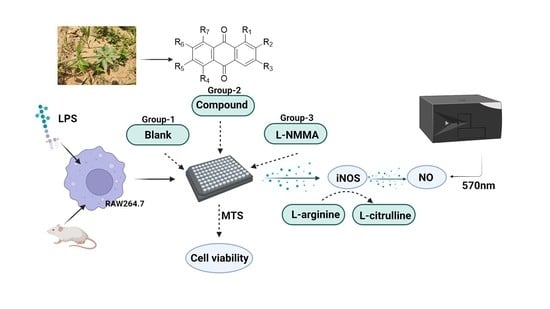
2. Results and Discussion

Compound 1 was obtained as a yellow powder, and the molecular formula was determined as C16H12O4, on the basis of HR-ESIMS (m/z 267.0669 [M − H], calcd. for C16H11O4, 267.0663). The UV spectrum showed maximum absorptions at 288 nm, suggesting a quinone structure. The 1H NMR data (Table 1) exhibited resonances which attributed to a 1, 3, 4-substituted benzene ring (δH 8.13 (1H, d, J = 7.9 Hz), 8.01 (1H, d, J = 1.8 Hz) and 7.71 (1H, dd, J = 7.9, 1.8 Hz)). The other benzene ring showed two orthodox-coupled aromatic proton resonances at (δH 8.04 (1H, d, J = 8.5 Hz) and 7.38 (1H, d, J = 8.5 Hz)). Moreover, aromatic methyl group δH 2.55 (3H, s), aromatic methoxy group δH 3.95 (3H, s) and phenolic hydroxyl group δH 9.40 (1H, s) protons were also observed. The 13C NMR exhibited 16 carbon signals, which consisted of 14 carbons of anthraquinone nucleus, a methoxy and a methyl. The HMBC (Figure 1) correlations of H-7 with C-5, 5-OMe with C-5 and H-8 with C-6/C-9 revealed that the methoxy group and hydroxyl group were connected at C-5 and C-6, respectively. In addition, the methyl group was deduced to be located at C-2 based on the HMBC correlations of 2-Me with C-1/C-2/C-3 and H-1 with C-3/C-9. Thus, the structure of compound 1 was established as 2-methyl-5-methoxy-6-hydroxyanthraquinone and named Cordifoquinone A, as shown in Figure 1.
Compound 2 was obtained as a yellow powder, and the molecular formula was determined to be C16H12O6, on the basis of HR-ESIMS (m/z 299.0561 [M − H], calcd. for C16H11O6, 299.0560). The 1H NMR data (Table 1) exhibited two meta-coupled aromatic proton resonances at δH 7.79 (1H, d, J = 7.8 Hz) and 7.63 (1H, d, J = 7.8 Hz), two aromatic proton singlets at δH 7.54 and 7.44 (each 1H, s), an aromatic methine proton signals at δH 4.62 (2H, s), an aromatic methoxy group δH 3.95 (3H, s) and a phenolic hydroxyl group δH 13.03 (1H, s). The HMBC (Figure 1) correlations from the aromatic methine protons to C-1/C-2/C-3 and from H-4 to C-2/C-10, together with the 1H NMR data, implied the presence of a phenolic hydroxyl group at the C-1 and a hydroxymethyl group at the C-2 position. Moreover, the methoxy group was deduced to be located at C-7, and a phenolic hydroxyl group was deduced to be located at C-6, based on the HMBC correlations of the 7-OMe with C-7, H-5 with C-7/C-10 and H-8 with C-6/C-9. Therefore, the structure of compound 2 was confirmed as 1,6-dihydroxy-2-(hydroxymethyl)-7-methoxyanthraquinone and named Cordifoquinone B.
Compound 3 was obtained as a yellow powder and had the same molecular formula of C16H12O6 as compound 2, based on HR-ESIMS (m/z 299.0561 [M − H], calcd. for C16H11O6, 299.0560). According to the NMR data (Table 1), 3 and 2 were found to have similar structures, except that the phenolic hydroxyl group on the hydroxymethyl side was moved to the methoxy side. The 1H NMR data exhibited resonances attributed to a 1, 3, 4-substituted benzene ring (δH 8.12 (1H, d, J = 1.7 Hz), 8.06 (1H, d, J = 7.9 Hz) and 7.76 (1H, dd, J = 7.9, 1.7 Hz)), an aromatic proton singlet at δH 7.19 (1H, s), an aromatic methine proton signals at δH 4.67 (2H, s), an aromatic methoxy group δH 3.83 (3H, s) and an aromatic hydroxyl group δH 13.03 (1H, s). The methoxy group was deduced to be located at C-7 based on the HMBC (Figure 1) correlations of 7-OMe with C-7 and H-5 with C-7/C-10. Moreover, the HMBC correlations from the aromatic methine protons to C-1/C-2/C-3, from H-4 to C-2/C-10 and from H-1 to C-9 suggested that the hydroxymethyl group was directly connected to C-2. Based on the data mentioned above, the structure of compound 3 was defined to be 6,8-dihydroxy-2-(hydroxymethyl)-7-methoxyanthraquinone and named Cordifoquinone C.
Compound 4 was obtained as a yellow powder and had the same molecular formula of C16H12O6 as compound 3, based on HR-ESIMS (m/z 299.0561 [M − H], calcd for C16H11O6, 299.0560). Its 1H and 13C NMR data (Table 1) were very similar to those of compound 3. The difference between them was that two phenolic hydroxyl groups and the methoxy group position of compound 4 were connected to C-5/C-7 and C-6, respectively, which were supported by the HMBC (Figure 1) correlations from the aromatic methine protons to C-1/C-2/C-3, H-1 to C-9/C-3, 6-OMe to C-6 and H-8 to C-6/C-9. Accordingly, the whole structure of compound 4 was finally determined as 5,7-dihydroxy-2-(hydroxymethyl)-6-methoxyanthraquinone and named Cordifoquinone D.
Additionally, 16 known anthraquinones, pustuline [27], 5,7-dihydroxy-6-methoxy-2-methylanthraquinone [28], soranjidiol [29], 1,6-dihydroxy-5-methoxy-2-methylanthraquinone [29], 1,5-dihydroxy-6-methoxy-2-methylanthraquinone [29], copareolatin 6-methylether [30], rubiadin [29], 2-methyl-1,3,6-trihydroxyanthraquinone [19], 7-hydroxy-2-(hydroxymethyl)-6-methoxyanthraquinone [31], 1-hydroxy-2-(hydroxymethyl)anthraquinone [32], 1,6-dihydroxy-2-(hydroxymethyl)-5-methoxyanthraquinone [33], 1,6-dihydroxy-2-(hydroxymethyl)-5,7-dimethoxyanthraquinone [34], 1,3-dihydroxy-2-hydroxymethyl-6-methoxyanthraquinone [35], 1,3-dihydroxy-2-(hydroxymethyl)-5,6-dimethoxyanthraquinone [36], 3,6-dihydroxy-2-(hydroxymethyl)-1-methoxyanthraquinone [37] and alizarin 1-methyl ether [38] were isolated from the aerial parts of R. cordifolia, as shown in Table 2. Their structures were elucidated by comparison of the NMR and ESI data with those reported in the literature. It is worth mentioning that anthraquinones in R. cordifolia are considered to be of the Rubia type; rings A and B are derived from horismite and α-ketoglutarate [39,40], whereas ring C is formed from isopentenyl diphosphate (IPP) [41]. Anthraquinone biosynthesized by this pathway has a methylation site in the C ring [42]. The aryl methyl group at site 2 can be further oxidized to hydroxymethyl. So, we hypothesized that compounds 120 might be synthesized by this pathway, as shown in Scheme 1.
All isolated compounds 120 were evaluated for their inhibitory effects on NO production in LPS-activated RAW264.7 macrophage cells. Cell viability was first examined by MTT assay to exclude false positive results caused by the potential cytotoxicity of the tested compounds. NG-Methyl-L-arginine acetate salt, an inhibitor of NO synthase, was used as a positive control (IC50 42.36 ± 2.47 μmol·L−1). As a result, compounds 1, 3 and 10 showed significant inhibitory effects against NO production, with IC50 values of 14.05 ± 0.48, 23.48 ± 1.05 and 29.23 ± 0.34 μmol·L−1 (Table 3), respectively, which were more potent than the positive control. By comparing the structure and activity of compounds 1, 3, 10, we found that the orthoposition of methoxyl and phenolic hydroxyl groups in the A ring of anthraquinone was essential for the NO inhibitory activity, and the hydroxyl group at position C-6 or C-7 provided a greater contribution. It is known that the synthesis of NO requires the addition of superoxide anions [43]. So, we hypothesized that the hydroxyl group in anthraquinone may capture superoxide anions, which are necessary for the synthesis of NO. Compound 1 showed stronger NO inhibitory activity than compounds 3 and 10, indicating that the activity was not necessarily related to the number of hydroxyl groups but may be related to the position of hydroxyl groups. We hypothesized that it may be due to the formation of intramolecular hydrogen bonds between hydroxyl group at position C-5 or C-8 and ketone carbonyl group at position C-9 or C-10, which makes the compound structure more stable and reduces its ability to capture superoxide anions. Through this speculation, it is reasonable to explain why compound 3 showed slightly higher NO inhibitory activity than compound 10. In addition, all compounds were evaluated for their antibacterial activities against four bacteria: Escherichia coli (ATCC 25922), Staphylococcus aureus subsp. aureus (ATCC 29213), Salmonella enterica subsp. enterica (ATCC 14028), and Pseudomonas aeruginosa (ATCC27853). However, no antibacterial activity was observed for these compounds at concentrations below 128 μg·mL−1 in this bioassay.

3. Experimental

3.1. General Experimental Procedures

UV spectra were taken on a Shimadzu UV-2401 spectrophotometer (Shimadzu, Kyoto, Japan). IR (KBr) spectra were recorded on a Bruker Vertex 70 (Bruker, Bremerhaven, Germany). The 1D and 2D NMR experiments were carried out using Avance III-400 and III-600 spectrometers (Bruker, Bremerhaven, Germany) with TMS as an internal standard. HR-ESIMS data were recorded on an Agilent 6540 Q-TOF mass spectrometer (Agilent Technologies, Santa Clara, CA, USA). Silica gel (200–300 mesh, Linyi Haixiang Co. Ltd., Linyi, China) and Sephadex LH-20 gel (Amersham Biosciences, Uppsala, Sweden) were employed for column chromatography. Thin-layer chromatography (TLC) analyses were performed on silica gel GF254 plates (Haohong, Chemical Co. Ltd., Yantai, China). HPLC purification was achieved on an Agilent 1100 instrument (Agilent Technologies, Santa Clara, CA, USA) with an Agilent ZORBAX SB-C18 column (5 μm, 9.4 × 250 mm, Agilent Technologies, Santa Clara, CA, USA).

3.2. Plant Materials

The aerial parts of Rubia cordifolia Linn. Were collected in June 2020 from Huaiyuan County, Anhui Province, China, and authenticated by Prof. Shou-Jin Liu from Anhui University of Traditional Chinese Medicine (Anhui, China). A voucher specimen (No. 20200601QC) was deposited at the Chinese Medicine Resource Center, Anhui University of Traditional Chinese Medicine (ACM).

3.3. Extraction and Isolation

The air-dried and powdered aerial parts of R. cordifolia (22 kg) were extracted with 95% EtOH at room temperature. After removal of the solvent under reduced pressure, the EtOH extract (22 kg) was suspended in water and partitioned with EtOAc. The EtOAc fraction (1.6 kg) was dissolved in EtOAc and chromatographed on silica gel column chromatography (Si CC), eluted with petroleum ether−acetone (1:0 to 0:1) to yield eight fractions, Fr. A–H (86 g, 60 g, 130 g, 123 g, 105 g, 178 g, 168 g, and 208 g). Fr. E (105 g) was dissolved in acetone and subjected to Si CC eluted with a gradient of CHCl3–MeOH (1:0 to 0:1) to obtain four major fractions, Fr. E1 to Fr. E4 (30 g, 8 g, 14 g, and 26 g). The extraction and separation process is shown in Figure 2.
Fr. E4 (26 g) was dissolved in acetone and further chromatographed over Si CC using petroleum ether−acetone (1:0 to 0:1) to give three subfractions, Fr. E4-1 to Fr. E4-3 (4.2 g, 3.5 g, 7.3 g). Compounds 8 (2 mg), 10 (8 mg), 11 (7 mg) and 17 (3 mg) were purified from Fr. E 4-1 (4.2 g) by Sephadex LH-20 (MeOH-CHCl3, 1:1) and semipreparative HPLC (H2O-MeOH, 75:25, 2.0 mL/min). Fr. E 4-2 (3.5 g) was applied to Sephadex LH-20 (CHCl3-MeOH, 1:1) to yield four subfractions (Fr. E4-2-1 to Fr. E4-2-4). Fr. E4-2-2 (127 mg) was separated by semipreparative HPLC (H2O-MeOH, 70:30, 2.0 mL·min−1) to obtain 12 (6 mg) and 18 (5 mg). Fr. E4-2-3 (238 mg) was separated by semipreparative HPLC (H2O-MeOH, 60:40, 2.0 mL·min−1) to afford 1 (5 mg), 2 (4 mg), 3 (8 mg), 5 (15 mg) and 15 (7 mg). Fr. E4-2-4 (182 mg) was separated by semipreparative HPLC (H2O-MeOH, 60:40, 2.0 mL·min−1) to afford 4 (4 mg), 6 (5 mg), 13 (2 mg), 16 (2 mg) and 19 (3 mg). Fr. E4-3 (7.3 g) gave 7 (2 mg), 9 (3 mg), 14 (4 mg) and 20 (6 mg) after repeated chromatography over Sephadex LH-20 (CHCl3-MeOH, 1:1) and Si CC gradient of CHCl3-MeOH (1:0 to 0:1).
Cordifoquinone A (1): Yellow powder; UV (MeOH) λmax (log ε) 251 (4.01), 264 (3.97) nm; IR (KBr) νmax 3423, 2955, 1666, 1629, 1581, 1428, 1363, 1302, 1256, 1024, 799 cm−1; 1H and 13C NMR data, see Table 1 and Figure S1; HR-ESIMS (m/z 267.0669 [M − H], calcd. for C16H11O4, 267.0663).
Cordifoquinone B (2): Yellow powder; UV (MeOH) λmax (log ε) 288 (3.89), 249 (3.53) nm; IR (KBr) νmax 3433, 2936, 1632, 1591, 1518, 1424, 1384, 1308, 1221, 1116, 1064, 754 cm−1; 1H and 13C NMR data, see Table 1 and Figure S2; HR-ESIMS (m/z 299.0561 [M − H], calcd. for C16H11O4, 299.0560).
Cordifoquinone C (3): Yellow powder; UV (MeOH) λmax (log ε) 248 (3.74), 285 (3.73) nm; IR (KBr) νmax 3443, 2952, 1662, 1632, 1596, 1452, 1385, 1340, 1279, 1219, 1134, 988 cm−1; 1H and 13C NMR data, see Table 1 and Figure S3; HR-ESIMS (m/z 299.0561 [M − H], calcd. for C16H11O6, 299.0560).
Cordifoquinone D (4): Yellow powder; UV (MeOH) λmax (log ε) 248 (3.47), 285 (3.41) nm; IR (KBr) νmax 3439, 2926, 1633, 1601, 1577, 1384, 1282, 1162, 975, 851 cm−1; 1H and 13C NMR data, see Table 1 and Figure S4; HR-ESIMS (m/z 299.0561 [M − H], calcd. for C16H11O6, 299.0560).

3.4. NO Inhibitory Assay

Murine macrophage cell line RAW264.7 was obtained from Shanghai Cell Bank of Chinese Academy of Sciences. RAW264.7 cells were seeded in 96-well cell culture plates (1.5 × 10⁵ cells/well), stimulated with 1 μg·mL−1 LPS (Sigma, St. Louis, MO, USA) for 18 h, and treated with serial dilutions of the compounds with a maximum concentration of 50 μM in triplicate during this period. NG-Methyl-L-arginine acetate salt (L-NMMA, Sigma), a well-known nitric oxide synthase (NOS) inhibitor, was used as a positive control [44]. NO production in the supernatant was assessed by Griess reagents (Reagent A and Reagent B, respectively, Sigma). The absorbance at 570 nm was measured with a microplate reader (Thermo, Waltham, MA, USA). The viability of RAW264.7 cells was evaluated by the MTS assay simultaneously to exclude the interference of the cytotoxicity of the test compounds. All tests were performed in triplicate, and the results were expressed as IC50 values.

3.5. Antibacterial Assay

Escherichia coli (ATCC 25922), Staphylococcus aureus subsp. aureus (ATCC 29213), Sal-monella enterica subsp. enterica (ATCC 14028), and Pseudomonas aeruginosa (ATCC 27853) were obtained from China General Microbiological Culture Collection Center. The inoculated strains and suspension were cultured for 24 h and adjusted to 0.5 McFarlane standard turbidity. A 96-well plate was prepared by injecting 100 µL of Muller Hinton broth/Luria-Bertani into each well. A volume of 100 µL of stock solution from the initial prepared samples was added to the first well. Then, 100 µL from their two-fold serial dilutions were transferred into consecutive wells, and 100 µL of the inoculums was added to achieve a final inoculum concentration of 5 × 10⁵ CFU/mL. The final volume in each well was 200 µL. Ceftazidime (Yuanye Co. Ltd., Shanghai, China) and sodium penicillin G (Labgic Co. Ltd., Beijing, China) were used as positive controls. After incubation at 37 °C for 24 h, growth was monitored by a Microplate Reader at 625 nm (PowerWave XS, BioTek, Winooski, VT, USA) [45].

4. Conclusions

Four new anthraquinones (14) and 16 known anthraquinones (520) were isolated from a 95% EtOH extract of aerial prat from R. cordifolia and elucidated by UV, IR, HR-ESIMS and NMR spectroscopic data. Among the isolated compounds, compounds 1, 3 and 10 displayed NO inhibitory activity with IC50 values of 14.05, 23.48 and 29.23 μmol·L−1. The structure and activity relationship analysis revealed that the orthoposition of methoxyl and phenolic hydroxyl groups in the A ring of anthra-quinone was essential for the NO inhibitory activity. The hydroxyl group at position C-6 or C-7 may have promoted the NO inhibitory activity, whereas the hydroxyl group at position C-5 or C-8 had the opposite effect. In addition, this series of anthraquinones showed no activity in tests of antibacterial activity. This study proved the reasonableness of using the aerial part of R. cordifolia as a medicine to treat diarrhea in Traditional Chinese medicine. It also provides scientific evidence and a foundation for the understanding of the anti-inflammatory effects and further utilization of R. cordifolia. In future research, both the extract and the natural products of R. cordifolia can be considered a drug or a health supplement to treat diarrhea.

Supplementary Materials

The following supporting information can be downloaded online, Figure S1: 1H, 13C and HMBC NMR spectra of compound 1; Figure S2: 1H, 13C and HMBC NMR spectra of compound 2; Figure S3: 1H, 13C and HMBC NMR spectra of compound 3; Figure S4: 1H, 13C and HMBC NMR spectra of compound 4.

Author Contributions

Conceptualization, S.-J.L. and J.-M.H.; methodology, H.L. and F.-C.R.; software, H.L.; validation, W.Q., H.Z. and W.-T.F.; investigation, H.L., Q.-H.K., and J.-M.Z.; re-sources, C.-W.F.; writing—original draft preparation, L.H; writing—review and editing, H.L. and F.-C.R.; visualization, L.Y.; supervision, J.-M.H.; project administration, S.-J.L.; funding acquisition, S.-J.L. All authors have read and agreed to the published version of the manuscript.

Funding

Please add: This research was funded by the Special Subsidies for TCM public health services, grant number RH2100000743. The APC was funded by National Administration of Traditional Chinese Medicine (NATCM).

Institutional Review Board Statement

Not applicable.

Informed Consent Statement

Not applicable.

Data Availability Statement

The data presented in this study are available in the Supplementary Materials.

Conflicts of Interest

The authors declare no conflict of interest.

Sample Availability

Samples of the compounds compound 1 to 20 are available from the authors.

Abbreviations

IDinfectious diarrhea
NOnitric oxide
iNOSinducible nitric oxide synthase
TCMTraditional Chinese Medicine
HR-ESIMShigh resolution electrospray ionization mass spectroscopy
UVultraviolet-visible
NMRnuclear magnetic resonance
HMBCheteronuclear multiple bond correlation
MTT3-(4,5-dimethylthiazol-2-yl)-2,5-diphenyltetrazolium bromid
IRinfrared ra
Q-TOFquadrupole-time of flight
HPLChigh performance liquid chromatography
TLCthin-layer chromatography
Si CCsilica gel column chromatography
EtOHethyl alcohol
EtOAcethyl acetate
MeOHMethanol
USAThe United States of America

References

  1. Snyder, J.D.; Merson, M. The magnitude of the global problem of acute diarrhoeal disease: A review of active surveillance data. Bull. WHO 1982, 60, 605–613. [Google Scholar]
  2. Liu, L.; Johnson, H.L.; Cousens, S.; Perin, J.; Scott, S.; Lawn, J.E.; Rudan, I.; Campbell, H.; Cibulskis, R.; Li, M.; et al. Global, regional, and national causes of child mortality: An updated systematic analysis for 2010 with time trends since 2000. Lancet 2012, 379, 2151–2161. [Google Scholar] [CrossRef]
  3. Stephen, J. Pathogenesis of infectious diarrhea. Can. J. Gastroenterol. 2001, 15, 669–683. [Google Scholar] [CrossRef] [PubMed] [Green Version]
  4. Moncada, S.; Higgs, A. The L-arginine-nitric oxide pathway. N. Engl. J. Med. 1993, 2, 2002–2012. [Google Scholar]
  5. Ignarro, L.J.; Buga, G.M.; Wood, K.S.; Byrns, R.E.; Chaudhuri, G. Endothelium-derived relaxing factor produced and released from artery and vein is nitric oxide. Proc. Natl. Acad. Sci. USA 1987, 84, 9265–9269. [Google Scholar] [CrossRef] [Green Version]
  6. Wink, D.A.; Mitchell, J.B. Chemical biology of nitric oxide: Insights into regulatory, cytotoxic, and cytoprotective mechanisms of nitric oxide. Free Radic. Biol. Med. 1998, 25, 434–456. [Google Scholar] [CrossRef]
  7. Grisham, M.B.; Jourd’Heuil, D.; Wink, D.A. Nitric oxide—I. Physiological chemistry of nitric oxide and its metabolites: Implications in inflammation. Am. J. Physiol. 1999, 276, 315–321. [Google Scholar] [CrossRef]
  8. Umer, S.; Tekewe, A.; Kebede, N. Antidiarrhoeal and antimicrobial activity of Calpurnia aurea leaf extract. BMC Complementary Altern. Med. 2013, 13, 21. [Google Scholar] [CrossRef] [Green Version]
  9. Orsi, P.R.; Bonamin, F.; Severi, J.A.; Santos, R.C.; Vilegas, W.; Hiruma-Lima, C.A.; Di Stasi, L.C. Hymenaea stigonocarpa Mart. ex Hayne: A Brazilian medicinal plant with gastric and duodenal anti-ulcer and antidiarrheal effects in experimental rodent models. J. Ethnopharmacol. 2012, 143, 81–90. [Google Scholar] [CrossRef] [Green Version]
  10. Ali, N.; Alam, H.; Khan, A.; Ahmed, G.; Shah, W.A.; Nabi, M.; Junaid, M. Antispasmodic and antidiarrhoeal activity of the fruit of Rosa moschata. BMC Complementary Altern. Med. 2014, 14, 485. [Google Scholar] [CrossRef]
  11. Atta, A.H.; Mouneir, S.M. Antidiarrhoeal activity of some Egyptian medicinal plant extracts. J. Ethnopharmacol. 2004, 92, 303–309. [Google Scholar] [CrossRef] [PubMed]
  12. Mbagwu, H.O.C.; Adeyemi, O.O. Anti-diarrhoeal activity of the aqueous extract of Mezoneuron benthamianum Baill (Caesalpiniaceae). J. Ethnopharmacol. 2008, 116, 16–20. [Google Scholar] [CrossRef]
  13. Praxedes de Sales, I.R.; Frota Machado, F.D.; Marinho, A.F.; Suassuna Carneiro Lucio, A.S.; Barbosa Filho, J.M.; Batista, L.M. Cissampelos sympodialis Eichl. (Menispermaceae), a medicinal plant, presents antimotility and antidiarrheal activity in vivo. BMC Complement. Altern. Med. 2015, 15, 253. [Google Scholar]
  14. Chinese Academy of Sciences Flora of China Commission. Flora of China; Science Press: Beijing, China, 1999; Volume 71, pp. 287–315. [Google Scholar]
  15. Itokawa, H.; Takeya, K.; Mihara, K.; Mori, N.; Hamanaka, T.; Sonobe, T.; Iitaka, Y. Studies on the antitumor cyclic hexapeptides obtained from Rubiae radix. Chem. Pharm. Bull. 1983, 31, 1424–1427. [Google Scholar] [CrossRef] [PubMed] [Green Version]
  16. Gong, X.P.; Sun, Y.Y.; Chen, W.; Guo, X.; Guan, J.K.; Li, D.Y.; Du, G. Anti-diarrheal and anti-inflammatory activities of aqueous extract of the aerial part of Rubia cordifolia. BMC Complementary Altern. Med. 2017, 17, 20. [Google Scholar] [CrossRef] [Green Version]
  17. Wang, K.; Gao, L.; Zhang, Q.; Zhang, Y.; Yao, W.F.; Zhang, M.; Tang, Y.P.; Ding, A.W.; Zhang, L. Revealing the mechanisms and the material basis of Rubia cordifolia L. on abnormal uterine bleeding with uniting simultaneous determination of four components and systematic pharmacology approach-experimental validation. J. Pharm. Biomed. Anal. 2020, 189, 113475. [Google Scholar] [CrossRef]
  18. Chinese Pharmacopoeia Commission. Pharmacopoeia of the People’s Republic of China; China Medical Science Press: Beijing, China, 2020; Volume 1, p. 245. [Google Scholar]
  19. Itokawa, H.; Mihara, K.; Takeya, K. Studies on a novel anthraquinone and its glycosides isolated from Rubia cordifolia and R. akane. Chem. Pharm. Bull. 1983, 31, 2353–2358. [Google Scholar] [CrossRef] [Green Version]
  20. Hu, Y.Y.; Zhang, X.J.; Zhang, Z.H.; Wang, Z.; Tan, N.H. Qualitative and quantitative analyses of quinones in multi-origin Rubia species by ultra-performance liquid chromatography-tandem mass spectrometry combined with chemometrics. J. Pharm. Biomed. Anal. 2020, 189, 113471. [Google Scholar] [CrossRef]
  21. Arisawa, M.; Ueno, H.; Nimura, M.; Hayashi, T.; Morita, N. Rubiatriol, a new triterpenoid from the Chinese drug Qian Cao Gen, Rubia cordifolia. J. Nat. Prod. 1986, 49, 1114–1116. [Google Scholar] [CrossRef]
  22. Chen, X.Q.; Zhao, S.M.; Wang, Z.; Zeng, G.Z.; Huang, M.B.; Tan, N.H. Rubicordins A-C, new cyclopeptides from Rubia cordifolia with cytotoxicity and inhibiting NF-kappa B signaling pathway. Tetrahedron 2015, 71, 9673–9678. [Google Scholar] [CrossRef]
  23. Zhang, M.T.; Yang, L.; Hu, J.M.; Zhang, H.; Shi, X.Q.; Liu, S.J. Study on lignan constituents from aerial part of Rubia cordifolia. Chin. Tradit. Herb. Drugs. 2017, 48, 4856–4859. [Google Scholar]
  24. Qiao, Y.F.; Wang, S.K.; Wu, L.J.; Li, X.; Zhu, T.R. Antibacterial constituents from the roots of Rubia cordifolia L. Acta. Pharm. Sin. 1990, 25, 834–839. [Google Scholar]
  25. Tao, J.; Morikawa, T.; Ando, S.; Matsuda, H.; Yoshikawa, M. Bioactive constituents from chinese natural medicines. XI. Inhibitors on NO production and degranulation in RBL-2H3 from Rubia yunnanensis: Structures of rubianosides II, III, and IV, rubianol-g, and rubianthraquinone. Chem. Pharm. Bull. 2003, 51, 654–662. [Google Scholar] [CrossRef] [PubMed] [Green Version]
  26. Son, J.K.; Jung, S.J.; Jung, J.H.; Fang, Z.; Lee, C.S.; Seo, C.S.; Moon, D.C.; Min, B.S.; Kim, M.R.; Woo, M.H. Anticancer constituents from the roots of Rubia cordifolia L. Chem. Pharm. Bull. 2008, 56, 213–216. [Google Scholar] [CrossRef] [PubMed] [Green Version]
  27. Nunez Montoya, S.C.; Agnese, A.M.; Cabrera, J.L. Anthraquinone Derivatives from Heterophyllaea pustulata. J. Nat. Prod. 2006, 69, 801–803. [Google Scholar] [CrossRef] [PubMed]
  28. El-Lakany, A.M.; Aboul-Ela, M.A.; Abdel-Kader, M.S.; Badr, J.M.; Sabri, N.N.; Goher, Y. Anthraquinones with antibacterial activities from Crucianella maritima L. growing in Egypt. Nat. Prod. Sci. 2004, 10, 63–68. [Google Scholar]
  29. Ismail, N.H.; Ali, A.M.; Aimi, N.; Kitajima, M.; Takayama, H.; Lajis, N.H. Anthraquinones from Morinda elliptica. Phytochemistry 1997, 45, 1723–1725. [Google Scholar] [CrossRef] [Green Version]
  30. Koyama, J.; Ogura, T.; Tagahara, K. Anthraquinones of Galium spurium. Phytochemistry 1993, 33, 1540–1542. [Google Scholar] [CrossRef]
  31. Kang, X.D.; Li, X.; Zhao, C.C.; Mao, Y. Two new anthraquinones from Hedyotis diffusa W. J. Asian. Nat. Prod. Res. 2008, 10, 193–197. [Google Scholar] [CrossRef]
  32. Itokawa, H.; Ibraheim, Z.Z.; Qiao, Y.F.; Takeya, K. Anthraquinones, naphthohydroquinones and naphthohydroquinone dimers from Rubia cordifolia and their cytotoxic activity. Chem. Pharm. Bull. 1993, 41, 1869–1872. [Google Scholar] [CrossRef] [Green Version]
  33. Longue Ekon, J.P.; Zra, T.; Lobe Songue, J.; Wondja Ngoko, M.L.; Ngassoum, M.B.; Talla, E.; Kamdem Waffo, A.F.; Sewald, N.; Wansi, J.D. New anthraquinone derivatives from the stem barks of Morinda lucida Benth. Phytochem. Lett. 2020, 39, 94–98. [Google Scholar] [CrossRef]
  34. El-Gamal, A.A.; Takeya, K.; Itokawa, H.; Halim, A.F.; Amer, M.M.; Saad, H.E.A.; Awad, S.A. Anthraquinones from the polar fractions of Galium sinaicum. Phytochemistry 1996, 42, 1149–1155. [Google Scholar] [CrossRef]
  35. Banthorpe, D.V.; White, J.J. Novel anthraquinones from undifferentiated cell cultures of Galium verum. Phytochemistry 1995, 38, 107–111. [Google Scholar] [CrossRef]
  36. Wisetsai, A.; Lekphrom, R.; Schevenels, F.T. New anthracene and anthraquinone metabolites from Prismatomeris filamentosa and their antibacterial activities. Nat. Prod. Res. 2021, 35, 1582–1589. [Google Scholar] [CrossRef]
  37. Ling, S.K.; Komorita, A.; Tanaka, T.; Fujioka, T.; Mihashi, K.; Kouno, I. Iridoids and anthraquinones from the Malaysian medicinal plant, Saprosma scortechinii (Rubiaceae). Chem. Pharm. Bull. 2002, 50, 1035–1040. [Google Scholar] [CrossRef] [Green Version]
  38. Halim, A.F.; Abd El-Fattah, H.; El-Gamal, A.A.; Thomson, R.H. Anthraquinones from Galium sinaicum. Phytochemistry 1991, 31, 355–356. [Google Scholar] [CrossRef]
  39. Leistner, E. Second pathway leading to anthraquinones in higher plants. Phytochemistry 1971, 10, 3015–3020. [Google Scholar] [CrossRef]
  40. Sieweke, H.J.; Leistner, E. o-Succinylbenzoate:coenzyme A ligase from anthraquinone producing cell suspension cultures of Galium mollugo. Phytochemistry 1992, 31, 2329–2335. [Google Scholar] [CrossRef]
  41. Lv, X.; Wang, F.; Zhou, P.; Ye, L.; Xie, W.; Xu, H.; Yu, H. Dual regulation of cytoplasmic and mitochondrial acetyl-CoA utilization for improved isoprene production in Saccharomyces cerevisiae. Nat. Commun. 2016, 7, 12851. [Google Scholar] [CrossRef]
  42. Han, Y.S.; van der Heijden, R.; Lefeber, A.W.M.; Erkelens, C.; Verpoorte, R. Biosynthesis of anthraquinones in cell cultures of Cinchona ‘Robusta’ proceeds via the methylerythritol 4-phosphate pathway. Phytochemistry 2002, 59, 45–55. [Google Scholar] [CrossRef]
  43. Ignarro, L.J. Biosynthesis and metabolism of endothelium-derived nitric oxide. Annu. Rev. Pharmacol. Toxicol. 1990, 30, 535–560. [Google Scholar] [CrossRef] [PubMed]
  44. Reif, D.W.; McCreedy, S.A. N-Nitro-L-arginine and N-monomethyl-L-arginine exhibit a different pattern of inactivation toward the three nitric oxide synthases. Arch. Biochem. Biophys. 1995, 320, 170–176. [Google Scholar] [CrossRef] [PubMed]
  45. Walker, R.D. Standards for antimicrobial susceptibility testing. Am. J. Vet. Res. 1999, 60, 64–65. [Google Scholar]
Figure 1. Key HMBC of compounds 14.
Molecules 27 01730 g001
Scheme 1. Plausible biosynthetic pathway for compounds 120.
Molecules 27 01730 sch001
Figure 2. The extraction and separation process of the aerial parts of Rubia cordifolia L.
Molecules 27 01730 g002
Table 1. 1H and 13C NMR spectroscopic data (δ in ppm, J in Hz) for compounds 14.
No1 a2 a3 b4 a
δHδCδHδCδHδCδHδC
18.01, d (1.8)127.2 158.68.12, d (1.7)123.58.06, d (1.7)124.0
2 145.5 138.3 149.8 149.5
37.71, dd (7.9, 1.8)135.47.63, d (7.8)133.17.76, dd (7.9, 1.7)131.67.78, dd (7.9, 1.7)131.7
48.13, d (7.9)127.87.79, d (7.8)118.88.06, d (7.9)126.88.13, d (7.9)126.3
4 a 133.5 131.8 131.7 132.1
5 148.47.54, s113.47.19, s109.9 157.0
6 158.1 155.1 157.0 139.8
77.38, d (8.5)121.6 153.5 139.6 160.3
88.04, d (8.5)125.87.44, s108.8 159.27.16, s110.5
8 a 127.9 125.2 109.6 129.3
9 182.6 187.8 186.1 181.9
9 a 133.7 115.2 133.2 132.9
10 182.7 181.8 181.4 185.5
10 a 127.5 129.2 129.1 109.0
2-Me2.53, s21.6
2-CH2OH 4.62, s57.94.67, s62.24.66, s62.2
5-OMe3.95, s61.7
6-OMe 3.82, s59.8
7-OMe 3.95, s56.33.83, s59.9
a 1H at 400 MHz and 13C at 150 MHz in Acetone-d6. b 1H at 400 MHz and 13C at 150 MHz in DMSO-d6.
Table 2. Chemical structures of isolated compounds 120 from Rubia cordifolia.
Molecules 27 01730 i001
R1R2R3R4R5R6R7
1HMeHOMeOHHH
2OHCH2OHHHOHOMeH
3HCH2OHHHOHOMeOH
4HCH2OHHOHOMeOHH
5HMeHHOMeOHH
6HMeHOHOMeOHH
7OHMeHHOHHH
8OHMeHOMeOHHH
9OHMeHOHOMeHH
10OHMeHOHOMeOHH
11OHOMeOHHHHH
12OHOMeOHHOHHH
13HCH2OHHHOMeOHH
14OHCH2OHHHHHH
15OHCH2OHHOMeOHHH
16OHCH2OHHOMeOHOMeH
17OHCH2OHOHHOMeHH
18OHCH2OHOHOMeOMeHH
19OMeCH2OHOHHOHHH
20OMeOHHHHHH
Table 3. NO inhibitory activities of compounds 1, 3 and 10.
CompoundsNO Inhibitory Effects
(IC50/μmol·L−1)
RAW 264.7 Cell
Viability a (%)
114.05 ± 0.48105.69 ± 0.25
323.48 ± 1.0597.67 ± 1.21
1029.23 ±0.34101.80 ± 1.10
L-NMMA b42.36 ± 2.4798.72 ± 0.94
a RAW 264.7 cells treated with samples at 50 μmol·L−1. b Positive control.
Publisher’s Note: MDPI stays neutral with regard to jurisdictional claims in published maps and institutional affiliations.

